gpr125 modulates dishevelled distribution and planar cell...
TRANSCRIPT

RESEARCH ARTICLE3028
Development 140, 3028-3039 (2013) doi:10.1242/dev.094839© 2013. Published by The Company of Biologists Ltd
INTRODUCTIONDuring embryogenesis, gastrulation establishes the three germlayers and the animal body plan. Vertebrate gastrulation relies onpolarized cell behaviors to drive convergence and extension (C&E)movements that narrow embryonic tissues mediolaterally andelongate them anteroposteriorly (Keller et al., 2000; Solnica-Krezel,2005; Yin et al., 2009; Gray et al., 2011). In dorsal regions ofXenopus and zebrafish gastrulae, cells become elongated and alignalong the mediolateral embryonic axis, allowing preferentialintercalation between their anterior and posterior neighbors to driveC&E (Keller et al., 2000; Topczewski et al., 2001; Jessen et al.,2002; Marlow et al., 2002; Lin et al., 2005). Modulation of celladhesion and intercellular signaling have been proposed to instructsuch complex cell behaviors (Yin et al., 2009). However, themolecules implementing these actions have not been fullyidentified.
Currently, the Wnt/PCP signaling system, which is equivalent tothe PCP pathway coordinating wing hair and ommatidia orientationin Drosophila (Simons and Mlodzik, 2008; Goodrich and Strutt,2011), is the best-studied pathway regulating C&E movements invertebrates (Tada and Kai, 2009; Yin et al., 2009; Gray et al., 2011).Polarized cell behaviors that underlie C&E, including directed cellmigration and polarized planar and radial intercalations, areexquisitely sensitive to PCP signaling levels, as excess or
insufficient Wnt/PCP pathway component function impairs C&Emovements (Wallingford et al., 2000; Jessen et al., 2002; Marlow etal., 2002; Carreira-Barbosa et al., 2003). In addition to regulatingC&E, a subset of Wnt/PCP components also regulates the caudaltangential migration of facial branchiomotor neurons (FBMN) inzebrafish and mouse (Jessen et al., 2002; Carreira-Barbosa et al.,2003; Wada et al., 2005; Wada et al., 2006; Wada and Okamoto,2009).
PCP pathway components are known to localize asymmetricallyin multiple tissues that manifest planar polarity. In the fly wingepithelia, the receptor Frizzled and cytoplasmic proteinsDishevelled (Dsh/Dvl in vertebrates) and Diego localize to the distalside of the cell, where the wing hair will eventually emerge. Bycontrast, the transmembrane protein Van gogh/Strabismus andcytoplasmic protein Prickle (Pk) localize proximally, whereas theseven transmembrane protocadherin Flamingo/Starry night ispresent at both cell edges (Axelrod, 2001; Feiguin et al., 2001; Struttet al., 2002; Tree et al., 2002; Bastock et al., 2003). This stereotypedasymmetric localization of Pk and Dvl on opposing anterior andposterior membranes has been observed in the neural plate anddorsal mesodermal cells undergoing C&E in zebrafish (Ciruna etal., 2006; Yin et al., 2008). Such molecular asymmetries areconsidered to be either a consequence of cell polarization or anessential step in the process of Wnt/PCP-mediated cell polarization(Simons and Mlodzik, 2008; McNeill, 2010; Goodrich and Strutt,2011; Gray et al., 2011).
Asymmetric localization of PCP components in polarizedepithelia and protein interaction studies supports a model wherebyPCP components interact in asymmetric membrane complexesspanning the juxtaposed cells to generate planar polarization(McNeill, 2010; Goodrich and Strutt, 2011). Recently, Dsh wasshown to cluster PCP complexes into membrane subdomains incells of Drosophila pupal wings (Strutt et al., 2011), raising thepossibility that clustering of asymmetric PCP complexes intomembrane subdomains might provide a local self-enhancementmechanism that establishes and/or maintains planar polarity (Strutt
1Neuroscience Graduate Program, Vanderbilt University School of Medicine,Nashville, TN 37232, USA. 2Department of Developmental and Molecular Biology,Albert Einstein College of Medicine, Bronx, NY 10461, USA. 3Department ofDevelopmental Biology, Washington University School of Medicine, St Louis, MO63110, USA. 4Department of Surgery, New York Hospital Medical Center of Queens,Flushing, NY 11355, USA. 5Department of Pharmacology, Vanderbilt UniversitySchool of Medicine, Nashville, TN 37232, USA. 6Department of Biological Sciences,Vanderbilt University, Nashville, TN 37232, USA.
*Authors for correspondence ([email protected];[email protected])
Accepted 2 May 2013
SUMMARYDuring vertebrate gastrulation, Wnt/planar cell polarity (PCP) signaling orchestrates polarized cell behaviors underlying convergenceand extension (C&E) movements to narrow embryonic tissues mediolaterally and lengthen them anteroposteriorly. Here, we haveidentified Gpr125, an adhesion G protein-coupled receptor, as a novel modulator of the Wnt/PCP signaling system. Excess Gpr125impaired C&E movements and the underlying cell and molecular polarities. Reduced Gpr125 function exacerbated the C&E and facialbranchiomotor neuron (FBMN) migration defects of embryos with reduced Wnt/PCP signaling. At the molecular level, Gpr125 recruitedDishevelled to the cell membrane, a prerequisite for Wnt/PCP activation. Moreover, Gpr125 and Dvl mutually clustered one anotherto form discrete membrane subdomains, and the Gpr125 intracellular domain directly interacted with Dvl in pull-down assays.Intriguingly, Dvl and Gpr125 were able to recruit a subset of PCP components into membrane subdomains, suggesting that Gpr125may modulate the composition of Wnt/PCP membrane complexes. Our study reveals a role for Gpr125 in PCP-mediated processes andprovides mechanistic insight into Wnt/PCP signaling.
KEY WORDS: Gastrulation movements, Convergence and extension, Facial branchiomotor neuron, Zebrafish
Gpr125 modulates Dishevelled distribution and planar cellpolarity signalingXin Li1,2, Isabelle Roszko3, Diane S. Sepich3, Mingwei Ni4, Heidi E. Hamm1,5, Florence L. Marlow2,* and Lilianna Solnica-Krezel1,3,6,*
DEVELO
PMENT

3029RESEARCH ARTICLEGpr125 modulates PCP signaling
et al., 2011). Interestingly, membrane clustering of PCP componentsoccurs between Xenopus Van gogh-like 2 (Vangl2, vertebratehomolog of Van gogh/Strabismus) and Drosophila Pk expressed inXenopus animal cap explants, and among zebrafish Frizzled7(Fzd7), Wnt11 and Xenopus Dvl expressed in zebrafish blastula(Jenny et al., 2003; Witzel et al., 2006). In the latter case, subdomainformation correlates with increased persistence of membranecontacts partially dependent on vertebrate Flamingo homologues,Cadherin EGF LAG seven-pass G-type receptors (Celsrs) (Witzel etal., 2006).
Celsrs belong to the family of adhesion G protein-coupledreceptors (GPCRs), which are chimeras of adhesion molecules andtransmembrane signal transducer GPCRs (Fredriksson et al., 2003).Owing to their unique structure, adhesion GPCRs are postulated toplay dual roles in cell adhesion and signal transduction (Yona et al.,2008). Recent studies of GPR56, GPR124 and Gpr126 implicateadhesion GPCRs in diverse developmental processes, includingbrain development, blood vessel formation and myelination inzebrafish and mammals (Kuhnert et al., 2010; Piao et al., 2004;Monk et al., 2009; Monk et al., 2011). As components of the PCPpathway, Celsr adhesion GPCRs have been reported to regulatezebrafish gastrulation and FBMN migration (Formstone and Mason,2005; Wada et al., 2006; Carreira-Barbosa et al., 2009).
To better understand the molecular mechanisms underlyinggastrulation movements and to uncover the functions ofuncharacterized adhesion GPCRs, we surveyed adhesion GPCRsfor candidate regulators of zebrafish gastrulation. Here, we haveidentified Gpr125 adhesion GPCR as a modulator of C&Egastrulation movements and FBMN migration. Gpr125 functionallyinteracts with multiple Wnt/PCP components and directly interactsvia its intracellular domain with Dvl. Mutual redistribution ofGpr125 and Dvl fusion proteins into discrete membrane subdomainsand their ability to selectively recruit additional PCP componentsinto these domains suggest that Gpr125 might act as a componentof the PCP membrane complexes and modulator of Wnt/PCPsignaling in vertebrates.
MATERIALS AND METHODSZebrafish linesAB, ABWIK, llkRW468, trivu67 (a nonsense allele; Chunyue Yin, Jason R.Jessen, I.R. and L.S.K., unpublished), slbtz216, tp53M214K and Tg(isl1:GFP)were used in this study (Heisenberg et al., 2000; Jessen et al., 2002;Berghmans et al., 2005; Wada et al., 2005). Embryos obtained from naturalspawnings were staged according to morphological criteria (Kimmel et al.,1995).
RT-PCR and cloning of zebrafish gpr125Total RNA was extracted with TRIZOL LS reagent (Invitrogen) from wild-type embryos at the indicated stages. cDNA was produced with SuperScriptIII first-strand synthesis system (Invitrogen). To detect gpr125 transcripts,PCR was performed using gpr125-q primers (supplementary material TableS1) with GoTaq Flexi DNA polymerase (Promega). The full-length gpr125-coding sequence (GenBank Accession Number KC996731) was amplifiedusing gpr125-fl primers (supplementary material Table S1) with Easy-Ahigh-fidelity PCR cloning enzyme (Agilent Technologies) and subclonedinto pCR8 vector (Invitrogen), from which various deletion forms ofGpr125 were amplified with the primers listed in supplementary materialTable S1 and subcloned into pCR8 and subsequently into pCS-based vectors(Villefranc et al., 2007) or E. coli expression vector pDEST 15 (Invitrogen)with Gateway LR clonase II enzyme mix or LR clonase II plus enzyme(Invitrogen).
RNA and antisense morpholino oligonucleotide (MO) injectionCapped RNA was synthesized using mMessage mMachine Kit (Ambion).Two non-overlapping MOs (MO1-gpr125 and MO2-gpr125) targeting the
5�UTR region were used. The effectiveness of each MO in blocking thetranslation of RNA encoding GFP fused to the MO target sequence (GFPreporter) was determined. The non-specific toxicity of MO1-gpr125 wasconfirmed by complete suppression of cell death in p53M214K/M214K nullmutants (supplementary material Fig. S2J,K). Sequences of all MOs usedare listed in supplementary material Table S1.
Anteroposterior (AP) axis, notochord and somite measurementsEmbryos were imaged using Olympus SZ61 or Zeiss Discovery dissectingmicroscopes and Olympus or Zeiss AxioCam MRM cameras inPictureFrame or Axiovision Rel 4.6 (Zeiss). For AP axis length, embryoswere traced from the forebrain to the tip of the tail fin. For notochord width,straight lines were drawn perpendicular to the AP axis between the lateralborders of the notochord at level of first somites. For somite length, the firstsomites were traced. The distance was measured with ImageJ software(NIH) (Marlow et al., 1998).
Whole-mount in situ hybridizationAntisense probes were synthesized with RNA labeling kits (Roche). DNAfragments amplified with gpr125-probe1 primers and gpr125-probe2primers (supplementary material Table S1) were used as templates forgpr125 probe synthesis. Whole-mount in situ hybridization analyses wereperformed as described previously (Marlow et al., 1998).
Whole-mount immunostainingEmbryos were fixed in 100% Prefer fixative (Anatech) for 40 minutes atroom temperature. Immunostaining was performed with a standard protocol.Antibodies were diluted in blocking buffer containing 0.5% BSA, 10%serum, 0.1% Triton X-100 and 2% DMSO in PBS. Primary antibodies usedwere: anti-zebrafish Tri/Vangl2 (rabbit, 1:500, made by the VanderbiltUniversity Antibody Core), anti-GFP (mouse, 1:500, Clontech, #632375)and rat anti-RFP (1:1000, Chromotek, clone 5F8). Secondary antibodieswere: Alexa Fluor 568 goat anti-rat, Alexa Fluor 647 goat anti-rabbit andAlexa Fluor 488 goat anti-mouse (1:500, Invitrogen).
Cell polarity analysesMeasurements and analyses of length-width ratios (LWRs) and mediolateralalignment were performed according to Myers et al. (Myers et al., 2002).Wild-type embryos were injected with 200 pg gpr125-Cherry + 100 pgmembraneEGFP (mEGFP), or 300 pg of mEGFP synthetic RNA.membraneCherry (mCherry) RNA (150 pg) was injected into vangl2/triembryos for membrane labeling. Embryos were fixed overnight in 4% PFAand confocal stacks were collected. Image analysis was performed inImageJ (NIH) and Fiji (Schindelin et al., 2012), where cells were outlinedby hand. LWRs and angles of the long axis were measured with Fit Ellipse.Rose diagrams were drawn using Rose.NET (Todd A. Thompson;http://mypage.iu.edu/~tthomps/programs/home.htm).
In vivo subcellular protein localization analysesPk localization experiments: one-cell embryos were injected with 200 or300 pg gpr125 and 100 pg mCherry synthetic RNA, or an equivalentamount of mCherry RNA. At the 16-cell stage, one cell was injected with16-19 pg of pk-GFP RNA (Yin et al., 2008). At tailbud to two-somite stage,z-stacks were collected using Quorum Spinning disc Confocal/ IX81-Olympus inverted microscope. Image analysis was performed in ImageJ(NIH) and Fiji.
To monitor protein localization in late blastulae, one-cell stage embryoswere injected with specific combinations of RNA at doses specified in thefigures. In mosaic expression experiments, Histone2B-RFP (H2B-RFP) andgpr125FL RNA were injected into one blastomere at the 16- to 32-cell stage.The superficial layer of live or immunostained blastulae at 4-5 hours post-fertilization (hpf) was imaged with a Zeiss Axioobserver Apotomefluorescence microscope equipped with a 40× oil lens (Zeiss) and MRmdigital camera (Zeiss), and Axiovision Rel 4.7 software (Zeiss), Leica TCSSP5 confocal or Quorum Spinning disc Confocal/ IX81-Olympus invertedmicroscope and Metamorph Acquisition software. For Dvl-GFP subdomainquantification, embryos were imaged using identical settings for the greenchannel and cells from five embryos of each group were randomly selectedfor measuring membrane length, Dvl-GFP particle size and number in ImageJ D
EVELO
PMENT

3030
(NIH). The average threshold of three membranes expressing Dvl-GFP alonewas used to set the background threshold for subdomain analysis. To monitorprotein localization in the gastrulae, RNA were injected at the specified dosesat the one-cell stage and embryos were fixed at 10 hpf for immunostaining.Images were acquired with a Quorum Spinning disc Confocal/IX81-Olympusinverted microscope and Metamorph Acquisition software.
Pull-down and western blot analysesControl glutathione S-transferase (GST) protein was produced from pGEX-4T-1 (GE Healthcare) in XL-1Blue E. coli and GST-fusion proteins wereproduced from pDEST 15-based vectors in BL21-AI E. coli according tomanufacturer’s protocol (Invitrogen). Pierce glutathione magnetic beads(Thermo Scientific) were used in purification and subsequent pull-downexperiments. Dvl-GFP and EGFP proteins were translated in vitro in TNTcoupled reticulocyte lysate systems (Promega). Pull-down was performedaccording to the Promega Protocols & Applications Guide with the followingmodifications: cells were lysed in lysis buffer [50 mM NaH2PO4, 300 mMNaCl, 1 mM EDTA and 1% TritonX-100 (pH 8.0)] with freshly addedLysozyme (200 µg/ml), DTT (1 mM) and complete Mini EDTA-free proteaseinhibitor cocktail tablets, and washed in lysis buffer without lysozyme in thepresence of 5% glycerol. All pull-down procedures were performed at 4°C.Denville Blue Protein Stain (Denville Scientific) was used to detect GST-fusion proteins in SDS-PAGE gels (Fisher Bioreagents). Western blot analysiswas conducted with primary antibodies [mouse monoclonal anti-GFPantibody (1:2000, Roche, clones 7.1 and 13.1) and mouse monoclonal anti-GST antibody HRP conjugate (1:8000, Santa Cruz, sc-138HRP)] and asecondary antibody: goat polyclonal anti-mouse HRP conjugate (1:10,000,Millipore, 12-349). An Amersham ECL plus western blotting detectionsystem (GE Healthcare) was used and signals were detected with AmershamHyperfilm ECL (GE Healthcare) or Fujifilm LAS-3000.
Statistical analysesData analyses were performed in GraphPad Prism (GraphPad Software)and Excel (Microsoft). All results are expressed as mean±s.e.m. Differencesbetween two groups were analyzed using a two-tailed Student’s t-test.Differences among three groups were analyzed by one-way ANOVA,followed by Bonferroni’s post hoc test. Statistical significance was set atP<0.05.
RESULTSExcess Gpr125 disrupts C&E movements andunderlying cell polarityLike other adhesion GPCRs, Gpr125 has a long extracellularsubunit with protein-protein interacting domains and a GPCRsubunit (supplementary material Fig. S1A). The last four aminoacids (ETTV) of Gpr125 constitute a PDZ-binding motif(supplementary material Fig. S1A), which is also found in thetransmembrane PCP pathway components Frizzled and Vangl2(Hering and Sheng, 2002; Jessen et al., 2002). Using semi-quantitative RT-PCR and whole-mount in situ hybridizationanalyses, we determined that gpr125 transcripts were maternallyprovided and uniformly distributed at blastula and gastrula stages(supplementary material Fig. S1B-D). Notably, at 25 hpf, gpr125expression became enriched in the rostral region, including thehindbrain at the level of the otic vesicles, where tangential migrationof FBMN occurs (supplementary material Fig. S1E) (Wada et al.,2005; Wada and Okamoto, 2009).
As gastrulation movements are sensitive to both elevated andreduced levels of their regulators (Wallingford et al., 2000; Jessenet al., 2002; Marlow et al., 2002; Carreira-Barbosa et al., 2003; Zenget al., 2007; Lin et al., 2009), we investigated Gpr125 functionthrough both gain- and loss-of-function approaches. Microinjectionof synthetic gpr125 RNA into wild-type zygotes caused dose-dependent shortening of the AP axis and synophthalmia or cyclopia(Fig. 1A-K), phenotypes suggestive of C&E defects (Marlow et al.,
RESEARCH ARTICLE Development 140 (14)
1998; Heisenberg et al., 2000; Topczewski et al., 2001; Jessen etal., 2002; Marlow et al., 2002; Carreira-Barbosa et al., 2003;Formstone and Mason, 2005). To determine whether suchdysmorphologies were due to an earlier C&E gastrulation defect,we compared expression of tissue-specific markers in gpr125 RNA-injected and control embryos at late gastrulation (two-somite stage)(Fig. 1L-O). The expression of distal-less homeobox 3 (dlx3) in theborder of the neural ectoderm, and paraxial protocadherin (papc) inthe adaxial and paraxial mesoderm revealed mediolaterally broaderbut anteroposteriorly shorter neural ectoderm, notochord andsomites (89%, n=37; Fig. 1M,O). In addition, the prechordalmesoderm, marked by hatching gland 1 (hgg1), failed to migratebeyond the anterior edge of the neural ectoderm and was abnormallyelongated in 35% of gpr125-injected embryos (n=37; Fig. 1M).Compromised anterior movement of the prechordal mesodermrelative to the overlying neural ectoderm has been proposed to causesynophthalmia or cyclopia in embryos with deficient or excess PCPpathway components (Marlow et al., 1998; Heisenberg et al., 2000;Marlow et al., 2002). At high doses of gpr125 RNA (i.e. 400 pg), asmall fraction of embryos exhibited dorsoventral axis patterningdefects, including expansion of dorsal markers at 5 hpf (X.L.,F.L.M. and L.S.K., unpublished) and tail truncation at 24 hpf(Fig. 1E). Therefore, Gpr125 gain-of-function phenocopies C&Edefects reported for gain- and loss-of-function of PCP pathwaycomponents (Topczewski et al., 2001; Jessen et al., 2002; Marlowet al., 2002; Formstone and Mason, 2005; Carreira-Barbosa et al.,2009) and disrupts patterning only when expressed in great excess.
At the cellular level, Gpr125-Cherry overexpressing embryos hadextra columns of cells in the notochord when compared withcontrols, indicating a deficiency in mediolateral intercalation(Fig. 1P,Q). Indeed, morphometric analysis revealed defects inmediolateral cell elongation and alignment, two PCP-dependentpolarized cell behaviors essential for mediolateral intercalation(Keller et al., 2000; Gray et al., 2011). In one-somite stage controlembryos, 55% of dorsal ectodermal cells oriented their long axeswithin a 20° arc perpendicular to the notochord and exhibited anaverage length-to-width ratio (LWR) of 1.72±0.04 (n=158; Fig. 1R),and 76% of notochord cells oriented mediolaterally with an averageLWR of 2.14±0.07 (n=131; Fig. 1T), consistent with previous reports(Topczewski et al., 2001; Jessen et al., 2002; Marlow et al., 2002).By contrast, in gpr125-Cherry RNA-injected embryos, only 32% ofectodermal cells and 30% of notochord cells exhibited normalmediolateral alignment (Fig. 1S,U) and showed reduced LWRs of1.56±0.02 (n=266; P<0.001) and 1.55±0.03 (n=220; P<0.001),respectively. In addition, we analyzed Drosophila Pk-GFPlocalization in Gpr125-overexpressing ectodermal cells (Fig. 1V-Z).Consistent with previous reports, Pk-GFP puncta preferentiallylocalized at the anterior edge of ectodermal cells in control gastrulae(Fig. 1V,X) (Ciruna et al., 2006; Yin et al., 2008). However, inembryos overexpressing Gpr125, the percentage of cells withanterior Pk-GFP puncta decreased concomitant with an increase incells with cytoplasmic Pk-GPF (Fig. 1W,X). These results indicatethat gpr125 gain of function impaired both molecular andmorphological planar cell polarities during C&E movements.
Reduced Gpr125 enhances C&E defects of PCPmutantsTo determine whether gpr125 is essential for C&E movements, weused two antisense morpholino oligonucleotides (MOs) to disrupt itstranslation. Both MOs blocked translation of synthetic RNAencoding GFP fused to gpr125 MO target sequences(supplementary material Fig. S2A-I). However, given that MO1- D
EVELO
PMENT

3031RESEARCH ARTICLEGpr125 modulates PCP signaling
Fig. 1. Excess Gpr125 causes C&E movement defects. (A-E) Lateral views of uninjected or gpr125 RNA injected embryos at 1 day post-fertilization(dpf ). Anterior is leftwards. AP axis length phenotypic categories. Blue, greater than 95%; green, 80-95%; yellow, 40-79%; red, smaller than 40% of theaverage AP axis length of control embryos. Arrowheads in D and E indicate the cyclopic eyes and change of head position. (F-J) Ventral views ofuninjected or gpr125 RNA-injected embryos at 3 dpf. Anterior is leftwards. Eye fusion defects were categorized into five groups (I-V) according toMarlow et al. (Marlow et al., 1998). (K) Quantification of AP axis shortening and eye fusion phenotypes. The colored bars correspond to the AP axislength phenotypic categories shown in B-E. Eye fusion phenotypes were quantified by cyclopia index (CI) according to Marlow et al. (Marlow et al.,1998). CI values are above the bars and numbers of embryos analyzed inside the bars. (L-O) Whole-mount in situ hybridization analyses of marker geneexpression in uninjected or 200 pg gpr125 RNA-injected embryos at the two-somite stage. (L,M) Animal pole views, ventral upwards. (N,O) Dorsal views,anterior upwards. n, notochord; ne, neural ectoderm border; pm, prechordal mesoderm; s, somites. Red line indicates the width of the notochord at thefirst somites. (P,Q) mEGFP-labeled notochord (n) of control or 200 pg gpr125-Cherry RNA-injected embryos at the one-somite stage. Anterior is upwards.All measured notochord cells are outlined and the notochord boundary of the gpr125-injected embryo is marked with dashed lines. (R-U) Analyses ofLWR and mediolateral (ML) alignment in the ectoderm or notochord of control (n=3 embryos, 158 and 131 cells, respectively) or 200 pg gpr125-CherryRNA-injected embryos (n=6 embryos, 266 and 220 cells, respectively) at the one-somite stage. Rose diagrams depict cell orientation relative to the APaxis (vertical dashed line). Corresponding LWRs are expressed as mean±s.e.m. in the lower right corners. (V,W) Punctate and cytosolic distribution of Pk-GFP in control or gpr125 RNA-injected embryos. (X) Classes of Pk-GFP distribution in control or gpr125 RNA-injected embryos (155 or 183 cells,respectively). (Y,Z) ML alignment and LWR of ectodermal cells analyzed for Pk-GFP localization in control or gpr125 RNA-injected gastrulae. D
EVELO
PMENT

3032
gpr125 caused non-specific cell death, which was suppressed byconcurrent loss of p53 function (supplementary material Fig. S2J,K)(Robu et al., 2007), MO2-gpr125 was mainly used in this study.
RESEARCH ARTICLE Development 140 (14)
Although gpr125 MOs did not cause specific morphological defectsin wild-type embryos (supplementary material Fig. S2L-W), theyenhanced the phenotypes of PCP mutants (Figs 2-4). PCP
Fig. 2. Knockdown of gpr125 enhances defects of scrbl1/llk and vangl2/tri mutants. (A-B�) Lateral views of uninjected, control MO- or MO2-gpr125-injected MZscrb1/llkrw468/rw468 or vangl2/trivu67/vu67 homozygotes at 1 dpf. Anterior is leftwards. The bracket in A marks the posterior body. Arrowheads in A�and B� indicate the cyclopic eyes and change of head position. Fractions of affected embryos are indicated. (C-D�) Ventral views of uninjected or 3.4 ngMO2-gpr125-injected MZscrb1/llkrw468/rw468 or vangl2/trivu67/vu67 embryos at 3 dpf. Anterior is leftwards. (E-G) Quantification of CI of MZscrb1/llkrw468/rw468,vangl2/trivu67/vu67 and MZwnt11/slbtz216/tz216 embryos at 3 dpf injected with gpr125 MOs and/or RNA or water. The numbers of analyzed embryos are insidethe bars. Brown bars represent results from three independent experiments with error bars indicating s.e.m. Yellow and blue bars show results from singleexperiments with results of additional repetitions shown in supplementary material Tables S2 and S3. *P<0.05, **P<0.01, ***P<0.001. (H-I�) Lateral views ofuninjected, 3.4 ng control MO injected or 3.4 ng MO2-gpr125-injected MZscrb1/llkrw468/+ or vangl2/trivu67/+ heterozygotes at 1 dpf. Anterior is leftwards.Fractions of affected embryos are indicated, except for I and I�, where more than 50 embryos were analyzed. (J) Quantification of AP axis length inscrb1/llkrw468/+ and vangl2/trivu67/+ embryos at 1 dpf. The numbers of analyzed embryos are inside the bars. Error bars indicate ±s.e.m. ***P<0.001. (K-L�) Lateral views of uninjected, 3.4 ng MO2-gpr125, or 3.4 ng MO2-gpr125 and 11.5 pg gpr125 RNA co-injected vangl2/trivu67/vu67 or vangl2/trivu67/+
embryos at 1 dpf. Anterior is leftwards. (M) Quantification of the impacts of gpr125 MO and RNA on the AP axis defects of vangl2/trivu67/vu67 vangl2/trivu67/+
embryos at 1 dpf. The numbers of analyzed embryos are inside the bars. Error bars indicate ±s.e.m. *P<0.05, **P<0.01, ***P<0.001. DEVELO
PMENT
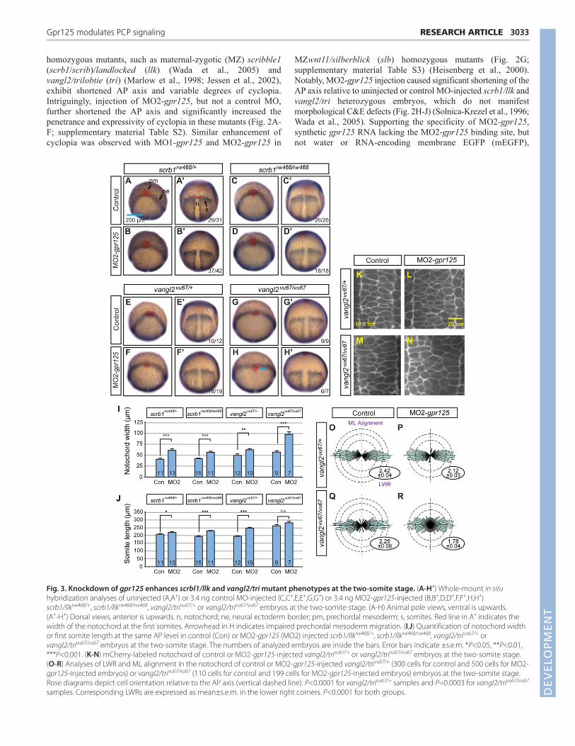
homozygous mutants, such as maternal-zygotic (MZ) scribble1(scrb1/scrib)/landlocked (llk) (Wada et al., 2005) andvangl2/trilobtie (tri) (Marlow et al., 1998; Jessen et al., 2002),exhibit shortened AP axis and variable degrees of cyclopia.Intriguingly, injection of MO2-gpr125, but not a control MO,further shortened the AP axis and significantly increased thepenetrance and expressivity of cyclopia in these mutants (Fig. 2A-F; supplementary material Table S2). Similar enhancement ofcyclopia was observed with MO1-gpr125 and MO2-gpr125 in
3033RESEARCH ARTICLEGpr125 modulates PCP signaling
MZwnt11/silberblick (slb) homozygous mutants (Fig. 2G;supplementary material Table S3) (Heisenberg et al., 2000).Notably, MO2-gpr125 injection caused significant shortening of theAP axis relative to uninjected or control MO-injected scrb1/llk andvangl2/tri heterozygous embryos, which do not manifestmorphological C&E defects (Fig. 2H-J) (Solnica-Krezel et al., 1996;Wada et al., 2005). Supporting the specificity of MO2-gpr125,synthetic gpr125 RNA lacking the MO2-gpr125 binding site, butnot water or RNA-encoding membrane EGFP (mEGFP),
Fig. 3. Knockdown of gpr125 enhances scrbl1/llk and vangl2/tri mutant phenotypes at the two-somite stage. (A-H�) Whole-mount in situhybridization analyses of uninjected (A,A�) or 3.4 ng control MO-injected (C,C�,E,E�,G,G�) or 3.4 ng MO2-gpr125-injected (B,B�,D,D�,F,F�,H,H�)scrb1/llkrw468/+, scrb1/llkrw468/rw468, vangl2/trivu67/+ or vangl2/trivu67/vu67 embryos at the two-somite stage. (A-H) Animal pole views, ventral is upwards. (A�-H�) Dorsal views, anterior is upwards. n, notochord; ne, neural ectoderm border; pm, prechordal mesoderm; s, somites. Red line in A� indicates thewidth of the notochord at the first somites. Arrowhead in H indicates impaired prechordal mesoderm migration. (I,J) Quantification of notochord widthor first somite length at the same AP level in control (Con) or MO2-gpr125 (MO2) injected scrb1/llkrw468/+, scrb1/llkrw468/rw468, vangl2/trivu67/+ orvangl2/trivu67/vu67 embryos at the two-somite stage. The numbers of analyzed embryos are inside the bars. Error bars indicate ±s.e.m. *P<0.05, **P<0.01,***P<0.001. (K-N) mCherry-labeled notochord of control or MO2-gpr125-injected vangl2/trivu67/+ or vangl2/trivu67/vu67 embryos at the two-somite stage.(O-R) Analyses of LWR and ML alignment in the notochord of control or MO2-gpr125-injected vangl2/trivu67/+ (300 cells for control and 500 cells for MO2-gpr125-injected embryos) or vangl2/trivu67/vu67 (110 cells for control and 199 cells for MO2-gpr125-injected embryos) embryos at the two-somite stage.Rose diagrams depict cell orientation relative to the AP axis (vertical dashed line). P<0.0001 for vangl2/trivu67/+ samples and P=0.0003 for vangl2/trivu67/vu67
samples. Corresponding LWRs are expressed as mean±s.e.m. in the lower right corners. P<0.0001 for both groups. DEVELO
PMENT
3034
significantly suppressed MO2-gpr125 enhancement of AP axisshortening of both vangl2/tri homozygotes and heterozygotes(Fig. 2K-M), and cyclopia defects of vangl2/tri homozygotes(Fig. 2F; supplementary material Table S2). Consistent with theenhanced axis shortening at 1 dpf, MO2-gpr125 injection causedwider and shorter neural ectoderm and axial and paraxial mesodermin scrb1/llk and vangl2/tri homozygotes, and heterozygotes at thetwo-somite stage (Fig. 3A-J). At the cellular level, reduced Gpr125function caused significant reduction of LWR and mediolateralalignment of cells in the notochord compared with control vangl2/triheterozygotes and homozygotes (Fig. 3K-R). In summary, theseresults indicate that when PCP signaling is reduced, Gpr125function becomes crucial for polarized cell behaviors underlyingC&E.
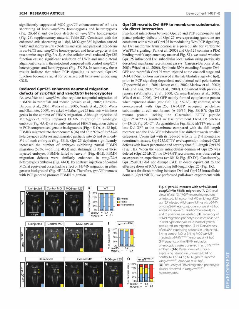
Reduced Gpr125 enhances neuronal migrationdefects of scrb1/llk and vangl2/tri heterozygotesAs scrb1/llk and vangl2/tri also regulate tangential migration ofFBMNs in zebrafish and mouse (Jessen et al., 2002; Carreira-Barbosa et al., 2003; Wada et al., 2005; Wada et al., 2006; Wadaand Okamoto, 2009), we asked whether gpr125 interacts with thesegenes in the context of FBMN migration. Although injection ofMO2-gpr125 rarely impaired FBMN migration in wild-typeembryos (Fig. 4A-D), it strongly enhanced FBMN migration defectsin PCP compromised genetic backgrounds (Fig. 4E-O). At 48 hpf,FBMNs migrated into rhombomere 6 (r6) and r7 in 92% of scrb1/llkheterozygous embryos and migrated partially into r5 and r6 in only8% of such embryos (Fig. 4E,I). Gpr125 depletion significantlyincreased the number of embryos exhibiting partial FBMNmigration (57%, n=65; Fig. 4G,I) and, strikingly, in 35% of theseinjected embryos, FBMNs failed to leave r4 (Fig. 4H,I). FBMNmigration defects were similarly enhanced in vangl2/triheterozygous embryos (Fig. 4J-O). By contrast, injection of controlMOs at equivalent doses had no effect on FBMN migration in eithergenetic background (Fig. 4F,I,L,M,O). Therefore, gpr125 interactswith PCP genes to promote FBMN migration.
RESEARCH ARTICLE Development 140 (14)
Gpr125 recruits Dvl-GFP to membrane subdomainsvia direct interactionFunctional interactions between Gpr125 and PCP components andplanar polarity defects of Gpr125 overexpressing gastrulae areconsistent with a role of Gpr125 in modulating Wnt/PCP signaling.As Dvl membrane translocation is a prerequisite for vertebrateWnt/PCP signaling (Park et al., 2005) and Gpr125 contains a PDZbinding motif (supplementary material Fig. S1), we tested whetherGpr125 influenced Dvl subcellular localization using previouslydescribed membrane recruitment assays (Carreira-Barbosa et al.,2003; Witzel et al., 2006). Synthetic RNA encoding Xenopus Dvl-GFP and zebrafish Gpr125 were injected at the one-cell stage andDvl-GFP distribution was assayed at the late blastula stage (4-5 hpf),prior to PCP signaling-dependent mediolateral cell polarization(Topczewski et al., 2001; Jessen et al., 2002; Marlow et al., 2002;Tada and Kai, 2009; Yin et al., 2009). Consistent with previousreports (Wallingford et al., 2000; Carreira-Barbosa et al., 2003;Witzel et al., 2006), Dvl-GFP mainly formed cytoplasmic punctawhen expressed alone (n=20/20; Fig. 5A-A�). By contrast, whenco-expressed with Gpr125, Dvl-GFP occupied patch-likesubdomains at cell membranes (n=36/36; Fig. 5B-B�). Gpr125mutant protein lacking the C-terminal ETTV peptide(gpr125DETTV) resulted in less prominent Dvl-GFP patches(n=13/13; Fig. 5C-C�). As quantified in Fig. 5E,F, ∆ETTV recruitedless Dvl-GFP to the membrane compared with the full-lengthreceptor, and the Dvl-GFP subdomain size shifted towards smallercategories. Consistent with its reduced activity in Dvl membranerecruitment assays, Gpr125∆ETTV overexpression induced C&Edefects with lower penetrance and severity than full-length Gpr125(Fig. 1K). When the entire intracellular domain of Gpr125 wasdeleted (Gpr125∆ICD), no Dvl-GFP recruitment was observed inco-expression experiments (n=18/18; Fig. 5D-D�). Consistently,Gpr125∆ICD did not disrupt C&E at doses equivalent to theeffective doses of RNA encoding full-length Gpr125 (Fig. 1K).
To test for direct binding between Dvl and Gpr125 intracellulardomain (Gpr125ICD), we performed pull-down experiments with
Fig. 4. gpr125 interacts with scrb1/llk andvangl2/tri in FBMN migration. (A-C) Dorsalviews of islet1(isl1):GFP-expressing neurons inuninjected, 3.4 ng control MO or 3.4 ng MO2-gpr125 injected wild-type siblings of scrb1/llkor vangl2/tri heterozygous embryos at 48 hpf.Anterior is upwards. r4 (rhombomere 4), r5and r6 positions are labeled. (D) Frequency ofFBMN migration phenotypic classes observedin wild-type embryos. Blue, normal; yellow,partial; red, no migration. (E-H) Dorsal viewsof isl1:GFP-expressing neurons in uninjected,3.4 ng control MO or 3.4 ng MO2-gpr125-injected scrb1/llkrw468/+ embryos at 48 hpf. (I) Frequency of the FBMN migrationphenotypic classes observed in scrb1/llkrw468/+
embryos. (J-N) Dorsal views of isl1:GFP-expressing neurons in uninjected, 3.4 ngcontrol MO or 3.4 ng MO2-gpr125-injectedvangl2/trivu67/+ embryos at 48 hpf. (O) Frequency of FBMN migration phenotypicclasses observed in vangl2/trivu67/+
heterozygotes.
DEVELO
PMENT

purified GST-Gpr125ICD, GST-Gpr125ICD∆ETTV fusion proteinsand in vitro translated Xenopus Dvl-GFP (Fig. 5G,H). We foundthat GST-Gpr125ICD pulled down Dvl-GFP, indicative of a directinteraction. The ∆ETTV form pulled down less Dvl-GFP (Fig. 5H),suggesting that ETTV promotes Gpr125ICD binding to Dvl. Takentogether, our results suggest that Gpr125 modulates PCP signalingby interacting with Dvl and promoting its accumulation inmembrane subdomains.
Dvl clusters Gpr125 and select PCP componentsinto membrane subdomainsCytoplasmic core PCP components, including Dvl, cluster PCPcomplexes in the cell membranes of Drosophila pupal wings (Struttet al., 2011). Given that Dvl-GFP localized to membranesubdomains when co-expressed with Gpr125, we asked whetherGpr125 colocalized with Dvl in these membrane subdomains usinga Gpr125 C-terminal Cherry fusion protein (Gpr125-Cherry),which, when overexpressed, impaired C&E movements andunderlying cell polarity similarly to the wild-type protein(Fig. 1K,Q,S,U). In zebrafish blastulae, Gpr125-Cherry expressedalone displayed uniform membrane distribution (n=17/17; Fig. 6A-A�), but it colocalized with Dvl-GFP in prominent membranesubdomains in co-expression experiments (n=12/12; Fig. 6B-B�;supplementary material Movie 1). Similarly, when co-expressed,
3035RESEARCH ARTICLEGpr125 modulates PCP signaling
Dvl-GFP clustered Gpc4/Kny-GFP into membrane subdomains inlate blastulae (n=10/12, Fig. 6D-D�; supplementary material Movie2). Interestingly, these Dvl-mediated PCP membrane subdomainspreferentially localized at the central regions of cell contactsbetween neighboring blastomeres (Fig. 6E). In addition, Dvl-GFPpromoted uniform membrane localization of endogenous Vangl2/Triin late blastulae (n=4/5; Fig. 6G�), when endogenous Tri/Vangl2was mainly cytoplasmic in uninjected embryos (Fig. 6F).
Next, we investigated whether Gpr125 influenced the distributionof other PCP components, including Fzd7-CFP, Gpc4/Kny-GFP andendogenous Vangl2 in blastulae expressing Gpr125-Cherry, but nochange of their distribution was observed (supplementary materialFig. S3). By contrast, when co-expressed with Dvl-YFP, Gpr125-Cherry, Fzd7-CFP and Dvl-YFP colocalized in membranesubdomains (n=17/22; Fig. 7A-A�). Moreover, mosaic expression ofGpr125 enhanced Gpc4/Kny-GFP clustering when Dvl wasoverexpressed (n=6/10; Fig. 7B-B�). In contrast to Fzd7 and Gpc4,neither endogenous nor overexpressed zebrafish Vangl2 wasenriched in Gpr125-Cherry and Dvl-GFP-containing subdomains(C-F; n=15/15 for endogenous Vangl2; n=5/5 for overexpressedVangl2). These results suggest that, analogous to Drosophila,distinct PCP complexes can form in vertebrates, and Fzd7 andGpc4/Kny may be components of large Dvl-containing proteincomplexes, the formation of which is promoted by Gpr125.
Fig. 5. Gpr125 promotes Dvl-GFP localization in discrete membrane subdomains via direct interaction. (A-D�) Animal pole views of live embryosat 4-5 hpf co-injected with 150 pg dvl-GFP and 50 pg mCherry RNA in the absence (A-A�) and presence of 380 pg gpr125FL (B-B�), gpr125ΔETTV (C-C�) orgpr125ΔICD RNA (D-D�). Arrows in B� indicate Dvl-GFP membrane subdomains. (E) Ratio of Dvl-GFP membrane area to the length of the membranemeasured on embryos expressing full-length or ΔETTV Gpr125. Numbers of membranes analyzed are inside bars. Data are mean±s.e.m. ***P<0.001. (F) Size distribution of Dvl-GFP membrane subdomains in embryos expressing full-length or ΔETTV Gpr125. Numbers of membranes analyzed areinside bars. (G,H) Pull-down assay with GST- and GFP-fusion proteins. Ten percent of GFP-fusion protein inputs were blotted with anti-GFP antibody and100% GST fusion protein inputs were stained with Denville Blue Protein Stain (G). Pull-down results were analyzed by western blotting using anti-GFPand anti-GST antibodies (H).
DEVELO
PMENT

3036
To assess the interaction between Gpr125, Dvl and Vangl2 duringC&E movements, we examined the relative distribution of Gpr125-Cherry, Dvl-GFP and endogenous Vangl2 at 10 hpf. Gpr125-Cherrylocalized to cell membranes and formed puncta both on themembrane and in the cytosol (Fig. 8A�,A�). Endogenous Vangl2localized mainly to the cell membranes (Fig. 8B) and membranestaining was not observed in MZvang/trivu67/vu67 mutants (Fig. 8C).Intriguingly, when co-expressed during gastrulation, Gpr125-Cherryand Dvl-GFP colocalized in large membrane patches, butendogenous Vangl2 was not enriched in Gpr125-Cherry:Dvl-GFPpatches (Fig. 8D-F�). Therefore, Gpr125 might primarily interactwith Dvl-containing protein complexes during C&E movements.
DISCUSSIONPreviously, the expression of Gpr125 was reported in various tissuesof mouse embryos and adults, including the pluripotentspermatogonial progenitor cells (Seandel et al., 2007; Homma et al.,2009; Pickering et al., 2008); however, its function was not known.Here, we have identified zebrafish Gpr125 as a novel modulator ofC&E gastrulation movements and tangential FBMN migration, twoprocesses evolutionarily conserved among vertebrates that requirePCP signaling (Wada and Okamoto, 2009; Gray et al., 2011). Towardselucidating the genetic and cellular mechanisms by which Gpr125regulates these processes, we showed that excess Gpr125 impairedWnt/PCP-dependent cellular polarities underlying normal C&Egastrulation movements. Moreover, reduction of gpr125 expressionexacerbated C&E and neuronal migration defects of several Wnt/PCPcomponent mutants. At the molecular level, we showed that Gpr125
RESEARCH ARTICLE Development 140 (14)
interacted with and recruited Dvl into membrane subdomains, andpromoted accumulation of select PCP components in such membranesubdomains.
We created a gpr125 loss-of-function condition with twoantisense MOs, which effectively blocked translation of syntheticRNA encoding GFP fused to gpr125 MO target sequences.However, the effectiveness of the MOs in blocking translation ofendogenous Gpr125 protein could not be evaluated because Gpr125antibodies are unavailable. Nevertheless, these MOs probablycreated at least a partial loss-of-function condition, as they enhancedthe C&E gastrulation and FBMN migration defects of homozygousand heterozygous PCP mutants, whereas a control MO did not(Figs 2-4). Similar to the interaction between gpr125 and PCPpathway genes reported here, exacerbation of C&E defects has beenreported for compound PCP pathway mutants compared with singlemutants (Marlow et al., 1998; Carreira-Barbosa et al., 2003; Kilianet al., 2003). More importantly, co-injecting gpr125 RNA lackingthe MO targeting sequence partially suppressed the exacerbatedC&E defects in MO2-gpr125-injected PCP mutants. Similar togpr125 MO-injected wild-type zebrafish embryos, Gpr125 knock-in null mice are grossly normal and fertile (Seandel et al., 2007).As gpr125 RNA is maternally deposited and we could not determinethe abundance of maternal protein, the lack of early developmentaldefects in gpr125 morphants could be due to maternal proteincontribution. Alternatively, as observed for celsr/flamingo genes,redundancy with other adhesion GPCRs or PCP pathwaycomponents might mask the loss of Gpr125 function (Carreira-Barbosa et al., 2009).
Fig. 6. Dvl clusters Gpr125 andGpc4/Kny into membranesubdomains and promotesuniform Vangl2/Tri membranelocalization in late blastulae (4-5hpf). (A-B�) Animal pole views oflive blastulae co-injected with 267pg gpr125-Cherry RNA and either150 pg mEGFP RNA (A-A�) or 150 pgdvl-GFP RNA (B-B�). Arrows in B�indicate Gpr125-Cherry:Dvl-GFPmembrane subdomains. (C-D�) Animal pole views of liveblastulae co-injected with 60 pggpc4/kny-GFP RNA, 50 pg mCherryRNA (C-C�) and 150 pg dvl RNA (D-D�). Arrows in D� indicate Gpc4/Kny-GFP membrane subdomains. (E) Graphic representation of therelative distribution of Dvl-GFP:Gpr125-Cherry and Gpc4/Kny-GFP:Dvl subdomains along cellmembranes. Membrane length wasnormalized as one. (F,F�) Animalpole views of a whole-mountimmunostained wild-type blastulawith Vangl2 and β-cateninantibodies. (G-G�) Animal poleviews of a 50 pg mCherry and 150pg dvl-GFP RNA-injected blastulaimmunostained for GFP, RFP andVangl2.
DEVELO
PMENT

We showed that the Gpr125 intracellular domain interacteddirectly with Dvl in pull-down experiments (Fig. 5) and wasrequired for Dvl recruitment into membrane subdomains uponGpr125 overexpression in zebrafish blastula (Fig. 5). Given that Dvlmembrane translocation is essential for vertebrate Wnt/PCPsignaling (Park et al., 2005) and C&E movements are altered byexcess PCP components (Wallingford et al., 2000; Topczewski etal., 2001; Jessen et al., 2002; Marlow et al., 2002; Carreira-Barbosaet al., 2003; Formstone and Mason, 2005), this interaction probablyin part accounts for C&E defects caused by Gpr125 gain of functionand possibly the exacerbated C&E defects caused by Gpr125 loss
3037RESEARCH ARTICLEGpr125 modulates PCP signaling
of function in PCP mutants. Interestingly, we did not detectsignificant differences in mediolateral elongation or cell orientationamong cells with distinct Pk distribution patterns in either controlgastrulae or those overexpressing Gpr125 (data not shown).Moreover, we observed little change in Pk-GFP distribution due toGpr125 depletion in both wild type and vangl2/tri heterozygotes(data not shown). Therefore, it is plausible that Pk does not interactdirectly with Gpr125. It will be important to examine whetherGpr125 loss of function would influence Dvl distribution. As Dvl isnot required for FBMN migration (Jessen et al., 2002; Wada andOkamoto, 2009), Gpr125 and the relevant PCP componentsprobably regulate FBMN migration and C&E via distinctmechanisms.
We found that the PDZ-binding motif of Gpr125 was partiallyresponsible for Dvl binding and recruitment (Fig. 5). Therequirement of the PDZ-binding motif for Dvl binding varies amongdifferent proteins. It is dispensable for binding of Fzd or Vangl2/Trito Dvl (Umbhauer et al., 2000; Park and Moon, 2002; Wong et al.,2003). However, the PDZ-binding motif mediates direct binding
Fig. 7. Gpr125 promotes localization of select PCP components inDvl-containing membrane subdomains in late blastulae (4-5 hpf).(A-A�) Live blastula co-injected with 110 pg fzd7-CFP, 150 pg dvl-YFP and300 pg gpr125-Cherry RNA. (B-B�) Live blastula co-injected with 60 pggpc4/kny-GFP, 150 pg dvl and 50 pg mCherry at the one-cell stage, and 4pg H2B-RFP and 20 pg gpr125FL RNA in one blastomere at the 16- to ~32-cell stage. The star in B� marks an H2B-RFP-positive nucleus and arrowsindicate membrane subdomains. (C-D�) Blastula injected with 300 pggpr125-Cherry, 150 pg dvl-GFP (C-C�) and 50 pg zebrafish vangl2/tri RNA(D-D�) immunostained for GFP, RFP and Vangl2. Animal pole views in A-D.(E,F) Quantification of fluorescence intensity ratios inside/outside domainfor Vangl2, Gpr125-Cherry and Dvl-GFP in embryos injected with 300 pggpr125-Cherry, 150 pg dvl-GFP (E) and 50 pg zebrafish vangl2 RNA (F). Dataare mean±s.e.m.
Fig. 8. Gpr125-Cherry and Dvl-GFP colocalize during gastrulation(10 hpf). (A-A�) Dorsal views of a gastrula injected with 50 pg gpr125-Cherry and 150 pg mEGFP RNA. Broken white lines outline the notochord.(B-C�) Whole-mount immunostaining for endogenous Vangl2 and β-catenin in wild-type (B) or MZvang/trivu67/vu67 gastrulae (C-C�). (D-F�) Dorsal mesoderm of gastrulae co-injected with 150 pg mEGFP and300 pg gpr125-Cherry RNA (D-D�), 150 pg dvl-GFP and 50 pg mCherry RNA(E-E�), or 150 pg dvl-GFP RNA and 300 pg gpr125-Cherry RNA (F-F�), andimmunostained for GFP, RFP and Vangl2.
DEVELO
PMENT

3038
between Xenopus Dvl and its cytoplasmic interacting proteinDapper/Dact (Cheyette et al., 2002; Gloy et al., 2002; Wong et al.,2003; Teran et al., 2009). As in Gpr125, the Dapper PDZ-bindingmotif, is –TTV and the threonine at the −2 position is reported to bewithin hydrogen bonding distance of a highly conserved arginine325 residue present in Dvl proteins, and is also essential for Dvlinteraction with Dapper (Cheyette et al., 2002). Additional Gpr125motif(s) mediating Dvl binding remain to be defined.
Previous reports show that Fzd7 recruits Dvl uniformly to the cellmembrane when overexpressed in the zebrafish blastula andpromotes Dvl accumulation into discrete membrane subdomainswhen co-expressed with Wnt11 (Witzel et al., 2006). We observedthat Dvl clustered Gpr125 into membrane subdomains, and viceversa, even without co-expression of Wnt11 (Figs 5, 6). Notably,Gpr125 promoted accumulation of Fzd7 and Gpc4/Kny in thesubdomains (Fig. 7). These observations are consistent with arecently discovered role for endogenous Dsh in clustering PCPcomplexes into membrane subdomains in Drosophila wing epithelia(Strutt et al., 2011). Moreover, our study raises the possibility thatother proteins such as Gpr125 cooperate with Dvl to promoteformation of such membrane subdomains. Interestingly, DrosophilaPk forms membrane clusters when co-expressed with XenopusVangl2 in Xenopus animal cap explants (Jenny et al., 2003), but inzebrafish blastula, Pk co-expression inhibits Fzd7-mediatedrecruitment of Dvl to the cell membrane possibly by destabilizingDvl (Carreira-Barbosa et al., 2003). Based on additional evidencethat Pk and Dvl fusion proteins localize to opposing cell edges inzebrafish gastrula (Ciruna et al., 2006; Yin et al., 2008), it istempting to speculate that distinct clusters of endogenous PCPcomplexes might exist during C&E movements in vertebrates.Moreover, because the membrane subdomains containing Gpr125and Dvl were enriched in Fzd and Gpc4/Kny but not in Vangl2/Tri,it is also possible that Gpr125 could be involved in formation ofasymmetric PCP complexes. As proposed for Drosophila PCPsignaling, clustering of PCP complexes could afford a self-enhancement mechanism contributing to the establishment and/ormaintenance of planar polarity (Strutt et al., 2011). During C&E inparticular, as mesenchymal cells are moving and changing theircontacts rather frequently, the local organization of PCP proteinsinto subdomains could facilitate efficient establishment of planarpolarity in the context of dynamic cell rearrangements.
It is unclear how clustering of PCP complexes might contributeto polarized cell behaviors driving C&E movements. Nevertheless,formation of Wnt11:Fzd7:Dvl subdomains has been correlated withincreased persistence of membrane contacts. In addition, Celsrshave been demonstrated to contribute substantially to this effect,likely via their ability to mediate adhesion (Usui et al., 1999; Shimaet al., 2004; Witzel et al., 2006). Like Celsrs, Gpr125 is an adhesionGPCR and its extracellular domain contains protein modules knownto mediate protein-ligand interactions suitable for regulatingintercellular communication and cell adhesion (de Wit et al., 2011;Pal et al., 2012). Therefore, it is worth testing in the future whetherGpr125 might function in PCP subdomains to regulate celladhesion.
In summary, we identified zebrafish Gpr125 as a novel modulatorof C&E gastrulation movements and tangential FBMN migration.Gpr125 influences Wnt/PCP pathway activity in part via interactingwith and modulating the distribution of Dvl. Our discovery thatGpr125 contributes to C&E during gastrulation, a processes whereall known PCP components act, and later during FBMN migration,where only a subset of PCP genes are required, opens up excitingavenues for further studies of Gpr125 function, in particular towards
RESEARCH ARTICLE Development 140 (14)
understanding how Wnt/PCP signaling regulates cell and tissuepolarity in distinct contexts.
AcknowledgementsWe thank Drs Andreas Jenny, Carl-Philipp Heisenberg, Fang Lin, HitoshiOkamoto, John Wallingford, Lei Feng and Avik Choudhuri for their generosityin sharing reagents and fish lines; Drs Andreas Jenny, Adrian Santos-Ledo, KellyMonk and Ryan S. Gray for comments on the manuscript; Linda Lobos forediting; and Analytical Imaging Facility at Albert Einstein College of Medicine,Dr Y. G. Yeung, and F.L.M. and L.S.K. lab members for helpful discussions andtechnical support. We acknowledge the research assistants in our fish facilitiesfor fish care.
FundingThis work was supported in part by National Institutes of Health grants[R01GM089979 to F.L.M. and R01GM77770 and GM55101 to L.S.K.].Deposited in PMC for release after 12 months.
Competing interests statementThe authors declare no competing financial interests.
Author contributionsAll authors conceived and designed the experiments, analyzed and discussedthe data, and discussed the manuscript. X.L., I.R., D.S.S. and M.N. performedthe experiments. H.E.H., F.L.M. and L.S.-K. contributed reagents, materials andanalysis tools. X.L. wrote the manuscript with the assistance of L.S.-K., F.L.M.,I.R. and D.S.S.
Supplementary materialSupplementary material available online athttp://dev.biologists.org/lookup/suppl/doi:10.1242/dev.094839/-/DC1
ReferencesAxelrod, J. D. (2001). Unipolar membrane association of Dishevelled mediates
Frizzled planar cell polarity signaling. Genes Dev. 15, 1182-1187.Bastock, R., Strutt, H. and Strutt, D. (2003). Strabismus is asymmetrically
localised and binds to Prickle and Dishevelled during Drosophila planarpolarity patterning. Development 130, 3007-3014.
Berghmans, S., Murphey, R. D., Wienholds, E., Neuberg, D., Kutok, J. L.,Fletcher, C. D., Morris, J. P., Liu, T. X., Schulte-Merker, S., Kanki, J. P. et al.(2005). tp53 mutant zebrafish develop malignant peripheral nerve sheathtumors. Proc. Natl. Acad. Sci. USA 102, 407-412.
Carreira-Barbosa, F., Concha, M. L., Takeuchi, M., Ueno, N., Wilson, S. W. andTada, M. (2003). Prickle 1 regulates cell movements during gastrulation andneuronal migration in zebrafish. Development 130, 4037-4046.
Carreira-Barbosa, F., Kajita, M., Morel, V., Wada, H., Okamoto, H., MartinezArias, A., Fujita, Y., Wilson, S. W. and Tada, M. (2009). Flamingo regulatesepiboly and convergence/extension movements through cell cohesive andsignalling functions during zebrafish gastrulation. Development 136, 383-392.
Cheyette, B. N., Waxman, J. S., Miller, J. R., Takemaru, K., Sheldahl, L. C.,Khlebtsova, N., Fox, E. P., Earnest, T. and Moon, R. T. (2002). Dapper, aDishevelled-associated antagonist of beta-catenin and JNK signaling, isrequired for notochord formation. Dev. Cell 2, 449-461.
Ciruna, B., Jenny, A., Lee, D., Mlodzik, M. and Schier, A. F. (2006). Planar cellpolarity signalling couples cell division and morphogenesis duringneurulation. Nature 439, 220-224.
de Wit, J., Hong, W., Luo, L. and Ghosh, A. (2011). Role of leucine-rich repeatproteins in the development and function of neural circuits. Annu. Rev. Cell Dev.Biol. 27, 697-729.
Feiguin, F., Hannus, M., Mlodzik, M. and Eaton, S. (2001). The ankyrin repeatprotein Diego mediates Frizzled-dependent planar polarization. Dev. Cell 1, 93-101.
Formstone, C. J. and Mason, I. (2005). Combinatorial activity of Flamingoproteins directs convergence and extension within the early zebrafish embryovia the planar cell polarity pathway. Dev. Biol. 282, 320-335.
Fredriksson, R., Lagerström, M. C., Lundin, L. G. and Schiöth, H. B. (2003).The G-protein-coupled receptors in the human genome form five mainfamilies. Phylogenetic analysis, paralogon groups, and fingerprints. Mol.Pharmacol. 63, 1256-1272.
Gloy, J., Hikasa, H. and Sokol, S. Y. (2002). Frodo interacts with Dishevelled totransduce Wnt signals. Nat. Cell Biol. 4, 351-357.
Goodrich, L. V. and Strutt, D. (2011). Principles of planar polarity in animaldevelopment. Development 138, 1877-1892.
Gray, R. S., Roszko, I. and Solnica-Krezel, L. (2011). Planar cell polarity:coordinating morphogenetic cell behaviors with embryonic polarity. Dev. Cell21, 120-133. D
EVELO
PMENT

3039RESEARCH ARTICLEGpr125 modulates PCP signaling
Heisenberg, C. P., Tada, M., Rauch, G. J., Saúde, L., Concha, M. L., Geisler, R.,Stemple, D. L., Smith, J. C. and Wilson, S. W. (2000). Silberblick/Wnt11mediates convergent extension movements during zebrafish gastrulation.Nature 405, 76-81.
Hering, H. and Sheng, M. (2002). Direct interaction of Frizzled-1, -2, -4, and -7with PDZ domains of PSD-95. FEBS Lett. 521, 185-189.
Homma, S., Shimada, T., Hikake, T. and Yaginuma, H. (2009). Expressionpattern of LRR and Ig domain-containing protein (LRRIG protein) in the earlymouse embryo. Gene Expr. Patterns 9, 1-26.
Jenny, A., Darken, R. S., Wilson, P. A. and Mlodzik, M. (2003). Prickle andStrabismus form a functional complex to generate a correct axis during planarcell polarity signaling. EMBO J. 22, 4409-4420.
Jessen, J. R., Topczewski, J., Bingham, S., Sepich, D. S., Marlow, F.,Chandrasekhar, A. and Solnica-Krezel, L. (2002). Zebrafish trilobite identifiesnew roles for Strabismus in gastrulation and neuronal movements. Nat. CellBiol. 4, 610-615.
Keller, R., Davidson, L., Edlund, A., Elul, T., Ezin, M., Shook, D. and Skoglund,P. (2000). Mechanisms of convergence and extension by cell intercalation.Philos. Trans. R. Soc. B 355, 897-922.
Kilian, B., Mansukoski, H., Barbosa, F. C., Ulrich, F., Tada, M. andHeisenberg, C. P. (2003). The role of Ppt/Wnt5 in regulating cell shape andmovement during zebrafish gastrulation. Mech. Dev. 120, 467-476.
Kimmel, C. B., Ballard, W. W., Kimmel, S. R., Ullmann, B. and Schilling, T. F.(1995). Stages of embryonic development of the zebrafish. Dev. Dyn. 203, 253-310.
Kuhnert, F., Mancuso, M. R., Shamloo, A., Wang, H. T., Choksi, V., Florek, M.,Su, H., Fruttiger, M., Young, W. L., Heilshorn, S. C. et al. (2010). Essentialregulation of CNS angiogenesis by the orphan G protein-coupled receptorGPR124. Science 330, 985-989.
Lin, F., Sepich, D. S., Chen, S., Topczewski, J., Yin, C., Solnica-Krezel, L. andHamm, H. (2005). Essential roles of Galpha12/13 signaling in distinct cellbehaviors driving zebrafish convergence and extension gastrulationmovements. J. Cell Biol. 169, 777-787.
Lin, F., Chen, S., Sepich, D. S., Panizzi, J. R., Clendenon, S. G., Marrs, J. A.,Hamm, H. E. and Solnica-Krezel, L. (2009). Galpha12/13 regulate epiboly byinhibiting E-cadherin activity and modulating the actin cytoskeleton. J. CellBiol. 184, 909-921.
Marlow, F., Zwartkruis, F., Malicki, J., Neuhauss, S. C., Abbas, L., Weaver, M.,Driever, W. and Solnica-Krezel, L. (1998). Functional interactions of genesmediating convergent extension, knypek and trilobite, during the partitioningof the eye primordium in zebrafish. Dev. Biol. 203, 382-399.
Marlow, F., Topczewski, J., Sepich, D. and Solnica-Krezel, L. (2002). ZebrafishRho kinase 2 acts downstream of Wnt11 to mediate cell polarity and effectiveconvergence and extension movements. Curr. Biol. 12, 876-884.
McNeill, H. (2010). Planar cell polarity: keeping hairs straight is not so simple.Cold Spring Harb. Perspect. Biol. 2, a003376.
Monk, K. R., Naylor, S. G., Glenn, T. D., Mercurio, S., Perlin, J. R., Dominguez,C., Moens, C. B. and Talbot, W. S. (2009). A G protein-coupled receptor isessential for Schwann cells to initiate myelination. Science 325, 1402-1405.
Monk, K. R., Oshima, K., Jörs, S., Heller, S. and Talbot, W. S. (2011). Gpr126 isessential for peripheral nerve development and myelination in mammals.Development 138, 2673-2680.
Myers, D. C., Sepich, D. S. and Solnica-Krezel, L. (2002). Bmp activity gradientregulates convergent extension during zebrafish gastrulation. Dev. Biol. 243,81-98.
Pal, K., Melcher, K. and Xu, H. E. (2012). Structure and mechanism forrecognition of peptide hormones by Class B G-protein-coupled receptors. ActaPharmacol. Sin. 33, 300-311.
Park, M. and Moon, R. T. (2002). The planar cell-polarity gene stbm regulatescell behaviour and cell fate in vertebrate embryos. Nat. Cell Biol. 4, 20-25.
Park, T. J., Gray, R. S., Sato, A., Habas, R. and Wallingford, J. B. (2005).Subcellular localization and signaling properties of dishevelled in developingvertebrate embryos. Curr. Biol. 15, 1039-1044.
Piao, X., Hill, R. S., Bodell, A., Chang, B. S., Basel-Vanagaite, L., Straussberg,R., Dobyns, W. B., Qasrawi, B., Winter, R. M., Innes, A. M. et al. (2004). Gprotein-coupled receptor-dependent development of human frontal cortex.Science 303, 2033-2036.
Pickering, C., Hägglund, M., Szmydynger-Chodobska, J., Marques, F., Palha,J. A., Waller, L., Chodobski, A., Fredriksson, R., Lagerström, M. C. andSchiöth, H. B. (2008). The Adhesion GPCR GPR125 is specifically expressed inthe choroid plexus and is upregulated following brain injury. BMC Neurosci. 9,97.
Robu, M. E., Larson, J. D., Nasevicius, A., Beiraghi, S., Brenner, C., Farber, S.A. and Ekker, S. C. (2007). p53 activation by knockdown technologies. PLoSGenet. 3, e78.
Schindelin, J., Arganda-Carreras, I., Frise, E., Kaynig, V., Longair, M.,Pietzsch, T., Preibisch, S., Rueden, C., Saalfeld, S., Schmid, B. et al. (2012).
Fiji: an open-source platform for biological-image analysis. Nat. Methods 9,676-682.
Seandel, M., James, D., Shmelkov, S. V., Falciatori, I., Kim, J., Chavala, S.,Scherr, D. S., Zhang, F., Torres, R., Gale, N. W. et al. (2007). Generation offunctional multipotent adult stem cells from GPR125+ germline progenitors.Nature 449, 346-350.
Shima, Y., Kengaku, M., Hirano, T., Takeichi, M. and Uemura, T. (2004).Regulation of dendritic maintenance and growth by a mammalian 7-passtransmembrane cadherin. Dev. Cell 7, 205-216.
Simons, M. and Mlodzik, M. (2008). Planar cell polarity signaling: from flydevelopment to human disease. Annu. Rev. Genet. 42, 517-540.
Solnica-Krezel, L. (2005). Conserved patterns of cell movements duringvertebrate gastrulation. Curr. Biol. 15, R213-R228.
Solnica-Krezel, L., Stemple, D. L., Mountcastle-Shah, E., Rangini, Z.,Neuhauss, S. C., Malicki, J., Schier, A. F., Stainier, D. Y., Zwartkruis, F.,Abdelilah, S. et al. (1996). Mutations affecting cell fates and cellularrearrangements during gastrulation in zebrafish. Development 123, 67-80.
Strutt, D., Johnson, R., Cooper, K. and Bray, S. (2002). Asymmetric localizationof frizzled and the determination of notch-dependent cell fate in theDrosophila eye. Curr. Biol. 12, 813-824.
Strutt, H., Warrington, S. J. and Strutt, D. (2011). Dynamics of core planarpolarity protein turnover and stable assembly into discrete membranesubdomains. Dev. Cell 20, 511-525.
Tada, M. and Kai, M. (2009). Noncanonical Wnt/PCP signaling during vertebrategastrulation. Zebrafish 6, 29-40.
Teran, E., Branscomb, A. D. and Seeling, J. M. (2009). Dpr Acts as a molecularswitch, inhibiting Wnt signaling when unphosphorylated, but promoting Wntsignaling when phosphorylated by casein kinase Idelta/epsilon. PLoS ONE 4,e5522.
Topczewski, J., Sepich, D. S., Myers, D. C., Walker, C., Amores, A., Lele, Z.,Hammerschmidt, M., Postlethwait, J. and Solnica-Krezel, L. (2001). Thezebrafish glypican knypek controls cell polarity during gastrulationmovements of convergent extension. Dev. Cell 1, 251-264.
Tree, D. R., Shulman, J. M., Rousset, R., Scott, M. P., Gubb, D. and Axelrod, J.D. (2002). Prickle mediates feedback amplification to generate asymmetricplanar cell polarity signaling. Cell 109, 371-381.
Umbhauer, M., Djiane, A., Goisset, C., Penzo-Méndez, A., Riou, J. F.,Boucaut, J. C. and Shi, D. L. (2000). The C-terminal cytoplasmic Lys-thr-X-X-X-Trp motif in frizzled receptors mediates Wnt/beta-catenin signalling. EMBO J.19, 4944-4954.
Usui, T., Shima, Y., Shimada, Y., Hirano, S., Burgess, R. W., Schwarz, T. L.,Takeichi, M. and Uemura, T. (1999). Flamingo, a seven-pass transmembranecadherin, regulates planar cell polarity under the control of Frizzled. Cell 98,585-595.
Villefranc, J. A., Amigo, J. and Lawson, N. D. (2007). Gateway compatiblevectors for analysis of gene function in the zebrafish. Dev. Dyn. 236, 3077-3087.
Wada, H. and Okamoto, H. (2009). Roles of noncanonical Wnt/PCP pathwaygenes in neuronal migration and neurulation in zebrafish. Zebrafish 6, 3-8.
Wada, H., Iwasaki, M., Sato, T., Masai, I., Nishiwaki, Y., Tanaka, H., Sato, A.,Nojima, Y. and Okamoto, H. (2005). Dual roles of zygotic and maternalScribble1 in neural migration and convergent extension movements inzebrafish embryos. Development 132, 2273-2285.
Wada, H., Tanaka, H., Nakayama, S., Iwasaki, M. and Okamoto, H. (2006).Frizzled3a and Celsr2 function in the neuroepithelium to regulate migration offacial motor neurons in the developing zebrafish hindbrain. Development 133,4749-4759.
Wallingford, J. B., Rowning, B. A., Vogeli, K. M., Rothbächer, U., Fraser, S. E.and Harland, R. M. (2000). Dishevelled controls cell polarity during Xenopusgastrulation. Nature 405, 81-85.
Witzel, S., Zimyanin, V., Carreira-Barbosa, F., Tada, M. and Heisenberg, C. P.(2006). Wnt11 controls cell contact persistence by local accumulation ofFrizzled 7 at the plasma membrane. J. Cell Biol. 175, 791-802.
Wong, H. C., Bourdelas, A., Krauss, A., Lee, H. J., Shao, Y., Wu, D., Mlodzik,M., Shi, D. L. and Zheng, J. (2003). Direct binding of the PDZ domain ofDishevelled to a conserved internal sequence in the C-terminal region ofFrizzled. Mol. Cell 12, 1251-1260.
Yin, C., Kiskowski, M., Pouille, P. A., Farge, E. and Solnica-Krezel, L. (2008).Cooperation of polarized cell intercalations drives convergence and extensionof presomitic mesoderm during zebrafish gastrulation. J. Cell Biol. 180, 221-232.
Yin, C., Ciruna, B. and Solnica-Krezel, L. (2009). Convergence and extensionmovements during vertebrate gastrulation. Curr. Top. Dev. Biol. 89, 163-192.
Yona, S., Lin, H. H., Siu, W. O., Gordon, S. and Stacey, M. (2008). Adhesion-GPCRs: emerging roles for novel receptors. Trends Biochem. Sci. 33, 491-500.
Zeng, X. X., Wilm, T. P., Sepich, D. S. and Solnica-Krezel, L. (2007). Apelin andits receptor control heart field formation during zebrafish gastrulation. Dev. Cell12, 391-402.
DEVELO
PMENT