goussia janae n. sp. (apicomplexa, eimeriorina) in dace
TRANSCRIPT
Vol. 8: 85-90. 1990 l DISEASES OF AQUATIC ORGANISMS Dis. aquat. Org. 1 Published June 7
Goussia janae n. sp. (Apicomplexa, Eimeriorina) in dace Leuciscus leuciscus and chub L. cephalus
Julius LukeS, Iva Dykova
Institute of Parasitology, Czechoslovak Academy of Sciences, BraniSovska 31,370 05 c e s k e Budejovice, Czechoslovakia
ABSTRACT: Goussia janae n. sp. is described from the intestine of dace Leuciscus leuciscus and chub L. cephalus. Merogonic, gamogonic and sporogonic stages are localized intracellularly in the apical part of the epithelial cells just beneath the cell membrane, i.e. in the 'epicellular' position. Ellipsoidal oocysts have a mean size of 18.1 x 12.7 pm and are without oocyst residuum; ellipsoidal sporocysts (13.5 x 5.0 pm) lack Stieda body and sporocyst residuum.
INTRODUCTION
In higher vertebrates, all hitherto described coccid- ian species are localized beneath the cell membrane in the apices of intestinal epithelia1 cells and belong to the genus Cryptosporidium (Current 1986). In fishes, in addition to 2 species of the genus Cryptosporidium, species of 3 more genera - Eimeria, Epieimena and Goussia - were noted to possess a similar localization. There are marked differences among life cycles of individual species.
Dykova & Lom (1981) created a new genus Epieimeria for species with 'epicellular' merogony and gamogony, intracellular sporogony, and sporocysts having a Stieda body, with Epieimeria anguillae as the type species. The scope of the genus has been expanded to accommodate new species from marine fishes (Lom & Dykova 1982, Daoudi 1987, Daoudi et al. 1987). Coccidlans with Eimeria-type sporocysts lacking a Stieda body have also been shown to complete their life cycle 'epicellularly' in the gut epithelium; Eimeria pigra exhibits exogenous sporulation (Leger & Bory 1932), whereas Eimeria catalana sporulates within the host (Lom & Dykova 1981). Finally, exogenously sporulating Eimeria vanasi seems to be unique in this group in possessing endodyogeny, 2 types of oocysts and both epi- and intracellular stages (Landsberg & Paperna 1987). Recently, Goussia acipenseris (Molnar 1986), G. girellae (Kent et al. 1988), G. langdoni (Mol- nar & Rohde 1988), G. pannonica (Molnar 1989) and G. zarnowski (Jastrzebski et al. 1988) were observed to live 'epicellularly' on the mucosal epithelium. All these
Q Inter-Research/Printed in F. R. Germany
species sporulate exogenously. Merogonic stages of G. girellae were also found in extraintestinal sites, while gamonts were localized in the intestinal epithelium only (Kent et al. 1988).
This paper presents a light microscopic study of a new 'epicellular' coccidium from the gut of chub and dace.
MATERIALS AND METHODS
Four fortuitous histological findings of 'epicellularly' localized developmental stages of coccidia in the intes- tine of chub from ponds in the vicinity of BeneSov, Czechoslovakia, in March 1982 and March 1984 were follotved by more intensive study of this infection in the same and related hosts. In total, 21 chub and 78 dace of different age categories from MalSe, Vltava and Blanice rivers, South Bohemia, were sampled from December 1988 to September 1989.
Mucosa of the anterior, central and posterior parts of the intestine were scraped to examine fresh material. Selected tissue samples were fixed in 1 0 % neutral buffered formalin and processed for routine histology. Tissue samples fixed in 2 % osmic acid in 0.1 M cacody- late buffer and embedded in Epon-Araldite for electron microscopy were used for light microscopy after toluidine blue staining of semithin sections.
Description of the new coccidian species was based on the study of 4 infected chub and 36 infected dace.
The study of endogenous development was com- pleted by observation of exogenous sporulation at 4 , 10 and 20°C.
86 Dis aquat. Org. 8: 85-90, 1990
While all measurements of merogonic, gamogonic and sporogonic stages were taken in histological sec- tions, oocysts were measured in the fresh state (n = 20).
RESULTS
In histological sections of the intestine of the first 4 infected chub collected in March 1982 and 1984, only merogonial stages were observed. Transmission elec- tron microscopy (TEM) of the same tissue samples re- embedded in Epon-Araldite revealed no attachment organelles typical of Cryptospondium spp. and thus suggested that the parasite belonged to the family Eimeriidae. Comparison of this material with our later findings from dace and chub has shown that both parasites were identical, without any differences in merogonic sequence. When observed in light micro- scope, these coccidia appeared epicellular. Electron microscopy, however, revealed that the parasite was in fact covered by the cell membrane of the epithelial cell and hence its localization is intracellular. The relation- ship between developing stages of this coccidian species and the host cell membrane will be analysed in a forthcoming ultrastructural study.
In 1989, naturally infected fish were collected in February, March and April. The first specimen found to be positive in February had been fed on dry food in a laboratory aquarium at 10°C since the beginning of Deceniber proving that infection had to be contracted in November or earlier.
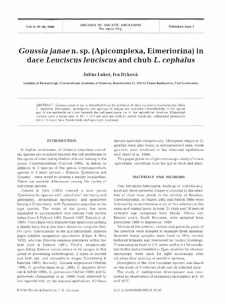
Small, early meronts found in the anterior and mid- dle part of the intestine measured 3.8 (2.3 to 7.3) X 2.8 (2.2 to 5.7) km (n = 20) (Fig. 1). Under the light micro- scope they appeared attached to the rnicrovillar surface of epithelial cells. Large oval or rounded meronts reached 9.6 (7.5 to 11.3) X 7.5 (6.5 to 11.3) pm in size (n = 20) (Fig. 2). When observed fresh, they contained 1 to 3 oval vacuoles. (Fig. 3). In stained sections, large nuclei with a dense nucleoli were observed. Merozoites developed in the same location as meronts, i.e. in the 'eplcellular' position. (Figs. 4 to 6). They were elon- gated, 6.9 (6.0 to 7.5) X 1.9 (1.6 to 2.2) pm (n = 20), with a tapered anterior end. Their average number was 8, with a range of 4 to 10. The number of merogonial generations was not established since only one type
of meront and merozoite was observed in natural infections.
Both early and differentiated gamonts were localized 'epicellularly' (Figs. 8 and 9). Occasionally, a few gam- onts were found deep in the epithelial layer In the vicinity of the basal membrane (Fig. 7). Ellipsoidal macrogamonts, 12.0 (11.5 to 13.0) X 9.6 (8.0 to 10.7) pm (n = 20), protruded considerably into the intestinal lumen. The cytoplasm was rich in small granules and vacuoles, with a large central nucleus possessing a prominent nucleolus (Fig. 8). Multinucleate developing microgamonts were ovoid 12.9 (11.5 to 15.0) X 10.3 (9.5 to 12.5) pm (n = 20) (Fig. 9). When mature, they contained 40 to 90 flagellated microgametes surround- ing a large residual body. Exflagellation was not observed. The proportion of macro- to rnicrogamonts was about 3 to 1.
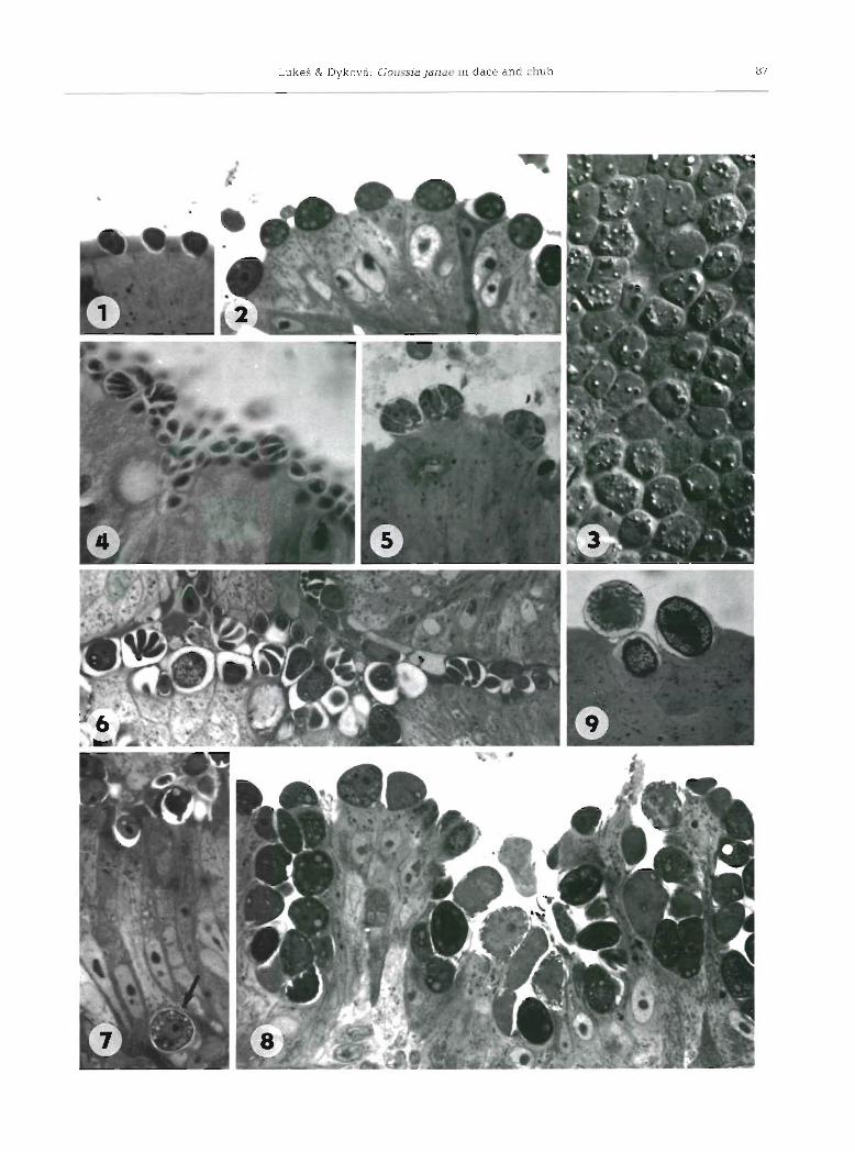
Sporonts could be distinguished from younger developmental stages by their oval shape and more dense cytoplasm containing numerous granules. They were also found in the 'epicellular' position (Fig. 9) and in the intestinal contents (Figs. 10 and 11).
Immature oocysts were spherical (Fig. 12). The spor- ont cytoplasm gradually detached from the thin oocyst wall and divided into 4 globular or broadly oval sporo- blasts. Later, the diameter of the oocyst decreased slightly and the thin oocyst wall was apposed to the sporoblast so that the oocyst lost its spherical shape.
Rarely were sporulated oocysts found in the posterior part of the intestine. As a rule, oocysts were shed unsporulated. Exogenous sporulation was completed within 48 h at 10°C. At 4 and 20°C, however, only about 5 and 1 % of oocysts sporulated, respectively.
When transferred from 10 to 20°C, heavily infected fish discharged white casts measuring several centime- ters in length containing a great number of sporogonial stages and remnants of host epithelial cells.
Mature oocysts of irregularly ellipsoidal shape were 18.1 (14.2 to 22.0) X 12.7 (11.0 to 14.5) pm (n = 20) (Figs. 13 to 15 and 19). The thin oocyst wall was stretched tightly over the sporocysts. Ellipsoidal sporo- cysts, 13.5 (12.5 to 14.5) x 5.0 (4.3 to 5.8) pm (n = 20), contained 2 elongated sporozoites 10.7 (10.0 to 11.5) X
2.8 (2.5 to 3.2) lbm (n = 20) slightly curved along the sporocyst wall in a head to tail position; sporocyst residuum were absent. A Goussia-type sporocyst wall,
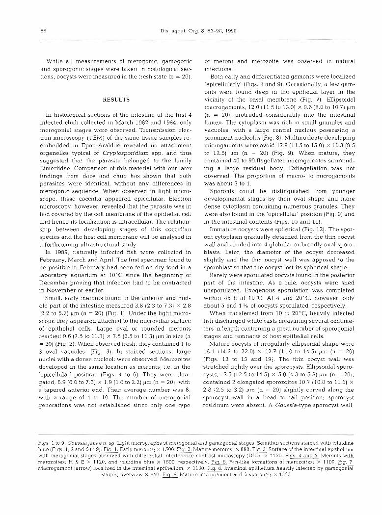
Figs. 1 to 9. Goussia janae n. sp. Light micrographs of merogonial and gamogonial stages. Semithin sections stained with toluidine blue (Figs. 1, 2 and 5 to 9) . Flg Early meronts; X 1500. G Mature meronts; X 890. Fig Surface of the intestinal epithelium with merogonial stages observed with differential interference contrast microscopy (DIC); x 1120. Figs. 4 and 5. Meronts with merozoites; H & E X 1120, and toluidine blue X 1600, respectively. Fig. 6, Fan-like formations of merozoites; X 1100. Macrogarnont (arrow) localized in the intestinal epithelium; X 1130. Flg Intestinal epithelium heavily infected by gamogonial
stages; overview X 950. Plg. Mature microgamont and 2 sporonts; X 1350

Luke? & Dykova. Goussia janae in dace and chub 8 9
Fig 19. Goussja janae n. sp. Diagrammatic representation of a sporulated oocyst
i.e. the presence of a suture connecting the 2 valves of the sporocyst wall, was observed by TEM (Fig. 16). It was not possible to release sporozoites from fresh sporocysts, even when considerable pressure was applied to the coverslip.
The lesions associated with the early phase of Gous- sia janae infection, i .e . with the merogony phase of the life cycle, were in fact restricted to individual epithelial cells. Light microscopy revealed microvillar atrophy of affected epithelial cells. The architecture of the epithe- lial lining, well preserved in the initlal stage of infec- tion, was markedly altered during gamogony and sporogony (Figs. 17 and 18). The formation of multiple secondary mucosal folds due to progressive and regres- sive changes of epithelium was observed along with its extensive desquamation.
At present, limited information is available on the histopathology of the cast formations, i.e. massive dls- charge of sporogonial stages potentiated by the change of temperature.
The extent of intestinal lesions reflects the intensity of infection. During gamogony and sporogony, there were sharply delimited regions in the intestine where almost all eplthelial cells were infected.
DISCUSSION
Since we could not, either in fresh mounts or in histological sections, discern the presence of a suture in the sporocyst wall, we resorted to an electron micro- scopic study. In this way, its presence could be estab-
lished, and our species could be assigned to the genus Goussia.
In the intestine of flsh of the genus Leuascus, 4 coccidian species have been found: Eimeria schulmani in L. idus (Kulemina 1969) and L. cephalus (Jastrzebski 1984); Goussia carpel11 in L. cephalus cabeda (Alvarez- Pellitero & Gonzales-Lanza 1986) and L. leuciscus (Lukei unpubl.) and 2 Goussia species designated as Goussia sp. 111 and Goussja sp. IX in L. cephalus (Mol- nar 1989). Our new species differs from E. schulmani by the absence of a sporocyst residual body and from G. carpelli in the dimensions of the oocysts. Oocysts of Goussia sp. IX are smaller, whereas those of Goussia sp. I11 are similar and may be identical. However, agglomerations of oocysts reported by Molnar (1989) as 'nodules' were not observed in our material. G. janae n. sp. resembles E. aurati from the goldfish in the exoge- nous sporulation, size of oocysts and sporocysts, but can be distinguished by the thinner sporocyst wall, more elongated oocysts and different host species. Sporogonic stages and oocysts of species in question are similar to those of E. vanasi (Landsberg & Paperna 1987). However in the merogonial and gamogonial sequence, there are differences in the number of merozoites and microgametes per meront and micro- gamont, respectively. Moreover, host species are phy- logenetically different. In the light microscope, meronts and merozoites of G. janae n. sp. of a single type were observed. Beca.use we studied only natural infections, we were unable to establish the number of merogonial generations in the life cycle. When the number of merozoites and the process of ectomerogony are con- cerned, G. janae n. sp , resembles the first merogonial generation of G. sinensis (Molnjr 1976) and G. iroquina (Paterson & Desser 1982). However, in the latter 2 species, second merogonial generations with many merozoites were described. A relatively low number of flagellated microgametes per microgamont is rare and among fish coccidia, has been reported only in G. iroquina (Paterson & Desser 1982) and G. aculeati (Jas- trzebski 1989).
Except for Eimeria catalana and members of the genus Epieimeria, all previously described 'epicellular' species sporulate exogenously. This mode of sporula- tion is rare amongst fish coccidia localized deep in the epithelium (Dykovb & Lom 1981). The sporonts of Goussia janae discharged either individually in feces or massively in casts. The latter pattern of excretion was
Figs. 10 to 18. Goussia janae n. sp. Sporogonial stages of fresh preparations observed with normal (Figs. 10 to 15) and differential interference contrast (DIC) optics (Figs. 11 and 13 to 14). Figs. 10 and 11. Sporonts discharged along with desquamated epithelial cells in a form of casts, X 280 and x 900, respectively. Fig. 12. Unsporulated oocysts; x 1110 Figs. l 3 and-13. Mature oocysts; X
1780. Fig. 15. Oocysts spor.ulated in casts, X 500. Fig. 16 Electron micrograph of the sporocyst wall demonstrating the suture of the 2 shell valves (arrow); X 100 000. Flgs 17 and 18. Alterations of the intestinal epithelium; H & E x 900, and toluidine blue X
930, respectively
90 Dis. aquat. Org. 8: 85-90, 1990 - -
described for Eimeria aurati by Hoffman (1965). The restriction of exogenous sporulation of Goussia janae to a temperature around 10°C (Luke5 unpubl.) might indicate a seasonal occurrence.
DIAGNOSIS OF GOUSSIA JANAE N. SP.
Type host: Dace, Leuciscus leuciscus (Linnaeus, 1758).
Type locality: MalSe river, South Bohemia, Czecho- slovakia.
Site of infection: Anterior and middle part of intes- tine.
Type slides: H - Pa - 034 have been deposited in the type collection of the Institute of Parasitology, Czecho- slovak Academy of Sciences, ceske Budejovice.
Description: Irregularly ellipsoidal oocysts 18.1 (14.2 to 22.0) X 12.7 (11.0 to 14.5) pm with no oocyst residuum. Ellipsoidal sporocysts 13.5 (12.5 to 14.5) X
5.0 (4.3 to 5.8) !.(m which lack a Stieda body and sporocyst residuum. Elongated sporozoites 10.7 (10.0 to 11.5) X 2.8 (2.5 to 3.2) pm. Endogenous life cycle takes place 'epicellularly' in the apical part of epithelia1 cells; meronts produce 4 to 10 merozoites; sporulation is exogenous and temperature dependent.
Etymology: The species was named in honour of Jana , wife of the senior author.
LITERATURE CITED
AIvarez-P~llitero, M. P, , Gonzales-Lanza, M C. (1986). Gous- sia carpel11 (Protozoa. Apicomplexa) in cyprinid fish of the Duero basin (NW Spain). Aspects of host-parasitr relation- ships. J. appl. Ichthyol. 3: 125-130
Current, W. L. (1986). Cr~ptosporidium: its biology and poten- tial for enviromental transmission CRC critical Rev envir Control 17: 21-51
Daoudi, F (1987). Coccidies et coccidioses d c poissons mediteranedns: systi?matique, ultrastnlcturc ct h~ologie. Doctoral thesis, I\.lontpcllier
Daoudi, F , RadujkoviC, B., Marques, A., Bouix, G. (1987) Nouvelles especes de Cnccidies / Apicomplexa,
Responsible Subject Editor: Professor W. Kurtinq, Hannover, F. R. Germany
Elnieri~dacl des gpnres Eimeria Schneider, 1875, et E p i ~ i m r n a Dykovi et Lorn, 1981, parasites de Poissons manns de 13 hale de Kotor (Youqoslavicl. Bull. hlus. Hist nitt., Pans 9 (Sect. A): 321-332
Dykovii, I., Lom, J . (1981). Fish coccidia: rritical notes on life cycles, clazsrl~c.ation and pathogenic~ty. J . Fish Dis. 4 . 187-505
Hoffrnan, G . L. (1865). Eimpria aurati n. sp. (Protozoa: Eimeriidae) from goldfish (Clarassius auratusl in North America. J . Protozool. 12: 273-275
Jastrxehski, hl. (1984). Coccidiofauna of cultured and feral fishcs in fish farms. Wiad. parazyt. 30: 141-163
Jastrzcbski, h. (1989). Ultrastructural study on the develop- ment of Goussia aculedti, a coccidium parasitizing the three-spined stickleback, Gasterosteus aculeatus. Dis aquat Org. 6 . 45-53
Jastrzebski, XI., Pastuszko. J., Kurska, E., Badowska, M. (1988) Kokcydia ciernika - Casterosteus aculeatus (L . ) Wiad. parazyt. 34: 55-63 (In Pol~sh)
Kent, 51. L., Fournie. J . W., Snodgrass, R. E., Elston, R. A. (1988). Goussia glrellae n sp. (Apicomplexa. Eirneriorina) in the opaleye; Girella nigricans. J. Protozool. 35: 287-290
Kulemina, I . V (1969). New species of endoparasitic protozoa of young fish of Lake Selinger. Zool. Zh. 48: 1295-1298 (in Russian)
Landsherg, J . H., Paperna, I (1987). Intestinal infections by Eimcria (S. 1.) vanasi n. sp. (Eimeriidae, Apicomplexa, Protozoa) in cichlid fish Ann. Parasitol. Hum.. Comp. 62. 283-293
Leger, M., Bory, T (1932). Ein~ei-ia pigra n sp . , nouvelle Coccidie juxtaCpitheliale, parasite du Gardon rouge. C.r. hebd. Slanc. Acad. Sci., Paris 194: 1710-1712
Lom, J. , Dykova. 1. (1981). New species of the genus Eimena f.Apicomplexa, Coccidia) from marine fish. Z. ParasitKde 66: 207-220
Lom, J. . Dykova, I. (1982). Some marine fish coccidla of the genera Eimeria Schneider, Epieimena Dykova et Lom and Goussia LabbB. J . Fish Dis. 5: 309-321
Moln6r. K. (1976). Histological study of coccidiosis caused in the silver carp and the bighead by Eimeria sinensis Chen, 1956. Acta vet. Acad. SCI. hung. 26: 303-312
Molnar. K. (1986). Occurence of two new Goussia species in Ihc intestine of the sterlet (Acipenser ruthenus). Acta vet. hung. 34: 168-174
h.lolnar, K. (1989). Nodular and epicellular coccidiosis in the intrstine of cyprtnld fishes. Dis. aquat. Org. 7: 1-11
\lolnar, K., Rohde, K. (1988). NCM~ coccidians from freshwater fishcs of Austra.lia J. Fish DIS 11: 161-169
Patergon, W. B., Desser, S. S. (1982). The biology of two Eirncria species (Protista: Apicornplexa) In their mutual fish hosts In Ontar~o. Can. J . Zool. 60: 761-775
Mdnuscript first received: January 16, 1990 R e v i s ~ d I-crsion ~ ~ c c e p t e d : April 2, 1990