glycobiology e book
TRANSCRIPT
Chapter 12
Classics in Carbohydrate Chemistryand Glycobiology
Increasingly, the greatest advances in science are being made at the interfaces of
disciplines. In the areas of carbohydrate chemistry and glycobiology, synergistic
interactions among synthetic organic chemists, biochemists and biologists are
leading to new therapies that promise to improve human health through
combating disease. The ultimate goal is the design of new therapeutics with
exquisite selectivity and without deleterious side-effects. These studies are being
enabled by the development of detailed molecular-level insights into the role of
carbohydrates in various disease states and the power of chemical synthesis to
deliver molecules of remarkable complexity on demand. This last chapter
presents four accounts of interdisciplinary research aimed at the development
of carbohydrate-based therapeutics where synthetic chemistry efforts have
played a vital role.
The Immucillins: Transition-state Analogue Inhibitors
of Enzymic N-Ribosyl Transfer Reactions
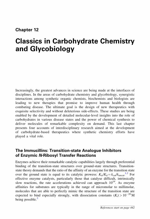
Enzymes achieve their remarkable catalytic capabilities largely through preferential
binding of the transition-state structures over ground-state structures. Transition-
state theory demands that the ratio of the affinity of an enzyme for the transition state
over the ground state is equal to its catalytic prowess: Kts/Kd = kcat/kuncat.1,2 For
effective enzyme catalysts, particularly those that catalyse difficult, intrinsically
slow reactions, the rate accelerations achieved can approach 1019. As enzyme
affinities for substrates are typically in the range of micromolar to millimolar,
molecules that are able to perfectly mimic the structure of the transition state are
expected to bind especially strongly, with dissociation constants (Ki)> 10�20 M
being possible.3
References start on page 442
In practice a transition-state structure has partial bonds, unnatural bond angles
and non-integer charge distributions, meaning that it is essentially impossible to
create a perfect chemically stable mimic. Nonetheless, transition-state mimicry
remains a highly sought ideal. One exceptionally successful application of this
principle has arisen from a 15-year collaboration between the research groups of
Vernon Schramm at the Albert Einstein College of Medicine, USA, and Richard
Furneaux at Industrial Research Limited, New Zealand, leading to some of the most
effective inhibitors known for any enzyme, and a series of promising new drugs that
are in clinical trials.4 The approach has been to (1) conduct basic studies to
determine the structure of the transition state for the enzyme of interest, (2) design
and synthesize molecules that mimic the transition state and, finally, (3) integrate
medicinal chemistry principles to mature rationally designed lead compounds into
clinical candidates.
N-Ribosyl transfer reactions occur widely in a range of processes essential
for cellular function. N-Ribosyl hydrolases catalyse the hydrolytic cleavage of
purine, pyrimidine and related bases from nucleosides and nucleotides. These
enzymes are involved in DNA repair, RNA depurination by plant toxins
(e.g. ricin) and salvage pathways that allow cellular recycling. Nucleoside
phosphorylases are related enzymes that cleave nucleosides by phosphorolysis
and have roles in nucleoside salvage, with the resulting ribosyl phosphates
re-entering cellular biosynthetic processes. These two classes of enzymes can
act by inversion or retention of stereochemistry, the latter being achieved by a
two-step mechanism involving stepwise inversions by an enzymic nucleophile.
The transition states for these processes can differ substantially, even for
enzymes that share a common mechanism. Design of an inhibitor requires
detailed information about the structure of the transition state for the enzyme
of interest – a blueprint for inhibitor design. The two enzymes to be discussed
here are purine nucleoside phosphorylase (PNP), and 50-methylthioadenosine-
S-adenosylhomocysteine nucleosidase (MTAN).
414 12 Classics in Carbohydrate Chemistry and Glycobiology
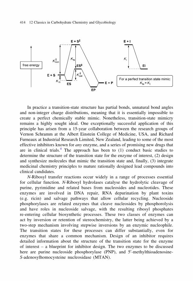
PNP is a human enzyme that catalyses the phosphorolysis of N-ribosidic bonds
of 6-oxypurine nucleosides and 6-oxypurine-20-deoxynucleosides and is involved in
nucleoside recycling. A rare genetic deficiency of PNP results in accumulation of
deoxynucleoside substrates, causing a disease termed T-cell deficiency. Inhibition of
this pathway has the potential to treat T-cell proliferative diseases such as T-cell
lymphomas.
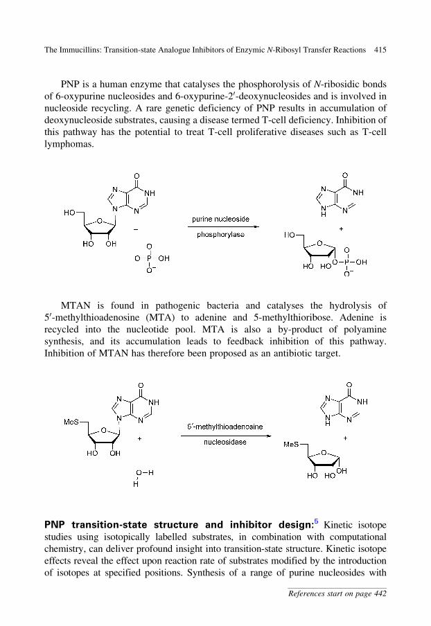
MTAN is found in pathogenic bacteria and catalyses the hydrolysis of
50-methylthioadenosine (MTA) to adenine and 5-methylthioribose. Adenine is
recycled into the nucleotide pool. MTA is also a by-product of polyamine
synthesis, and its accumulation leads to feedback inhibition of this pathway.
Inhibition of MTAN has therefore been proposed as an antibiotic target.
PNP transition-state structure and inhibitor design:5 Kinetic isotope
studies using isotopically labelled substrates, in combination with computational
chemistry, can deliver profound insight into transition-state structure. Kinetic isotope
effects reveal the effect upon reaction rate of substrates modified by the introduction
of isotopes at specified positions. Synthesis of a range of purine nucleosides with
The Immucillins: Transition-state Analogue Inhibitors of Enzymic N-Ribosyl Transfer Reactions 415
References start on page 442
isotopes at specific positions allows the investigation of kinetic isotope effects for the
enzyme-catalysed reaction. However, as the enzyme initially chosen for study, bovine
PNP, has a large forward commitment factor for phosphorolysis, the arsenolysis
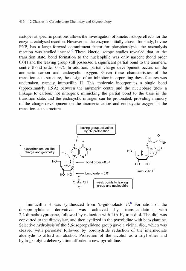
reaction was studied instead.6 These kinetic isotope studies revealed that, at the
transition state, bond formation to the nucleophile was only nascent (bond order
0.01) and the leaving group still possessed a significant partial bond to the anomeric
centre (bond order 0.37). In addition, partial charge development occurs on the
anomeric carbon and endocyclic oxygen. Given these characteristics of the
transition-state structure, the design of an inhibitor incorporating these features was
undertaken, namely immucillin H. This molecule incorporates a single bond
(approximately 1.5 A) between the anomeric centre and the nucleobase (now a
linkage to carbon, not nitrogen), mimicking the partial bond to the base in the
transition state, and the endocyclic nitrogen can be protonated, providing mimicry
of the charge development on the anomeric centre and endocyclic oxygen in the
transition-state structure.
Immucillin H was synthesized from ‘D-gulonolactone’.8 Formation of the
diisopropylidene derivative was achieved by transacetalation with
2,2-dimethoxypropane, followed by reduction with LiAlH4 to a diol. The diol was
converted to the dimesylate, and then cyclized to the pyrrolidine with benzylamine.
Selective hydrolysis of the 5,6-isopropylidene group gave a vicinal diol, which was
cleaved with periodate followed by borohydride reduction of the intermediate
aldehyde to afford an alcohol. Protection of the alcohol as a silyl ether and
hydrogenolytic debenzylation afforded a new pyrrolidine.
416 12 Classics in Carbohydrate Chemistry and Glycobiology
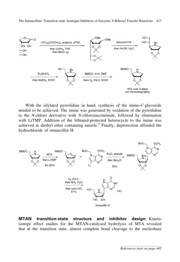
With the silylated pyrrolidine in hand, synthesis of the imino-C-glycoside
needed to be achieved. The imine was generated by oxidation of the pyrrolidine
to the N-chloro derivative with N-chlorosuccinimide, followed by elimination
with LiTMP. Addition of the lithiated-protected heterocycle to the imine was
achieved in diethyl ether containing anisole.9 Finally, deprotection afforded the
hydrochloride of immucillin H.
MTAN transition-state structure and inhibitor design: Kinetic
isotope effect studies for the MTAN-catalysed hydrolysis of MTA revealed
that at the transition state, almost complete bond cleavage to the nucleobase
The Immucillins: Transition-state Analogue Inhibitors of Enzymic N-Ribosyl Transfer Reactions 417
References start on page 442
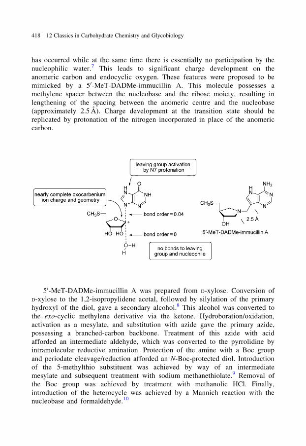
has occurred while at the same time there is essentially no participation by the
nucleophilic water.7 This leads to significant charge development on the
anomeric carbon and endocyclic oxygen. These features were proposed to be
mimicked by a 50-MeT-DADMe-immucillin A. This molecule possesses a
methylene spacer between the nucleobase and the ribose moiety, resulting in
lengthening of the spacing between the anomeric centre and the nucleobase
(approximately 2.5 A). Charge development at the transition state should be
replicated by protonation of the nitrogen incorporated in place of the anomeric
carbon.
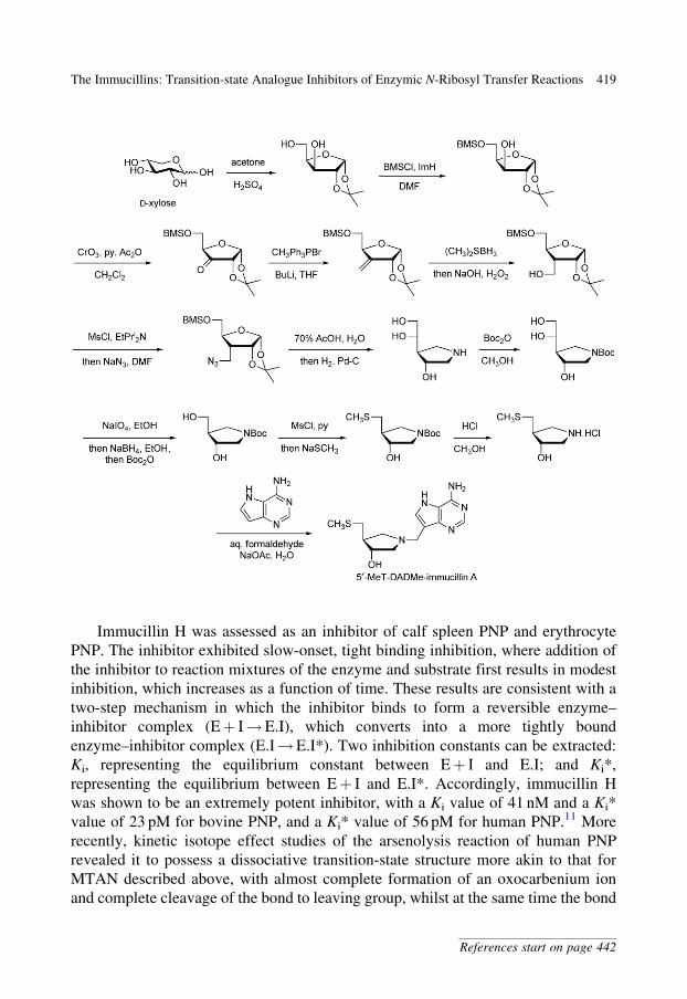
50-MeT-DADMe-immucillin A was prepared from D-xylose. Conversion of
D-xylose to the 1,2-isopropylidene acetal, followed by silylation of the primary
hydroxyl of the diol, gave a secondary alcohol.8 This alcohol was converted to
the exo-cyclic methylene derivative via the ketone. Hydroboration/oxidation,
activation as a mesylate, and substitution with azide gave the primary azide,
possessing a branched-carbon backbone. Treatment of this azide with acid
afforded an intermediate aldehyde, which was converted to the pyrrolidine by
intramolecular reductive amination. Protection of the amine with a Boc group
and periodate cleavage/reduction afforded an N-Boc-protected diol. Introduction
of the 5-methylthio substituent was achieved by way of an intermediate
mesylate and subsequent treatment with sodium methanethiolate.9 Removal of
the Boc group was achieved by treatment with methanolic HCl. Finally,
introduction of the heterocycle was achieved by a Mannich reaction with the
nucleobase and formaldehyde.10
418 12 Classics in Carbohydrate Chemistry and Glycobiology
Immucillin H was assessed as an inhibitor of calf spleen PNP and erythrocyte
PNP. The inhibitor exhibited slow-onset, tight binding inhibition, where addition of
the inhibitor to reaction mixtures of the enzyme and substrate first results in modest
inhibition, which increases as a function of time. These results are consistent with a
two-step mechanism in which the inhibitor binds to form a reversible enzyme–
inhibitor complex (Eþ I!E.I), which converts into a more tightly bound
enzyme–inhibitor complex (E.I!E.I*). Two inhibition constants can be extracted:
Ki, representing the equilibrium constant between Eþ I and E.I; and Ki*,
representing the equilibrium between Eþ I and E.I*. Accordingly, immucillin H
was shown to be an extremely potent inhibitor, with a Ki value of 41 nM and a Ki*
value of 23 pM for bovine PNP, and a Ki* value of 56 pM for human PNP.11 More
recently, kinetic isotope effect studies of the arsenolysis reaction of human PNP
revealed it to possess a dissociative transition-state structure more akin to that for
MTAN described above, with almost complete formation of an oxocarbenium ion
and complete cleavage of the bond to leaving group, whilst at the same time the bond
The Immucillins: Transition-state Analogue Inhibitors of Enzymic N-Ribosyl Transfer Reactions 419
References start on page 442

to nucleophile is essentially non-existent. Accordingly, the ‘second generation’
inhibitor, DADMe-immucillin H, was investigated as an inhibitor of human PNP
and was found to be an 8.5 pM inhibitor of this enzyme.12
X-ray crystallographic analysis of the enzyme–inhibitor complex of bovine PNP
and immucillin H reveals that essentially every hydrogen-bond donor/acceptor site is
fully engaged in favourable interactions, providing a structural rationale for the
effective inhibition of the enzyme by this compound.13
The exceptional potency of immucillin H and DADMe-immucillin H
suggested their potential for the inhibition of T-lymphocytes. Previous studies
with nanomolar inhibitors of PNP had failed to inhibit T-lymphocytes because of
lack of potency – it is known that >95% inhibition of PNP is required for
significant reduction in T-cell function. Gratifyingly, immucillin H was an
inhibitor of human T-lymphocytes, suggesting that it may have utility in the
treatment of diseases characterized by abnormal T-cell growth or activation.14
Oral dosing of immucillin H in mice resulted in effective inhibition of PNP
activity within minutes, and it required 4 days for recovery to 50% PNP activity
from a single dose (0.8 mg kg�1).15 Oral administration of DADMe-immucillin H
at the same dosage leads to similarly effective inhibition of PNP activity. How-
ever, the rate of recovery of PNP activity after a single dose of DADMe-
immucillin H was lower, with 11.5 days being required for recovery to 50%
activity, a rate that approximately matches the rate of protein resynthesis,
suggesting that the off-rate for inhibitor dissociation is not higher than resynthesis
of the target enzyme! At the time of writing, immucillin H (Fodosine) is in Phase II
clinical trials for relapsed/resistant T-cell leukaemia and cutaneous T-cell leukae-
mia.16 Further investigations have shown that related acyclic derivatives are
effective inhibitors of human PNP.17 These ‘third generation’ PNP inhibitors
include a triol that has been found to be as effective as DADMe-immucillin H
for inhibition of human PNP, with a Ki* value of 8.6 pM, and an achiral deriva-
tive of ‘tris base’ that is a sub-nanomolar inhibitor of malarial PNP. More recent
studies have reported the synthesis of an achiral azetidine that is a sub-nanomolar
inhibitor of PNPs.18
420 12 Classics in Carbohydrate Chemistry and Glycobiology
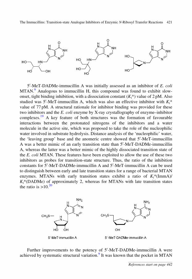
50-MeT-DADMe-immucillin A was initially assessed as an inhibitor of E. coli
MTAN.9 Analogous to immucillin H, this compound was found to exhibit slow-
onset, tight binding inhibition, with a dissociation constant (Ki*) value of 2 pM. Also
studied was 50-MeT-immucillin A, which was also an effective inhibitor with Ki*
value of 77 pM. A structural rationale for inhibitor binding was provided for these
two inhibitors and the E. coli enzyme by X-ray crystallography of enzyme–inhibitor
complexes.19 A key feature of both structures was the formation of favourable
interactions between the protonated nitrogens of the inhibitors and a water
molecule in the active site, which was proposed to take the role of the nucleophilic
water involved in substrate hydrolysis. Distance analysis of the ‘nucleophilic’ water,
the ‘leaving group’ base and the anomeric centre showed that 50-MeT-immucillin
A was a better mimic of an early transition state than 50-MeT-DADMe-immucillin
A, whereas the latter was a better mimic of the highly dissociated transition state of
the E. coli MTAN. These features have been exploited to allow the use of these two
inhibitors as probes for transition-state structure. Thus, the ratio of the inhibition
constants for 50-MeT-DADMe-immucillin A and 50-MeT-immucillin A can be used
to distinguish between early and late transition states for a range of bacterial MTAN
enzymes. MTANs with early transition states exhibit a ratio of Ki*(ImmA)/
Ki*(DADMe) of approximately 2, whereas for MTANs with late transition states
the ratio is >10.20
Further improvements to the potency of 50-MeT-DADMe-immucillin A were
achieved by systematic structural variation.9 It was known that the pocket in MTAN
The Immucillins: Transition-state Analogue Inhibitors of Enzymic N-Ribosyl Transfer Reactions 421
References start on page 442
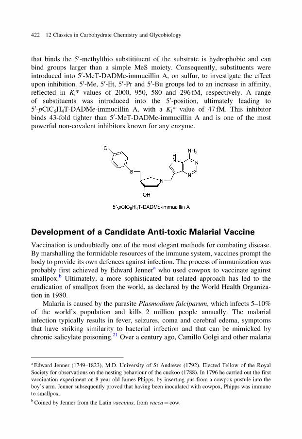
that binds the 50-methylthio substitituent of the substrate is hydrophobic and can
bind groups larger than a simple MeS moiety. Consequently, substituents were
introduced into 50-MeT-DADMe-immucillin A, on sulfur, to investigate the effect
upon inhibition. 50-Me, 50-Et, 50-Pr and 50-Bu groups led to an increase in affinity,
reflected in Ki* values of 2000, 950, 580 and 296 fM, respectively. A range
of substituents was introduced into the 50-position, ultimately leading to
50-pClC6H4T-DADMe-immucillin A, with a Ki* value of 47 fM. This inhibitor
binds 43-fold tighter than 50-MeT-DADMe-immucillin A and is one of the most
powerful non-covalent inhibitors known for any enzyme.
Development of a Candidate Anti-toxic Malarial Vaccine
Vaccination is undoubtedly one of the most elegant methods for combating disease.
By marshalling the formidable resources of the immune system, vaccines prompt the
body to provide its own defences against infection. The process of immunization was
probably first achieved by Edward Jennera who used cowpox to vaccinate against
smallpox.b Ultimately, a more sophisticated but related approach has led to the
eradication of smallpox from the world, as declared by the World Health Organiza-
tion in 1980.
Malaria is caused by the parasite Plasmodium falciparum, which infects 5–10%
of the world’s population and kills 2 million people annually. The malarial
infection typically results in fever, seizures, coma and cerebral edema, symptoms
that have striking similarity to bacterial infection and that can be mimicked by
chronic salicylate poisoning.21 Over a century ago, Camillo Golgi and other malaria
a Edward Jenner (1749–1823), M.D. University of St Andrews (1792). Elected Fellow of the Royal
Society for observations on the nesting behaviour of the cuckoo (1788). In 1796 he carried out the first
vaccination experiment on 8-year-old James Phipps, by inserting pus from a cowpox pustule into the
boy’s arm. Jenner subsequently proved that having been inoculated with cowpox, Phipps was immune
to smallpox.b Coined by Jenner from the Latin vaccinus, from vacca = cow.
422 12 Classics in Carbohydrate Chemistry and Glycobiology
scientists demonstrated that malarial fever is synchronous with the developmental
cycle of the blood-stage parasite. Together, these observations support the hypoth-
esis that the pathological reactions that occur upon infection by the parasite are
initiated by a malarial toxin.22 Glycosylphosphatidylinositol (GPI) of the parasite,
which is expressed in free and protein conjugated forms, has the properties predicted
for a toxin, can induce cytokine and adhesion expression in macrophages and
vascular endothelium, and can be lethal in vivo.23,24 Anti-toxin vaccines are effec-
tive public health measures that are used for protection against tetanus and diphtheria
and prevent subjects from becoming ill, rather than preventing parasite multiplica-
tion. These vaccines use a ‘toxoid’, an inactivated form of the toxin, to induce host
immunity and protect against subsequent toxin. This section will describe work by
the groups of Louis Schofield at the Walter and Eliza Hall Medical Institute,
Australia and Peter Seeberger, at the Massachusetts Institute of Technology, USA,
and the ETH, Switzerland, to develop a candidate anti-toxin vaccine for malaria.
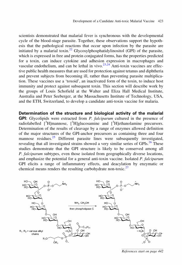
Determination of the structure and biological activity of the malarial
GPI: Glycolipids were extracted from P. falciparum cultured in the presence of
radiolabelled [3H]mannose, [3H]glucosamine and [3H]ethanolamine precursors.
Determination of the results of cleavage by a range of enzymes allowed definition
of the major structures of the GPI-anchor precursors as containing three and four
mannose residues.25 Different parasite lines were subsequently investigated,
revealing that all investigated strains showed a very similar series of GPIs.26 These
studies demonstrate that the GPI structure is likely to be conserved among all
P. falciparum subtypes, even those isolated from geographically diverse locations,
and emphasize the potential for a general anti-toxin vaccine. Isolated P. falciparum
GPI elicits a range of inflammatory effects, and deacylation by enzymatic or
chemical means renders the resulting carbohydrate non-toxic.27
Development of a Candidate Anti-toxic Malarial Vaccine 423
References start on page 442
A delipidated glycan can be generated by sequential treatment of the GPI with
methanolic ammonia and phosphatidylinositol phospholipase C.25 This non-toxic
carbohydrate structure was chosen as the antigen from which to develop a vaccine
candidate.
A widely used approach to the development of synthetic vaccines is to conjugate
the antigen (the hapten) to an immunogenic carrier protein. Conjugation to generate
a large molecular species ensures long-term residence of the resultant conjugate in
the blood system, and the immunogenicity of the carrier protein ensures that the
hapten is recognized by the immune system and results in an immune response.
However, acquisition of the glycan in homogeneous form and sufficient quantities
requires chemical synthesis.
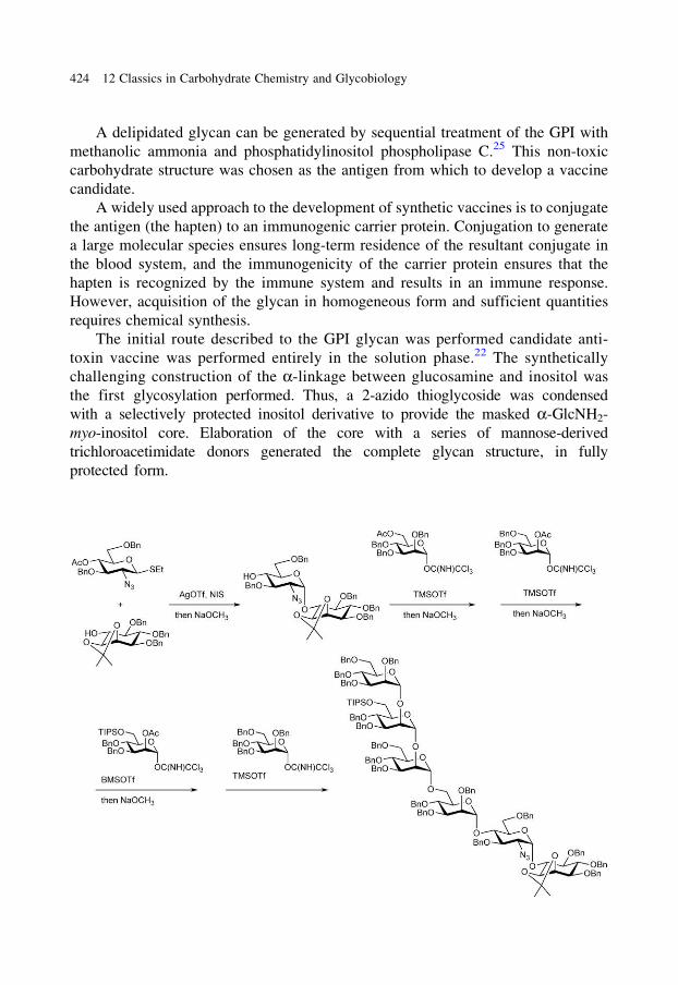
The initial route described to the GPI glycan was performed candidate anti-
toxin vaccine was performed entirely in the solution phase.22 The synthetically
challenging construction of the a-linkage between glucosamine and inositol was
the first glycosylation performed. Thus, a 2-azido thioglycoside was condensed
with a selectively protected inositol derivative to provide the masked a-GlcNH2-
myo-inositol core. Elaboration of the core with a series of mannose-derived
trichloroacetimidate donors generated the complete glycan structure, in fully
protected form.
424 12 Classics in Carbohydrate Chemistry and Glycobiology
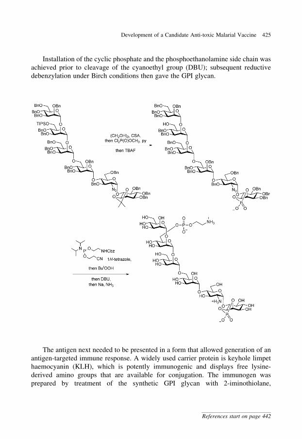
Installation of the cyclic phosphate and the phosphoethanolamine side chain was
achieved prior to cleavage of the cyanoethyl group (DBU); subsequent reductive
debenzylation under Birch conditions then gave the GPI glycan.
The antigen next needed to be presented in a form that allowed generation of an
antigen-targeted immune response. A widely used carrier protein is keyhole limpet
haemocyanin (KLH), which is potently immunogenic and displays free lysine-
derived amino groups that are available for conjugation. The immunogen was
prepared by treatment of the synthetic GPI glycan with 2-iminothiolane,
Development of a Candidate Anti-toxic Malarial Vaccine 425
References start on page 442
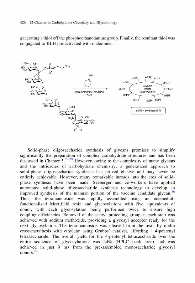
generating a thiol off the phosphoethanolamine group. Finally, the resultant thiol was
conjugated to KLH pre-activated with maleimide.
Solid-phase oligosaccharide synthesis of glycans promises to simplify
significantly the preparation of complex carbohydrate structures and has been
discussed in Chapter 5.28,29 However, owing to the complexity of many glycans
and the intricacies of carbohydrate chemistry, a generalized approach to
solid-phase oligosaccharide synthesis has proved elusive and may never be
entirely achievable. However, many remarkable inroads into the area of solid-
phase synthesis have been made. Seeberger and co-workers have applied
automated solid-phase oligosaccharide synthesis technology to develop an
improved synthesis of the mannan portion of the vaccine candidate glycan.30
Thus, the tetramannoside was rapidly assembled using an octenediol-
functionalized Merrifield resin and glycosylations with five equivalents of
donor, with each glycosylation being performed twice to ensure high
coupling efficiencies. Removal of the acetyl protecting group at each step was
achieved with sodium methoxide, providing a glycosyl acceptor ready for the
next glycosylation. The tetramannoside was cleaved from the resin by olefin
cross-metathesis with ethylene using Grubbs’ catalyst, affording a 4-pentenyl
tetrasaccharide. The overall yield for the 4-pentenyl tetrasaccharide over the
entire sequence of glycosylations was 44% (HPLC peak area) and was
achieved in just 9 hrs from the pre-assembled monosaccharide glycosyl
donors.29
426 12 Classics in Carbohydrate Chemistry and Glycobiology
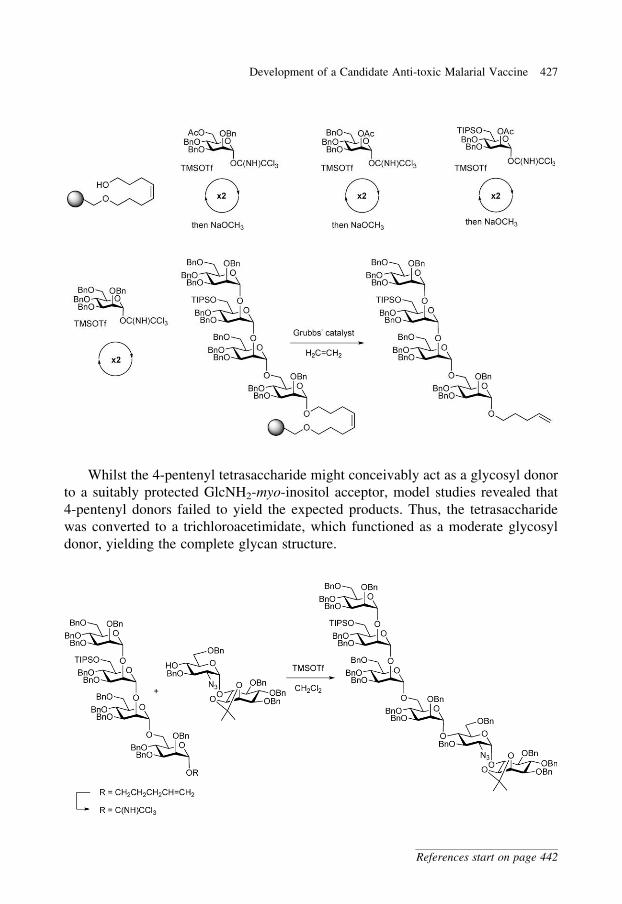
Whilst the 4-pentenyl tetrasaccharide might conceivably act as a glycosyl donor
to a suitably protected GlcNH2-myo-inositol acceptor, model studies revealed that
4-pentenyl donors failed to yield the expected products. Thus, the tetrasaccharide
was converted to a trichloroacetimidate, which functioned as a moderate glycosyl
donor, yielding the complete glycan structure.
Development of a Candidate Anti-toxic Malarial Vaccine 427
References start on page 442
The synthetic GPI–KLH conjugate was used to vaccinate mice, and gave rise to
antibodies with activity against glycan.22 Notably, the anti-GPI antibodies did not
cross-react with mammalian GPIs, possibly owing to the structural differences
between malarial and mammalian GPI structures. Mice immunized with the
synthetic GPI–KLH conjugate were significantly protected against severe malaria,
as indicated by improved rates of survival, when challenged with Plasmodium
berghei, a murine model for human malaria. Consistent with the mode of action of
the vaccine being against the malaria toxin, parasite levels in immunized animals
were not significantly different from non-immunized animals. Current work is
focused on defining the optimal structure and presentation of the glycan
antigen,31,32 developing an effective immunogenic carrier protein (of parasite or
bacterial origin), and establishing optimum dosing regimes in pre-clinical studies.24
Ultimately, clinical trials will be needed to evaluate whether a synthetic GPI anti-
toxin vaccine will reduce malarial mortality and morbidity in the field with a suitable
safety profile.
Synthetic Carbohydrate Anti-tumour Vaccines
Aberrant glycosylation is a hallmark of the cancer phenotype. Thus, a
carbohydrate vaccine against tumour cells has long been a keenly sought
goal.33,34 Unlike anti-infection vaccines, which protect against future infections,
anti-cancer vaccines aim to assist in the eradication of malignant cells post-
diagnosis and face a long list of potential problems including tumour cell–induced
immunosuppression even in the presence of immunogenic antigens, the intrinsi-
cally poor immunogenicity of most carbohydrates and the fact that many tumour
antigens are self-antigens that are present in lower amounts on healthy tissues.
The following section details research aimed at exploiting the identification of
specific glycolipid and glycoprotein glycans that are over-expressed on the sur-
faces of malignant cells. The work has aimed to develop a cell-free, fully
synthetic anti-tumour vaccine that could be used to elicit highly specific and
robust immune responses against malignant cells that bear the requisite glycans.
The goal is to induce the body to produce antibodies that could act within the
blood system to clear circulating tumour cells and micrometastases, through
complement-mediated cell lysis, or other mechanisms.
Globo-H is a hexasaccharide that was originally isolated as a ceramide-linked
glycolipid from the human breast cancer cell line MCF-7.34 The glycolipid is found at
the cell surface and has been characterized with the aid of a monoclonal antibody, MBr1.
Immunohistological analysis using MBr1 demonstrated that Globo-H is found in a range
of other cancers including pancreas, stomach, uterine endometrium and prostate. While
there is some evidence that Globo-H is found on normal tissues, it is believed to be
localized on these tissues in areas where the immune system has restricted access.
428 12 Classics in Carbohydrate Chemistry and Glycobiology
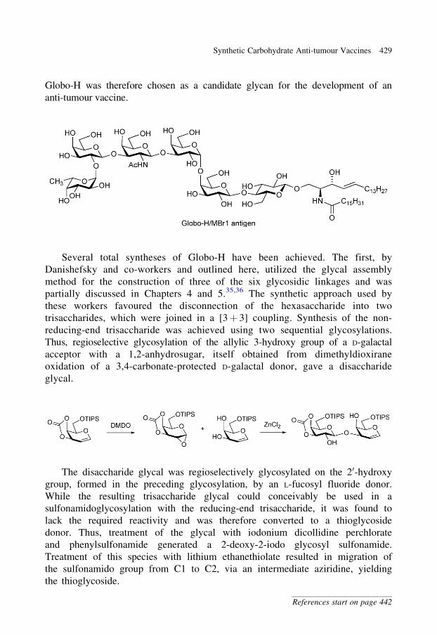
Globo-H was therefore chosen as a candidate glycan for the development of an
anti-tumour vaccine.
Several total syntheses of Globo-H have been achieved. The first, by
Danishefsky and co-workers and outlined here, utilized the glycal assembly
method for the construction of three of the six glycosidic linkages and was
partially discussed in Chapters 4 and 5.35,36 The synthetic approach used by
these workers favoured the disconnection of the hexasaccharide into two
trisaccharides, which were joined in a [3þ 3] coupling. Synthesis of the non-
reducing-end trisaccharide was achieved using two sequential glycosylations.
Thus, regioselective glycosylation of the allylic 3-hydroxy group of a D-galactal
acceptor with a 1,2-anhydrosugar, itself obtained from dimethyldioxirane
oxidation of a 3,4-carbonate-protected D-galactal donor, gave a disaccharide
glycal.
The disaccharide glycal was regioselectively glycosylated on the 20-hydroxy
group, formed in the preceding glycosylation, by an L-fucosyl fluoride donor.
While the resulting trisaccharide glycal could conceivably be used in a
sulfonamidoglycosylation with the reducing-end trisaccharide, it was found to
lack the required reactivity and was therefore converted to a thioglycoside
donor. Thus, treatment of the glycal with iodonium dicollidine perchlorate
and phenylsulfonamide generated a 2-deoxy-2-iodo glycosyl sulfonamide.
Treatment of this species with lithium ethanethiolate resulted in migration of
the sulfonamido group from C1 to C2, via an intermediate aziridine, yielding
the thioglycoside.
Synthetic Carbohydrate Anti-tumour Vaccines 429
References start on page 442
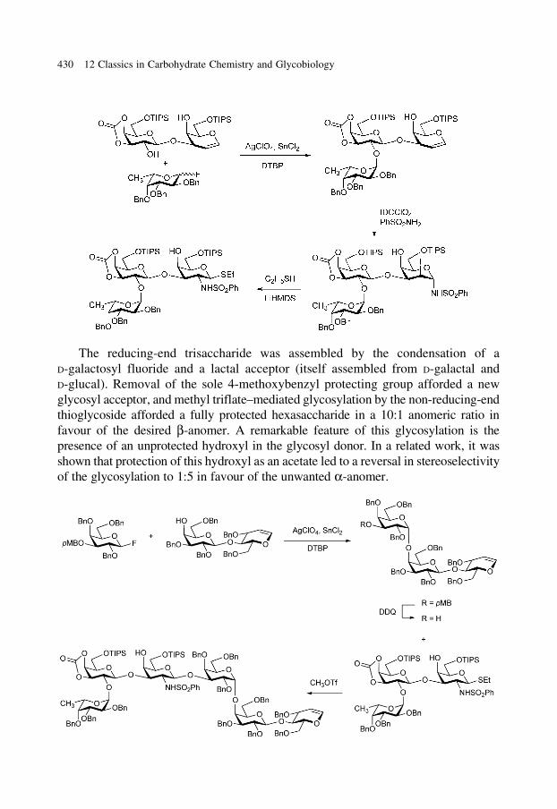
The reducing-end trisaccharide was assembled by the condensation of a
D-galactosyl fluoride and a lactal acceptor (itself assembled from D-galactal and
D-glucal). Removal of the sole 4-methoxybenzyl protecting group afforded a new
glycosyl acceptor, and methyl triflate–mediated glycosylation by the non-reducing-end
thioglycoside afforded a fully protected hexasaccharide in a 10:1 anomeric ratio in
favour of the desired b-anomer. A remarkable feature of this glycosylation is the
presence of an unprotected hydroxyl in the glycosyl donor. In a related work, it was
shown that protection of this hydroxyl as an acetate led to a reversal in stereoselectivity
of the glycosylation to 1:5 in favour of the unwanted a-anomer.
430 12 Classics in Carbohydrate Chemistry and Glycobiology
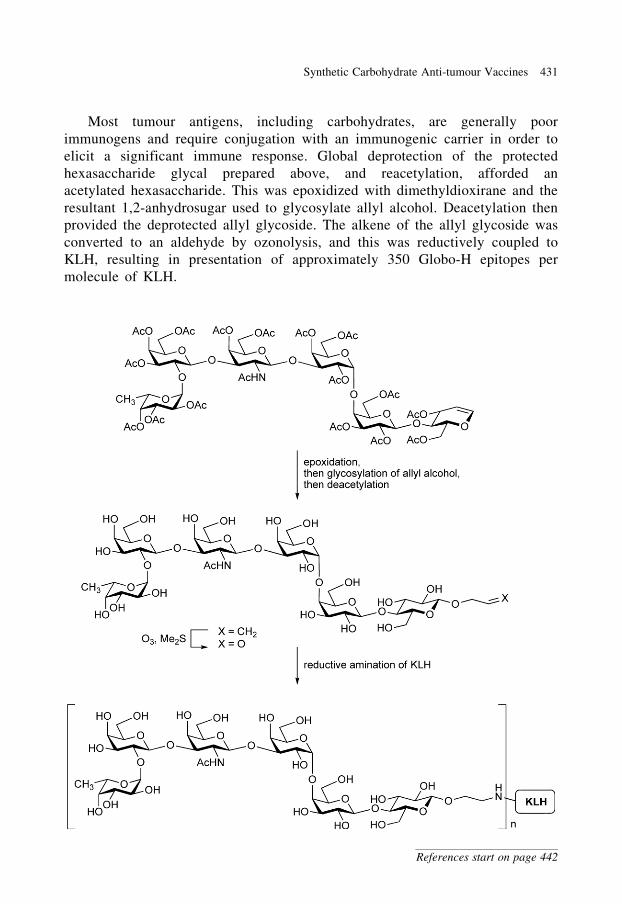
Most tumour antigens, including carbohydrates, are generally poor
immunogens and require conjugation with an immunogenic carrier in order to
elicit a significant immune response. Global deprotection of the protected
hexasaccharide glycal prepared above, and reacetylation, afforded an
acetylated hexasaccharide. This was epoxidized with dimethyldioxirane and the
resultant 1,2-anhydrosugar used to glycosylate allyl alcohol. Deacetylation then
provided the deprotected allyl glycoside. The alkene of the allyl glycoside was
converted to an aldehyde by ozonolysis, and this was reductively coupled to
KLH, resulting in presentation of approximately 350 Globo-H epitopes per
molecule of KLH.
Synthetic Carbohydrate Anti-tumour Vaccines 431
References start on page 442
As mentioned above, the carbohydrate antigen of Globo-H was conjugated
to a carrier protein with the expectation that this would enhance its
immunogenicity. Even so, such subunit vaccines are inherently less
immunogenic than those employing attenuated microorganisms. Many antigens
are therefore administrated with adjuvants, immunostimulatory molecules that
are themselves not immunogenic but serve to enhance or prolong the immune
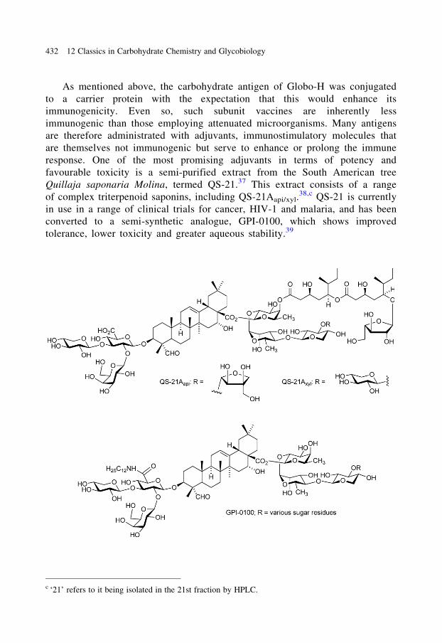
response. One of the most promising adjuvants in terms of potency and
favourable toxicity is a semi-purified extract from the South American tree
Quillaja saponaria Molina, termed QS-21.37 This extract consists of a range
of complex triterpenoid saponins, including QS-21Aapi/xyl.38,c QS-21 is currently
in use in a range of clinical trials for cancer, HIV-1 and malaria, and has been
converted to a semi-synthetic analogue, GPI-0100, which shows improved
tolerance, lower toxicity and greater aqueous stability.39
c ‘21’ refers to it being isolated in the 21st fraction by HPLC.
432 12 Classics in Carbohydrate Chemistry and Glycobiology
Immunization of mice with the Globo-H-KLH conjugate in the presence of
QS-21 resulted in high-titre IgM and IgG responses against the Globo-H
antigen.40 The vaccine also elicits high-titre IgM antibodies against Globo-H
in humans and was shown to be safe. At the time of writing, this conjugate is
scheduled for Phase II/III clinical trial evaluation for the treatment of breast
cancer.
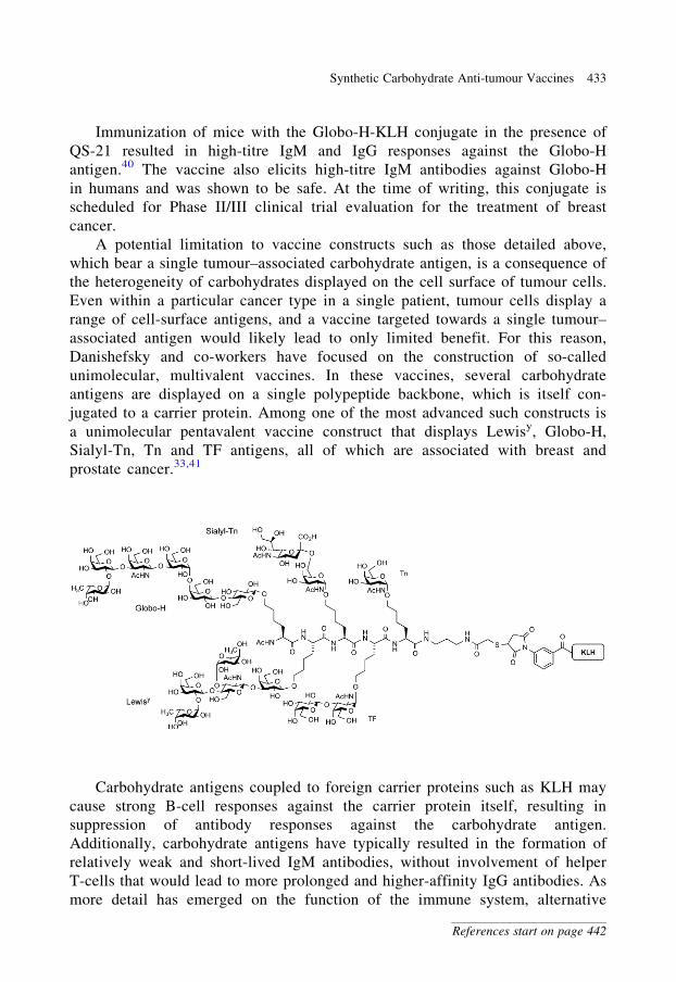
A potential limitation to vaccine constructs such as those detailed above,
which bear a single tumour–associated carbohydrate antigen, is a consequence of
the heterogeneity of carbohydrates displayed on the cell surface of tumour cells.
Even within a particular cancer type in a single patient, tumour cells display a
range of cell-surface antigens, and a vaccine targeted towards a single tumour–
associated antigen would likely lead to only limited benefit. For this reason,
Danishefsky and co-workers have focused on the construction of so-called
unimolecular, multivalent vaccines. In these vaccines, several carbohydrate
antigens are displayed on a single polypeptide backbone, which is itself con-
jugated to a carrier protein. Among one of the most advanced such constructs is
a unimolecular pentavalent vaccine construct that displays Lewisy, Globo-H,
Sialyl-Tn, Tn and TF antigens, all of which are associated with breast and
prostate cancer.33,41
Carbohydrate antigens coupled to foreign carrier proteins such as KLH may
cause strong B-cell responses against the carrier protein itself, resulting in
suppression of antibody responses against the carbohydrate antigen.
Additionally, carbohydrate antigens have typically resulted in the formation of
relatively weak and short-lived IgM antibodies, without involvement of helper
T-cells that would lead to more prolonged and higher-affinity IgG antibodies. As
more detail has emerged on the function of the immune system, alternative
Synthetic Carbohydrate Anti-tumour Vaccines 433
References start on page 442
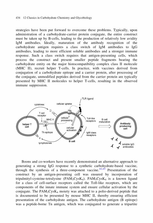
strategies have been put forward to overcome these problems. Typically, upon
administration of a carbohydrate-carrier protein conjugate, the entire construct
must be taken up by B-cells, leading to the production of relatively low avidity
IgM antibodies. Ideally, maturation of the antibody recognition of the
carbohydrate antigen requires a class switch of IgM antibodies to IgG
antibodies, leading to more efficient soluble antibodies and a stronger immune
response. Such a class switch requires that antigen-presenting cells, which
process the construct and present smaller peptide fragments bearing the
carbohydrate entity on the major histocompatibility complex class II molecule
(MHC II), recruit helper T-cells. In practice, with vaccines derived from
conjugation of a carbohydrate epitope and a carrier protein, after processing of
the conjugate, unmodified peptides derived from the carrier protein are typically
presented by MHC II molecules to helper T-cells, resulting in the observed
immune suppression.
Boons and co-workers have recently demonstrated an alternative approach to
generating a strong IgG response to a synthetic carbohydrate-based vaccine,
through the synthesis of a three-component vaccine.42,43 Presentation of the
construct by an antigen-presenting cell was ensured by incorporation of
tripalmityl-cysteine-tetralysine (PAM3CysK4). PAM3CysK4 is a known ligand
for a class of cell-surface receptors called the Toll-like receptors, which are
components of the innate immune system and ensure cellular activation by the
conjugate. The PAM3CysK4 moiety was attached to a polio-derived peptide that
is documented to be presented by mouse MHC II, thereby ensuring efficient
presentation of the carbohydrate antigen. The carbohydrate antigen (B epitope)
was a peptide-borne Tn antigen, which was conjugated to generate a tripartite
434 12 Classics in Carbohydrate Chemistry and Glycobiology
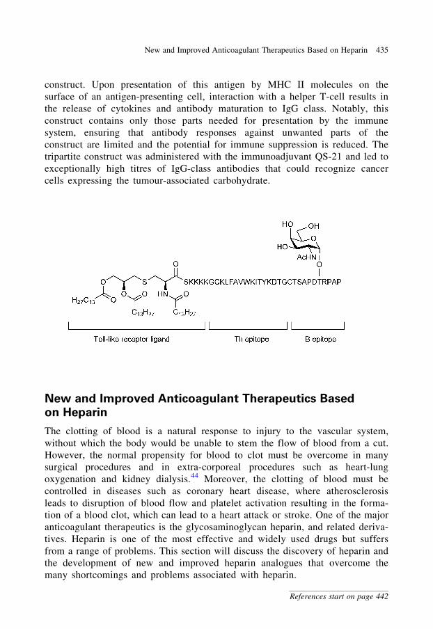
construct. Upon presentation of this antigen by MHC II molecules on the
surface of an antigen-presenting cell, interaction with a helper T-cell results in
the release of cytokines and antibody maturation to IgG class. Notably, this
construct contains only those parts needed for presentation by the immune
system, ensuring that antibody responses against unwanted parts of the
construct are limited and the potential for immune suppression is reduced. The
tripartite construct was administered with the immunoadjuvant QS-21 and led to
exceptionally high titres of IgG-class antibodies that could recognize cancer
cells expressing the tumour-associated carbohydrate.
New and Improved Anticoagulant Therapeutics Based
on Heparin
The clotting of blood is a natural response to injury to the vascular system,
without which the body would be unable to stem the flow of blood from a cut.
However, the normal propensity for blood to clot must be overcome in many
surgical procedures and in extra-corporeal procedures such as heart-lung
oxygenation and kidney dialysis.44 Moreover, the clotting of blood must be
controlled in diseases such as coronary heart disease, where atherosclerosis
leads to disruption of blood flow and platelet activation resulting in the forma-
tion of a blood clot, which can lead to a heart attack or stroke. One of the major
anticoagulant therapeutics is the glycosaminoglycan heparin, and related deriva-
tives. Heparin is one of the most effective and widely used drugs but suffers
from a range of problems. This section will discuss the discovery of heparin and
the development of new and improved heparin analogues that overcome the
many shortcomings and problems associated with heparin.
New and Improved Anticoagulant Therapeutics Based on Heparin 435
References start on page 442
The discovery and development of heparin: In a search for substances
from mammalian tissues that cause blood to clot, in 1916 McLean and Howell
found a substance, first termed ‘heparophosphatide’, and later heparin, which
prevented blood coagulation.44,45 More than 50 years passed before even the
most rudimentary structural characterization was finally completed, revealing
that heparin was a sulfated polymer of D-glucosamine, L-iduronic acid and
D-glucuronic acid.44 Nonetheless, the therapeutic potential of heparin was
rapidly recognized, and a number of groups attempted to develop a
commercial process for its production, first from dog liver and then from
more abundant beef liver. However, the concurrent development of a pet food
industry in Canada and the USA drove up the cost of this raw material and
required the development of an alternative source. A collaboration between the
University of Toronto and Connaught Laboratories led to a commercial process
for heparin production from beef lung and intestines (and later porcine
intestines), allowing preparation of sufficient material for human trials.44,46
Heparin was approved for use in humans in 1937, 1 year before the Federal
Food, Drug, and Cosmetic Act of 1938 was passed by the US Congress,
containing new provisions including the requirement that new drugs must be
shown safe before marketing. Possibly, as a consequence of the lax rules in
place during the time of its approval, heparin use has many serious side-effects
including a risk of osteoporosis, haemorrhagic complications and heparin-
induced thrombocytopoenia (HIT), which in severe cases can lead to death.
Heparin has been cited as the drug responsible for a majority of drug-related
deaths in patients who are otherwise ‘reasonably healthy’.47 Owing to its
complex dose/activity profile and narrow safety window, heparin is typically
only used in in-patients, in which case, careful monitoring of the patient can be
performed.
The coagulation cascade:44,48 Blood coagulation is a complex process
that involves signal amplification of a blood-clotting stimulus by a series of
enzymes where enzyme precursors (denoted by Roman numerals) are converted
to active enzymes (denoted by Roman numerals, followed by lowercase ‘a’),
which then act on the next set of enzyme precursors, converting them in turn
into active enzymes. The intrinsic (or contact activation) pathway is activated by
the contact of blood with foreign surfaces and to sub-endothelial surfaces
exposed during blood vessel damage. The extrinsic (or tissue factor) pathway
is activated by the excretion of tissue factor by injured cells. Both of these
pathways converge to form factor Xa, an enzyme that catalyses the formation of
a burst of thrombin. Thrombin catalyses the conversion of soluble fibrinogen to
insoluble strands of fibrin, which are cross-linked by a transglutaminase,
factor XIIIa, to form a cross-linked blood clot.
436 12 Classics in Carbohydrate Chemistry and Glycobiology
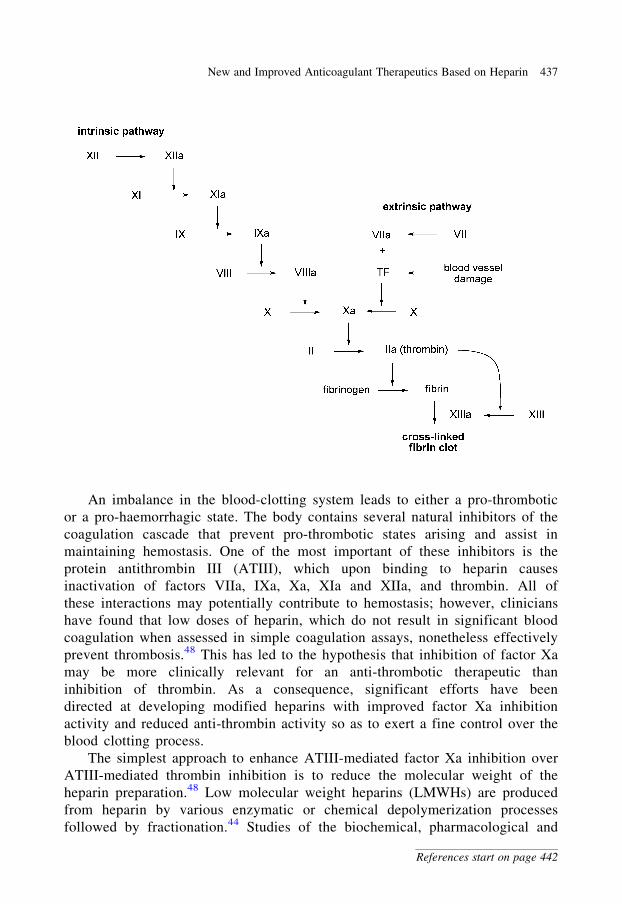
An imbalance in the blood-clotting system leads to either a pro-thrombotic
or a pro-haemorrhagic state. The body contains several natural inhibitors of the
coagulation cascade that prevent pro-thrombotic states arising and assist in
maintaining hemostasis. One of the most important of these inhibitors is the
protein antithrombin III (ATIII), which upon binding to heparin causes
inactivation of factors VIIa, IXa, Xa, XIa and XIIa, and thrombin. All of
these interactions may potentially contribute to hemostasis; however, clinicians
have found that low doses of heparin, which do not result in significant blood
coagulation when assessed in simple coagulation assays, nonetheless effectively
prevent thrombosis.48 This has led to the hypothesis that inhibition of factor Xa
may be more clinically relevant for an anti-thrombotic therapeutic than
inhibition of thrombin. As a consequence, significant efforts have been
directed at developing modified heparins with improved factor Xa inhibition
activity and reduced anti-thrombin activity so as to exert a fine control over the
blood clotting process.
The simplest approach to enhance ATIII-mediated factor Xa inhibition over
ATIII-mediated thrombin inhibition is to reduce the molecular weight of the
heparin preparation.48 Low molecular weight heparins (LMWHs) are produced
from heparin by various enzymatic or chemical depolymerization processes
followed by fractionation.44 Studies of the biochemical, pharmacological and
New and Improved Anticoagulant Therapeutics Based on Heparin 437
References start on page 442
chemical structures of LMWHs from different sources demonstrate that they are
not chemically or biochemically equivalent and have different pharmacological
properties.49 Despite the potential complications that these variations may cause,
LMWHs have largely displaced heparin from the drug market, and their
improved safety profile, more predictable dose/activity response and prolonged
anti-thrombotic activity allow their use in an outpatient setting. However,
LMWHs still suffer from some side-effects, in particular HIT.50
In the 1970s several crucial observations were made that helped cast light on
the mechanism of action of heparin.51 It was found that only a small fraction of
heparin molecules bind with high affinity to the blood-clotting inhibitor ATIII.
Moreover, the high affinity fraction accounted for essentially all of the anti-
coagulant activity of unfractionated heparin. This suggested that there was a
specific chemical structure within some heparin chains that was the preferred
ligand for ATIII. A Herculean effort by a number of research groups led to the
identification of several related pentasaccharides, which are the minimal
structure required for high-affinity binding to ATIII.51 The biological activity of
these pentasaccharides was confirmed by their synthesis,52–54 which showed that
they were not only able to bind to ATIII (with an affinity of 50 nM) but also were
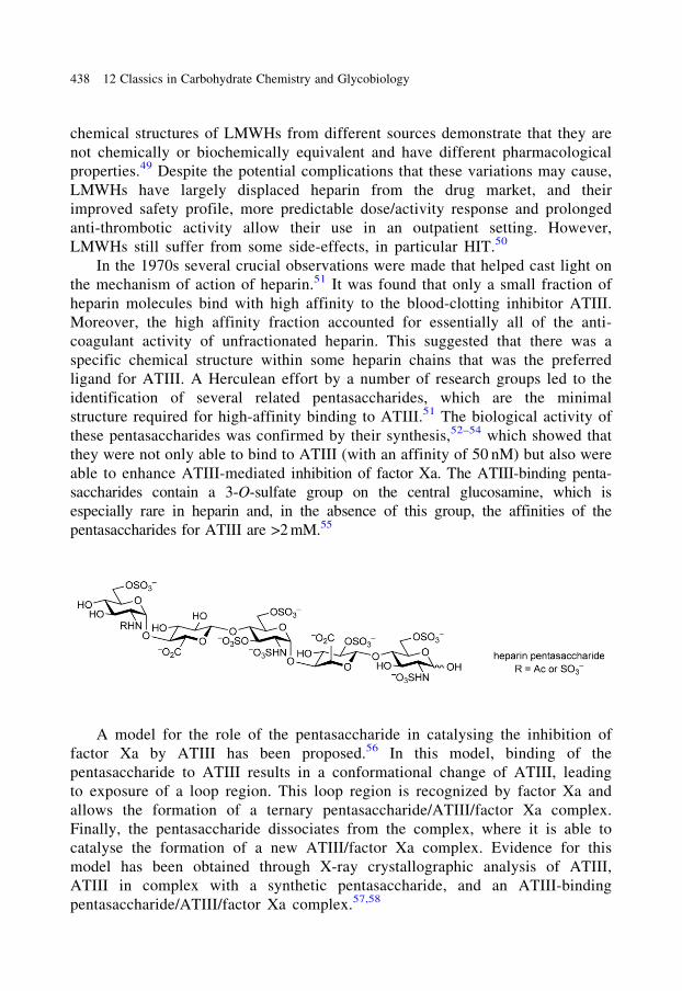
able to enhance ATIII-mediated inhibition of factor Xa. The ATIII-binding penta-
saccharides contain a 3-O-sulfate group on the central glucosamine, which is
especially rare in heparin and, in the absence of this group, the affinities of the
pentasaccharides for ATIII are >2 mM.55
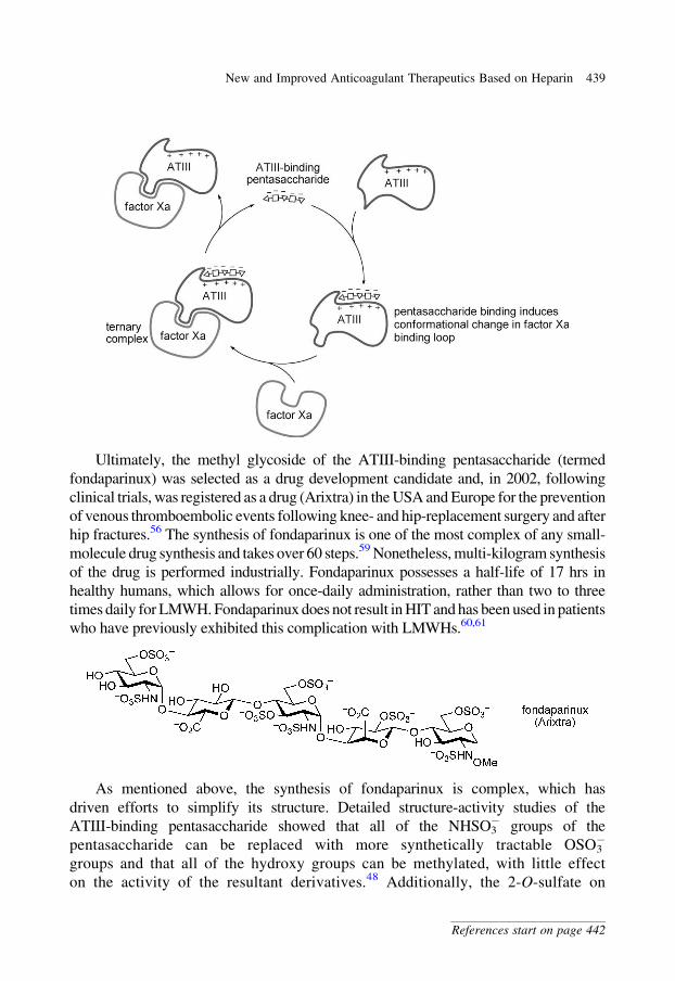
A model for the role of the pentasaccharide in catalysing the inhibition of
factor Xa by ATIII has been proposed.56 In this model, binding of the
pentasaccharide to ATIII results in a conformational change of ATIII, leading
to exposure of a loop region. This loop region is recognized by factor Xa and
allows the formation of a ternary pentasaccharide/ATIII/factor Xa complex.
Finally, the pentasaccharide dissociates from the complex, where it is able to
catalyse the formation of a new ATIII/factor Xa complex. Evidence for this
model has been obtained through X-ray crystallographic analysis of ATIII,
ATIII in complex with a synthetic pentasaccharide, and an ATIII-binding
pentasaccharide/ATIII/factor Xa complex.57,58
438 12 Classics in Carbohydrate Chemistry and Glycobiology
Ultimately, the methyl glycoside of the ATIII-binding pentasaccharide (termed
fondaparinux) was selected as a drug development candidate and, in 2002, following
clinical trials, was registered as a drug (Arixtra) in the USA and Europe for the prevention
of venous thromboembolic events following knee- and hip-replacement surgery and after
hip fractures.56 The synthesis of fondaparinux is one of the most complex of any small-
molecule drug synthesis and takes over 60 steps.59 Nonetheless, multi-kilogram synthesis
of the drug is performed industrially. Fondaparinux possesses a half-life of 17 hrs in
healthy humans, which allows for once-daily administration, rather than two to three
times daily for LMWH. Fondaparinux does not result in HIT and has been used in patients
who have previously exhibited this complication with LMWHs.60,61
As mentioned above, the synthesis of fondaparinux is complex, which has
driven efforts to simplify its structure. Detailed structure-activity studies of the
ATIII-binding pentasaccharide showed that all of the NHSO3� groups of the
pentasaccharide can be replaced with more synthetically tractable OSO3�
groups and that all of the hydroxy groups can be methylated, with little effect
on the activity of the resultant derivatives.48 Additionally, the 2-O-sulfate on
New and Improved Anticoagulant Therapeutics Based on Heparin 439
References start on page 442
the L-iduronic acid moiety can be replaced by a 2-O-methyl group. Finally,
sulfation of the 3-hydroxy group on the reducing-end sugar provides an
enhancement of ATIII-binding ability. When all of these modifications were
incorporated into a single molecule, the resulting pentasaccharide, termed
idraparinux, is not only much easier to synthesize than fondaparinux (approximately
25 steps) but also possesses higher activity and a longer duration of action (half-life
in humans of 120 hrs), potentially allowing once-weekly administration. Idraparinux
is currently in Phase III clinical trials.
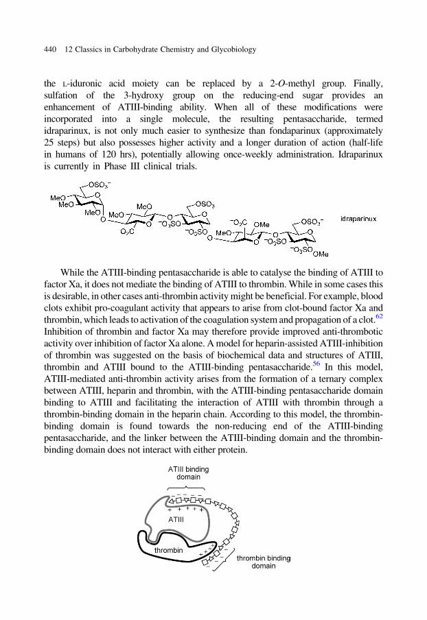
While the ATIII-binding pentasaccharide is able to catalyse the binding of ATIII to
factor Xa, it does not mediate the binding of ATIII to thrombin. While in some cases this
is desirable, in other cases anti-thrombin activity might be beneficial. For example, blood
clots exhibit pro-coagulant activity that appears to arise from clot-bound factor Xa and
thrombin, which leads to activation of the coagulation system and propagation of a clot.62
Inhibition of thrombin and factor Xa may therefore provide improved anti-thrombotic
activity over inhibition of factor Xa alone. A model for heparin-assisted ATIII-inhibition
of thrombin was suggested on the basis of biochemical data and structures of ATIII,
thrombin and ATIII bound to the ATIII-binding pentasaccharide.56 In this model,
ATIII-mediated anti-thrombin activity arises from the formation of a ternary complex
between ATIII, heparin and thrombin, with the ATIII-binding pentasaccharide domain
binding to ATIII and facilitating the interaction of ATIII with thrombin through a
thrombin-binding domain in the heparin chain. According to this model, the thrombin-
binding domain is found towards the non-reducing end of the ATIII-binding
pentasaccharide, and the linker between the ATIII-binding domain and the thrombin-
binding domain does not interact with either protein.
440 12 Classics in Carbohydrate Chemistry and Glycobiology
Consideration of the heparin/ATIII/thrombin model led to the proposal of an
ingenious approach to overcome one of the most serious problems with heparin,
HIT.63 HIT is caused by an immunoallergic reaction to the immunogenic
platelet factor 4–heparin complex.50 Platelet factor 4 (PF4) appears to bind to
any anionic heparin sequence, with the size for optimal PF4 binding being a
16-mer, and with at least eight consecutive negatively charged sugars being
required for binding. The model described above for the heparin/ATIII/thrombin
ternary complex suggests that at least 14 sugar residues are required for
successful formation of the ternary complex, and biochemical studies of model
compounds demonstrated that in fact at least a 15-mer was required. Thus, at
first glance it might appear that it would be impossible to find an
oligosaccharide that exhibits thrombin inhibition without interacting with PF4.
However, the model for the pentasaccharide/ATIII/thrombin complex
described above suggests that the sugars linking the ATIII-binding domain and
the thrombin-binding domain do not interact with either protein. Taken together,
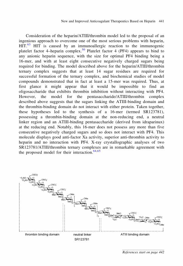
these hypotheses led to the synthesis of a 16-mer (termed SR123781),
possessing a thrombin-binding domain at the non-reducing end, a neutral
linker region and an ATIII-binding pentasaccharide (derived from idraparinux)
at the reducing end. Notably, this 16-mer does not possess any more than five
consecutive negatively charged sugars and so does not interact with PF4. This
molecule displays good anti-factor Xa activity, superior anti-thrombin activity to
heparin and no interaction with PF4. X-ray crystallographic analyses of two
SR123781/ATIII/thrombin ternary complexes are in remarkable agreement with
the proposed model for their interaction.64,65
New and Improved Anticoagulant Therapeutics Based on Heparin 441
References start on page 442
References
1. Mader, M.M. and Bartlett, P.A. (1997). Chem. Rev., 97, 1281.
2. Wolfenden, R. and Snider, M.J. (2001). Acc. Chem. Res., 34, 938.
3. Withers, S.G., Namchuk, M. and Mosi, R. (1999). In Iminosugars as Glycosidase Inhibitors:
Nojirimycin and Beyond (A.E. Stutz, ed.) p. 188. Wiley-VCH: Weinheim.
4. Evans, G.B. (2005). Aust. J. Chem., 57, 837.
5. Taylor, E.A. and Schramm, V.L. (2005). Curr. Top. Med. Chem., 5, 1237.
6. Schramm, V.L. (2003). Acc. Chem. Res., 36, 588.
7. Singh, V., Lee, J.E., Nunez, S., Howell, P.L. and Schramm, V.L. (2005). Biochemistry, 44, 11647
8. Filichev, V.V., Brandt, M. and Pedersen, E.B. (2001). Carbohydr. Res., 333, 115.
9. Singh, V., Evans, G.B., Lenz, D.H., Mason, J.M., Clinch, K., Mee, S., Painter, G.F., Tyler, P.C.,
Furneaux, R.H., Lee, J.E., Howell, P.L. and Schramm, V.L. (2005). J. Biol. Chem., 280, 18265.
10. Evans, G.B., Furneaux, R.H., Tyler, P.C. and Schramm, V.L. (2003). Org. Lett., 5, 3639.
11. Miles, R.W., Tyler, P.C., Furneaux, R.H., Bagdassarian, C.K., Schramm, V.L. (1998). Biochem-
istry, 37, 8615.
12. Evans, G.B.; Furneaux, R. H.; Lewandowicz, A., Schramm, V.L., Tyler, P.C. (2003). J. Med.
Chem., 46, 5271.
13. Kicska, G.A., Tyler, P.C., Evans, G.B.,Furneaux, R.H., Shi, W., Fedorov, A., Lewandowicz, A.,
Cahill, S.M., Almo, S.C. and Schramm, V.L. (2002). Biochemistry, 41, 14489.
14. Kicska, G.A., Long, L., Horig, H. Fairchild, C., Tyler, P.C., Furneaux, R.H., Schramm, V.L. and
Kaufman, H.L. (2001). Proc. Natl. Acad. Sci. USA, 98, 4593.
15. Lewandowicz, A., Tyler, P.C., Evans, G.B., Furneaux, R.H., Schramm, V.L. (2003). J. Biol. Chem.,
278, 31465.
16. Korycka, A., Blonski, J.Z. and Robak, T. (2007). Mini-Rev. Med. Chem., 7, 976.
17. Taylor, E.A., Clinch, K., Kelly, P.M. Li, L., Evans, G.B., Tyler, P.C. and Schramm, V.L. (2007).
J. Am. Chem. Soc., 129, 6984.
18. Evans, G.B., Furneaux, R.H., Greatrex, B., Murkin, A.S., Schramm, V.L., Tyler, P.C. (2008).
J. Med. Chem., 51, 948.
19. Lee, J.E., Singh, V., Evans, G.B., Tyler, P.C., Furneaux, R.H., Cornell, K.A., Riscoe, M.K.,
Schramm, V.L. and Howell, P.L. (2005). J. Biol. Chem., 280, 18274.
20. Gutierrez, J.A., Luo, M., Singh, V, Li, L., Brown, R.L., Norris, G.E., Evans, G.B., Furneaux, R.H.,
Tyler, P.C., Painter, G.F., Lenz, D. H. and Schramm, V.L. (2007). ACS Chem. Biol., 2, 725.
21. Clark, I.A. and Schofield, L. (2000). Parasitol. Today, 16, 451.
22. Schofield, L., Hewitt, M.C., Evans, K., Siomos, M.A. and Seeberger, P.H. (2002). Nature, 418, 785.
23. Schofield, L., McConville, M.J., Hansen, D., Campbell, A.S., Fraser-Reid, B., Grusby, M.J. and
Tachado, S.D. (1999). Science, 283, 225.
24. Schofield, L. (2007). Microbes Infect., 9, 784.
25. Gerold, P., Dieckmann-Schuppert, A. and Schwarz, R.T. (1994). J. Biol. Chem., 269, 2597.
26. Berhe, S., Schofield, L., Schwarz, R.T. and Gerold, P. (1999). Mol. Biochem. Parasitol., 103, 273.
27. Schofield, L. and Hackett, F. (1993). J. Exp. Med., 177, 145.
28. Plante, O.J., Palmacci, E.R. and Seeberger, P.H. (2001). Science, 291, 1523.
29. Seeberger, P.H. (2003). Chem. Commun., 1115.
30. Hewitt, M.C., Snyder, D.A. and Seeberger, P.H. (2002). J. Am. Chem. Soc., 124, 13434.
31. Seeberger, P.H., Soucy, R.L., Kwon, Y.U., Snyder, D.A. and Kanemitsu, T. (2004). Chem.
Commun., 1706.
32. Kwon, Y.U., Soucy, R.L., Snyder, D.A. and Seeberger, P.H. (2005). Chem. Eur. J., 11, 2493.
33. Ouerfelli, O., Warren, J.D., Wilson, R.M. and Danishefsky, S.J. (2005). Expert Rev. Vaccines, 4, 677.
442 12 Classics in Carbohydrate Chemistry and Glycobiology
34. Danishefsky, S.J. and Allen, J.R. (2000). Angew. Chem. Int. Ed., 39, 836.
35. Bilodeau, M.T., Park, T.K., Hu, S., Randolph, J.T., Danishefsky, S.J., Livingston, P.O. and Zhang, S.
(1995). J. Am. Chem. Soc., 117, 7840.
36. Park, T.K., Kim, I.J., Hu, S., Bilodeau, M.T., Randolph, J.T., Kwon, O. and Danishefsky, S.J.
(1996). J. Am. Chem. Soc., 118, 11488.
37. Galonic, D.P. and Gin, D.Y. (2007). Nature, 446, 1000.
38. Kim, Y.J., Wang, P., Navarro-Villalobos, M., Rohde, B.D., Derryberry, J., Gin, D.Y. (2006). J. Am.
Chem. Soc., 128, 11906.
39. Marciani, D.J., Press, J.B., Reynolds, R.C., Pathak, A.K., Pathak, V., Gundy, L.E., Farmer, J.T.,
Koratich, M.S. and May, R.D. (2000). Vaccine, 18, 3141.
40. Slovin, S.F., Ragupathi, G., Adluri, S., Ungers, G., Terry, K., Kim, S., Spassova, M., Bornmann,
W.G., Fazzari, M., Dantis, L., Olkiewicz, K., Lloyd, K.O., Livingston, P.O., Danishefsky, S.J. and
Scher, H.I. (1999). Proc. Natl. Acad. Sci. USA, 96, 5710.
41. Ragupathi, G., Koide, F., Livingston, P.O., Cho, Y.S., Endo, A., Wan, Q., Spassova, M.K., Keding,
S.J., Allen, J., Ouerfelli, O., Wilson, R.M. and Danishefsky, S.J. (2006). J. Am. Chem. Soc., 128, 2715.
42. Ingale, S., Wolfert, M.A., Gaekwad, J., Buskas, T. and Boons, G.J. (2007). Nat. Chem. Biol., 3, 663.
43. Bundle, D.R. (2007). Nat. Chem. Biol., 3, 605.
44. Linhardt, R.J. (2003). J. Med. Chem., 46, 2551.
45. Linhardt, R.J. (1991). Chem. Ind., 2, 45.
46. Rutty, C.J. (1996). CONNTACT, 9.
47. Porter, J. and Jick, H. (1977). JAMA, 237, 879.
48. van Boeckel, C.A.A. and Petitou, M. (1993). Angew. Chem. Int. Ed. Engl., 32, 1671.
49. Linhardt, R.J., Loganathan, D., al-Hakim, A., Wang, H.M., Walenga, J.M., Hoppensteadt, D. and
Fareed, J. (1990). J. Med. Chem., 33, 1639.
50. Baglin, T.P. (2001). J. Clin. Pathol., 54, 272.
51. Petitou, M., Casu, B. and Lindahl, U. (2003). Biochimie, 85, 83.
52. Petitou, M., Duchaussoy, P., Lederman, I., Choay, J., Sinay, P., Jacquinet, J.C. and Torri, G.
(1986). Carbohydr. Res., 147, 221.
53. Sinay, P., Jacquinet, J.C., Petitou, M., Duchaussoy, P., Lederman, I., Choay, J. and Torri, G.
(1984). Carbohydr. Res., 132, C5.
54. van Boeckel, C.A., Beetz, T., Vos, J.N., de Jong, A.J.M., van Aelst, S.F., van den Bosch, R.H.,
Mertens, J.M.R. and van der Vlugt, F.A. (1985). J. Carbohydr. Chem., 4, 293.
55. Petitou, M., Duchaussoy, P., Lederman, I., Choay, J. and Sinay, P. (1988). Carbohydr. Res., 179, 163.
56. Petitou, M. and van Boeckel, C.A.A. (2004). Angew. Chem. Int. Ed., 43, 3118.
57. Imberty, A., Lortat-Jacob, H. and Perez, S. (2007). Carbohydr. Res., 342, 430.
58. Johnson, D.J.D., Li, W., Adams, T.E. and Huntington, J.A. (2006). EMBO J., 25, 2029.
59. Codee, J.D.C., Overkleeft, H.S., van der Marel, G.A. and van Boeckel, C.A.A. (2004). Drug
Discov. Today: Technologies, 1, 317.
60. Kuo, K.H. and Kovacs, M.J. (2005). Hematology, 10, 271.
61. Giangrande, P.L.F. (2002). Int. J. Clin. Prac., 56, 615.
62. Herault, J.-P., Cappelle, M., Bernat, A., Millet, L., Bono, F., Schaeffer, P. and Herbert, J.-M.
(2003). J. Thromb. Haemost., 1, 1959.
63. Petitou, M., Herault, J.-P., Bernat, A., Driguez, P.-A., Duchaussoy, P., Lormeau, J.-C. and Herbert,
J.-M. (1999). Nature, 398, 417.
64. Li, W., Johnson, D.J., Esmon, C.T. and Huntington, J.A. (2004). Nat. Struct. Mol. Biol., 11, 857.
65. Dementiev, A., Petitou, M., Herbert, J.-M. and Gettins, P.G.W. (2004). Nat. Struct. Mol. Biol., 11, 863.
References 443
Chapter 9
Disaccharides, Oligosaccharidesand Polysaccharides
Monosaccharides may be linked together in an almost limitless number of ways to
form disaccharides, oligosaccharides and polysaccharides. There are over 134 million
ways to link the eight D-hexopyranoses together into a linear hexasaccharide and, if
different ring forms (e.g. furanose), L-sugars, deoxy sugars, aminodeoxy sugars, chain
branching and assorted modifying groups are allowed, the number of potential struc-
tures increases combinatorially. That said there is a much smaller set of structures
commonly found in nature. This chapter will cover the common di- and trisaccharides
and oligomers and polymers derived from (mostly) single carbohydrate monomers.
Later chapters will cover more complex oligo- and polysaccharides comprised of more
than one sugar monomer and bearing various functional group adornments. To begin we
provide a list of the commonly accepted names for a range of disaccharides (Table 1);
many of these compounds are not found in appreciable amounts in nature but are
derived by controlled hydrolysis of more complex polysaccharides.
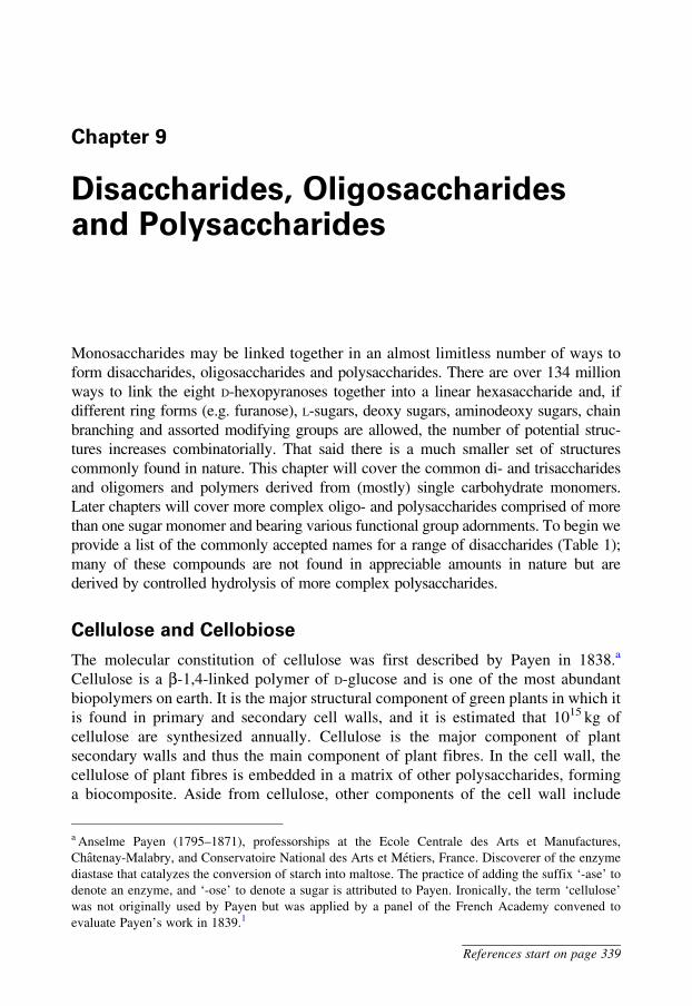
Cellulose and Cellobiose
The molecular constitution of cellulose was first described by Payen in 1838.a
Cellulose is a b-1,4-linked polymer of D-glucose and is one of the most abundant
biopolymers on earth. It is the major structural component of green plants in which it
is found in primary and secondary cell walls, and it is estimated that 1015 kg of
cellulose are synthesized annually. Cellulose is the major component of plant
secondary walls and thus the main component of plant fibres. In the cell wall, the
cellulose of plant fibres is embedded in a matrix of other polysaccharides, forming
a biocomposite. Aside from cellulose, other components of the cell wall include
a Anselme Payen (1795–1871), professorships at the Ecole Centrale des Arts et Manufactures,
Chatenay-Malabry, and Conservatoire National des Arts et Metiers, France. Discoverer of the enzyme
diastase that catalyzes the conversion of starch into maltose. The practice of adding the suffix ‘-ase’ to
denote an enzyme, and ‘-ose’ to denote a sugar is attributed to Payen. Ironically, the term ‘cellulose’
was not originally used by Payen but was applied by a panel of the French Academy convened to
evaluate Payen’s work in 1839.1
References start on page 339
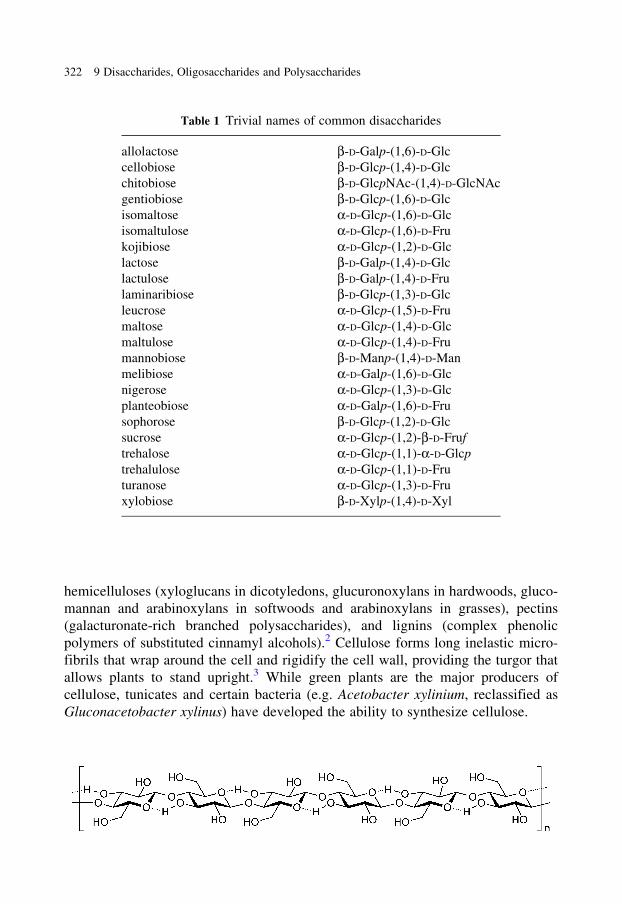
hemicelluloses (xyloglucans in dicotyledons, glucuronoxylans in hardwoods, gluco-
mannan and arabinoxylans in softwoods and arabinoxylans in grasses), pectins
(galacturonate-rich branched polysaccharides), and lignins (complex phenolic
polymers of substituted cinnamyl alcohols).2 Cellulose forms long inelastic micro-
fibrils that wrap around the cell and rigidify the cell wall, providing the turgor that
allows plants to stand upright.3 While green plants are the major producers of
cellulose, tunicates and certain bacteria (e.g. Acetobacter xylinium, reclassified as
Gluconacetobacter xylinus) have developed the ability to synthesize cellulose.
Table 1 Trivial names of common disaccharides
allolactose b-D-Galp-(1,6)-D-Glc
cellobiose b-D-Glcp-(1,4)-D-Glc
chitobiose b-D-GlcpNAc-(1,4)-D-GlcNAc
gentiobiose b-D-Glcp-(1,6)-D-Glc
isomaltose a-D-Glcp-(1,6)-D-Glc
isomaltulose a-D-Glcp-(1,6)-D-Fru
kojibiose a-D-Glcp-(1,2)-D-Glc
lactose b-D-Galp-(1,4)-D-Glc
lactulose b-D-Galp-(1,4)-D-Fru
laminaribiose b-D-Glcp-(1,3)-D-Glc
leucrose a-D-Glcp-(1,5)-D-Fru
maltose a-D-Glcp-(1,4)-D-Glc
maltulose a-D-Glcp-(1,4)-D-Fru
mannobiose b-D-Manp-(1,4)-D-Man
melibiose a-D-Galp-(1,6)-D-Glc
nigerose a-D-Glcp-(1,3)-D-Glc
planteobiose a-D-Galp-(1,6)-D-Fru
sophorose b-D-Glcp-(1,2)-D-Glc
sucrose a-D-Glcp-(1,2)-b-D-Fruf
trehalose a-D-Glcp-(1,1)-a-D-Glcp
trehalulose a-D-Glcp-(1,1)-D-Fru
turanose a-D-Glcp-(1,3)-D-Fru
xylobiose b-D-Xylp-(1,4)-D-Xyl
322 9 Disaccharides, Oligosaccharides and Polysaccharides
Cellulose structure has been the subject of much investigation, and a complete
crystallographic description remains elusive. There is evidence from powder
diffraction studies of cellulose for the presence of a hydrogen bond between the
endocyclic oxygen of one sugar and the 3-OH of its neighbour. This hydrogen bond
results from a structure where every second sugar residue is ‘flipped’ with respect to
the first, forming a ribbon-like chain. Each of these chains pack together in the
crystal in either a parallel or an anti-parallel sense, forming cellulose I or II,
respectively.4 Cellulose I is the natural form found in plant fibres, and cellulose II
is formed upon recrystallization of cellulose.
Cellulose biosynthesis in plants is orchestrated by glycosyltransferases termed
cellulase synthases, using UDP-glucose as a substrate, itself derived from sucrose.
Cellulose synthase enzymes are believed to be arranged in sixfold symmetric
rosettes, each comprised of six catalytic subunits, in the plasma membrane of the
plant cell.5 Consistent with this biosynthetic hypothesis, individual cellulose fibres
are proposed to consist of 36 parallel b-glucan chains.
Cellulose is the major constituent of paper and is therefore used heavily by the
pulp and paper industry. In the textile industry, cellulose is the major component of
fabrics made from cotton, linen and other plant fibres. In recent years there has been
great interest in moving away from chemical-intensive traditional bleaching pro-
cesses for fibre modification, towards enzyme-based procedures. Glycoside hydro-
lases, termed xylanases, are useful for fibre modification and assist in the removal of
lignins from crude kraftb pulps by degrading unwanted hemicellulose components.6,7
Cellulose is particularly challenging to degrade, and organisms capable of doing
so require many enzymes to achieve the task. Enzyme complexes, termed cellulo-
somes, have been found in cellulolytic fungi and bacteria.8 These complexes contain
endo- and exo-cellulases, and b-glucosidases, as well as carbohydrate-binding pro-
teins that are commonly fused to a glycoside hydrolase. Vertebrates are unable to
degrade cellulose, and the digestive tracts of herbivores contain symbiotic cellulo-
lytic bacteria. Similarly, termites achieve the degradation of cellulose either through
gut microflora or through the practice of fungiculture.9 In fungiculture, termites
apply pre-digested plant material to a fungal colony, and older cultured material is
removed and consumed by the termite.
Cellulolytic enzymes may be of potential use in cellulose degradation for
bioethanol production. However, to date, limited success has been achieved in
developing commercial processes capable of degrading cellulose economically,
with the bulk of bioethanol being produced from more easily fermented precursors
containing starch, such as grains. Cellulases have been developed as additives for
laundry detergents.10 During wear, microfibres rise from the surface of cotton thread
and affect the feel and brightness of cotton fabrics. Alkali-stable cellulases have been
b Kraft. noun (German), strength or power.
Cellulose and Cellobiose 323
References start on page 339
developed that degrade these microfibres, restoring the feel and brightening the
appearance of the garment.
Cellulose is a feedstock for the preparation of a wide range of materials. The
nitric acid ester of cellulose, cellulose nitrate or ‘nitrocellulose’, is the oldest
cellulose derivative and is prepared by treatment of cellulose with nitric acid,
sulfuric acid and water.11 It has excellent film-forming properties and is used in
lacquers and thermoplastics. A combination of nitrocellulose and camphor provides
celluloid, the first synthetic thermoplastic material. Nitrocellulose is combustible
and explosive, and was used in gun cotton and various military devices. However, its
shock sensitivity led to a range of industrial disasters in plants and mines. Nobelc
discovered a more stable combination of cellulose nitrate, termed blasting gelatine or
gelignite, comprised of ‘nitroglycerine’, sodium nitrate and wood pulp, and also a
smokeless military explosive termed ballistite, composed of cellulose nitrate and
nitroglycerine blended with a stabilizer.11 Cellulose acetate is formed by acetylation
of cellulose with a mixture of acetic acid, acetic anhydride and sulfuric acid.11
Cellulose acetate is used as an electrical insulating film, as a lacquer, in adhesives
and in the production of cellulose acetate fibres, which are used in the textile
industry. Cellulose may be converted to rayon fibres or cellophane sheets by the
viscose process, which involves dissolution of cellulose in alkali and carbon
disulfide, and reprecipitation using acid.11
Carboxymethylcellulose is an ether derivative of cellulose prepared by reaction
of cellulose and chloroacetic acid in alkali.12 It is used in the food industry as a
thickener and stabilizer, in the mining industry in drilling muds, and in the paper and
textile industry to alter surface characteristics of fibres. Controlled acetolysis
of cellulose with acetic anhydride and sulfuric acid leads to a-cellobiose
octaacetate.13
Starch, Amylopectin, Amylose and Maltose
Starch is the major source of dietary energy for the world’s human population. Plants
produce a mixture of a-glucans that are deposited in the cytoplasm as insoluble,
semicrystalline granules and that act as an energy storage reserve. Starch granules
are composed of a linear polymer, termed amylose, of several thousand a-1,4-linked
D-glucose residues, and a branched polymer, termed amylopectin, consisting of
mainly a-1,4-linked D-glucose residues, with an a-1,6-linked branch every 24–30
residues.14 Amylopectin molecules are much larger than amylose; each amylopectin
molecule contains up to 106D-glucose units. Amylose adopts a helical conformation
c Alfred Nobel (1833–1896), Swedish inventor and industrialist noted for the inventions of dynamite,
gelignite and ballistite, and the creation of the Nobel Foundation by bequest of his will.
324 9 Disaccharides, Oligosaccharides and Polysaccharides

with six D-glucose residues per turn, an outer diameter of 13 A and an inner diameter
of 5 A. This inner hydrophobic cavity can occlude lipophilic molecules such as
butanol;14 the starch–iodine complex provides a sensitive means for the detection
of iodine and results from the binding of polyiodide ion (likely to be I5–) within this
cavity, thereby generating a characteristic blue colour.15
The biosynthesis of amylose and amylopectin in plants requires a
glycosyltransferase (starch synthase), a transglycosidase (branching enzyme) and a
glycoside hydrolase (debranching enzyme).16 Starch synthase condenses the glycosyl
donor ADP-glucose to give a-1,4-linked linear chains of D-glucose residues.
ADP-glucose itself is synthesized from glucose-1-phosphate and ATP by the action
of ADP-glucose pyrophosphorylase.16 Starch-branching enzymes cleave internal a-1,4-
linkages within the long chains and transfer the released chains to the 6-position of a
terminal sugar residue, resulting in the formation of branch points within amylopectin.
Debranching enzymes are involved in amylopectin biosynthesis and cleave a-1,6-
glucose linkages, removing branches that are inappropriately positioned within
amylopectin and that affect its ability to pack into a dense, semicrystalline structure.
Starch is widely used in foods for controlling texture and structure, and for
making gels. Maltose syrups and maltodextrins are produced from starch by partial
hydrolysis using acid or enzymes and consist of mixtures of D-glucose, maltose and
maltose oligosaccharides. They are used as a fermentation substrate in brewing, for
improving ‘mouth feel’ of foods, and for altering the physical properties of food-
stuffs through their gelling and crystallization prevention properties.17 Starch may be
isomerized to form high-fructose corn syrups, which possess sweetness similar to
invert syrup derived from sucrose (see Epilogue). Such syrups can be used to
maintain the texture of baked goods during storage and to prevent crystallization
in frozen foods such as ice cream.18 Aside from its use as a dietary component, starch
Starch, Amylopectin, Amylose and Maltose 325
References start on page 339
finds various industrial applications as a textile sizing agent, in adhesives, in the
pharmaceutical industry as an excipient and in cosmetics. Finally, a growing use of
starch is as a renewable feedstock providing the so-called biofuels, which are an
alternative to fuels derived from fossil reserves. The most significant biofuel is
bioethanol, which in the United States is in large part derived from fermentation of
starch from corn.19 In the United States alone, 15 megalitres of bioethanol are
produced annually.20 Despite much interest, no process for conversion of cellulosic
material into ethanol is yet in large-scale use.
Glycogen
Glycogen is a branched polymer of D-glucose that serves as a store of energy and
carbon in vertebrates, and is found largely in the liver and skeletal muscle.15 It
possesses considerable structural similarity to amylopectin but is more highly
branched, and is sometimes referred to as animal starch. Glycogen particles contain
107–108D-glucose molecules per unit, and varying amounts of associated proteins.
The basic polymerization occurs through a-1,4-linkages, with branch points intro-
duced through a-1,6-linkages. After feeding in mammals, elevated blood glucose
stimulates secretion of insulin, promoting glucose uptake into tissues and production
of glycogen. When blood glucose levels fall, the hormone glucagon stimulates the
breakdown of glycogen in the liver.
Glycogen particles contain at their core a dimeric protein termed glycogenin.
Glycogenin is a glycosyltransferase that initiates glycogen biosynthesis and acts as a
primer for the glycogen molecule.21 Glycogenin catalyses the transfer of D-glucose
from UDP-glucose to a tyrosine residue (Tyr194) on glycogenin to form an
a-glucosyl tyrosine. Additional catalytic cycles lead to elongation of the a-glucosyl
tyrosine, building an a-1,4-linked D-glucose chain of about 10 residues.22 There is
evidence suggesting that one molecule of glycogenin within the dimer glucosylates
the other partner,23 although an analysis of an X-ray crystal structure of glycogenin
suggested that self-glucosylation may occur for the earliest glucosylation events.21
Following the initiation of glycogen biosynthesis by glycogenin, an a-glucosyltrans-
ferase, glycogen synthase, elongates the short chains formed on glycogenin, leading
to extended a-1,4-linked chains. A transglycosidase, glycogen-branching enzyme,
introduces a-1,6-linked branches into the glycogen structure, through transfer of the
terminal seven glucosyl residues from an a-1,4-linked chain to O6 of a glucosyl
residue.24,25 This reaction introduces a branch point, and both chains can then be
extended by glycogen synthase. Introduction of branches increases the number of
terminal D-glucose residues and affects the rate of release of D-glucose during
glycogen metabolism. Additionally, introduction of branches into glycogen increases
its solubility.
326 9 Disaccharides, Oligosaccharides and Polysaccharides
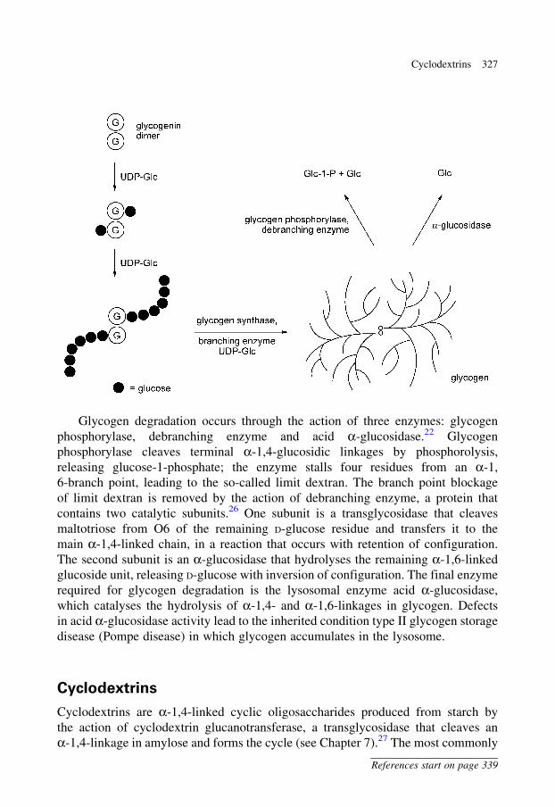
Glycogen degradation occurs through the action of three enzymes: glycogen
phosphorylase, debranching enzyme and acid a-glucosidase.22 Glycogen
phosphorylase cleaves terminal a-1,4-glucosidic linkages by phosphorolysis,
releasing glucose-1-phosphate; the enzyme stalls four residues from an a-1,
6-branch point, leading to the so-called limit dextran. The branch point blockage
of limit dextran is removed by the action of debranching enzyme, a protein that
contains two catalytic subunits.26 One subunit is a transglycosidase that cleaves
maltotriose from O6 of the remaining D-glucose residue and transfers it to the
main a-1,4-linked chain, in a reaction that occurs with retention of configuration.
The second subunit is an a-glucosidase that hydrolyses the remaining a-1,6-linked
glucoside unit, releasing D-glucose with inversion of configuration. The final enzyme
required for glycogen degradation is the lysosomal enzyme acid a-glucosidase,
which catalyses the hydrolysis of a-1,4- and a-1,6-linkages in glycogen. Defects
in acid a-glucosidase activity lead to the inherited condition type II glycogen storage
disease (Pompe disease) in which glycogen accumulates in the lysosome.
Cyclodextrins
Cyclodextrins are a-1,4-linked cyclic oligosaccharides produced from starch by
the action of cyclodextrin glucanotransferase, a transglycosidase that cleaves an
a-1,4-linkage in amylose and forms the cycle (see Chapter 7).27 The most commonly
Cyclodextrins 327
References start on page 339
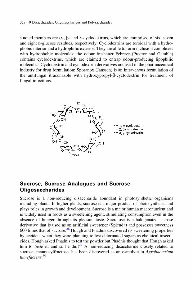
studied members are a-, b- and �-cyclodextrins, which are comprised of six, seven
and eight D-glucose residues, respectively. Cyclodextrins are toroidal with a hydro-
phobic interior and a hydrophilic exterior. They are able to form inclusion complexes
with hydrophobic molecules; the odour freshener Febreze (Proctor and Gamble)
contains cyclodextrins, which are claimed to entrap odour-producing lipophilic
molecules. Cyclodextrin and cyclodextrin derivatives are used in the pharmaceutical
industry for drug formulation; Sporanox (Janssen) is an intravenous formulation of
the antifungal itraconazole with hydroxypropyl-b-cyclodextrin for treatment of
fungal infections.
Sucrose, Sucrose Analogues and Sucrose
Oligosaccharides
Sucrose is a non-reducing disaccharide abundant in photosynthetic organisms
including plants. In higher plants, sucrose is a major product of photosynthesis and
plays roles in growth and development. Sucrose is a major human macronutrient and
is widely used in foods as a sweetening agent, stimulating consumption even in the
absence of hunger through its pleasant taste. Sucralose is a halogenated sucrose
derivative that is used as an artificial sweetener (Splenda) and possesses sweetness
600 times that of sucrose.28 Hough and Phadnis discovered its sweetening properties
by accident when they were planning to test chlorinated sugars as chemical insecti-
cides. Hough asked Phadnis to test the powder but Phadnis thought that Hough asked
him to taste it, and so he did!29 A non-reducing disaccharide closely related to
sucrose, mannosylfructose, has been discovered as an osmolyte in Agrobacterium
tumefaciens.30
328 9 Disaccharides, Oligosaccharides and Polysaccharides
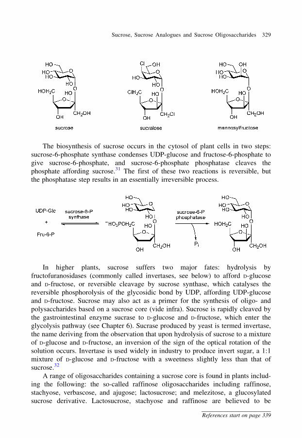
The biosynthesis of sucrose occurs in the cytosol of plant cells in two steps:
sucrose-6-phosphate synthase condenses UDP-glucose and fructose-6-phosphate to
give sucrose-6-phosphate, and sucrose-6-phosphate phosphatase cleaves the
phosphate affording sucrose.31 The first of these two reactions is reversible, but
the phosphatase step results in an essentially irreversible process.
In higher plants, sucrose suffers two major fates: hydrolysis by
fructofuranosidases (commonly called invertases, see below) to afford D-glucose
and D-fructose, or reversible cleavage by sucrose synthase, which catalyses the
reversible phosphorolysis of the glycosidic bond by UDP, affording UDP-glucose
and D-fructose. Sucrose may also act as a primer for the synthesis of oligo- and
polysaccharides based on a sucrose core (vide infra). Sucrose is rapidly cleaved by
the gastrointestinal enzyme sucrase to D-glucose and D-fructose, which enter the
glycolysis pathway (see Chapter 6). Sucrase produced by yeast is termed invertase,
the name deriving from the observation that upon hydrolysis of sucrose to a mixture
of D-glucose and D-fructose, an inversion of the sign of the optical rotation of the
solution occurs. Invertase is used widely in industry to produce invert sugar, a 1:1
mixture of D-glucose and D-fructose with a sweetness slightly less than that of
sucrose.32
A range of oligosaccharides containing a sucrose core is found in plants includ-
ing the following: the so-called raffinose oligosaccharides including raffinose,
stachyose, verbascose, and ajugose; lactosucrose; and melezitose, a glucosylated
sucrose derivative. Lactosucrose, stachyose and raffinose are believed to be
Sucrose, Sucrose Analogues and Sucrose Oligosaccharides 329
References start on page 339

largely undigestible and may cause flatulence in humans.33 They are used as low-
calorie, non-cariogenic sugar substitutes in a range of foods.34,35 Additionally,
lactosucrose may have effects in promoting the growth of beneficial bacteria in the
gut of humans and canine and feline pets.36 The raffinose oligosaccharides are
of almost ubiquitous occurrence in plant seeds and are biosynthesized from sucrose
and UDP-galactose, by the intermediacy of various galactosylinositols, including
galactinol.37 These oligosaccharides, along with the fructans discussed later in this
chapter, represent rare examples of oligosaccharides that are synthesized without the
direct involvement of sugar 1-phosphate donors.37
Sucrose is a precursor for the production of a variety of disaccharides used in the
food industry including isomaltulose (Palatinose) by rearrangement with the enzyme
trehalulose synthase,38 and isomalt (Palatinit), a mixture of two diastereoisomeric
disaccharides derived by reduction of isomaltulose (see Chapter 3).39
330 9 Disaccharides, Oligosaccharides and Polysaccharides
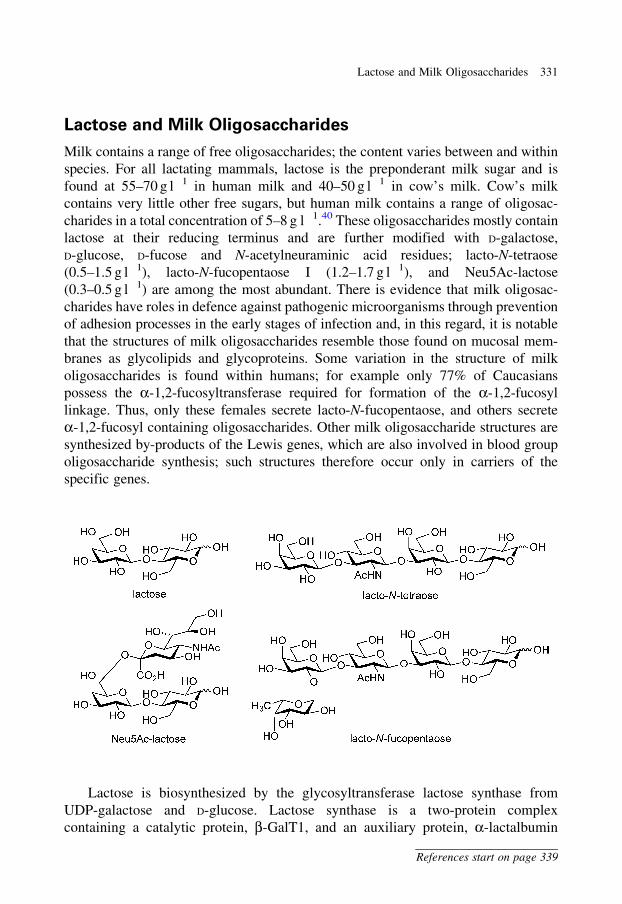
Lactose and Milk Oligosaccharides
Milk contains a range of free oligosaccharides; the content varies between and within
species. For all lactating mammals, lactose is the preponderant milk sugar and is
found at 55–70 g l�1 in human milk and 40–50 g l�1 in cow’s milk. Cow’s milk
contains very little other free sugars, but human milk contains a range of oligosac-
charides in a total concentration of 5–8 g l�1.40 These oligosaccharides mostly contain
lactose at their reducing terminus and are further modified with D-galactose,
D-glucose, D-fucose and N-acetylneuraminic acid residues; lacto-N-tetraose
(0.5–1.5 g l�1), lacto-N-fucopentaose I (1.2–1.7 g l�1), and Neu5Ac-lactose
(0.3–0.5 g l�1) are among the most abundant. There is evidence that milk oligosac-
charides have roles in defence against pathogenic microorganisms through prevention
of adhesion processes in the early stages of infection and, in this regard, it is notable
that the structures of milk oligosaccharides resemble those found on mucosal mem-
branes as glycolipids and glycoproteins. Some variation in the structure of milk
oligosaccharides is found within humans; for example only 77% of Caucasians
possess the a-1,2-fucosyltransferase required for formation of the a-1,2-fucosyl
linkage. Thus, only these females secrete lacto-N-fucopentaose, and others secrete
a-1,2-fucosyl containing oligosaccharides. Other milk oligosaccharide structures are
synthesized by-products of the Lewis genes, which are also involved in blood group
oligosaccharide synthesis; such structures therefore occur only in carriers of the
specific genes.
Lactose is biosynthesized by the glycosyltransferase lactose synthase from
UDP-galactose and D-glucose. Lactose synthase is a two-protein complex
containing a catalytic protein, b-GalT1, and an auxiliary protein, a-lactalbumin
Lactose and Milk Oligosaccharides 331
References start on page 339
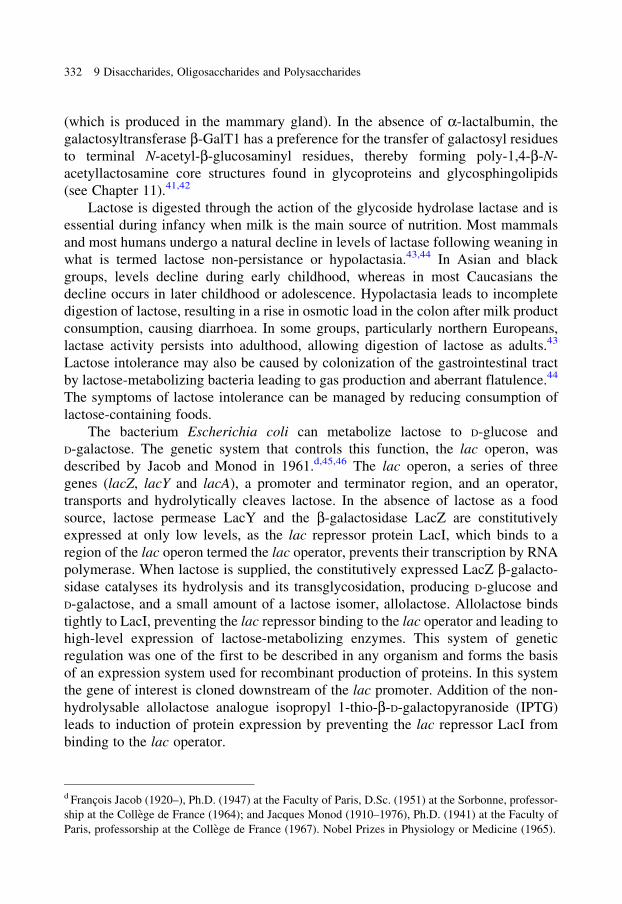
(which is produced in the mammary gland). In the absence of a-lactalbumin, the
galactosyltransferase b-GalT1 has a preference for the transfer of galactosyl residues
to terminal N-acetyl-b-glucosaminyl residues, thereby forming poly-1,4-b-N-
acetyllactosamine core structures found in glycoproteins and glycosphingolipids
(see Chapter 11).41,42
Lactose is digested through the action of the glycoside hydrolase lactase and is
essential during infancy when milk is the main source of nutrition. Most mammals
and most humans undergo a natural decline in levels of lactase following weaning in
what is termed lactose non-persistance or hypolactasia.43,44 In Asian and black
groups, levels decline during early childhood, whereas in most Caucasians the
decline occurs in later childhood or adolescence. Hypolactasia leads to incomplete
digestion of lactose, resulting in a rise in osmotic load in the colon after milk product
consumption, causing diarrhoea. In some groups, particularly northern Europeans,
lactase activity persists into adulthood, allowing digestion of lactose as adults.43
Lactose intolerance may also be caused by colonization of the gastrointestinal tract
by lactose-metabolizing bacteria leading to gas production and aberrant flatulence.44
The symptoms of lactose intolerance can be managed by reducing consumption of
lactose-containing foods.
The bacterium Escherichia coli can metabolize lactose to D-glucose and
D-galactose. The genetic system that controls this function, the lac operon, was
described by Jacob and Monod in 1961.d,45,46 The lac operon, a series of three
genes (lacZ, lacY and lacA), a promoter and terminator region, and an operator,
transports and hydrolytically cleaves lactose. In the absence of lactose as a food
source, lactose permease LacY and the b-galactosidase LacZ are constitutively
expressed at only low levels, as the lac repressor protein LacI, which binds to a
region of the lac operon termed the lac operator, prevents their transcription by RNA
polymerase. When lactose is supplied, the constitutively expressed LacZ b-galacto-
sidase catalyses its hydrolysis and its transglycosidation, producing D-glucose and
D-galactose, and a small amount of a lactose isomer, allolactose. Allolactose binds
tightly to LacI, preventing the lac repressor binding to the lac operator and leading to
high-level expression of lactose-metabolizing enzymes. This system of genetic
regulation was one of the first to be described in any organism and forms the basis
of an expression system used for recombinant production of proteins. In this system
the gene of interest is cloned downstream of the lac promoter. Addition of the non-
hydrolysable allolactose analogue isopropyl 1-thio-b-D-galactopyranoside (IPTG)
leads to induction of protein expression by preventing the lac repressor LacI from
binding to the lac operator.
d Francois Jacob (1920–), Ph.D. (1947) at the Faculty of Paris, D.Sc. (1951) at the Sorbonne, professor-
ship at the College de France (1964); and Jacques Monod (1910–1976), Ph.D. (1941) at the Faculty of
Paris, professorship at the College de France (1967). Nobel Prizes in Physiology or Medicine (1965).
332 9 Disaccharides, Oligosaccharides and Polysaccharides
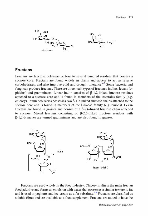
Fructans
Fructans are fructose polymers of four to several hundred residues that possess a
sucrose core. Fructans are found widely in plants and appear to act as reserve
carbohydrates, and also improve cold and drought tolerance.47 Some bacteria and
fungi can produce fructans. There are three main types of fructans: inulins, levans (or
phleins) and gramminans. Linear inulin consists of b-1,2-linked fructose residues
attached to a sucrose core and is found in members of the Asterales family (e.g.
chicory). Inulin neo-series possesses two b-1,2-linked fructose chains attached to the
sucrose core and is found in members of the Liliacae family (e.g. onions). Levan
fructans are found in grasses and consist of a b-2,6-linked fructose chain attached
to sucrose. Mixed fructans consisting of b-2,6-linked fructose residues with
b-1,2-branches are termed gramminans and are also found in grasses.
Fructans are used widely in the food industry. Chicory inulin is the main fructan
food additive and forms an emulsion with water that possesses a similar texture to fat
and is used in yoghurts and ice-cream as a fat substitute.48 Fructans are classified as
soluble fibres and are available as a food supplement. Fructans are touted to have the
Fructans 333
References start on page 339
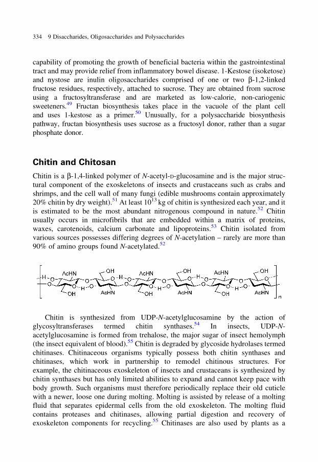
capability of promoting the growth of beneficial bacteria within the gastrointestinal
tract and may provide relief from inflammatory bowel disease. 1-Kestose (isoketose)
and nystose are inulin oligosaccharides comprised of one or two b-1,2-linked
fructose residues, respectively, attached to sucrose. They are obtained from sucrose
using a fructosyltransferase and are marketed as low-calorie, non-cariogenic
sweeteners.49 Fructan biosynthesis takes place in the vacuole of the plant cell
and uses 1-kestose as a primer.50 Unusually, for a polysaccharide biosynthesis
pathway, fructan biosynthesis uses sucrose as a fructosyl donor, rather than a sugar
phosphate donor.
Chitin and Chitosan
Chitin is a b-1,4-linked polymer of N-acetyl-D-glucosamine and is the major struc-
tural component of the exoskeletons of insects and crustaceans such as crabs and
shrimps, and the cell wall of many fungi (edible mushrooms contain approximately
20% chitin by dry weight).51 At least 1013 kg of chitin is synthesized each year, and it
is estimated to be the most abundant nitrogenous compound in nature.52 Chitin
usually occurs in microfibrils that are embedded within a matrix of proteins,
waxes, carotenoids, calcium carbonate and lipoproteins.53 Chitin isolated from
various sources possesses differing degrees of N-acetylation – rarely are more than
90% of amino groups found N-acetylated.52
Chitin is synthesized from UDP-N-acetylglucosamine by the action of
glycosyltransferases termed chitin synthases.54 In insects, UDP-N-
acetylglucosamine is formed from trehalose, the major sugar of insect hemolymph
(the insect equivalent of blood).55 Chitin is degraded by glycoside hydrolases termed
chitinases. Chitinaceous organisms typically possess both chitin synthases and
chitinases, which work in partnership to remodel chitinous structures. For
example, the chitinaceous exoskeleton of insects and crustaceans is synthesized by
chitin synthases but has only limited abilities to expand and cannot keep pace with
body growth. Such organisms must therefore periodically replace their old cuticle
with a newer, loose one during molting. Molting is assisted by release of a molting
fluid that separates epidermal cells from the old exoskeleton. The molting fluid
contains proteases and chitinases, allowing partial digestion and recovery of
exoskeleton components for recycling.55 Chitinases are also used by plants as a
334 9 Disaccharides, Oligosaccharides and Polysaccharides
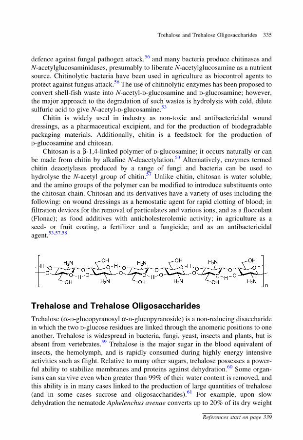
defence against fungal pathogen attack,56 and many bacteria produce chitinases and
N-acetylglucosaminidases, presumably to liberate N-acetylglucosamine as a nutrient
source. Chitinolytic bacteria have been used in agriculture as biocontrol agents to
protect against fungus attack.56 The use of chitinolytic enzymes has been proposed to
convert shell-fish waste into N-acetyl-D-glucosamine and D-glucosamine; however,
the major approach to the degradation of such wastes is hydrolysis with cold, dilute
sulfuric acid to give N-acetyl-D-glucosamine.53
Chitin is widely used in industry as non-toxic and antibactericidal wound
dressings, as a pharmaceutical excipient, and for the production of biodegradable
packaging materials. Additionally, chitin is a feedstock for the production of
D-glucosamine and chitosan.
Chitosan is a b-1,4-linked polymer of D-glucosamine; it occurs naturally or can
be made from chitin by alkaline N-deacetylation.53 Alternatively, enzymes termed
chitin deacetylases produced by a range of fungi and bacteria can be used to
hydrolyse the N-acetyl group of chitin.57 Unlike chitin, chitosan is water soluble,
and the amino groups of the polymer can be modified to introduce substituents onto
the chitosan chain. Chitosan and its derivatives have a variety of uses including the
following: on wound dressings as a hemostatic agent for rapid clotting of blood; in
filtration devices for the removal of particulates and various ions, and as a flocculant
(Flonac); as food additives with anticholesterolemic activity; in agriculture as a
seed- or fruit coating, a fertilizer and a fungicide; and as an antibactericidal
agent.53,57,58
Trehalose and Trehalose Oligosaccharides
Trehalose (a-D-glucopyranosyl a-D-glucopyranoside) is a non-reducing disaccharide
in which the two D-glucose residues are linked through the anomeric positions to one
another. Trehalose is widespread in bacteria, fungi, yeast, insects and plants, but is
absent from vertebrates.59 Trehalose is the major sugar in the blood equivalent of
insects, the hemolymph, and is rapidly consumed during highly energy intensive
activities such as flight. Relative to many other sugars, trehalose possesses a power-
ful ability to stabilize membranes and proteins against dehydration.60 Some organ-
isms can survive even when greater than 99% of their water content is removed, and
this ability is in many cases linked to the production of large quantities of trehalose
(and in some cases sucrose and oligosaccharides).61 For example, upon slow
dehydration the nematode Aphelenchus avenae converts up to 20% of its dry weight
Trehalose and Trehalose Oligosaccharides 335
References start on page 339
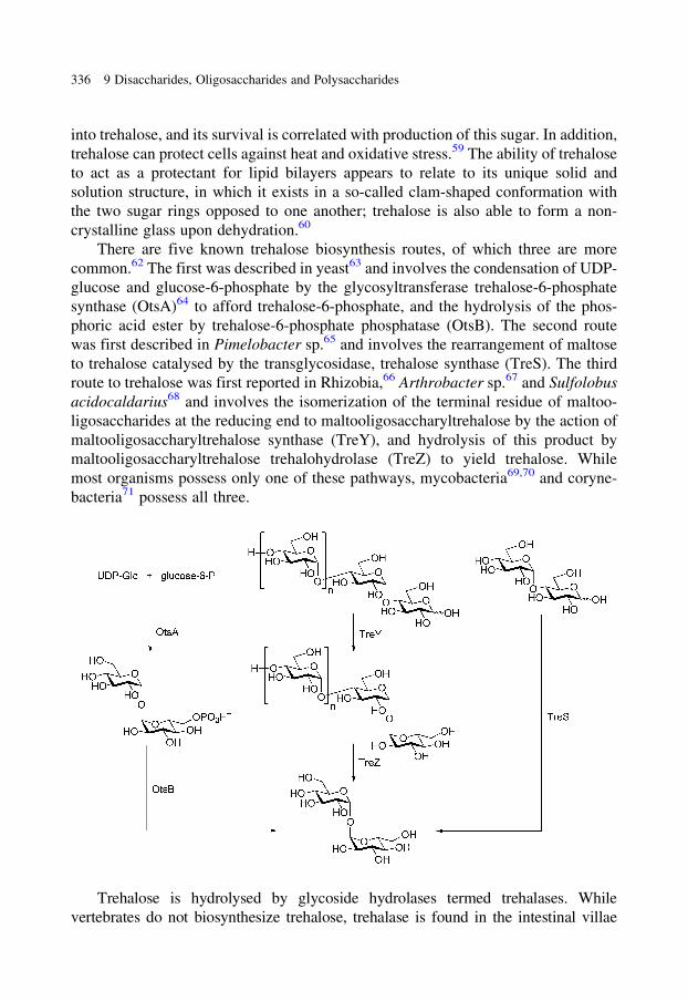
into trehalose, and its survival is correlated with production of this sugar. In addition,
trehalose can protect cells against heat and oxidative stress.59 The ability of trehalose
to act as a protectant for lipid bilayers appears to relate to its unique solid and
solution structure, in which it exists in a so-called clam-shaped conformation with
the two sugar rings opposed to one another; trehalose is also able to form a non-
crystalline glass upon dehydration.60
There are five known trehalose biosynthesis routes, of which three are more
common.62 The first was described in yeast63 and involves the condensation of UDP-
glucose and glucose-6-phosphate by the glycosyltransferase trehalose-6-phosphate
synthase (OtsA)64 to afford trehalose-6-phosphate, and the hydrolysis of the phos-
phoric acid ester by trehalose-6-phosphate phosphatase (OtsB). The second route
was first described in Pimelobacter sp.65 and involves the rearrangement of maltose
to trehalose catalysed by the transglycosidase, trehalose synthase (TreS). The third
route to trehalose was first reported in Rhizobia,66 Arthrobacter sp.67 and Sulfolobus
acidocaldarius68 and involves the isomerization of the terminal residue of maltoo-
ligosaccharides at the reducing end to maltooligosaccharyltrehalose by the action of
maltooligosaccharyltrehalose synthase (TreY), and hydrolysis of this product by
maltooligosaccharyltrehalose trehalohydrolase (TreZ) to yield trehalose. While
most organisms possess only one of these pathways, mycobacteria69,70 and coryne-
bacteria71 possess all three.
Trehalose is hydrolysed by glycoside hydrolases termed trehalases. While
vertebrates do not biosynthesize trehalose, trehalase is found in the intestinal villae
336 9 Disaccharides, Oligosaccharides and Polysaccharides
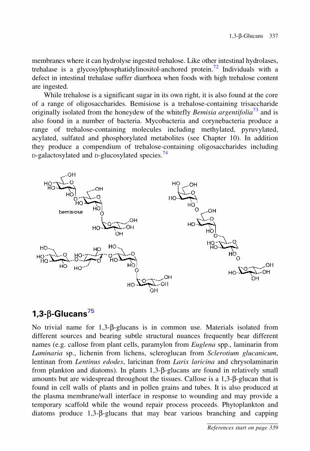
membranes where it can hydrolyse ingested trehalose. Like other intestinal hydrolases,
trehalase is a glycosylphosphatidylinositol-anchored protein.72 Individuals with a
defect in intestinal trehalase suffer diarrhoea when foods with high trehalose content
are ingested.
While trehalose is a significant sugar in its own right, it is also found at the core
of a range of oligosaccharides. Bemisiose is a trehalose-containing trisaccharide
originally isolated from the honeydew of the whitefly Bemisia argentifolia73 and is
also found in a number of bacteria. Mycobacteria and corynebacteria produce a
range of trehalose-containing molecules including methylated, pyruvylated,
acylated, sulfated and phosphorylated metabolites (see Chapter 10). In addition
they produce a compendium of trehalose-containing oligosaccharides including
D-galactosylated and D-glucosylated species.74
1,3-b-Glucans75
No trivial name for 1,3-b-glucans is in common use. Materials isolated from
different sources and bearing subtle structural nuances frequently bear different
names (e.g. callose from plant cells, paramylon from Euglena spp., laminarin from
Laminaria sp., lichenin from lichens, scleroglucan from Sclerotium glucanicum,
lentinan from Lentinus edodes, laricinan from Larix laricina and chrysolaminarin
from plankton and diatoms). In plants 1,3-b-glucans are found in relatively small
amounts but are widespread throughout the tissues. Callose is a 1,3-b-glucan that is
found in cell walls of plants and in pollen grains and tubes. It is also produced at
the plasma membrane/wall interface in response to wounding and may provide a
temporary scaffold while the wound repair process proceeds. Phytoplankton and
diatoms produce 1,3-b-glucans that may bear various branching and capping
1,3-b-Glucans 337
References start on page 339
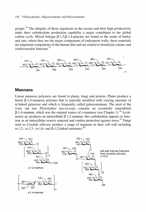
groups.76 The ubiquity of these organisms in the oceans and their high productivity
make their carbohydrate production capability a major contributor to the global
carbon cycle. Mixed linkage b-1,3;b-1,4-glucans are found in the seeds of barley
and oats, where they are the major components of endosperm walls; these materials
are important components of the human diet and are related to beneficial colonic and
cardiovascular function.77
Mannans
Linear mannose polymers are found in plants, fungi and protists. Plants produce a
linear b-1,4-mannose polymer that is typically modified with varying amounts of
a-linked galactose and which is frequently called galactomannan. The seed of the
ivory nut tree Phytelephas macrocarpa contains an essentially unmodified
b-1,4-mannan, which was the original source of D-mannose (see Chapter 1).78 Leish-
mania sp. produces an intracellular b-1,2-mannan; this carbohydrate appears to func-
tion as an intracellular reserve material and confers protection against stress.79 Fungi
such as Candida albicans produce a range of mannans in their cell wall including
a-1,2-, a-1,3-, a-1,6- and b-1,2-linked structures.80
338 9 Disaccharides, Oligosaccharides and Polysaccharides

References
1. Marchessault, R.H. and Sundararajan, P.R. (1983). In The polysaccharides (G.O. Aspinall, ed.)
Vol. 2, p. 11. Academic Press: New York.
2. Teeri, T.T., Brumer, H., 3rd, Daniel, G. and Gatenholm, P. (2007). Trends Biotechnol., 25, 299.
3. Somerville, C. (2006). Annu. Rev. Cell Dev. Biol., 22, 53.
4. Koyama, M., Helbert, W., Imai, T., Sugiyama, J. and Henrissat, B. (1997). Proc. Natl. Acad. Sci.
USA, 94, 9091.
5. Williamson, R.E., Burn, J.E. and Hocart, C.H. (2002). Trends Plant Sci., 7, 461.
6. Woodward, J. (1984). In Topics in Enzyme and Fermentation Biotechnology. Vol. 8, p. 9. John
Wiley & Sons: New York.
7. Gilbert, H.J. and Hazlewood, G.P. (1993). J. Gen. Microbiol., 139, 187.
8. Bayer, E.A., Chanzy, H., Lamed, R. and Shoham, Y. (1998). Curr. Opin. Struct. Biol., 8, 548.
9. Aanen, D.K., Eggleton, P., Rouland-Lefevre, C., Guldberg-Froslev, T., Rosendahl, S. and
Boomsma, J.J. (2002). Proc. Natl. Acad. Sci. USA, 99, 14887.
10. Ito, S. (1997). Extremophiles, 1, 61.
11. Balser, K., Hoppe, L., Eicher, T., Wandel, M. and Astheimer, H.-J. (1986). In Ullmann’s Ency-
clopedia of Industrial Chemistry (W. Gerhartz, ed.) Vol. A5, p. 419. Wiley-VCH: Weinheim.
12. Brandt, L. (1986). In Ullmann’s Encyclopedia of Industrial Chemistry (W. Gerhartz, ed.)
Vol. A5, p. 461. Wiley-VCH: Weinheim.
13. Braun, G. (1943). Org. Syn. Coll. Vol. 2, 124.
14. Daniel, J.R. (1986). In Ullmann’s Encyclopedia of Industrial Chemistry (W. Gerhartz, ed.)
Vol. A5, p. 1. Wiley-VCH: Weinheim.
15. Saenger, W. (1984). Naturwissenschaften, 71, 31.
16. James, M.G., Denyer, K. and Myers, A.M. (2003). Curr. Opin. Plant Biol., 6, 215.
17. Chronakis, I.S. (1998). Crit. Rev. Food Sci. Nutr., 38, 599.
18. Guilnot, A. and Mercier, C. (1983). In The Polysaccharides (G.O. Aspinall, ed.) Vol. 3, p. 209.
Academic Press: New York.
19. Ragauskas, A.J., Williams, C.K., Davison, B.H., Britovsek, G., Cairney, J., Eckert, C.A., Frederick,
W.J., Jr., Hallett, J.P., Leak, D.J., Liotta, C.L., Mielenz, J.R., Murphy, R., Templer, R. and
Tschaplinski, T. Science, 311, 484.
20. Kim, S. and Dale, B.E. (2005). Biomass Bioenergy, 29, 426.
21. Gibbons, B.J., Roach, P.J. and Hurley, T.D. (2002). J. Mol. Biol., 319, 463.
22. Roach, P.J. (2002). Curr. Mol. Med., 2, 101.
23. Lin, A., Mu, J., Yang, J. and Roach, P.J. (1999). Arch. Biochem. Biophys., 363, 163.
24. Thon, V.J., Khalil, M. and Cannon, J.F. (1993). J. Biol. Chem., 268, 7509.
25. Verhue, W. and Hers, H.G. (1966). Biochem. J., 99, 222.
26. Liu, W., Madsen, N.B., Braun, C. and Withers, S.G. (1991). Biochemistry, 30, 1419.
27. Biwer, A., Antranikian, G. and Heinzle, E. (2002). Appl. Microbiol. Biotechnol., 59, 609.
28. Hough, L. and Phadnis, S.P. (1976). Nature, 263, 800.
29. Bilger, B. (2006). New Yorker, 82, 40.
30. Torres, L.L. and Salerno, G.L. (2007). Proc. Natl. Acad. Sci. USA, 104, 14318.
31. Salerno, G.L. and Curatti, L. (2003). Trends Plant Sci., 8, 63.
32. Sale, J.W. and Skinner, W.W. (1922). J. Ind. Eng. Chem., 14, 522.
33. Vinjamoori, D.V., Byrum, J.R., Hayes, T. and Das, P.K. (2004). J. Anim. Sci., 82, 319.
34. Nakakuki, T. (2002). Pure Appl. Chem., 74, 1245.
35. Kanauchi, O., Mitsuyama, K., Araki, Y. and Andoh, A. (2003). Curr. Pharm. Des., 9, 333.
36. Rastall, R.A. (2004). J. Nutr., 134, 2022S.
37. Peterbauer, T. and Richter, A. (2001). Seed Sci. Res., 11, 185.
References 339
38. Ravaud, S., Robert, X., Watzlawick, H., Haser, R., Mattes, R. and Aghajari, N. J. Biol. Chem, 282,
28126.
39. Strater, P. (1986). In Alternative sweeteners (L.O. Nabors and R.C. Gelardi, eds) p. 217.
Marcel Dekker: New York.
40. Kunz, C., Rudloff, S., Baier, W., Klein, N. and Strobel, S. (2000). Annu. Rev. Nutr., 20, 699.
41. Brodbeck, U., Denton, W.L., Tanahashi, N. and Ebner, K.E. (1967). J. Biol. Chem., 242, 1391.
42. Ramakrishnan, B. and Qasba, P.K. (2001). J. Mol. Biol., 310, 205.
43. Swallow, D.M. (2003). Annu. Rev. Genet., 37, 197.
44. Vesa, T.H., Marteau, P. and Korpela, R. (2000). J. Am. Coll. Nutr., 19, 165S.
45. Jacob, F. and Monod, J. (1961). J. Mol. Biol., 3, 318.
46. Lewis, M. (2005). C. R. Biol., 328, 521.
47. Ritsema, T. and Smeekens, S. (2003). Curr. Opin. Plant Biol., 6, 223.
48. Ritsema, T. and Smeekens, S.C. (2003). J. Plant. Physiol., 160, 811.
49. Tokunaga, T., Oku, T. and Hosoya, N. (1989). J. Nutr., 119, 553.
50. Vijn, I., van Dijken, A., Sprenger, N., van Dun, K., Weisbeek, P., Wiemken, A. and Smeekens, S.
(1997). Plant J., 11, 387.
51. Hirano, S. (1986). In Ullmann’s Encyclopedia of Industrial Chemistry (W. Gerhartz, ed.) Vol. A6,
p. 231. Wiley-VCH: Weinheim.
52. Muzzarelli, R.A.A. (1999). In Chitin and Chitinases (P. Jolles and R.A.A. Muzzarelli, eds) p. 1.
Basel: Birkhauser Verlag.
53. Synowiecki, J. and Al-Khateeb, N.A. (2003). Crit. Rev. Food Sci. Nutr., 43, 145.
54. Merzendorfer, H. (2006). J. Comp. Physiol. B, 176, 1.
55. Merzendorfer, H. and Zimoch, L. (2003). J. Exp. Biol., 206, 4393.
56. Bhattacharya, D., Nagpure, A. and Gupta, R.K. (2007). Crit. Rev. Biotechnol., 27, 21.
57. Tsigos, I., Martinou, A., Kafetzopoulos, D. and Bouriotis, V. (2000). Trends Biotechnol., 18, 305.
58. Cuero, R.G. (1999). In Chitin and Chitinases (P. Jolles, and R.A.A. Muzzarelli, eds) p. 315.
Birkhauser Verlag: Basel.
59. Elbein, A.D., Pan, Y.T., Pastuszak, I. and Carroll, D. (2003). Glycobiology, 13, 17R.
60. Albertorio, F., Chapa, V.A., Chen, X., Diaz, A.J. and Cremer, P.S. (2007). J. Am. Chem. Soc., 129, 10567.
61. Crowe, J.H., Hoekstra, F.A. and Crowe, L.M. (1992). Annu. Rev. Physiol., 54, 579.
62. Paul, M. (2007). Curr. Opin. Plant Biol., 10, 303.
63. Cabib, E. and Leloir, L.F. (1958). J. Biol. Chem., 231, 259.
64. Gibson, R.P., Turkenburg, J.P., Charnock, S.J., Lloyd, R. and Davies, G.J. (2002). Chem. Biol., 9,
1337.
65. Tsusaki, K., Nishimoto, T., Nakada, T., Kubota, M., Chaen, H., Sugimoto, T. and Kurimoto, M.
(1996). Biochim. Biophys. Acta, 1290, 1.
66. Maruta, K., Hattori, K., Nakada, T., Kubota, M., Sugimoto, T. and Kurimoto, M. (1996). Biosci.
Biotechnol. Biochem., 60, 717.
67. Maruta, K., Hattori, K., Nakada, T., Kubota, M., Sugimoto, T. and Kurimoto, M. (1996). Biochim.
Biophys. Acta, 1289, 10.
68. Maruta, K., Mitsuzumi, H., Nakada, T., Kubota, M., Chaen, H., Fukuda, S., Sugimoto, T. and
Kurimoto, M. (1996). Biochim. Biophys. Acta, 1291, 177.
69. De Smet, K.A., Weston, A., Brown, I.N., Young, D.B. and Robertson, B.D. (2000). Microbiology,
146, 199.
70. Woodruff, P.J., Carlson, B.L., Siridechadilok, B., Pratt, M.R., Senaratne, R.H., Mougous, J.D.,
Riley, L.W., Williams, S.J. and Bertozzi, C.R. (2004). J. Biol. Chem., 279, 28835.
71. Tzvetkov, M., Klopprogge, C., Zelder, O. and Liebl, W. (2003). Microbiology, 149, 1659.
72. Ruf, J., Wacker, H., James, P., Maffia, M., Seiler, P., Galand, G., von Kieckebusch, A., Semenza, G.
and Matei, N. (1990). J. Biol. Chem., 265, 15034.
340 9 Disaccharides, Oligosaccharides and Polysaccharides

73. Hendrix, D.L. and Wei, Y.A. (1994). Carbohydr. Res., 253, 329.
74. Ohta, M., Pan, Y.T., Laine, R.A. and Elbein, A.D. (2002). Eur. J. Biochem., 269, 3142.
75. Stone, B.A. and Clarke, A.E. (1992). Chemistry and Biology of (1,3)-�-Glucans. Victoria:
La Trobe University Press.
76. Alderkamp, A.-C., Buma, A.G.J. and van Rijssel, M. (2007). Biogeochem., 83, 99.
77. Burton, R.A., Wilson, S.M., Hrmova, M., Harvey, A.J., Shirley, N.J., Medhurst, A., Stone, B.A.,
Newbigin, E.J., Bacic, A. and Fincher, G.B. (2006). Science, 311, 1940.
78. Stephen, A.M. (1983). In The polysaccharides (G.O. Aspinall, ed.) Vol. 2, p. 97. Academic Press:
New York.
79. Ralton, J.E., Naderer, T., Piraino, H.L., Bashtannyk, T.A., Callaghan, J.M. and McConville, M.J.
(2003). J. Biol. Chem., 278, 40757.
80. Shibata, N., Suzuki, A., Kobayashi, H. and Okawa, Y. (2007). Biochem. J., 404, 365.
References 341
Chapter 11
Glycoproteins and Proteoglycans
Proteins produced by the cell may be modified with a range of carbohydrate
structures. The modifications may occur prior to delivery to intracellular organelles,
secretion from the cell or incorporation into membranes, or they may occur on a
mature protein present in the cytosol or nucleus of the cell. There are three main
types of protein modification: N-linked glycosylation, O-linked glycosylation and
the attachment of glycosylphosphatidylinositol (GPI) anchors. A relatively rare
modification is C-linked glycosylation. The reasons for protein glycosylation are at
best only partly understood but include roles in ensuring proper folding of proteins
and their degradation when incompetently folded, ensuring correct targeting of
glycoproteins to intra- and extracellular destinations, altering susceptibility to degra-
dation, modulation of immunological properties and the engagement of lectins. This
chapter will review the major types of protein glycosylation found in higher organ-
isms, with a particular emphasis on mammalian biology. While an immense number
of structures is possible through the connection of the 10 common mammalian
monosaccharides (Glc, Gal, Man, Sia, GlcNAc, GalNAc, Fuc, Xyl, GlcA and
IdoA), systematic structural analysis reveals that only a small number of monosac-
charide connections accounts for the majority of the linkages. Werz et al. have
reported that of a set of 3299 oligosaccharides obtained from 38 mammalian species,
11 monosaccharide connections accounted for >75% of all glycosidic linkages.1
Glycoproteins are proteins that bear one or more O-, N- or C-linked glycan
chains. These chains comprise typically up to 20% but, occasionally, more than 90%
of the mass of the resultant conjugate. A proteoglycan is a peptide or protein that
bears one or more covalently attached glycosaminoglycan chains. As has been noted,
the distinction between a glycoprotein and a proteoglycan is somewhat arbitrary,
as the same polypeptide may bear both glycosaminoglycan chains and O-, N- or
C-linked glycans.2,3 A mucin is a glycoprotein that carries many O-linked glycan
chains that are clustered together. A glycosphingolipid is a mono- or oligosaccharide
that is (usually) attached via glucose or galactose to ceramide. A ganglioside is an
anionic glycolipid that contains one or more residues of sialic acid. A GPI anchor is
a glycan structure that links a phosphatidylinositol (PI) to a phosphoethanolamine,
attached as an amide, to the C-terminus of a protein.
References start on page 410
Eukaryotic cells contain an interconnected network of membrane-bound cister-
nae, vesicles and vacuoles that are connected to the plasma membrane and comprise
the secretory and endocytic pathways.4 Newly synthesized proteins destined for the
cell surface, secretion or other compartments in the secretory pathway are initially
translocated into the endoplasmic reticulum (ER). The ER comprises the start of the
secretory pathway and is a relatively large, highly dynamic organelle, which is
adjoined to the nucleus of the cell and can be divided into three domains: the
ribosome-studded rough ER, the ribosome-free smooth ER and the nuclear envelope.
Proteins are assembled by translation in the rough ER by ribosomes, multicomponent
systems comprised of proteins and RNA. Protein folding occurs in the ER, and
various carbohydrates may also be added here (e.g. N-linked glycan core structures
and GPI anchors). Proteins may leave the ER directly or by passage through
the Golgi apparatus,a which contains a complex post-translational modification
machinery. Alternatively, folding-incompetent proteins are detected and degraded
by an ER-associated degradation (ERAD) pathway. The Golgi apparatus is a polar
organelle comprised of a stack of membrane-bound sacs termed cisternae. The
cis face accepts vesicular traffic from the ER, and the trans face acts as the exit
site for vesicular traffic. In the Golgi apparatus a complex array of protein modifica-
tions may occur including glycosylation, deglycosylation, O- and N-sulfation, and
O-acetylation, resulting in the formation of O-linked glycans (e.g. mucin-type
O-linked glycans and proteoglycans) and the elaboration of N-linked glycans.
Additionally, specific carbohydrate structures (mannose-6-phosphate groups) may
be added to ensure targeting of various, generally catabolic, enzymes to an intracel-
lular organelle termed the lysosome. The lysosome is a digestive organelle rich in
hydrolases that receives (glyco)proteins from the cytosol, the plasma membrane and
the extracellular milieu that are destined for degradation, thereby allowing their
constituents to re-enter cellular metabolic processes.
N-Linked Glycosylation5
N-Linked glycans are added to the majority of proteins that enter the secretory
pathway and are important for their folding, stability and function. N-Linked
glycosylation involves en bloc transfer of preformed glycans (see below) through a
b-linkage to the side-chain nitrogen of asparagine. The asparagine residue is found in
the highly conserved sequence Asn-Xxx-Thr, where Xxx is any residue but proline.
While N-linked glycans possess complex and diverse structures dependent on their
origin, when they are initially added to growing peptides, these glycans share a
a Camillo Golgi (1843–1926), M.D. (1865). Professor at Pavia University, Italy. Nobel Prize in
Physiology or Medicine (1906).
370 11 Glycoproteins and Proteoglycans
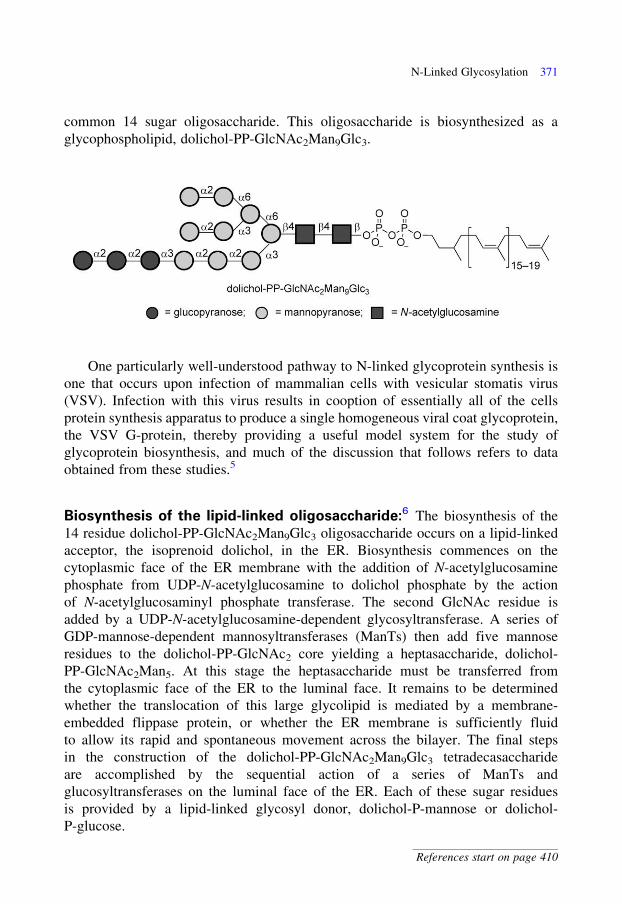
common 14 sugar oligosaccharide. This oligosaccharide is biosynthesized as a
glycophospholipid, dolichol-PP-GlcNAc2Man9Glc3.
One particularly well-understood pathway to N-linked glycoprotein synthesis is
one that occurs upon infection of mammalian cells with vesicular stomatis virus
(VSV). Infection with this virus results in cooption of essentially all of the cells
protein synthesis apparatus to produce a single homogeneous viral coat glycoprotein,
the VSV G-protein, thereby providing a useful model system for the study of
glycoprotein biosynthesis, and much of the discussion that follows refers to data
obtained from these studies.5
Biosynthesis of the lipid-linked oligosaccharide:6 The biosynthesis of the
14 residue dolichol-PP-GlcNAc2Man9Glc3 oligosaccharide occurs on a lipid-linked
acceptor, the isoprenoid dolichol, in the ER. Biosynthesis commences on the
cytoplasmic face of the ER membrane with the addition of N-acetylglucosamine
phosphate from UDP-N-acetylglucosamine to dolichol phosphate by the action
of N-acetylglucosaminyl phosphate transferase. The second GlcNAc residue is
added by a UDP-N-acetylglucosamine-dependent glycosyltransferase. A series of
GDP-mannose-dependent mannosyltransferases (ManTs) then add five mannose
residues to the dolichol-PP-GlcNAc2 core yielding a heptasaccharide, dolichol-
PP-GlcNAc2Man5. At this stage the heptasaccharide must be transferred from
the cytoplasmic face of the ER to the luminal face. It remains to be determined
whether the translocation of this large glycolipid is mediated by a membrane-
embedded flippase protein, or whether the ER membrane is sufficiently fluid
to allow its rapid and spontaneous movement across the bilayer. The final steps
in the construction of the dolichol-PP-GlcNAc2Man9Glc3 tetradecasaccharide
are accomplished by the sequential action of a series of ManTs and
glucosyltransferases on the luminal face of the ER. Each of these sugar residues
is provided by a lipid-linked glycosyl donor, dolichol-P-mannose or dolichol-
P-glucose.
N-Linked Glycosylation 371
References start on page 410
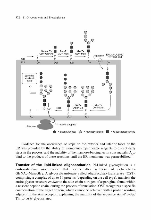
Evidence for the occurrence of steps on the exterior and interior faces of the
ER was provided by the ability of membrane-impermeable reagents to disrupt early
steps in the process, and the inability of the mannose-binding lectin concanavalin A to
bind to the products of these reactions until the ER membrane was permeabilized.7
Transfer of the lipid-linked oligosaccharide: N-Linked glycosylation is a
co-translational modification that occurs after synthesis of dolichol-PP-
GlcNAc2Man9Glc3. A glycosyltransferase called oligosaccharyltransferase (OST),
comprising a complex of up to 10 proteins (depending on the cell type), transfers the
entire glycan structure en bloc to the side-chain nitrogen of asparagine, found within
a nascent peptide chain, during the process of translation. OST recognizes a specific
conformation of the target protein, which cannot be achieved with a proline residing
adjacent to the Asn acceptor, explaining the inability of the sequence Asn-Pro-Ser/
Thr to be N-glycosylated.
372 11 Glycoproteins and Proteoglycans
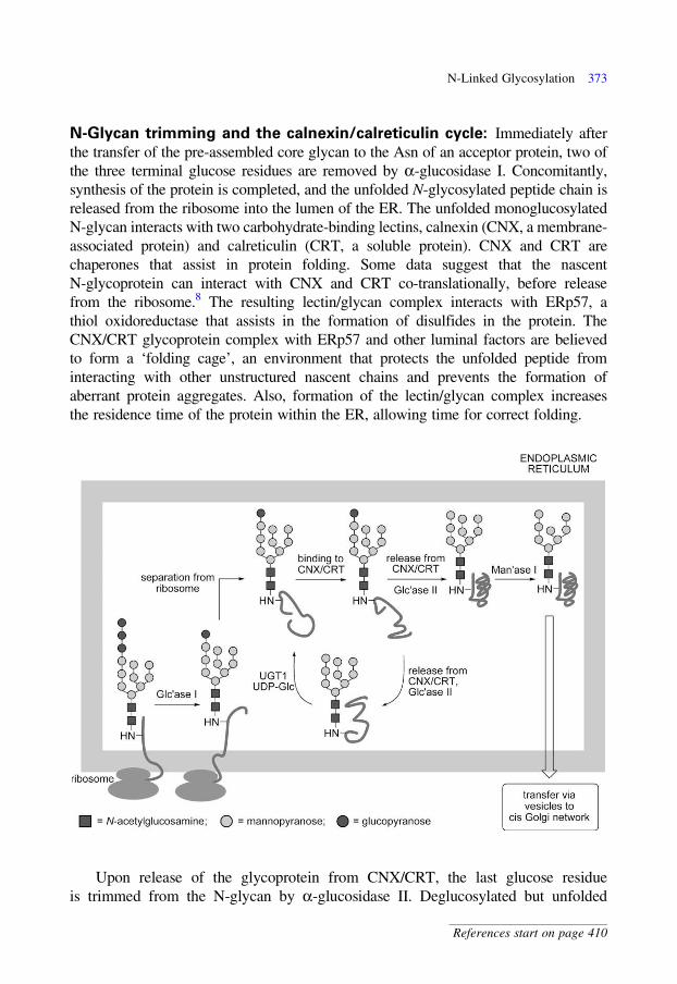
N-Glycan trimming and the calnexin/calreticulin cycle: Immediately after
the transfer of the pre-assembled core glycan to the Asn of an acceptor protein, two of
the three terminal glucose residues are removed by a-glucosidase I. Concomitantly,
synthesis of the protein is completed, and the unfolded N-glycosylated peptide chain is
released from the ribosome into the lumen of the ER. The unfolded monoglucosylated
N-glycan interacts with two carbohydrate-binding lectins, calnexin (CNX, a membrane-
associated protein) and calreticulin (CRT, a soluble protein). CNX and CRT are
chaperones that assist in protein folding. Some data suggest that the nascent
N-glycoprotein can interact with CNX and CRT co-translationally, before release
from the ribosome.8 The resulting lectin/glycan complex interacts with ERp57, a
thiol oxidoreductase that assists in the formation of disulfides in the protein. The
CNX/CRT glycoprotein complex with ERp57 and other luminal factors are believed
to form a ‘folding cage’, an environment that protects the unfolded peptide from
interacting with other unstructured nascent chains and prevents the formation of
aberrant protein aggregates. Also, formation of the lectin/glycan complex increases
the residence time of the protein within the ER, allowing time for correct folding.
Upon release of the glycoprotein from CNX/CRT, the last glucose residue
is trimmed from the N-glycan by a-glucosidase II. Deglucosylated but unfolded
N-Linked Glycosylation 373
References start on page 410
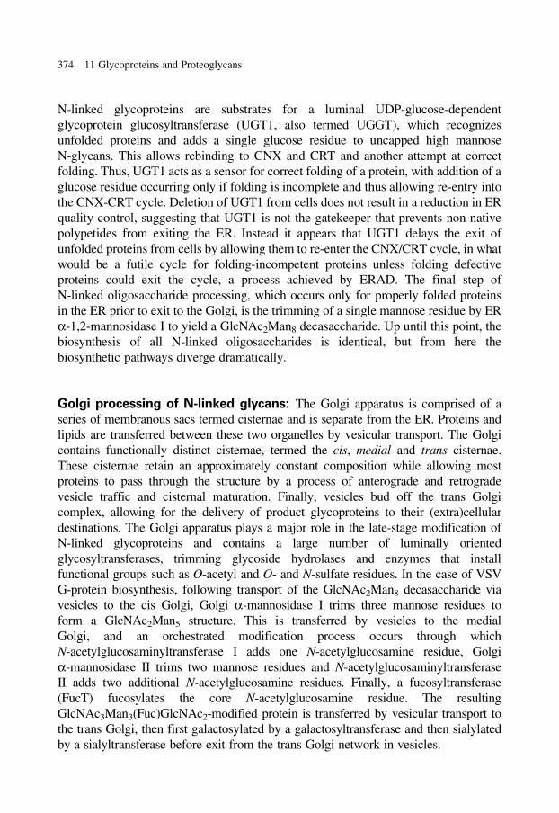
N-linked glycoproteins are substrates for a luminal UDP-glucose-dependent
glycoprotein glucosyltransferase (UGT1, also termed UGGT), which recognizes
unfolded proteins and adds a single glucose residue to uncapped high mannose
N-glycans. This allows rebinding to CNX and CRT and another attempt at correct
folding. Thus, UGT1 acts as a sensor for correct folding of a protein, with addition of a
glucose residue occurring only if folding is incomplete and thus allowing re-entry into
the CNX-CRT cycle. Deletion of UGT1 from cells does not result in a reduction in ER
quality control, suggesting that UGT1 is not the gatekeeper that prevents non-native
polypetides from exiting the ER. Instead it appears that UGT1 delays the exit of
unfolded proteins from cells by allowing them to re-enter the CNX/CRT cycle, in what
would be a futile cycle for folding-incompetent proteins unless folding defective
proteins could exit the cycle, a process achieved by ERAD. The final step of
N-linked oligosaccharide processing, which occurs only for properly folded proteins
in the ER prior to exit to the Golgi, is the trimming of a single mannose residue by ER
a-1,2-mannosidase I to yield a GlcNAc2Man8 decasaccharide. Up until this point, the
biosynthesis of all N-linked oligosaccharides is identical, but from here the
biosynthetic pathways diverge dramatically.
Golgi processing of N-linked glycans: The Golgi apparatus is comprised of a
series of membranous sacs termed cisternae and is separate from the ER. Proteins and
lipids are transferred between these two organelles by vesicular transport. The Golgi
contains functionally distinct cisternae, termed the cis, medial and trans cisternae.
These cisternae retain an approximately constant composition while allowing most
proteins to pass through the structure by a process of anterograde and retrograde
vesicle traffic and cisternal maturation. Finally, vesicles bud off the trans Golgi
complex, allowing for the delivery of product glycoproteins to their (extra)cellular
destinations. The Golgi apparatus plays a major role in the late-stage modification of
N-linked glycoproteins and contains a large number of luminally oriented
glycosyltransferases, trimming glycoside hydrolases and enzymes that install
functional groups such as O-acetyl and O- and N-sulfate residues. In the case of VSV
G-protein biosynthesis, following transport of the GlcNAc2Man8 decasaccharide via
vesicles to the cis Golgi, Golgi a-mannosidase I trims three mannose residues to
form a GlcNAc2Man5 structure. This is transferred by vesicles to the medial
Golgi, and an orchestrated modification process occurs through which
N-acetylglucosaminyltransferase I adds one N-acetylglucosamine residue, Golgi
a-mannosidase II trims two mannose residues and N-acetylglucosaminyltransferase
II adds two additional N-acetylglucosamine residues. Finally, a fucosyltransferase
(FucT) fucosylates the core N-acetylglucosamine residue. The resulting
GlcNAc3Man3(Fuc)GlcNAc2-modified protein is transferred by vesicular transport to
the trans Golgi, then first galactosylated by a galactosyltransferase and then sialylated
by a sialyltransferase before exit from the trans Golgi network in vesicles.
374 11 Glycoproteins and Proteoglycans
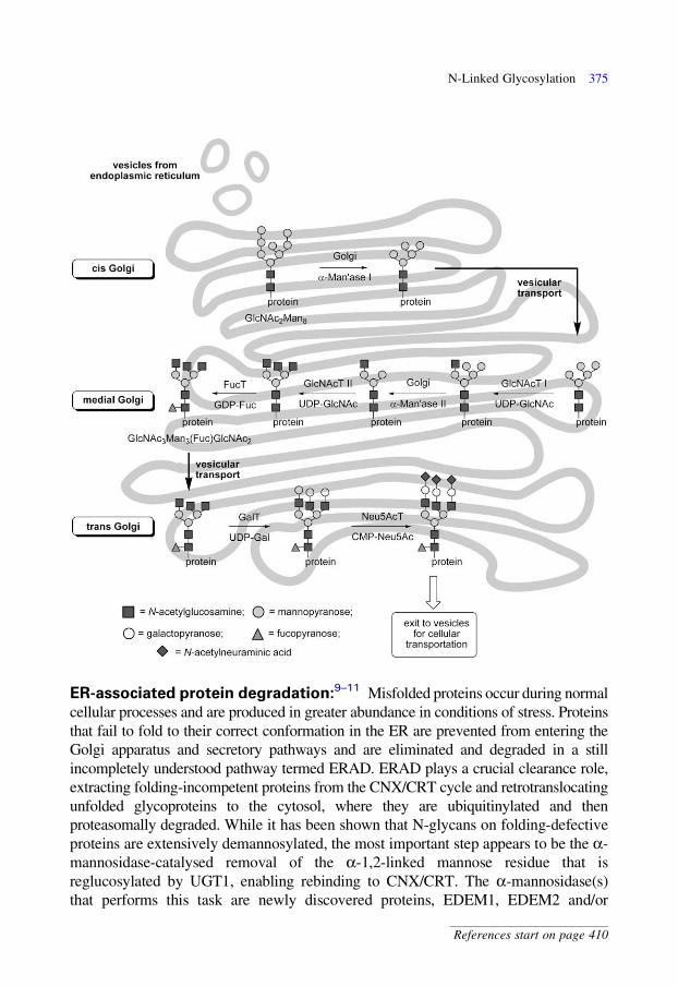
ER-associated protein degradation:9–11 Misfolded proteins occur during normal
cellular processes and are produced in greater abundance in conditions of stress. Proteins
that fail to fold to their correct conformation in the ER are prevented from entering the
Golgi apparatus and secretory pathways and are eliminated and degraded in a still
incompletely understood pathway termed ERAD. ERAD plays a crucial clearance role,
extracting folding-incompetent proteins from the CNX/CRT cycle and retrotranslocating
unfolded glycoproteins to the cytosol, where they are ubiquitinylated and then
proteasomally degraded. While it has been shown that N-glycans on folding-defective
proteins are extensively demannosylated, the most important step appears to be the a-
mannosidase-catalysed removal of the a-1,2-linked mannose residue that is
reglucosylated by UGT1, enabling rebinding to CNX/CRT. The a-mannosidase(s)
that performs this task are newly discovered proteins, EDEM1, EDEM2 and/or
N-Linked Glycosylation 375
References start on page 410

EDEM3.8 Removal of the acceptor mannose residue, which can be reglucosylated by
UGT1, results in irreversible interruption of futile folding attempts in the CNX/CRT
cycle. The steps leading to further degradation of the N-glycan, and retrotranslocation of
the incompetently folded protein to the cytosol for degradation are poorly understood.
Diversity of N-linked glycans: The above discussion describes the biosynthesis of
only one N-linked glycan. In nature, N-linked glycans display an extraordinary
diversity, and even N-linked glycans on the same protein may have different
structures. However, the processing of all N-linked oligosaccharides is identical up to
the formation of the GlcNAc2Man8 structure formed immediately after the CNX/CRT
cycle, so that all N-linked glycans share a common GlcNAc2Man3 pentasaccharide core
(five non-core Man residues are cleaved in the case of VSV G-protein glycans). The
oligosaccharides that result from this common pentasaccharide can be classified into
three groups. High mannose oligosaccharides have two to six mannose residues added
to the core pentasaccharide; complex oligosaccharides contain varying numbers of
sialylated N-acetyllactosamine units as well as a fucose residue attached to the core
GlcNAc, and a bisecting GlcNAc residue linked to the b-linked mannose residue;
hybrid oligosaccharides contain features of both the complex and the high mannose
structures and usually contain a bisecting GlcNAc. The complex oligosaccharides may
be further modified by the addition of extra branches on the a-linked mannose residues
or the addition of extra sugars that elongate and introduce branching into the outer
chains. Further post-glycosylational modifications occur through the action of Golgi-
resident sulfotransferases and O-acetyltransferases that add N- and O-sulfate and
O-acetyl groups, respectively, to maturing N-linked oligosaccharides.
376 11 Glycoproteins and Proteoglycans

Inhibitors of N-linked glycoprotein biosynthesis: Inhibitors of N-linked
glycoprotein biosynthesis and degradation have proven important tools in the study
of these pathways. Tunicamycin inhibits the synthesis of dolichol-PP-GlcNAc, the
first lipid-linked carbohydrate in the pathway for the synthesis of dolichol-PP-
GlcNAc2Man9, through inhibition of GlcNAc phosphorotransferase. Tunicamycin
therefore decreases the amount of dolichol-PP-GlcNAc available for the synthesis of
N-linked glycoproteins and is a widely used reagent for the study of the effect of the
complete loss of N-linked glycosylation. Bacitracin, a cyclic polypeptide, forms a
complex with dolichol-PP that prevents its dephosphorylation, thereby preventing
the biosynthesis of lipid-linked glycoprotein precursors. Amphomycin, also a
peptide antibiotic, forms a complex with dolichol-P, thereby preventing its
utilization in N-linked glycoprotein biosynthesis.12 As mentioned in Chapter 8,
Imperiali and co-workers have reported the development of a membrane-
permeable cyclic peptide inhibitor of OST. This compound was designed with
knowledge of the preference of this enzyme for a specific conformation of the
Asn-Xxx-Ser/Thr consensus sequence and has been modified by the inclusion of a
membrane-permeable peptide to ensure that the inhibitor crosses the intracellular
membrane of the ER.13
Various sugar-shaped nitrogen heterocycles are especially useful in blocking
processing glycosidases involved in N-linked glycan biosynthesis and degradation.14,15
These inhibitors do not interfere with protein folding but lead to glycoproteins bearing
truncated N-glycans. Examples include glucosidase inhibitors: australine, an inhibitor
of a-glucosidase I, and castanospermine and deoxynojirimycins (see Chapter 7),
inhibitors of a-glucosidase I and II. These inhibitors lead to accumulation of
N-glycans bearing varying numbers of glucose residues. Swainsonine and mannostatin
N-Linked Glycosylation 377
References start on page 410
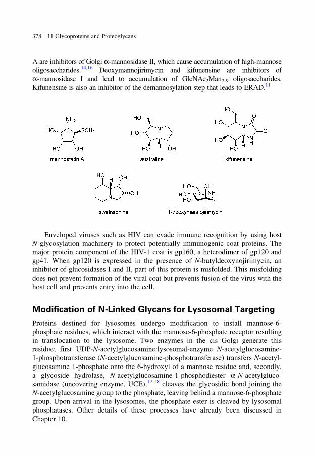
A are inhibitors of Golgi a-mannosidase II, which cause accumulation of high-mannose
oligosaccharides.14,16 Deoxymannojirimycin and kifunensine are inhibitors of
a-mannosidase I and lead to accumulation of GlcNAc2Man7-9 oligosaccharides.
Kifunensine is also an inhibitor of the demannosylation step that leads to ERAD.11
Enveloped viruses such as HIV can evade immune recognition by using host
N-glycosylation machinery to protect potentially immunogenic coat proteins. The
major protein component of the HIV-1 coat is gp160, a heterodimer of gp120 and
gp41. When gp120 is expressed in the presence of N-butyldeoxynojirimycin, an
inhibitor of glucosidases I and II, part of this protein is misfolded. This misfolding
does not prevent formation of the viral coat but prevents fusion of the virus with the
host cell and prevents entry into the cell.
Modification of N-Linked Glycans for Lysosomal Targeting
Proteins destined for lysosomes undergo modification to install mannose-6-
phosphate residues, which interact with the mannose-6-phosphate receptor resulting
in translocation to the lysosome. Two enzymes in the cis Golgi generate this
residue; first UDP-N-acetylglucosamine:lysosomal-enzyme N-acetylglucosamine-
1-phosphotransferase (N-acetylglucosamine-phosphotransferase) transfers N-acetyl-
glucosamine 1-phosphate onto the 6-hydroxyl of a mannose residue and, secondly,
a glycoside hydrolase, N-acetylglucosamine-1-phosphodiester a-N-acetylgluco-
samidase (uncovering enzyme, UCE),17,18 cleaves the glycosidic bond joining the
N-acetylglucosamine group to the phosphate, leaving behind a mannose-6-phosphate
group. Upon arrival in the lysosomes, the phosphate ester is cleaved by lysosomal
phosphatases. Other details of these processes have already been discussed in
Chapter 10.
378 11 Glycoproteins and Proteoglycans
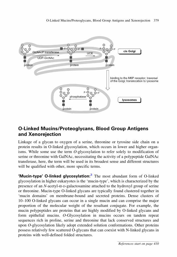
O-Linked Mucins/Proteoglycans, Blood Group Antigens
and Xenorejection
Linkage of a glycan to oxygen of a serine, threonine or tyrosine side chain on a
protein results in O-linked glycosylation, which occurs in lower and higher organ-
isms. While some use the term O-glycosylation to refer solely to modification of
serine or threonine with GalNAc, necessitating the activity of a polypeptide GalNAc
transferase, here, the term will be used in its broadest sense and different structures
will be qualified with other, more specific terms.
‘Mucin-type’ O-linked glycosylation:3 The most abundant form of O-linked
glycosylation in higher eukaryotes is the ‘mucin-type’, which is characterized by the
presence of an N-acetyl-a-D-galactosamine attached to the hydroxyl group of serine
or threonine. Mucin-type O-linked glycans are typically found clustered together in
‘mucin domains’ on membrane-bound and secreted proteins. Dense clusters of
10–100 O-linked glycans can occur in a single mucin and can comprise the major
proportion of the molecular weight of the resultant conjugate. For example, the
mucin polypeptides are proteins that are highly modified by O-linked glycans and
form epithelial mucins. O-Glycosylation in mucins occurs on tandem repeat
sequences rich in proline, serine and threonine that lack conserved structures and
upon O-glycosylation likely adopt extended solution conformations. Other proteins
possess relatively few scattered O-glycans that can coexist with N-linked glycans in
proteins with well-defined folded structures.
O-Linked Mucins/Proteoglycans, Blood Group Antigens and Xenorejection 379
References start on page 410
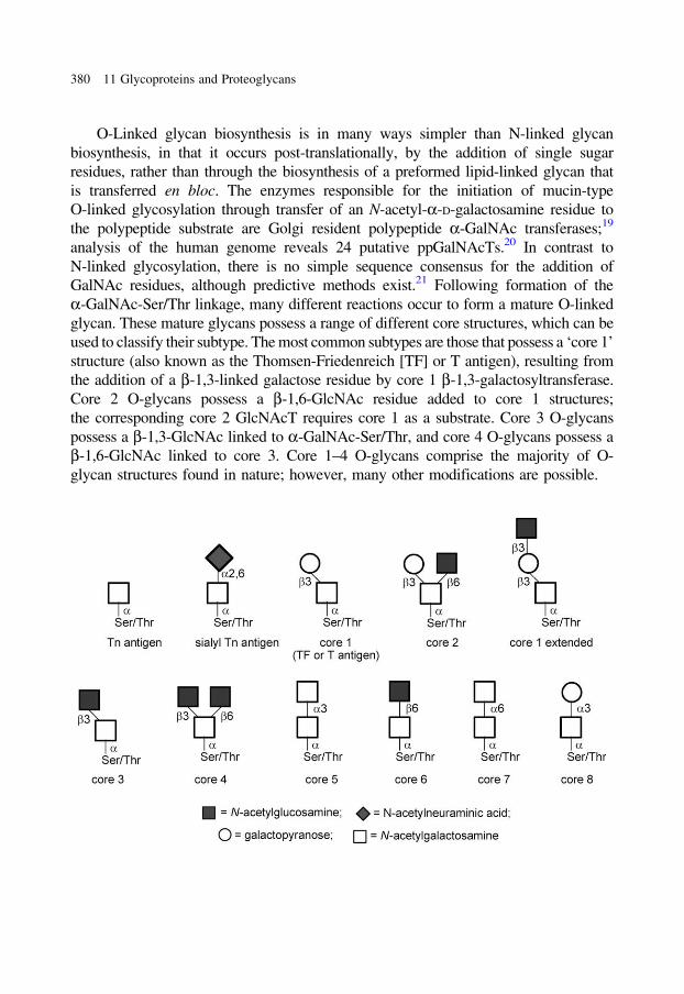
O-Linked glycan biosynthesis is in many ways simpler than N-linked glycan
biosynthesis, in that it occurs post-translationally, by the addition of single sugar
residues, rather than through the biosynthesis of a preformed lipid-linked glycan that
is transferred en bloc. The enzymes responsible for the initiation of mucin-type
O-linked glycosylation through transfer of an N-acetyl-a-D-galactosamine residue to
the polypeptide substrate are Golgi resident polypeptide a-GalNAc transferases;19
analysis of the human genome reveals 24 putative ppGalNAcTs.20 In contrast to
N-linked glycosylation, there is no simple sequence consensus for the addition of
GalNAc residues, although predictive methods exist.21 Following formation of the
a-GalNAc-Ser/Thr linkage, many different reactions occur to form a mature O-linked
glycan. These mature glycans possess a range of different core structures, which can be
used to classify their subtype. The most common subtypes are those that possess a ‘core 1’
structure (also known as the Thomsen-Friedenreich [TF] or T antigen), resulting from
the addition of a b-1,3-linked galactose residue by core 1 b-1,3-galactosyltransferase.
Core 2 O-glycans possess a b-1,6-GlcNAc residue added to core 1 structures;
the corresponding core 2 GlcNAcT requires core 1 as a substrate. Core 3 O-glycans
possess a b-1,3-GlcNAc linked to a-GalNAc-Ser/Thr, and core 4 O-glycans possess a
b-1,6-GlcNAc linked to core 3. Core 1–4 O-glycans comprise the majority of O-
glycan structures found in nature; however, many other modifications are possible.
380 11 Glycoproteins and Proteoglycans
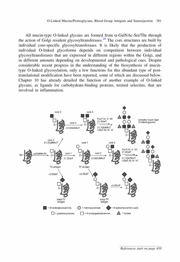
All mucin-type O-linked glycans are formed from a-GalNAc-Ser/Thr through
the action of Golgi resident glycosyltransferases.20 The core structures are built by
individual core-specific glycosyltransferases. It is likely that the production of
individual O-linked glycoforms depends on competition between individual
glycosyltransferases that are expressed in different regions within the Golgi, and
in different amounts depending on developmental and pathological cues. Despite
considerable recent progress in the understanding of the biosynthesis of mucin-
type O-linked glycosylation, only a few functions for this abundant type of post-
translational modification have been reported, some of which are discussed below.
Chapter 10 has already detailed the function of another example of O-linked
glycans, as ligands for carbohydrate-binding proteins, termed selectins, that are
involved in inflammation.
O-Linked Mucins/Proteoglycans, Blood Group Antigens and Xenorejection 381
References start on page 410
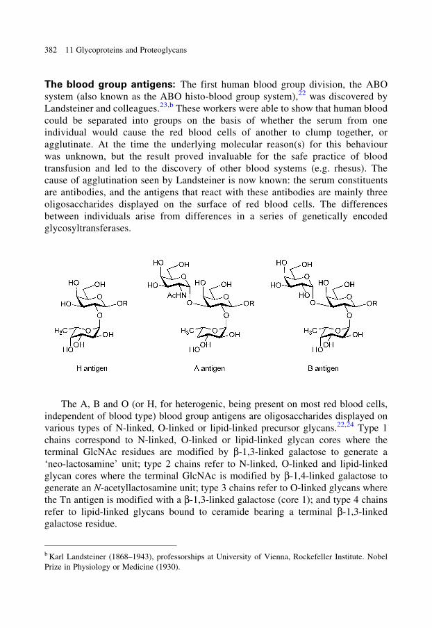
The blood group antigens: The first human blood group division, the ABO
system (also known as the ABO histo-blood group system),22 was discovered by
Landsteiner and colleagues.23,b These workers were able to show that human blood
could be separated into groups on the basis of whether the serum from one
individual would cause the red blood cells of another to clump together, or
agglutinate. At the time the underlying molecular reason(s) for this behaviour
was unknown, but the result proved invaluable for the safe practice of blood
transfusion and led to the discovery of other blood systems (e.g. rhesus). The
cause of agglutination seen by Landsteiner is now known: the serum constituents
are antibodies, and the antigens that react with these antibodies are mainly three
oligosaccharides displayed on the surface of red blood cells. The differences
between individuals arise from differences in a series of genetically encoded
glycosyltransferases.
The A, B and O (or H, for heterogenic, being present on most red blood cells,
independent of blood type) blood group antigens are oligosaccharides displayed on
various types of N-linked, O-linked or lipid-linked precursor glycans.22,24 Type 1
chains correspond to N-linked, O-linked or lipid-linked glycan cores where the
terminal GlcNAc residues are modified by b-1,3-linked galactose to generate a
‘neo-lactosamine’ unit; type 2 chains refer to N-linked, O-linked and lipid-linked
glycan cores where the terminal GlcNAc is modified by b-1,4-linked galactose to
generate an N-acetyllactosamine unit; type 3 chains refer to O-linked glycans where
the Tn antigen is modified with a b-1,3-linked galactose (core 1); and type 4 chains
refer to lipid-linked glycans bound to ceramide bearing a terminal b-1,3-linked
galactose residue.
b Karl Landsteiner (1868–1943), professorships at University of Vienna, Rockefeller Institute. Nobel
Prize in Physiology or Medicine (1930).
382 11 Glycoproteins and Proteoglycans
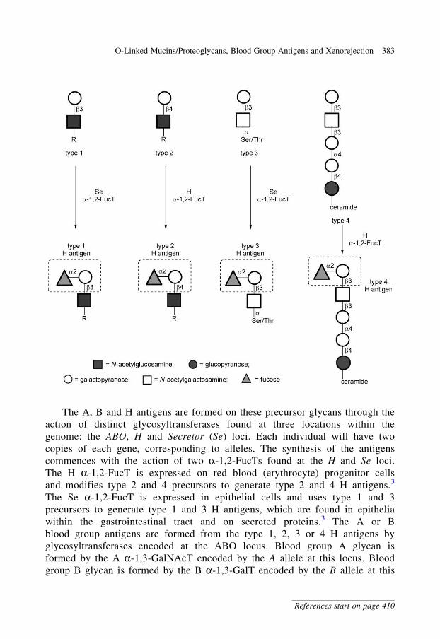
The A, B and H antigens are formed on these precursor glycans through the
action of distinct glycosyltransferases found at three locations within the
genome: the ABO, H and Secretor (Se) loci. Each individual will have two
copies of each gene, corresponding to alleles. The synthesis of the antigens
commences with the action of two a-1,2-FucTs found at the H and Se loci.
The H a-1,2-FucT is expressed on red blood (erythrocyte) progenitor cells
and modifies type 2 and 4 precursors to generate type 2 and 4 H antigens.3
The Se a-1,2-FucT is expressed in epithelial cells and uses type 1 and 3
precursors to generate type 1 and 3 H antigens, which are found in epithelia
within the gastrointestinal tract and on secreted proteins.3 The A or B
blood group antigens are formed from the type 1, 2, 3 or 4 H antigens by
glycosyltransferases encoded at the ABO locus. Blood group A glycan is
formed by the A a-1,3-GalNAcT encoded by the A allele at this locus. Blood
group B glycan is formed by the B a-1,3-GalT encoded by the B allele at this
O-Linked Mucins/Proteoglycans, Blood Group Antigens and Xenorejection 383
References start on page 410
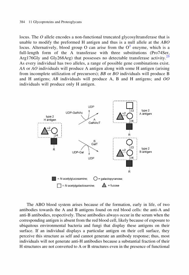
locus. The O allele encodes a non-functional truncated glycosyltransferase that is
unable to modify the preformed H antigen and thus is a null allele at the ABO
locus. Alternatively, blood group O can arise from the O2 enzyme, which is a
full-length form of the A transferase with three substitutions (Pro74Ser,
Arg176Gly and Gly268Arg) that possesses no detectable transferase activity.25
As every individual has two alleles, a range of possible gene combinations exist.
AA or AO individuals will produce A antigen along with some H antigen (arising
from incomplete utilization of precursors); BB or BO individuals will produce B
and H antigens; AB individuals will produce A, B and H antigens; and OO
individuals will produce only H antigen.
The ABO blood system arises because of the formation, early in life, of two
antibodies towards the A and B antigens found on red blood cells: the anti-A and
anti-B antibodies, respectively. These antibodies always occur in the serum when the
corresponding antigen is absent from the red blood cell, likely because of exposure to
ubiquitous environmental bacteria and fungi that display these antigens on their
surface. If an individual displays a particular antigen on their cell surface, they
perceive this structure as self and cannot generate an antibody response; thus, most
individuals will not generate anti-H antibodies because a substantial fraction of their
H structures are not converted to A or B structures even in the presence of functional
384 11 Glycoproteins and Proteoglycans
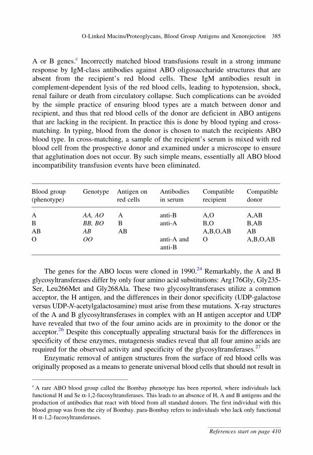
A or B genes.c Incorrectly matched blood transfusions result in a strong immune
response by IgM-class antibodies against ABO oligosaccharide structures that are
absent from the recipient’s red blood cells. These IgM antibodies result in
complement-dependent lysis of the red blood cells, leading to hypotension, shock,
renal failure or death from circulatory collapse. Such complications can be avoided
by the simple practice of ensuring blood types are a match between donor and
recipient, and thus that red blood cells of the donor are deficient in ABO antigens
that are lacking in the recipient. In practice this is done by blood typing and cross-
matching. In typing, blood from the donor is chosen to match the recipients ABO
blood type. In cross-matching, a sample of the recipient’s serum is mixed with red
blood cell from the prospective donor and examined under a microscope to ensure
that agglutination does not occur. By such simple means, essentially all ABO blood
incompatibility transfusion events have been eliminated.
The genes for the ABO locus were cloned in 1990.24 Remarkably, the A and B
glycosyltransferases differ by only four amino acid substitutions: Arg176Gly, Gly235-
Ser, Leu266Met and Gly268Ala. These two glycosyltransferases utilize a common
acceptor, the H antigen, and the differences in their donor specificity (UDP-galactose
versus UDP-N-acetylgalactosamine) must arise from these mutations. X-ray structures
of the A and B glycosyltransferases in complex with an H antigen acceptor and UDP
have revealed that two of the four amino acids are in proximity to the donor or the
acceptor.26 Despite this conceptually appealing structural basis for the differences in
specificity of these enzymes, mutagenesis studies reveal that all four amino acids are
required for the observed activity and specificity of the glycosyltransferases.27
Enzymatic removal of antigen structures from the surface of red blood cells was
originally proposed as a means to generate universal blood cells that should not result in
Blood group
(phenotype)
Genotype Antigen on
red cells
Antibodies
in serum
Compatible
recipient
Compatible
donor
A AA, AO A anti-B A,O A,AB
B BB, BO B anti-A B,O B,AB
AB AB AB A,B,O,AB AB
O OO anti-A and
anti-B
O A,B,O,AB
c A rare ABO blood group called the Bombay phenotype has been reported, where individuals lack
functional H and Se a-1,2-fucosyltransferases. This leads to an absence of H, A and B antigens and the
production of antibodies that react with blood from all standard donors. The first individual with this
blood group was from the city of Bombay. para-Bombay refers to individuals who lack only functional
H a-1,2-fucosyltransferases.
O-Linked Mucins/Proteoglycans, Blood Group Antigens and Xenorejection 385
References start on page 410
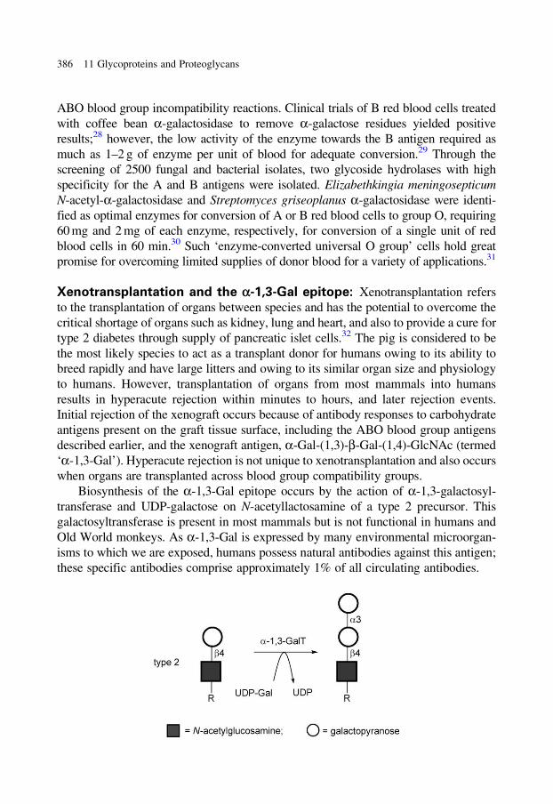
ABO blood group incompatibility reactions. Clinical trials of B red blood cells treated
with coffee bean a-galactosidase to remove a-galactose residues yielded positive
results;28 however, the low activity of the enzyme towards the B antigen required as
much as 1–2 g of enzyme per unit of blood for adequate conversion.29 Through the
screening of 2500 fungal and bacterial isolates, two glycoside hydrolases with high
specificity for the A and B antigens were isolated. Elizabethkingia meningosepticum
N-acetyl-a-galactosidase and Streptomyces griseoplanus a-galactosidase were identi-
fied as optimal enzymes for conversion of A or B red blood cells to group O, requiring
60 mg and 2 mg of each enzyme, respectively, for conversion of a single unit of red
blood cells in 60 min.30 Such ‘enzyme-converted universal O group’ cells hold great
promise for overcoming limited supplies of donor blood for a variety of applications.31
Xenotransplantation and the a-1,3-Gal epitope: Xenotransplantation refers
to the transplantation of organs between species and has the potential to overcome the
critical shortage of organs such as kidney, lung and heart, and also to provide a cure for
type 2 diabetes through supply of pancreatic islet cells.32 The pig is considered to be
the most likely species to act as a transplant donor for humans owing to its ability to
breed rapidly and have large litters and owing to its similar organ size and physiology
to humans. However, transplantation of organs from most mammals into humans
results in hyperacute rejection within minutes to hours, and later rejection events.
Initial rejection of the xenograft occurs because of antibody responses to carbohydrate
antigens present on the graft tissue surface, including the ABO blood group antigens
described earlier, and the xenograft antigen, a-Gal-(1,3)-b-Gal-(1,4)-GlcNAc (termed
‘a-1,3-Gal’). Hyperacute rejection is not unique to xenotransplantation and also occurs
when organs are transplanted across blood group compatibility groups.
Biosynthesis of the a-1,3-Gal epitope occurs by the action of a-1,3-galactosyl-
transferase and UDP-galactose on N-acetyllactosamine of a type 2 precursor. This
galactosyltransferase is present in most mammals but is not functional in humans and
Old World monkeys. As a-1,3-Gal is expressed by many environmental microorgan-
isms to which we are exposed, humans possess natural antibodies against this antigen;
these specific antibodies comprise approximately 1% of all circulating antibodies.
386 11 Glycoproteins and Proteoglycans

Hyperacute rejection can be prevented by absorbing a-1,3-Gal antibodies in
vivo through competitive binding to externally administered soluble a-Gal
epitope or ex vivo by immunoabsorption.32 However, a secondary rejection
process, acute humoral xenograft rejection, develops days or weeks after
transplantation. A more promising route to reducing xenograft rejection has
been to genetically modify the donor animal. The greatest attention has
focussed on deleting or modifying the a-1,3-Gal epitope. Pigs with
homozygous deletion of a-1,3-galactosyltransferase have been developed, and
these pigs are negative for a-1,3-Gal as determined using a lectin.33 These pigs
have been used as xenograft donors in transplantation experiments into baboons;
transplanted hearts survived up to 6 months, and kidney transplants survived for
up to 83 days.34 However, these experiments used powerful immunosuppression
techniques that are not feasible for humans. In the future, a-1,3-Gal-deficient
pigs should provide an experimental model for studying the finer details of
xenograft destruction and a genetic platform for engineering additional genetic
modifications to overcome other barriers to xenograft survival.
O-Linked N-Acetyl-b-D-glucosamine35–37
O-Linked N-acetyl-b-D-glucosamine (O-GlcNAc) on Ser or Thr is found on a
wide variety of nuclear and cytoplasmic proteins within plant and animal cells.
O-GlcNAc has several differences from classical N-linked glycosylation and
O-linked glycosylation discussed earlier. First, O-GlcNAc is not formed within
the luminal compartments of the ER or Golgi apparatus but occurs within the
cytoplasm or nucleoplasm. Secondly, O-GlcNAc is not elongated through the
addition of further sugars. Finally, O-GlcNAc is a dynamic modification and can
be added and removed on a timescale similar to that of protein phosphorylation.
Cycling of O-GlcNAc occurs through the action of two proteins, the UDP-N-
acetylglucosamine-dependent O-GlcNAc transferase, and O-GlcNAcase, a
retaining glycoside hydrolase that uses neighbouring group participation by the
2-acetamido group of the GlcNAc residue.38
A clear and demonstrable role for O-GlcNAc has remained elusive; however,
many interesting features of this dynamic post-translational modification have been
noted. O-GlcNAc has been observed to exhibit several types of competitive interplay
with O-phosphate.35 Reciprocal same-site occupancy, where the two post-
translational modifications compete at the same site within a protein, occurs in
several proteins including the transcription factor c-Myc and endothelial nitric
oxide synthase. Adjacent-site occupancy can occur where O-GlcNAc and
O-phosphate are found at adjacent sites, and occupancy of one site by one modifica-
tion precludes the other occurring at the adjacent site. This situation has been
observed in the tumour suppressor p53.
O-Linked N-Acetyl-b-D-glucosamine 387
References start on page 410
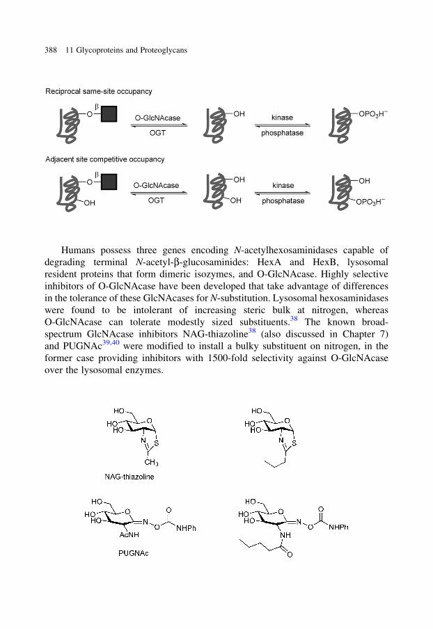
Humans possess three genes encoding N-acetylhexosaminidases capable of
degrading terminal N-acetyl-b-glucosaminides: HexA and HexB, lysosomal
resident proteins that form dimeric isozymes, and O-GlcNAcase. Highly selective
inhibitors of O-GlcNAcase have been developed that take advantage of differences
in the tolerance of these GlcNAcases for N-substitution. Lysosomal hexosaminidases
were found to be intolerant of increasing steric bulk at nitrogen, whereas
O-GlcNAcase can tolerate modestly sized substituents.38 The known broad-
spectrum GlcNAcase inhibitors NAG-thiazoline38 (also discussed in Chapter 7)
and PUGNAc39,40 were modified to install a bulky substituent on nitrogen, in the
former case providing inhibitors with 1500-fold selectivity against O-GlcNAcase
over the lysosomal enzymes.
388 11 Glycoproteins and Proteoglycans
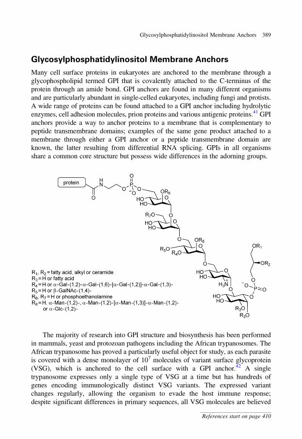
Glycosylphosphatidylinositol Membrane Anchors
Many cell surface proteins in eukaryotes are anchored to the membrane through a
glycophospholipid termed GPI that is covalently attached to the C-terminus of the
protein through an amide bond. GPI anchors are found in many different organisms
and are particularly abundant in single-celled eukaryotes, including fungi and protists.
A wide range of proteins can be found attached to a GPI anchor including hydrolytic
enzymes, cell adhesion molecules, prion proteins and various antigenic proteins.41 GPI
anchors provide a way to anchor proteins to a membrane that is complementary to
peptide transmembrane domains; examples of the same gene product attached to a
membrane through either a GPI anchor or a peptide transmembrane domain are
known, the latter resulting from differential RNA splicing. GPIs in all organisms
share a common core structure but possess wide differences in the adorning groups.
The majority of research into GPI structure and biosynthesis has been performed
in mammals, yeast and protozoan pathogens including the African trypanosomes. The
African trypanosome has proved a particularly useful object for study, as each parasite
is covered with a dense monolayer of 107 molecules of variant surface glycoprotein
(VSG), which is anchored to the cell surface with a GPI anchor.42 A single
trypanosome expresses only a single type of VSG at a time but has hundreds of
genes encoding immunologically distinct VSG variants. The expressed variant
changes regularly, allowing the organism to evade the host immune response;
despite significant differences in primary sequences, all VSG molecules are believed
Glycosylphosphatidylinositol Membrane Anchors 389
References start on page 410
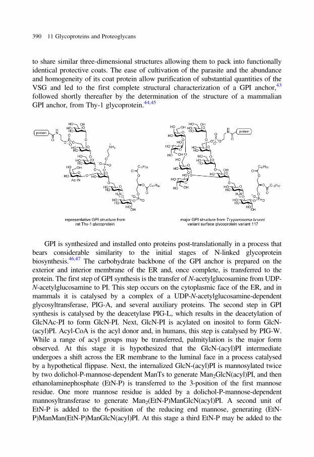
to share similar three-dimensional structures allowing them to pack into functionally
identical protective coats. The ease of cultivation of the parasite and the abundance
and homogeneity of its coat protein allow purification of substantial quantities of the
VSG and led to the first complete structural characterization of a GPI anchor,43
followed shortly thereafter by the determination of the structure of a mammalian
GPI anchor, from Thy-1 glycoprotein.44,45
GPI is synthesized and installed onto proteins post-translationally in a process that
bears considerable similarity to the initial stages of N-linked glycoprotein
biosynthesis.46,47 The carbohydrate backbone of the GPI anchor is prepared on the
exterior and interior membrane of the ER and, once complete, is transferred to the
protein. The first step of GPI synthesis is the transfer of N-acetylglucosamine from UDP-
N-acetylglucosamine to PI. This step occurs on the cytoplasmic face of the ER, and in
mammals it is catalysed by a complex of a UDP-N-acetylglucosamine-dependent
glycosyltransferase, PIG-A, and several auxiliary proteins. The second step in GPI
synthesis is catalysed by the deacetylase PIG-L, which results in the deacetylation of
GlcNAc-PI to form GlcN-PI. Next, GlcN-PI is acylated on inositol to form GlcN-
(acyl)PI. Acyl-CoA is the acyl donor and, in humans, this step is catalysed by PIG-W.
While a range of acyl groups may be transferred, palmitylation is the major form
observed. At this stage it is hypothesized that the GlcN-(acyl)PI intermediate
undergoes a shift across the ER membrane to the luminal face in a process catalysed
by a hypothetical flippase. Next, the internalized GlcN-(acyl)PI is mannosylated twice
by two dolichol-P-mannose-dependent ManTs to generate Man2GlcN(acyl)PI, and then
ethanolaminephosphate (EtN-P) is transferred to the 3-position of the first mannose
residue. One more mannose residue is added by a dolichol-P-mannose-dependent
mannosyltransferase to generate Man2(EtN-P)ManGlcN(acyl)PI. A second unit of
EtN-P is added to the 6-position of the reducing end mannose, generating (EtN-
P)ManMan(EtN-P)ManGlcN(acyl)PI. At this stage a third EtN-P may be added to the
390 11 Glycoproteins and Proteoglycans
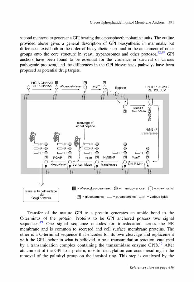
second mannose to generate a GPI bearing three phosphoethanolamine units. The outline
provided above gives a general description of GPI biosynthesis in mammals, but
differences exist both in the order of biosynthetic steps and in the attachment of other
groups onto the core structure in yeast, trypanosomes and other protozoa.42,48 GPI
anchors have been found to be essential for the virulence or survival of various
pathogenic protozoa, and the differences in the GPI biosynthesis pathways have been
proposed as potential drug targets.
Transfer of the mature GPI to a protein generates an amide bond to the
C-terminus of the protein. Proteins to be GPI anchored possess two signal
sequences.49 One signal sequence encodes for translocation across the ER
membrane and is common to secreted and cell surface membrane proteins. The
other is a C-terminal sequence that encodes for its own cleavage and replacement
with the GPI anchor in what is believed to be a transamidation reaction, catalysed
by a transamidation complex containing the transamidase enzyme GPI8.49 After
attachment of the GPI to a protein, inositol deacylation can occur resulting in the
removal of the palmityl group on the inositol ring. This step is catalysed by the
Glycosylphosphatidylinositol Membrane Anchors 391
References start on page 410
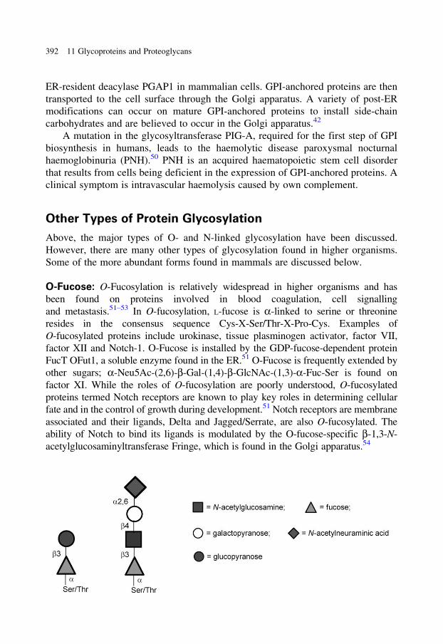
ER-resident deacylase PGAP1 in mammalian cells. GPI-anchored proteins are then
transported to the cell surface through the Golgi apparatus. A variety of post-ER
modifications can occur on mature GPI-anchored proteins to install side-chain
carbohydrates and are believed to occur in the Golgi apparatus.42
A mutation in the glycosyltransferase PIG-A, required for the first step of GPI
biosynthesis in humans, leads to the haemolytic disease paroxysmal nocturnal
haemoglobinuria (PNH).50 PNH is an acquired haematopoietic stem cell disorder
that results from cells being deficient in the expression of GPI-anchored proteins. A
clinical symptom is intravascular haemolysis caused by own complement.
Other Types of Protein Glycosylation
Above, the major types of O- and N-linked glycosylation have been discussed.
However, there are many other types of glycosylation found in higher organisms.
Some of the more abundant forms found in mammals are discussed below.
O-Fucose: O-Fucosylation is relatively widespread in higher organisms and has
been found on proteins involved in blood coagulation, cell signalling
and metastasis.51–53 In O-fucosylation, L-fucose is a-linked to serine or threonine
resides in the consensus sequence Cys-X-Ser/Thr-X-Pro-Cys. Examples of
O-fucosylated proteins include urokinase, tissue plasminogen activator, factor VII,
factor XII and Notch-1. O-Fucose is installed by the GDP-fucose-dependent protein
FucT OFut1, a soluble enzyme found in the ER.51 O-Fucose is frequently extended by
other sugars; a-Neu5Ac-(2,6)-b-Gal-(1,4)-b-GlcNAc-(1,3)-a-Fuc-Ser is found on
factor XI. While the roles of O-fucosylation are poorly understood, O-fucosylated
proteins termed Notch receptors are known to play key roles in determining cellular
fate and in the control of growth during development.51 Notch receptors are membrane
associated and their ligands, Delta and Jagged/Serrate, are also O-fucosylated. The
ability of Notch to bind its ligands is modulated by the O-fucose-specific b-1,3-N-
acetylglucosaminyltransferase Fringe, which is found in the Golgi apparatus.54
392 11 Glycoproteins and Proteoglycans

C-Mannose: An unusual form of protein glycosylation is C-mannosylation
wherein C2 of tryptophan is mannosylated through an a-C-glycosidic linkage.
C-Mannosylation was first reported in the mid-1990s, and relatively little is
known of its function. C-Mannosylation occurs through a microsomal
mannosyltransferase that utilizes dolichol-P-Man as donor;55 this suggests that
C-mannosylation occurs within the ER.56 The consensus sequence for
C-mannosylation is Trp-Xxx-Xxx-Trp, with C-mannosylation occurring on the
first tryptophan residue. C-Mannosylation may occur in closely positioned repeat
sequences in one protein and can coexist with other forms of protein glycosylation.
The protein thrombospondin-1 is a platelet protein that possesses four C-mannosyl
groups and two O-linked fucose residues.57
O-Mannose glycans: O-Mannosylation of serine and threonine residues is a
modification common to fungi such as Saccharomyces cerevisiae and Pichia
pastoris.58 These structures include high mannose mannans that possess linear
a-Man-(1,3)-a-Man-(1,3)-a-Man-(1,2)-a-Man-(1,2)-a-Man-Ser/Thr and that may
also be modified by mannose-1-phosphate residues. The majority of mammalian
O-mannose glycans are variations of the tetrasaccharide a-Neu5Ac-(2,3)-b-Gal-
(1,4)-b-GlcNAc-(1,2)-a-Man-Ser/Thr. O-Mannose glycans have been found in
brain glycopeptides and in a-dystroglycan, a highly glycosylated transmembrane
protein that is part of the dystrophin-associated glycoprotein complex and that is
involved in the pathogenesis of many forms of muscular dystrophy. The biosynthesis
of O-mannose glycans is best understood in S. cerevisiae where the initial step is the
formation of an a-mannosyl serine or threonine catalysed by a dolichol-P-Man-
dependent protein, O-mannosyltransferase, in the ER.59 The S. cerevisiae protein
O-mannosyltransferase PMT4 will only glycosylate proteins that are membrane
associated with a Ser/Thr-rich domain facing the lumen of the ER.60 Further
mannosylation events occur in the Golgi apparatus. Defects in the synthesis of
O-mannose chains lead to several forms of congenital muscular dystrophies
including Walker–Warburg syndrome (WWS), Fukuyama congenital muscular
dystrophy and muscle-eye-brain disease.58 These diseases lead to abnormal
glycosylation of a-dystroglycan, and mutations in the human protein
O-mannosyltransferase gene have been found in some sufferers of WWS.
Other Types of Protein Glycosylation 393
References start on page 410
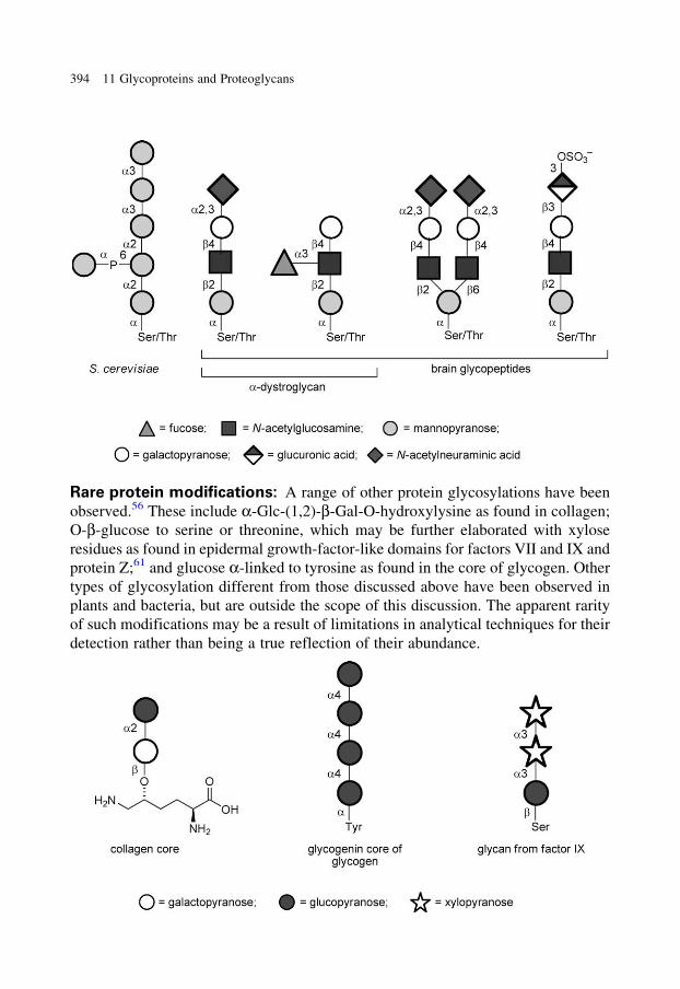
Rare protein modifications: A range of other protein glycosylations have been
observed.56 These include a-Glc-(1,2)-b-Gal-O-hydroxylysine as found in collagen;
O-b-glucose to serine or threonine, which may be further elaborated with xylose
residues as found in epidermal growth-factor-like domains for factors VII and IX and
protein Z;61 and glucose a-linked to tyrosine as found in the core of glycogen. Other
types of glycosylation different from those discussed above have been observed in
plants and bacteria, but are outside the scope of this discussion. The apparent rarity
of such modifications may be a result of limitations in analytical techniques for their
detection rather than being a true reflection of their abundance.
394 11 Glycoproteins and Proteoglycans
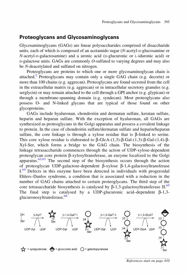
Proteoglycans and Glycosaminoglycans
Glycosaminoglycans (GAGs) are linear polysaccharides comprised of disaccharide
units, each of which is composed of an acetamido sugar (N-acetyl-D-glucosamine or
N-acetyl-D-galactosamine) and a uronic acid (D-glucuronic or L-iduronic acid) or
D-galactose units. GAGs are commonly O-sulfated to varying degrees and may also
be N-deacetylated and sulfated on nitrogen.
Proteoglycans are proteins to which one or more glycosaminoglycan chain is
attached.2 Proteoglycans may contain only a single GAG chain (e.g. decorin) or
more than 100 chains (e.g. aggrecan). Proteoglycans are found secreted from the cell
in the extracellular matrix (e.g. aggrecan) or in intracellular secretory granules (e.g.
serglycin) or may remain attached to the cell through a GPI anchor (e.g. glypican) or
through a membrane-spanning domain (e.g. syndecan). Most proteoglycans also
possess O- and N-linked glycans that are typical of those found on other
glycoproteins.
GAGs include hyaluronan, chondroitin and dermatan sulfate, keratan sulfate,
heparin and heparan sulfate. With the exception of hyaluronan, all GAGs are
synthesized as proteoglycans in the Golgi apparatus and possess a covalent linkage
to protein. In the case of chondroitin sulfate/dermatan sulfate and heparin/heparan
sulfate, the core linkage is through a xylose residue that is b-linked to serine.
This core xylose residue is elaborated to b-GlcA-(1,3)-b-Gal-(1,3)-b-Gal-(1,4)-b-
Xyl-Ser, which forms a bridge to the GAG chain. The biosynthesis of the
linkage tetrasaccharide commences through the action of UDP-xylose-dependent
proteoglycan core protein b-xylosyltransferase, an enzyme localized to the Golgi
apparatus.62,63 The second step of the biosynthesis occurs through the action
of proteoglycan UDP-galactose-dependent b-xylose b-1,4-galactosyltransferase
I.64 Defects in this enzyme have been detected in individuals with progeroidal
Ehlers–Danlos syndrome, a condition that is associated with a reduction in the
number of GAG chains attached to certain proteoglycans. The third step of the
core tetrasaccharide biosynthesis is catalysed by b-1,3-galactosyltransferase II.65
The final step is catalysed by a UDP-glucuronic acid–dependent b-1,3-
glucuronosyltransferase.66
Proteoglycans and Glycosaminoglycans 395
References start on page 410
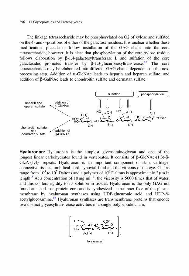
The linkage tetrasaccharide may be phosphorylated on O2 of xylose and sulfated
on the 4- and 6-positions of either of the galactose residues. It is unclear whether these
modifications precede or follow installation of the GAG chain onto the core
tetrasaccharide; however, it is clear that phosphorylation of the core xylose residue
follows elaboration by b-1,4-galactosyltransferase I, and sulfation of the core
galactosides promotes transfer by b-1,3-glucuronosyltransferase.67 The core
tetrasaccharide may be elaborated into different GAG chains dependent on the next
processing step. Addition of a-GlcNAc leads to heparin and heparan sulfate, and
addition of b-GalNAc leads to chondroitin sulfate and dermatan sulfate.
Hyaluronan: Hyaluronan is the simplest glycosaminoglycan and one of the
longest linear carbohydrates found in vertebrates. It consists of b-GlcNAc-(1,3)-b-
GlcA-(1,4)- repeats. Hyaluronan is an important component of skin, cartilage,
connective tissues, umbilical cord, synovial fluid and the vitreous of the eye. Chains
range from 105 to 107 Daltons and a polymer of 106 Daltons is approximately 2 mm in
length.3 At a concentration of 10 mg ml�1, the viscosity is 5000 times that of water,
and this confers rigidity to its solution in tissues. Hyaluronan is the only GAG not
found attached to a protein core and is synthesized at the inner face of the plasma
membrane by hyaluronan synthases using UDP-glucuronic acid and UDP-N-
acetylglucosamine.68 Hyaluronan synthases are transmembrane proteins that encode
two distinct glycosyltransferase activities in a single polypeptide chain.
396 11 Glycoproteins and Proteoglycans
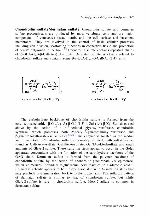
Chondroitin sulfate/dermatan sulfate: Chondroitin sulfate and dermatan
sulfate proteoglycans are produced by most vertebrate cells and are major
components of connective tissue matrix and the cell surface and basement
membranes. They are involved in the control of basic cellular processes
including cell division, scaffolding functions in connective tissue and promotion
of neurite outgrowth in the brain.69 Chondroitin sulfate contains repeating chains
of b-GlcA-(1,3)-b-GalNAc-(1,4)- units. Dermatan sulfate is closely related to
chondroitin sulfate and contains some b-L-IdoA-(1,3)-b-GalNAc-(1,4)- units.
The carbohydrate backbone of chondroitin sulfate is formed from the
core tetrasaccharide b-GlcA-(1,3)-b-Gal-(1,3)-b-Gal-(1,4)-b-Xyl-Ser discussed
above by the action of a bifunctional glycosyltransferase, chondroitin
synthase, which possesses both N-acetyl-b-galactosaminyltransferase and
b-glucuronosyltransferase activities.69,70 This enzyme is located in the medial
and trans Golgi. Chondroitin sulfate is variably sulfated, with sulfate esters
found as GalNAc-4-sulfate, GalNAc-6-sulfate, GalNAc-4,6-disulfate and small
amounts of GlcA-2-sulfate. These sulfation steps appear to occur in the Golgi
apparatus concomitant with the formation of the carbohydrate backbone of the
GAG chain. Dermatan sulfate is formed from the polymer backbone of
chondroitin sulfate by the action of chondroitin-glucuronate C5 epimerase,
which epimerizes individual D-glucuronic acid residues to L-iduronic acid.71
Epimerase activity appears to be closely associated with O-sulfation steps that
may preclude re-epimerization back to D-glucuronic acid. The sulfation pattern
of dermatan sulfate is similar to that of chondroitin sulfate, but while
GlcA-2-sulfate is rare in chondroitin sulfate, IdoA-2-sulfate is common in
dermatan sulfate.
Proteoglycans and Glycosaminoglycans 397
References start on page 410
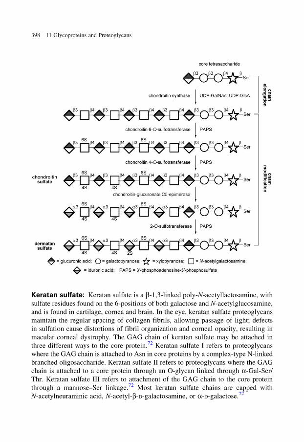
Keratan sulfate: Keratan sulfate is a b-1,3-linked poly-N-acetyllactosamine, with
sulfate residues found on the 6-positions of both galactose and N-acetylglucosamine,
and is found in cartilage, cornea and brain. In the eye, keratan sulfate proteoglycans
maintain the regular spacing of collagen fibrils, allowing passage of light; defects
in sulfation cause distortions of fibril organization and corneal opacity, resulting in
macular corneal dystrophy. The GAG chain of keratan sulfate may be attached in
three different ways to the core protein.72 Keratan sulfate I refers to proteoglycans
where the GAG chain is attached to Asn in core proteins by a complex-type N-linked
branched oligosaccharide. Keratan sulfate II refers to proteoglycans where the GAG
chain is attached to a core protein through an O-glycan linked through a-Gal-Ser/
Thr. Keratan sulfate III refers to attachment of the GAG chain to the core protein
through a mannose–Ser linkage.72 Most keratan sulfate chains are capped with
N-acetylneuraminic acid, N-acetyl-b-D-galactosamine, or a-D-galactose.72
398 11 Glycoproteins and Proteoglycans
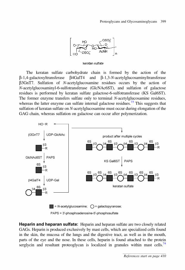
The keratan sulfate carbohydrate chain is formed by the action of the
b-1,4-galactosyltransferase b4GalT4 and b-1,3-N-acetylglucosaminyltransferase
b3GnT7. Sulfation of N-acetylglucosamine residues occurs by the action of
N-acetylglucosaminyl-6-sulfotransferase (GlcNAc6ST), and sulfation of galactose
residues is performed by keratan sulfate galactose-6-sulfotransferase (KS Gal6ST).
The former enzyme transfers sulfate only to terminal N-acetylglucosamine residues,
whereas the latter enzyme can sulfate internal galactose residues.73 This suggests that
sulfation of keratan sulfate on N-acetylglucosamine must occur during elongation of the
GAG chain, whereas sulfation on galactose can occur after polymerization.
Heparin and heparan sulfate: Heparin and heparan sulfate are two closely related
GAGs. Heparin is produced exclusively by mast cells, which are specialized cells found
in the skin, the mucosa of the lungs and the digestive tract, as well as in the mouth,
parts of the eye and the nose. In these cells, heparin is found attached to the protein
serglycin and resultant proteoglycan is localized in granules within mast cells.74
Proteoglycans and Glycosaminoglycans 399
References start on page 410
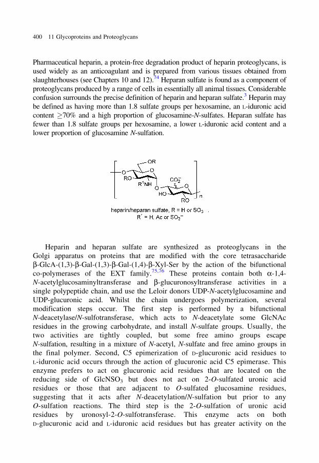
Pharmaceutical heparin, a protein-free degradation product of heparin proteoglycans, is
used widely as an anticoagulant and is prepared from various tissues obtained from
slaughterhouses (see Chapters 10 and 12).74 Heparan sulfate is found as a component of
proteoglycans produced by a range of cells in essentially all animal tissues. Considerable
confusion surrounds the precise definition of heparin and heparan sulfate.3 Heparin may
be defined as having more than 1.8 sulfate groups per hexosamine, an L-iduronic acid
content �70% and a high proportion of glucosamine-N-sulfates. Heparan sulfate has
fewer than 1.8 sulfate groups per hexosamine, a lower L-iduronic acid content and a
lower proportion of glucosamine N-sulfation.
Heparin and heparan sulfate are synthesized as proteoglycans in the
Golgi apparatus on proteins that are modified with the core tetrasaccharide
b-GlcA-(1,3)-b-Gal-(1,3)-b-Gal-(1,4)-b-Xyl-Ser by the action of the bifunctional
co-polymerases of the EXT family.75,76 These proteins contain both a-1,4-
N-acetylglucosaminyltransferase and b-glucuronosyltransferase activities in a
single polypeptide chain, and use the Leloir donors UDP-N-acetylglucosamine and
UDP-glucuronic acid. Whilst the chain undergoes polymerization, several
modification steps occur. The first step is performed by a bifunctional
N-deacetylase/N-sulfotransferase, which acts to N-deacetylate some GlcNAc
residues in the growing carbohydrate, and install N-sulfate groups. Usually, the
two activities are tightly coupled, but some free amino groups escape
N-sulfation, resulting in a mixture of N-acetyl, N-sulfate and free amino groups in
the final polymer. Second, C5 epimerization of D-glucuronic acid residues to
L-iduronic acid occurs through the action of glucuronic acid C5 epimerase. This
enzyme prefers to act on glucuronic acid residues that are located on the
reducing side of GlcNSO3 but does not act on 2-O-sulfated uronic acid
residues or those that are adjacent to O-sulfated glucosamine residues,
suggesting that it acts after N-deacetylation/N-sulfation but prior to any
O-sulfation reactions. The third step is the 2-O-sulfation of uronic acid
residues by uronosyl-2-O-sulfotransferase. This enzyme acts on both
D-glucuronic acid and L-iduronic acid residues but has greater activity on the
400 11 Glycoproteins and Proteoglycans
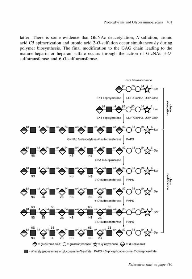
latter. There is some evidence that GlcNAc deacetylation, N-sulfation, uronic
acid C5 epimerization and uronic acid 2-O-sulfation occur simultaneously during
polymer biosynthesis. The final modification to the GAG chain leading to the
mature heparin or heparan sulfate occurs through the action of GlcNAc 3-O-
sulfotransferase and 6-O-sulfotransferase.
Proteoglycans and Glycosaminoglycans 401
References start on page 410
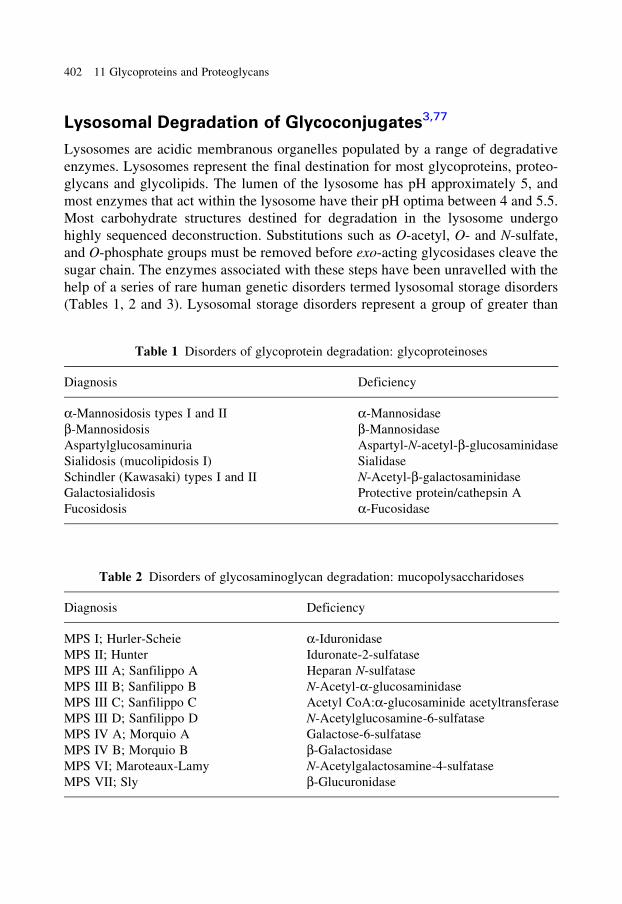
Lysosomal Degradation of Glycoconjugates3,77
Lysosomes are acidic membranous organelles populated by a range of degradative
enzymes. Lysosomes represent the final destination for most glycoproteins, proteo-
glycans and glycolipids. The lumen of the lysosome has pH approximately 5, and
most enzymes that act within the lysosome have their pH optima between 4 and 5.5.
Most carbohydrate structures destined for degradation in the lysosome undergo
highly sequenced deconstruction. Substitutions such as O-acetyl, O- and N-sulfate,
and O-phosphate groups must be removed before exo-acting glycosidases cleave the
sugar chain. The enzymes associated with these steps have been unravelled with the
help of a series of rare human genetic disorders termed lysosomal storage disorders
(Tables 1, 2 and 3). Lysosomal storage disorders represent a group of greater than
Table 1 Disorders of glycoprotein degradation: glycoproteinoses
Diagnosis Deficiency
a-Mannosidosis types I and II a-Mannosidase
b-Mannosidosis b-Mannosidase
Aspartylglucosaminuria Aspartyl-N-acetyl-b-glucosaminidase
Sialidosis (mucolipidosis I) Sialidase
Schindler (Kawasaki) types I and II N-Acetyl-b-galactosaminidase
Galactosialidosis Protective protein/cathepsin A
Fucosidosis a-Fucosidase
Table 2 Disorders of glycosaminoglycan degradation: mucopolysaccharidoses
Diagnosis Deficiency
MPS I; Hurler-Scheie a-Iduronidase
MPS II; Hunter Iduronate-2-sulfatase
MPS III A; Sanfilippo A Heparan N-sulfatase
MPS III B; Sanfilippo B N-Acetyl-a-glucosaminidase
MPS III C; Sanfilippo C Acetyl CoA:a-glucosaminide acetyltransferase
MPS III D; Sanfilippo D N-Acetylglucosamine-6-sulfatase
MPS IV A; Morquio A Galactose-6-sulfatase
MPS IV B; Morquio B b-Galactosidase
MPS VI; Maroteaux-Lamy N-Acetylgalactosamine-4-sulfatase
MPS VII; Sly b-Glucuronidase
402 11 Glycoproteins and Proteoglycans
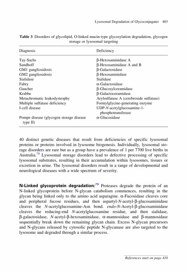
40 distinct genetic diseases that result from deficiencies of specific lysosomal
proteins or proteins involved in lysosome biogenesis. Individually, lysosomal sto-
rage disorders are rare but as a group have a prevalence of 1 per 7700 live births in
Australia.78 Lysosomal storage disorders lead to defective processing of specific
lysosomal substrates, resulting in their accumulation within lysosomes, tissues or
excretion in urine. The lysosomal disorders result in a range of developmental and
neurological diseases with a wide spectrum of severity.
N-Linked glycoprotein degradation:79 Proteases degrade the protein of an
N-linked glycoprotein before N-glycan catabolism commences, resulting in the
glycan being linked only to the amino acid asparagine. a-Fucosidase cleaves core
and peripheral fucose residues, and then aspartyl-N-acetyl-b-glucosaminidase
cleaves the N-acetylglucosamine-Asn bond. endo-N-Acetyl-b-glucosaminidase
cleaves the reducing-end N-acetylglucosamine residue, and then sialidase,
b-galactosidase, N-acetyl-b-hexosaminidase, a-mannosidase and b-mannosidase
sequentially break down the remaining glycan chain. Excess N-glycan precursors
and N-glycans released by cytosolic peptide N-glycanase are also targeted to the
lysosome and degraded through a similar process.
Table 3 Disorders of glycolipid, O-linked mucin-type glycosylation degradation, glycogen
storage or lysosomal targeting
Diagnosis Deficiency
Tay-Sachs b-Hexosaminidase A
Sandhoff b-Hexosaminidase A and B
GM1 gangliosidosis b-Galactosidase
GM2 gangliosidosis b-Hexosaminidase
Sialidase Sialidase
Fabry a-Galactosidase
Gaucher b-Glucosylceramidase
Krabbe b-Galactoceramidase
Metachromatic leukodystrophy Arylsulfatase A (cerebroside sulfatase)
Multiple sulfatase deficiency Formylglycine-generating enzyme
I-cell disease UDP-N-acetylglucosamine-1-
phosphotransferase
Pompe disease (glycogen storage disease
type II)
a-Glucosidase
Lysosomal Degradation of Glycoconjugates 403
References start on page 410
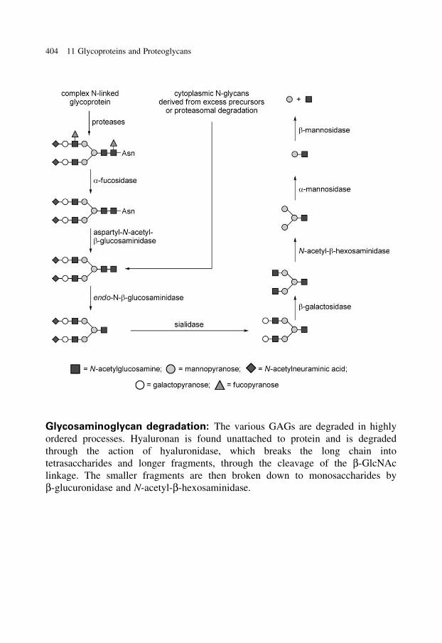
Glycosaminoglycan degradation: The various GAGs are degraded in highly
ordered processes. Hyaluronan is found unattached to protein and is degraded
through the action of hyaluronidase, which breaks the long chain into
tetrasaccharides and longer fragments, through the cleavage of the b-GlcNAc
linkage. The smaller fragments are then broken down to monosaccharides by
b-glucuronidase and N-acetyl-b-hexosaminidase.
404 11 Glycoproteins and Proteoglycans
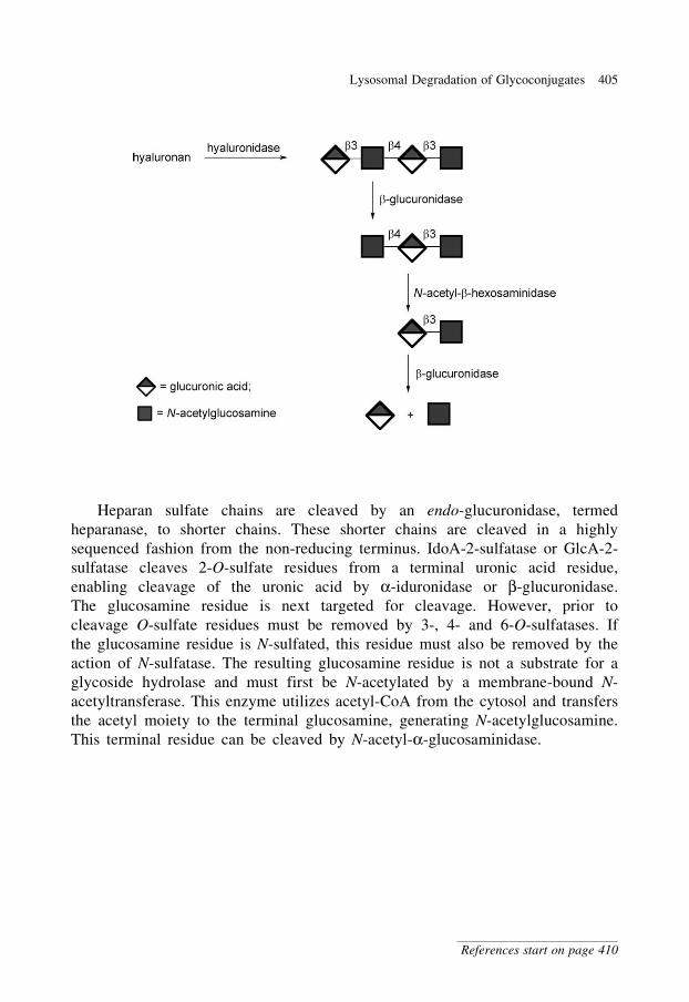
Heparan sulfate chains are cleaved by an endo-glucuronidase, termed
heparanase, to shorter chains. These shorter chains are cleaved in a highly
sequenced fashion from the non-reducing terminus. IdoA-2-sulfatase or GlcA-2-
sulfatase cleaves 2-O-sulfate residues from a terminal uronic acid residue,
enabling cleavage of the uronic acid by a-iduronidase or b-glucuronidase.
The glucosamine residue is next targeted for cleavage. However, prior to
cleavage O-sulfate residues must be removed by 3-, 4- and 6-O-sulfatases. If
the glucosamine residue is N-sulfated, this residue must also be removed by the
action of N-sulfatase. The resulting glucosamine residue is not a substrate for a
glycoside hydrolase and must first be N-acetylated by a membrane-bound N-
acetyltransferase. This enzyme utilizes acetyl-CoA from the cytosol and transfers
the acetyl moiety to the terminal glucosamine, generating N-acetylglucosamine.
This terminal residue can be cleaved by N-acetyl-a-glucosaminidase.
Lysosomal Degradation of Glycoconjugates 405
References start on page 410
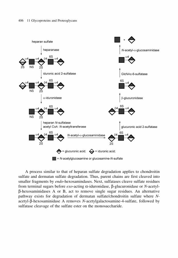
A process similar to that of heparan sulfate degradation applies to chondroitin
sulfate and dermatan sulfate degradation. Thus, parent chains are first cleaved into
smaller fragments by endo-hexosamindases. Next, sulfatases cleave sulfate residues
from terminal sugars before exo-acting a-iduronidase, b-glucuronidase or N-acetyl-
b-hexosaminidases A or B, act to remove single sugar residues. An alternative
pathway exists for degradation of dermatan sulfate/chondroitin sulfate where N-
acetyl-b-hexosaminidase A removes N-acetylgalactosamine-4-sulfate, followed by
sulfatase cleavage of the sulfate ester on the monosaccharide.
406 11 Glycoproteins and Proteoglycans
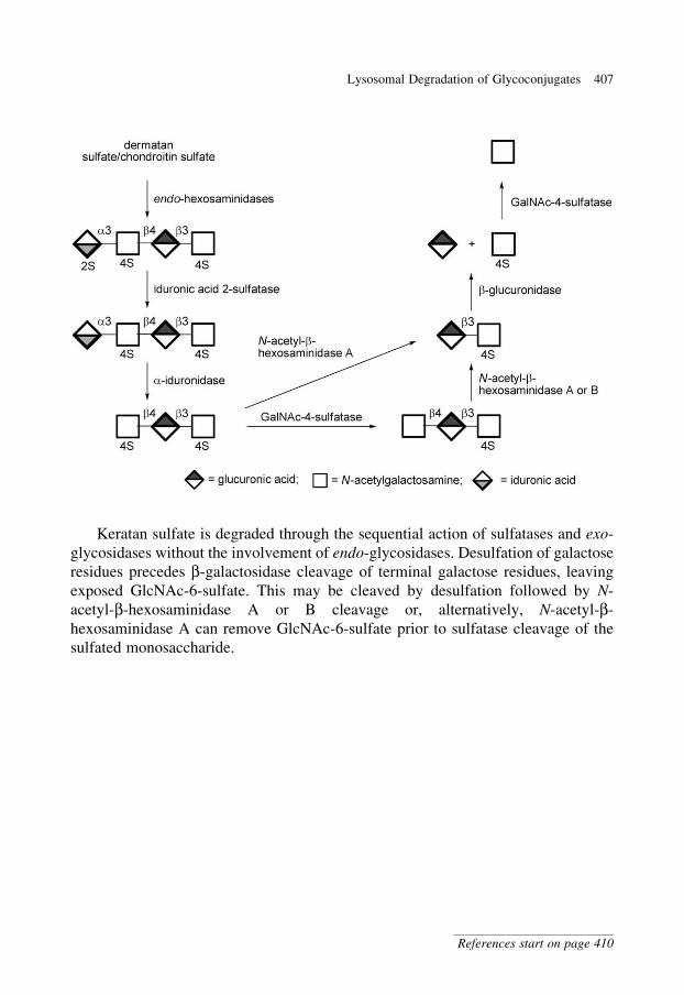
Keratan sulfate is degraded through the sequential action of sulfatases and exo-
glycosidases without the involvement of endo-glycosidases. Desulfation of galactose
residues precedes b-galactosidase cleavage of terminal galactose residues, leaving
exposed GlcNAc-6-sulfate. This may be cleaved by desulfation followed by N-
acetyl-b-hexosaminidase A or B cleavage or, alternatively, N-acetyl-b-
hexosaminidase A can remove GlcNAc-6-sulfate prior to sulfatase cleavage of the
sulfated monosaccharide.
Lysosomal Degradation of Glycoconjugates 407
References start on page 410
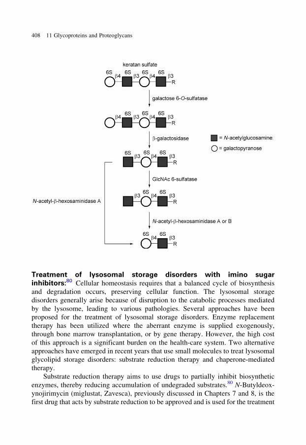
Treatment of lysosomal storage disorders with imino sugar
inhibitors:80 Cellular homeostasis requires that a balanced cycle of biosynthesis
and degradation occurs, preserving cellular function. The lysosomal storage
disorders generally arise because of disruption to the catabolic processes mediated
by the lysosome, leading to various pathologies. Several approaches have been
proposed for the treatment of lysosomal storage disorders. Enzyme replacement
therapy has been utilized where the aberrant enzyme is supplied exogenously,
through bone marrow transplantation, or by gene therapy. However, the high cost
of this approach is a significant burden on the health-care system. Two alternative
approaches have emerged in recent years that use small molecules to treat lysosomal
glycolipid storage disorders: substrate reduction therapy and chaperone-mediated
therapy.
Substrate reduction therapy aims to use drugs to partially inhibit biosynthetic
enzymes, thereby reducing accumulation of undegraded substrates.80 N-Butyldeox-
ynojirimycin (miglustat, Zavesca), previously discussed in Chapters 7 and 8, is the
first drug that acts by substrate reduction to be approved and is used for the treatment
408 11 Glycoproteins and Proteoglycans
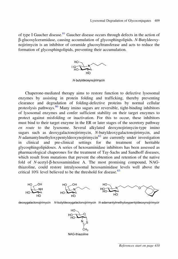
of type I Gaucher disease.81 Gaucher disease occurs through defects in the action of
b-glucosylceramidase, causing accumulation of glycosphingolipids. N-Butyldeoxy-
nojirimycin is an inhibitor of ceramide glucosyltransferase and acts to reduce the
formation of glycosphingolipids, preventing their accumulation.
Chaperone-mediated therapy aims to restore function to defective lysosomal
enzymes by assisting in protein folding and trafficking, thereby preventing
clearance and degradation of folding-defective proteins by normal cellular
proteolysis pathways.80 Many imino sugars are reversible, tight-binding inhibitors
of lysosomal enzymes and confer sufficient stability on their target enzymes to
protect against misfolding or inactivation. For this to occur, these inhibitors
must bind to their target enzyme in the ER or later stages of the secretory pathway
en route to the lysosome. Several alkylated deoxynojirimycin-type imino
sugars such as deoxygalactonojirimycin, N-butyldeoxygalactonojirimycin, and
N-adamantylmethyloxypentyldeoxynojirimycin82 are currently under investigation
in clinical and pre-clinical settings for the treatment of heritable
glycosphingolipidoses. A series of hexosaminidase inhibitors has been assessed as
pharmacological chaperones for the treatment of Tay-Sachs and Sandhoff diseases,
which result from mutations that prevent the obtention and retention of the native
fold of N-acetyl-b-hexosaminidase A. The most promising compound, NAG-
thiazoline, could restore intralysosomal hexosaminidase levels well above the
critical 10% level believed to be the threshold for disease.83
Lysosomal Degradation of Glycoconjugates 409
References start on page 410
References
1. Werz, D.B., Ranzinger, R., Herget, S., Adibekian, A., von der Lieth, C.-W. and Seeberger, P.H.
(2007). ACS Chem. Biol., 2, 685.
2. Kjellen, L. and Lindahl, U. (1991). Annu. Rev. Biochem., 60, 443.
3. Varki, A., Cummings, R., Esko, J., Hart, G. and Marth, J., eds (1999) Essentials of Glycobiology.
Cold Spring Harbor: New York.
4. Roth, J. (2002). Chem. Rev., 102, 285.
5. Kornfeld, R. and Kornfeld, S. (1985). Annu. Rev. Biochem., 54, 631.
6. Burda, P. and Aebi, M. (1999). Biochim. Biophys. Acta, 1426, 239.
7. Snider, M.D. and Robbins, P.W. (1982). J. Biol. Chem., 257, 6796.
8. Moreman, K.W. and Molinari, M. (2006). Curr. Opin. Chem. Biol., 16, 592.
9. Helenius, A. and Aebi, M. (2001). Science, 291, 2364.
10. Rudd, P.M., Elliott, T., Cresswell, P., Wilson, I.A. and Dwek, R.A. (2001). Science, 291, 2370.
11. Ruddock, L.W. and Molinari, M. (2006). J. Cell Sci., 119, 4373.
12. Banerjee, D.K. (1989). J. Biol. Chem., 264, 2024.
13. Eason, P.D. and Imperiali, B. (1999). Biochemistry, 38, 5430.
14. Elbein, A.D. (1991). FASEB J., 5, 3055.
15. Winchester, B. and Fleet, G.W. (1992). Glycobiology, 2, 199.
16. Kawatkar, S.P., Kuntz, D.A., Woods, R.J., Rose, D.R. and Boons, G.J. (2006). J. Am. Chem. Soc.,
128, 8310.
17. Rohrer, J. and Kornfeld, R. (2001). Mol. Biol. Cell, 12, 1623.
18. Varki, A., Sherman, W. and Kornfeld, S. (1983). Arch. Biochem. Biophys., 222, 145.
19. Abeijon, C. and Hirschberg, C.B. (1987). J. Biol. Chem., 262, 4153.
20. Hang, H.C. and Bertozzi, C.R. (2005). Bioorg. Med. Chem., 13, 5021.
21. Julenius, K., Mølgaard, A., Gupta, R. and Brunak, S. (2005). Glycobiology, 15, 153.
22. Clausen, H. and Hakomori, S.-i. (1989). Vox Sang., 56, 1.
23. Watkins, W.M. (1966). Science, 152, 172.
24. Hakomori, S. (1999). Biochim. Biophys. Acta, 1473, 247.
25. Lee, H.J., Barry, C.H., Borisova, S.N., Seto, N.O., Zheng, R.B., Blancher, A., Evans, S.V. and
Palcic, M.M. (2005). J. Biol. Chem., 280, 525.
26. Patenaude, S.I., Seto, N.O., Borisova, S.N., Szpacenko, A., Marcus, S.L., Palcic, M.M. and Evans,
S.V. (2002). Nat. Struct. Biol., 9, 685.
27. Rose, N.L., Palcic, M.M. and Evans, S.V. (2005). J. Chem. Ed., 82, 1846.
28. Kruskall, M.S., AuBuchon, J.P., Anthony, K.Y., Herschel, L., Pickard, C., Biehl, R., Horowitz, M.,
Brambilla, D.J. and Popovsky, M.A. (2000). Transfusion, 40, 1290.
29. Goldstein, J., Siviglia, G., Hurst, R., Lenny, L. and Reich, L. (1982). Science, 215, 168.
30. Liu, Q.P., Sulzenbacher, G., Yuan, H., Bennett, E.P., Pietz, G., Saunders, K., Spence, J., Nudelman,
E., Levery, S.B., White, T., Neveu, J.M., Lane, W.S., Bourne, Y., Olsson, M.L., Henrissat, B. and
Clausen, H. (2007). Nat. Biotechnol., 25, 454.
31. Daniels, G. and Withers, S.G. (2007). Nat. Biotechnol., 25, 427.
32. Yang, Y.-G. and Sykes, M. (2007). Nat. Rev. Immunol., 7, 519.
33. Phelps, C.J., Koike, C., Vaught, T.D., Boone, J., Wells, K.D., Chen, S.H., Ball, S., Specht, S.M.,
Polejaeva, I.A., Monahan, J.A., Jobst, P.M., Sharma, S.B., Lamborn, A.E., Garst, A.S., Moore, M.,
Demetris, A.J., Rudert, W.A., Bottino, R., Bertera, S., Trucco, M., Starzl, T.E., Dai, Y. and Ayares,
D.L. (2003). Science, 299, 411.
34. Milland, J., Christiansen, D. and Sandrin, M.S. (2005). Immunol. Cell Biol., 83, 687.
35. Wells, L., Vosseller, K. and Hart, G.W. (2001). Science, 291, 2376.
36. Hart, G.W., Housley, M.P. and Slawson, C. (2007). Nature, 446, 1017.
410 11 Glycoproteins and Proteoglycans
37. Zachara, N.E. and Hart, G.W. (2002). Chem. Rev., 102, 431.
38. Macauley, M.S., Whitworth, G.E., Debowski, A.W., Chin, D. and Vocadlo, D.J. (2005). J. Biol.
Chem., 280, 25313.
39. Kim, E.J., Perreira, M., Thomas, C.J. and Hanover, J.A. (2006). J. Am. Chem. Soc., 128, 4234.
40. Stubbs, K.A., Zhang, N. and Vocadlo, D.J. (2006). Org. Biomol. Chem., 4, 839.
41. Ferguson, M.A. and Williams, A.F. (1988). Annu. Rev. Biochem., 57, 285.
42. Ferguson, M.A. (1999). J. Cell Sci., 112, 2799.
43. Ferguson, M.A., Homans, S.W., Dwek, R.A. and Rademacher, T.W. (1988). Science, 239, 753.
44. Homans, S.W., Ferguson, M.A., Dwek, R.A., Rademacher, T.W., Anand, R. and Williams, A.F.
(1988). Nature, 333, 269.
45. Tse, A.G., Barclay, A.N., Watts, A. and Williams, A.F. (1985). Science, 230, 1003.
46. Kinoshita, T. and Inoue, N. (2000). Curr. Opin. Chem. Biol., 4, 632.
47. Maeda, Y., Ashida, H. and Kinoshita, T. (2006). Methods Enzymol., 416, 182.
48. McConville, M.J. and Ferguson, M.A. (1993). Biochem. J., 294, 305.
49. Zacks, M.A. and Garg, N. (2006). Mol. Membr. Biol., 23, 209.
50. Kinoshita, T., Ohishi, K. and Takeda, J. (1997). J. Biochem., 122, 251.
51. Okajima, T. and Matsuda, T. (2006). Methods Enzymol., 417, 111.
52. Harris, R.J. and Spellman, M.W. (1993). Glycobiology, 3, 219.
53. Peter-Katalinic, J. (2005). Methods Enzymol., 405, 139.
54. Haltiwanger, R.S. and Stanley, P. (2002). Biochim. Biophys. Acta, 1573, 328.
55. Doucey, M.A., Hess, D., Cacan, R. and Hofsteenge, J. (1998). Mol. Biol. Cell, 9, 291.
56. Spiro, R.G. (2002). Glycobiology, 12, 43R.
57. Hofsteenge, J., Huwiler, K.G., Macek, B., Hess, D., Lawler, J., Mosher, D.F. and Peter-
Katalinic, J. (2001). J. Biol. Chem., 276, 6485.
58. Willer, T., Valero, M.C., Tanner, W., Cruces, J. and Strahl, S. (2003). Curr. Opin. Struct. Biol.,
13, 621.
59. Lehle, L., Strahl, S. and Tanner, W. (2006). Angew. Chem. Int. Ed., 45, 6802.
60. Hutzler, J., Schmid, M., Bernard, T., Henrissat, B. and Strahl, S. (2007). Proc. Natl. Acad. Sci.
USA, 104, 7827.
61. Nishimura, H., Kawabata, S., Kisiel, W., Hase, S., Ikenaka, T., Takao, T., Shimonishi, Y. and
Iwanaga, S. (1989). J. Biol. Chem., 264, 20320.
62. Gotting, C., Kuhn, J., Zahn, R., Brinkmann, T. and Kleesiek, K. (2000). J. Mol. Biol., 304, 517.
63. Schon, S., Prante, C., Bahr, C., Kuhn, J., Kleesiek, K. and Gotting, C. (2006). J. Biol. Chem.,
281, 14224.
64. Almeida, R., Levery, S.B., Mandel, U., Kresse, H., Schwientek, T., Bennett, E.P. and Clausen, H.
(1999). J. Biol. Chem., 274, 26165.
65. Bai, X., Zhou, D., Brown, J.R., Crawford, B.E., Hennet, T. and Esko, J.D. (2001). J. Biol. Chem.,
276, 48189.
66. Wei, G., Bai, X., Sarkar, A.K. and Esko, J.D. (1999). J. Biol. Chem., 274, 7857.
67. Gulberti, S., Lattard, V., Fondeur, M., Jacquinet, J.C., Mulliert, G., Netter, P., Magdalou, J.,
Ouzzine, M. and Fournel-Gigleux, S. (2005). J. Biol. Chem., 280, 1417.
68. Itano, N. and Kimata, K. (2002). IUBMB Life, 54, 195.
69. Sugahara, K., Mikami, T., Uyama, T., Mizuguchi, S., Nomura, K. and Kitagawa, H. (2003). Curr.
Opin. Struct. Biol., 13, 612.
70. Silbert, J.E. and Sugumaran, G. (2002). IUBMB Life, 54, 177.
71. Maccarana, M., Olander, B., Malmstrom, J., Tiedemann, K., Aebersold, R., Lindahl, U., Li, J.P.
and Malmstrom, A. (2006). J. Biol. Chem., 281, 11560.
72. Funderburgh, J.L. (2002). IUBMB Life, 54, 187.
73. Kitayama, K., Hayashida, Y., Nishida, K. and Akama, T.O. (2007). J. Biol. Chem., 282, 30085.
References 411
74. Linhardt, R.J. (2003). J. Med. Chem., 46, 2551.
75. Esko, J.D. and Selleck, S.B. (2002). Annu. Rev. Biochem., 71, 435.
76. Sugahara, K. and Kitagawa, H. (2002). IUBMB Life, 54, 163.
77. Scriver, C.R., Beaudet, A.L., Valle, D. and Sly, W.S., eds (2006). Metabolic and Molecular Bases
of Inherited Diseases Online, 8th Ed., acccessed 22/11/07. McGraw-Hill.
78. Meikle, P.J., Hopwood, J.J., Clague, A.E. and Carey, W.F. (1999). JAMA, 281, 249.
79. Winchester, B. (2005). Glycobiology, 15, 1R.
80. Butters, T.D., Dwek, R.A. and Platt, F.M. (2005). Glycobiology, 15, 43R.
81. Elstein, D., Hollak, C., Aerts, J.M.F.G., van Weely, S., Maas, M., Cox, T.M., Lachmann, R.H.,
Hrebicek, M., Platt, F.M., Butters, T.D., Dwek, R.A. and Zimran, A. (2004). J. Inherit. Metab. Dis.,
27, 757.
82. Wennekes, T., Berg, R.J., Donker, W., Marel, G.A., Strijland, A., Aerts, J.M., Overkleeft, H.S.
(2007). J. Org. Chem., 72, 1088.
83. Tropak, M.B., Reid, S.P., Guiral, M., Withers, S.G. and Mahuran, D. (2004). J. Biol. Chem., 279,
13478.
412 11 Glycoproteins and Proteoglycans
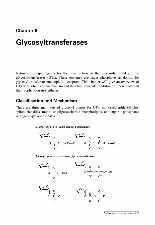
Chapter 8
Glycosyltransferases
Nature’s principal agents for the construction of the glycosidic bond are the
glycosyltransferases (GTs). These enzymes use sugar phosphates as donors for
glycosyl transfer to nucleophilic acceptors. This chapter will give an overview of
GTs with a focus on mechanism and structure, reagents/inhibitors for their study and
their application in synthesis.
Classification and Mechanism
There are three main sets of glycosyl donors for GTs: monosaccharide (di)pho-
sphonucleosides, mono- or oligosaccharide phospholipids, and sugar-1-phosphates
or sugar-1-pyrophosphates.
References start on page 316
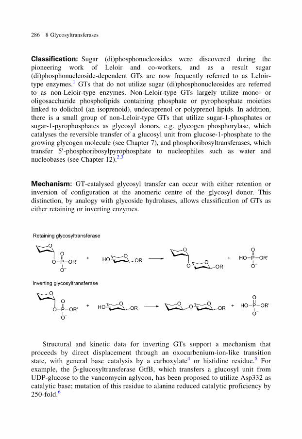
Classification: Sugar (di)phosphonucleosides were discovered during the
pioneering work of Leloir and co-workers, and as a result sugar
(di)phosphonucleoside-dependent GTs are now frequently referred to as Leloir-
type enzymes.1 GTs that do not utilize sugar (di)phosphonucleosides are referred
to as non-Leloir-type enzymes. Non-Leloir-type GTs largely utilize mono- or
oligosaccharide phospholipids containing phosphate or pyrophosphate moieties
linked to dolichol (an isoprenoid), undecaprenol or polyprenol lipids. In addition,
there is a small group of non-Leloir-type GTs that utilize sugar-1-phosphates or
sugar-1-pyrophosphates as glycosyl donors, e.g. glycogen phosphorylase, which
catalyses the reversible transfer of a glucosyl unit from glucose-1-phosphate to the
growing glycogen molecule (see Chapter 7), and phosphoribosyltransferases, which
transfer 50-phosphoribosylpyrophosphate to nucleophiles such as water and
nucleobases (see Chapter 12).2,3
Mechanism: GT-catalysed glycosyl transfer can occur with either retention or
inversion of configuration at the anomeric centre of the glycosyl donor. This
distinction, by analogy with glycoside hydrolases, allows classification of GTs as
either retaining or inverting enzymes.
Structural and kinetic data for inverting GTs support a mechanism that
proceeds by direct displacement through an oxocarbenium-ion-like transition
state, with general base catalysis by a carboxylate4 or histidine residue.5 For
example, the b-glucosyltransferase GtfB, which transfers a glucosyl unit from
UDP-glucose to the vancomycin aglycon, has been proposed to utilize Asp332 as
catalytic base; mutation of this residue to alanine reduced catalytic proficiency by
250-fold.6
286 8 Glycosyltransferases
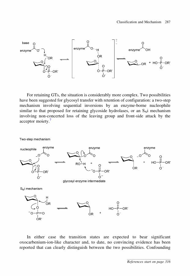
For retaining GTs, the situation is considerably more complex. Two possibilities
have been suggested for glycosyl transfer with retention of configuration: a two-step
mechanism involving sequential inversions by an enzyme-borne nucleophile
similar to that proposed for retaining glycoside hydrolases, or an SNi mechanism
involving non-concerted loss of the leaving group and front-side attack by the
acceptor moiety.7
In either case the transition states are expected to bear significant
oxocarbenium-ion-like character and, to date, no convincing evidence has been
reported that can clearly distinguish between the two possibilities. Confounding
Classification and Mechanism 287
References start on page 316
the situation, crystal structures of retaining GTs are ambiguous in identifying
possible candidate nucleophiles, with some lacking any suitable nucleophile
situated close enough to the glycosyl donor to be able to form a covalent
intermediate. Analysis of some structures has identified possible candidates,
which mutagenesis studies have supported;8 however, in other cases,
mutagenesis of the candidate residue to a non-nucleophilic amino acid has not
resulted in the expected, dramatic loss in catalytic prowess.7,9 Attempts to trap
covalent intermediates on wild-type GTs have proven unsuccessful;10 in one case,
mutation of the proposed aspartate nucleophile to a non-nuclophilic asparagine
residue resulted in the formation of a covalently labelled enzyme bearing a
glycosyl moiety on yet another carboxylate residue.9 The technique of chemical
rescue, whereby a small exogenous nucleophile is used to reactivate an inactive
enzyme mutant, was applied to an inactive retaining galactosyltransferase mutant
lacking the proposed glutamate nucleophile and led to a more than 100-fold
increase in activity and the formation of b-D-galactosyl azide.11 This result was
taken to support a double-displacement mechanism but does not definitively
exclude the SNi mechanism.
Detailed kinetic analyses have been performed on many retaining and
inverting GTs. Generally, ordered kinetic mechanisms have been seen with
either the glycosyl donor or the glycosyl acceptor binding to the enzyme first,
followed by a catalytic step, and then either the (di)phosphonucleoside or
glycosylated product departing from the active site first.12 For example, the
UDP-xylose-dependent core protein b-xylosyltransferase operates by way of an
ordered single displacement with UDP-xylose as the leading substrate and
the xylosylated peptide as the first released product.13 By contrast, OleD, a
UDP-glucose-dependent b-glucosyltransferase from Streptomyces antibioticus,
operates through an ordered mechanism where the acceptor, the macrolide
lankamycin, binds before UDP-glucose, and UDP is released prior to b-glucosylated
lankamycin.14
Many GTs utilize divalent metal ions such as Mn2þ and Mg2þ as cofactors.
These metals are found mainly in GTs that are diphosphonucleoside dependent, and
an analysis by X-ray crystallography has shown that the metal ion is coordinated to
an oxygen of each of the two phosphate groups, as well as to side-chain carboxyl
groups of the protein. Much has been made of the so-called DXD amino acid
sequence as an identifier of GTs, where the aspartate residues of this sequence are
presumed to comprise the metal-binding residues of the active site;15 in this respect,
it is to be noted that no part of the DXD motif is invariant among GTs and that
this motif is present in more than 50% of all protein sequences examined.16 More-
over, it is emphasized that many GTs are metal-ion independent, and thus do not
bind metals at the active site.4
288 8 Glycosyltransferases
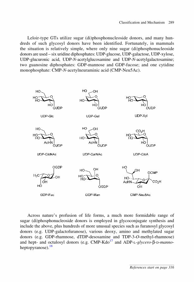
Leloir-type GTs utilize sugar (di)phosphonucleoside donors, and many hun-
dreds of such glycosyl donors have been identified. Fortunately, in mammals
the situation is relatively simple, where only nine sugar (di)phosphonucleoside
donors are used – six uridine diphosphates: UDP-glucose, UDP-galactose, UDP-xylose,
UDP-glucuronic acid, UDP-N-acetylglucosamine and UDP-N-acetylgalactosamine;
two guanosine diphosphates: GDP-mannose and GDP-fucose; and one cytidine
monophosphate: CMP-N-acetylneuraminic acid (CMP-Neu5Ac).
Across nature’s profusion of life forms, a much more formidable range of
sugar (di)phosphonucleoside donors is employed in glycoconjugate synthesis and
include the above, plus hundreds of more unusual species such as furanosyl glycosyl
donors (e.g. UDP-galactofuranose), various deoxy, amino and methylated sugar
donors (e.g. GDP-rhamnose, dTDP-desosamine and TDP-3-O-methyl-rhamnose)
and hept- and octulosyl donors (e.g. CMP-Kdo17 and ADP-L-glycero-b-D-manno-
heptopyranose).18
Classification and Mechanism 289
References start on page 316
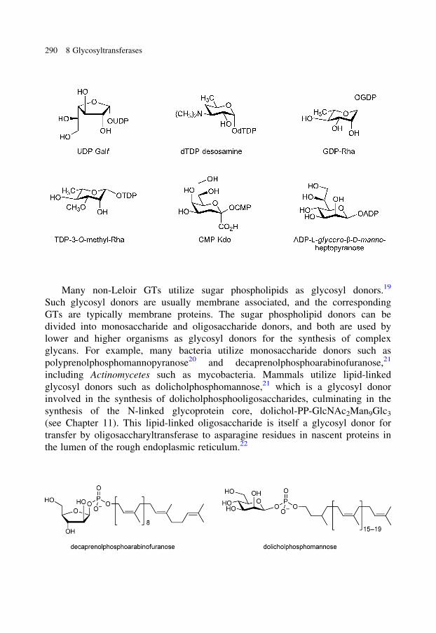
Many non-Leloir GTs utilize sugar phospholipids as glycosyl donors.19
Such glycosyl donors are usually membrane associated, and the corresponding
GTs are typically membrane proteins. The sugar phospholipid donors can be
divided into monosaccharide and oligosaccharide donors, and both are used by
lower and higher organisms as glycosyl donors for the synthesis of complex
glycans. For example, many bacteria utilize monosaccharide donors such as
polyprenolphosphomannopyranose20 and decaprenolphosphoarabinofuranose,21
including Actinomycetes such as mycobacteria. Mammals utilize lipid-linked
glycosyl donors such as dolicholphosphomannose,21 which is a glycosyl donor
involved in the synthesis of dolicholphosphooligosaccharides, culminating in the
synthesis of the N-linked glycoprotein core, dolichol-PP-GlcNAc2Man9Glc3
(see Chapter 11). This lipid-linked oligosaccharide is itself a glycosyl donor for
transfer by oligosaccharyltransferase to asparagine residues in nascent proteins in
the lumen of the rough endoplasmic reticulum.22
290 8 Glycosyltransferases
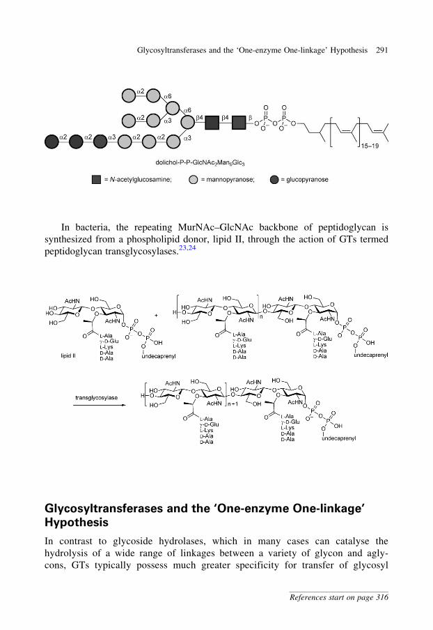
In bacteria, the repeating MurNAc–GlcNAc backbone of peptidoglycan is
synthesized from a phospholipid donor, lipid II, through the action of GTs termed
peptidoglycan transglycosylases.23,24
Glycosyltransferases and the ‘One-enzyme One-linkage’
Hypothesis
In contrast to glycoside hydrolases, which in many cases can catalyse the
hydrolysis of a wide range of linkages between a variety of glycon and agly-
cons, GTs typically possess much greater specificity for transfer of glycosyl
Glycosyltransferases and the ‘One-enzyme One-linkage’ Hypothesis 291
References start on page 316
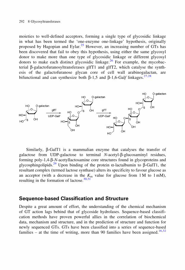
moieties to well-defined acceptors, forming a single type of glycosidic linkage
in what has been termed the ‘one-enzyme one-linkage’ hypothesis, originally
proposed by Hagopian and Eylar.25 However, an increasing number of GTs has
been discovered that fail to obey this hypothesis, using either the same glycosyl
donor to make more than one type of glycosidic linkage or different glycosyl
donors to make each distinct glycosidic linkage.26 For example, the mycobac-
terial b-galactofuranosyltransferases glfT1 and glfT2, which catalyse the synth-
esis of the galactofuranose glycan core of cell wall arabinogalactan, are
bifunctional and can synthesize both b-1,5 and b-1,6-Galf linkages.27,28
Similarly, b-GalT1 is a mammalian enzyme that catalyses the transfer of
galactose from UDP-galactose to terminal N-acetyl-b-glucosaminyl residues,
forming poly-1,4-b-N-acetyllactosamine core structures found in glycoproteins and
glycosphingolipids.29 Upon binding of the protein a-lactalbumin to b-GalT1, the
resultant complex (termed lactose synthase) alters its specificity to favour glucose as
an acceptor (with a decrease in the Km value for glucose from 1 M to 1 mM),
resulting in the formation of lactose.30,31
Sequence-based Classification and Structure
Despite a great amount of effort, the understanding of the chemical mechanism
of GT action lags behind that of glycoside hydrolases. Sequence-based classifi-
cation methods have proven powerful allies in the correlation of biochemical
data, mechanism and structure, and in the prediction of structure and function for
newly sequenced GTs. GTs have been classified into a series of sequence-based
families – at the time of writing, more than 90 families have been assigned.16,32
292 8 Glycosyltransferases
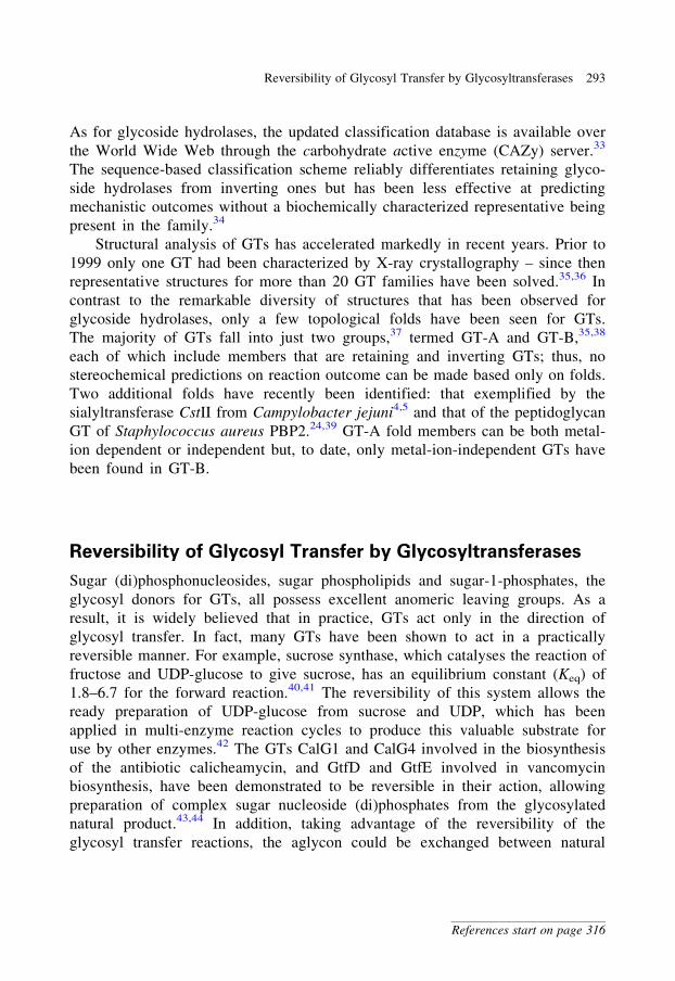
As for glycoside hydrolases, the updated classification database is available over
the World Wide Web through the carbohydrate active enzyme (CAZy) server.33
The sequence-based classification scheme reliably differentiates retaining glyco-
side hydrolases from inverting ones but has been less effective at predicting
mechanistic outcomes without a biochemically characterized representative being
present in the family.34
Structural analysis of GTs has accelerated markedly in recent years. Prior to
1999 only one GT had been characterized by X-ray crystallography – since then
representative structures for more than 20 GT families have been solved.35,36 In
contrast to the remarkable diversity of structures that has been observed for
glycoside hydrolases, only a few topological folds have been seen for GTs.
The majority of GTs fall into just two groups,37 termed GT-A and GT-B,35,38
each of which include members that are retaining and inverting GTs; thus, no
stereochemical predictions on reaction outcome can be made based only on folds.
Two additional folds have recently been identified: that exemplified by the
sialyltransferase CstII from Campylobacter jejuni4,5 and that of the peptidoglycan
GT of Staphylococcus aureus PBP2.24,39 GT-A fold members can be both metal-
ion dependent or independent but, to date, only metal-ion-independent GTs have
been found in GT-B.
Reversibility of Glycosyl Transfer by Glycosyltransferases
Sugar (di)phosphonucleosides, sugar phospholipids and sugar-1-phosphates, the
glycosyl donors for GTs, all possess excellent anomeric leaving groups. As a
result, it is widely believed that in practice, GTs act only in the direction of
glycosyl transfer. In fact, many GTs have been shown to act in a practically
reversible manner. For example, sucrose synthase, which catalyses the reaction of
fructose and UDP-glucose to give sucrose, has an equilibrium constant (Keq) of
1.8–6.7 for the forward reaction.40,41 The reversibility of this system allows the
ready preparation of UDP-glucose from sucrose and UDP, which has been
applied in multi-enzyme reaction cycles to produce this valuable substrate for
use by other enzymes.42 The GTs CalG1 and CalG4 involved in the biosynthesis
of the antibiotic calicheamycin, and GtfD and GtfE involved in vancomycin
biosynthesis, have been demonstrated to be reversible in their action, allowing
preparation of complex sugar nucleoside (di)phosphates from the glycosylated
natural product.43,44 In addition, taking advantage of the reversibility of the
glycosyl transfer reactions, the aglycon could be exchanged between natural
Reversibility of Glycosyl Transfer by Glycosyltransferases 293
References start on page 316
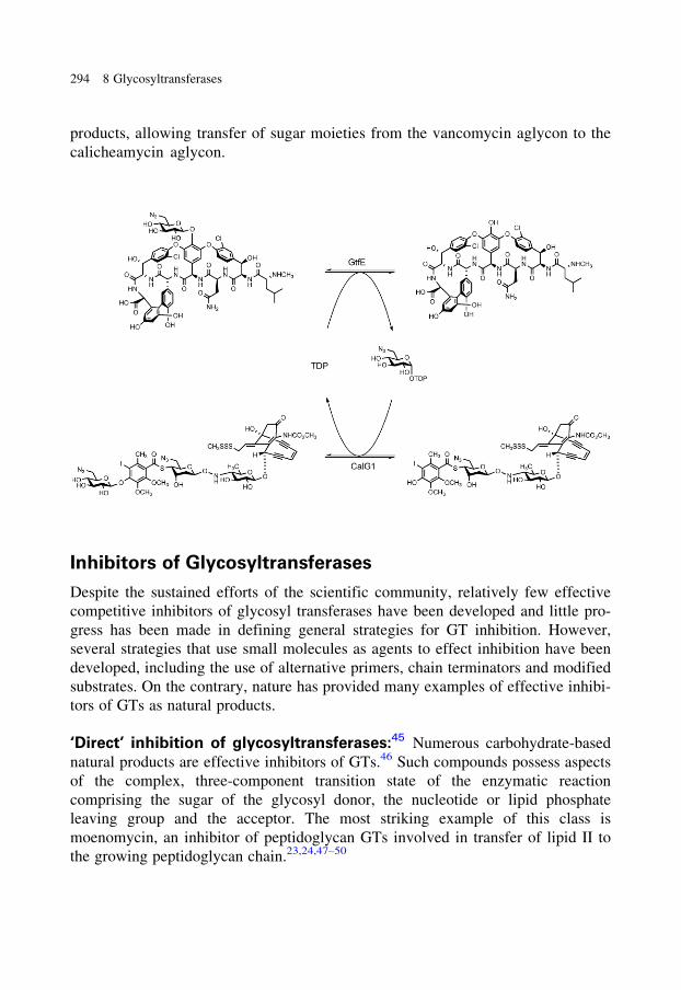
products, allowing transfer of sugar moieties from the vancomycin aglycon to the
calicheamycin aglycon.
Inhibitors of Glycosyltransferases
Despite the sustained efforts of the scientific community, relatively few effective
competitive inhibitors of glycosyl transferases have been developed and little pro-
gress has been made in defining general strategies for GT inhibition. However,
several strategies that use small molecules as agents to effect inhibition have been
developed, including the use of alternative primers, chain terminators and modified
substrates. On the contrary, nature has provided many examples of effective inhibi-
tors of GTs as natural products.
‘Direct’ inhibition of glycosyltransferases:45 Numerous carbohydrate-based
natural products are effective inhibitors of GTs.46 Such compounds possess aspects
of the complex, three-component transition state of the enzymatic reaction
comprising the sugar of the glycosyl donor, the nucleotide or lipid phosphate
leaving group and the acceptor. The most striking example of this class is
moenomycin, an inhibitor of peptidoglycan GTs involved in transfer of lipid II to
the growing peptidoglycan chain.23,24,47–50
294 8 Glycosyltransferases
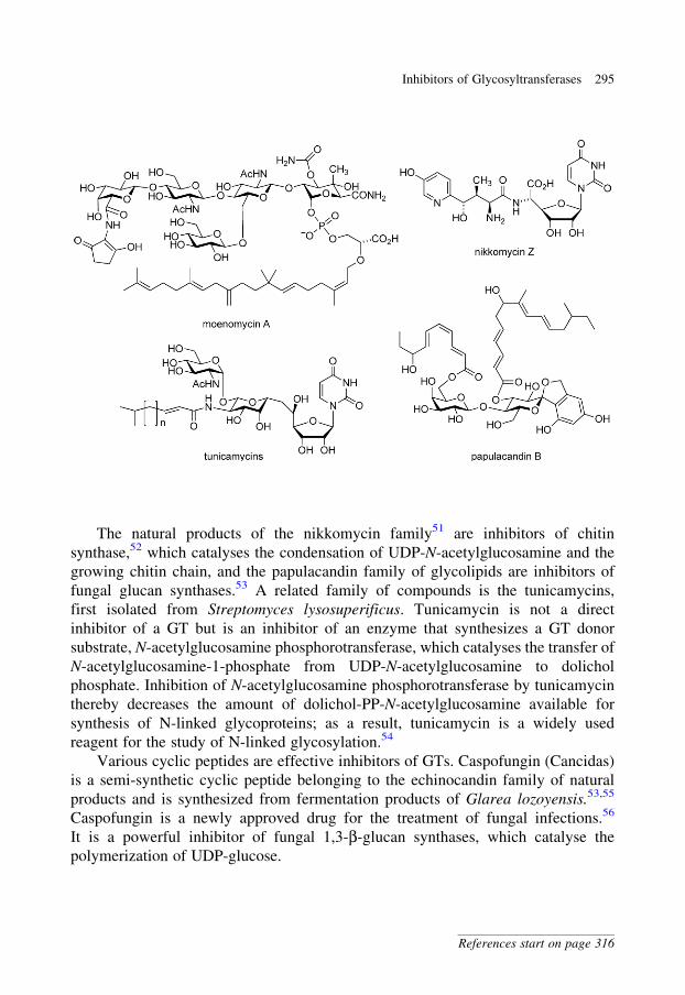
The natural products of the nikkomycin family51 are inhibitors of chitin
synthase,52 which catalyses the condensation of UDP-N-acetylglucosamine and the
growing chitin chain, and the papulacandin family of glycolipids are inhibitors of
fungal glucan synthases.53 A related family of compounds is the tunicamycins,
first isolated from Streptomyces lysosuperificus. Tunicamycin is not a direct
inhibitor of a GT but is an inhibitor of an enzyme that synthesizes a GT donor
substrate, N-acetylglucosamine phosphorotransferase, which catalyses the transfer of
N-acetylglucosamine-1-phosphate from UDP-N-acetylglucosamine to dolichol
phosphate. Inhibition of N-acetylglucosamine phosphorotransferase by tunicamycin
thereby decreases the amount of dolichol-PP-N-acetylglucosamine available for
synthesis of N-linked glycoproteins; as a result, tunicamycin is a widely used
reagent for the study of N-linked glycosylation.54
Various cyclic peptides are effective inhibitors of GTs. Caspofungin (Cancidas)
is a semi-synthetic cyclic peptide belonging to the echinocandin family of natural
products and is synthesized from fermentation products of Glarea lozoyensis.53,55
Caspofungin is a newly approved drug for the treatment of fungal infections.56
It is a powerful inhibitor of fungal 1,3-b-glucan synthases, which catalyse the
polymerization of UDP-glucose.
Inhibitors of Glycosyltransferases 295
References start on page 316
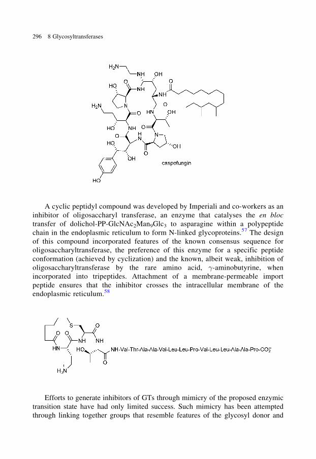
A cyclic peptidyl compound was developed by Imperiali and co-workers as an
inhibitor of oligosaccharyl transferase, an enzyme that catalyses the en bloc
transfer of dolichol-PP-GlcNAc2Man9Glc3 to asparagine within a polypeptide
chain in the endoplasmic reticulum to form N-linked glycoproteins.57 The design
of this compound incorporated features of the known consensus sequence for
oligosaccharyltransferase, the preference of this enzyme for a specific peptide
conformation (achieved by cyclization) and the known, albeit weak, inhibition of
oligosaccharyltransferase by the rare amino acid, �-aminobutyrine, when
incorporated into tripeptides. Attachment of a membrane-permeable import
peptide ensures that the inhibitor crosses the intracellular membrane of the
endoplasmic reticulum.58
Efforts to generate inhibitors of GTs through mimicry of the proposed enzymic
transition state have had only limited success. Such mimicry has been attempted
through linking together groups that resemble features of the glycosyl donor and
296 8 Glycosyltransferases
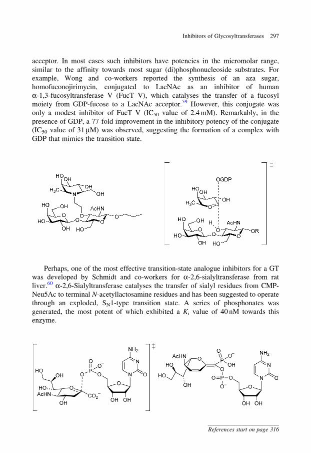
acceptor. In most cases such inhibitors have potencies in the micromolar range,
similar to the affinity towards most sugar (di)phosphonucleoside substrates. For
example, Wong and co-workers reported the synthesis of an aza sugar,
homofuconojirimycin, conjugated to LacNAc as an inhibitor of human
a-1,3-fucosyltransferase V (FucT V), which catalyses the transfer of a fucosyl
moiety from GDP-fucose to a LacNAc acceptor.59 However, this conjugate was
only a modest inhibitor of FucT V (IC50 value of 2.4 mM). Remarkably, in the
presence of GDP, a 77-fold improvement in the inhibitory potency of the conjugate
(IC50 value of 31 mM) was observed, suggesting the formation of a complex with
GDP that mimics the transition state.
Perhaps, one of the most effective transition-state analogue inhibitors for a GT
was developed by Schmidt and co-workers for a-2,6-sialyltransferase from rat
liver.60 a-2,6-Sialyltransferase catalyses the transfer of sialyl residues from CMP-
Neu5Ac to terminal N-acetyllactosamine residues and has been suggested to operate
through an exploded, SN1-type transition state. A series of phosphonates was
generated, the most potent of which exhibited a Ki value of 40 nM towards this
enzyme.
Inhibitors of Glycosyltransferases 297
References start on page 316

Hindsgaul and co-workers have described the design of a bisubstrate analogue
comprised of a conjugate of the acceptor sugar and the diphosphonucleoside portion
of the donor. The conjugate was an inhibitor (Ki value of 2.3 mM) of porcine
a-1,2-fucosyltransferase, which transfers a fucosyl moiety from GDP-fucose
to b-galactosides.61
Hashimoto and co-workers have reported a bisubstrate analogue that is again a
conjugate of a sugar diphosphonucleoside and the glycosyl acceptor. This compound
was found to be a reasonably potent inhibitor (Ki value of 1.35 mM relative to the
acceptor, and Ki value of 3.3 mM relative to the donor, UDP-galactose) of
b-1,4-galactosyltransferase from bovine milk.62
Lowary and Hindsgaul have reported the synthesis of a powerful inhibitor of
blood group A GT obtained by simple modification of the acceptor substrate.63
Blood group A GT catalyses the synthesis of the blood group A determinant,
a-GalNAc-(1,3)-[a-Fuc-(1,2)-]-b-Gal-OR, by transfer of an N-acetylgalactosaminyl
residue from UDP-N-acetylgalactosamine to the H determinant, a-Fuc-(1,2)-b-Gal-OR.
a-Fuc-(1,2)-b-Gal-O-octyl is an effective artificial substrate for this GT, and
modification of the acceptor hydroxyl to an amino group results in a powerful
inhibitor (Ki value of 200 nM) of this GT. This compound was subsequently
shown to be effective in inhibiting blood group A determinant synthesis in cell
culture.64
298 8 Glycosyltransferases
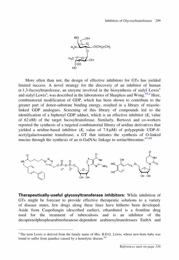
More often than not, the design of effective inhibitors for GTs has yielded
limited success. A novel strategy for the discovery of an inhibitor of human
a-1,3-fucosyltransferase, an enzyme involved in the biosynthesis of sialyl Lewisx
and sialyl Lewisa, was described in the laboratories of Sharpless and Wong.65,a Here,
combinatorial modification of GDP, which has been shown to contribute to the
greater part of donor-substrate binding energy, resulted in a library of triazole-
linked GDP analogues. Screening of this library of compounds led to the
identification of a biphenyl GDP adduct, which is an effective inhibitor (Ki value
of 62 nM) of the target fucosyltransferase. Similarly, Bertozzi and co-workers
reported the synthesis of a targeted combinatorial library of uridine derivatives that
yielded a uridine-based inhibitor (Ki value of 7.8 mM) of polypeptide UDP-N-
acetylgalactosamine transferase, a GT that initiates the synthesis of O-linked
mucins through the synthesis of an a-GalNAc linkage to serine/threonine.67,68
Therapeutically-useful glycosyltransferase inhibitors: While inhibition of
GTs might be forecast to provide effective therapeutic solutions to a variety
of disease states, few drugs along these lines have hitherto been developed.
Aside from Caspofungin (described earlier), ethambutol is a frontline drug
used for the treatment of tuberculosis and is an inhibitor of the
decaprenolphosphoarabinofuranose-dependent arabinosyltransferases EmbA and
a The term Lewis is derived from the family name of Mrs. H.D.G. Lewis, whose new-born baby was
found to suffer from jaundice caused by a hemolytic disease.66
Inhibitors of Glycosyltransferases 299
References start on page 316
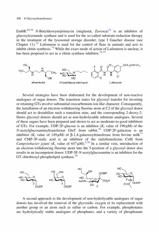
EmbB.69,70 N-Butyldeoxynojirimycin (miglustat, Zavesca)71 is an inhibitor of
glucosylceramide synthase and is used for the so-called substrate-reduction therapy
in the treatment of the lysosomal storage disorder, type I Gaucher disease (see
Chapter 11).72 Lufenuron is used for the control of fleas in animals and acts to
inhibit chitin synthesis.73 While the exact mode of action of Lufenuron is unclear, it
has been proposed to act as a chitin synthase inhibitor.74,75
Several strategies have been elaborated for the development of non-reactive
analogues of sugar donors. The transition states for glycosyl transfer for inverting
or retaining GTs involve substantial oxocarbenium-ion-like character. Consequently,
the installation of an electron-withdrawing fluorine atom at C2 of the glycosyl donor
should act to destabilize such a transition state, and the corresponding 2-deoxy-2-
fluoro glycosyl donors should act as non-hydrolysable substrate analogues. Several
of these sugars have been prepared and shown to act as moderate-to-good inhibitors
of GTs. For example, UDP-2F-glucose is an inhibitor (Ki value of 190 mM) of the
N-acetylglucosaminyltransferase GlnT from rabbit,76 UDP-2F-galactose is an
inhibitor (Ki value of 149 mM) of b-1,4-galactosyltransferase from bovine milk77
and CMP-3F-sialic acid is an inhibitor of the sialyltransferase CstII from
Campylobacter jejuni (Ki value of 657 mM).5,78 In a similar vein, introduction of
an electron-withdrawing fluorine atom into the 5-position of a glycosyl donor also
results in an incompetent donor; UDP-5F-N-acetylglucosamine is an inhibitor for the
GT chitobiosyl-phospholipid synthase.79
A second approach to the development of non-hydrolysable analogues of sugar
donors has involved the removal of the glycosidic oxygen or its replacement with
another group or an atom such as sulfur or carbon. For example, phosphonates
are hydrolytically stable analogues of phosphates, and a variety of phosphonate
300 8 Glycosyltransferases
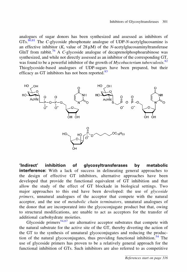
analogues of sugar donors has been synthesized and assessed as inhibitors of
GTs.80,81 The C-glycoside phosphonate analogue of UDP-N-acetylglucosamine is
an effective inhibitor (Ki value of 28 mM) of the N-acetylglucosaminyltransferase
GlnT from rabbit.76 A C-glycoside analogue of decaprenolphosphoarabinose was
synthesized, and while not directly assessed as an inhibitor of the corresponding GT,
was found to be a powerful inhibitor of the growth of Mycobacterium tuberculosis.82
Thioglycoside-based analogues of UDP-sugars have been prepared, but their
efficacy as GT inhibitors has not been reported.83
‘Indirect’ inhibition of glycosyltransferases by metabolic
interference: With a lack of success in delineating general approaches to
the design of effective GT inhibitors, alternative approaches have been
developed that provide the functional equivalent of GT inhibition and that
allow the study of the effect of GT blockade in biological settings. Two
major approaches to this end have been developed: the use of glycoside
primers, unnatural analogues of the acceptor that compete with the natural
acceptor, and the use of metabolic chain terminators, unnatural analogues of
the donor that are incorporated into the glycoconjugate product but that, owing
to structural modifications, are unable to act as acceptors for the transfer of
additional carbohydrate moieties.
Glycoside primers54,67 are alternative acceptor substrates that compete with
the natural substrate for the active site of the GT, thereby diverting the action of
the GT to the synthesis of unnatural glycoconjugates and reducing the produc-
tion of the natural glycoconjugates, thus providing functional inhibition.54 The
use of glycoside primers has proven to be a relatively general approach for the
functional inhibition of GTs. Such inhibitors are also referred to as competitive
Inhibitors of Glycosyltransferases 301
References start on page 316
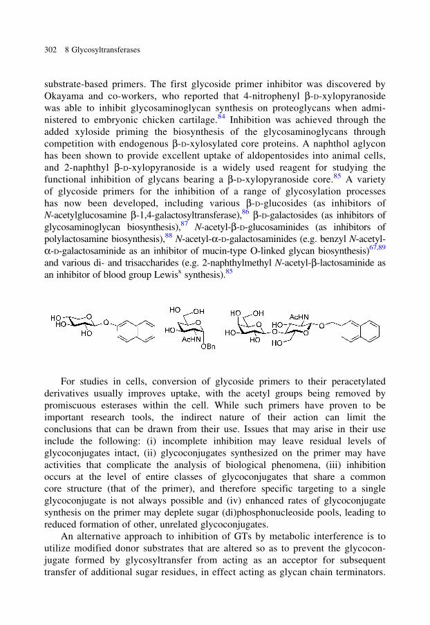
substrate-based primers. The first glycoside primer inhibitor was discovered by
Okayama and co-workers, who reported that 4-nitrophenyl b-D-xylopyranoside
was able to inhibit glycosaminoglycan synthesis on proteoglycans when admi-
nistered to embryonic chicken cartilage.84 Inhibition was achieved through the
added xyloside priming the biosynthesis of the glycosaminoglycans through
competition with endogenous b-D-xylosylated core proteins. A naphthol aglycon
has been shown to provide excellent uptake of aldopentosides into animal cells,
and 2-naphthyl b-D-xylopyranoside is a widely used reagent for studying the
functional inhibition of glycans bearing a b-D-xylopyranoside core.85 A variety
of glycoside primers for the inhibition of a range of glycosylation processes
has now been developed, including various b-D-glucosides (as inhibitors of
N-acetylglucosamine b-1,4-galactosyltransferase),86 b-D-galactosides (as inhibitors of
glycosaminoglycan biosynthesis),87 N-acetyl-b-D-glucosaminides (as inhibitors of
polylactosamine biosynthesis),88 N-acetyl-a-D-galactosaminides (e.g. benzyl N-acetyl-
a-D-galactosaminide as an inhibitor of mucin-type O-linked glycan biosynthesis)67,89
and various di- and trisaccharides (e.g. 2-naphthylmethyl N-acetyl-b-lactosaminide as
an inhibitor of blood group Lewisx synthesis).85
For studies in cells, conversion of glycoside primers to their peracetylated
derivatives usually improves uptake, with the acetyl groups being removed by
promiscuous esterases within the cell. While such primers have proven to be
important research tools, the indirect nature of their action can limit the
conclusions that can be drawn from their use. Issues that may arise in their use
include the following: (i) incomplete inhibition may leave residual levels of
glycoconjugates intact, (ii) glycoconjugates synthesized on the primer may have
activities that complicate the analysis of biological phenomena, (iii) inhibition
occurs at the level of entire classes of glycoconjugates that share a common
core structure (that of the primer), and therefore specific targeting to a single
glycoconjugate is not always possible and (iv) enhanced rates of glycoconjugate
synthesis on the primer may deplete sugar (di)phosphonucleoside pools, leading to
reduced formation of other, unrelated glycoconjugates.
An alternative approach to inhibition of GTs by metabolic interference is to
utilize modified donor substrates that are altered so as to prevent the glycocon-
jugate formed by glycosyltransfer from acting as an acceptor for subsequent
transfer of additional sugar residues, in effect acting as glycan chain terminators.
302 8 Glycosyltransferases
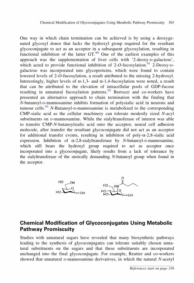
One way in which chain termination can be achieved is by using a deoxyge-
nated glycosyl donor that lacks the hydroxyl group required for the resultant
glycoconjugate to act as an acceptor in a subsequent glycosylation, resulting in
functional inhibition of the latter GT.90 One of the earliest examples of this
approach was the supplementation of liver cells with ‘2-deoxy-D-galactose’,
which acted to provide functional inhibition of 2-O-fucosylation.91 2-Deoxy-D-
galactose was incorporated into glycoproteins, which were found to contain
lowered levels of 2-O-fucosylation, a result attributed to the missing 2-hydroxyl.
Interestingly, higher levels of a-1,3- and a-1,4-fucosylation were noted, a result
that can be attributed to the elevation of intracellular pools of GDP-fucose
resulting in unnatural fucosylation patterns.92 Bertozzi and co-workers have
presented an alternative approach to chain termination with the finding that
N-butanoyl-D-mannosamine inhibits formation of polysialic acid in neurons and
tumour cells.93 N-Butanoyl-D-mannosamine is metabolized to the corresponding
CMP-sialic acid as the cellular machinery can tolerate modestly sized N-acyl
substituents on D-mannosamine. While the sialyltransferase of interest was able
to transfer CMP-N-butanoylsialic acid onto the acceptor, neural cell adhesion
molecule, after transfer the resultant glycoconjugate did not act as an acceptor
for additional transfer events, resulting in inhibition of poly-a-2,8-sialic acid
expression. Inhibition of a-2,8-sialyltransferase by N-butanoyl-D-mannosamine,
which still bears the hydroxyl group required to act as acceptor once
incorporated into a glycoconjugate, likely results from a lack of tolerance by
the sialyltransferase of the sterically demanding N-butanoyl group when found in
the acceptor.
Chemical Modification of Glycoconjugates Using Metabolic
Pathway Promiscuity
Studies with unnatural sugars have revealed that many biosynthetic pathways
leading to the synthesis of glycoconjugates can tolerate suitably chosen unna-
tural substituents on the sugars and that these substituents are incorporated
unchanged into the final glycoconjugate. For example, Reutter and co-workers
showed that unnatural D-mannosamine derivatives, in which the natural N-acetyl
Chemical Modification of Glycoconjugates Using Metabolic Pathway Promiscuity 303
References start on page 316

group was replaced by N-propanoyl, N-butanoyl or N-pentanoyl, are converted
into the corresponding N-acylsialoses and incorporated into sialic acid–containing
glycoconjugates in cell culture and in rats.94,95 An elegant extension of this
observation is to incorporate metabolically silent functional groups that, following
incorporation into glycoconjugates, can react selectively with reagents to enable
the detection or isolation of the chemically labelled glycoconjugate. This approach
has been termed ‘metabolic labelling of glycans’. Of particular importance to this
approach is the identification of so-called bio-orthogonal conjugations, reactions
that result in the ligation of two species with a chemoselectivity that allows them
to be performed in a biological context.96,97 Several functional groups have been
utilized to achieve metabolic labelling of glycans and are well tolerated by
entire metabolic pathways leading to the glycoconjugates, including azido,98
thiol,99 keto100 and alkynyl groups.101 Of this set of functional groups, the
azido group is the premier choice, being distinguished by its lack of reactivity
in cellular systems, its rarity in biological settings and its exquisite specificity
for clean, high-yielding reactions with suitable reactive probes. For example,
N-azidoacetyl-D-mannosamine can be converted into azide-modified cell-surface
sialic acid residues by way of the respective biosynthesis pathway.98,102 The azido
group is not altered as it passes through the biosynthetic pathway but, once in
place, can react with azide-specific reagents such as bifunctional phosphines
bearing electrophilic traps (the Staudinger ligation), terminal alkynes in the
presence of copper(I) catalysis (the CuAAC reaction) and strain-promoted
Huisgen [3þ 2] cycloaddition with cycloalkynes.103–106 By such ligation reac-
tions, fluorescent dyes and epitope tags, including biotin and antigenic peptides,
can be conjugated to facilitate detection and enable enrichment/isolation
by immuno-precipitation.
304 8 Glycosyltransferases
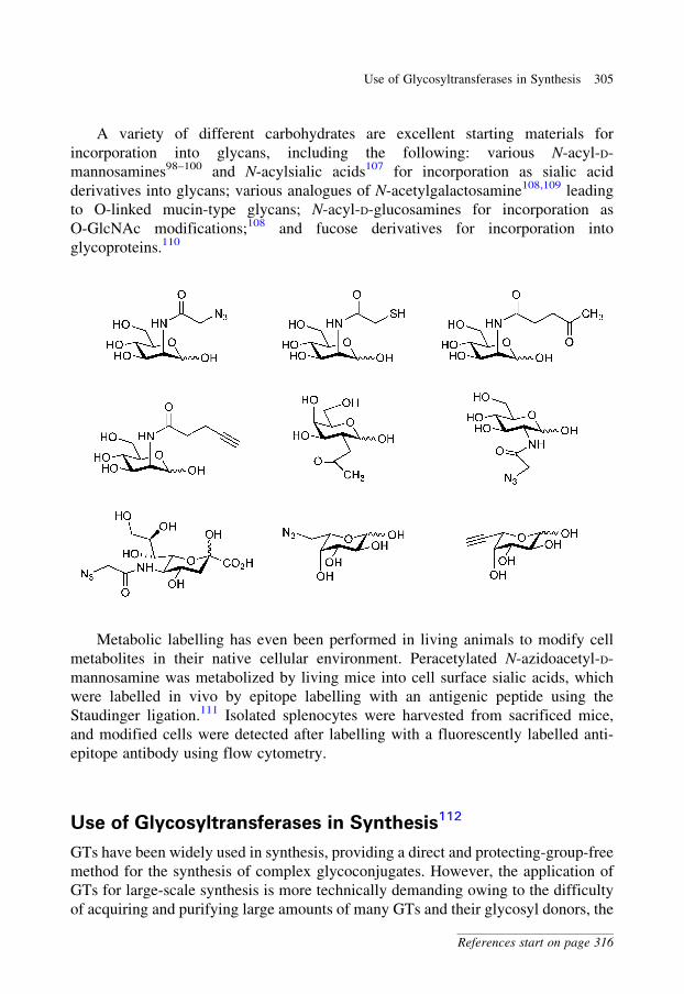
A variety of different carbohydrates are excellent starting materials for
incorporation into glycans, including the following: various N-acyl-D-
mannosamines98–100 and N-acylsialic acids107 for incorporation as sialic acid
derivatives into glycans; various analogues of N-acetylgalactosamine108,109 leading
to O-linked mucin-type glycans; N-acyl-D-glucosamines for incorporation as
O-GlcNAc modifications;108 and fucose derivatives for incorporation into
glycoproteins.110
Metabolic labelling has even been performed in living animals to modify cell
metabolites in their native cellular environment. Peracetylated N-azidoacetyl-D-
mannosamine was metabolized by living mice into cell surface sialic acids, which
were labelled in vivo by epitope labelling with an antigenic peptide using the
Staudinger ligation.111 Isolated splenocytes were harvested from sacrificed mice,
and modified cells were detected after labelling with a fluorescently labelled anti-
epitope antibody using flow cytometry.
Use of Glycosyltransferases in Synthesis112
GTs have been widely used in synthesis, providing a direct and protecting-group-free
method for the synthesis of complex glycoconjugates. However, the application of
GTs for large-scale synthesis is more technically demanding owing to the difficulty
of acquiring and purifying large amounts of many GTs and their glycosyl donors, the
Use of Glycosyltransferases in Synthesis 305
References start on page 316
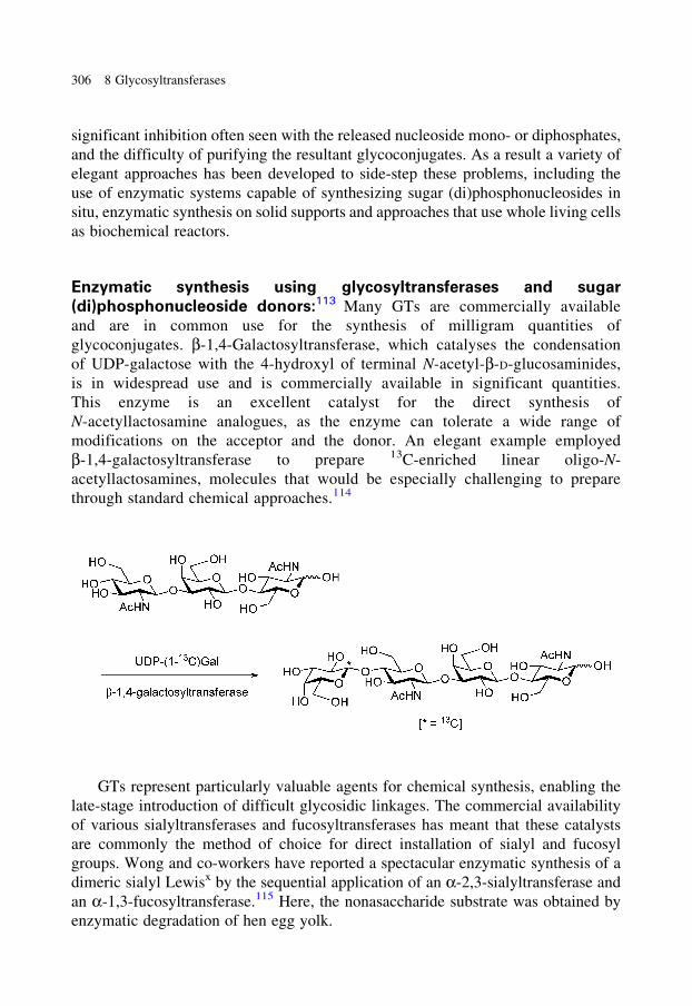
significant inhibition often seen with the released nucleoside mono- or diphosphates,
and the difficulty of purifying the resultant glycoconjugates. As a result a variety of
elegant approaches has been developed to side-step these problems, including the
use of enzymatic systems capable of synthesizing sugar (di)phosphonucleosides in
situ, enzymatic synthesis on solid supports and approaches that use whole living cells
as biochemical reactors.
Enzymatic synthesis using glycosyltransferases and sugar
(di)phosphonucleoside donors:113 Many GTs are commercially available
and are in common use for the synthesis of milligram quantities of
glycoconjugates. b-1,4-Galactosyltransferase, which catalyses the condensation
of UDP-galactose with the 4-hydroxyl of terminal N-acetyl-b-D-glucosaminides,
is in widespread use and is commercially available in significant quantities.
This enzyme is an excellent catalyst for the direct synthesis of
N-acetyllactosamine analogues, as the enzyme can tolerate a wide range of
modifications on the acceptor and the donor. An elegant example employed
b-1,4-galactosyltransferase to prepare 13C-enriched linear oligo-N-
acetyllactosamines, molecules that would be especially challenging to prepare
through standard chemical approaches.114
GTs represent particularly valuable agents for chemical synthesis, enabling the
late-stage introduction of difficult glycosidic linkages. The commercial availability
of various sialyltransferases and fucosyltransferases has meant that these catalysts
are commonly the method of choice for direct installation of sialyl and fucosyl
groups. Wong and co-workers have reported a spectacular enzymatic synthesis of a
dimeric sialyl Lewisx by the sequential application of an a-2,3-sialyltransferase and
an a-1,3-fucosyltransferase.115 Here, the nonasaccharide substrate was obtained by
enzymatic degradation of hen egg yolk.
306 8 Glycosyltransferases
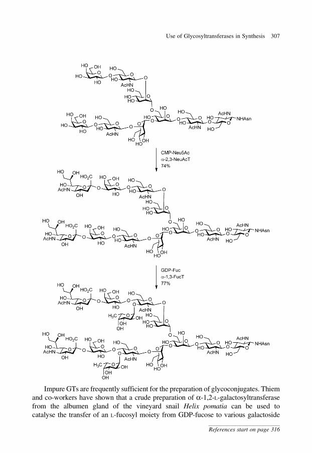
Impure GTs are frequently sufficient for the preparation of glycoconjugates. Thiem
and co-workers have shown that a crude preparation of a-1,2-L-galactosyltransferase
from the albumen gland of the vineyard snail Helix pomatia can be used to
catalyse the transfer of an L-fucosyl moiety from GDP-fucose to various galactoside
Use of Glycosyltransferases in Synthesis 307
References start on page 316
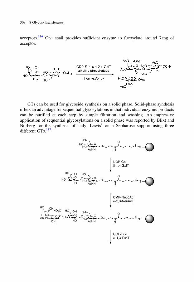
acceptors.116 One snail provides sufficient enzyme to fucosylate around 7 mg of
acceptor.
GTs can be used for glycoside synthesis on a solid phase. Solid-phase synthesis
offers an advantage for sequential glycosylations in that individual enzymic products
can be purified at each step by simple filtration and washing. An impressive
application of sequential glycosylations on a solid phase was reported by Blixt and
Norberg for the synthesis of sialyl Lewisx on a Sepharose support using three
different GTs.117
308 8 Glycosyltransferases
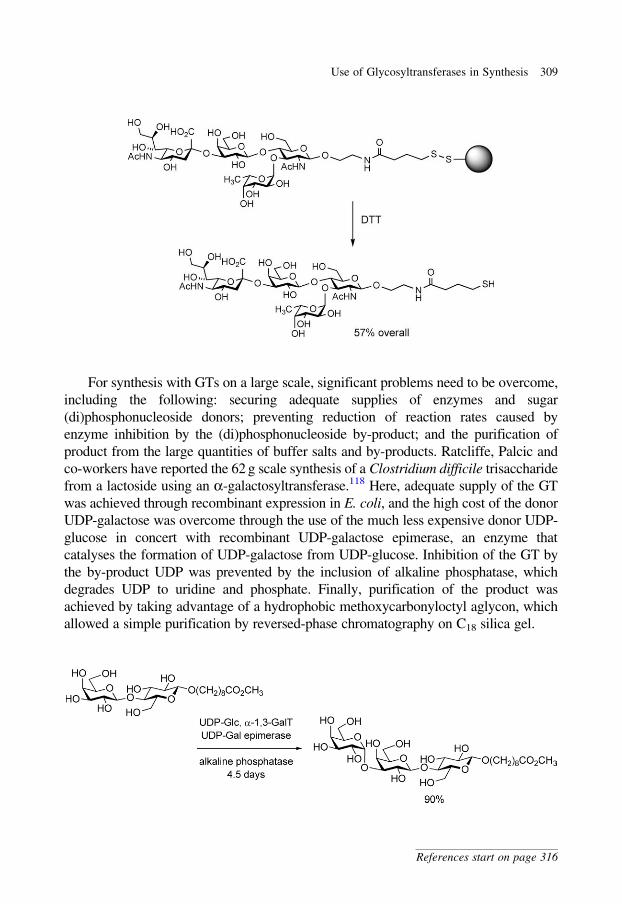
For synthesis with GTs on a large scale, significant problems need to be overcome,
including the following: securing adequate supplies of enzymes and sugar
(di)phosphonucleoside donors; preventing reduction of reaction rates caused by
enzyme inhibition by the (di)phosphonucleoside by-product; and the purification of
product from the large quantities of buffer salts and by-products. Ratcliffe, Palcic and
co-workers have reported the 62 g scale synthesis of a Clostridium difficile trisaccharide
from a lactoside using an a-galactosyltransferase.118 Here, adequate supply of the GT
was achieved through recombinant expression in E. coli, and the high cost of the donor
UDP-galactose was overcome through the use of the much less expensive donor UDP-
glucose in concert with recombinant UDP-galactose epimerase, an enzyme that
catalyses the formation of UDP-galactose from UDP-glucose. Inhibition of the GT by
the by-product UDP was prevented by the inclusion of alkaline phosphatase, which
degrades UDP to uridine and phosphate. Finally, purification of the product was
achieved by taking advantage of a hydrophobic methoxycarbonyloctyl aglycon, which
allowed a simple purification by reversed-phase chromatography on C18 silica gel.
Use of Glycosyltransferases in Synthesis 309
References start on page 316
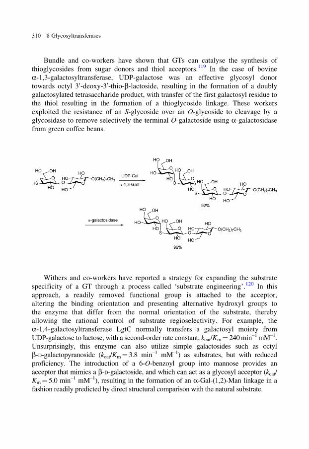
Bundle and co-workers have shown that GTs can catalyse the synthesis of
thioglycosides from sugar donors and thiol acceptors.119 In the case of bovine
a-1,3-galactosyltransferase, UDP-galactose was an effective glycosyl donor
towards octyl 30-deoxy-30-thio-b-lactoside, resulting in the formation of a doubly
galactosylated tetrasaccharide product, with transfer of the first galactosyl residue to
the thiol resulting in the formation of a thioglycoside linkage. These workers
exploited the resistance of an S-glycoside over an O-glycoside to cleavage by a
glycosidase to remove selectively the terminal O-galactoside using a-galactosidase
from green coffee beans.
Withers and co-workers have reported a strategy for expanding the substrate
specificity of a GT through a process called ‘substrate engineering’.120 In this
approach, a readily removed functional group is attached to the acceptor,
altering the binding orientation and presenting alternative hydroxyl groups to
the enzyme that differ from the normal orientation of the substrate, thereby
allowing the rational control of substrate regioselectivity. For example, the
a-1,4-galactosyltransferase LgtC normally transfers a galactosyl moiety from
UDP-galactose to lactose, with a second-order rate constant, kcat/Km = 240 min–1 mM–1.
Unsurprisingly, this enzyme can also utilize simple galactosides such as octyl
b-D-galactopyranoside (kcat/Km = 3.8 min–1 mM–1) as substrates, but with reduced
proficiency. The introduction of a 6-O-benzoyl group into mannose provides an
acceptor that mimics a b-D-galactoside, and which can act as a glycosyl acceptor (kcat/
Km = 5.0 min–1 mM–1), resulting in the formation of an a-Gal-(1,2)-Man linkage in a
fashion readily predicted by direct structural comparison with the natural substrate.
310 8 Glycosyltransferases
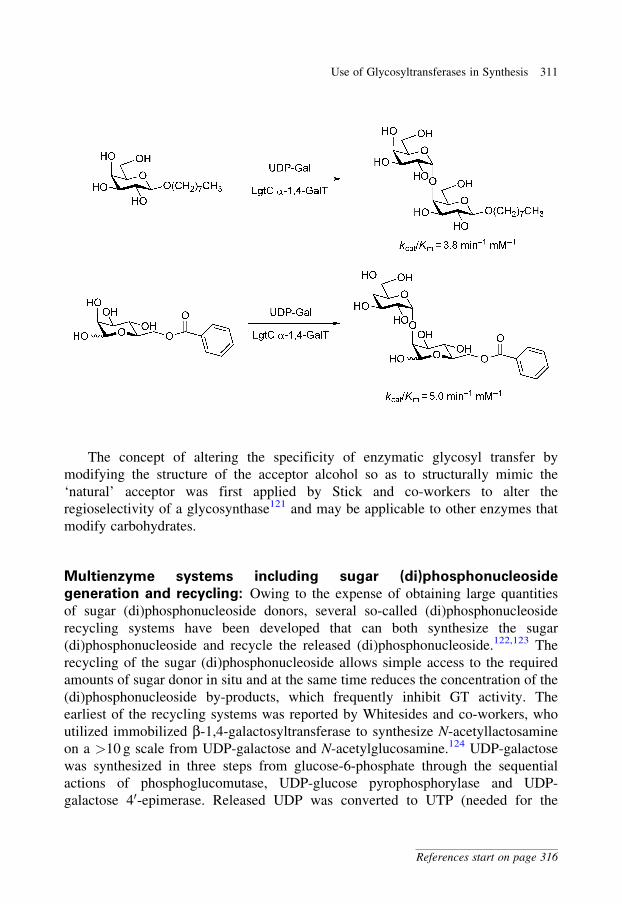
The concept of altering the specificity of enzymatic glycosyl transfer by
modifying the structure of the acceptor alcohol so as to structurally mimic the
‘natural’ acceptor was first applied by Stick and co-workers to alter the
regioselectivity of a glycosynthase121 and may be applicable to other enzymes that
modify carbohydrates.
Multienzyme systems including sugar (di)phosphonucleoside
generation and recycling: Owing to the expense of obtaining large quantities
of sugar (di)phosphonucleoside donors, several so-called (di)phosphonucleoside
recycling systems have been developed that can both synthesize the sugar
(di)phosphonucleoside and recycle the released (di)phosphonucleoside.122,123 The
recycling of the sugar (di)phosphonucleoside allows simple access to the required
amounts of sugar donor in situ and at the same time reduces the concentration of the
(di)phosphonucleoside by-products, which frequently inhibit GT activity. The
earliest of the recycling systems was reported by Whitesides and co-workers, who
utilized immobilized b-1,4-galactosyltransferase to synthesize N-acetyllactosamine
on a >10 g scale from UDP-galactose and N-acetylglucosamine.124 UDP-galactose
was synthesized in three steps from glucose-6-phosphate through the sequential
actions of phosphoglucomutase, UDP-glucose pyrophosphorylase and UDP-
galactose 40-epimerase. Released UDP was converted to UTP (needed for the
Use of Glycosyltransferases in Synthesis 311
References start on page 316
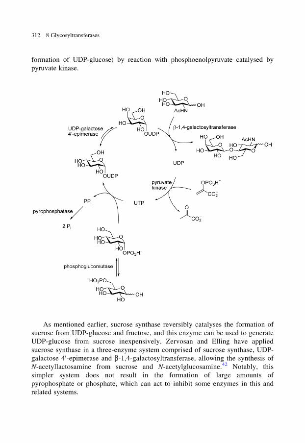
formation of UDP-glucose) by reaction with phosphoenolpyruvate catalysed by
pyruvate kinase.
As mentioned earlier, sucrose synthase reversibly catalyses the formation of
sucrose from UDP-glucose and fructose, and this enzyme can be used to generate
UDP-glucose from sucrose inexpensively. Zervosan and Elling have applied
sucrose synthase in a three-enzyme system comprised of sucrose synthase, UDP-
galactose 40-epimerase and b-1,4-galactosyltransferase, allowing the synthesis of
N-acetyllactosamine from sucrose and N-acetylglucosamine.42 Notably, this
simpler system does not result in the formation of large amounts of
pyrophosphate or phosphate, which can act to inhibit some enzymes in this and
related systems.
312 8 Glycosyltransferases
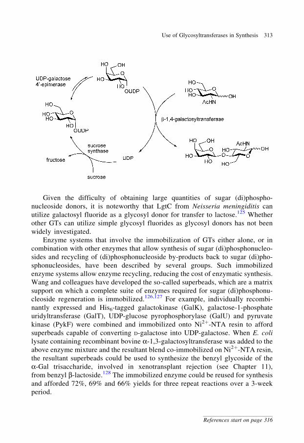
Given the difficulty of obtaining large quantities of sugar (di)phospho-
nucleoside donors, it is noteworthy that LgtC from Neisseria meningiditis can
utilize galactosyl fluoride as a glycosyl donor for transfer to lactose.125 Whether
other GTs can utilize simple glycosyl fluorides as glycosyl donors has not been
widely investigated.
Enzyme systems that involve the immobilization of GTs either alone, or in
combination with other enzymes that allow synthesis of sugar (di)phosphonucleo-
sides and recycling of (di)phosphonucleoside by-products back to sugar (di)pho-
sphonucleosides, have been described by several groups. Such immobilized
enzyme systems allow enzyme recycling, reducing the cost of enzymatic synthesis.
Wang and colleagues have developed the so-called superbeads, which are a matrix
support on which a complete suite of enzymes required for sugar (di)phosphonu-
cleoside regeneration is immobilized.126,127 For example, individually recombi-
nantly expressed and His6-tagged galactokinase (GalK), galactose-1-phosphate
uridyltransferase (GalT), UDP-glucose pyrophosphorylase (GalU) and pyruvate
kinase (PykF) were combined and immobilized onto Ni2þ-NTA resin to afford
superbeads capable of converting D-galactose into UDP-galactose. When E. coli
lysate containing recombinant bovine a-1,3-galactosyltransferase was added to the
above enzyme mixture and the resultant blend co-immobilized on Ni2þ-NTA resin,
the resultant superbeads could be used to synthesize the benzyl glycoside of the
a-Gal trisaccharide, involved in xenotransplant rejection (see Chapter 11),
from benzyl b-lactoside.128 The immobilized enzyme could be reused for synthesis
and afforded 72%, 69% and 66% yields for three repeat reactions over a 3-week
period.
Use of Glycosyltransferases in Synthesis 313
References start on page 316
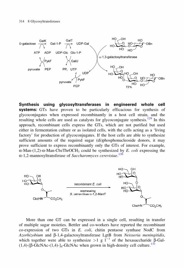
Synthesis using glycosyltransferases in engineered whole cell
systems: GTs have proven to be particularly efficacious for synthesis of
glycoconjugates when expressed recombinantly in a host cell strain, and the
resulting whole cells are used as catalysts for glycoconjugate synthesis.129 In this
approach, recombinant cells express the GTs, which are not purified but used
either in fermentation culture or as isolated cells, with the cells acting as a ‘living
factory’ for production of glycoconjugates. If the host cells are able to synthesize
sufficient amounts of the required sugar (di)phosphonucleoside donors, it may
prove sufficient to express recombinantly only the GTs of interest. For example,
a-Man-(1,2)-a-Man-CbzThrOCH3 could be synthesized by E. coli expressing the
a-1,2-mannosyltransferase of Saccharomyces cerevisiae.130
More than one GT can be expressed in a single cell, resulting in transfer
of multiple sugar moieties. Bettler and co-workers have reported the recombinant
co-expression of two GTs in E. coli, chitin pentaose synthase NodC from
Azorhizobium and b-1,4-galactosyltransferase LgtB from Neisseria meningitidis,
which together were able to synthesize >1 g l�1 of the hexasaccharide b-Gal-
(1,4)-[b-GlcNAc-(1,4)-]4-GlcNAc when grown in high-density cell culture.131
314 8 Glycosyltransferases
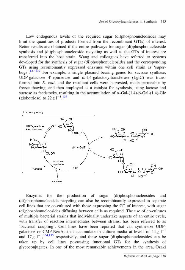
Low endogenous levels of the required sugar (di)phosphonucleosides may
limit the quantities of products formed from the recombinant GT(s) of interest.
Better results are obtained if the entire pathways for sugar (di)phosphonucleoside
synthesis and (di)phosphonucleoside recycling as well as the GTs of interest are
transferred into the host strain. Wang and colleagues have referred to systems
developed for the synthesis of sugar (di)phosphonucleosides and the corresponding
GTs using recombinantly expressed enzymes within one cell strain as ‘super-
bugs’.127,132 For example, a single plasmid bearing genes for sucrose synthase,
UDP-galactose 40-epimerase and a-1,4-galactosyltransferase (LgtC) was trans-
formed into E. coli, and the resultant cells were harvested, made permeable by
freeze thawing, and then employed as a catalyst for synthesis, using lactose and
sucrose as feedstocks, resulting in the accumulation of a-Gal-(1,4)-b-Gal-(1,4)-Glc
(globotriose) to 22 g l�1.133
Enzymes for the production of sugar (di)phosphonucleosides and
(di)phosphonucleoside recycling can also be recombinantly expressed in separate
cell lines that are co-cultured with those expressing the GT of interest, with sugar
(di)phosphonucleosides diffusing between cells as required. The use of co-cultures
of multiple bacterial strains that individually undertake aspects of an entire cycle,
with transfer of reaction intermediates between strains, has been referred to as
‘bacterial coupling’. Cell lines have been reported that can synthesize UDP-
galactose or CMP-NeuAc that accumulate in culture media at levels of 44 g l�1
and 17 g l�1,134,135 respectively, and these sugar (di)phosphonucleosides can be
taken up by cell lines possessing functional GTs for the synthesis of
glycoconjugates. In one of the most remarkable achievements in the area, Ozaki
Use of Glycosyltransferases in Synthesis 315
References start on page 316
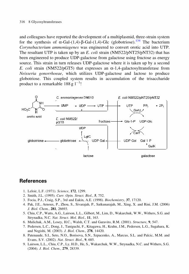
and colleagues have reported the development of a multiplasmid, three-strain system
for the synthesis of a-Gal-(1,4)-b-Gal-(1,4)-Glc (globotriose).134 The bacterium
Corynebacterium ammoniagenes was engineered to convert orotic acid into UTP.
The resultant UTP is taken up by an E. coli strain (NM522/pNT25/pNT32) that has
been engineered to produce UDP-galactose from galactose using fructose as energy
source. This strain in turn releases UDP-galactose where it is taken up by a second
E. coli strain (NM522/pGT5) that expresses an a-1,4-galactosyltransferase from
Neisseria gonorrhoeae, which utilizes UDP-galactose and lactose to produce
globotriose. This coupled system results in accumulation of the trisaccharide
product to a remarkable 188 g l�1!
References
1. Leloir, L.F. (1971). Science, 172, 1299.
2. Smith, J.L. (1995). Curr. Opin. Struct. Biol., 5, 752.
3. Focia, P.J., Craig, S.P., 3rd and Eakin, A.E. (1998). Biochemistry, 37, 17120.
4. Pak, J.E., Arnoux, P., Zhou, S., Sivarajah, P., Satkunarajah, M., Xing, X. and Rini, J.M. (2006)
J. Biol. Chem., 281, 26693.
5. Chiu, C.P., Watts, A.G., Lairson, L.L., Gilbert, M., Lim, D., Wakarchuk, W.W., Withers, S.G. and
Strynadka, N.C. Nat. Struct. Mol. Biol., 11, 163.
6. Mulichak, A.M., Losey, H.C., Walsh, C.T. and Garavito, R.M. (2001). Structure, 9, 547.
7. Pedersen, L.C., Dong, J., Taniguchi, F., Kitagawa, H., Krahn, J.M., Pedersen, L.G., Sugahara, K.
and Negishi, M. (2003). J. Biol. Chem., 278, 14420.
8. Patenaude, S.I., Seto, N.O., Borisova, S.N., Szpacenko, A., Marcus, S.L. and Palcic, M.M. and
Evans, S.V. (2002). Nat. Struct. Biol., 9, 685.
9. Lairson, L.L., Chiu, C.P., Ly, H.D., He, S., Wakarchuk, W.W., Strynadka, N.C. and Withers, S.G.
(2004). J. Biol. Chem., 279, 28339.
316 8 Glycosyltransferases
10. Ly, H.D., Lougheed, B., Wakarchuk, W.W. and Withers, S.G. (2002). Biochemistry, 41, 5075.
11. Monegal, A. and Planas, A. (2006). J. Am. Chem. Soc., 128, 16030.
12. Chen, L., Men, H., Ha, S., Ye, X.-Y., Brunner, L., Hu, Y. and Walker, S. (2002). Biochemistry,
41, 6824.
13. Kearns, A.E., Campbell, S.C., Westley, J. and Schwartz, N.B. (1991). Biochemistry, 30, 7477.
14. Quiros, L.M., Carbajo, R.J., Brana, A.F. and Salas, J.A. (2000). J. Biol. Chem., 275, 11713.
15. Breton, C., Bettler, E., Joziasse, D.H., Geremia, R.A. and Imberty, A. (1998). J. Biochem., 123,
1000.
16. Coutinho, P.M., Deleury, E., Davies, G.J. and Henrissat, B. (2003). J. Mol. Biol., 328, 307.
17. Lin, C.-H., Murray, B.W., Ollmann, I.R. and Wong, C.-H. (1997). Biochemistry, 36, 780.
18. Zamyatina, A., Gronow, S., Oertelt, C., Puchberger, M., Brade, H. and Kosma, P. (2000). Angew.
Chem. Int. Ed., 39, 4150.
19. Waechter, C.J. and Lennarz, W.J. (1976). Annu. Rev. Biochem., 45, 95.
20. Besra, G.S., Morehouse, C.B., Rittner, C.M., Waechter, C.J. and Brennan, P.J. (1997). J. Biol.
Chem., 272, 18460.
21. Scherman, M.S., Kalbe-Bournonville, L., Bush, D., Xin, Y., Deng, L. and McNeil, M. (1996).
J. Biol. Chem., 271, 29652.
22. Silberstein, S. and Gilmore, R. (1996). FASEB J., 10, 849.
23. van Heijenoort, J. (2001). Glycobiology, 11, 25R.
24. Lovering, A.L., de Castro, L.H., Lim, D. and Strynadka, N.C. (2007). Science, 315, 1402.
25. Hagopian, A. and Eylar, E.H. (1968). Arch. Biochem. Biophys., 128, 422.
26. Rose, N.L., Completo, G.C., Lin, S.J., McNeil, M., Palcic, M.M. and Lowary, T.L. (2006). J. Am.
Chem. Soc., 128, 6721.
27. Kremer, L., Dover, L.G., Morehouse, C., Hitchin, P., Everett, M., Morris, H.R., Dell, A., Brennan,
P.J., McNeil, M.R., Flaherty, C., Duncan, K. and Besra, G.S. (2001). J. Biol. Chem., 276, 26430.
28. Belanova, M., Dianiskova, P., Brennan, P.J., Completo, G.C., Rose, N.L., Lowary, T.L. and
Mikusova, K. (2008). J. Bacteriol., 190, 1141.
29. Gastinel, L.N., Cambillau, C. and Bourne, Y. (1999). EMBO J., 18, 3546.
30. Brodbeck, U., Denton, W.L., Tanahashi, N. and Ebner, K.E. (1967). J. Biol. Chem., 242, 1391.
31. Ramakrishnan, B. and Qasba, P.K. (2001). J. Mol. Biol., 310, 205.
32. Campbell, J.A., Davies, G.J., Bulone, V. and Henrissat, B. (1997). Biochem. J., 326, 929.
33. Coutinho, P.M. and Henrissat, B. Carbohydrate-Active Enzymes Server at URL:
http://www.cazy.org accessed 19/6/08.
34. Ullman, C.G. and Perkins, S.J. (1997). Biochem. J., 326, 929.
35. Davies, G.J., Gloster, T.M. and Henrissat, B. (2005). Curr. Opin. Struct. Biol., 15, 637.
36. Breton, C., Snajdrova, L., Jeanneau, C., Koca, J. and Imberty, A. (2006). Glycobiology, 16, 29R.
37. Unligil, U.M. and Rini, J.M. (2000). Curr. Opin. Struct. Biol., 10, 510.
38. Hu, Y. and Walker, S. (2002). Chem. Biol., 9, 1287.
39. Yuan, Y., Barrett, D., Zhang, Y., Kahne, D., Sliz, P. and Walker, S. (2007). Proc. Natl. Acad. Sci.
USA, 104, 5348.
40. Geigenberger, P. and Stitt, M. (1993). Planta, 189, 329.
41. Cardini, C.E., Leloir, L.F. and Chiriboga, J. (1955). J. Biol. Chem., 214, 149.
42. Zervosan, A. and Elling, L. (1996). J. Am. Chem. Soc., 118, 1836.
43. Zhang, C., Griffith, B.R., Fu, Q., Albermann, C., Fu, X., Lee, I. K., Li, L. and Thorson, J.S.
(2006). Science, 313, 1291.
44. Melancon, C.E., III, Thibodeaux, C.J. and Liu, H.-w. (2006). ACS Chem. Biol., 1, 499.
45. Qian, X. and Palcic, M.M. (2000). In Carbohydrates in Chemistry and Biology (B. Ernst, G.W.
Hart, P. Sinay, eds.) Wiley-VCH: Weinheim, Vol. 3, 2, p. 293.
46. Knapp, S. (1995). Chem. Rev., 95, 1859.
References 317
47. Ostash, B. and Walker, S. (2005). Curr. Opin. Chem. Biol., 9, 459.
48. Halliday, J., McKeveney, D., Muldoon, C., Rajaratnam, P. and Meutermans, W. (2006). Biochem.
Pharmacol., 71, 957.
49. Taylor, J.G., Li, X., Oberthur, M., Zhu, W. and Kahne, D.E. (2006). J. Am. Chem. Soc., 128, 15084.
50. Welzel, P. (2005). Chem. Rev., 105, 4610.
51. Dahn, U., Hagenmaier, H., Hohne, H., Konig, W.A., Wolf, G. and Zahner, H. (1976). Arch.
Microbiol., 107, 143.
52. Cabib, E. (1991). Antimicrob. Agents Chemother., 35, 170.
53. Debono, M. and Gordee, R.S. (1994). Annu. Rev. Microbiol., 48, 471.
54. Varki, A., Cummings, R., Esko, J., Hart, G. and Marth, J., eds. (1999). Essentials of Glycobiology.
New York: Cold Spring Harbor.
55. Journet, M., Cai, D., DiMichele, L.M., Hughes, D.L., Larsen, R.D., Verhoeven, T.R. and Reider,
P.J. (1999). J. Org. Chem., 64, 2411.
56. Letscher-Bru, V. and Herbrecht, R. (2003). J. Antimicrob. Chemother., 51, 513.
57. Hendrickson, T.L., Spencer, J.R., Kato, M. and Imperiali, B. (1996). J. Am. Chem. Soc., 118, 7636.
58. Eason, P.D. and Imperiali, B. (1999). Biochemistry, 38, 5430.
59. Qiao, L., Murray, B.W., Shimazaki, M., Schultz, J.E. and Wong, C.-H. (1996). J. Am. Chem. Soc.,
118, 7653.
60. Muller, B., Schaub, C. and Schmidt, R.R. (1998). Angew. Chem. Int. Ed., 37, 2893.
61. Palcic, M.M., Heerze, L.D., Srivastava, O.P. and Hindsgaul, O. (1989). J. Biol. Chem., 264,
17174.
62. Hashimoto, H., Endo, T. and Kajihara, Y. (1997). J. Org. Chem., 62, 1914.
63. Lowary, T.L. and Hindsgaul, O. (1994). Carbohydr. Res., 251, 33.
64. Laferte, S., Chan, N.W., Sujino, K., Lowary, T.L. and Palcic, M.M. (2000). Eur. J. Biochem., 267,
4840.
65. Lee, L.V., Mitchell, M.L., Huang, S.J., Fokin, V.V., Sharpless, K.B. and Wong, C.H. (2003).
J. Am. Chem. Soc., 125, 9588.
66. Mourant, A.E. (1946). Nature, 4007, 237.
67. Hang, H.C. and Bertozzi, C.R. (2005). Bioorg. Med. Chem., 13, 5021.
68. Hang, H.C., Yu, C., Ten Hagen, K.G., Tian, E., Winans, K.A., Tabak, L.A. and Bertozzi, C.R.
(2004). Chem. Biol., 11, 337.
69. Belanger, A.E., Besra, G.S., Ford, M.E., Mikusova, K., Belisle, J.T., Brennan, P.J. and Inamine,
J.M. (1996). Proc. Natl. Acad. Sci. USA, 93, 11919.
70. Lee, R.E., Protopopova, M., Crooks, E., Slayden, R.A., Terrot, M. and Barry, C.E., 3rd J. Comb.
Chem., 5, 172.
71. Wennekes, T., Berg, R.J., Donker, W., Marel, G.A., Strijland, A., Aerts, J.M. and Overkleeft, H.S.
(2007). J. Org. Chem., 72, 1088.
72. Elstein, D., Hollak, C., Aerts, J.M.F.G., van Weely, S., Maas, M., Cox, T.M., Lachmann, R.H.,
Hrebicek, M., Platt, F.M., Butters, T.D., Dwek, R.A. and Zimran, A. (2004). J. Inherit. Metab.
Dis., 27, 757.
73. Meinke, P.T. (2001). J. Med. Chem., 44, 641.
74. Hajjar, N.P. and Casida, J.E. (1978). Science, 200, 1499.
75. Wilson, T.G. and Cryan, J.R. (1997). J. Exp. Zool., 278, 37.
76. Gordon, R.D., Sivarajah, P., Satkunarajah, M., Ma, D., Tarling, C.A., Vizitiu, D., Withers, S.G.
and Rini, J.M. (2006). J. Mol. Biol., 360, 67.
77. Hayashi, T., Murray, B.W., Wang, R. and Wong, C.-H. (1997). Bioorg. Med. Chem., 5, 497.
78. Watts, A.G. and Withers, S.G. (2004). Can. J. Chem., 82, 1581.
79. Hartman, M.C., Jiang, S., Rush, J.S., Waechter, C.J. and Coward, J.K. (2007). Biochemistry, 46,
11630.
318 8 Glycosyltransferases
80. Zou, W. (2005). Curr. Top. Med. Chem., 5, 1363.
81. Qian, X. and Palcic, M.M. (2000) In Carbohydrates in Chemistry and Biology (B. Ernst,
G.W. Hart, P. Sinay, eds.) Wiley-VCH: Weinheim, Vol. 2, 3, p. 293.
82. Centrone, C.A. and Lowary, T.L. (2002). J. Org. Chem., 67, 8862.
83. Zhu, X., Stolz, F. and Schmidt, R.R. (2004). J. Org. Chem., 69, 7367.
84. Okayama, M., Kimata, K. and Suzuki, S. (1973). J. Biochem., 74, 1069.
85. Sarkar, A.K., Fritz, T.A., Taylor, W.H. and Esko, J.D. (1995). Proc. Natl. Acad. Sci. USA, 92,
3323.
86. Portner, A., Etchison, J.R., Sampath, D. and Freeze, H.H. (1996). Glycobiology, 6, 7.
87. Robinson, H.C., Brett, M.J., Tralaggan, P.J., Lowther, D.A. and Okayama, M. (1975).
Biochem. J., 148, 25.
88. Neville, D.C., Field, R.A. and Ferguson, M.A. (1995). Biochem. J., 307, 791.
89. Kuan, S.F., Byrd, J.C., Basbaum, C. and Kim, Y.S. (1989). J. Biol. Chem., 264, 19271.
90. Danac, R., Ball, L., Gurr, S.J., Muller, T. and Fairbanks, A.J. (2007). ChemBioChem, 8, 1241.
91. Buchsel, R., Hassels-Vischer, B., Tauber, R. and Reutter, W. (1980). Eur. J. Biochem., 111, 445.
92. Geilen, C.C., Kannicht, C., Orthen, B., Heidrich, C., Paul, C., Grunow, D., Nuck, R. and Reutter,
W. (1992). Arch. Biochem. Biophys., 296, 108.
93. Mahal, L.K., Charter, N.W., Angata, K., Fukuda, M., Koshland, D.E., Jr. and Bertozzi, C.R.
(2001). Science, 294, 380.
94. Kayser, H., Zeitler, R., Kannicht, C., Grunow, D., Nuck, R. and Reutter, W. (1992). J. Biol.
Chem., 267, 16934.
95. Keppler, O.T., Stehling, P., Herrmann, M., Kayser, H., Grunow, D., Reutter, W. and Pawlita, M.
(1995). J. Biol. Chem., 270, 1308.
96. Laughlin, S.T., Agard, N.J., Baskin, J.M., Carrico, I.S., Chang, P.V., Ganguli, A.S., Hangauer,
M.J., Lo, A., Prescher, J.A. and Bertozzi, C.R. (2006). Methods Enzymol., 415, 230.
97. Saxon, E. and Bertozzi, C.R. (2001). Annu. Rev. Cell Dev. Biol., 17, 1.
98. Saxon, E. and Bertozzi, C.R. (2000). Science, 287, 2007.
99. Sampathkumar, S.G., Li, A.V., Jones, M.B., Sun, Z. and Yarema, K.J. (2006). Nat. Chem. Biol., 2,
149.
100. Mahal, L.K., Yarema, K.J. and Bertozzi, C.R. (1997). Science, 276, 1125.
101. Hsu, T.L., Hanson, S.R., Kishikawa, K., Wang, S.K., Sawa, M. and Wong, C.H. (2007). Proc.
Natl. Acad. Sci. USA, 104, 2614.
102. Saxon, E., Luchansky, S.J., Hang, H.C., Yu, C., Lee, S.C. and Bertozzi, C.R. (2002). J. Am. Chem.
Soc., 124, 14893.
103. Agard, N.J., Baskin, J.M., Prescher, J.A., Lo, A. and Bertozzi, C.R. (2006). ACS Chem. Biol., 1,
644.
104. Baskin, J.M. and Bertozzi, C.R. (2007). QSAR Comb. Sci., 26, 1211.
105. Baskin, J.M., Prescher, J.A., Laughlin, S.T., Agard, N.J., Chang, P.V., Miller, I.A., Lo, A.,
Codelli, J.A. and Bertozzi, C.R. (2007). Proc. Natl. Acad. Sci. USA, 104, 16793.
106. Ning, X., Guo, J., Wolfert, M.A. and Boons, G.-J. (2008). Angew. Chem. Int. Ed., 47, 2253.
107. Goon, S., Schilling, B., Tullius, M.V., Gibson, B.W. and Bertozzi, C.R. (2003). Proc. Natl. Acad.
Sci. USA, 100, 3089.
108. Vocadlo, D.J., Hang, H.C., Kim, E.J., Hanover, J.A. and Bertozzi, C.R. (2003). Proc. Natl. Acad.
Sci. USA, 100, 9116.
109. Hang, H.C. and Bertozzi, C.R. (2001). J. Am. Chem. Soc., 123, 1242.
110. Rabuka, D., Hubbard, S.C., Laughlin, S.T., Argade, S.P. and Bertozzi, C.R. (2006). J. Am. Chem.
Soc., 128, 12078.
111. Prescher, J.A., Dube, D.H. and Bertozzi, C.R. (2004). Nature, 430, 873.
112. Koeller, K.M. and Wong, C.H. (2000). Chem. Rev., 100, 4465.
References 319
113. Qian, X., Sujino, K., Palcic, M.M. and Ratcliffe, R.M. (2002). J. Carbohydr. Chem., 21, 911.
114. Di Virgilio, S., Glushka, J., Moremen, K. and Pierce, M. (1999). Glycobiology, 9, 353.
115. Lin, C.H., Shimazaki, M., Wong, C.H., Koketsu, M., Juneja, L.R. and Kim, M. (1995). Bioorg.
Med. Chem., 3, 1625.
116. Scheppokat, A.M., Bretting, H. and Thiem, J. (2003). Carbohydr. Res., 338, 2083.
117. Blixt, O. and Norberg, T. (1998). J. Org. Chem., 63, 2705.
118. Ratcliffe, R.M., Kamath, V.P., Yeske, R.E., Gregson, J.M., Fang, Y.R. and Palcic, M.M. (2004).
Synthesis, 2293.
119. Rich, J.R., Szpacenko, A., Palcic, M.M. and Bundle, D.R. (2004). Angew. Chem. Int. Ed., 43, 613.
120. Lairson, L.L., Watts, A.G., Wakarchuk, W.W. and Withers, S.G. (2006). Nat. Chem. Biol., 2, 724.
121. Stick, R.V., Stubbs, K.A. and Watts, A.G. (2004). Aust. J. Chem., 57, 779.
122. Bulter, T. and Elling, L. (1999). Glycoconj. J., 16, 147.
123. Koeller, K.M. and Wong, C.-H. (2000) In Carbohydrates in Chemistry and Biology (B. Ernst,
G. Hart, P. Sinay, eds) Wiley-VCH: Weinheim, Vol. 3, 2, p. 663.
124. Wong, C.-H., Haynie, S.L. and Whitesides, G.M. (1982). J. Org. Chem., 47, 5416.
125. Lougheed, B., Ly, H.D., Wakarchuk, W.W. and Withers, S.G. (1999). J. Biol. Chem., 274, 37717.
126. Nahalka, J., Liu, Z., Chen, X. and Wang, P.G. (2003). Chem. Eur. J., 9, 372.
127. Zhang, J., Chen, X., Shao, J., Liu, Z., Kowal, P., Lu, Y. and Wang, P.G. (2003). Methods
Enzymol., 362, 106.
128. Chen, X., Fang, J., Zhang, J., Liu, Z., Shao, J., Kowal, P., Andreana, P. and Wang, P.G. (2001).
J. Am. Chem. Soc., 123, 2081.
129. Endo, T. and Koizumi, S. (2000). Curr. Opin. Struct. Biol., 10, 536.
130. Herrmann, G.F., Wang, P., Shen, G.-J. and Wong, C.-H. (1994). Angew. Chem. Int. Ed., 33, 1241.
131. Bettler, E., Samain, E., Chazalet, V., Bosso, C., Heyraud, A., Joziasse, D.H., Wakarchuk, W.W.,
Imberty, A. and Geremia, A.R. (1999). Glycoconj. J., 16, 205.
132. Chen, X., Liu, Z., Zhang, J., Zhang, W., Kowal, P. and Wang, P.G. (2002). ChemBioChem, 3, 47.
133. Chen, X., Zhang, J., Kowal, P., Liu, Z., Andreana, P.R., Lu, Y. and Wang, P. (2001). G. J. Am.
Chem. Soc., 123, 8866.
134. Koizumi, S., Endo, T., Tabata, K. and Ozaki, A. (1998). Nat. Biotechnol., 16, 847.
135. Endo, T., Koizumi, S., Tabata, K. and Ozaki, A. (2000). Appl. Microbiol. Biotechnol., 53, 257.
320 8 Glycosyltransferases
Chapter 10
Modifications of Glycansand Glycoconjugates
Glycans and glycoconjugates possess diversity over and above that of polymers of
amino acids and of nucleic acids through their capacity for stereochemical (a and
b), linkage (1,2-, 1,3-, 1,4-, etc.) and ring (furanose, pyranose) isomerism, as well
as the possibility of branching of carbohydrate chains. Additional diversity is
possible through the installation of various organic and inorganic esters onto the
carbohydrate units, or as linking groups between them. Examples of such groups
include simple acetic acid esters found on bacterial metabolites, sulfate esters
found on various glycosaminoglycans (GAGs) and, less commonly, phosphate
esters located on, or within, glycan chains. Alkylation of hydroxyl groups and
epimerization reactions are also common modifications of mature carbohydrate
structures. Modified glycans and glycoconjugates have three possible origins: pre-
glycosylational, where the modification is performed prior to the construction of a
glycosidic linkage; co-glycosylational, where the modifying group is installed
during the construction of, and forms part of, a linkage between neighbouring
sugars; and post-glycosylational, where the modifying group or epimerization
occurs on a mature glycan or glycoconjugate.1 Examples of each of these are
known, with the post-glycosylational modification being the most widely encoun-
tered. This chapter will give an overview of the occurrence, function (where
known) and biosynthesis of co- and post-glycosylationally modified carbohydrates
that are modified by carboxylate, sulfate and phosphate esters, by methylation and
by epimerization reactions. Pre-glycosylational modifications of simple monosac-
charide derivatives, or modifications involved in central metabolism, will not be
discussed here as they were discussed in Chapter 6.
Before we begin, it is worth commenting that many of the problems that
plague advances in studying glycoconjugates in general, such as structure
elucidation and supply of homogeneous materials, are compounded in the case
of modified glycoconjugates. As a result, while a great many modified glyco-
conjugates are known, a detailed knowledge of their roles and functions is clear
only for a few.
References start on page 364
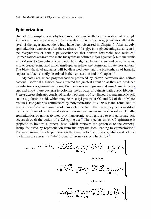
Epimerization
One of the simplest carbohydrate modifications is the epimerization of a single
stereocentre in a sugar residue. Epimerizations may occur pre-glycosylationally at the
level of the sugar nucleotide, which have been discussed in Chapter 6. Alternatively,
epimerizations can occur after the synthesis of the glycan or glycoconjugate, as seen in
the biosynthesis of certain polysaccharides that contain hexuronic acid residues.2
Epimerizations are involved in the biosynthesis of three major glycans: b-D-mannuronic
acid (ManA) to a-L-guluronic acid (GulA) in alginate biosynthesis, and b-D-glucuronic
acid to a-L-iduronic acid in heparin/heparan sulfate and dermatan sulfate biosynthesis.
The biosynthesis of alginates will be discussed here, and the biosynthesis of heparin/
heparan sulfate is briefly described in the next section and in Chapter 11.
Alginates are linear polysaccharides produced by brown seaweeds and certain
bacteria. Bacterial alginates have attracted the greatest attention as they are produced
by infectious organisms including Pseudomonas aeruginosa and Burkholderia cepa-
cia, and allow these bacteria to colonize the airways of patients with cystic fibrosis.3
P. aeruginosa alginates consist of random polymers of 1,4-linked b-D-mannuronic acid
and a-L-guluronic acid, which may bear acetyl groups at O2 and O3 of the b-ManA
residues. Biosynthesis commences by polymerization of GDP-D-mannuronic acid to
give a linear b-D-mannuronic acid homopolymer. Next, the linear polymer is modified
by the addition of acetic acid esters to some D-mannuronic acid residues. Finally,
epimerization of non-acetylated b-D-mannuronic acid residues to a-L-guluronic acid
occurs through the action of a C5 epimerase.4 The mechanism of C5 epimerase is
proposed to involve a general base, which removes the proton a to the carboxyl
group, followed by reprotonation from the opposite face, leading to epimerization.2
The mechanism of such epimerases is thus similar to that of lyases, which instead lead
to elimination across the C4–C5 bond of uronates (see Chapter 7).2
344 10 Modifications of Glycans and Glycoconjugates
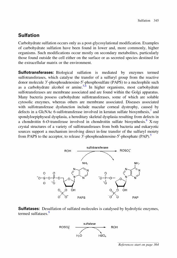
Sulfation
Carbohydrate sulfation occurs only as a post-glycosylational modification. Examples
of carbohydrate sulfation have been found in lower and, more commonly, higher
organisms. Such modifications occur mostly on secondary metabolites, particularly
those found outside the cell either on the surface or as secreted species destined for
the extracellular matrix or the environment.
Sulfotransferases: Biological sulfation is mediated by enzymes termed
sulfotransferases, which catalyse the transfer of a sulfuryl group from the reactive
donor molecule 30-phosphoadenosine-50-phosphosulfate (PAPS) to a nucleophile such
as a carbohydrate alcohol or amine.5,6 In higher organisms, most carbohydrate
sulfotransferases are membrane associated and are found within the Golgi apparatus.
Many bacteria possess carbohydrate sulfotransferases, some of which are soluble
cytosolic enzymes, whereas others are membrane associated. Diseases associated
with sulfotransferase dysfunction include macular corneal dystrophy, caused by
defects in a GlcNAc 6-sulfotransferase involved in keratan sulfate biosynthesis,7 and
spondyloepiphyseal dysplasia, a hereditary skeletal dysplasia resulting from defects in
a chondroitin 6-O-transferase involved in chondroitin sulfate biosynthesis.8 X-ray
crystal structures of a variety of sulfotransferases from both bacteria and eukaryotic
sources support a mechanism involving direct in-line transfer of the sulfuryl moiety
from PAPS to the acceptor, to release 30-phosphoadenosine-50-phosphate (PAP).5
Sulfatases: Desulfation of sulfated molecules is catalysed by hydrolytic enzymes,
termed sulfatases.9
Sulfation 345
References start on page 364
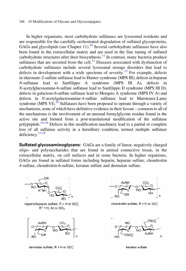
In higher organisms, most carbohydrate sulfatases are lysosomal residents and
are responsible for the carefully orchestrated degradation of sulfated glycoproteins,
GAGs and glycolipids (see Chapter 11).10 Several carbohydrate sulfatases have also
been found in the extracellular matrix and are used in the fine tuning of sulfated
carbohydrate structures after their biosynthesis.11 In contrast, many bacteria produce
sulfatases that are secreted from the cell.12 Diseases associated with dysfunction of
carbohydrate sulfatases include several lysosomal storage disorders that lead to
defects in development with a wide spectrum of severity.13 For example, defects
in iduronate-2-sulfate sulfatase lead to Hunter syndrome (MPS III), defects in heparan
N-sulfatase lead to Sanfilippo A syndrome (MPS III A), defects in
N-acetylglucosamine-6-sulfate sulfatase lead to Sanfilippo D syndrome (MPS III D),
defects in galactose-6-sulfate sulfatase lead to Morquio A syndrome (MPS IV A) and
defects in N-acetylgalactosamine-4-sulfate sulfatase lead to Maroteaux-Lamy
syndrome (MPS VI).10 Sulfatases have been proposed to operate through a variety of
mechanisms, none of which have definitive evidence in their favour – common to all of
the mechanisms is the involvement of an unusual formylglycine residue found in the
active site and formed from a post-translational modification of the sulfatase
polypeptide.14–16 Defects in this modification machinery lead to a partial or complete
loss of all sulfatase activity in a hereditary condition, termed multiple sulfatase
deficiency.17,18
Sulfated glycosaminoglycans: GAGs are a family of linear, negatively charged
oligo- and polysaccharides that are found in animal connective tissue, in the
extracellular matrix, on cell surfaces and in some bacteria. In higher organisms,
GAGs are found in sulfated forms including heparin, heparan sulfate, chondroitin
4-sulfate, chondroitin-6-sulfate, keratan sulfate and dermatan sulfate.
346 10 Modifications of Glycans and Glycoconjugates
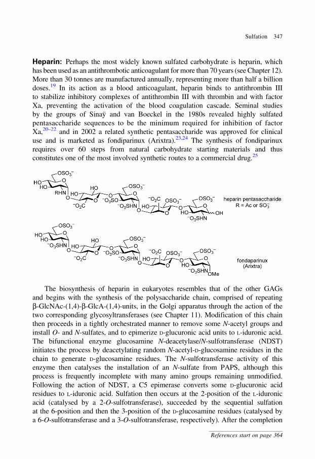
Heparin: Perhaps the most widely known sulfated carbohydrate is heparin, which
has been used as an antithrombotic anticoagulant for more than 70 years (see Chapter 12).
More than 30 tonnes are manufactured annually, representing more than half a billion
doses.19 In its action as a blood anticoagulant, heparin binds to antithrombin III
to stabilize inhibitory complexes of antithrombin III with thrombin and with factor
Xa, preventing the activation of the blood coagulation cascade. Seminal studies
by the groups of Sinay and van Boeckel in the 1980s revealed highly sulfated
pentasaccharide sequences to be the minimum required for inhibition of factor
Xa,20–22 and in 2002 a related synthetic pentasaccharide was approved for clinical
use and is marketed as fondiparinux (Arixtra).23,24 The synthesis of fondiparinux
requires over 60 steps from natural carbohydrate starting materials and thus
constitutes one of the most involved synthetic routes to a commercial drug.25
The biosynthesis of heparin in eukaryotes resembles that of the other GAGs
and begins with the synthesis of the polysaccharide chain, comprised of repeating
b-GlcNAc-(1,4)-b-GlcA-(1,4)-units, in the Golgi apparatus through the action of the
two corresponding glycosyltransferases (see Chapter 11). Modification of this chain
then proceeds in a tightly orchestrated manner to remove some N-acetyl groups and
install O- and N-sulfates, and to epimerize D-glucuronic acid units to L-iduronic acid.
The bifunctional enzyme glucosamine N-deacetylase/N-sulfotransferase (NDST)
initiates the process by deacetylating random N-acetyl-D-glucosamine residues in the
chain to generate D-glucosamine residues. The N-sulfotransferase activity of this
enzyme then catalyses the installation of an N-sulfate from PAPS, although this
process is frequently incomplete with many amino groups remaining unmodified.
Following the action of NDST, a C5 epimerase converts some D-glucuronic acid
residues to L-iduronic acid. Sulfation then occurs at the 2-position of the L-iduronic
acid (catalysed by a 2-O-sulfotransferase), succeeded by the sequential sulfation
at the 6-position and then the 3-position of the D-glucosamine residues (catalysed by
a 6-O-sulfotransferase and a 3-O-sulfotransferase, respectively). After the completion
Sulfation 347
References start on page 364
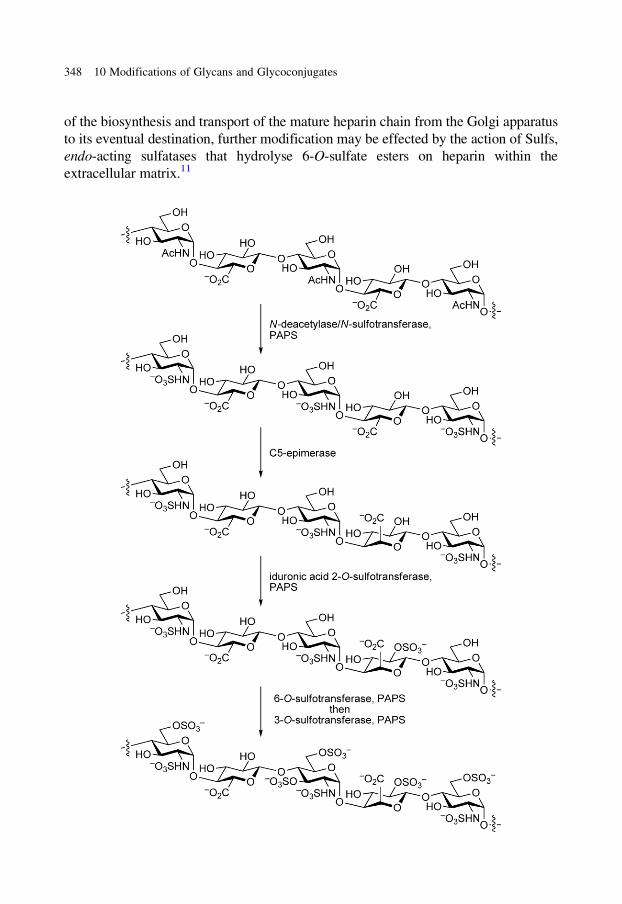
of the biosynthesis and transport of the mature heparin chain from the Golgi apparatus
to its eventual destination, further modification may be effected by the action of Sulfs,
endo-acting sulfatases that hydrolyse 6-O-sulfate esters on heparin within the
extracellular matrix.11
348 10 Modifications of Glycans and Glycoconjugates
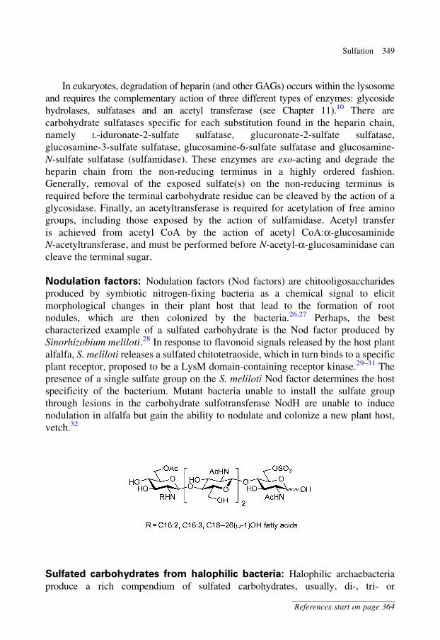
In eukaryotes, degradation of heparin (and other GAGs) occurs within the lysosome
and requires the complementary action of three different types of enzymes: glycoside
hydrolases, sulfatases and an acetyl transferase (see Chapter 11).10 There are
carbohydrate sulfatases specific for each substitution found in the heparin chain,
namely L-iduronate-2-sulfate sulfatase, glucuronate-2-sulfate sulfatase,
glucosamine-3-sulfate sulfatase, glucosamine-6-sulfate sulfatase and glucosamine-
N-sulfate sulfatase (sulfamidase). These enzymes are exo-acting and degrade the
heparin chain from the non-reducing terminus in a highly ordered fashion.
Generally, removal of the exposed sulfate(s) on the non-reducing terminus is
required before the terminal carbohydrate residue can be cleaved by the action of a
glycosidase. Finally, an acetyltransferase is required for acetylation of free amino
groups, including those exposed by the action of sulfamidase. Acetyl transfer
is achieved from acetyl CoA by the action of acetyl CoA:a-glucosaminide
N-acetyltransferase, and must be performed before N-acetyl-a-glucosaminidase can
cleave the terminal sugar.
Nodulation factors: Nodulation factors (Nod factors) are chitooligosaccharides
produced by symbiotic nitrogen-fixing bacteria as a chemical signal to elicit
morphological changes in their plant host that lead to the formation of root
nodules, which are then colonized by the bacteria.26,27 Perhaps, the best
characterized example of a sulfated carbohydrate is the Nod factor produced by
Sinorhizobium meliloti.28 In response to flavonoid signals released by the host plant
alfalfa, S. meliloti releases a sulfated chitotetraoside, which in turn binds to a specific
plant receptor, proposed to be a LysM domain-containing receptor kinase.29–31 The
presence of a single sulfate group on the S. meliloti Nod factor determines the host
specificity of the bacterium. Mutant bacteria unable to install the sulfate group
through lesions in the carbohydrate sulfotransferase NodH are unable to induce
nodulation in alfalfa but gain the ability to nodulate and colonize a new plant host,
vetch.32
Sulfated carbohydrates from halophilic bacteria: Halophilic archaebacteria
produce a rich compendium of sulfated carbohydrates, usually, di-, tri- or
Sulfation 349
References start on page 364
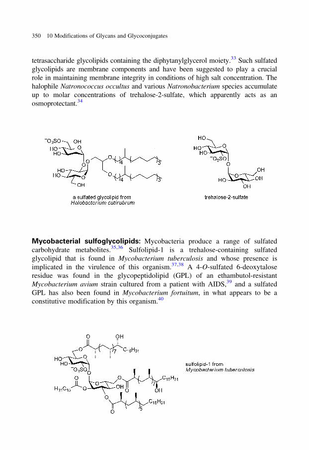
tetrasaccharide glycolipids containing the diphytanylglycerol moiety.33 Such sulfated
glycolipids are membrane components and have been suggested to play a crucial
role in maintaining membrane integrity in conditions of high salt concentration. The
halophile Natronococcus occultus and various Natronobacterium species accumulate
up to molar concentrations of trehalose-2-sulfate, which apparently acts as an
osmoprotectant.34
Mycobacterial sulfoglycolipids: Mycobacteria produce a range of sulfated
carbohydrate metabolites.35,36 Sulfolipid-1 is a trehalose-containing sulfated
glycolipid that is found in Mycobacterium tuberculosis and whose presence is
implicated in the virulence of this organism.37,38 A 4-O-sulfated 6-deoxytalose
residue was found in the glycopeptidolipid (GPL) of an ethambutol-resistant
Mycobacterium avium strain cultured from a patient with AIDS,39 and a sulfated
GPL has also been found in Mycobacterium fortuitum, in what appears to be a
constitutive modification by this organism.40
350 10 Modifications of Glycans and Glycoconjugates
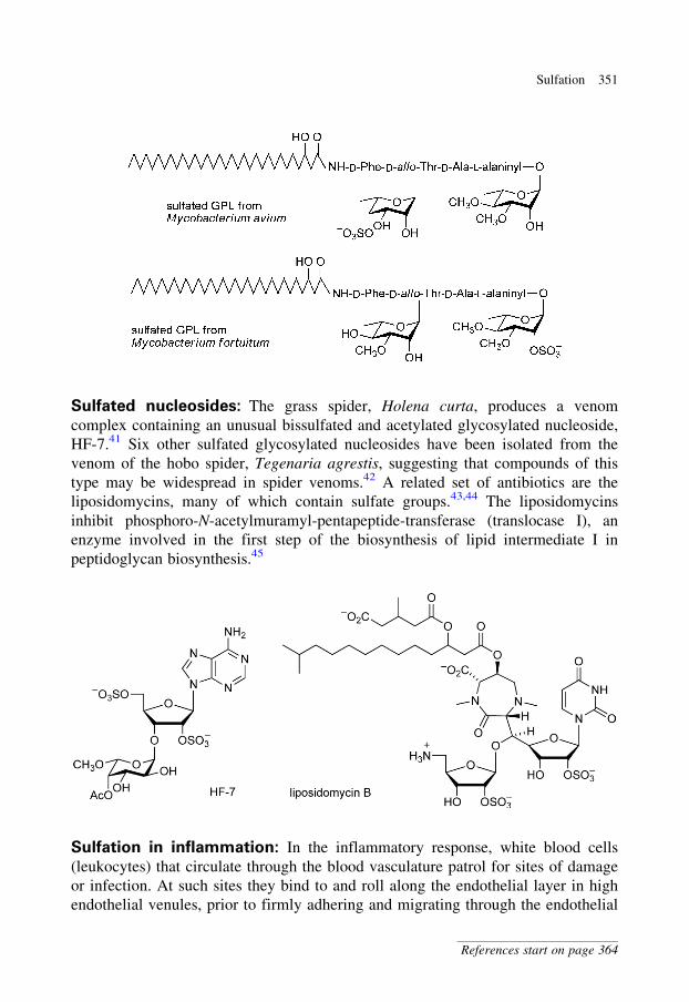
Sulfated nucleosides: The grass spider, Holena curta, produces a venom
complex containing an unusual bissulfated and acetylated glycosylated nucleoside,
HF-7.41 Six other sulfated glycosylated nucleosides have been isolated from the
venom of the hobo spider, Tegenaria agrestis, suggesting that compounds of this
type may be widespread in spider venoms.42 A related set of antibiotics are the
liposidomycins, many of which contain sulfate groups.43,44 The liposidomycins
inhibit phosphoro-N-acetylmuramyl-pentapeptide-transferase (translocase I), an
enzyme involved in the first step of the biosynthesis of lipid intermediate I in
peptidoglycan biosynthesis.45
Sulfation in inflammation: In the inflammatory response, white blood cells
(leukocytes) that circulate through the blood vasculature patrol for sites of damage
or infection. At such sites they bind to and roll along the endothelial layer in high
endothelial venules, prior to firmly adhering and migrating through the endothelial
Sulfation 351
References start on page 364
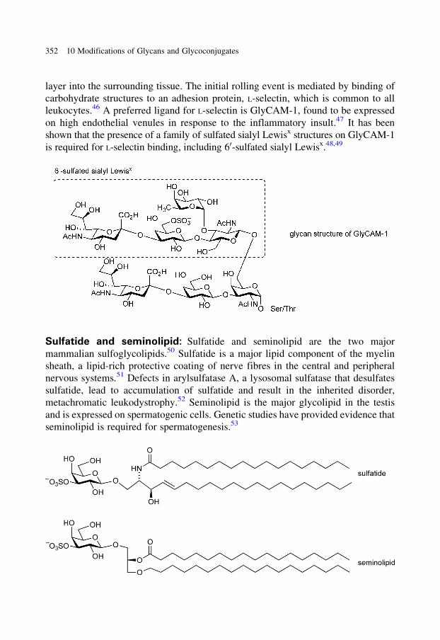
layer into the surrounding tissue. The initial rolling event is mediated by binding of
carbohydrate structures to an adhesion protein, L-selectin, which is common to all
leukocytes.46 A preferred ligand for L-selectin is GlyCAM-1, found to be expressed
on high endothelial venules in response to the inflammatory insult.47 It has been
shown that the presence of a family of sulfated sialyl Lewisx structures on GlyCAM-1
is required for L-selectin binding, including 60-sulfated sialyl Lewisx.48,49
Sulfatide and seminolipid: Sulfatide and seminolipid are the two major
mammalian sulfoglycolipids.50 Sulfatide is a major lipid component of the myelin
sheath, a lipid-rich protective coating of nerve fibres in the central and peripheral
nervous systems.51 Defects in arylsulfatase A, a lysosomal sulfatase that desulfates
sulfatide, lead to accumulation of sulfatide and result in the inherited disorder,
metachromatic leukodystrophy.52 Seminolipid is the major glycolipid in the testis
and is expressed on spermatogenic cells. Genetic studies have provided evidence that
seminolipid is required for spermatogenesis.53
352 10 Modifications of Glycans and Glycoconjugates
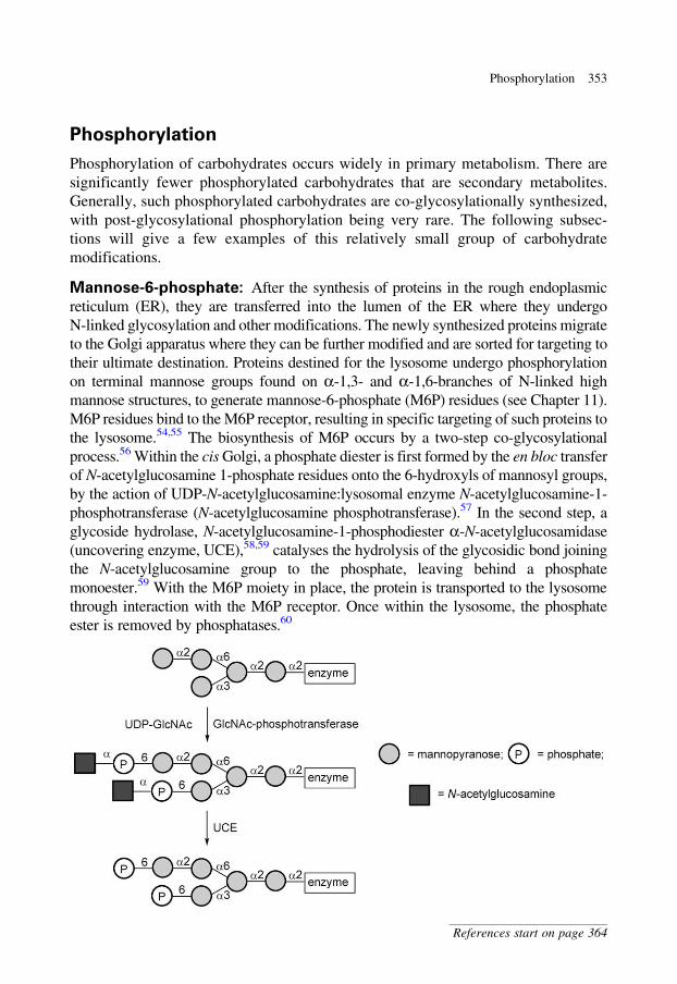
Phosphorylation
Phosphorylation of carbohydrates occurs widely in primary metabolism. There are
significantly fewer phosphorylated carbohydrates that are secondary metabolites.
Generally, such phosphorylated carbohydrates are co-glycosylationally synthesized,
with post-glycosylational phosphorylation being very rare. The following subsec-
tions will give a few examples of this relatively small group of carbohydrate
modifications.
Mannose-6-phosphate: After the synthesis of proteins in the rough endoplasmic
reticulum (ER), they are transferred into the lumen of the ER where they undergo
N-linked glycosylation and other modifications. The newly synthesized proteins migrate
to the Golgi apparatus where they can be further modified and are sorted for targeting to
their ultimate destination. Proteins destined for the lysosome undergo phosphorylation
on terminal mannose groups found on a-1,3- and a-1,6-branches of N-linked high
mannose structures, to generate mannose-6-phosphate (M6P) residues (see Chapter 11).
M6P residues bind to the M6P receptor, resulting in specific targeting of such proteins to
the lysosome.54,55 The biosynthesis of M6P occurs by a two-step co-glycosylational
process.56 Within the cis Golgi, a phosphate diester is first formed by the en bloc transfer
of N-acetylglucosamine 1-phosphate residues onto the 6-hydroxyls of mannosyl groups,
by the action of UDP-N-acetylglucosamine:lysosomal enzyme N-acetylglucosamine-1-
phosphotransferase (N-acetylglucosamine phosphotransferase).57 In the second step, a
glycoside hydrolase, N-acetylglucosamine-1-phosphodiester a-N-acetylglucosamidase
(uncovering enzyme, UCE),58,59 catalyses the hydrolysis of the glycosidic bond joining
the N-acetylglucosamine group to the phosphate, leaving behind a phosphate
monoester.59 With the M6P moiety in place, the protein is transported to the lysosome
through interaction with the M6P receptor. Once within the lysosome, the phosphate
ester is removed by phosphatases.60
Phosphorylation 353
References start on page 364
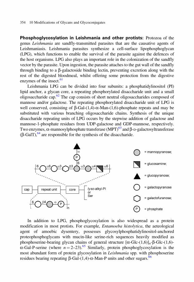
Phosphoglycosylation in Leishmania and other protists: Protozoa of the
genus Leishmania are sandfly-transmitted parasites that are the causative agents of
Leishmaniasis. Leishmania parasites synthesize a cell-surface lipophosphoglycan
(LPG), which functions to enable the survival of the parasite against the defences of
the host organisms. LPG also plays an important role in the colonization of the sandfly
vector by the parasite. Upon ingestion, the parasite attaches to the gut wall of the sandfly
through binding to a b-galactoside binding lectin, preventing excretion along with the
rest of the digested bloodmeal, whilst offering some protection from the digestive
enzymes of the insect.61
Leishmania LPG can be divided into four subunits: a phosphatidylinositol (PI)
lipid anchor, a glycan core, a repeating phosphorylated disaccharide unit and a small
oligosaccharide cap.62 The cap consists of short neutral oligosaccharides composed of
mannose and/or galactose. The repeating phosphorylated disaccharide unit of LPG is
well conserved, consisting of b-Gal-(1,4)-a-Man-(1,6)-phosphate repeats and may be
substituted with various branching oligosaccharide chains. Synthesis of the unique
disaccharide repeating units of LPG occurs by the stepwise addition of galactose and
mannose-1-phosphate residues from UDP-galactose and GDP-mannose, respectively.
Two enzymes, a-mannosylphosphate transferase (MPT)63 and b-D-galactosyltransferase
(b-GalT),64 are responsible for the synthesis of the disaccharide.
In addition to LPG, phosphoglycosylation is also widespread as a protein
modification in most protists. For example, Entamoeba histolytica, the aeteological
agent of amoebic dysentery, possesses glycosylphosphatidylinositol-anchored
proteophosphoglycans with mucin-like serine-rich sequences heavily modified as
phosphoserine-bearing glycan chains of general structure [a-Glc-(1,6)]n-b-Glc-(1,6)-
a-Gal-P-serine (where n = 2–23).65 Similarly, protein phosphoglycosylation is the
most abundant form of protein glycosylation in Leishmania spp. with phosphoserine
residues bearing repeating b-Gal-(1,4)-a-Man-P units and other sugars.66
354 10 Modifications of Glycans and Glycoconjugates
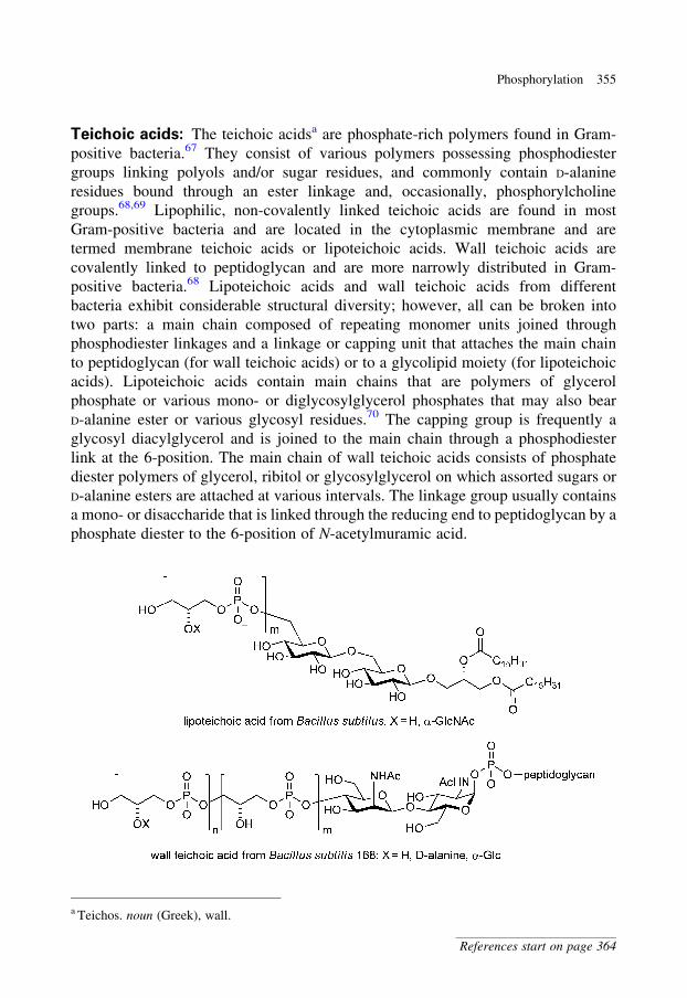
Teichoic acids: The teichoic acidsa are phosphate-rich polymers found in Gram-
positive bacteria.67 They consist of various polymers possessing phosphodiester
groups linking polyols and/or sugar residues, and commonly contain D-alanine
residues bound through an ester linkage and, occasionally, phosphorylcholine
groups.68,69 Lipophilic, non-covalently linked teichoic acids are found in most
Gram-positive bacteria and are located in the cytoplasmic membrane and are
termed membrane teichoic acids or lipoteichoic acids. Wall teichoic acids are
covalently linked to peptidoglycan and are more narrowly distributed in Gram-
positive bacteria.68 Lipoteichoic acids and wall teichoic acids from different
bacteria exhibit considerable structural diversity; however, all can be broken into
two parts: a main chain composed of repeating monomer units joined through
phosphodiester linkages and a linkage or capping unit that attaches the main chain
to peptidoglycan (for wall teichoic acids) or to a glycolipid moiety (for lipoteichoic
acids). Lipoteichoic acids contain main chains that are polymers of glycerol
phosphate or various mono- or diglycosylglycerol phosphates that may also bear
D-alanine ester or various glycosyl residues.70 The capping group is frequently a
glycosyl diacylglycerol and is joined to the main chain through a phosphodiester
link at the 6-position. The main chain of wall teichoic acids consists of phosphate
diester polymers of glycerol, ribitol or glycosylglycerol on which assorted sugars or
D-alanine esters are attached at various intervals. The linkage group usually contains
a mono- or disaccharide that is linked through the reducing end to peptidoglycan by a
phosphate diester to the 6-position of N-acetylmuramic acid.
a Teichos. noun (Greek), wall.
Phosphorylation 355
References start on page 364
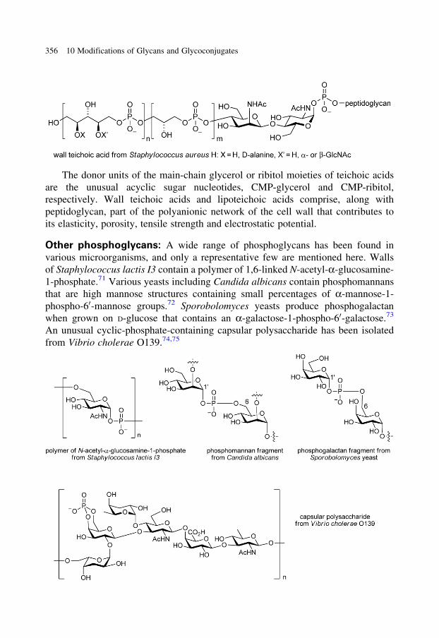
The donor units of the main-chain glycerol or ribitol moieties of teichoic acids
are the unusual acyclic sugar nucleotides, CMP-glycerol and CMP-ribitol,
respectively. Wall teichoic acids and lipoteichoic acids comprise, along with
peptidoglycan, part of the polyanionic network of the cell wall that contributes to
its elasticity, porosity, tensile strength and electrostatic potential.
Other phosphoglycans: A wide range of phosphoglycans has been found in
various microorganisms, and only a representative few are mentioned here. Walls
of Staphylococcus lactis I3 contain a polymer of 1,6-linked N-acetyl-a-glucosamine-
1-phosphate.71 Various yeasts including Candida albicans contain phosphomannans
that are high mannose structures containing small percentages of a-mannose-1-
phospho-60-mannose groups.72 Sporobolomyces yeasts produce phosphogalactan
when grown on D-glucose that contains an a-galactose-1-phospho-60-galactose.73
An unusual cyclic-phosphate-containing capsular polysaccharide has been isolated
from Vibrio cholerae O139.74,75
356 10 Modifications of Glycans and Glycoconjugates
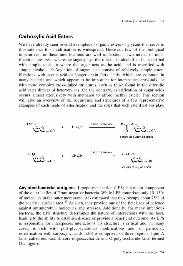
Carboxylic Acid Esters
We have already seen several examples of organic esters of glycans that serve to
illustrate that this modification is widespread. However, few of the biological
imperatives for these modifications are well understood. Two modes of mod-
ifications are seen: where the sugar plays the role of an alcohol and is esterified
with simple acids, or where the sugar acts as the acid, and is esterified with
simple alcohols. O-Acylation of sugars can consist of relatively simple ester-
ifications with acetic acid or longer chain fatty acids, which are common in
many bacteria and which appear to be important for interspecies cross-talk, or
with more complex cross-linked structures, such as those found in the diferulic
acid ester dimers of heteroxylans. On the contrary, esterification of sugar acids
occurs almost exclusively with methanol to afford methyl esters. This section
will give an overview of the occurrence and structures of a few representative
examples of each mode of esterification and the roles that such esterifications play.
Acylated bacterial antigens: Lipopolysaccharide (LPS) is a major component
of the outer leaflet of Gram-negative bacteria. While LPS comprises only 10–15%
of molecules in the outer membrane, it is estimated that they occupy about 75% of
the bacterial surface area.28 As such, they provide one of the first lines of defence
against antimicrobial molecules and stresses. Additionally, for many infectious
bacteria, the LPS structure determines the nature of interactions with the host,
leading to the ability to establish disease or provide a beneficial outcome. As LPS
is responsible for interspecies interactions, its structure is critical and, in many
cases, is rich with post-glycosylational modifications and, in particular,
esterification with carboxylic acids. LPS is comprised of three regions: lipid A
(also called endotoxin), core oligosaccharide and O-polysaccharide (also termed
O-antigen).
Carboxylic Acid Esters 357
References start on page 364
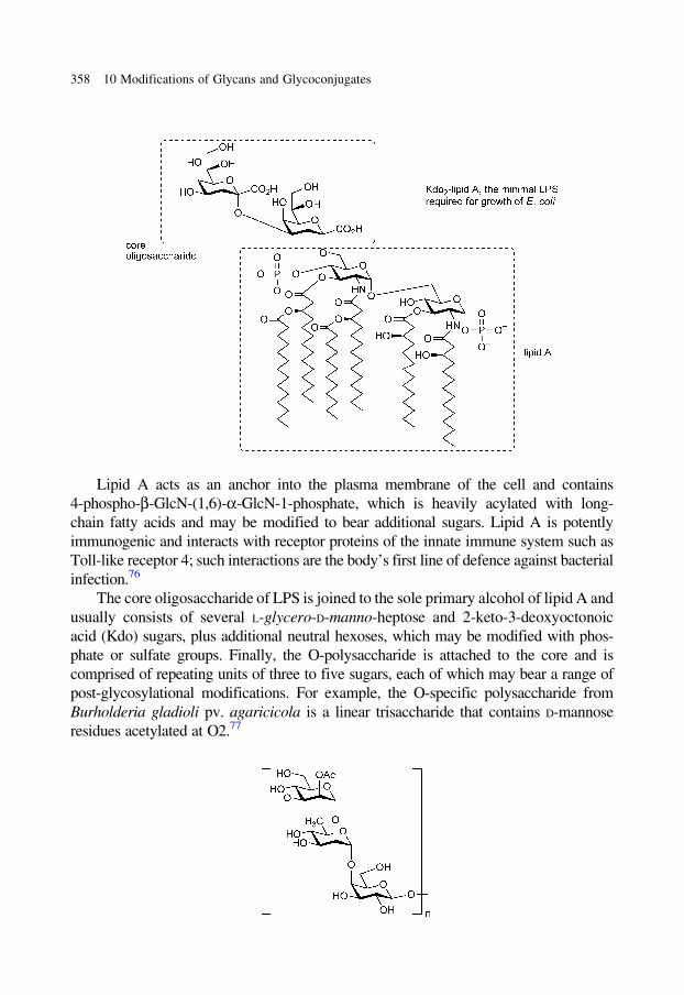
Lipid A acts as an anchor into the plasma membrane of the cell and contains
4-phospho-b-GlcN-(1,6)-a-GlcN-1-phosphate, which is heavily acylated with long-
chain fatty acids and may be modified to bear additional sugars. Lipid A is potently
immunogenic and interacts with receptor proteins of the innate immune system such as
Toll-like receptor 4; such interactions are the body’s first line of defence against bacterial
infection.76
The core oligosaccharide of LPS is joined to the sole primary alcohol of lipid A and
usually consists of several L-glycero-D-manno-heptose and 2-keto-3-deoxyoctonoic
acid (Kdo) sugars, plus additional neutral hexoses, which may be modified with phos-
phate or sulfate groups. Finally, the O-polysaccharide is attached to the core and is
comprised of repeating units of three to five sugars, each of which may bear a range of
post-glycosylational modifications. For example, the O-specific polysaccharide from
Burholderia gladioli pv. agaricicola is a linear trisaccharide that contains D-mannose
residues acetylated at O2.77
358 10 Modifications of Glycans and Glycoconjugates
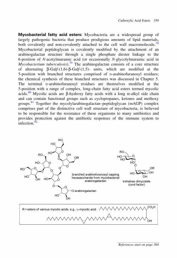
Mycobacterial fatty acid esters: Mycobacteria are a widespread group of
largely pathogenic bacteria that produce prodigious amounts of lipid materials,
both covalently and non-covalently attached to the cell wall macromolecule.78
Mycobacterial peptidoglycan is covalently modified by the attachment of an
arabinogalactan structure through a single phosphate diester linkage to the
6-position of N-acetylmuramic acid (or occasionally N-glycolylmuramic acid in
Mycobacterium tuberculosis).79 The arabinogalactan consists of a core structure
of alternating b-Galf-(1,6)-b-Galf-(1,5)- units, which are modified at the
5-position with branched structures comprised of D-arabinofuranosyl residues;
the chemical synthesis of these branched structures was discussed in Chapter 5.
The terminal D-arabinofuranosyl residues are themselves modified at the
5-position with a range of complex, long-chain fatty acid esters termed mycolic
acids.80 Mycolic acids are b-hydroxy fatty acids with a long a-alkyl side chain
and can contain functional groups such as cyclopropanes, ketones and methoxy
groups.81 Together the mycolylarabinogalactan–peptidoglycan (mAGP) complex
comprises part of the distinctive cell wall structure of mycobacteria, is believed
to be responsible for the resistance of these organisms to many antibiotics and
provides protection against the antibiotic responses of the immune system to
infection.82
Carboxylic Acid Esters 359
References start on page 364

Good evidence has accumulated that movement of mycolic acids from their
point of synthesis within the cytoplasm to their destination in the cell wall is
facilitated by a carrier molecule, the disaccharide trehalose. Mycolic acids are
transferred onto the primary hydroxyls of trehalose to generate trehalose
dimycolate (TDM, also termed cord factor).83 TDM is transported out of the cell
where it forms a significant non-covalently-associated portion of the cell wall.84
Transfer of mycolic acid to the arabinan of the mAGP complex is catalysed by a set
of transesterases, the antigen 85 proteins (85A, 85B and 85C).85 The role of the
antigen 85 transesterases is complex; aside from catalysing the transfer of mycolic
acid from TDM to the mAGP complex, these enzymes can also catalyse the transfer
of mycolic acids between TDM and trehalose to form two molecules of trehalose
monomycolate, a reaction that presumably provides a ready flux of mycolyl groups
for delivery to the cell wall. The antigen 85 proteins are major secreted proteins of
M. tuberculosis, consistent with their role in cell wall manufacture. They share a
common a/b hydrolase fold and possess a catalytic triad, Ser-Glu-His, which they
share with lipases.86,87
Carboxylic acid esters in hemicelluloses: Plant cell wall polysaccharides
are the most abundant organic materials on earth. Hemicelluloses are second
only to cellulose as the most common type of polysaccharide found within
plants.88 Hemicelluloses include a range of non-cellulosic components (e.g.
arabinoxylans, pectins, galactomannans, xyloglucans and b-glucans) and can
again be modified to form esters in two possible ways: where the sugar acts as
an alcohol and is esterified with various carboxylic acids (e.g. heteroxylans) or
where the sugar is the acid and is esterified with the simple alcohol, methanol
(e.g. pectins).89
Heteroxylans include various heterogeneous branched polymers consisting
of a b-1,4-linked xylan core appended with L-arabinose, hexoses (D-mannose,
D-glucose, D-galactose) and uronic acids (D-glucuronic, 4-O-methyl-D-glucuronic
acids); these polymer chains may be further O-methylated, or esterified with
acetic, ferulic (4-hydroxy-3-methoxycinnamic acid) or p-coumaric acids. The
major roles of heteroxylans are to confer structural rigidity upon plant cell
walls and provide protection against microbial and herbivore attack.90 Ferulic
acid esters enhance the strength of the plant cell wall by forming diferulyl
cross-links between glycan polymer chains or with lignin, a complex polyphe-
nolic compound found in the extracellular matrix. In vivo, peroxidase catalyses
the oxidative coupling of ferulic acid side chains, resulting in the formation of
ferulic acid dehydrodimers.90
360 10 Modifications of Glycans and Glycoconjugates

Pectins are heterogeneous, branched and highly hydrated polysaccharides
that are rich in D-galacturonic acid residues. Homogalacturonans have a core
structure composed of repeating a-1,4-linked galacturonic acid residues, and
rhamnogalacturonan I pectins have a core of a-L-Rha-(1,4)-a-GalA-(1,2)-residues.88
The pectin cores are modified with other carbohydrates and are heavily modified with
methyl esters. They are synthesized in the Golgi of the plant cell and are de-esterified in
the cell wall by enzymes termed pectin methylesterases.91
Biological degradation of hemicelluloses occurs through the synergistic action
of main-chain degrading glycosidases and a series of carbohydrate esterases, includ-
ing acetylesterases, methylesterases, p-coumaryl esterases, ferulyl esterases and
pectin methylesterases.92,93 Carbohydrate esterases are found within bacteria and
fungi that catabolize hemicelluloses and have also been identified through sequen-
cing efforts in plant genomes, where they presumably function to allow remodelling
of cell wall hemicellulose during development.94 X-ray crystal structures of
most ferulyl, cinnamyl and acetylxylan esterases, and pectin methylesterases, show
an a/b hydrolase fold and a conserved catalytic triad, Ser-His-Asp, which is common
to lipases, suggesting that these enzymes are a subclass of this group.95,96 A smaller
subset of carbohydrate esterases is metal-ion dependent.93,97
Carboxylic Acid Esters 361
References start on page 364
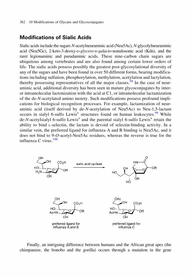
Modifications of Sialic Acids
Sialic acids include the sugars N-acetylneuraminic acid (Neu5Ac), N-glycolylneuraminic
acid (Neu5Gc), 2-keto-3-deoxy-D-glycero-D-galacto-nonulosonic acid (Kdn), and the
rarer legionaminic and pseudaminic acids. These nine-carbon chain sugars are
ubiquitous among vertebrates and are also found among certain lower orders of
life. The sialic acids possess possibly the greatest post-glycosylational diversity of
any of the sugars and have been found in over 50 different forms, bearing modifica-
tions including sulfation, phosphorylation, methylation, acetylation and lactylation,
thereby possessing representatives of all the major classes.98 In the case of neur-
aminic acid, additional diversity has been seen in mature glycoconjugates by inter-
or intramolecular lactonization with the acid at C1, or intramolecular lactamization
of the de-N-acetylated amino moiety. Such modifications possess profound impli-
cations for biological recognition processes. For example, lactamization of neur-
aminic acid (itself derived by de-N-acetylation of Neu5Ac) to Neu-1,5-lactam
occurs in sialyl 6-sulfo Lewisx structures found on human leukocytes.99 While
de-N-acetylsialyl 6-sulfo Lewisx and the parental sialyl 6-sulfo Lewisx retain the
ability to bind L-selectin, the lactam is devoid of selectin-binding activity. In a
similar vein, the preferred ligand for influenza A and B binding is Neu5Ac, and it
does not bind to 9-O-acetyl-Neu5Ac residues, whereas the reverse is true for the
influenza C virus.100
Finally, an intriguing difference between humans and the African great apes (the
chimpanzee, the bonobo and the gorilla) occurs through a mutation in the gene
362 10 Modifications of Glycans and Glycoconjugates
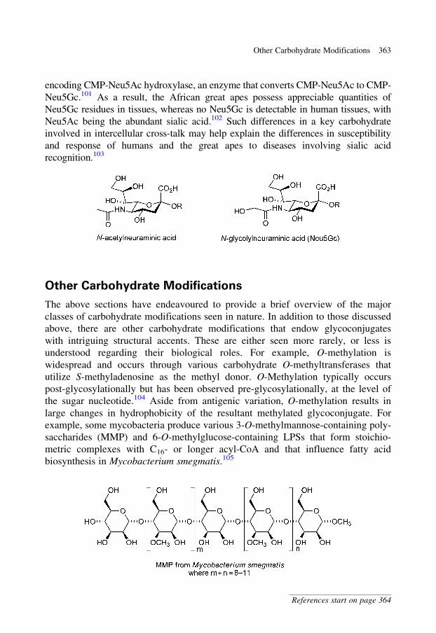
encoding CMP-Neu5Ac hydroxylase, an enzyme that converts CMP-Neu5Ac to CMP-
Neu5Gc.101 As a result, the African great apes possess appreciable quantities of
Neu5Gc residues in tissues, whereas no Neu5Gc is detectable in human tissues, with
Neu5Ac being the abundant sialic acid.102 Such differences in a key carbohydrate
involved in intercellular cross-talk may help explain the differences in susceptibility
and response of humans and the great apes to diseases involving sialic acid
recognition.103
Other Carbohydrate Modifications
The above sections have endeavoured to provide a brief overview of the major
classes of carbohydrate modifications seen in nature. In addition to those discussed
above, there are other carbohydrate modifications that endow glycoconjugates
with intriguing structural accents. These are either seen more rarely, or less is
understood regarding their biological roles. For example, O-methylation is
widespread and occurs through various carbohydrate O-methyltransferases that
utilize S-methyladenosine as the methyl donor. O-Methylation typically occurs
post-glycosylationally but has been observed pre-glycosylationally, at the level of
the sugar nucleotide.104 Aside from antigenic variation, O-methylation results in
large changes in hydrophobicity of the resultant methylated glycoconjugate. For
example, some mycobacteria produce various 3-O-methylmannose-containing poly-
saccharides (MMP) and 6-O-methylglucose-containing LPSs that form stoichio-
metric complexes with C16- or longer acyl-CoA and that influence fatty acid
biosynthesis in Mycobacterium smegmatis.105
Other Carbohydrate Modifications 363
References start on page 364
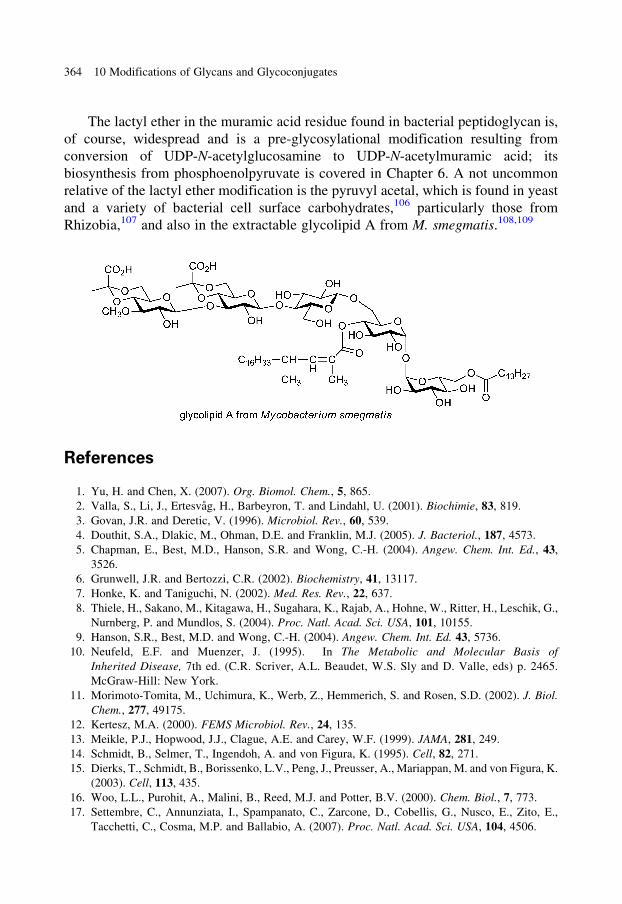
The lactyl ether in the muramic acid residue found in bacterial peptidoglycan is,
of course, widespread and is a pre-glycosylational modification resulting from
conversion of UDP-N-acetylglucosamine to UDP-N-acetylmuramic acid; its
biosynthesis from phosphoenolpyruvate is covered in Chapter 6. A not uncommon
relative of the lactyl ether modification is the pyruvyl acetal, which is found in yeast
and a variety of bacterial cell surface carbohydrates,106 particularly those from
Rhizobia,107 and also in the extractable glycolipid A from M. smegmatis.108,109
References
1. Yu, H. and Chen, X. (2007). Org. Biomol. Chem., 5, 865.
2. Valla, S., Li, J., Ertesvag, H., Barbeyron, T. and Lindahl, U. (2001). Biochimie, 83, 819.
3. Govan, J.R. and Deretic, V. (1996). Microbiol. Rev., 60, 539.
4. Douthit, S.A., Dlakic, M., Ohman, D.E. and Franklin, M.J. (2005). J. Bacteriol., 187, 4573.
5. Chapman, E., Best, M.D., Hanson, S.R. and Wong, C.-H. (2004). Angew. Chem. Int. Ed., 43,
3526.
6. Grunwell, J.R. and Bertozzi, C.R. (2002). Biochemistry, 41, 13117.
7. Honke, K. and Taniguchi, N. (2002). Med. Res. Rev., 22, 637.
8. Thiele, H., Sakano, M., Kitagawa, H., Sugahara, K., Rajab, A., Hohne, W., Ritter, H., Leschik, G.,
Nurnberg, P. and Mundlos, S. (2004). Proc. Natl. Acad. Sci. USA, 101, 10155.
9. Hanson, S.R., Best, M.D. and Wong, C.-H. (2004). Angew. Chem. Int. Ed. 43, 5736.
10. Neufeld, E.F. and Muenzer, J. (1995). In The Metabolic and Molecular Basis of
Inherited Disease, 7th ed. (C.R. Scriver, A.L. Beaudet, W.S. Sly and D. Valle, eds) p. 2465.
McGraw-Hill: New York.
11. Morimoto-Tomita, M., Uchimura, K., Werb, Z., Hemmerich, S. and Rosen, S.D. (2002). J. Biol.
Chem., 277, 49175.
12. Kertesz, M.A. (2000). FEMS Microbiol. Rev., 24, 135.
13. Meikle, P.J., Hopwood, J.J., Clague, A.E. and Carey, W.F. (1999). JAMA, 281, 249.
14. Schmidt, B., Selmer, T., Ingendoh, A. and von Figura, K. (1995). Cell, 82, 271.
15. Dierks, T., Schmidt, B., Borissenko, L.V., Peng, J., Preusser, A., Mariappan, M. and von Figura, K.
(2003). Cell, 113, 435.
16. Woo, L.L., Purohit, A., Malini, B., Reed, M.J. and Potter, B.V. (2000). Chem. Biol., 7, 773.
17. Settembre, C., Annunziata, I., Spampanato, C., Zarcone, D., Cobellis, G., Nusco, E., Zito, E.,
Tacchetti, C., Cosma, M.P. and Ballabio, A. (2007). Proc. Natl. Acad. Sci. USA, 104, 4506.
364 10 Modifications of Glycans and Glycoconjugates
18. Sardinello, M., Annunziata, I., Roma, G. and Ballabio, A. (2005). Hum. Mol. Genet., 14, 3203.
19. Witczak, Z.J. and Nieforth, K.A., eds (1997). Carbohydrates in Drug Design. Marcel Dekker, Inc:
New York, USA.
20. Sinay, P., Jacquinet, J.C., Petitou, M., Duchaussoy, P., Lederman, I., Choay, J. and Torri, G.
(1984). Carbohydr. Res., 132, C5.
21. van Boeckel, C.A., Beetz, T., Vos, J.N., de Jong, A.J.M., van Aelst, S.F., van den Bosch, R.H.,
Mertens, J.M.R. and van der Vlugt, F.A. (1985). J. Carbohydr. Chem., 4, 293.
22. Petitou, M., Duchaussoy, P., Lederman, I., Choay, J., Sinay, P., Jacquinet, J.C. and Torri, G.
(1986). Carbohydr. Res., 147, 221.
23. Petitou, M. and van Boeckel, C.A.A. (2004). Angew. Chem. Int. Ed., 43, 3118.
24. van Boeckel, C.A.A. and Petitou, M. (1993). Angew. Chem. Int. Ed., 32, 1671.
25. Codee, J.D.C., Overkleeft, H.S., van der Marel, G.A. and van Boeckel, C.A.A. (2004). Drug
Discov. Today: Technologies, 1, 317.
26. Cullimore, J.V., Ranjeva, R. and Bono, J.J. (2001). Trends Plant Sci., 6, 24.
27. D’Haeze, W. and Holsters, M. (2002). Glycobiology, 12, 79R.
28. Lerouge, P., Roche, P., Faucher, C., Maillet, F., Truchet, G., Prome, J.C. and Denarie, J. (1990).
Nature, 344, 781.
29. Radutoiu, S., Madsen, L.H., Madsen, E.B., Felle, H.H., Umehara, Y., Gronlund, M., Sato, S.,
Nakamura, Y., Tabata, S., Sandal, N. and Stougaard, J. (2003). Nature, 425, 585.
30. Limpens, E., Franken, C., Smit, P., Willemse, J., Bisseling, T. and Geurts, R. (2003). Science,
302, 630.
31. Madsen, E.B., Madsen, L.H., Radutoiu, S., Olbryt, M., Rakwalska, M., Szczyglowski, K., Sato,
S., Kaneko, T., Tabata, S., Sandal, N. and Stougaard, J. (2003). Nature, 425, 637.
32. Roche, P., Debelle, F., Maillet, F., Lerouge, P., Faucher, C., Truchet, G., Denarie, J. and Prome,
J.C. (1991). Cell, 67, 1131.
33. Kates, M. (1993). Experientia, 49, 1027.
34. Desmarais, D., Jablonski, P.E., Fedarko, N.S. and Roberts, M.F. (1997). J. Bacteriol., 179, 3146.
35. Mougous, J.D., Green, R.E., Williams, S.J., Brenner, S.E. and Bertozzi, C.R. (2002). Chem. Biol.,
9, 767.
36. Schelle, M.W. and Bertozzi, C.R. (2006). ChemBioChem, 7, 1516.
37. Mougous, J.D., Petzold, C.J., Senaratne, R.H., Lee, D.H., Akey, D.L., Lin, F.L., Munchel, S.E.,
Pratt, M.R., Riley, L.W., Leary, J.A., Berger, J.M. and Bertozzi, C.R. (2004). Nat. Struct. Mol.
Biol., 11, 721.
38. Leigh, C.D. and Bertozzi, C.R. (2008). J. Org. Chem., 73, 1008.
39. Khoo, K.H., Jarboe, E., Barker, A., Torrelles, J., Kuo, C.W. and Chatterjee, D. (1999). J. Biol.
Chem., 274, 9778.
40. Lopez Marın, L.M., Laneelle, M.A., Prome, D., Laneelle, G., Prome, J.C. and Daffe, M. (1992).
Biochemistry, 31, 11106.
41. McCormick, J., Li, Y.B., McCormick, K., Duynstee, H.I., van Engen, A.K., van der Marel, G.A.,
Ganem, B., van Boom, J.H. and Meinwald, J. (1999). J. Am. Chem. Soc., 121, 5661.
42. Taggi, A.E., Meinwald, J. and Schroeder, F.C. (2004). J. Am. Chem. Soc., 126, 10364.
43. Ubukata, M., Isono, K., Kimura, K.-i., Nelson, C. and McCloskey, J.A. (1988). J. Am. Chem. Soc.,
110, 4416.
44. Knapp, S., Morriello, G.J. and Doss, G.A. (2002). Org. Lett., 4, 603.
45. Kimura, K. and Bugg, T.D. (2003). Nat. Prod. Rep., 20, 252.
46. Bertozzi, C.R. (1995). Chem. Biol., 2, 703.
47. Hemmerich, S. and Rosen, S.D. (2000). Glycobiology, 10, 849.
48. Hemmerich, S. and Rosen, S.D. (1994). Biochemistry, 33, 4830.
49. Hemmerich, S., Bertozzi, C.R., Leffler, H. and Rosen, S.D. (1994). Biochemistry, 33, 4820.
References 365
50. Ishizuka, I. (1997). Prog. Lipid Res., 36, 245.
51. Coetzee, T., Suzuki, K. and Popko, B. (1998). Trends Neurosci., 21, 126.
52. Gieselmann, V., Matzner, U., Hess, B., Lullmann-Rauch, R., Coenen, R., Hartmann, D.,
D’Hooge, R., DeDeyn, P. and Nagels, G. (1998). J. Inherit. Metab. Dis., 21, 564.
53. Honke, K., Zhang, Y., Cheng, X., Kotani, N. and Taniguchi, N. (2004). Glycoconj. J., 21, 59.
54. Kornfeld, S. (1987). FASEB J., 1, 462.
55. von Figura, K. and Hasilik, A. (1986). Annu. Rev. Biochem., 55, 167.
56. Hasilik, A., Klein, U., Waheed, A., Strecker, G. and von Figura, K. (1980). Proc. Natl. Acad. Sci.
USA, 77, 7074.
57. Reitman, M.L. and Kornfeld, S. (1981). J. Biol. Chem., 256, 11977.
58. Rohrer, J. and Kornfeld, R. (2001). Mol. Biol. Cell., 12, 1623.
59. Varki, A., Sherman, W. and Kornfeld, S. (1983). Arch. Biochem. Biophys., 222, 145.
60. Tabas, I. and Kornfeld, S. (1980). J. Biol. Chem., 255, 6633.
61. Descoteaux, A. and Turco, S.J. (1999). Biochim. Biophys. Acta, 1455, 341.
62. Turco, S.J. and Descoteaux, A. (1992). Annu. Rev. Microbiol., 46, 65.
63. Carver, M.A. and Turco, S.J. (1992). Arch. Biochem. Biophys., 295, 309.
64. Carver, M.A. and Turco, S.J. (1991). J. Biol. Chem., 266, 10974.
65. Moody-Haupt, S., Patterson, J.H., Mirelman, D. and McConville, M.J. (2000). J. Mol. Biol., 297,
409.
66. McConville, M.J., Mullin, K.A., Ilgoutz, S.C. and Teasdale, R.D. (2002). Microbiol. Mol. Biol.
Rev., 66, 122.
67. Archibald, A.R. and Baddiley, J. (1966). Adv. Carbohydr. Chem., 21, 323.
68. Neuhaus, F.C. and Baddiley, J. (2003). Microbiol. Mol. Biol. Rev., 67, 686.
69. Ward, J.B. (1981). Microbiol. Rev., 45, 211.
70. Stadelmaier, A., Morath, S., Hartung, T. and Schmidt, R.R. (2003). Angew. Chem. Int. Ed., 42,
916.
71. Archibald, A.R., Baddiley, J., Button, D., Heptinstall, S. and Stafford, G.H. (1968). Nature, 219,
855.
72. Cutler, J.E. (2001). Med. Mycol., 39, 75.
73. Slodki, M.E. (1966). J. Biol. Chem., 241, 2700.
74. Knirel, Y.A., Paredes, L., Jansson, P.-E., Weintraub, A., Widmalm, G. and Albert, M.J. (1995).
Eur. J. Biochem., 232, 391.
75. Ruttens, B. and Kovac, P. (2006). Carbohydr. Res., 341, 1077.
76. Miller, S.I., Ernst, R.K. and Bader, M.W. (2005). Nat. Rev. Microbiol., 3, 36.
77. Karapetyan, G., Kaczynski, Z., Iacobellis, N.S., Evidente, A. and Holst, O. (2006). Carbohydr.
Res., 341, 930.
78. Takayama, K., Wang, C. and Besra, G.S. (2005). Clin. Microbiol. Rev., 18, 81.
79. Crick, D.C., Mahapatra, S. and Brennan, P.J. (2001). Glycobiology, 11, 107R.
80. D’Souza, F.W. and Lowary, T.L. (2000). Org. Lett., 2, 1493.
81. Barry, C.E., 3rd, Lee, R.E., Mdluli, K., Sampson, A.E., Schroeder, B.G., Slayden, R.A. and Yuan, Y.
(1998). Prog. Lipid Res., 37, 143.
82. Brennan, P.J. and Nikaido, H. (1995). Annu. Rev. Biochem., 64, 29.
83. Kremer, L., Baulard, A.R., and Besra, G.S. (2000). In Molecular Genetics of Mycobacteria (G.H.
Hatful and W.R. Jacobs, Jr., eds) p. 173. ASM Press: Washington, DC.
84. Ryll, R., Kumazawa, Y. and Yano, I. (2001). Microbiol. Immunol., 45, 801.
85. Belisle, J.T., Vissa, V.D., Sievert, T., Takayama, K., Brennan, P.J. and Besra, G.S. (1997).
Science, 276, 1420.
86. Ronning, D.R., Klabunde, T., Besra, G.S., Vissa, V.D., Belisle, J.T. and Sacchettini, J.C. (2000).
Nat. Struct. Biol., 7, 141.
366 10 Modifications of Glycans and Glycoconjugates
87. Ronning, D.R., Vissa, V.D., Besra, G.S., Belisle, J.T. and Sacchettini, J.C. (2004). J. Biol. Chem.,
279, 36771.
88. Carpita, N. and McCann, M. (2002). In Biochemistry and Molecular Biology of Plants
(B.B. Buchanan, W. Gruissem and R.L. Jones, eds) p. 52. American Society of Plant Physiolo-
gists: Rockville, MD.
89. Henrissat, B., Coutinho, P.M. and Davies, G.J. (2001). Plant Mol. Biol., 47, 55.
90. Mathew, S. and Abeaham, T.E. (2004). Crit. Rev. Biotechnol., 24, 59.
91. Micheli, F. (2001). Trends Plant Sci., 6, 414.
92. Saha, B.C. (2003). J. Ind. Microbiol. Biotechnol., 30, 279.
93. Davies, G.J., Gloster, T.M. and Henrissat, B. (2005). Curr. Opin. Struct. Biol., 15, 637.
94. Markovic, O. and Janecek, S. (2004). Carbohydr. Res., 339, 2281.
95. Kroon, P.A., Williamson, G., Fish, N.M., Archer, D.B. and Belshaw, N.J. (2000). Eur.
J. Biochem., 267, 6740.
96. Faulds, C.B., Molina, R., Gonzalez, R., Husband, F., Juge, N., Sanz-Aparicio, J. and Hermoso,
J.A. (2005). FEBS J., 272, 4362.
97. Taylor, E.J., Gloster, T.M., Turkenburg, J.P., Vincent, F., Brzozowski, A.M., Dupont, C.,
Shareck, F., Centeno, M.S., Prates, J.A., Puchart, V., Ferreira, L.M., Fontes, C.M., Biely, P. and
Davies, G.J. (2006). J. Biol. Chem., 281, 10968.
98. Angata, T. and Varki, A. (2002). Chem. Rev., 102, 439.
99. Mitsuoka, C., Ohmori, K., Kimura, N., Kanamori, A., Komba, S., Ishida, H., Kiso, M. and
Kannagi, R. (1999). Proc. Natl. Acad. Sci. USA, 96, 1597.
100. Rogers, G.N., Herrler, G., Paulson, J.C. and Klenk, H.D. (1986). J. Biol. Chem., 261, 5947.
101. Chou, H.H., Takematsu, H., Diaz, S., Iber, J., Nickerson, E., Wright, K.L., Muchmore, E.A.,
Nelson, D.L., Warren, S.T. and Varki, A. (1998). Proc. Natl. Acad. Sci. USA, 95, 11751.
102. Muchmore, E.A., Diaz, S. and Varki, A. (1998). Am. J. Phys. Anthropol., 107, 187.
103. Angata, T., Varki, N.M. and Varki, A. (2001). J. Biol. Chem., 276, 40282.
104. Weisman, L.S. and Ballou, C.E. (1984). J. Biol. Chem., 259, 3464.
105. Cheon, H.S., Wang, Y., Ma, J. and Kishi, Y. (2007). ChemBioChem, 8, 353.
106. Fontaine, T., Talmont, F., Dutton, G.G. and Fournet, B. (1991). Anal. Biochem., 199, 154.
107. Eckhardt, E. and Ziegler, T. (1994). Carbohydr. Res., 264, 253.
108. Kamisango, K., Saadat, S., Dell, A. and Ballou, C.E. (1985). J. Biol. Chem., 260, 4117.
109. Ziegler, T., Eckhardt, E. and Birault, V. (1993). J. Org. Chem., 58, 1090.
References 367