glossectomy in the severe maxillofacial vascular
TRANSCRIPT

CASE REPORT Open Access
Glossectomy in the severe maxillofacialvascular malformation with jaw deformity:a rare case reportMin-Hyeog Park1, Chul-Man Kim1, Dong-Young Chung1 and Jun-Young Paeng1,2*
Abstract
In the field of oral-maxillofacial surgery, vascular malformations present in various forms. Abnormalities in the size ofthe tongue by vascular malformations can cause mandibular prognathism and skeletal deformity. The risk in surgicaltreatment for patients with vascular malformation is high, due to bleeding from vascular lesions. We report a rare case ofmacroglossia that was treated by partial glossectomy, resulting in an improvement in the swallowing and masticationfunctions in the patient. A 25-year-old male patient with severe open-bite and mandibular prognathism presented toour department for the management of macroglossia. The patient had a difficulty in food intake because of the largetongue. Orthognathic surgery was not indicated because the patient had severe jaw bone destruction and alveolar boneresorption. Therefore, the patient underwent partial glossectomy under general anesthesia. There was severehemorrhaging during the surgery, but the bleeding was controlled by local procedures.
Keywords: Macroglossia, Glossectomy, Venous malformation
BackgroundVenous malformations are anomalies of veins or lymphvessels or both veins and lymph vessels. They arepresent at birth and manifest at different ages. Venousmalformations in the tongue cause significant clinicalproblems such as swallowing difficulties and speech andairway obstruction. The incidence of vascular malforma-tion has been reported in approximately 7 % of all be-nign tumors, the majority of which develop in the headand neck region. However, when localized on thetongue, in most cases, these lesions can cause clinicalproblems such as active bleeding and airway obstructionfrom the mouth [1–3].The current methods of treatment include electrocoagu-
lation, cryotherapy, sclerotherapy, surgical excision, orcombinations of these treatments. The treatment of super-ficial, localized venous malformation is relatively simpleand effective. However, the treatment of deep, extensivevenous malformation remains difficult and presents
various complications. For complicated cases, the resultsachieved with a single treatment method are not satisfac-tory. Therefore, several approaches are required to achieveacceptable results. In most cases, a surgical excision is con-sidered as the first choice to improve the function and ap-pearance. However, for large lesions, partial glossectomycan be considered after sclerotherapy to improve swallow-ing, chewing, and speech. During the surgery, care shouldbe taken to regulate the hemorrhage and the airway [4–6].We report a case of surgical treatment using partial
glossectomy in a patient who had venous malformationof the tongue.
Case presentationA 25-year-old man with a medical history of macroglos-sia was referred to our department for the managementof the condition on October 25, 2011. The tongue wasinterpositioned between the teeth, interfering with chew-ing. He had discomfort in swallowing, chewing, andspeech because the vascular mass accounted for most ofthe tongue. He had been treated previously at a vascularsurgery clinic. Surgical resection was performed undergeneral anesthesia during the plastic surgery on January5, 1996. An excisional biopsy was performed, and the
* Correspondence: [email protected] of Oral and Maxillofacial Surgery, School of Dentistry,Kyungpook National University, Daegu, Republic of Korea2Department of Oral and Maxillofacial Surgery, Kyungpook NationalUniversity Hospital, 2175 Dalgubeoldae-ro, Daegu 700-705, Korea
© 2015 Park et al. Open Access This article is distributed under the terms of the Creative Commons Attribution 4.0International License (http://creativecommons.org/licenses/by/4.0/), which permits unrestricted use, distribution, andreproduction in any medium, provided you give appropriate credit to the original author(s) and the source, provide a link tothe Creative Commons license, and indicate if changes were made.
Park et al. Maxillofacial Plastic and Reconstructive Surgery (2015) 37:42 DOI 10.1186/s40902-015-0043-z

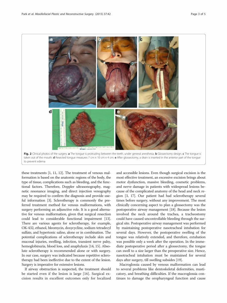
diagnosis of a venous malformation was made. Between1997 and 2005, ethanol sclerotherapy was performed overten times. However, the effect of sclerotherapy was unsatis-factory, and the patient continued to experience discomfortin eating because of the large tongue. He exhibited a severeopen-bite and mandible prognathism due to the largetongue, at his first visit to our department (Fig. 1a, b, e). Inaddition, a large amount of phleboliths were observed scat-tered around the mandible (Fig. 1c, d). Cervical magneticresonance imaging (MRI) revealed lesions on the mouthfloor, the glottis, and the supraglottic area, in addition tothe entire tongue. There was no significant interval changein the massive venous malformation in the face and necksince August 11, 2005. Partial glossectomy was performedusing the keyhole technique, under general anesthesia, onDecember 29, 2011 (Fig. 2a, b, c). The incision was per-formed with a number 15 blade and by electrosurgical co-agulation. During the surgery, the excessive tongue masswas removed. The specimen measured 7 cm× 10 cm×4 cm in size (Fig. 2d). The remaining tongue was suturedwith 5-0 Vicryl® sutures, using a half-circle cutting needle.A Penrose drain was inserted in the anterior part of thetongue to prevent edema (Fig. 2e). The endotracheal intub-ation was retained for 1 week to prevent airway obstructiondue to the swelling of the tongue. After the operation, thepatient showed mild exudation from the dead space nec-rotic tissue. Two weeks postoperatively, the patient was dis-charged without any serious complications. During thefollow-up, the patient complained of an impaired sense oftaste and a mild, sharp pain in the tongue without any
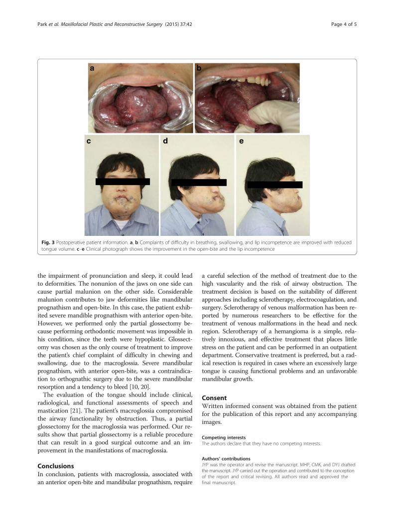
complications of the surgical wound. We prescribed gaba-pentin (300 mg/day) and cetamadol (975 mg/112.5 mg/day) for the pain, under the impression that it was a neuro-pathic pain. These medications improved the patient’s pain.Although he experienced some taste impairment, heshowed an improvement in swallowing and chewing. Therewas no recurrence of the lesions on the tongue in a follow-up, 9 months postoperatively (Fig. 3a–e).
DiscussionVenous malformations are common vascular malforma-tions, presenting at any location, including the head andneck region. They are present at birth, expand slowly dur-ing childhood, and often enlarge during trauma, puberty,and pregnancy, due to the hormonal changes occurringduring these periods. They are composed of an abnormalcollection of veins, which are thin-walled, sponge-like chan-nels of variable size, lacking in smooth muscle. In general,they are a bluish compressible mass and tend to slowly ex-pand with time [7, 8]. For example, a venous infiltration ofthe tongue results in macroglossia, which presents a risk ofswelling or bleeding and may impair swallowing and eating.The tongue plays an important role in swallowing, speech,and breathing, as well as in occlusion and skeletal growth.Therefore, a tongue anomaly may cause a malocclusionand result in changes in skeletal growth, such as open-bitedeformity and mandibular prognathism [9, 10].Venous malformations can be managed by observation,
irradiation, electrocoagulation, cryotherapy, low-dose as-pirin, sclerotherapy, surgical excision, or combinations of
Fig. 1 Preoperative patient information. a The tongue is interpositioned between the teeth, interfering with chewing. b The patient shows severemandible prognathism, with anterior open-bite. c 3-D facial computed tomography view reveals the calcification to be away from the mandiblebody. d Panoramic image shows phleboliths around the mandible body. e Clinical photograph shows the macroglossia
Park et al. Maxillofacial Plastic and Reconstructive Surgery (2015) 37:42 Page 2 of 5
these treatments [1, 11, 12]. The treatment of venous mal-formation is based on the anatomic regions of the body, thetype of tissue, complications such as bleeding, and the func-tional factors. Therefore, Doppler ultrasonography, mag-netic resonance imaging, and direct injection venographymay be required to confirm the diagnosis and provide use-ful information [3]. Sclerotherapy is commonly the pre-ferred treatment method for venous malformations, withsurgery performing an adjunctive role. It is a good alterna-tive for venous malformation, given that surgical resectioncould lead to considerable functional impairment [13].There are various agents for sclerotherapy, for example,OK-432, ethanol, bleomycin, doxycycline, sodium tetradecylsulfate, and hypertonic saline, alone or in combination. Thepotential complications of sclerotherapy include skin andmucosal injuries, swelling, infection, transient nerve palsy,hemoglobinuria, blood loss, and anaphylaxis [14, 15]. Abso-lute sclerotherapy is recommended alone or with surgery.In our case, surgery was indicated because repetitive sclero-therapy had been ineffective due to the extent of the lesion.Surgery is imperative for extensive lesions.If airway obstruction is suspected, the treatment should
be started even if the lesion is large [16]. Surgical ex-cision results in excellent outcomes only for localized
and accessible lesions. Even though surgical excision is themost effective treatment, an excessive excision brings aboutmotor dysfunction, massive bleeding, cosmetic problems,and nerve damage in patients with widespread lesions be-cause of the complicated anatomy of the head and neck re-gion [3, 17]. Our patient had had sclerotherapy severaltimes before surgery, without any improvement. The mostclinically concerning aspect to plan a glossectomy was thepostoperative airway management [18]. Because the lesioninvolved the neck around the trachea, a tracheostomycould have caused uncontrollable bleeding through the sur-gical site. Postoperative airway management was performedby maintaining postoperative nasotracheal intubation forseveral days. However, the postoperative swelling of thetongue was relatively extended, and therefore, extubationwas possible only a week after the operation. In the imme-diate postoperative period after a glossectomy, the tonguecan swell to a size larger than the preoperative size. Hence,nasotracheal intubation must be maintained for severaldays after surgery, till swelling subsides [19].Macroglossia caused by venous malformation can lead
to several problems like dentoskeletal deformities, masti-catory, and breathing difficulties. If the macroglossia con-tinues to damage the oropharyngeal function and cause
Fig. 2 Clinical photos of the surgery. a The tongue is protruding between the teeth, under general anesthesia. b Glossectomy design. c The tongue istaken out of the mouth. d Resected tongue measures 7 cm× 10 cm× 4 cm. e After glossectomy, a drain is inserted in the anterior part of the tongueto prevent edema
Park et al. Maxillofacial Plastic and Reconstructive Surgery (2015) 37:42 Page 3 of 5
the impairment of pronunciation and sleep, it could leadto deformities. The nonunion of the jaws on one side cancause partial malunion on the other side. Considerablemalunion contributes to jaw deformities like mandibularprognathism and open-bite. In this case, the patient exhib-ited severe mandible prognathism with anterior open-bite.However, we performed only the partial glossectomy be-cause performing orthodontic movement was impossible inhis condition, since the teeth were hypoplastic. Glossect-omy was chosen as the only course of treatment to improvethe patient’s chief complaint of difficulty in chewing andswallowing, due to the macroglossia. Severe mandibularprognathism, with anterior open-bite, was a contraindica-tion to orthognathic surgery due to the severe mandibularresorption and a tendency to bleed [10, 20].The evaluation of the tongue should include clinical,
radiological, and functional assessments of speech andmastication [21]. The patient’s macroglossia compromisedthe airway functionality by obstruction. Thus, a partialglossectomy for the macroglossia was performed. Our re-sults show that partial glossectomy is a reliable procedurethat can result in a good surgical outcome and an im-provement in the manifestations of macroglossia.
ConclusionsIn conclusion, patients with macroglossia, associated withan anterior open-bite and mandibular prognathism, require
a careful selection of the method of treatment due to thehigh vascularity and the risk of airway obstruction. Thetreatment decision is based on the suitability of differentapproaches including sclerotherapy, electrocoagulation, andsurgery. Sclerotherapy of venous malformation has been re-ported by numerous researchers to be effective for thetreatment of venous malformations in the head and neckregion. Sclerotherapy of a hemangioma is a simple, rela-tively innoxious, and effective treatment that places littlestress on the patient and can be performed in an outpatientdepartment. Conservative treatment is preferred, but a rad-ical resection is required in cases where an excessively largetongue is causing functional problems and an unfavorablemandibular growth.
ConsentWritten informed consent was obtained from the patientfor the publication of this report and any accompanyingimages.
Competing interestsThe authors declare that they have no competing interests.
Authors’ contributionsJYP was the operator and revise the manuscript. MHP, CMK, and DYJ draftedthe manuscript. JYP carried out the operation and contributed to the conceptionof the report and critical revising. All authors read and approved thefinal manuscript.
Fig. 3 Postoperative patient information. a, b Complaints of difficulty in breathing, swallowing, and lip incompetence are improved with reducedtongue volume. c–e Clinical photograph shows the improvement in the open-bite and the lip incompetence
Park et al. Maxillofacial Plastic and Reconstructive Surgery (2015) 37:42 Page 4 of 5

Authors’ informationAll of the authors have no affiliations with or involvement in any organization orentity with any financial interest or non-financial interest in this manuscript. Thismanuscript represents original works and is not being considered for publicationelsewhere.
Received: 30 September 2015 Accepted: 10 November 2015
References1. Bowman J, Johnson J, McKusick M, Gloviczki P, Driscoll D (2013) Outcomes
of sclerotherapy and embolization for arteriovenous and venousmalformations. In: Seminars in vascular surgery, vol 1. Elsevier, pp 48-54
2. Kobayashi K, Nakao K, Kishishita S, Tamaruya N, Monobe H, Ki S et al (2013)Vascular malformations of the head and neck. Auris Nasus Larynx 40(1):89–92
3. Zheng JW, Mai HM, Zhang L, Wang YA, Fan XD, Su LX et al (2013) Guidelinesfor the treatment of head and neck venous malformations. Int J ClinExp Med 6(5):377
4. de Lorimier AA (1995) Sclerotherapy for venous malformations. J PediatrSurg 30(2):188–194
5. Rabe E, Pannier F (2013) Sclerotherapy in venous malformation. Phlebology28(suppl 1):188–191
6. Hammer FD, Boon LM, Mathurin P, Vanwijck RR (2001) Ethanolsclerotherapy of venous malformations: evaluation of systemic ethanolcontamination. J Vasc Interv Radiol 12(5):595–600
7. Mullican J, Young A (1988) Vascular birthmarks: hemangiomas andmalformation. PA Saunders
8. Waner M, Suen JY (1999) Hemangiomas and vascular malformations of thehead and neck, vol 487. Wiley-liss
9. Turvey TA, Journot V, Epker BN (1976) Correction of anterior open bitedeformity: a study of tongue function, speech changes, and stability.J Maxillofac Surg 4:93–101
10. Wolford LM, Cottrell DA (1996) Diagnosis of macroglossia and indicationsfor reduction glossectomy. Am J Orthod Dentofacial Orthop 110(2):170–177
11. Nguyen JT, Koerper MA, Hess CP, Dowd CF, Hoffman WY, Dickman M et al(2014) Aspirin therapy in venous malformation: a retrospective cohort studyof benefits, side effects, and patient experiences. Pediatr Dermatol 31(5):556–560
12. Rubin BA, Brunswick A, Riina H, Kondziolka D (2014) Advances in radiosurgeryfor arteriovenous malformations of the brain. Neurosurgery 74:S50–S59
13. Lee B, Do Y, Byun H, Choo I, Kim D, Huh S (2003) Advanced managementof venous malformation with ethanol sclerotherapy: mid-term results. J VascSurg 37(3):533–538
14. Richter GT, Friedman AB (2012) Hemangiomas and vascular malformations:current theory and management. Int J Pediatr 2012:1-10.
15. Wiegand S, Eivazi B, Zimmermann AP, Sesterhenn AM, Werner JA (2011)Sclerotherapy of lymphangiomas of the head and neck. Head Neck 33(11):1649–1655
16. Adams DM, Lucky AW (2006) Cervicofacial vascular anomalies. I. Hemangiomasand other benign vascular tumors. In: Seminars in pediatric surgery, vol 2.Elsevier, pp 124-132
17. Costa SAP, Brinhole MCP, Silva RA, Santos DH, Tanabe MN (2013) Surgicaltreatment of congenital true macroglossia. Case reports in dentistry 2013
18. Glade RS, Richter GT, James CA, Suen JY, Buckmiller LM (2010) Diagnosisand management of pediatric cervicofacial venous malformations:retrospective review from a vascular anomalies center. Laryngoscope120(2):229–235
19. Nargozian C (2004) The airway in patients with craniofacial abnormalities.Pediatr Anesth 14(1):53–59
20. X-c J (2005) Surgical management of lymphangiomatous orlymphangiohemangiomatous macroglossia. J Oral MaxillofacSurg 63(1):15–19
21. Lazarus CL, Logemann JA, Pauloski BR, Rademaker AW, Larson CR, Mittal BBet al (2000) Swallowing and tongue function following treatment for oraland oropharyngeal cancer. J Speech Lang Hear Res 43(4):1011–1023
Submit your manuscript to a journal and benefi t from:
7 Convenient online submission
7 Rigorous peer review
7 Immediate publication on acceptance
7 Open access: articles freely available online
7 High visibility within the fi eld
7 Retaining the copyright to your article
Submit your next manuscript at 7 springeropen.com
Park et al. Maxillofacial Plastic and Reconstructive Surgery (2015) 37:42 Page 5 of 5