glomerular mesangial cell matrix in - ucl …discovery.ucl.ac.uk/1306802/1/1306802.pdf · 1...
TRANSCRIPT

1
INTERACTION OF MONOCYTES WITH
GLOMERULAR MESANGIAL CELL MATRIX IN
THE PATHOGENESIS OF GLOMERULAR INJURY
ENAM UR RAHMAN
A THESIS SUBMITTED TO THE FACULTY OF MEDICINE
UNIVERSITY OF LONDON
FOR THE DEGREE OF
DOCTOR OF PHILOSOPHY
CENTRE FOR NEPHROLOGY
UNIVERSITY COLLEGE LONDON MEDICAL SCHOOL
ROWLAND HILL STREET, HAMPSTEAD
LONDON NW3 2PF

2
ABSTRACT
Acute inflammatory kidney diseases may resolve, leaving limited residual
damage or progress to cause chronic renal scarring characterized by glomerulosclerosis
and interstitial fibrosis. Understanding the mechanisms that control inflammation within
the kidney may facilitate the development of treatment strategies to prevent irreversible
kidney damage and slow progression of chronic kidney disease. Infiltration of
mononuclear cells is recognized as an early event in many different conditions that may
ultimately lead to kidney injury. Having extravasated from blood vessels at sites of
injury, these multifunctional cells differentiate into tissue macrophages, which
depending on their phenotype, have the potential to both promote resolution of
inflammation or to cause scarring, making them an attractive target for therapy. Having
left the glomerular capillary lumen, mononuclear cells are very likely to encounter the
mesangial matrix. It was therefore hypothesized that interactions between monocytes
and matrix components might modify the behavior of the infiltrating cells and thereby
modify the outcome of the inflammatory process.
The work presented in this thesis demonstrates that mesangial matrix activates
monocytes leading to expression of peroxisome proliferators activated receptor γ and the
CD36 scavenger receptor, both markers of macrophage differentiation. Since LDL
accumulation in the mesangium may contribute to glomerular injury, the interaction
between this lipoprotein and the matrix was also examined. These studies demonstrated
that LDL becomes oxidized when exposed to matrix components, possibly due to loss of
protective antioxidants. The presence of oxidized LDL has the potential to induce
3
mesangial cell chemokine production, which is likely to promote further monocyte
influx into the glomerulus. Furthermore, matrix-activated monocytes internalized
oxidized LDL via CD36 scavenger receptor, leading to foam cell formation, a
recognized characteristic feature of glomerular injury. Foam cell formation may in turn
amplify and perpetuate the disease process by driving further production of cytokines
and growth factors.
Finally, to establish that these observations were relevant to human glomerular
disease, the presence of macrophages expressing PPAR-γ and the CD36 scavenger
receptor in human kidney biopsy samples taken from patients with inflammatory
glomerular disease was demonstrated, using sections from non-inflamed kidneys as
controls. These observations imply that monocyte-matrix interactions are important in
the context of glomerular disease and may represent a potential target for therapies
designed to limit injury resulting from glomerular inflammation.

4
ACKNOWLEDGEMENTS
The experimental studies presented in this thesis were carried out at the Centre
for Nephrology, Royal Free Campus, University College London. The work was
conducted by the author unless stated otherwise and supervised by Dr. David Wheeler.
I am grateful to Dr. Jill Norman, Dr. Zac Varghese, Professor Stephen Powis and
Professor James Owen for their kind support during the execution of this work. I thank
Dr. Ravinder Chana and Dr. Xiong-Zhong Ruan who taught me the techniques of
mesangial cell culture and who provided invaluable advice and encouragement
throughout the project. I also thank Mr. James Gaya for his help with staining human
kidney sections for macrophage activation markers and Professor Alan Phillips for
allowing me access to his departmental scanning electron microscope.
Finally, I am heartily thankful to my supervisor, Dr. David Wheeler for the
encouragement and guidance provided whilst conducting my experimental work and
preparing this thesis.

5
PUBLICATIONS ARISING FROM THIS WORK
Rahman EU, Ruan XZ, Chana RS, Brunskill NJ, Gaya J, Powis SH, Varghese Z,
Moorhead JF, Wheeler DC. Mesangial matrix-activated monocytes express functional
scavenger receptors and accumulate intracellular lipid. Nephrol Dial Transplant. 2008
Jun;23(6):1876-85.
Chana RS, Martin J, Rahman EU, Wheeler DC. Monocyte adhesion to mesangial
matrix modulates cytokine and metalloproteinase production. Kidney Int. 2003
Mar;63(3):889-98.

6
CONTENTS
ABSTRACT......................................................................................................................2
ACKNOWLEDGEMENTS.............................................................................................4
PUBLICATIONS ARISING FROM THIS WORK .....................................................5
LIST OF FIGURES .......................................................................................................12
LIST OF TABLES .........................................................................................................16
LIST OF ABBREVIATIONS .......................................................................................17
CHAPTER 1. GENERAL INTRODUCTION ............................................................21
1.1. THE NORMAL GLOMERULUS ..................................................................................22
1.2. GLOMERULAR MESANGIAL CELLS .........................................................................26
1.3. THE MESANGIAL CELL MATRIX.............................................................................27
1.4. THE GLOMERULUS IN DISEASE...............................................................................29
1.5. MESANGIAL CELL MATRIX IN GLOMERULAR DISEASE ..........................................30
1.6. THE MONOCYTE – MACROPHAGE LINEAGE............................................................32
1.7. MONOCYTES ..........................................................................................................33
1.8. MACROPHAGES ......................................................................................................34
1.8.1. Macrophage Heterogeneity............................................................................35
1.9. MONOCYTES/MACROPHAGES IN GLOMERULAR INJURY.........................................38

7
1.9.1. The Potential For Monocyte-Matrix Interactions To Influence Glomerular
Injury........................................................................................................................38
1.10. LIPIDS IN GLOMERULAR INJURY...........................................................................40
1.11. MACROPHAGE DEACTIVATION AS A TARGET FOR THERAPY ................................42
1.12. AIMS OF THIS WORK ............................................................................................44
CHAPTER 2. GENERAL METHODS........................................................................46
2.1. HUMAN MESANGIAL CELL CULTURE .....................................................................47
2.1.1. Materials ........................................................................................................48
2.1.2. Method for Human Mesangial Cell (HMC) culture.......................................49
2.1.3. Media and growth condition ..........................................................................50
2.1.4. Passaging cells...............................................................................................50
2.2. MESANGIAL CELL MATRIX ISOLATION ..................................................................51
2.3. IMMUNOHISTOCHEMICAL LABELLING ....................................................................52
2.4. FIBRONECTIN ASSAY..............................................................................................53
2.5. CULTURE OF HUMAN PERIPHERAL BLOOD MONONUCLEAR CELLS .......................53
2.5.1. Materials ........................................................................................................53
2.5.2. Methods of peripheral human blood mononuclear cell culture.....................54
2.6. CULTURE OF THE HUMAN MONOCYTE CELL LINE THP-1.....................................55
2.6.1. Materials ........................................................................................................55
2.6.2. Method of human monocyte THP-1 cell culture ............................................56
2.7. CULTURE OF THE HUMAN MONOCYTE CELL LINE U937.......................................57

8
2.8. FLOW CYTOMETRY ANALYSIS OF MONOCYTES .....................................................57
2.9. SCANNING ELECTRON MICROSCOPY ......................................................................58
2.10. MONOCYTE ADHESION ASSAY.............................................................................59
2.11. CYTOKINE PRODUCTION.......................................................................................60
2.12. THYMIDINE INCORPORATION................................................................................61
2.13. GELATINOLYTIC ACTIVITY...................................................................................62
2.14. ANALYSIS OF TIMP I AND TIMP II .....................................................................62
2.15. RNA ISOLATION...................................................................................................63
2.15.1. Materials ......................................................................................................63
2.15.2. Steps to avoid Ribonuclease contamination.................................................65
2.15.3. Total RNA purification.................................................................................65
2.16. RT-PCR...............................................................................................................66
2.16.1. Materials ......................................................................................................67
2.16.2. Method of RT-PCR.......................................................................................67
2.17. ANALYSIS OF PPAR-γ, CD36 AND SCAVENGER RECEPTOR-A GENE EXPRESSION
USING RT-PCR.............................................................................................................68
2.18. ANALYSIS OF PPAR-γ PROTEIN EXPRESSION USING WESTERN BLOT .................69
2.19. MODIFIED LOWRY ASSAY FOR LIPOPROTEIN AND CELL MEMBRANE PROTEIN
ESTIMATION..................................................................................................................70
2.19.1. Materials ......................................................................................................70
2.19.2. Lowry Assay .................................................................................................71
2.20. PREPARATION OF HUMAN LOW DENSITY LIPOPROTEIN (LDL) ...........................71

9
2.20.1. Materials ......................................................................................................71
2.20.2. LDL isolation ...............................................................................................72
2.20.3. Agarose gel electrophoresis of lipoproteins ................................................74
2.20.3.1. Materials ...................................................................................................74
2.20.3.2. Preparation of reagents ............................................................................75
2.20.3.3. Agarose gel electrophoresis......................................................................75
2.21. LDL ACETYLATION .............................................................................................76
2.22. LDL OXIDATION BY MESANGIAL CELL MATRIX.................................................76
2.23. ANALYSIS OF SCAVENGER RECEPTOR-A ACTIVITY.............................................77
2.24. MORPHOLOGICAL EXAMINATION OF FUNCTIONAL SCAVENGER RECEPTORS ......78
2.25. STAINING OF HUMAN KIDNEY BIOPSY MATERIAL FOR MACROPHAGE
ACTIVATION MARKERS.................................................................................................78
2.26. DATA ANALYSIS ..................................................................................................79
CHAPTER 3. MONOCYTE ADHESION TO MESANGIAL MATRIX
MODULATES CYTOKINE AND METALLOPROTEINASE PRODUCTION....80
3.1. INTRODUCTION.................................................................................................81
3.3. RESULTS..............................................................................................................82
3.3.1. Matrix composition ........................................................................................82
3.3.2. Monocyte characteristics ...............................................................................83
3.3.3. Adherence of monocytes to mesangial cell matrix.........................................83
3.3.4. Inhibition of monocyte adhesion to matrix ....................................................86

10
3.3.5. Adhesion to mesangial matrix stimulates monocyte cytokine secretion ........88
3.3.6. Effects of antibodies on cytokine production .................................................91
3.3.7. Mesangial cell matrix stimulation of MMP release.......................................93
3.3.8. Effect of matrix and soluble proteins on monocyte TIMP secretion..............96
3.4. DISCUSSION .......................................................................................................98
CHAPTER 4. MESANGIAL MATRIX-ACTIVATED MONOCYTES EXPRESS
FUNCTIONAL SCAVENGER RECEPTORS AND ACCUMULATE
INTRACELLULAR LIPID ........................................................................................104
4.1. INTRODUCTION...............................................................................................105
4.3. RESULTS............................................................................................................107
4.3.1. PPAR-γ expression by matrix-activated monocytes.....................................107
4.3.2. CD36 expression by matrix-activated monocytes ........................................110
4.3.3. Scavenger receptor expression by matrix-activated monocytes ..................112
4.3.4. Uptake of modified lipoproteins by matrix-activated monocytes.................115
4.3.5. Oxidation of LDL by mesangial cell matrix.................................................118
4.3.6. Identification of macrophage activation markers in human kidney biopsy
material ..................................................................................................................120
4.4. DISCUSSION .....................................................................................................122

11
CHAPTER 5. GENERAL DISCUSSION AND CONCLUSION ............................128
5.1. RESEARCH QUESTIONS ADDRESSED IN THIS THESIS ............................................129
5.2. LIMITATIONS OF THE EXPERIMENTAL WORK ......................................................130
5.2.1. Monocyte Binding Studies............................................................................130
5.2.2. Monocyte Activation Studies ........................................................................132
5.2.3. Disease Specific Matrix Modification..........................................................133
5.2.4. Mesangial Cell Matrix Sequesters Cytokines and Growth Factors.............134
5.2.5. Scavenger receptor: Protein Level Expression............................................134
5.3. IMPLICATIONS OF MAJOR FINDINGS.....................................................................134
5.4. POTENTIAL THERAPEUTIC IMPLICATIONS.............................................................135
5.5. CONCLUSION ........................................................................................................136
REFERENCES.............................................................................................................137

12
LIST OF FIGURES
CHAPTER 1
Figure 1.1A The Normal Glomerulus 23
Figure 1.1B Schematic Diagram of a Single Capillary Tuft within
Bowman’s Capsule 24
CHAPTER 2
Figure 2.1 Separation of Human Glomeruli by Differential Sieving 49
CHAPTER 3
Figure 3.1A Electron micrograph of mesangial matrix 84
Figure 3.1B U-937 monocytes adherent to cell matrix 84
Figure 3.2 Adhesion of U-937 monocytes to matrix synthesized by
mesangial cells prestimulated with TGF-β, TNFα and
TGF-β/TNFα 85
Figure 3.3A Inhibition of U-937 monocyte adhesion to matrix produced
by unstimulated mesangial cells 87
Figure 3.3B Inhibition of U-937 monocyte adhesion to matrix produced
by mesangial cells prestimulated with TGF-β/TNFα 87
Figure 3.4A IL-1β production by peripheral blood-derived monocytes
cultured in the presence of mesangial cell matrix 89

13
Figure 3.4B IL-6 production by peripheral blood-derived monocytes
cultured in the presence of mesangial cell matrix 89
Figure 3.4C TNFα production by peripheral blood-derived monocytes
cultured in the presence of mesangial cell matrix 89
Figure 3.5A IL-1β production by monocytes cultured in the presence
of soluble matrix and its protein components 90
Figure 3.5B IL-6 production by monocytes cultured in the presence
of soluble matrix and its protein components 90
Figure 3.5C TNFα production by monocytes cultured in the
presence of soluble matrix and its protein components 90
Figure 3.6A Matrix metalloproteinase (MMP) released by monocytes
cultured in the presence of mesangial cell matrix 94
Figure 3.6B Relative change in metalloproteinase secretion compared
to control (100%) 94
Figure 3.7A Matrix metalloproteinase (MMP) release by monocytes
cultured in the presence of fibronectin 95
Figure 3.7B Relative change in metalloproteinase secretion compared
to control (100%) 95
CHAPTER 4
Figure 4.1A Time-dependent expression of PPAR-γ mRNA by THP-1
monocytes examined by RT-PCR 108

14
Figure 4.1B Density analysis of bands of PPAR-γ mRNA normalised by
subtracting BSA protein control and comparison with
β-actin mRNA 108
Figure 4.2A Western analysis of PPAR-γ protein expression by THP-1
monocytes 109
Figure 4.2B Density analysis of bands of PPAR-γ protein normalised by
comparison with α-actin protein 109
Figure 4.3A Time-dependent expression of CD36 mRNA in response to
mesangial matrix 111
Figure 4.3B Density analysis of CD36 mRNA normalised by subtracting
BSA protein control and comparison with β-actin mRNA 111
Figure 4.4A Scavenger receptor A mRNA expression by THP-1
monocytes incubated in the presence of increasing mesangial
matrix for 48 hours 113
Figure 4.4B Density analysis of scavenger receptor A mRNA normalised
by comparison with β-actin mRNA 113
Figure 4.5A Time-dependent expression of scavenger receptor A
mRNA in response to mesangial matrix 114
Figure 4.5B Density analysis of scavenger receptor A mRNA normalised
by subtracting BSA protein control and comparison with
β-actin mRNA 114
Figure 4.6 Effects of matrix on Ac-LDL uptake by monocyte/macrophages 116

15
Figure 4.7A Visualisation of Ac-LDL uptake by THP-1 monocytes
following exposure to mesangial matrix 117
Figure 4.7B Visualisation of Ac-LDL uptake by THP-1 monocytes
following exposure to BSA, negative control 117
Figure 4.8 Agarose gel electrophoresis demonstrating oxidation of
LDL by mesangial matrix 119
Figure 4.9 Staining of human kidney sections for macrophage
activation markers 121

16
LIST OF TABLES
CHAPTER 1
Table 1.1 Functions of the Glomerular Mesangium 25
Table 1.2 Stimuli for classically and alternatively activated macrophages 37
CHAPTER 3
Table 3.1 Production of cytokines by human macrophages exposed to
anti-VLA-4 (10 μg/ml) and anti-VLA-5 (5 μg/ml) antibodies
for 24 hours 92
Table 3.2 Release of tissue inhibitors of metalloproteinases by human
macrophages incubated on plastic culture plates (control), with
mesangial cell matrix, and LPS for 24 hours 97
17
LIST OF ABBREVIATIONS
Ac-LDL: Acetylated LDL
AgII: Angiotensin II
BHT: Butylated hydroxytoluene
BSA: Bovine serum albumin
CD: Cluster of differentiation
CM: Conditioned medium
CR3: Complement receptor type 3
Dil-Ac-LDL: Ac-LDL, labeled with 1,1’-dioctadecy-3,3,3’,3’,-
tetramethylindocarbocyanine
DMSO: Dimethyl sulphoxide
DNA: Deoxyribonucleic acid
ECM: Extracellular matrix
EDTA: Ethylene diamine tetra acetic acid
ESRD: End-stage renal disease
FACS: Fluorescence-activated cell sorter analysis
FCS: Foetal calf serum
FITC: Fluorescein isothiocyanate
FN: Fibronectin
GBM: Glomerular basement membrane
GFR: Glomerular filtration rate

18
Gp: Glycoprotein
GS: Glomerulosclerosis
GM-CSF: Granulocyte monocyte colony stimulating factor
H2O2 Hydrogen peroxide
Hep Heparin
HLA-DR: Histocompatibility leukocyte antigen-differentiation
region
HMCL: Human mesangial cell line
ICAM: Intracellular adhesion molecule
IL: Interleukin
Ig: Immunoglobulin
IL-1β: Interleukin-1β
LDL: Low density lipoprotein
LDH: Lactate dehydrogenase
LFA: Lymphocyte functional-associated antigen
LPS: Lipopolysaccharide
MadCAM: Mucosal addressin cell adhesion molecule
MAC-1: Adhesion molecule CD11b/CD18
MFI: Mean fluorescence intensity
MC: Mesangial cell
M-CSF: Monocyte colony stimulating factor
MCP-1: Monocyte chemotactic protein

19
MDA: Malondialdehyde
MMP: Matrix metalloproteinase
NaBr: Sodium bromide
NaCl: Sodium chloride
Ox-LDL: Oxidised low-density lipoprotein
PBMC: Peripheral blood mononuclear cell
PBS: Phosphate buffered saline
PCR: Polymerase chain reaction
PDGF: Platelet derived growth factor
PGE: Prostaglandin E
PKC: Phosphokinase C
PMA: Phorbol 12-myristate 13-acetate
Poly I: Polyinosinic acid
PPAR: Peroxisome proliferator activated receptor
RGD: Arginine-Glycine-Aspartic Acid
RNA: Ribonucleic acid
RPMI 1640 Medium: Roswell Park Memorial Institute 1640 medium
RT-PCR: Reverse transcriptase-polymerase chain reaction
SCr: Scavenger receptor
SDS-PAGE: Sodium dodecyl sulphate-polyacrylamide gel
electrophoresis
SD: Standard deviation
SMC: Smooth muscle cell

20
TBARS: Thiobarbituric acid reactive substances
TBA: Thiobarbituric acid
TBS: Tris buffered saline
TGF-β: Tumour growth factor-β
Th: T-helper
TIMP: Tissue inhibitors of metalloproteinase
TNF-α: Tumour necrosis factor-α
UKTS: United Kingdom transplant sharing scheme
VCAM: Vascular Cell Adhesion Molecule
VLA: Very Late Antigen

21
CHAPTER 1
GENERAL INTRODUCTION

22
Glomeruli are susceptible to a variety of inflammatory, metabolic, haemodynamic,
toxic and infectious insults which induce similar clinicopathologic presentations.
Despite advances in understanding the factors that initiate glomerular injury, efforts to
stop or slow the progression of established chronic renal disease have proved largely
unsuccessful. The fact that multiple pathogenic mechanisms result in a similar
histological endpoint suggests that the glomerulus has only a limited repertoire of
responses to injury and that renal scarring can be considered to represent a secondary
phenomenon rather than a specific disease process. Glomerulosclerosis is the final result
of a number of interrelated events leading to permanent glomerular injury. Histological
features of many chronic progressive renal diseases are evidenced by the accumulation
of matrix, macrophages and cholesterol in sclerotic glomeruli. Several factors act
independently or together to play a pivotal role in determining whether the final outcome
of an acute inflammatory glomerular lesion leads to complete resolution or permanent
scarring. The study of these factors may suggest new targets for therapeutic intervention.
1.1. THE NORMAL GLOMERULUS
Blood enters the glomerulus through the afferent arteriole, which branches into
tiny clusters of looping blood vessels, which comprise the glomerular (“capillary”) tuft
(which is actually a highly specialized section of an arteriole). As blood passes through
the tuft, the plasma is filtered through fenestrations in the endothelial cells and the
glomerular basement membrane (GBM), then through spaces between the podocyte foot
processes (in the slit membrane) into Bowman’s space. The capillary endothelium, GBM
and the slit membrane constitute the filtration barrier that collectively filters plasma

23
allowing passage of water, small solutes such as sodium, urea, and glucose, small
proteins and small organic molecules. The filtered blood is then drained from the
glomerular tuft through the efferent arteriole. The GBM is continuous throughout the
glomerulus, surrounding each capillary loop and the ‘stalk’ region of the glomerular tuft.
At the vascular pole, the GBM is continuous with Bowman’s capsule. Between the
glomerular capillaries lie the mesangial cells and the mesangial matrix, which together
provide structural support by surrounding the glomerular capillaries (Figure 1.1a and
1.1b). Some mesangial cells are located outside the glomerulus, between the afferent and
efferent arterioles and are known as Lacis or Goormaghtigh cells.
Figure 1.1a. The Normal Glomerulus
(Image courtesy of PB Works nephrology images)

24
Figure 1.1b. Schematic Diagram of a Single Capillary Tuft within Bowman’s
Capsule
Much of the original work describing the mesangium was concerned with the
ultra-structural appearance of cross-sections of the glomerulus using electron and light
microscopy. According to current understanding, the mesangium consists of mesangial
cells and mesangial cell matrix, which are capable of various tasks Table 1.1. The
interstitial areas of each lobule join at the glomerular stalk and are thereby in direct
continuity with the renin-secreting juxtaglomerular apparatus.
(Image courtesy of PB Works nephrology images)

25
Table 1.1. Functions of the Glomerular Mesangium
The table below gives a summary of the various functions of the Mesangial cell and
Mesangial cell matrix within the mesangium, as reviewed by Professor Detlef
Schlondorff (Schlondorff 1996; Schlondorff and Banas 2009).
Component of Mesangium Function
Mesangial Cell • Production of Matrix.
• Production of tissue inhibitor of metalloproteinases
(TIMPs) and matrix metalloproteinases (MMPs)
which control turnover of mesangial matrix.
• Influence GFR by regulating blood flow through the
glomerular capillaries or by altering capillary surface
area.
• Biological handling and clearance of
macromolecules; advanced glycated end-products
(AGE’s), immune complexes and lipids.
• Exhibit phagocytic activity.
• Production of vasoactive agents; angiotensin II (Ag
II), nitric oxide (NO), prostaglandin E2 (PGE2) and
inflammatory mediators.
Mesangial Cell Matrix • Structural support.
• May potentially trap LDL.
• Has the capacity to oxidise LDL.
• Interacts with mesangial cells and infiltrating cells
via ligands.
• Sequesters cytokines and growth factors.

26
1.2. GLOMERULAR MESANGIAL CELLS
Mesangial cells are similar to vascular smooth muscle cells and are believed to
have several functions in vivo. They contain large amounts of actin, myosin and
tropomyosin indicative of a contractile function. The presence of receptors for the
vasoactive peptide angiotensin II (AgII) support this role (Kreisberg 1983), which is
important in the regulation of glomerular haemodynamics. When glomeruli are
damaged, mesangial cells produce chemotactic factors such as monocyte chemotactic
protein (MCP-1) (Cushing, Berliner et al. 1990), monocyte colony stimulating factor (m-
CSF) (Rajavashisth, Andalibi et al. 1990) and interleukin-1β (IL-1β) (Ku, Thomas et al.
1992). Mesangial cells also secrete matrix, which creates the structural framework for
the glomerular tuft as well as enzymes (and enzyme inhibitors) that maintain the balance
between synthesis and degradation (Michael, Keane et al. 1980; Davies, Coles et al.
1990; Sugiyama, Kashihara et al. 1998; Fogo 1999). Additionally the mesangial cell
possesses phagocytic properties that contribute to the clearance and uptake of
macromolecules from the glomerulus (Schreiner, Kiely et al. 1981; Davies 1994). Thus,
inappropriate activation of mesangial cells may lead to excess matrix production, as well
as release of chemotactic factors resulting in monocyte influx into the mesangium.
Expansion of the mesangium, both due to the deposition of matrix proteins and
an increase of mesangial cell numbers, is seen in kidney disease. Estimates of
proliferation of mesangial cells in vivo suggest a low rate of about 1% per day (Pabst
and Sterzel 1983; Davies 1994). Mesangial cell proliferation appears to play a role in the
progression of glomerular pathology, particularly in the early stages. Matrix expansion is

27
generally preceded by mesangial cell proliferation in experimental models of
glomerulonephritis. Persistent mesangial cell hyperplasia, caused by repeated injury, is
believed to lead to irreversible scarring and eventual loss of glomerular function (Floege,
Eng et al. 1993; Shimizu, Kawachi et al. 1999). The proliferation of mesangial cells is
presumed to be a necessary physiological response required for the reconstitution of
renal tissue.
1.3. THE MESANGIAL CELL MATRIX
The mesangial extracellular matrix fills the spaces between the mesangial cell
and the perimesangial basement membrane, in addition a small amount of matrix may be
found beneath the endothelium. In ultra-structural studies, this matrix has been
characterized as a dense network of elastic microfibrils similar to the connective tissue
of many other organs, and contains a network of intercellular channels that traffic
macromolcules. In immunohistochemical studies, fibronectin is detected within the
mesangium, along with laminin and type IV collagen, whilst type III collagen is found in
the tubulointerstitium and type V collagen in the mesangial interstitium and the GBM
(Oomura, Nakamura et al. 1989; Sugiyama, Kashihara et al. 1998). Fibronectin and
Collagen type IV comprise the major protein components within the mesangial matrix,
and were therefore the components studied in the experimental work described in this
thesis. Minor components present in mesangial matrix include proteins such as laminin,
vitronectin, entactin and proteoglycans. Fibronectin is the only component to be present
exclusively within mesangial matrix and is specifically localized to areas immediately

28
surrounding mesangial cell processes. The other protein components mentioned are
found distributed throughout the mesangial matrix, tubular basement membranes and
Bowman's capsule (Madri, Roll et al. 1980).
Fibronectin is closely associated with microfibrils, thus providing a link between
mesangial cells and other matrix structures (Reale, Luciano et al. 1981; Brown, Andres
et al. 1982; Cohen and Ku 1984). This microfibrillar network also appears to provide a
solid base of contact between mesangial cells and the perimesangial GBM. Microfibrils
are attached to sites in the mesangial cell membrane that serve to anchor intracellular
actin filaments and penetrate the lamina densa to connect with the GBM. Not only does
fibronectin serve to interconnect cells and matrix components, but also interconnects the
microfibrils at their crossing points, so further stabilizing the entire matrix (Schwartz,
Goldfischer et al. 1985). As a result of these interconnections, the microfibrillar network
has sufficient three-dimensional tensile strength to balance distending forces acting in all
directions. The importance of the mesangial matrix as a connecting structure between
mesangial cells and the GBM is demonstrated by studies in which the failure of such
connections produces miroaneurysms of the glomerular capillary tuft (Mosher 1984;
Cohen, Saini et al. 1987; Proctor 1987; Yasuda, Kondo et al. 1996).
Due to its nature, mesangial matrix has the potential to trap large molecules
including lipoproteins such as LDL (Gupta, Rifici et al. 1992; Wheeler and Chana
1993). The involvement of additional factors such as intra-renal hypertension, and
inflammation are necessary for the induction and progression of lipid-induced renal
dysfunction. Foam cells and lipid deposits are found in focal segmental sclerosis in
human renal biopsies (Lee, Lee et al. 1991). Many of the features of progressive
29
glomerular and tubulo-interstitial diseases share biological mechanisms with those of
atherosclerosis.
This may be relevant in the pathogenesis of glomerular disease, particularly
following an insult that has altered the glomerular filtration barrier permeability,
allowing excess amounts of LDL to penetrate the mesangium. This process may
potentially contribute to the formation of foam cells within the mesangium as explored
later in the thesis.
1.4. THE GLOMERULUS IN DISEASE
Glomerular injury results from an initial pathogenic insult and may heal without
consequences or lead to altered function of intrinsic glomerular cells and invasion of
monocytes/macrophages from the circulation. Such changes result initially in mesangial
cell proliferation, but if not resolved, continued cell proliferation leads to hyperplasia
and the concomitant increase in matrix production to glomerulosclerosis. The term
glomerulosclerosis is a non-specific finding on light microscopic examination that can
be seen in any primary glomerular, tubulointerstitial, or vascular kidney disease.
The process may initially involve only a small proportion of glomeruli (focal)
and within these, only certain segments of the tuft (segmental). These localized lesions
may progress to involve the whole glomerulus. The sclerotic areas consist of collapsed
capillary loops obscured by an excess of mesangial matrix.
A variety of early changes are recognized to precede glomerular obliteration. The
basement membrane become thickened, is often detached from the overlying epithelial

30
cells and may be adherent to Bowman’s capsule. Epithelial cells are hypertrophied or
absent at the sites of adhesions and electron microscopy reveals fusion of foot processes.
Subendothelial eosinophilic deposits of hyaline material are found in both sclerotic and
non-sclerotic areas of the glomerular tuft. Mesangial hypercellularity may result both
from an increase in the number of contractile mesangial cells and from invasion of
inflammatory macrophages. The mesangial area is also expanded by an excess
deposition of mesangial cell matrix (Couchman, Beavan et al. 1994).
Lipid deposition is seen within mesangial and epithelial cells both of sclerotic
and non-sclerotic capillary loops but may also occur in interstitial regions (Chana,
Wheeler et al. 2000). These histological changes correlate with the clinical
manifestations of progressive glomerulosclerosis (Klahr, Schreiner et al. 1988; Magil
and Frohlich 1991; Moorhead 1991). Proteinuria usually precedes a reduction in
glomerular filtration rate and renal blood flow. Deterioration of renal function
progresses and is frequently associated with the development of hypertension. Tubular
atrophy leads to a reduction of the renal concentrating ability and impaired acid
secretion.
1.5. MESANGIAL CELL MATRIX IN GLOMERULAR DISEASE
The mesangial matrix is no longer seen as a static scaffold in which cells reside;
but has been shown to be involved in cell proliferation, migration and cell-cell
interactions. Turnover of the different extracellular matrix components is recognised as
an active process with multiple levels of regulation (Sterzel, Schulze-Lohoff et al. 1992;
31
Yasuda, Kondo et al. 1996). Net deposition of matrix proteins, as seen in
glomerulosclerosis, results from both quantitative and qualitative changes to mesangial
matrix (Bruijn, Hogendoorn et al. 1988; Klahr, Schreiner et al. 1988; Olgemoller and
Schleicher 1993; Harendza, Schneider et al. 1999). Changes in mesangial matrix may
modify glomerular function by changing cell-cell interactions and by promoting
infiltration and entrapment of macrophages.
Matrix construction and remodelling involves three factors, matrix
metalloproteinases (MMPs), plasmins that activate latent MMPs and tissue inhibitors of
MMPs (TIMPs) (Raines 2000; Keeling and Herrera 2008). In inflammatory conditions,
levels of growth factors such as TGF-β increase and can act to suppress the expression
of matrix degrading plasminogen-activator inhibitor (PAI), and increase the activity of
tissue inhibitors of metalloproteinases (TIMPs), thus favoring matrix accumulation.
One consequence of mesangial matrix remodelling is that changes within this
complex three-dimensional structure can reveal hidden sites previously unrecognizable
to various adhesion receptors on the surface of cells coming into contact with the matrix.
For example the RGD sequence of the fibronectin molecule is able to bind very late
antigen (VLA)-5 present on the cell surface of monocytes (Pierschbacher and Ruoslahti
1984; Hemler 1990). Also the CS-1 domain of fibronectin binds VLA-4 on monocytes
by an RGD-independent mechanism (Wayner, Garcia-Pardo et al. 1989). Matrix
remodelling, by exposing these sites, may promote monocyte adhesion to this matrix
component.

32
1.6. THE MONOCYTE – MACROPHAGE LINEAGE
Monocytes and macrophages comprise a family of phagocytic cells that are
widely distributed throughout the body and are generally referred to as the mononuclear
phagocyte system because of their common origin, similar morphology and common
functions. Monocytes originate in the bone marrow, but become widely distributed in
tissues where they mature into macrophages and take on specialist roles. Macrophages
are well recognized for their ability to phagocytose, a property that enables them to
eliminate pathogens and other foreign materials. However, these cells play a pivotal role
in a variety of processes including inflammation, the induction and regulation of specific
immune responses and tissue remodelling and repair. There are several basic properties
of these cells that are relevant to their role in glomerular injury.
Firstly, mononuclear phagocytes are highly mobile and have the capacity to
adhere to various biological substrates, a function that facilitates their migration to sites
of inflammation. Secondly, these cells secrete a range of soluble mediators that
modulate functions of many other different types of cells. Thirdly, mononuclear
phagocytes ingest and degrade various materials including senescent cells and tissue
debris. Finally, mononuclear phagocytes can be activated by the external environment.
Whilst mononuclear phagocytes play a critical role in host defense, these cells
may also injure the host while exercising their defensive role. For example, monocytes
have been shown to contribute to tissue damage by releasing proteolytic enzymes
(Campbell, Silverman et al. 1989; Senior, Connolly et al. 1989), toxic oxygen
metabolites (Carp and Janoff 1980; Campbell, Senior et al. 1982), pro-fibrotic cytokines

33
(Martinet, Rom et al. 1987; Shaw 1991) and other mediators (Tracey, Lowry et al.
1986).
Extravasation of monocytes from the vasculature into the mesangium involves
directional migration of cells in response to chemoattractant factors. Since the
mesangium forms the core of each tuft and is in direct contact with plasma constituents
without an intervening membrane, monocytes directly encounter matrix during the
migration stage. It is clear that regulated and reversible adherence of monocytes to
extracellular matrix components is a prerequisite for the accumulation of these cells at
sites of tissue inflammation (Snyderman and Goetzl 1981); however, little is known
about the biological and pathological factors that regulate monocyte adherence to
extracellular matrix and the resulting changes that occur to the monocyte.
1.7. MONOCYTES
Monocytes represent 3 to 8% of peripheral blood leukocytes. These mature cells
measure 12 to 15 μM in diameter and posses a characteristic kidney-shaped nucleus.
Their cytoplasm contains a well-developed Golgi apparatus, numerous lysosomal
granules, microtubules and actin-containing filaments (which are cross-linked by actin-
binding protein and myosin). Monocytes are slowly motile, exhibit phagocytic activity
and have a strong tendency to adhere and spread on glass surfaces (Lasser 1983).
Monocytes give a positive reaction for non-specific esterases and contain peroxidase,
acid phosphatase, lysozyme, aryl sulphatase and glucuronic acid (Yam, Li et al. 1971).
Monocytes express HLA-DR antigens on their surface (McKinney, Boto et al. 1980;
34
Smith and Ault 1981) along with receptors for Fc component of IgG, complement C3
(Huber, Polley et al. 1968; Schwartz, Bianco et al. 1975) and insulin (Schwartz, Bianco
et al. 1975).
1.8. MACROPHAGES
Macrophages measure 20 to 80μM in diameter and contain a large vacuolated
nucleus often with prominent nucleoli. Their cytoplasm contains a large well-developed
Golgi apparatus, abundant rough endoplasmic reticulum and ribosomes, large
mitochondria, microtubules, microfilaments and numerous lysosomes rich in hydrolytic
enzymes.
The transition from monocyte to macrophage is associated with increases in: 1)
the number of lysosomes and mitochondria; 2) the activity of mitochondrial enzymes
and the rate of cellular respiration; 3) phagocytic activity; 4) protein synthesis; 5) the
capacity to interact with lymphocytes (Lasser 1983); 6) the expression of Scavenger
receptor-A (Xu, Yu et al. 2006) and 7) the expression of Peroxisome proliferator-
activated receptor-gamma (PPAR-γ) (von Knethen and Brune 2003).
Macrophages are also facultative anaerobes, with the exception of the pulmonary
alveolar macrophage. They are highly motile and have marked phagocytic activity. In
contrast to monocytes, macrophages have been shown to proliferate in response to
certain stimuli in vitro (Diesselhoff-den Dulk, Crofton et al. 1979; Lasser 1983). As with
monocytes, macrophages also express receptors for the Fc component of IgG, C3
(Griffin, Spertini et al. 1990) and insulin (Bar, Kahn et al. 1977) however macrophages

35
also express receptors for IgE which is important in mediating host immunity to various
parasites (Dessaint, Torpier et al. 1979; Melewicz and Spiegelberg 1980). Some
macrophages express HLA-DR antigens and can function as antigen presenting cells for
lymphocytes. The expression of HLA-DR antigen varies with the type of macrophage;
only 15% of peritoneal macrophages express HLA-DR antigen compared with 50% of
spleen and thymus macrophages (Cowing, Schwartz et al. 1978; Beller and Unanue
1980).
Macrophages are widely distributed throughout the body, but are particularly
prominent in the spleen, lymph nodes, liver (Kupffer Cells), peritoneum, skin
(Langerhans cells) and pulmonary alveoli. Macrophages resident in different tissues
have widely differing morphological and functional properties. It has been postulated
that the profile of local stimuli, to which macrophages are exposed in a particular tissue,
influences their maturation and thereby accounts for their diversity of form and function
(Cline, Lehrer et al. 1978).
1.8.1. Macrophage Heterogeneity
Local factors are important in determining the phenotype adopted by the
recruited monocyte. The resultant tissue macrophages can be broadly divided into two
groups; ‘resident macrophages’ and ‘inflammatory macrophages’(Gordon 2003).
Tissue macrophages are heterogeneous and those isolated from different
anatomical sites differ in function, presumably because of adaptive responses to the local
microenvironment. Inflammatory macrophages are derived largely from circulating

36
monocytes which infiltrate damaged tissue; some also arise by local cell division.
Different macrophage activation states have been recognized and result from exposure to
specific stimuli that initiate differentiation into A) classically or B) alternatively
activated macrophages (see table 1.2).
Classically activated macrophages exhibit a Th1-like phenotype, promoting
inflammation, extracellular matrix destruction, and apoptosis, while alternatively
activated macrophages display a Th2-like phenotype, promoting extracellular matrix
construction, cell proliferation, and angiogenesis (Erwig and Rees 1999; Duffield 2003).
Although both phenotypes are important for clearance of pathogens and apoptotic cells,
the classically activated macrophage tends to elicit chronic inflammation and tissue
injury whereas the alternatively activated macrophage tends to resolve inflammation and
facilitate wound healing.

37
Table 1.2. Stimuli for classically and alternatively activated macrophages.
The table below summarises the various stimuli giving rise to a particular macrophage
activation status as reviewed by Dr. Jeremy Duffield, a prominent author within the field
inflammation research (Duffield 2003).
Macrophage Activation Status Stimulus
Classical Activation
Pro-inflammatory cytokines
Abnormal matrix
Hypoxia
Bacterial DNA
Alternative Activation
IL-4
IL-10
IL-13
TGF-β
As a result of their opposing phenotypic states, macrophages play a central role
in innate protection both through the clearance of infective pathogens and through the
repair of tissue injury that occurs, in part, as a consequence of this response. For
example, the initial response of infiltrating monocytes to bacterial infection results in
macrophage differentiation favoring a classically activated phenotype and so is cytotoxic
and proinflammatory; then, once the infection is under control, macrophages
phagocytose cellular debris and apoptotic bodies and begin tissue repair (Duffield 2003;
Erwig, Kluth et al. 2003).

38
1.9. MONOCYTES/MACROPHAGES IN GLOMERULAR INJURY
In the normal kidney there are small numbers of interstitial leukocytes thought to
perform an immune surveillance function. These are predominantly monocytes. Only a
small fraction of leukocytes in the normal kidney comprise B cells, T cells, natural killer
cells, and neutrophils.
In an acutely inflamed glomerulus, the predominant leukocyte is the macrophage.
In human glomerular disease, macrophage numbers correlate with the extent of
histological damage at the time of biopsy and predict renal outcome in certain disease
settings (Ootaka, Saito et al. 1997). Macrophages expressing activation and proliferation
markers have been identified in more aggressive forms of human and experimental
glomerulonephritis and correlate with disease severity (Kerr, Nikolic-Paterson et al.
1994; Lan, Nikolic-Paterson et al. 1995; Yang, Isbel et al. 1998). Strategies that limit
disease progression in this setting include (A) the systemic depletion of macrophages;
(B) Inhibition of pro-inflammatory cytokines that both activate and are produced by
activated macrophages; and (C) the blocking of factors that promote the recruitment of
macrophages to tissue sites (e.g; blockade of cytokines and adhesion molecules).
1.9.1. The Potential For Monocyte-Matrix Interactions To Influence Glomerular
Injury
The mesangial matrix has the potential to play a key role in monocyte
differentiation (Sugiyama, Kashihara et al. 1998; Jacob, Shastry et al. 2002). There are

39
two main mechanisms by which matrix can influence cell behavior. Firstly matrix
harbours growth factors and growth factor binding proteins (Ignotz, Heino et al. 1989;
Lee and Streuli 1999). These factors are passively sequestering but may be actively
released by remodelling enzymes such as the MMPs and may thereby influence the
behaviour of cells that are exposed to matrix components. Secondly, matrix can directly
regulate cells via receptor-mediated signalling (Schoecklmann, Rupprecht et al. 1996;
Gauer, Yao et al. 1997; Hamerski and Santoro 1999). Since monocytes enter the
mesangium through the fenestrated endothelium, they are unlikely to have undergone
endothelial activation and therefore may encounter mesangial matrix in an inactivated
state. The first activation signals that these cells encounter may therefore take the form
of receptor-mediated signalling by matrix components. This monocyte-matrix
interaction may play a key role in influencing monocyte behaviour within the
glomerulus and the resulting macrophage phenotype is likely to be highly dependant on
the local microenvironment within the glomerulus. For example, in a disease situation,
matrix remodelling may be disrupted, leading to qualitative and quantitative changes in
matrix composition which in turn may influence the sequestration of growth factors. A
change in the composition of matrix components may potentially trigger different signal
transduction cascades which, in turn, may influence monocyte behavior. (Wesley, Meng
et al. 1998; Ingram, Ly et al. 1999; Urushihara, Takamatsu et al. 2010). Furthermore,
subtle changes in the matrix composition resulting from enzymatic digestion may release
bioactive matrix fragments or expose sequestered growth factors.
As an example, fibronectin, one of the major matrix proteins, binds to very late
antigen (VLA)-5 integrin subunits on the surface of monocytes. Binding of fibronectin

40
to this receptor depends on its conformation. Since fibronectin production is increased in
diseased mesangial matrix, it is conceivable that an excess of this matrix component or
conformational changes might influence these signalling pathways and thereby modulate
monocyte activation and behaviour. The same may be true of collagen type IV, another
matrix protein that is increased in diseased mesangial cell matrix. The RGD sequence of
the fibronectin molecule binds VLA-5 present on the cell surface of monocytes thus
signalling to the cell (Pierschbacher and Ruoslahti 1984; Hemler 1990). Likewise the
CS-1 domain of fibronectin binds VLA-4 on monocytes by an RGD-independent
mechanism (Wayner, Garcia-Pardo et al. 1989).
1.10. LIPIDS IN GLOMERULAR INJURY
The pathological effects of lipoprotein in progressive kidney disease may be
similar to those in atherosclerosis. In recent years, an improved understanding of
atherosclerosis has illuminated the pathology of glomerulosclerosis and supported the
concept of lipoproteins as mediators of renal disease (Moorhead, Brunton et al. 1997).
The possible role of lipoproteins in progressive renal disease may be understood in the
more familiar context of atherosclerosis.
Atherosclerosis results from a complex sequence of events in which normal
cycling of LDL through the vascular endothelium is altered, leading to trapping of LDL.
In addition, monocytes are recruited from the blood, smooth muscle cells proliferate and
fibrous tissue is deposited. Trapped LDL may become oxidized, partly as a result of pro-
oxidant factors produced by monocytes (Parthasarathy, Printz et al. 1986; Quinn,

41
Parthasarathy et al. 1987; Boullier, Bird et al. 2001). The similarity between
atherosclerosis and glomerulosclerosis is based on the assumption that the glomerulus
posses cell types which are known to respond to lipoprotein injury, namely monocytes,
or which resemble smooth muscle, namely the mesangial cell. Thus the mechanisms
involved in atherosclerosis may also apply to glomerulosclerosis.
Oxidised LDL (Ox-LDL) has been demonstrated to be more cytotoxic when
compared with unmodified native LDL (Fernando, Varghese et al. 1993). Ox-LDL has
also been proven to be important in lesion progression in atherosclerosis since uptake by
macrophages via scavenger receptors causes the generation of foam cells. A similar
mechanism may play an important role in lipid-mediated glomerulosclerosis. Ox-LDL
has also been shown to stimulate monocyte influx (Pai, Kirschenbaum et al. 1995) by
inducing mesangial cells to release chemotactic cytokines.
Another prominent feature of lipid-induced glomerular injury is the accumulation
of mesangial cell matrix. Studies in vitro indicate that lipid-activated mesangial cells
produce excess matrix (Schlondorff 1993; Wheeler and Chana 1993; Lee 1999) as has
been described in the atheromatous artery (Ross 1984). Lee et al has demonstrated that
LDL stimulates mesangial cells through the induction of the phosphokinase C (PKC)
pathway to synthesize TGF-β, which favours matrix production (Lee 1999). Chana et al
reported that LDL also selectively enhances the synthesis of specific proteoglycans and
hyaluronan in mesangial cells (Chana, Wheeler et al. 2000). The incubation of mesangial
cells with native LDL (25-100 μg/ml) increased the synthesis and secretion of both
fibronectin and laminin in a dose-dependant manner. Similarly, oxidized forms of LDL
42
(25-100 μg/ml) increased fibronectin and laminin and had a greater effect than native
LDL (Roh, Kamanna et al. 1998).
Therefore, lipoproteins and modified lipoproteins may get trapped in the
mesangial matrix, become oxidized and get taken up by infiltrating
monocyte/macrophages which form foam cells.
1.11. MACROPHAGE DEACTIVATION AS A TARGET FOR THERAPY
The exact mechanism of macrophage activation and accumulation within the glomerulus
is largely unknown, although there is a considerable amount of experimental evidence
implicating adhesion molecules as being relevant in the setting of glomerulosclerosis.
Many of the interactions between adhesion molecules and infiltrating macrophages have
been successfully blocked and could serve as targets for therapeutic interventions (Adler
and Brady 1999; Allen, McHale et al. 1999; Chana and Wheeler 1999; Cook, Khan et al.
2002). For example, the beta-1 integrin, α4β1, also known as very late antigen 4 (VLA-
4), is present on macrophages and binds to VCAM-1, which has been shown to be up-
regulated in the glomerular endothelium in experimental glomerulonephritis. Blocking
using anti-α4 antibodies can prevent experimental crescentic glomerulonephritis as
demonstrated by Allen et al (Allen, McHale et al. 1999) and has been shown to halt
progression of established disease (Khan, Allen et al. 2003), making it a very attractive
candidate for therapy. The humanized version of this anti-α4 monoclonal antibody
known as Natalizumab has been used successfully in multicenter double-blind controlled
studies in Crohn's disease and multiple sclerosis (Ghosh, Goldin et al. 2003; Miller,

43
Khan et al. 2003; Miller, Soon et al. 2007; Targan, Feagan et al. 2007). Despite its initial
approval Natalizumab was withdrawn from the market by its manufacturer after it was
linked with three cases of the rare neurological condition progressive multifocal
leukoencephalopathy (PML) when administered in combination with interferon β-1a,
another immunosuppressive drug often used in the treatment of multiple sclerosis (Tyler
and Khalili 2005; Ransohoff 2007). After a review of safety information and no further
deaths, the drug was returned to the US market in 2006 under a special prescription
program. As of June 2009, ten cases of PML were known. However, twenty-four cases
of PML had been reported since its reintroduction by October 2009, showing a sharp rise
in the number of fatalities and prompting a review of the chemical for human use. By
January 2010, 31 cases of PML were attributed to natalizumab, however it was not
withdrawn from the market because its clinical benefits outweighed the risks involved
(Ransohoff 2010; Steiner 2010).
The beta-2 integrins include CD11a/CD18 (LFA-1) and CD11b/CD18 (Mac-
1/Complement receptor 3). A humanized monoclonal antibody to CD18 known as
Efalizumab, which blocks CD11a/CD18 and CD11b/CD18 has been reported to reduce
infiltrating leukocytes and improve vasculitic ulcers in four of five patients with
systemic vasculitis (Lockwood, Elliott et al. 1999). Despite these beneficial effects
Efalizumab was withdrawn from the market ten years later as it was associated in some
cases with fatal brain infections (Major 2010).

44
1.12. AIMS OF THIS WORK
Monocyte/macrophage accumulation within the glomerular mesangium is a
recognized feature of glomerular injury in man. Although the mechanisms of
macrophage trafficking and activation within the glomerulus are not properly
understood, it is generally assumed that these cells are derived from circulating
monocytes that migrate from the glomerular capillary lumen. During this process the
monocyte encounters many extracellular signals that promote differentiation to a
macrophage and other cellular responses. Recent studies suggest that such interactions
may program macrophages, thereby potentially modifying their behavior in the setting
of acute or chronic glomerular disease (Erwig, Kluth et al. 1998; Min, Lyons et al.
2009).
To begin to address the pathobiological importance of alterations in monocyte
phenotype following interaction with matrix components, there is the need to firstly
identify the nature of the interaction taking place and the resulting changes in monocyte
phenotype. A representative matrix component (fibronectin) was used to conduct
blocking studies to examine the extent to which binding to matrix modifies the secretory
behavior of monocytes.
Secondly to address the extent of monocyte differentiation into macrophages
upon exposure to matrix, three macrophage specific markers were studied: a) the
peroxisomal proliferator-activated receptor-γ (PPAR−γ), a nuclear receptor that acts as a
transcriptional mediator for genes involved in lipid metabolism and adipogenesis
45
((Moore, Rosen et al. 2001), b) CD36, a class B scavenger receptor and c) Scavenger
receptor class-A.
Finally since LDL accumulation in the mesangium may contribute to glomerular
injury, the interaction between this lipoprotein and the mesangial matrix was examined.

46
CHAPTER 2
GENERAL METHODS

47
2.1. HUMAN MESANGIAL CELL CULTURE
Techniques used for the isolation and maintenance of mesangial cells in vitro
have been refined since these cells were first cultured in vitro in the late 1970’s. The two
methods for mesangial cell culture used are enzymatic isolation (Striker and Striker
1985) and explantation (Kreisberg and Karnovsky 1983; Striker, Lange et al. 1987). The
starting material used for both methods is glomeruli isolated by differential sieving
techniques.
Enzymatic isolation uses collagenases to partially digest away the glomeruli, thus
exposing the glomerular ‘cores’ comprising capillary loops and mesangium depleted of
endothelial cells and the majority of epithelial cells. These cores are then explanted in to
plastic cell culture flasks, and give rise to a heterogeneous outgrowth of cells within 2-4
days.
The choice of a culture medium with a high serum concentration (10-20%)
promotes growth of mesangial cells rather than endothelial or glomerular epithelial cells,
which require lower serum concentrations. The fact that mesangial cells attach more
readily to plastic than endothelial and epithelial cells also aids in their purification. Thus
within 2-3 passages, homogenous mesangial cell cultures are obtained.
The alternative method of mesangial cell isolation (the explantation method),
involves plating of undigested glomeruli into plastic flasks and use of high
concentrations of foetal calf serum (FCS). Due to the different growth rates of intrinsic
glomerular cell, the timing of subculture is used to select the cell type of interest. Both
the enzymatic isolation and explantation methods provide reproducible and reliable
48
mesangial cell cultures, although cells have to be subcultured several times before
sufficient numbers can be obtained for use in experiments. These cells retain many of
the morphological and functional characteristics of mesangial cells in vivo (Lee 1995).
2.1.1. Materials
1) RPMI 1640 (Gibco BRL, Paisley, UK)
2) Foetal calf serum (Gibco BRL, Paisley, UK)
3) Glutamine (Sigma, Dorset, UK)
4) Penicillin (Sigma, Dorset, UK)
5) Streptomycin (Sigma, Dorset, UK)
6) ITS (insulin-human transferrin-sodium selenite) (Sigma, Dorset, UK)
7) Trypsin, ethylene diamine tetra acetic acid (EDTA) (0.025% and 0.01% respectively)
8) 75 cm2 and 25 cm2 conical flasks (Falcon, UK)
9) Sterile stainless steel sieves with the following mesh sizes: 425μm, 180μm and
125μm.
10) Glass syringe plunger (20ml)
11) Collagenase (Type 1A) (Sigma, Dorset, UK)

49
2.1.2. Method for Human Mesangial Cell (HMC) culture
Human cadaver kidneys that could not be used for transplantation for technical
reasons were used as a source of cultured cells with permission from the United
Kingdom Transplant Sharing Scheme (UKTS). Under sterile conditions, the capsule was
removed and the cortex excised from the underlying medulla. Cortical fragments were
minced to a pulp and pushed through a 425μm mesh stainless steel sieve using a plastic
syringe (20ml) plunger. The material on the underside of the sieve was washed through a
180μm sieve and the glomeruli retained on the top surface of a third sieve with a mesh
diameter of 125μm (Figure 2.1).
Figure 2.1. Separation of Human Glomeruli by Differential Sieving
These glomeruli were collected by aspiration, pelleted by centrifugation at
1000g, placed in collagenase (type 1A) solution (400-600 units/ml) and digested for 20
minutes at 37oC. The digestion was arrested by adding medium containing 20% FCS, the

50
glomeruli were harvested by centrifugation and plated on 25cm2 or 75cm2 Falcon tissue
culture flasks depending upon the number of glomeruli obtained.
2.1.3. Media and growth condition
Growth medium consisted of 80% RPMI-1640 (Gibco BRL, Paisley, UK) and
20% FCS (Life Technologies, Paisley, UK), supplemented with insulin (5μg/ml) human
transferrin (5μg/ml), and sodium selenite (5ng/ml) (Insulin-Transferrin-Sodium selenite
media supplement, Sigma). Benzyl penicillin (100 units/ml), and Streptomycin sulphate
(50 μg/ml) were added to minimise the risks of infection.
2.1.4. Passaging cells
Primary cultures were left undisturbed for 9 days after which time the growth
medium was changed. Glomeruli attached to the plastic within 3-4 days and after an
initial outgrowth of epithelial cells (day 7-14), human mesangial cells began to
predominate and outgrew the epithelial cells within 3 weeks of plating (Fernando,
Varghese et al. 1993). Cells were subcultured when they reached confluence. Growth
medium was removed and cells washed with PBS. Trypsin and ethylene diamine tetra
acetic acid (EDTA), (0.025% and 0.01% respectively dissolved in sterile PBS, Life
Technologies) was then added and the cells incubated for 3-5 minutes at 37oC. Cell
detachment was assessed by phase-contrast microscopy and enhanced by vigorous

51
agitation. The enzymatic action of trypsin was arrested by adding growth medium; next
the detached cells were pelleted by centrifugation at 1000g for 10 minutes. The cell
pellet was then re-suspended in fresh growth medium. The cells detached from one
25cm2 flask were placed in two new flasks of the same size at a plating density of 1-5 x
106 cells/ml. Medium was changed every 3-4 days and subsequent passages carried out
at 7-14 day intervals when confluence was reached. Mesangial cells from passages 2-10
were used in characterisation studies and for the experiments described in the following
chapters.
2.2. MESANGIAL CELL MATRIX ISOLATION
Mesangial cells were grown to approximately 90% confluence, washed 3 times
with RPMI medium then growth arrested in serum-free RPMI medium for 48 hours. The
cell layer was removed by addition of 2.5 mM NH4OH and 0.1% Triton X-100 for 3
minutes, leaving behind cell matrix (Weiss and Regiani 1984). This matrix layer was
then washed 3 times with PBS before commencing adhesion experiments.
For experiments requiring solubilised matrix, the isolated matrix layer described
above was collected by mechanical scraping and sonicated for 30 seconds using an
ultrasonic probe to apply ultrasound energy.
Protein concentration of matrix was carried out using a modified Lowry method
(Lowry OH 1951), the matrix was then re-suspended in RPMI medium at concentrations
of 10, 50, 100 and 500 μg/ml. Matrix was isolated from mesangial cells derived from 4
different glomerular preparations and used on the day of isolation. The matrix contained
52
very low concentrations of TGF-β (<0.05 pg/μg), and virtually undetectable amounts of
TNF-α (<0.02 pg/μg), IL-1β (<0.003 pg/μg) and IL-6 (<0.003 pg/μg) as measured by
enzyme-linked immunosorbent assay (Felisaz, Boumediene et al.) (R&D Systems,
Abingdon, Oxon, UK) according to the manufacturer’s instructions. Individual matrix
proteins; collagen type IV, fibronectin and laminin were sourced from Sigma (Sigma
Chemical Co, Poole, Dorset, UK). All reagents and materials including matrix and
buffers were tested for endotoxin contamination using a Limulus amebocyte lysate test
kit (Sigma) and proved negative
2.3. IMMUNOHISTOCHEMICAL LABELLING
These studies were designed to confirm that matrix preparations were free of mesangial
cells and contained fibronectin. Human mesangial cells were grown on glass cover slips,
then removed from the underlying matrix as described in section 2.2. The remaining
matrix was then fixed with 100% ethanol and exposed to a mouse monoclonal anti-
human fibronectin antibody for 30 minutes at room temperature. Cells and matrix were
then exposed to a bridging rabbit anti-mouse antibody for one hour, followed by an
alkaline phosphatase-conjugated mouse anti-alkaline phosphatase complex. After
rinsing, the coverslips were developed using fast red for 5 mins and counter-stained for
nuclei with Mayer’s acid alum haematoxylin.

53
2.4. FIBRONECTIN ASSAY
The synthesis of fibronectin by mesangial cells was measured by ELISA
(Burton, Combe et al. 1996). After washing, the cell layer was solubilized overnight in
wash buffer (0.3M NaCl, 0.1% Triton X-100 in PBS) containing 1% P40.
The standards and appropriately diluted cell layer extracts were then incubated
overnight in wells that had been pre-coated with a rabbit polyclonal anti-human
fibronectin antibody (1:1000, Sigma). This was followed by the addition of a mouse
monoclonal anti-human fibronectin (1:500, Sigma) and a horseradish peroxidase-
conjugated anti-mouse antibody (1:1000, Dako) for 2 hours each.
Plates were then developed using a phenylenediamine substrate. After the color
had appeared, the reaction was stopped by adding 1M sulphuric acid and the absorbance
recorded at 492 nm. All fibronectin concentrations were measured as ng/ml and then
expressed as % of control (growth arrested cells).
2.5. CULTURE OF HUMAN PERIPHERAL BLOOD MONONUCLEAR CELLS
2.5.1. Materials
1) RPMI 1640 with L-glutamine containing 25mm HEPES
2) 10% FCS (fibronectin free)
3) 1% L-glutamine (200mM)
4) 1% Penicillin-streptomycin solution (10,000 units and 10,000 μg/ml respectively)
5) Ficoll-Hypaque (Pharmacia Biotech AB, Uppsala, Sweden

54
6) 0.15 mmol/L sterile NaCl solution
7) 5 mmol/L sterile EDTA
2.5.2. Methods of peripheral human blood mononuclear cell culture
Peripheral blood was collected into lithium-heparin tubes and diluted with an
equal volume of sterile 0.15 mol/L NaCl solution. The diluted blood (30ml) was layered
over ficoll-Hypaque (Pharmacia Biotech AB, Uppsala, Sweden) (15ml) and centrifuged
at 900 g for 30 min at room temperature. The mononuclear cells were harvested and
washed twice with 20 ml of sterile 0.15 mol/L NaCl solution. The cells were re-
suspended at 2 x 10⁶ cells/ml in supplemented RPMI 1640 medium, containing 10%
fibronectin free foetal calf serum, L-glutamine and penicillin-streptomycin. An adherent
step was then performed to separate the monocytes, which are the adherent cells, from
the lymphocytes which are the non-adherent cells (Ackerman and Douglas 1978). This
involved plating aliquots (1 ml) of the mononuclear suspension into 35 x 10 mm tissue
culture dishes. The dishes were incubated at 37oC for 2 hours in a humidified incubator
at 37oC, 5% CO2. Non-adherent cells were removed by washing four times with 2 ml of
sterile 0.15 mol/L NaCl solution. The adherent cells were then detached by incubating
the dishes with sterile 0.15 mol/L NaCl solution containing 5 mmol/L EDTA for 45 min
at 4oC whilst gently agitating. The dishes were again washed three times with 2 ml
sterile 0.15 mol/L NaCl solution. The suspension of previously adherent cells was
centrifuged (600 g for 5 min at 4oC) and the supernatant was discarded.

55
The cells were re-suspended in 2 ml of RPMI medium, counted three times using
a haemocytometer and the mean value was determined. Cell viability was shown to be
between 90 and 95% when assessed by cellular exclusion of trypan blue under phase-
contrast microscopy.
For experiments the cells were re-suspended at 1.5 x 10⁶ cells/ml in
supplemented RPMI medium with 5% fibronectin-free homologous serum. All reagents
and materials used in the experiment had been tested for endotoxin contamination using
a Limulus amebocyte lysate test kit (Sigma) and proved negative.
2.6. CULTURE OF THE HUMAN MONOCYTE CELL LINE THP-1
The THP-1 cell line was originally derived from the peripheral blood of a one
year old male with acute monocytic leukaemia (Tsuchiya, Yamabe et al. 1980). These
cells were sourced from ATCC (Middlesex, UK). The cells are grown in continuous
suspension in the medium described below and can be differentiated into macrophages
by exposure to phorbol esters (Tsuchiya, Yamabe et al. 1980; Tsuchiya, Kobayashi et al.
1982).
2.6.1. Materials
Medium A
1) RPMI 1640 with L-glutamine containing 25mm HEPES
2) 10% FCS

56
3) 1% L-glutamine (200mM)
4) 1% Penicillin-streptomycin solution (10,000 units and 10,000 μg/ml respectively)
5) 40 nmol/ml mercaptoethanol
Medium B
1) RPMI 1640 with L-glutamine containing 25mm HEPES
2) 10% FCS
3) 1% L-glutamine (200mM)
4) 1% Penicillin-streptomycin solution (10,000 units and 10,000 μg/ml respectively)
5) 40 nmol/ml mercaptoethanol
6) 125 nmol/ml phorbol ester myristate acetate (Chinetti, Griglio et al.)
2.6.2. Method of human monocyte THP-1 cell culture
Cells were grown to a concentration of 2-4 x 10⁶ cells/ml during a 7 day growth
period and were then harvested by centrifugation for 10 min at 400 g and re-suspended
in medium A to give a concentration of 1 x 10⁶ cell/ml.
To differentiate THP-1 monocytes into macrophages, cells were re-suspending in
medium B at a concentration of 5.25 x 10⁵ cells/ml and 1 ml of this suspension placed
into each well of a 12-well plate. The medium was then replaced with 1ml/well of
medium B every 2 days and the plates incubated in a humidified incubator at 37oC, 5%
CO2 for 5 days. For experiments the cells were re-suspended at 1.5 x 10⁶ cells/ml in

57
supplemented RPMI medium with 5% fibronectin-free homologous serum. All reagents
and materials used in the experiments including matrix and buffers had been tested for
endotoxin contamination using a Limulus amebocyte lysate test kit (Sigma) and proved
negative.
2.7. CULTURE OF THE HUMAN MONOCYTE CELL LINE U937
Human myelomonocytic leukaemia cells of the U-937 cell line (European
Collection of Cell Culture, Salisbury, UK) were grown in supplemented RPMI 1640
medium as above. Cultures were expanded by seeding approximately 2 x 10⁶ cells into
15 ml 20% foetal calf serum supplemented RPMI medium in T75 culture flasks (Falcon
Scientific supplies, London, UK) and medium changed every 4 days. For experiments,
cells were centrifuged at 180g for five minutes. The monocyte pellet was rinsed twice in
RPMI medium. For experiments the cells were re-suspended at 1.5 x 10⁶ cells/ml in
supplemented RPMI medium with 5% fibronectin-free homologous serum. All reagents
and materials used in the experiments including matrix and buffers had been tested for
endotoxin contamination using a Limulus amebocyte lysate test kit (Sigma) and proved
negative.
2.8. FLOW CYTOMETRY ANALYSIS OF MONOCYTES
Monocyte associated cell surface markers, CD 69 and late HLA-DR expression
were assessed on peripheral blood and U-937 monocytes. A change in the expression of

58
these antigens would allow us to identify if the monocytes have become activated.
Briefly, 200μl of monocyte cell suspension (1.0 x 10⁶ cells/ml) was aliquoted and
centrifuged at 400g for 5 minutes. The pellet was resuspended in 80μl of PBS/10%
FCS/0.1% sodium azide. Then 10μl of anti-CD 14 antibody coupled to fluorescein
isothiocyanate (FITC) (Becton Dickinson, Cowley, UK) together with 10μl of either
anti-CD 69 antibody coupled to phycoerythrin (PE) (Becton Dickinson, Cowley, UK) or
anti-HLA-DR PE antibody (Becton Dickinson, Cowley, UK) was added and samples
incubated in the dark at room temperature for 30 minutes. Cells were then washed with
PBS containing 1% BSA and fixed for flow cytometric analysis in equal amounts of
PBS/10% FCS/0.1% NaN3 and 2% paraformaldehyde solution. Data were acquired on a
FACS 440-flow cytometer using an argon ion laser at 488nm. FITC fluorescence was
assessed using a 530 + 15 nm filter and PE using a 575 + 15nm filter. Collected data
were edited and analysed using in house software.
2.9. SCANNING ELECTRON MICROSCOPY
I thank Professor Alan Phillips at the Department of Paediatric Gastroenterology,
Royal Free Hospital for the initial training and subsequent use of his departmental
scanning electron microscope. Coverslips measuring 1cm in diameter were placed at the
bottom of 24 well plates. Mesangial cells were grown to confluence within the wells and
relevant wells were subsequently treated with Trypsin and ethylene diamine tetra acetic
acid (EDTA), (0.025% and 0.01% respectively dissolved in sterile PBS, Life
59
Technologies) to expose the underlying matrix and incubated with human PBMCs for 48
hours in a humidified incubator at 37oC, 5% CO2.
After the culture period, tissue specimens were washed three times with fresh
culture medium to remove any non adherent cells, fixed in 3% phosphate-buffered
glutaraldehyde, and post fixed in 1% aqueous osmium tetroxide. For scanning electron
microscopy (SEM), specimens were taken through a graduated series of ethanol and
critical point dried in liquid CO2, using a Polaron E3000 critical point drying apparatus.
Samples were then sputter coated with gold-palladium in a Polaron E5100 series II
coating system and examined in a JOEL JSM-5300 scanning electron microscope. Scans
of 5 random fields of view for each specimen were taken.
2.10. MONOCYTE ADHESION ASSAY
The binding of U-937 and peripheral blood-derived monocytes to mesangial
matrix was determined using a colorimetric method as described by Mené et al (Mene,
Fais et al. 1995). Mesangial cells plated at a density of 25,000 cells/well in 24 well
plates were grown for 96 hours then growth arrested for 48 hours in serum free medium.
To determine the impact of matrix accumulation, mesangial cells were prestimulated
with TGF-β (10 ng/ml), TNFα (10 ng/ml) or a combination of both cytokines for 48
hours. After thorough washing, the cell layer was removed by adding 2.5 mM NH4OH
and 0.1% Triton X-100 for 10 minutes as described by Weiss and Regiani leaving
behind the cell matrix which was also washed extensively (Weiss and Regiani 1984).

60
Monocytes (2 x 10⁶ cells/well) were then incubated with the residual matrix produced by
the pre-stimulated or non-stimulated mesangial cells for one hour at 37oC in serum free
medium. Non-adhered monocytes were removed by washing. Adhered cells were fixed
with 1.0% glutaraldehyde and stained with a crystal violet solution (0.1% (w/v). Cells
were washed, solubilized overnight in Triton X-100 (1.0%) and absorbance readings
recorded at 595nm. The results obtained were corrected by subtracting background
staining of the underlying matrix. The corrected values were then taken to be
proportional to the number of monocytes bound to matrix.
To confirm the role of fibronectin and specific monocyte integrins in the binding
process, anti-human fibronectin (25 μg/ml, Sigma), anti-VLA-4 (10 μg/ml, AMS
Biotechnology) and anti-VLA-5 (5 μg/ml, AMS Biotechnology) antibodies were used in
blocking experiments. In each case, the optimal antibody concentrations were chosen on
the basis of preliminary experiments using a range of dilutions. The anti-human
fibronectin antibody was pre-incubated with matrix whilst either U937 or peripheral
blood-derived monocytes were pre-exposed to the anti-integrin antibodies for one hour
at room temperature prior to the adhesion assays.
2.11. CYTOKINE PRODUCTION
IL-1β, IL-6 and TNFα secreted by peripheral blood-derived monocytes
following exposure to matrix and individual protein components were measured by
ELISA (R&D Systems). Briefly monocytes (0.5 x 10⁶ cells/ml) were added to 24
multiwell plates containing mesangial matrix or to empty plastic wells for 24 hours.
61
Alternatively, solubilized matrix, fibronectin, laminin and collagen IV were added to
monocytes pre-plated in 24 well plates. For positive and negative controls, monocytes
were incubated in plastic wells for 24 hours with or without lipopolysaccharide (LPS, 10
μg/ml) respectively. In each case, the resulting conditioned medium was collected and
spun at 6500 rpm for 5 mins on a microfuge to remove non-adherent monocytes.
Samples were stored at -80oC prior to the analysis. All reagents and materials used in the
experiments including matrix, matrix components and buffers were tested for endotoxin
contamination using a Limulus amebocyte lysate test kit (Sigma) and proved negative.
2.12. THYMIDINE INCORPORATION
The ability of mesangial cell matrix to induce proliferation of blood-derived and
U-937 monocytes was assayed by measuring the incorporation of 3H-thymidine. Briefly,
3H-thymidine (3.6 Ci/ml) was added to peripheral-blood derived (0.25 x 10⁶ cells/ml) or
U-937 monocytes (1.25 x 10⁶ cells/ml) plated on matrix or in empty plastic culture
plates for up to 96 hours. At the end of the incubation period, the adherent and non-
adherent cells were collected, washed with PBS and treated with 50% methanol
containing 10% glacial acetic followed by 10% Trichloroacetic acid. After further
washes, the cells were solubilized in 0.1 M NaOH containing 1% SDS and the
radioactivity incorporated determined by liquid scintillation counting.
62
2.13. GELATINOLYTIC ACTIVITY
The secretion of gelatinolytic enzymes by peripheral blood-derived monocytes
incubated with mesangial cell matrix and matrix proteins was determined by
zymography as previously described (Martin, Davies et al. 1989). Briefly, mesangial
cells grown in 24 well plates were removed from the underlying matrix and incubated
with human monocytes (0.5 x 10⁶ cells/ml) for 24 hours in the absence of foetal calf
serum. The resulting conditioned medium was isolated, centrifuged and stored at –80oC
for batch analysis.
The enzyme activity of MMP-9 was assessed by SDS-PAGE using a 7.5% gel,
incorporating gelatin (1 mg/ml), under non-reducing conditions. Following
electrophoresis the gel was washed with 2.5% Triton X-100 for 1 hour and incubated in
50 mM Tris HCl pH 7.6 containing CaCl2 (10 mM) and Brij (0.05%) at 37oC overnight.
The gel was then fixed and stained with Coomassie blue and enzymatic activity
demonstrated by zones of lysis, analysed by comparison to controls and quantified by
densitometry.
2.14. ANALYSIS OF TIMP I AND TIMP II
Concentrations of TIMP I and TIMP II secreted by blood-derived monocytes into
conditioned medium were measured by ELISA (Baker, Tickle et al. 1994; Martin,
Steadman et al. 1998). Standards of TIMP 1 (0.15 to 150 ng/ml) and TIMP II (0.15 to
300 ng/ml) were prepared using purified TIMP kindly supplied by Prof. T. E. Cawston

63
(Department of Medicine, University of Newcastle, UK). Antibodies to TIMP were
kindly provided by Dr. A Docherty, Cell Tech, Slough, and Dr G Murphy, University of
East Anglia, Norwich, U.K.
2.15. RNA ISOLATION
Total RNA was isolated from cultured human mesangial cells by the
guanidinium method (Chirgwin, Przbyla et al. 1979). In this method, Guanidine
thiocyanate, in association with β-mecaptoethanol and N-lauroyl sarcosine powerfully
inhibit RNase and act to disrupt the nucleoprotein complex, allowing RNA to be
released into solution. Intact RNA was purified from contaminants by
phenol:chloroform extraction. RNA selectively partitions into the aqueous phase, free
from DNA and protein and was concentrated by precipitation with isopropanol (Perry,
La Torre et al. 1972; Chirgwin, Przbyla et al. 1979).
2.15.1. Materials
1) RNase-Free Water: ddH2O was treated with 0.1% DEPC (Sigma, Dorset, UK) at 37oC
overnight, then autoclaved.
2) 0.75M sodium citrate (pH 7.0): 11.029g of citrate 3 Na (Sigma, Dorset, UK) was
dissolved in dH2O and reconstituted to 50ml. Next 0.1ml of DEPC was added and the
mixture stirred for 2 hours, then autoclaved.

64
3) 10% Sarcosyl: 10g of n-lauroylsarosine sodium salt (Sigma, Dorset, UK) was
dissolved in dH2O and reconstituted to 100ml. Then 0.2ml of DEPC was added and the
mixture was stirred for about 2 hours, then autoclaved.
4) Denaturing solution: 250g of guanidinium thiocyanate (GTC) (Sigma, Dorset, UK)
(final concentration 4M) was mixed with 17.6ml of 0.75M sodium citrate (pH 7.0) (final
concentration 25mmol/l), 26.4ml of 10% sarcosyl (final concentration 0.5%) and 293ml
of RNase-free water. The mixture was stirred at 65oC until GTC was dissolved. Before
use, 2-mercaptoethanol was added (Sigma, Dorset, UK) 0.36ml per 5ml solution.
5) 2M Sodium acetate (pH 4.0): 27.216g sodium acetate (Sigma, Dorset, UK) was
dissolved in dH2O. The pH was adjusted using glacial acetic acid, reconstituted to
100ml, then filtered.
6) 0.5M EDTA: 186.1g of disodium ethylene diamine tetraacetate (Sigma, Dorset, UK)
was added to 800ml of dH2O and stirred vigorously. The pH was adjusted to 8.0 with
NaOH. The solution was dispensed into aliquots and sterilised by autoclaving.
7) 3M Sodium acetate (pH 5.2): 408.1g of sodium acetate was dissolved in dH2O and pH
adjusted to 5.2 with glacial acetic acid. The volume was adjusted to 1 litre. The solution
was dispensed into aliquots and sterilised by autoclaving.
8) Phenol: Chloroform:Isoamyl Alcohol (125:24.1, pH 4.7) (Sigma, Dorset, UK)
9) Isopropanol (Sigma, Dorset, UK)
10) Ice-cold 75% ethanol, RNase-free (Sigma, Dorset, UK)

65
2.15.2. Steps to avoid Ribonuclease contamination
Sterile disposable plastic ware was used for handling RNA. Non-disposable
glassware and plastic ware were treated before use to ensure that they were RNase-free.
Glassware was baked at 200oC overnight. Plastic ware was thoroughly rinsed with 0.1N
NaOH, 1mM EDTA and then with nuclease-free water. RNase-free materials were used
for weighing chemicals. Solutions were treated by the addition of diethyl pyrocarbonate
(DEPC) 0.1% overnight at room temperature, and then autoclaved for 30 minutes to
remove any traces of DEPC. Tris buffers were prepared using containers which were
designated for Tris only and which had been treated with DEPC and autoclaved.
2.15.3. Total RNA purification
Human mesangial cells were cultured in 75 cm2 flasks. Before RNA extraction,
cells were pelleted and washed once in ice-cold phosphate-buffered saline and lysed in
ice-cold denature solution using 600μl per 1 flask (about 5x10⁶). Cell lysates were
sheared eight times through a 21-gauge needle. Next, 60μl of 2M sodium acetate (pH
4.0) was added to the lysate in each flask and mixed thoroughly by inverting 4-5 times.
One volume of phenol:chloroform:isoamyl alcohol was then added. The mixture was
vortexed after each addition and for at least 10 seconds after the final step. The emulsion
was incubated on ice for 15 minutes and then centrifuged at 12,000 rpm for 20 minutes
at 4oC. The aqueous phase was carefully transferred to a fresh RNase free tube, taking

66
care not to touch the interface. To this an equal volume of isopropanol was added and
mixed, then precipitated by incubating at –20oC for 2 hours. The crude RNA pellet was
recovered by centrifugation at 12,000 rpm for 20 minutes at 4oC and washed by
resuspension in 1ml of 75% ice-cold ethanol.
The RNA was recovered by centrifugation at 12,000 rpm for 10 minutes at 4oC.
The pellet was then dried in air and resuspended in 150μl RNase free water. 10μl of 3M
sodium acetate (pH 5.2) was added and mixed thoroughly by inverting the tube 4-5
times. One volume (150μl) of phenol:chloroform:isoamylalcohol was added. The
mixture was then vortexed for 10 seconds, then centrifuged for 10 minutes at 4oC. The
aqueous phase was carefully transferred to a fresh RNase free tube, then an equal
volume of ethanol (100%) was added, this was then mixed and precipitated by
incubating at –20oC for 2 hours. The concentration of RNA was determined by
measuring the absorbance at 260nm using a spectrophotometer.
2.16. RT-PCR
The RNA sample was reverse transcribed to cDNA to provide the necessary
DNA template for the thermostable polymerase. (Becker-Andre and Hahlbrock 1989).

67
2.16.1. Materials
All reagents were obtained from Perkin-Elmer (PE Applied Biosystems Ltd,
Warrington, Cheshire, UK)
1) 10x PCR buffer II: 500mmol/l KCl, 100mmol/l Tris/HCl
2) 25 mmol/l MgCl2 solution
3) dNTPs: 10mmol/l deaoxyribonucleoside triphosphates
4) Random hexamers 50μmol/l
5) RNase inhibitor (20 Unit/�l)
6) M-MLV reverse transcriptase (50 Unit/�l)
7) Taq DNA polymerase (5 Unit/�l)
2.16.2. Method of RT-PCR
Total RNA (500ng) was used as a template for RT-PCR. The RT reaction was set
up in a 20μl mixture containing 50mmol/l KCl, 10mmol/l Tris/HCl, 5mmol/l MgCl2, 1
mmol/l of each of the dNTPs, 2.5μmol/l random hexamers, 20 U RNase inhibitor
(RNAsin), and 50 U of M-MLV reverse transcriptase. Incubations were performed in a
DNA Thermal Cycler (Perkin-Elmer 9600) for 10 minutes at room temperature,
followed by 30 minutes at 42oC and 5 minutes at 99oC. After cDNA synthesis by RT,
the incubation mixture was split into two 10μl aliquots for separate amplification of

68
cDNA using specific primers. For PCR, the final concentrations of the PCR reaction
mixture were 50mmol/l KCL, 10mmol/l Tris/HCl, 2mmol/l MgCl2, 200μmol/l dNTPs,
0.125-0.25μmol/l of primers, 1.25U Taq DNA polymerase. After incubation at 145
seconds at 95oC, 30 seconds at 55-65oC and 60 seconds at 72oC twenty microlitres of
each PCR reaction were subjected to electrophoresis in a 2% agarose gel.
2.17. ANALYSIS OF PPAR-γ, CD36 AND SCAVENGER RECEPTOR-A GENE EXPRESSION
USING RT-PCR
Based on previous experiments, monocytes were incubated in the presence of
soluble matrix for various times up to 120 hours (5 days) at 37oC in a humidified
atmosphere of 5% CO2. PMA (125 nM) and cell culture grade bovine serum albumin
(BSA, 500 μg/mL) (Sigma) served as positive and negative controls respectively. Cells
were trypsinised and recovered by centrifugation. Total RNA (approximately 500 ng)
was extracted from the cell pellet by a SDS/double phenol extraction method (Ruan,
Varghese et al. 1999). The RNA was used as a template for reverse transcriptase-
polymerase chain reaction (RT-PCR) and the resultant cDNA was amplified for PPAR-
γ, CD36 and scavenger receptor-A by PCR, using β-actin as a control. The following
primers were used: PPAR-γ upper primer 5’-GGC AAT TGA ATG TCG TGT CTG
TGG AGA TAA 3’ and PPAR-γ lower primer 5’-AGC TCC AGG GCT TGT AGC AGG
TTG TCT TGA-3’, CD36 upper primer 5’ CAG CCT CAT TTC CAC CTT TTG TT and
CD36 lower primer 5’ GTT GAC CTG CAG CCG TTT TG, scavenger receptor-A
upper primer 5' TCG CTC AAT GAC AGC TTT GC 3’ and scavenger receptor-A lower

69
primer 5’ CCA TGT TGC TCA TGT GTT CC 3’, β-actin upper primer 5'-ATG GAT
GAT GAT ATC GCC GCG-3’ and β-actin lower primer 5’-CTA GAA GCA TTT GCG
GTG GAC GAT GGA GGG GCC-3’ (Biogenesis, Poole, UK). Twenty microlitres of
the PCR product was subjected to electrophoresis on a 2% agarose gel and bands
visualised by staining with ethidium bromide under UV light. Gels were subjected to
densitometric analysis using a Gel Doc 2000 scanner (Bio-Rad, Herts, UK), and band
intensity normalised to β-actin to control for variations in loading then normalised to the
appropriate control conditions (100%).
2.18. Analysis Of PPAR-γ Protein Expression Using Western Blot
Modulation of PPAR-γ in THP-1 monocytes in response to mesangial cell matrix
was examined by Dr. Ravinder Chana using Western blot analysis. Monocytes seeded
into 24 well plates at a density of 1.5 x 10⁶ cells/well and incubated with matrix (500
μg/ml) for various times up to 48 hours. Supplemented RPMI media containing either
PMA or BSA served as a positive and negative control respectively. After stimulation,
cells were collected, washed twice with cold PBS and lysed in Laemmli buffer (60
mmol/L Tris, pH 6.8, 10% glycerol, 2% sodium deoxycholate, 100 mmol/L
dithiothreitol and 0.01% bromophenol blue). Cell lysates were heated at 100oC for 5
minutes and subjected to 10% SDS-polyacrylamide gel electrophoresis (Bio-Rad, Herts,
UK). Proteins were transferred on to Protran nitrocellulose membranes (Schleicher and
Schuell BioSciences GmbH, Dassel, Germany). PPAR-γ was detected using a
monoclonal antibody (SC7273; SantaCruz Biotech, Cambridge, UK) at 1:1000 dilution
70
and a secondary horseradish peroxidase-linked anti-mouse IgG antibody (A4416;
Sigma) at 1:2000. Bound antibodies were visualized using an enhanced
chemiluminescence system (Amersham Biosciences, Bucks, UK). For densitometric
analysis, bands from Western blots were scanned and quantified using Scion Image
version 4.0.2.
2.19. MODIFIED LOWRY ASSAY FOR LIPOPROTEIN AND CELL MEMBRANE PROTEIN
ESTIMATION
Protein concentration was estimated using a modified Lowry assay as described
by Markwell. (Markwell, Haas et al. 1978).
2.19.1. Materials
1) 100μg/ml stock solution of BSA (Sigma Ltd. Dorset UK)
2) Reagent A: consisting of 2% Na2CO3, 0.4% NaOH, 0.16% Sodium tartrate and 1%
SDS (Sigma Ltd. Dorset UK)
3) Reagent B containing 4% CuSO45H2O (Sigma Ltd. Dorset UK)
4) 1 N Folin-Ciocalteu solution (F-C) (Sigma Ltd. Dorset UK)

71
2.19.2. Lowry Assay
BSA stock was diluted in dH2O to obtain standards as follows: 0, 5, 10, 20, 40,
60, 80 and 100μg/ml. Next 3ml of reagent C (solution A: solution B at 100:1) were
added to 1 ml of sample containing 10-100μg of protein and incubated at room temp for
10 min. Next 0.3 ml of 1 N F-C reagent was added and vortex mixed then incubated for
45 min at room temperature. Finally, absorbance was read at 660nm using a
spectrophotometer and the content of sample protein calculated using the BSA standard
curve.
2.20. PREPARATION OF HUMAN LOW DENSITY LIPOPROTEIN (LDL)
LDL was isolated from plasma by sequential ultracentrifugation as described by
Havel (Havel, Eder et al. 1955).
2.20.1. Materials
1) 0.2M disodium EDTA (pH 7.4) (Merck Ltd. Lutterworth, Leics, UK)
2) 2.5% sodium azide (Sigma, Dorset, UK)
3) Benzyl penicillin 600 U/ml
4) Streptomycin sulphate 100 mg/ml
5) 2000 U/ml Kallikrein inactivator Aprotinin (Trasylol, Bayer UK Ltd, Newbury, UK)

72
6) 0.3M Sodium chloride (Merck Ltd. Lutterworth, Leics, UK)
7) Sodium bromide (Merck Ltd. Lutterworth, Leics, UK)
8) Visking tubing
9) 0.15M Phosphate buffered saline (PBS pH 7.4)
2.20.2. LDL isolation
4ml of 0.2M disodium EDTA was mixed with 1ml of 2.5% sodium azide,
0.125ml of benzyl penicillin (600 U/ml), 0.125ml streptomycin sulphate (100mg/ml),
0.125ml of kallikrein inactivator aprotinin (200 U/ml) and 7ml of 0.3M sodium chloride.
1ml of this preservative solution was added to universal tubes to prevent enzymatic
degradation of lipoprotein particles.
Venous blood (180ml) was then collected from normo-lipidaemic healthy
volunteers who had undergone an overnight fast (20ml of blood was added into each
universal container which had been pre filled with 1ml of the preservative solution). The
plasma was separated by centrifugation at 3000g for 10 minutes. LDL (density range
1.019-1.063g/ml) was isolated by ultracentrifugation in a Beckman L8-55 M or L8-80 M
ultracentrifuge fitted with a 50.3 Ti rotor, using NaBr for density adjustment. The
plasma was adjusted to a density of 1.019g/ml using formula 1 to calculate the amount
of NaBr to be added and centrifuged at 40000rpm for 20 hours at 4oC to remove
chylomicrons (CMs), very low-density lipoprotein (VLDL) and intermediate density
lipoprotein (IDL).

73
Formula 1
Amount of NaBr (g) to be added = vi (df –di)
1 – [V x df]
vi The initial volume
di The initial density of plasma
df Final density required
V Partial specific volume of NaBr (0.235)
Once the tubes had been removed from the rotor, they were placed on ice, the
tube caps removed and the supernatant containing the VLDL and IDL aspirated and
discarded using a 19 gauge needle attached to a 10ml syringe. The infranatants were
pooled, mixed thoroughly and adjusted to a density of 1.063g/ml by adding NaBr using
formula 1. The tubes were then re-capped and centrifuged at 40000rpm at 4oC for a
further 20 hours to obtain LDL, which could be visualised as an orange layer at the top
of the tube. LDL was harvested using a 19 gauge needle attached to a 10ml syringe,
concentrated and purified by centrifugation at 40000rpm at 4oC for a further 20 hours.
The LDL was harvested, placed within a dialysis membrane which had been softened by
boiling in distilled water, and dialysed in 5 L of PBS containing 1mM EDTA for 24
hours, changing dialysate twice. The EDTA acts as a chelating agent to sequester metal
ions. Twenty four hours prior to the start of an oxidation experiment, a portion of the

74
LDL was re-dialysed as described above but omitting the EDTA. LDL protein
concentration was measured using a Lowry assay as modified by Markwell et al (see
above) (Markwell, Haas et al. 1978).
2.20.3. Agarose gel electrophoresis of lipoproteins
Native and oxidised LDL samples were separated by electrophoresis according
to their net charge at pH 8.6 using low voltage electrophoresis (Noble 1968). Separated
lipoproteins were stained using Fat Red 7B. The electrophoretic mobility of each sample
was compared to that of a native LDL standard and the results expressed as relative
electrophoretic mobility (REM).
2.20.3.1. Materials
1) Barbitone buffer (0.05 mol/L pH 8.6) Sodium barbital 8.85g/L, barbital 1.3g/L,
sodium chloride 0.5g/L, sodium EDTA 0.35g/L. All chemicals are Analar grade (BDH
Ltd, Dorset, UK)
2) Pre-prepared agarose gels (10g/L) catalogue number 470100 (Ciba-Corning, UK)
3) Fat Red 7B stain (Sigma, Dorset, UK)
4) Methanol (Rathburn chemicals, UK)
5) Distilled water

75
2.20.3.2. Preparation of reagents
Barbitone buffer (2L) was prepared, pH adjusted to 8.6 and stored at 4oC for up
to 4 months. A stock solution of Fat Red 7B stain (0.225g/l) was prepared by stirring
0.045g of the stain in 100ml methanol in a conical flask for approximately 6 hours.
Immediately prior to use, a working stain solution was prepared by adding 2ml of
distilled water to 10ml of stain in a clean dry measuring cylinder. A de-stain was
prepared by mixing methanol with water (2:1).
2.20.3.3. Agarose gel electrophoresis
The agarose film was gently peeled away from its backing plate and placed on a
level surface. The wells were filled with approximately 0.8μl of sample or reference
control. Both the anode and cathode chambers of the Ciba-Corning electrophoresis tank
were filled with 100ml barbitone buffer and the gel placed in the holder and then
lowered into the buffer. The sample origin was placed on the cathode side as the samples
migrated towards the anode.
The electrophoresis chamber was plugged into the power pack and
electrophorised at a voltage of 100mV with a current of 4mA for 40 minutes. The
progress of the electrophoresis was followed by the movement of the tracking dye
(bromophenol blue). When electrophoresis was complete, the gel was removed from the
chamber and dried in an oven at 55oC for 20 minutes. The gel was then placed in a

76
staining tray and stained for 1-4 minutes with freshly prepared working stain, in order to
visualise the bands. The gel was then de-stained in methanol:water (2:1 v/v) and the
backing on the gel was wiped and dried in an oven for 20 minutes.
2.21. LDL ACETYLATION
LDL was acetylated by incubation with saturated sodium acetate solution at a
ratio of 1:2 and stirred continuously for 30 minutes at 4oC. Aliquots of acetic anhydride
(1.5 μl/mg LDL) were added to the mixture over 90 minutes. The Ac-LDL was then
dialysed against PBS containing 0.01% EDTA, pH 7.4, using a PD10 Column
(Amersham). Freshly isolated native LDL and Ac-LDL were then passed through a 0.2
μm filter and protein concentrations were measured using a modified Lowry method
(Lowry OH 1951). Acetylation of LDL was confirmed by assessing the changes in
mobility of modified lipoprotein using agarose gel electrophoresis.
2.22. LDL OXIDATION BY MESANGIAL CELL MATRIX
Relative electrophoretic mobility was used to assess oxidation of LDL by matrix
(Wheeler, Chana et al. 1994). Matrix (500 μg/ml) and native LDL (250 μg/ml) were co-
incubated in the absence of cells. As a positive control, native LDL (250 μg/ml) was
incubated in the presence of CuSO4 (10 μM). Native LDL co-incubated with BSA (500
μg/ml) and native LDL alone served as negative controls. The effect of the antioxidants
EDTA (100 μM) and butylated hydroxytoluene (BHT, 20 μM) on matrix co-incubated

77
with LDL was also assessed. Following incubation for 24 hours at 37oC, 5% CO2, the
protein fraction was adjusted to 50 μg/ml with PBS and 5 μl of each sample was loaded
on to a 0.5% agarose gel (Paragon Lipogel, Beckman, Austria) and subjected to
electrophoresis. Bands were visualised by staining gels according to the manufacturer’s
instructions.
2.23. ANALYSIS OF SCAVENGER RECEPTOR-A ACTIVITY
Based on previous experiments, THP-1 monocytic cells (1.5 x 10⁶ cells/ml) were
resuspended in supplemented RPMI medium and incubated with 500 μg/ml of
solubilized matrix or with 500 μg/ml BSA protein (negative protein control) or 125nm
PMA for 48 hours at 37oC. Cells were then exposed for a further 3 hours to 10 μg/ml
Ac-LDL labelled with 1,1’-dioctadecyl-3,3,3’3’-tetramethyllindocarbocynanine
perchlorate (Dil-labelled Ac-LDL, Biogenesis, Poole, UK) in the presence or absence of
an excess of unlabelled Ac-LDL (250 μg/ml) to confirm the specificity of receptor-
mediated uptake. After incubation, the cells were recovered by centrifugation at 350 x g
for 5 minutes, washed three times with PBS and fixed in 5% formalin solution in PBS.
Ac-LDL binding and uptake was assessed by flow cytometry (EPICS XL-MCL;
Beckman Coulter, Bucks, UK). Forward and side scatter gates were established to
exclude dead cells and cell debris from the analysis. Fluorescence signals from the
accumulated Dil associated with the cells were detected at 555-600 nm by a
photomultiplier, and then converted to digital format and processed. The mean

78
fluorescence intensity (MFI) of 5 x 10³ cells were analysed in each sample. Auto-
fluorescence signals generated by unlabelled cells were used as negative controls in each
experiment. The MFI of the Dil-labelled cells was calculated by subtracting the auto-
fluorescence intensity from the observed MFI of labelled cells. Each experiment was
carried out in duplicate, on four separate occasions with different preparations of cells
and Ac-LDL. The average of the duplicate determinations was used for statistical
analysis.
2.24. MORPHOLOGICAL EXAMINATION OF FUNCTIONAL SCAVENGER RECEPTORS
THP-1 monocytes were incubated in chamber slides (0.5 x 10⁶ cells/ml) with
solubilized matrix (500 μg/ml). PMA (125 nM) and BSA (500 μg/ml) served as positive
and negative controls respectively. Polyinosinic acid (poly I) (100μg/ml) was also added
as an inhibitor of the scavenger receptor. After 120 hours incubation at 37oC, cells were
further incubated with 50 μg/ml Ac-LDL for 48 hours. Cells were then washed with
PBS, fixed for 30 minutes with 5% formalin solution in PBS, stained with Oil Red O for
30 minutes and counter-stained with haematoxylin for another 5 minutes. Lipid
inclusion was assessed by observing at least 8 fields under a light microscope.
2.25. STAINING OF HUMAN KIDNEY BIOPSY MATERIAL FOR MACROPHAGE
ACTIVATION MARKERS
Ethical approval to use human kidney biopsy material was obtained. Sections of
formalin-fixed paraffin embedded kidney tissue were dewaxed and treated with

79
hydrogen peroxide to block endogenous peroxidase. Four kidney samples from patients
with non-inflammatory conditions (two with ischaemic nephropathy, one with thin
membrane nephropathy and one with myoglobinuria) and five from patients with
inflammatory diseases (three with pauci-immune necrotizing/vasculitic
glomerulonephritis, one with anti-glomerular basement membrane antibody disease and
one with lupus nephritis) were submitted to Mr. James Gaya who then conducted
staining for macrophage activation markers. Sections were then heated in TRIS-EDTA
buffer (pH 9.0) before being stained with either a mouse monoclonal antibody to CD68
diluted 1:200 (PG-M1 antibody, Dako, Cambridge UK), a mouse monoclonal antibody
to PPARγ diluted 1:100 (Santa Cruz), or a goat antibody to scavenger receptor diluted
1:500 (Abcam, Cambridge UK). In the case of the anti-CD68 antibody, samples were
pre-treated with trypsin for 10 minutes. After incubation with the first stage antibody for
1 hour, an Envision kit (Dako) was used for the second stage in the case of the mouse
antibodies and a peroxidase-conjugated rabbit anti-sheep antibody in the case of the goat
antibody. Hydrogen peroxide and diaminobenzidine were used as substrates and sections
were counterstained with haematoxylin.
2.26. DATA ANALYSIS
Groups of data in all experiments were evaluated for significance using a Mann-
Whitney unpaired non-parametric two tailed test or where indicated by paired student’s
t-test. Results are expressed as mean + SEM unless otherwise stated and p < 0.05 was
considered significant.

80
CHAPTER 3
MONOCYTE ADHESION TO MESANGIAL MATRIX
MODULATES CYTOKINE AND METALLOPROTEINASE
PRODUCTION
81
3.1. INTRODUCTION
Blocking signalling pathways that determine monocyte/macrophage function
represents an attractive target for therapeutic intervention. Such strategies have been
evaluated in animal models of acute glomerulonephritis. For example monoclonal
antibodies directed against leukocyte functional associated molecule-1 (LFA-1) and its
endothelial ligand, intercellular adhesion molecule-1 (ICAM-1) were found to block
monocyte trafficking and inhibit glomerular injury in rats given nephrotoxic serum
(Kawasaki, Yaoita et al. 1993). In the same model, blocking antibodies directed against
another monocyte integrin, very late antigen-4 (VLA-4), also attenuated renal injury but
without affecting the number of infiltrating cells, whilst blockade of vascular cell
adhesion molecule-1 (VCAM-1), a major ligand of VLA-4, had no effect (Allen,
McHale et al. 1999). These results suggest that binding of VLA-4 to a ligand other than
VCAM-1 within the glomerulus may be important in modulating monocyte function in
the context of glomerular injury. Previous studies by our group demonstrated that
adhesion of monocytes to activated mesangial cells was mediated, at least in part, by
cell-associated fibronectin (Chana and Wheeler 1999). It was postulated that this matrix
component may be an important VLA-4 ligand. The following experiments were
designed to test the hypothesis that interactions with mesangial matrix modulates the
secretory function of monocyte/macrophages. The results indicate that matrix may
promote mononuclear cell accumulation within the mesangium, and that interactions
between monocytes (in part mediated by VLA-4) and matrix components (particularly
fibronectin) promote secretion of monocyte products that may modify disease outcome.

82
3.3. RESULTS
3.3.1. Matrix composition
Staining studies demonstrated that after solubilization of mesangial cells, no
nuclear material remained and that fibronectin was present in the residual matrix.
Electron microscopy confirmed an absence of cellular debris (Figure 3.1A). In keeping
with previous published work, pre-stimulation of mesangial cells with TGF-β alone and
in combination with TNFα led to increased fibronectin production (Pawluczyk and
Harris 1998). At a concentration of 10 ng/ml, exposure to TGF-β increased cell-
associated fibronectin to (mean ± SEM)) 127.8% ± 10.1% (p < 0.05) compared to
control cells grown in serum-free medium (100%). The cell-associated fibronectin was
further increased to 256.7% (3.9%, p < 0.001) when a combination of 10 ng/ml of TNFα
and 10 ng/ml TGF-β was added suggesting that these cytokines had a synergistic effect
on matrix synthesis. TNFα alone had little effect on cell-associated fibronectin
production (114.5% + 4.1%, p = NS). When matrix is assessed for cytokine entrapment,
it was found to contain very low concentrations of TGF-β ( <0.05 pg/μg), and virtually
undetectable amounts of TNFα ( <0.02 pg/μg), IL-1β ( <0.003 pg/μg) and IL-6 ( <0.003
pg/μg).

83
3.3.2. Monocyte characteristics
U937 and peripheral blood-derived monocytes recovered after 24 hours
incubation on tissue culture plastic plates possessed both early (CD 69) and late (HLA-
DR) monocyte surface markers. There was no change in the expression of either cell
marker, even after the cells were cultured on matrix or plastic for up to 72 hours.
3.3.3. Adherence of monocytes to mesangial cell matrix
Both U-937 and peripheral blood-derived monocytes adhered strongly to matrix
synthesized by mesangial cells grown under standard conditions, following the removal
of the mesangial cells (Figure 3.1B). Binding occurred predominantly to the matrix
itself, with minimal adhesion to the uncoated plastic plate determined by subtracting
background staining to plastic alone. Using U937 cells it was demonstrated that pre-
stimulation of mesangial cells with TGF-β and TNFα led to increased monocyte binding
to the residual matrix (Figure 3.2). Compared to control matrix (i.e. that produced by
mesangial cells exposed to serum free medium alone, 100% binding), binding of U937
cells to matrix synthesized by cells pre-stimulated with TGF-β and TNFα was increased
by 127.5% ± 17.7%, (p < 0.05) and 123.6% ± 9.7% (p < 0.05) respectively. When a
combination of the two cytokines was used, binding increased to 188.0% ± 5.5% (p <
0.001) compared to control matrix.

84
Figure 3.1.
(A) Electron micrograph of mesangial matrix. The cell layer was removed from the
underlying matrix by adding 2.5mM NH4OH and 0.1% Triton X-100. The matrix was
then fixed and prepared for electron microscopy as described in the methods section.
Bar = 1 μm, original magnification X 5000.
(B) U-937 monocytes adherent to cell matrix. U-937 monocytes were incubated with
matrix for one hour. After washing, adherent monocytes were fixed and stained with
crystal violet as described in the methods section and visualised under a light
microscope. Original magnification X 100.

85
Figure 3.2. Adhesion of U-937 monocytes to matrix synthesized by mesangial cells
prestimulated with TGF-β, TNFα and TGF-β/TNFα. Monocytes were incubated for
one hour with matrix after removal of mesangial cells. Adherent monocytes were fixed,
stained, solubilized and quantified by colorimetry. The mean (SEM) of triplicate
absorbance readings, corrected for background staining of matrix, is expressed as a
percentage of control (matrix arising from unstimulated cells). The results are
representative of those obtained in 4 experiments. * p < 0.05, ** p < 0.001 vs. control.

86
3.3.4. Inhibition of monocyte adhesion to matrix
Antibodies directed against VLA-4 and VLA-5 integrins reduced U937
monocyte binding to mesangial matrix when compared to control (no antibody, 100%).
Using non-prestimulated mesangial cells, anti-VLA-5 antibody reduced monocyte
binding to 78.4% ± 6.0%, (p < 0.005) (Figure 3.3A) when compared to control matrix
(no antibody, 100%). In contrast, binding to matrix synthesized by TGF-β/TNFα pre-
stimulated mesangial cells was reduced by anti-VLA-5 antibody to 44.8% ± 2.5% (p <
0.0001) (Figure 3.3B). Once again, a reduction in binding was observed when matrix
from non-prestimulated mesangial cells was primed with anti-VLA-4 antibody, but not
with anti-fibronectin antibody (Figure 3.3A). Anti-VLA-4 and anti-fibronectin
antibodies also reduced monocyte adhesion to matrix produced by TGF-β/TNFα pre-
stimulated mesangial cells to 54.9% ± 4.9% (p < 0.0001) and 56.6% ± 5.6% (p <
0.0001), respectively, compared to control (Figure 3.3B). In a comparative experiment,
binding of peripheral blood-derived monocytes to mesangial matrix synthesised by non-
prestimulated mesangial cells was inhibited by 61.5 ± 4.3% and 58.5 ± 3.6% by anti-
VLA-4 and anti-VLA-5 antibodies respectively.

87
Figure 3.3. Inhibition of U-937 monocyte adhesion to matrix produced by
unstimulated mesangial cells (A) or mesangial cells prestimulated with TGF-
β/TNFα (B). The matrix was pre-incubated with anti-fibronectin (25 μg/ml) antibody for 2
hours before a suspension of U-937 monocytes was added. Alternatively monocytes were pre-
incubated with anti-VLA-4 (10 μg/ml) and anti-VLA-5 (5 μg/ml) antibodies for 1 hour before
being added to matrix. After washing, adherent monocytes were fixed, stained with crystal
violet, solubilized and quantified by densitometry. The results are expressed as mean (SEM)
absorbance, corrected for the background staining of matrix, and expressed as a percentage of
control (100%). These results are a representative of 4 experiments; each conducted in triplicate
at optimal antibody concentrations. * p <0.0001 vs. control.

88
3.3.5. Adhesion to mesangial matrix stimulates monocyte cytokine secretion
Adhesion of peripheral blood-derived monocytes to mesangial matrix led to an
increase in the production of IL-1β by 14-fold when compared to control monocytes
incubated in plastic wells (7.74 + 1.25 pg/ml vs. 0.55 + 0.23 pg/ml, p < 0.001) (Figure
3.4). In contrast, there was a 226-fold increase following LPS stimulation (124.7 + 15.26
pg/ml, p < 0.001). Similar results were obtained when the production of IL-6 and TNFα
was examined. There were approximately 37-fold and 6-fold increases in IL-6 and
TNFα production by monocytes bound to matrix when compared to control cells
incubated in plastic wells. These increases in cytokine levels cannot be attributed to the
change in monocyte cell number. Proliferation studies using 3H-thymidine showed
similar radiolabel incorporation into U-937 and peripheral blood-derived monocytes
incubated on either matrix or plastic culture plates.
Like whole matrix, individual matrix proteins stimulated cytokine production but
differed in terms of the magnitude of this effect. For example, there was approximately a
19-fold increase in IL-1β when peripheral blood-derived monocytes were incubated with
fibronectin (100μg/ml, p < 0.005) when compared to control (monocytes incubated in
plastic wells) (Figure 3.5A). In contrast, collagen type IV and laminin increased
cytokine production by approximately 14- and 5-fold respectively. This compared to a 9-
fold increase when solubilized whole matrix was added at the same concentration. These
proteins also stimulated monocyte IL-6 and TNFα secretion (Figure 3.5B and 3.5C) with
a similar pattern of response.
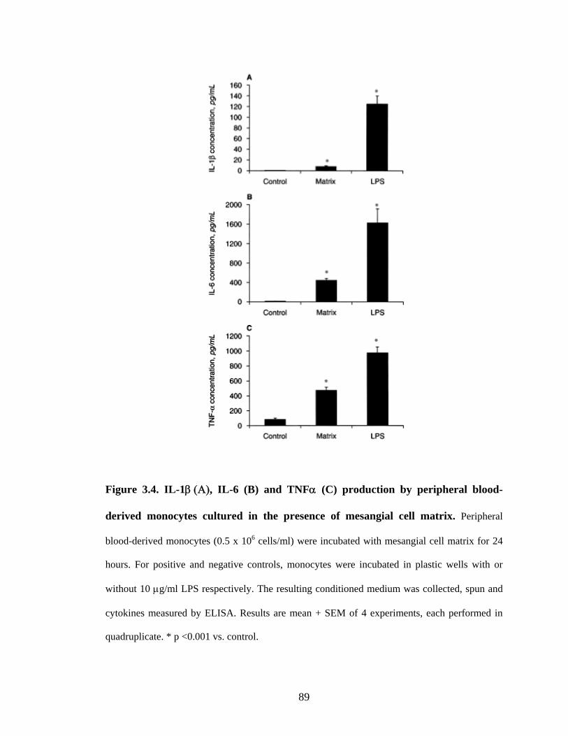
89
Figure 3.4. IL-1β (Α), IL-6 (B) and TNFα (C) production by peripheral blood-
derived monocytes cultured in the presence of mesangial cell matrix. Peripheral
blood-derived monocytes (0.5 x 10⁶ cells/ml) were incubated with mesangial cell matrix for 24
hours. For positive and negative controls, monocytes were incubated in plastic wells with or
without 10 μg/ml LPS respectively. The resulting conditioned medium was collected, spun and
cytokines measured by ELISA. Results are mean + SEM of 4 experiments, each performed in
quadruplicate. * p <0.001 vs. control.

90
Figure 3.5. IL-1β (A) and IL-6 (B) and TNFα (C) production by monocytes
cultured in the presence of soluble matrix and its protein components. PBMCs (0.5 x
106 cells/well) were incubated with solubilized mesangial cell matrix (100 μg/well) or its protein
components (100 μg/well) for 24 hours. For positive and negative controls, monocytes were
incubated in plastic wells with or without 10 μg/ml LPS respectively. The resulting conditioned
medium was collected, spun and cytokines measured by ELISA. Results (mean + SD) are
representative of 4 experiments, each performed in quadruplicate. *p <0.005 vs. control.

91
3.3.6. Effects of antibodies on cytokine production
The secretion of cytokines by peripheral blood-derived monocytes was mimicked
by incubation with anti-integrin antibodies. For example, incubation of monocytes in
plastic culture plates with anti-VLA-4 (10 μg/ml) and anti-VLA-5 (5 μg/ml) antibodies
increased IL-1β production compared to control monocytes in medium alone (Table
3.1). Similarly, TNFα secretion was increased by both antibodies when compared to
control.

92
Table 3.1. Production of cytokines by human macrophages exposed to anti-VLA-4
(10 μg/ml) and anti-VLA-5 (5 μg/ml) antibodies for 24 hours.
Antibody IL-1β (pg/ml) p value TNFα (pg/ml) p value
Control 2.0 + 2.4 - 64.2 + 6.0 -
anti-VLA-4 45.4 + 12.0 <0.005 2649.6 + 424.2 <0.0001
anti-VLA-5 93.3 + 13.5 <0.0001
<0.05*
3551.7 + 458.4 <0.0001
<0.005*
Cytokines were measured in the conditioned medium by ELISA. Results are mean
+SEM of 4 experiments, each performed in triplicate or quadruplicate. Statistical
analysis was performed using a paired student’s t-test. p vs. control. *p vs. anti VLA-4.
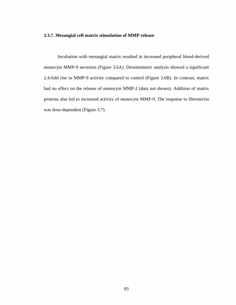
93
3.3.7. Mesangial cell matrix stimulation of MMP release
Incubation with mesangial matrix resulted in increased peripheral blood-derived
monocyte MMP-9 secretion (Figure 3.6A). Densitometric analysis showed a significant
2.4-fold rise in MMP-9 activity compared to control (Figure 3.6B). In contrast, matrix
had no effect on the release of monocyte MMP-2 (data not shown). Addition of matrix
proteins also led to increased activity of monocyte MMP-9. The response to fibronectin
was dose-dependent (Figure 3.7).

94
Figure 3.6. Matrix metalloproteinase (MMP) released by monocytes cultured in the
presence of mesangial cell matrix (A). Peripheral blood-derived monocytes (0.5 x 10⁶
cells/ml) were incubated with mesangial cell matrix for 24 hours in the absence of foetal
calf serum. For controls, monocytes were incubated in plastic wells with or without 10
μg/ml LPS. The resulting conditioned medium was collected, spun and analyzed by
zymography. Lane 1, control; Lane 2, cell matrix; Lane 3, LPS. Results are
representative of 5 experiments. Densitometric evaluation of zymograms (B) shows the
relative change in metalloproteinase secretion compared to control (100%).

95
Figure 3.7. Matrix metalloproteinase (MMP) release by monocytes cultured in the
presence of fibronectin (A). Peripheral blood-derived monocytes (0.5 x 10⁶ cells/ml)
were co-incubated with increasing concentrations of fibronectin for 24 hours in the
absence of foetal calf serum. The resulting conditioned medium was subjected to
zymography. Results are representative of 2 experiments. Densitometric evaluation of
zymograms (B) shows the relative change in metalloproteinase secretion compared to
control (100%).

96
3.3.8. Effect of matrix and soluble proteins on monocyte TIMP secretion
Matrix did not stimulate peripheral blood-derived monocyte TIMP I or TIMP II
secretion (Table 3.2), nor was there any effect seen with addition of fibronectin (data not
shown). The effect of anti-VLA-4 and anti-VLA-5 antibodies on monocyte secretion of
metalloproteinases or their inhibitors was not investigated.

97
Table 3.2. Release of tissue inhibitors of metalloproteinases by human macrophages
incubated on plastic culture plates (control), with mesangial cell matrix, and LPS
for 24 hours.
TIMP I (ng/ml) TIMP II (ng/ml)
Control 16.7 + 3.8 848.1 + 126.0
Matrix 14.3 + 1.3 725.5 + 102.1
LPS 22.5 + 1.4 630.3 + 99.3
TIMP I and II were measured in the conditioned medium by ELISA as described in the
methods section. Results are mean + SEM of 4 experiments (TIMP I) and 3 experiments
(TIMP II) each performed in quadruplicate.

98
3.4. DISCUSSION
This study demonstrates that monocytes specifically bind to mesangial matrix
and to its component proteins including fibronectin and to a lesser extent collagen type
IV and laminin. Monocyte binding to whole matrix is mediated, at least in part by
fibronectin. An increase in monocyte binding to mesangial matrix was shown to
accompany the enhanced synthesis and secretion of fibronectin induced by cytokine pre-
stimulation of mesangial cells. Incubation of monocytes with mesangial matrix and
individual matrix proteins led to the secretion of pro-inflammatory cytokines (IL-1β, IL-
6, and TNF-α) and to the activation of metalloproteinase (MMP-9). The release of
MMP-9 was not associated with a change in production of tissue inhibitors of
metalloproteinase indicating a net breakdown of matrix, thus exposing an increased
potential for the binding of infiltrating monocytes. By adding radiolabelled thymidine to
monocytes incubated with mesangial matrix it was possible to demonstrate that this
increased secretion of cytokines and metalloproteinase was not associated with cell
proliferation. These results therefore suggest that accumulation of mesangial matrix,
particularly fibronectin, promotes monocyte entrapment and that binding to matrix
proteins specifically stimulates monocyte secretion of inflammatory cytokines and
matrix degrading metalloproteinase. Such interactions may have important implications
in the pathogenesis of renal injury, particularly because the mechanisms by which
macrophages are activated is known to determine their functional characteristics (Song,
Ouyang et al. 2000).

99
Binding of monocytes to fibronectin is likely to be mediated by integrin
receptors which are composed of the α and β subunits (Hynes 1992). Among these
integrins, α4β1 (VLA-4) which binds to the CS1 and CS-5 regions of the IIICS domain
of the fibronectin (Humphries, Komoriya et al. 1987; Mould, Komoriya et al. 1991) and
α5β1 (VLA-5) which binds to the RGD sequence (Brown and Goodwin 1988; Brown,
Phillips et al. 1989) are found on activated monocytes (Ferreira, Garcia-Pardo et al.
1990; Hemler, Elices et al. 1990). The possible involvement of VLA-4, and VLA-5 was
therefore investigated. Anti-VLA-4 and VLA-5 antibodies caused a marked inhibition of
U937 and peripheral blood-derived monocyte adhesion to whole matrix, suggesting that
both cells shared common binding mechanisms involving both integrins. However,
neither antibody completely blocked the adhesion process thereby indicating that other
integrins and matrix components including collagen and laminin may participate in this
interaction. The demonstration of a common binding mechanism allowed us to use U937
cells, which were more readily available than peripheral blood-derived monocytes, for
binding studies although the latter were employed to examine the effects of binding on
secretory function.
Chronic glomerular diseases are characterised by the accumulation of
extracellular matrix composed of proteins including collagens, laminin and fibronectin
along with proteoglycans and glycosaminoglycans. In the healthy glomerulus, these
components not only provide structural support for glomerular cells but also influence
their behaviour (Border, Okuda et al. 1989). Fibronectin, one of the most abundant
mesangial matrix proteins, has been shown to have chemotactic properties for

100
monocytes, macrophages and fibroblast, and to be mitogenic to fibroblasts and
mesangial cells (Ruoslahti 1988). Since these results demonstrate that individual matrix
components differ in their ability to bind and activate macrophages, it follows that the
nature of disease-related changes in matrix composition might in turn influence the
extent of macrophage accumulation within the diseased glomerulus and the secretory
characteristics of these infiltrating cells. Such interactions could determine the outcome
of an acute or chronic inflammatory process.
Several animal and human studies suggest that monocytes/macrophages play a
critical role in the initiation and progression of renal diseases. For example, in rats with
remnant kidneys, macrophage accumulation was shown to strongly correlate with the
progression of focal sclerosis suggesting that these cells may play a role in the scarring
process (van Goor, Fidler et al. 1991). In humans, monocyte accumulation is seen in
most forms of glomerulonephritis, including those associated with progressive fibrosis
(Magil and Cohen 1989; Li, Hancock et al. 1990). Whilst monocyte infiltration may
have beneficial functions, for example, promotion of the resolution of inflammation by
apoptosis of infiltrating cells (Duffield, Erwig et al. 2000; Huynh, Fadok et al. 2002),
several monocyte/macrophage secretory products may have a detrimental influence on
the function of adjacent mesangial cells. These results demonstrate that binding of
monocytes to whole matrix and matrix proteins, particularly fibronectin, enhances
secretion of proinflammatory cytokines and matrix degrading metalloproteinases. These
findings are in agreement with those of other investigators who have reported enhanced
monocyte secretion of IL-1, IL-6, IL-8 and TNFα upon exposure of cells to matrix
101
proteins including fibronectin (Haskill, Johnson et al. 1988; Heinel, Singleton et al.
1995; Mahnke, Bhardwaj et al. 1995; Takizawa, Nishinarita et al. 1995). For example,
Takizawa et al demonstrated that addition of fibronectin to monocytes isolated from
human plasma stimulated production of IL-1, IL-6 and TNFα (Takizawa, Nishinarita et
al. 1995). There have also been reports of matrix proteins modulating secretion of
metalloproteinases by various cells including fibroblast (Huhtala, Humphries et al. 1995)
and keratinocytes (Larjava, Lyons et al. 1993). Studies by Martin et al have
demonstrated that specific matrix components enhance secretion of MMP-2 and MMP-9
by human mesangial cells and that membrane type metalloproteinase MTMMP, which is
selectively induced by fibronectin, is important in this process (Martin, Eynstone et al.
2001). The present study extends these findings by demonstrating that matrix produced
by glomerular cells may also modulate the accumulation and activation of infiltrating
inflammatory cells. Matrix-mediated effects may help to explain the changes in
metalloproteinase to inhibitor ratios observed by Mené et al in co-culture experiments
involving mesangial cells and monocytes (Mene, Caenazzo et al. 2001).
Since binding of both U937 and peripheral blood-derived monocytes to
mesangial matrix components involved the integrins VLA-4 and VLA-5, an
investigation into whether these molecules might be involved in signal transduction was
carried out. Stimulation of peripheral blood-derived monocytes with either anti-VLA4 or
anti-VLA5 antibodies mimicked the effects of matrix on cytokine production, as well as
blocking monocyte binding to matrix. Studies in other cell types have shown similar
effects, for example, activation of fibroblasts by a crosslinking anti-ICAM-1 antibody

102
(Clayton, Evans et al. 1998) and of fibrosarcoma cells by antibodies to α5, α6 and β1
integrin subunits (Stanton, Gavrilovic et al. 1998) was associated with activation. Whilst
the down stream events were not investigated in this study, in other experimental
settings the binding of integrin components to matrix induces activation of
phospholipases (Cybulsky, Carbonetto et al. 1993), kinase signalling pathways (Malik
and Parsons 1996) and the AP-1 transcription factor (Yamada, Nikaido et al. 1991).
Thus it is reasonable to propose that infiltrating monocytes may be activated by
interactions with matrix components via integrin receptors. Monocyte responses may be
influenced by pre-programming as has been observed following exposure to a variety of
cytokines (Erwig, Kluth et al. 1998; Erwig, Stewart et al. 2000; Song, Ouyang et al.
2000). Furthermore accumulation and disease-specific modification of matrix
components may alter monocyte/macrophage behavior and thereby potentially influence
disease outcome. For example, these results would suggest that accumulation of
fibronectin enhances metalloproteinase production, without increased inhibitor activity,
a situation that is likely to promote matrix degradation. Inhibition of monocyte responses
by blockade of these signalling pathways represents a potential target for therapeutic
intervention in human glomerular disease and has proved effective in recent animal
studies (Allen, McHale et al. 1999).
In summary, these results demonstrate that mesangial matrix plays a key role in
the immobilization and activation of monocytes within the glomerulus. Since matrix
proteins differ in their ability to modulate monocyte secretory functions, changes in
matrix composition or organization in glomerular disease may influence the behavior of

103
infiltrating cells and thereby the outcome of the disease process. Better understanding of
the potential importance of these processes may help in the design of treatment strategies
for chronic glomerular diseases.

104
CHAPTER 4
MESANGIAL MATRIX-ACTIVATED MONOCYTES
EXPRESS FUNCTIONAL SCAVENGER RECEPTORS
AND ACCUMULATE INTRACELLULAR LIPID

105
4.1. INTRODUCTION
Transendothelial migration of monocytes into the glomerular mesangium is a
recognized early feature of glomerular injury in man and in experimental models of
kidney disease (Brady 1993). These cells play a central role in orchestrating tissue
inflammation and may be critical in determining whether the final outcome of an acute
inflammatory glomerular lesion is complete resolution or permanent scarring (Duffield
2003). Interactions between monocytes and extracellular structures encountered during
the process of transmigration may play a critical role in determining the phenotype and
therefore the behaviour of the activated tissue macrophage. As described in section 1.3,
extracellular matrix is a highly ordered network of fibrous proteins and associated
glycoproteins embedded in a hydrated ground substance of glycosaminoglycans and
proteoglycans. It is recognised that matrix not only provides a structural framework, but
also influences cellular behaviour. For example, integrin-mediated adhesion of
monocytes to extracellular matrix may regulate expression of numerous inflammatory
and immune response genes (de Fougerolles and Koteliansky 2002). The importance of
this process is demonstrated by disease states thought to arise from dysregulation of
matrix-integrin interactions (Campbell, Senior et al. 1987).
In the previous chapter, it has been demonstrated that exposure of human
monocytes to both intact glomerular matrix (and to its individual components) enhanced
the production of a range of inflammatory cytokines and matrix-degrading
metalloproteinases. However, these experiments did not conclusively demonstrate that
such interactions induced monocyte to macrophage differentiation. The experiments

106
described in this chapter were designed to test the hypothesis that activation by
mesangial matrix converts monocytes to a macrophage phenotype. The expression of
three macrophage specific markers were studied: a) the peroxisomal proliferator-
activated receptor-γ (PPAR−γ), a nuclear receptor that acts as a transcriptional mediator
for genes involved in lipid metabolism and adipogenesis (Moore, Rosen et al. 2001), b)
CD36, a class B scavenger receptor and c) scavenger receptor class-A. Both these
scavenger receptors are located in the plasma membrane of the macrophage and are
involved in the cellular uptake of modified lipoproteins (Brown, Basu et al. 1980). Since
unregulated uptake of modified lipoproteins is a characteristic of the tissue macrophage,
the capacity of matrix-activated monocytes to accumulate intracellular lipid when
exposed to acetylated low density lipoprotein (Ac-LDL), a scavenger receptor ligand
was also tested. To further examine the role of matrix in foam cell formation, an
assessment of the capacity of matrix to modify LDL in the absence of cells to produce
oxidised LDL (ox-LDL), a naturally occurring scavenger receptor ligand identified in
diseased glomeruli, was conducted. To confirm the relevance of these observations to
human glomerular disease, human kidney biopsy sections from patients with
inflammatory and non-inflammatory glomerular disease were stained for macrophage
activation markers. These results demonstrate that mesangial cell matrix has the
potential both to induce monocyte to macrophage maturation and to oxidise LDL,
thereby indicating a likely modulatory role in glomerular inflammation and foam cell
formation. It was also possible to demonstrate activated macrophages in glomeruli
derived from patients with inflammatory glomerular disease.

107
4.3. RESULTS
4.3.1. PPAR-γ expression by matrix-activated monocytes
Whilst no PPAR-γ mRNA was detected by RT-PCR analysis of total RNA
extracted from freshly isolated THP-1 monocytes, message was detectable within 24
hours when cells were incubated with soluble mesangial matrix (500 μg/ml). Expression
was maximal at 48 hours, persisting over at least 5 days and was comparable to that
observed when cells were stimulated with PMA over a similar time period under
identical experimental conditions (Figure 4.1). Increased expression of PPAR-γ protein
within 24 hours of exposure of matrix stimulation was confirmed by Western analysis,
with levels of expression being similar to those observed following PMA stimulation
(Figure 4.2). No further increase in expression was observed when incubation was
extended beyond 48 hours to 7 days.

108
Figure 4.1. Time-dependent expression of PPAR-γ mRNA by THP-1 monocytes.
Monocytes were incubated with mesangial cell matrix (500 μg/mL) or PMA (125 nM)
for up to 120 hours. A) PPAR-γ mRNA expression was examined by RT-PCR. B)
Histogram showing analysis of mean ± SEM density of bands of PPAR-γ mRNA from 4
experiments, normalised by subtracting BSA protein control and comparison with β-
actin mRNA.

109
Figure 4.2. Western analysis of PPAR-γ protein expression by THP-1 monocytes.
Monocytes were incubated with 500 μg/mL mesangial cell matrix or PMA (125nM) for
0, 6, 24 and 48 hours. Cells were lysed and subjected to SDS-PAGE and proteins
transferred to nitrocellulose membrane. A) Western blot showing PPAR-γ detected using
an anti-PPAR-γ antibody. B) Histogram showing mean ± SEM density of bands of
PPAR-γ protein from quadruplicate wells, normalised by comparison with α-actin
protein, and expressed as percentage of control (0 hours). *p<0.005 vs. Control.

110
4.3.2. CD36 expression by matrix-activated monocytes
Enhanced expression of CD36 mRNA was detected by RT-PCR analysis of total
RNA extracted from THP-1 monocytes exposed to soluble mesangial matrix (500
μg/ml). An increase in message was detected after 48 hours of incubation, and was
comparable with that observed when cells were stimulated with PMA for the same time
period under identical experimental conditions (Figure 4.3). No further increase in
expression was observed when incubation was extended beyond 120 hours to 7 days
(data not shown).

111
Figure 4.3. Time-dependent expression of CD36 mRNA in response to mesangial
matrix.
Mesangial cell matrix (500 μg/mL) or PMA (125 nM) were incubated with monocytes
for 0, 6, 48 and 120 hours. A) RT-PCR analysis of CD36 mRNA expression. B)
Histogram showing analysis of mean ± SEM of CD36 mRNA bands from 4
experiments, normalised by subtracting BSA protein control and comparison with β-
actin mRNA. Results are expressed as a percentage of control (0 hours). *p<0.005 vs.
control.

112
4.3.3. Scavenger receptor expression by matrix-activated monocytes
Scavenger receptor-A mRNA expression increased in a concentration-dependent
manner when THP-1 monocytes were incubated with increasing concentrations of
soluble matrix protein for 48 hours with a maximal response at 100 μg/ml (Figure 4.4).
A time-dependent increase was observed with addition of 500 μg/ml matrix protein
increasing up to 120 hours incubation (Figure 4.5), with no further change up to 7 days
(data not shown). No expression was observed under baseline conditions prior to
stimulation, neither did equivalent concentrations of BSA induce detectable scavenger
receptor-A message, suggesting that the observed effect was specific.

113
Figure 4.4. Scavenger receptor A mRNA expression by THP-1 monocytes
incubated in the presence of increasing mesangial matrix for 48 hours.
Monocytes were incubated with increasing mesangial cell matrix concentrations of 0-
500 μg/mL or PMA (125 nM) for 48 hours. A) Scavenger receptor A mRNA expression
examined by RT-PCR. B) Histogram of mean ± SEM density of scavenger receptor A
mRNA bands from 4 experiments, normalised by comparison with β-actin mRNA, and
expressed as percentage of results obtained when equal amounts of BSA were added.
*p<0.005 vs. equivalent BSA control.

114
Figure 4.5. Time-dependent expression of scavenger receptor A mRNA in response
to mesangial matrix.
Mesangial cell matrix (500 μg/mL) or PMA (125 nM) were incubated with monocytes
for 0, 6, 48 and 120 hours. A) RT-PCR analysis of scavenger receptor A mRNA
expression. B) Histogram of mean ± SEM density of scavenger receptor A mRNA bands
from 4 experiments, normalised by subtracting BSA protein control and comparison
with β-actin mRNA. Expressed as a percentage of control (0 hours). *p<0.005 vs.
control.

115
4.3.4. Uptake of modified lipoproteins by matrix-activated monocytes
The presence of functional scavenger receptors was confirmed using flow
cytometry in which incubation of matrix-activated monocytes with Dil-labelled Ac-LDL
led to an increase in mean fluorescence intensity (MFI) (Figure 4.6). This effect was
largely reversed by addition of an excess of unlabelled ligand. The MFI of THP-1 cells
incubated with Dil-labelled Ac-LDL after exposure to matrix increased to 373±34.8%
(p<0.005) as compared to cells exposed to BSA (100%). PMA pre-stimulation of
monocytes, increased MFI to 423±55.5% (p<0.005). These increases in MFI induced by
matrix-and PMA activation were inhibited by the addition of excess unlabelled Ac-LDL
to 134±12.1% (p<0.001 vs. no excess of unlabelled lipoprotein) and to 170±16.1% (p
<0.001) respectively, suggesting specific binding of Dil-labelled Ac-LDL to scavenger
receptors.
Incubation of monocytes with unlabelled Ac-LDL following stimulation by
exposure to matrix for 120 hours led to intracellular accumulation of Oil Red O- stained
lipid droplets (Figure 4.7A). Lipid uptake did not occur following BSA stimulation
(Figure 4.7B). Prior exposure to PMA was also associated with intracellular lipid
deposition but no intracellular lipid staining was observed when poly I was added with
Ac-LDL following activation of monocytes by matrix or PMA.

116
Figure 4.6. Effects of matrix on Ac-LDL uptake by monocyte/macrophages.
THP-1 monocytes were incubated with matrix (500 μg/mL) for 48 hours. BSA (500
μg/mL) and PMA (125 nM) served as negative and positive controls, respectively.
Monocytes were recovered and incubated for a further 3 hours with 10 μg/mL DiI-
labelled Ac-LDL in the presence or absence of an excess (XS) of unlabelled Ac-LDL
(250 μg/mL). The mean fluorescence intensity (MFI) was calculated by subtracting the
auto-fluorescence intensity from the observed fluorescence intensity of labeled cells.
The histogram represents mean ± SEM MFI calculated from 4 experiments under the
conditions shown, carried out in duplicate and expressed as percentage above BSA
control (100%). *p<0.005 vs. BSA control, **p<0.001 vs. no excess unlabelled Ac-
LDL.
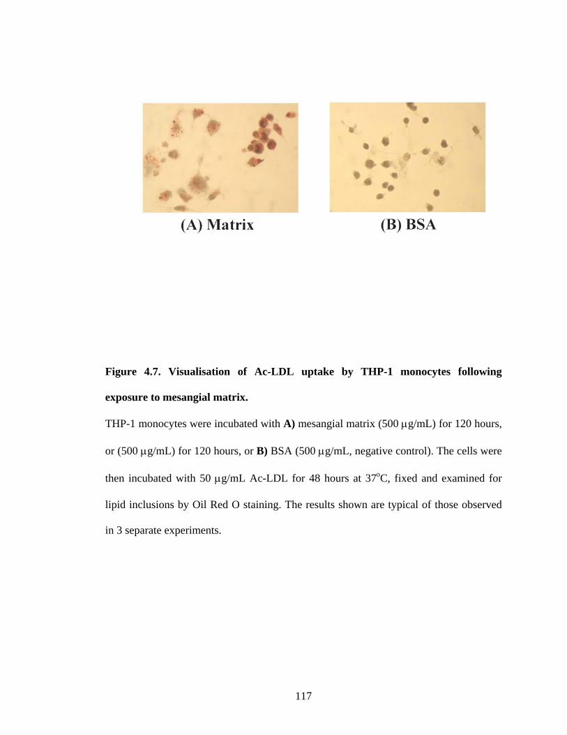
117
Figure 4.7. Visualisation of Ac-LDL uptake by THP-1 monocytes following
exposure to mesangial matrix.
THP-1 monocytes were incubated with A) mesangial matrix (500 μg/mL) for 120 hours,
or (500 μg/mL) for 120 hours, or B) BSA (500 μg/mL, negative control). The cells were
then incubated with 50 μg/mL Ac-LDL for 48 hours at 37oC, fixed and examined for
lipid inclusions by Oil Red O staining. The results shown are typical of those observed
in 3 separate experiments.

118
4.3.5. Oxidation of LDL by mesangial cell matrix
Incubation of LDL with mesangial cell matrix in the absence of cells led to
enhanced electrophoretic mobility of recovered lipoprotein on agarose gel (Figure 4.8).
A similar shift in mobility was seen when LDL was exposed to copper sulphate, a
powerful oxidising agent, but was blocked when the antioxidants EDTA (100 μM) and
BHT (20 μM) were added, suggesting that matrix induces LDL oxidation. In contrast,
incubation with BSA did not change the electrophoretic mobility of the lipoprotein.

119
Figure 4.8. Agarose gel electrophoresis demonstrating oxidation of LDL by
mesangial matrix.
Lane 1: Freshly isolated native LDL (negative control), Lane 2: LDL incubated with
BSA protein (500μg/mL), Lane 3: LDL incubated with CuSO4 (10 μM, positive
control), Lane 4: Matrix (500μg/mL) incubated with LDL, Lane 5: Matrix incubated
with LDL and with the antioxidants EDTA (100 μM) and BHT (20 μM). LDL incubated
with mesangial matrix had a mobility similar to that observed with the positive control
(CuSO4). This effect was abolished by the addition of EDTA and BHT, indicating that
matrix promotes LDL oxidation.

120
4.3.6. Identification of macrophage activation markers in human kidney biopsy
material
CD-68 positive cells were extremely difficult to identify in the normal kidney
section and there was no staining for PPAR-γ or scavenger receptor. In contrast, all three
markers were readily detected in the inflamed kidney, predominantly within the
glomeruli (Figure 4.9). CD68 and scavenger receptor were located in a cytoplasmic
distribution and PPAR-γ within nuclei in keeping with the cellular location of these
markers.

121
Non-inflamed: CD68 Inflamed: CD68
Non-inflamed: PPAR-γ Inflamed: PPAR-γ
Non-inflamed: Scavenger receptor Inflamed: Scavenger receptor Figure 4.9. Staining of human kidney sections for macrophage activation markers.
Sections of non-inflamed and inflamed human kidney were stained for the macrophage
antigen CD-68 and for the activation markers PPAR-γ and Scavenger receptor.

122
4.4. DISCUSSION
These results demonstrate that exposure of monocytes to mesangial cell matrix in
vitro induces expression of PPAR-γ, CD36 and scavenger receptor-A and promotes
phagocytic activity, characteristics usually associated with a macrophage phenotype.
This would suggest that conversion of infiltrating monocytes to mature tissue
macrophages within the glomerular mesangium in vivo may not necessarily depend on
the presence of mesangial cells, but may result from direct interactions with matrix
components. While circulating monocytes are relatively inert, the activated tissue
macrophage may take on a destructive role, inducing cell death by apoptosis and
degrading extracellular matrix. Alternatively, these cells may facilitate repair by
inducing cell proliferation and secretion of replacement matrix components (Duffield
2003). Macrophages also play a key role in the phagocytosis of cellular debris, lipids
and denatured proteins in inflamed tissue, a process that may result in the formation of
foam cells. Foam cells are characteristically seen at sites of tissue injury, for example in
the arterial intima in atherosclerosis, and are recognised in the kidney in damaged
glomeruli (Moorhead 1991). Given the pivotal role of the monocyte/macrophage in
modulating tissue injury and the diverse biological activities of these cells, these
findings may have important implications in the context of glomerular disease as
supported by the demonstration of both PPAR-γ and scavenger receptor expression in
inflamed human kidney sections. Thus, matrix-mediated activation may influence the
behaviour of monocytes that infiltrate the glomerular mesangium and thereby potentially
modify the outcome of an inflammatory process.

123
It is recognised that the phenotypic state adopted by a tissue macrophage is
influenced by the activation signals that naïve monocytes receive and that broadly, two
distinct activated phenotypes can be identified. The classical activation pathway results
from exposure to Th-1 type cytokines such as TNF-α, IL-1β and IL-6 and results in a
macrophage with pro-inflammatory properties, capable of further generation of pro-
inflammatory cytokines and the degradation of normal and abnormal matrix components
(Erwig, Kluth et al. 2001). Classically activated cells also possess the ability to take up
modified lipoproteins, potentially resulting in the formation of lipid-laden foam cells. In
contrast, the alternative activation pathway induced by Th-2 type cytokines such as IL-4
and IL-13 produces a macrophage that generates anti-inflammatory cytokines,
suppresses the synthesis of pro-inflammatory cytokines and is resistant to re-activation,
thus being responsible for co-ordinating resolution of an inflammatory process (Duffield
2003). Taken together with the previous chapter, which demonstrated that incubation of
monocytes with mesangial cell matrix stimulates secretion of the pro-inflammatory
cytokines IL-6, IL-1β and TNF-α as well as matrix-degrading metalloproteinases, it
seems reasonable to conclude that mesangial cell matrix activates macrophages via the
classical pathway.
In late 2005 the Th-1 type and Th-2 paradigm was further simplified. Monocytes
primed by Th-1 type cytokines, which promote differentiation into proatherogenic
‘Classical’ macrophages were termed M1 macrophages, while those primed by Th-2
type cytokines, which lead to an “Alternative” anti-inflammatory macrophage
phenotype, were termed M2 macrophages (Mantovani, Sica et al. 2005). Functional

124
polarization of macrophages into M1 or M2 cells was deemed as more operationally
useful and provided a simplified conceptual framework describing the plasticity of
mononuclear phagocytes (Mantovani, Sica et al. 2004). Genetic approaches have begun
to shed new light on mechanisms underlying macrophage differentiation and attempts to
dissect the actual in vivo significance of their polarization are currently being
investigated (Rauh, Ho et al. 2005; Biswas and Mantovani 2010).
These findings are highly relevant to the fate of monocytes that undergo
transmigration to become tissue macrophages, but not to cells that undergo reverse-
transmigration since they adopt the phenotype of an immature or mature dendritic cell
(depending on the absence or presence of inflammatory stimuli respectively) (Randolph,
Beaulieu et al. 1998).
Other macrophage-specific markers which have been previously explored by my
colleagues included CD69 and the HLA-DR antigens (Chana, Martin et al. 2003).
However it was discovered that both markers were expressed at low levels on THP-1
monocytes with no significant up-regulation occurring following stimulation with PMA,
an accepted and potent activator of monocytes. Other investigators have observed that
HLA-DR expression varies with the source of the macrophage, such that 15% of
peritoneal macrophages express the antigen compared to 50% of spleen and thymus-
derived cells (Lewis, Norris et al. 1990). Another potential candidate was Mac-1, a
member of the β2 integrin family also known as CD11b/CD18, however, this cell
surface adhesion receptor proved not to be specific to macrophages and was also
expressed at low levels by freshly isolated monocytes as demonstrated by other workers

125
(Miller, Bainton et al. 1987). PPAR-γ expression proved to be a more useful indicator
since this intracellular receptor showed very low levels of expression in monocytes, but
is strongly induced during their differentiation into mature macrophages, suggesting that
it may be involved in the differentiation process. In addition, PPAR-γ has been
implicated in the modulation of several macrophage functions including the regulation
of pro-inflammatory activities and stimulation of ox-LDL uptake further strengthening
the use of this factor as a macrophage marker (Moore, Fitzgerald et al. 2001). PPAR-γ is
also abundantly expressed in lesions such as atherosclerotic plaques where formation of
foam cells is observed (Tontonoz, Nagy et al. 1998). It should be emphasised that
PPAR-γ was used simply as a macrophage marker in these studies and that ligand-
induced activation of this receptor was not examined. It seems likely that other
signalling pathways are activated by matrix-monocyte interactions, particularly since
PPAR-γ activation does not explain the increase in cytokine production that has
previously been reported (Chana, Martin et al. 2003).
Scavenger receptor-A is a macrophage-specific cell surface protein that
specifically binds and internalises oxidised and chemically modified LDL particles,
similar to the class B scavenger receptor; CD36, which also bind modified forms of LDL
(Brown, Basu et al. 1980). Scavenger receptor expression is restricted to macrophages
thereby providing a reliable marker for the purpose of these studies. Scavenger receptor-
A has been implicated in mediating a variety macrophage functions, including
intracellular signalling, endocytosis, adhesion and phagocytosis. Unlike uptake of native
LDL via the LDL receptor, which is tightly controlled, scavenger receptor-A mediated

126
uptake of modified lipoprotein is not regulated by intracellular cholesterol levels and can
therefore potentially lead to intracellular cholesterol accumulation and the formation of
foam cells (Gough, Greaves et al. 1999). The class B scavenger receptor CD36 has also
been implicated in the process of lipid accumulation in macrophages and serves as an
adhesion receptor on macrophages for matrix components such as collagen and
thrombospondin (Tandon, Kralisz et al. 1989).
In keeping with the changes in lipoprotein receptor expression observed, matrix-
activated monocytes accumulated intracellular lipid when incubated with Ac-LDL, a
synthetic scavenger receptor ligand, as demonstrated by intracellular Oil Red O staining.
This phagocytic capacity was further confirmed by flow cytometry of matrix-activated
monocytes exposed to Dil-labelled Ac-LDL. Uptake of Ac-LDL was shown to be
specific, since it could be inhibited by addition of an excess of unlabelled acetylated
lipoprotein, thus confirming receptor involvement. Induction of phagocytic activity was
also observed following PMA-mediated activation, but not when an irrelevant protein
(BSA) was added.
Lipoproteins, including LDL, infiltrate the normal mesangium and are found
deposited in diseased glomeruli (Wheeler and Chana 1993). Having previously shown
that mesangial cells oxidise LDL in vitro (Wheeler, Chana et al. 1994), It has been
demonstrated here that exposure of LDL to mesangial matrix has a similar effect. Thus,
not only does matrix exposure induce a phagocytic macrophage phenotype in
monocytes, but also converts LDL to an appropriate scavenger receptor ligand thereby
potentially contributing to the development of foam cells. The mechanisms by which
matrix promotes LDL oxidation were not explored but may involve entrapment of

127
lipoprotein by glycosaminoglycans, thereby rendering particles more susceptible to the
effects of reactive oxidative species (Abuja 2002).
In conclusion, mesangial matrix has the capacity to convert monocytes to
macrophages displaying characteristics associated with a classically activated
phenotype. By inducing macrophage scavenger receptor expression and converting LDL
to an oxidised product, matrix may also play a key role in the formation of foam cells
within the glomerular mesangium. The impact of changes in matrix composition on
these interactions and the potential for such changes to modify the outcome of an
inflammatory process within the glomerulus warrant further investigation.

128
CHAPTER 5
GENERAL DISCUSSION AND CONCLUSION

129
5.1. RESEARCH QUESTIONS ADDRESSED IN THIS THESIS
Infiltration of mononuclear cells into the mesangium and their differentiation are
recognized as an early event in many diseases that ultimately lead to chronic kidney
failure. A variety of factors are likely to play key roles in orchestrating monocyte influx
and in determining the phenotype that the infiltrating monocytes ultimately adopt. In
particular, monocyte interactions with components of mesangial matrix may play a key
role in these processes.
The work presented in this thesis set out to dissect the nature of the interaction
between monocytes and mesangial cell matrix and the resulting changes in monocyte
phenotype. A representative matrix protein component, namely Fibronectin, which is up-
regulated when mesangial cells are exposed to TGF-β and TNF-α (Chana and Wheeler
1999) was used to conduct matrix binding and blocking studies to examine the extent to
which matrix exposure modified cytokine, MMP and TIMP production by monocytes.
Whole matrix, as well as other matrix protein components namely Collagen type IV and
Laminin were also examined for their effects on cytokine production.
To address the extent of monocyte differentiation into macrophages upon
exposure to mesangial cell matrix, one challenge was the identification of a suitably
robust marker of monocyte to macrophage conversion. After a thorough literature search
three reliable macrophage specific markers were studied: a) PPAR−γ, b) CD36 and c)
Scavenger receptor class-A.
Finally since LDL may play a central role in the pathogenesis of
glomerulosclerosis it was assessed whether this lipoprotein might become oxidized in

130
the mesangium by exposure to matrix, thereby becoming a ligand for uptake via
macrophage scavenger receptors and promoting foam cell formation.
5.2. LIMITATIONS OF THE EXPERIMENTAL WORK
5.2.1. Monocyte Binding Studies
From earlier experiments by our group (Chana and Wheeler 1999) it has
demonstrated that exposure of mesangial cells to LDL stimulates the production of the
monocyte chemoattractant MCP-1, indicating that the presence of LDL within the
injured glomerular mesangium might promote monocyte accumulation. It was also
demonstrated that LDL increases the synthesis of the matrix component fibronectin by
mesangial cells, thus providing an additional factor by which monocytes might be
retained. In the experiments described in this thesis, this work was taken forward by
examining ligand-integrin interactions involving fibronectin and monocytes. One
criticism is that most of these experiments were based on a monocyte cell line. However,
although U937 monocytes were used in the earlier experiments, these data were
strengthened by using the same blocking antibodies in experiments using whole matrix
and human PBMCs, and also found a decrease in monocyte adhesion to matrix through
the VLA-4 and VLA-5 integrin receptors. When examining the resulting secretory
behaviour of monocytes upon binding to whole matrix and individual matrix proteins
PBMCs were also used in some of these experiments. It can therefore be accepted that
the U937 cell line provided an appropriate model on which to base these experiments.

131
Ideally it would have been possible to extract individual component proteins of
mesangial matrix. However this would have required huge amounts of matrix and a
complex extraction process which would not have guaranteed pure protein components.
The other option was to qualitatively and quantitatively define the components of
mesangial cell matrix. Although attempts to do this were investigated, it was not a
feasible option given the focus of these investigations. Instead, a decision to use an equal
volume of the three major matrix proteins was taken, namely Collagen Type IV,
Fibronectin and Laminin and make a comparison with whole matrix when looking at
changes in inflammatory cytokine secretory behaviour and activation of MMP-9 and
TIMPs by PBMCs.
Since experiments involving cytokine and MMP secretion and TIMP activation
were performed at a single time-point of 24 hours, the possibility that at later time
points, matrix activated macrophages lose their pro-inflammatory capacity, and take on
the alternatively activated anti-inflammatory phenotype, cannot be excluded. In vivo,
both cell phenotypes are likely to be present in inflamed tissue and the balance between
them may be critical in determining the extent of subsequent fibrosis. It is also possible
that a classically-activated macrophage programmed by exposure to matrix does not
respond to the signals usually associated with the alternative activation pathway,
potentially resulting in an uncontrolled inflammatory response with limited subsequent
tissue repair (Duffield 2003). Further time-points up to 7 days may prove useful in
answering this question.

132
5.2.2. Monocyte Activation Studies
When designing these experiments one challenge was the identification of a
suitably robust marker of monocyte to macrophage conversion. The three macrophage
specific markers chosen were a) PPAR-γ, b) CD36 and c) Scavenger receptor class-A.
MAC-1 was considered but was abandoned due to there being low levels of expression
on freshly isolated monocytes, as also demonstrated by others (Bainton, Miller et al.
1987; Cifarelli, Libman et al. 2007; Yakubenko, Belevych et al. 2008).
The observed changes in macrophage specific markers following matrix
interaction were reproduced using the THP-1 monocyte cell line supplied by two
companies (ECACC and ATCC) both of which claim that these cells differentiate into
macrophages. In addition, a number of published studies appear to support the
differentiation of this cell line (Tsuchiya, Yamabe et al. 1980; Kritharides, Christian et
al. 1998; Kim, Studer et al. 2008). Taking into account the available literature and
discussions with experts working in the field of inflammation, it can be accepted that
these cells provided a reasonable model for these experiments.
To further validate the model, studies comparing the response of human
peripheral blood monocytes and THP-1 cells to mesangial matrix assessing the
production of inflammatory cytokines and MMP-9 were performed. These studies show
almost identical patterns of induction suggesting that the two cell types show similar
responsiveness. Furthermore, previous work by our group has demonstrated similarities

133
between monocyte cell lines and cells isolated from peripheral blood in terms of surface
markers and binding characteristics (Wheeler, Chana et al. 1994).
5.2.3. Disease Specific Matrix Modification
Matrix elaborated by healthy mesangial cells were used, however this did not
take account of the fact that disease-specific matrix modification may influence
monocyte activation, since there is strong evidence to suggest that monocyte-
macrophage differentiation is influenced by the nature of the matrix proteins (Laouar,
Collart et al. 1999) For example, matrix glycation that occurs in diabetes mellitus
influences the balance between matrix synthesis and degradation by mesangial cells,
promoting accumulation (Schleicher and Olgemoller 1992) and may also affect
monocyte to macrophage differentiation. Indeed many studies have shown that non-
enzymatically glycated matrix occurring in diabetes influences monocyte to macrophage
differentiation (Jacob, Shastry et al. 2001; Min, Lyons et al. 2009). A key matrix protein,
collagen type 1 when non-enzymatically glycated was shown to accelerate monocyte to
macrophage differentiation, leading to foam cell formation upon interaction with
oxidised LDL (Jacob, Shastry et al. 2001). Since mesangial cell matrix may be
particularly prone to glycation as a result of its prolonged lifespan, an extension to this
work would be to investigate various matrix modifications on monocyte differentiation
such as non-enzymatic glycation.

134
5.2.4. Mesangial Cell Matrix Sequesters Cytokines and Growth Factors
The possibility that these results are at least in part explained by retention of low
concentrations of cytokines within the matrix material cannot be ruled out. More
detailed experiments investigating the ability of matrix to sequester growth factors and
cytokines would be the next logical step in investigating the effects on monocyte
behaviour. For example investigating the effects of matrix impregnated with TGF-β,
matrix has been shown to sequester this growth factor and it has been shown to play an
important role in matrix production. (Gambaro and Baggio 1998).
5.2.5. Scavenger receptor: Protein Level Expression
Experiments were limited to examining the expression of scavenger receptors at
the message level without going on to provide protein expression data as was the case
with PPAR-γ expression. However, up-regulation of functional scavenger receptors in
the lipoprotein uptake studies was demonstrated and it is unlikely that further extending
these studies to examine scavenger receptor expression at the protein level would have
changed the conclusions.
5.3. IMPLICATIONS OF MAJOR FINDINGS
The work presented in this thesis help us to better understand how matrix
components contribute to changes in monocyte phenotype. Chapter 3 demonstrates that
mesangial matrix plays a key role in the immobilization and activation of monocytes

135
within the glomerulus. These results would also suggest that accumulation of fibronectin
enhances metalloproteinase production, without increased inhibitor activity, a situation
that is likely to promote matrix degradation.
In Chapter 4, initial findings were further strengthened by demonstrating that
mesangial matrix has the capacity to convert monocytes to macrophages displaying
characteristics associated with a classically activated phenotype. Since lipoproteins,
including LDL, infiltrate the normal mesangium and are found deposited in diseased
glomeruli (Wheeler and Chana 1993) mesangial cell matrix may play a key role in the
formation of foam cells within the glomerular mesangium through its capacity to oxidise
LDL as well as stimulate monocyte to macrophage differentiation.
Thus it has been demonstrated that interactions between mesangial matrix
components and infiltrating monocytes may play a key role in the progression of
glomerular injury.
5.4. POTENTIAL THERAPEUTIC IMPLICATIONS
Interactions between adhesion molecules and infiltrating macrophages have been
successfully blocked and could serve as targets for therapeutic interventions (Adler and
Brady 1999; Allen, McHale et al. 1999; Chana and Wheeler 1999; Cook, Khan et al.
2002). The humanized version of this anti-α4 monoclonal antibody known as
Natalizumab has been used successfully in multicenter double-blind controlled studies in
Crohn's disease and multiple sclerosis (Ghosh, Goldin et al. 2003; Miller, Khan et al.
2003; Miller, Soon et al. 2007; Targan, Feagan et al. 2007). Despite there being adverse

136
reactions in some patients to this drug it was given approval by the FDA as its clinical
benefits outweighed the risks involved (Ransohoff 2010; Steiner 2010). It would be interesting
to see the effects of this drug on patients suffering from glomerulosclerosis.
The beta-2 integrins include CD11a/CD18 (LFA-1) and CD11b/CD18 (Mac-
1/Complement receptor 3). A humanized monoclonal antibody to CD18 known as
Efalizumab, which blocks CD11a/CD18 and CD11b/CD18 has been reported to reduce
infiltrating leukocytes and improve vasculitic ulcers in patients with systemic vasculitis
(Lockwood, Elliott et al. 1999), however ten years on Efalizumab has been withdrawn
from the market as it was associated in some cases with fatal brain infections (Major
2010). Inhibition of monocyte responses by blockade of these signalling pathways
represents a potential target for therapeutic intervention in human glomerular disease.
5.5. CONCLUSION
In summary, enhanced monocyte adhesion to mesangial cell matrix results in
monocyte retention, activation and differentiation within the glomerular mesangium. In
the presence of LDL, macrophages are likely to accumulate lipid to form foam cells.
Inhibition of monocyte-matrix interactions represents a potential therapeutic intervention
that may prove protective in the setting of kidney disease. .

137
REFERENCES

138
Abuja, P. M. (2002). "Aggregation of LDL with chondroitin-4-sulfate makes LDL oxidizable in the presence of water-soluble antioxidants." FEBS Lett 512(1-3): 245-248.
Ackerman, S. K. and S. D. Douglas (1978). "Purification of human monocytes on microexudate-coated surfaces." J Immunol 120(4): 1372-1374.
Adler, S. and H. R. Brady (1999). "Cell adhesion molecules and the glomerulopathies." Am J Med 107(4): 371-386.
Allen, A., J. McHale, et al. (1999). "Endothelial expression of VCAM-1 in experimental crescentic nephritis and effect of antibodies to very late antigen-4 or VCAM-1 on glomerular injury." J Immunol 162: 5519-5527.
Bainton, D. F., L. J. Miller, et al. (1987). "Leukocyte adhesion receptors are stored in peroxidase-negative granules of human neutrophils." J Exp Med 166(6): 1641-1653.
Baker, T., S. Tickle, et al. (1994). "Serum metalloproteinases and their inhibitors: markers for malignant potential." Br J Cancer 70: 506-512.
Bar, R. S., C. R. Kahn, et al. (1977). "Insulin inhibition of antibody-dependent cytoxicity and insulin receptors in macrophages." Nature 265(5595): 632-635.
Becker-Andre, M. and K. Hahlbrock (1989). "Absolute mRNA quantification using the polymerase chain reaction (PCR). A novel approach by a PCR aided transcript titration assay (PATTY)." Nucleic Acids Res 17: 9437-9446.
Beller, D. I. and E. R. Unanue (1980). "IA antigens and antigen-presenting function of thymic macrophages." J Immunol 124(3): 1433-1440.
Biswas, S. K. and A. Mantovani (2010). "Macrophage plasticity and interaction with lymphocyte subsets: cancer as a paradigm." Nat Immunol 11(10): 889-896.
Border, W. A., S. Okuda, et al. (1989). "Extracellular matrix and glomerular diseases." Semin Nephrol 9: 307-317.
Boullier, A., D. A. Bird, et al. (2001). "Scavenger receptors, oxidized LDL, and atherosclerosis." Ann N Y Acad Sci 947: 214-222; discussion 222-213.
Brady, H. R. (1993). "Leukocyte adhesion molecules: potential targets for therapeutic intervention in kidney diseases." Curr Opin Nephrol Hypertens 2(2): 171-182.
139
Brown, D. L., D. R. Phillips, et al. (1989). "Synthesis and expression of the fibroblast fibronectin receptor in human monocytes." J Clin Invest 84(1): 366-370.
Brown, D. M., G. A. Andres, et al. (1982). "Kidney complications." Diabetes 31(Suppl 1 Pt 2): 71-81.
Brown, E. J. and J. L. Goodwin (1988). "Fibronectin receptors of phagocytes. Characterization of the Arg-Gly-Asp binding proteins of human monocytes and polymorphonuclear leukocytes." J Exp Med 167(3): 777-793.
Brown, M. S., S. K. Basu, et al. (1980). "The scavenger cell pathway for lipoprotein degradation: specificity of the binding site that mediates the uptake of negatively-charged LDL by macrophages." J Supramol Struct 13(1): 67-81.
Bruijn, J. A., P. C. Hogendoorn, et al. (1988). "The extracellular matrix in pathology." J Lab Clin Med 111(2): 140-149.
Burton, C. J., C. Combe, et al. (1996). "Fibronectin production by human tubular cells: The effect of apical protein." Kidney Int 50: 760-767.
Campbell, E. J., R. M. Senior, et al. (1982). "Proteolysis by neutrophils. Relative importance of cell-substrate contact and oxidative inactivation of proteinase inhibitors in vitro." J Clin Invest 70(4): 845-852.
Campbell, E. J., R. M. Senior, et al. (1987). "Extracellular matrix injury during lung inflammation." Chest 92(1): 161-167.
Campbell, E. J., E. K. Silverman, et al. (1989). "Elastase and cathepsin G of human monocytes. Quantification of cellular content, release in response to stimuli, and heterogeneity in elastase-mediated proteolytic activity." J Immunol 143(9): 2961-2968.
Carp, H. and A. Janoff (1980). "Potential mediator of inflammation. Phagocyte-derived oxidants suppress the elastase-inhibitory capacity of alpha 1-proteinase inhibitor in vitro." J Clin Invest 66(5): 987-995.
Chana, R. S., J. Martin, et al. (2003). "Monocyte adhesion to mesangial matrix modulates cytokine and metalloproteinase production." Kidney Int 63(3): 889-898.
Chana, R. S. and D. C. Wheeler (1999). "Fibronectin augments monocyte adhesion to low-density lipoprotein-stimulated mesangial cells." Kidney Int 55: 179-188.
140
Chana, R. S., D. C. Wheeler, et al. (2000). "Low-density lipoprotein stimulates mesangial cell proteoglycan and hyaluronan synthesis." Nephrol Dial Transplant 15(2): 167-172.
Chinetti, G., S. Griglio, et al. (1998). "Activation of proliferator-activated receptors alpha and gamma induces apoptosis of human monocyte-derived macrophages." J Biol Chem 273(40): 25573-25580.
Chirgwin, J. M., A. E. Przbyla, et al. (1979). "Isolation of biologically active ribonucleic acid from sources enriched in ribonuclease." Biochemistry 18: 5294-5299.
Cifarelli, V., I. M. Libman, et al. (2007). "Increased Expression of Monocyte CD11b (Mac-1) in Overweight Recent-Onset Type 1 Diabetic Children." Rev Diabet Stud 4(2): 112-117.
Clayton, A., R. A. Evans, et al. (1998). "Cellular activation through the ligation of intercellular adhesion molecule-1." J Cell Sci 111: 443-453.
Cline, M. J., R. I. Lehrer, et al. (1978). "UCLA Conference. Monocytes and macrophages: functions and diseases." Ann Intern Med 88(1): 78-88.
Cohen, M. P. and L. Ku (1984). "Inhibition of fibronectin binding to matrix components by nonenzymatic glycosylation." Diabetes 33(10): 970-974.
Cohen, M. P., R. Saini, et al. (1987). "Fibronectin binding to glomerular basement membrane is altered in diabetes." Diabetes 36(6): 758-763.
Cook, H. T., S. B. Khan, et al. (2002). "Treatment with an antibody to VLA-1 integrin reduces glomerular and tubulointerstitial scarring in a rat model of crescentic glomerulonephritis." Am J Pathol 161(4): 1265-1272.
Couchman, J. R., L. A. Beavan, et al. (1994). "Glomerular matrix: synthesis, turnover and role in mesangial expansion." Kidney Int 45(2): 328-335.
Cowing, C., B. D. Schwartz, et al. (1978). "Macrophage Ia antigens. I. macrophage populations differ in their expression of Ia antigens." J Immunol 120(2): 378-384.
Cushing, S. D., J. A. Berliner, et al. (1990). "Minimally modified low density lipoprotein induces monocyte chemotactic protein 1 in human endothelial cells and smooth muscle cells." Proc Natl Acad Sci U S A 87(13): 5134-5138.

141
Cybulsky, A. V., S. Carbonetto, et al. (1993). "Extracellular matrix-stimulated phospholipase activation is mediated by beta 1-integrin." Am J Physiol 264: C323-C332.
Davies, M. (1994). "The mesangial cell: a tissue culture view." Kidney Int 45(2): 320-327.
Davies, M., G. A. Coles, et al. (1990). "Proteinases and the glomerulus: their role in glomerular diseases." Klin Wochenschr 68(22): 1145-1149.
de Fougerolles, A. R. and V. E. Koteliansky (2002). "Regulation of monocyte gene expression by the extracellular matrix and its functional implications." Immunol Rev 186: 208-220.
Dessaint, J. P., G. Torpier, et al. (1979). "Cytophilic binding of IgE to the macrophage. I. Binding characteristics of IgE on the surface of macrophages in the rat." Cell Immunol 46(1): 12-23.
Diesselhoff-den Dulk, M. M., R. W. Crofton, et al. (1979). "Origin and kinetics of Kupffer cells during an acute inflammatory response." Immunology 37(1): 7-14.
Duffield, J. S. (2003). "The inflammatory macrophage: a story of Jekyll and Hyde." Clin Sci (Lond) 104(1): 27-38.
Duffield, J. S., L. P. Erwig, et al. (2000). "Activated macrophages direct apoptosis and suppress mitosis of mesangial cells." J Immunol 164(4): 2110-2119.
Erwig, L. P., D. C. Kluth, et al. (2001). "Macrophages in renal inflammation." Curr Opin Nephrol Hypertens 10(3): 341-347.
Erwig, L. P., D. C. Kluth, et al. (2003). "Macrophage heterogeneity in renal inflammation." Nephrol Dial Transplant 18(10): 1962-1965.
Erwig, L. P., D. C. Kluth, et al. (1998). "Initial cytokine exposure determines function of macrophages and renders them unresponsive to other cytokines." J Immunol 161(4): 1983-1988.
Erwig, L. P. and A. J. Rees (1999). "Macrophage activation and programming and its role for macrophage function in glomerular inflammation." Kidney Blood Press Res 22(1-2): 21-25.

142
Erwig, L. P., K. Stewart, et al. (2000). "Macrophages from inflamed but not normal glomeruli are unresponsive to anti-inflammatory cytokines." Am J Pathol 156(1): 295-301.
Felisaz, N., K. Boumediene, et al. (1999). "Stimulating effect of diacerein on TGF-beta1 and beta2 expression in articular chondrocytes cultured with and without interleukin-1." Osteoarthritis Cartilage 7(3): 255-264.
Fernando, R. L., Z. Varghese, et al. (1993). "Oxidation of low-density lipoproteins by rat mesangial cells and the interaction of oxidized low-density lipoproteins with rat mesangial cells in vitro." Nephrol Dial Transplant 8(6): 512-518.
Ferreira, O. C., A. Garcia-Pardo, et al. (1990). "Specific binding of the human monocytic cell line U937 to the alternatively spliced connecting segment (IIICS) of fibronectin." J Exp Med 171: 351-356.
Floege, J., E. Eng, et al. (1993). "Factors involved in the regulation of mesangial cell proliferation in vitro and in vivo." Kidney Int Suppl 39: S47-54.
Fogo, A. B. (1999). "Mesangial matrix modulation and glomerulosclerosis." Exp Nephrol 7(2): 147-159.
Gambaro, G. and B. Baggio (1998). "Growth factors and the kidney in diabetes mellitus." Crit Rev Clin Lab Sci 35(2): 117-151.
Gauer, S., J. Yao, et al. (1997). "Adhesion molecules in the glomerular mesangium." Kidney Int 51(5): 1447-1453.
Ghosh, S., E. Goldin, et al. (2003). "Natalizumab for active Crohn's disease." N Engl J Med 348(1): 24-32.
Gordon, S. (2003). "Alternative activation of macrophages." Nat Rev Immunol 3(1): 23-35.
Gough, P. J., D. R. Greaves, et al. (1999). "Analysis of macrophage scavenger receptor (SR-A) expression in human aortic atherosclerotic lesions." Arterioscler Thromb Vasc Biol 19(3): 461-471.
Griffin, J. D., O. Spertini, et al. (1990). "Granulocyte-macrophage colony-stimulating factor and other cytokines regulate surface expression of the leukocyte adhesion

143
molecule-1 on human neutrophils, monocytes, and their precursors." J Immunol 145(2): 576-584.
Gupta, S., V. Rifici, et al. (1992). "Interactions of LDL and modified LDL with mesangial cells and matrix." Kidney Int 41(5): 1161-1169.
Hamerski, D. A. and S. A. Santoro (1999). "Integrins and the kidney: biology and pathobiology." Curr Opin Nephrol Hypertens 8(1): 9-14.
Harendza, S., A. Schneider, et al. (1999). "Extracellular matrix deposition and cell proliferation in a model of chronic glomerulonephritis in the rat." Nephrol Dial Transplant 14(12): 2873-2879.
Haskill, S., C. Johnson, et al. (1988). "Adherence induces selective mRNA expression of monocyte mediators and proto-oncogenes." J Immunol 140: 1690-1694.
Havel, R. J., H. A. Eder, et al. (1955). "The distribution and chemical composition of ultracentrifugally seperated lipoproteins in human serum." J Clin Invest 34: 1345-1353.
Heinel, L. A., D. Singleton, et al. (1995). "Monocyte adherence to the subendothelial basement membrane increases interleukin-8 gene expression and antigen release." Inflammation 19: 517-527.
Hemler, M. E. (1990). "VLA proteins in the integrin family: structures, functions, and their role on leukocytes." Annu Rev Immunol 8: 365-400.
Hemler, M. E., M. J. Elices, et al. (1990). "Structure of the integrin VLA-4 and its cell-cell and cell-matrix adhesion functions." Immunol Rev 114: 45-65.
Huber, H., M. J. Polley, et al. (1968). "Human monocytes: distinct receptor sites for the third component of complement and for immunoglobulin G." Science 162(859): 1281-1283.
Huhtala, P., M. J. Humphries, et al. (1995). "Cooperative signaling by alpha 5 beta 1 and alpha 4 beta 1 integrins regulates metalloproteinase gene expression in fibroblasts adhering to fibronectin." J Cell Biol 129: 867-879.
Humphries, M. J., A. Komoriya, et al. (1987). "Identification of two distinct regions of the type III connecting segment of human plasma fibronectin that promote cell type-specific adhesion." J Biol Chem 262: 6886-6892.
144
Huynh, M. L., V. A. Fadok, et al. (2002). "Huynh ML, Fadok VA, Henson PM. Phosphatidylserine-dependent ingestion of apoptotic cells promotes TGF-beta1 secretion and the resolution of inflammation. J Clin Invest 109:41-50, 2002." J Clin Invest 109: 41-50.
Hynes, R. O. (1992). "Integrins: versatility, modulation, and signaling in cell adhesion." Cell 69: 11-25.
Ignotz, R. A., J. Heino, et al. (1989). "Regulation of cell adhesion receptors by transforming growth factor-beta. Regulation of vitronectin receptor and LFA-1." J Biol Chem 264(1): 389-392.
Ingram, A. J., H. Ly, et al. (1999). "Mesangial cell signaling cascades in response to mechanical strain and glucose." Kidney Int 56(5): 1721-1728.
Jacob, S. S., P. Shastry, et al. (2001). "Influence of non-enzymatically glycated collagen on monocyte-macrophage differentiation." Atherosclerosis 159(2): 333-341.
Jacob, S. S., P. Shastry, et al. (2002). "Monocyte-macrophage differentiation in vitro: modulation by extracellular matrix protein substratum." Mol Cell Biochem 233(1-2): 9-17.
Kawasaki, K., E. Yaoita, et al. (1993). "Antibodies against intracellular adhesion molecule-1 and lymphocyte function-associated antigen-1 prevent glomerular injury in rat experimental crescentic glomerulonephritis." J Immunol 150: 1074-1083.
Keeling, J. and G. A. Herrera (2008). "Human matrix metalloproteinases: characteristics and pathologic role in altering mesangial homeostasis." Microsc Res Tech 71(5): 371-379.
Kerr, P. G., D. J. Nikolic-Paterson, et al. (1994). "Deoxyspergualin suppresses local macrophage proliferation in rat renal allograft rejection." Transplantation 58(5): 596-601.
Khan, S. B., A. R. Allen, et al. (2003). "Blocking VLA-4 prevents progression of experimental crescentic glomerulonephritis." Nephron Exp Nephrol 95(3): e100-110.
Kim, J. H., R. K. Studer, et al. (2008). "Activated macrophage-like THP-1 cells modulate anulus fibrosus cell production of inflammatory mediators in response to cytokines." Spine (Phila Pa 1976) 33(21): 2253-2259.

145
Klahr, S., G. Schreiner, et al. (1988). "The progression of renal disease." N Engl J Med 318(25): 1657-1666.
Kreisberg, J. I. (1983). "Contractile properties of the glomerular mesangium." Fed Proc 42(14): 3053-3057.
Kreisberg, J. I. and M. J. Karnovsky (1983). "Glomerular cells in culture." Kidney Int 23(3): 439-447.
Kritharides, L., A. Christian, et al. (1998). "Cholesterol metabolism and efflux in human THP-1 macrophages." Arterioscler Thromb Vasc Biol 18(10): 1589-1599.
Ku, G., C. E. Thomas, et al. (1992). "Induction of interleukin 1 beta expression from human peripheral blood monocyte-derived macrophages by 9-hydroxyoctadecadienoic acid." J Biol Chem 267(20): 14183-14188.
Lan, H. Y., D. J. Nikolic-Paterson, et al. (1995). "Local macrophage proliferation in the progression of glomerular and tubulointerstitial injury in rat anti-GBM glomerulonephritis." Kidney Int 48(3): 753-760.
Laouar, A., F. R. Collart, et al. (1999). "Interaction between alpha 5 beta 1 integrin and secreted fibronectin is involved in macrophage differentiation of human HL-60 myeloid leukemia cells." J Immunol 162(1): 407-414.
Larjava, H., J. G. Lyons, et al. (1993). "Anti-integrin antibodies induce type IV collagenase expression in keratinocytes." J Cell Physiol 157: 190-200.
Lasser, A. (1983). "The mononuclear phagocytic system: a review." Hum Pathol 14(2): 108-126.
Lee, G. S. (1995). "Mesangial cell culture: its role in the understanding of the pathogenesis of glomerular disease." Ann Acad Med Singapore 24(6): 851-855.
Lee, H. S. (1999). "Oxidized LDL, glomerular mesangial cells and collagen." Diabetes Res Clin Pract 45(2-3): 117-122.
Lee, H. S., J. S. Lee, et al. (1991). "Intraglomerular lipid deposition in routine biopsies." Clin Nephrol 36(2): 67-75.

146
Lee, Y. J. and C. H. Streuli (1999). "Extracellular matrix selectively modulates the response of mammary epithelial cells to different soluble signaling ligands." J Biol Chem 274(32): 22401-22408.
Lewis, S. L., P. J. Norris, et al. (1990). "Phenotypic characterization of monocytes and macrophages from CAPD patients." ASAIO Trans 36(3): M575-577.
Li, H. L., W. W. Hancock, et al. (1990). "Mononuclear cell activation and decreased renal function in IgA nephropathy with crescents." Kidney Int 37: 1552-1556.
Lockwood, C. M., J. D. Elliott, et al. (1999). "Anti-adhesion molecule therapy as an interventional strategy for autoimmune inflammation." Clin Immunol 93(2): 93-106.
Lowry OH, R. N., Farr AL, Randall RJ (1951). "Protein measurement with the folin-phenol reagent." J Biol Chem 193: 265-275.
Madri, J. A., F. J. Roll, et al. (1980). "Ultrastructural localization of fibronectin and laminin in the basement membranes of the murine kidney." J Cell Biol 86(2): 682-687.
Magil, A. B. and A. H. Cohen (1989). "Monocytes and focal glomerulosclerosis." Lab Invest 61(4): 404-409.
Magil, A. B. and J. J. Frohlich (1991). "Monocytes and macrophages in focal glomerulosclerosis in Zucker rats." Nephron 59(1): 131-138.
Mahnke, K., R. Bhardwaj, et al. (1995). "Heterodimers of the calcium-binding proteins MRP8 and MRP14 are expressed on the surface of human monocytes upon adherence to fibronectin and collagen. Relation to TNF-a, IL-6 and superoxide production." J Leukoc Biol 57: 63-71.
Major, E. O. (2010). "Progressive multifocal leukoencephalopathy in patients on immunomodulatory therapies." Annu Rev Med 61: 35-47.
Malik, R. K. and J. T. Parsons (1996). "Integrin-dependent activation of the p70 ribosomal S6 kinase signaling pathway." J Biol Chem 271: 29785-29791.
Mantovani, A., A. Sica, et al. (2005). "Macrophage polarization comes of age." Immunity 23(4): 344-346.
Mantovani, A., A. Sica, et al. (2004). "The chemokine system in diverse forms of macrophage activation and polarization." Trends Immunol 25(12): 677-686.

147
Markwell, M. A., S. M. Haas, et al. (1978). "A modification of the Lowry procedure to simplify protein determination in membrane and lipoprotein samples." Anal Biochem 87(1): 206-210.
Martin, J., M. Davies, et al. (1989). "Human mesangial cells secrete a GBM-degrading neutral proteinase and a specific inhibitor." Kidney Int 36: 790-801.
Martin, J., L. Eynstone, et al. (2001). "Induction of metalloproteinases by glomerular mesangial cells stimulated by proteins of the extracellular matrix." J Am Soc Nephrol 12: 88-96.
Martin, J., R. Steadman, et al. (1998). "Differential regulation of matrix metalloproteinases and their inhibitors in human glomerular epithelial cells in vitro." J Am Soc Nephrol 9: 1629-1637.
Martinet, Y., W. N. Rom, et al. (1987). "Exaggerated spontaneous release of platelet-derived growth factor by alveolar macrophages from patients with idiopathic pulmonary fibrosis." N Engl J Med 317(4): 202-209.
McKinney, T. K., W. O. Boto, et al. (1980). "Membrane expression and synthesis of p23,30 (HLA-DR antigen) by human peripheral blood monocytes." Exp Hematol 8(6): 709-716.
Melewicz, F. M. and H. L. Spiegelberg (1980). "Fc receptors for IgE on a subpopulation of human peripheral blood monocytes." J Immunol 125(3): 1026-1031.
Mene, P., C. Caenazzo, et al. (2001). "Monocyte/mesangial cell interactions in high-glucose co-cultures." Nephrol Dial Transplant 16(5): 913-922.
Mene, P., S. Fais, et al. (1995). "Regulation of U-937 monocyte adhesion to cultured human mesangial cells by cytokines and vasoactive agents." Nephrol Dial Transplant 10(4): 481-489.
Michael, A. F., W. F. Keane, et al. (1980). "The glomerular mesangium." Kidney Int 17(2): 141-154.
Miller, D. H., O. A. Khan, et al. (2003). "A controlled trial of natalizumab for relapsing multiple sclerosis." N Engl J Med 348(1): 15-23.
Miller, D. H., D. Soon, et al. (2007). "MRI outcomes in a placebo-controlled trial of natalizumab in relapsing MS." Neurology 68(17): 1390-1401.

148
Miller, L. J., D. F. Bainton, et al. (1987). "Stimulated mobilization of monocyte Mac-1 and p150,95 adhesion proteins from an intracellular vesicular compartment to the cell surface." J Clin Invest 80(2): 535-544.
Min, D., J. G. Lyons, et al. (2009). "Mesangial cell-derived factors alter monocyte activation and function through inflammatory pathways: possible pathogenic role in diabetic nephropathy." Am J Physiol Renal Physiol 297(5): F1229-1237.
Moore, K. J., M. L. Fitzgerald, et al. (2001). "Peroxisome proliferator-activated receptors in macrophage biology: friend or foe?" Curr Opin Lipidol 12(5): 519-527.
Moore, K. J., E. D. Rosen, et al. (2001). "The role of PPAR-gamma in macrophage differentiation and cholesterol uptake." Nat Med 7(1): 41-47.
Moorhead, J. F. (1991). "Lipids and progressive kidney disease." Kidney Int Suppl 31: S35-40.
Moorhead, J. F., C. Brunton, et al. (1997). "Glomerular atherosclerosis." Miner Electrolyte Metab 23(3-6): 287-290.
Mosher, D. F. (1984). "Physiology of fibronectin." Annu Rev Med 35: 561-575.
Mould, A. P., A. Komoriya, et al. (1991). "The CS5 peptide is a second site in the IIICS region of fibronectin recognized by the integrin a4b1. Inhibition of a4b1 function by RGD peptide homologues." J Biol Chem 266: 3579-3585.
Noble, R. P. (1968). "Electrophoretic separation of plasma lipoproteins in agarose gel." J Lipid Res 9: 693-700.
Olgemoller, B. and E. Schleicher (1993). "Alterations of glomerular matrix proteins in the pathogenesis of diabetic nephropathy." Clin Investig 71(5 Suppl): S13-19.
Oomura, A., T. Nakamura, et al. (1989). "Alterations in the extracellular matrix components in human glomerular diseases." Virchows Arch A Pathol Anat Histopathol 415(2): 151-159.
Ootaka, T., T. Saito, et al. (1997). "Mechanism of infiltration and activation of glomerular monocytes/macrophages in IgA nephropathy." Am J Nephrol 17(2): 137-145.

149
Pabst, R. and R. B. Sterzel (1983). "Cell renewal of glomerular cell types in normal rats. An autoradiographic analysis." Kidney Int 24(5): 626-631.
Pai, R., M. A. Kirschenbaum, et al. (1995). "Low-density lipoprotein stimulates the expression of macrophage colony-stimulating factor in glomerular mesangial cells." Kidney Int 48(4): 1254-1262.
Parthasarathy, S., D. J. Printz, et al. (1986). "Macrophage oxidation of low density lipoprotein generates a modified form recognized by the scavenger receptor." Arteriosclerosis 6(5): 505-510.
Pawluczyk, I. Z. and K. P. Harris (1998). "Cytokine interactions promote synergistic fibronectin accumulation by mesangial cells." Kidney Int 54(1): 62-70.
Perry, R. P., J. La Torre, et al. (1972). "The lability of poly(A) sequences during extraction of messenger RNA from polyribosomes." Biochim Biophys Acta 262: 220-226.
Pierschbacher, M. D. and E. Ruoslahti (1984). "Cell attachment activity of fibronectin can be duplicated by small synthetic fragments of the molecule." Nature 309(5963): 30-33.
Proctor, R. A. (1987). "Fibronectin: a brief overview of its structure, function, and physiology." Rev Infect Dis 9 Suppl 4: S317-321.
Quinn, M. T., S. Parthasarathy, et al. (1987). "Oxidatively modified low density lipoproteins: a potential role in recruitment and retention of monocyte/macrophages during atherogenesis." Proc Natl Acad Sci U S A 84(9): 2995-2998.
Raines, E. W. (2000). "The extracellular matrix can regulate vascular cell migration, proliferation, and survival: relationships to vascular disease." Int J Exp Pathol 81(3): 173-182.
Rajavashisth, T. B., A. Andalibi, et al. (1990). "Induction of endothelial cell expression of granulocyte and macrophage colony-stimulating factors by modified low-density lipoproteins." Nature 344(6263): 254-257.
Randolph, G. J., S. Beaulieu, et al. (1998). "Differentiation of monocytes into dendritic cells in a model of transendothlial trafficking." Science 282: 480-483.

150
Ransohoff, R. M. (2007). ""Thinking without thinking" about natalizumab and PML." J Neurol Sci 259(1-2): 50-52.
Ransohoff, R. M. (2010). "PML risk and natalizumab: more questions than answers." Lancet Neurol 9(3): 231-233.
Rauh, M. J., V. Ho, et al. (2005). "SHIP represses the generation of alternatively activated macrophages." Immunity 23(4): 361-374.
Reale, E., L. Luciano, et al. (1981). "Glomerular basement membrane and mesangial matrix: a comparative study in different vertebrates." Ren Physiol 4(2-3): 85-89.
Roh, D. D., V. S. Kamanna, et al. (1998). "Oxidative modification of low-density lipoprotein enhances mesangial cell protein synthesis and gene expression of extracellular matrix proteins." Am J Nephrol 18(4): 344-350.
Ross, R. (1984). "Atherosclerosis." J Cardiovasc Pharmacol 6 Suppl 4: S714-719.
Ruan, X. Z., Z. Varghese, et al. (1999). "Human mesangial cells express inducible macrophage scavenger receptor: an Ap-1 and ets mediated response." Kidney Int Suppl 71: S163-166.
Ruoslahti, E. (1988). "Fibronectin and its receptors." Annu Rev Biochem 57: 375-413.
Schleicher, E. D. and B. Olgemoller (1992). "Glomerular changes in diabetes mellitus." Eur J Clin Chem Clin Biochem 30(10): 635-640.
Schlondorff, D. (1993). "Cellular mechanisms of lipid injury in the glomerulus." Am J Kidney Dis 22(1): 72-82.
Schlondorff, D. (1996). "Roles of the mesangium in glomerular function." Kidney Int 49(6): 1583-1585.
Schlondorff, D. and B. Banas (2009). "The mesangial cell revisited: no cell is an island." J Am Soc Nephrol 20(6): 1179-1187.
Schoecklmann, H. O., H. D. Rupprecht, et al. (1996). "Mesangial cell-matrix interactions in glomerular inflammation." Kidney Blood Press Res 19(3-4): 184-190.

151
Schreiner, G. F., J. M. Kiely, et al. (1981). "Characterization of resident glomerular cells in the rat expressing Ia determinants and manifesting genetically restricted interactions with lymphocytes." J Clin Invest 68(4): 920-931.
Schwartz, E., S. Goldfischer, et al. (1985). "Extracellular matrix microfibrils are composed of core proteins coated with fibronectin." J Histochem Cytochem 33(4): 268-274.
Schwartz, R. H., A. R. Bianco, et al. (1975). "Demonstration that monocytes rather than lymphocytes are the insulin-binding cells in preparations of humah peripheral blood mononuclear leukocytes: implications for studies of insulin-resistant states in man." Proc Natl Acad Sci U S A 72(2): 474-478.
Senior, R. M., N. L. Connolly, et al. (1989). "Elastin degradation by human alveolar macrophages. A prominent role of metalloproteinase activity." Am Rev Respir Dis 139(5): 1251-1256.
Shaw, R. J. (1991). "The role of lung macrophages at the interface between chronic inflammation and fibrosis." Respir Med 85(4): 267-273.
Shimizu, F., H. Kawachi, et al. (1999). "Role of mesangial cell damage in progressive renal disease." Kidney Blood Press Res 22(1-2): 5-12.
Smith, B. R. and K. A. Ault (1981). "Increase of surface Ia-like antigen expression on human monocytes independent of antigenic stimuli." J Immunol 127(5): 2020-2027.
Snyderman, R. and E. J. Goetzl (1981). "Molecular and cellular mechanisms of leukocyte chemotaxis." Science 213(4510): 830-837.
Song, E., N. Ouyang, et al. (2000). "Influence of alternatively and classically activated macrophages on fibrogenic activities of human fibroblasts." Cell Immunol 204(1): 19-28.
Stanton, H., J. Gavrilovic, et al. (1998). "The activation of ProMMP-2 (gelatinase A) by HT1080 fibrosarcoma cells is promoted by culture on a fibronectin substrate and is concomitant with an increase in processing of MT1-MMP (MMP-14) to a 45 kDa form." J Cell Sci 111: 2789-2798.
Steiner, I. (2010). "PML: underdiagnosed in MS patients on natalizumab." Lancet Neurol 9(6): 564; author reply 564-565.

152
Sterzel, R. B., E. Schulze-Lohoff, et al. (1992). "Interactions between glomerular mesangial cells, cytokines, and extracellular matrix." J Am Soc Nephrol 2(10 Suppl): S126-131.
Striker, G. E., M. A. Lange, et al. (1987). "Glomerular cells in vitro." Adv Nephrol Necker Hosp 16: 169-186.
Striker, G. E. and L. J. Striker (1985). "Glomerular cell culture." Lab Invest 53(2): 122-131.
Sugiyama, H., N. Kashihara, et al. (1998). "Regulation of survival and death of mesangial cells by extracellular matrix." Kidney Int 54(4): 1188-1196.
Takizawa, T., S. Nishinarita, et al. (1995). "Interaction of the cell-binding domain of fibronectin with VLA-5 integrin induces monokine production in cultured human monocytes." Clin Exp Immunol 101: 376-382.
Tandon, N. N., U. Kralisz, et al. (1989). "Identification of glycoprotein IV (CD36) as a primary receptor for platelet-collagen adhesion." J Biol Chem 264: 7576-7583.
Targan, S. R., B. G. Feagan, et al. (2007). "Natalizumab for the treatment of active Crohn's disease: results of the ENCORE Trial." Gastroenterology 132(5): 1672-1683.
Tontonoz, P., L. Nagy, et al. (1998). "PPARgamma promotes monocyte/macrophage differentiation and uptake of oxidized LDL." Cell 93(2): 241-252.
Tracey, K. J., S. F. Lowry, et al. (1986). "Cachectin/tumor necrosis factor mediates changes of skeletal muscle plasma membrane potential." J Exp Med 164(4): 1368-1373.
Tsuchiya, S., Y. Kobayashi, et al. (1982). "Induction of maturation in cultured human monocytic leukemia cells by a phorbol diester." Cancer Res 42(4): 1530-1536.
Tsuchiya, S., M. Yamabe, et al. (1980). "Establishment and characterization of a human acute monocytic leukemia cell line (THP-1)." Int J Cancer 26(2): 171-176.
Tyler, K. L. and K. Khalili (2005). "Natalizumab and progressive multifocal leukoencephalopathy: highlights of the International Workshop on JC Virus/PML and Multiple Sclerosis, June 3-4, 2005, Philadelphia PA." Rev Neurol Dis 2(3): 144-149.

153
Urushihara, M., M. Takamatsu, et al. (2010). "ERK5 activation enhances mesangial cell viability and collagen matrix accumulation in rat progressive glomerulonephritis." Am J Physiol Renal Physiol 298(1): F167-176.
van Goor, H., V. Fidler, et al. (1991). "Determinants of focal and segmental glomerulosclerosis in the rat after renal ablation. Evidence for involvement of macrophages and lipids." Lab Invest 64(6): 754-765.
von Knethen, A. and B. Brune (2003). "PPARgamma--an important regulator of monocyte/macrophage function." Arch Immunol Ther Exp (Warsz) 51(4): 219-226.
Wayner, E. A., A. Garcia-Pardo, et al. (1989). "Identification and characterization of the T lymphocyte adhesion receptor for an alternative cell attachment domain (CS-1) in plasma fibronectin." J Cell Biol 109(3): 1321-1330.
Weiss, S. J. and S. Regiani (1984). "Neutrophils degrade subendothelial matrices in the presence of alph-1-proteinase inhibitor. Cooperative use of lysosomal proteinases and oxygen metabolites." J Clin Invest 73: 1297-1303.
Wesley, R. B., 2nd, X. Meng, et al. (1998). "Extracellular matrix modulates macrophage functions characteristic to atheroma: collagen type I enhances acquisition of resident macrophage traits by human peripheral blood monocytes in vitro." Arterioscler Thromb Vasc Biol 18(3): 432-440.
Wheeler, D. C. and R. S. Chana (1993). "Interactions between lipoproteins, glomerular cells and matrix." Miner Electrolyte Metab 19(3): 149-164.
Wheeler, D. C., R. S. Chana, et al. (1994). "Oxidation of low density lipoprotein by mesangial cells may promote glomerular injury." Kidney Int 45(6): 1628-1636.
Xu, W., L. Yu, et al. (2006). "Resistin increases lipid accumulation and CD36 expression in human macrophages." Biochem Biophys Res Commun 351(2): 376-382.
Yakubenko, V. P., N. Belevych, et al. (2008). "The role of integrin alpha D beta2 (CD11d/CD18) in monocyte/macrophage migration." Exp Cell Res 314(14): 2569-2578.
Yam, L. T., C. Y. Li, et al. (1971). "Cytochemical identification of monocytes and granulocytes." Am J Clin Pathol 55(3): 283-290.

154
Yamada, A., T. Nikaido, et al. (1991). "Activation of human CD4 T lymphocytes. Interaction of fibronectin with VLA-5 receptor on CD4 cells induces the AP-1 transcription factor." J Immunol 146: 53-56.
Yang, N., N. M. Isbel, et al. (1998). "Local macrophage proliferation in human glomerulonephritis." Kidney Int 54(1): 143-151.
Yasuda, T., S. Kondo, et al. (1996). "Regulation of extracellular matrix by mechanical stress in rat glomerular mesangial cells." J Clin Invest 98(9): 1991-2000.