global atlas on asthma - rcot.org · with asthma * atopy and asthma ... are seven times more likely...
TRANSCRIPT

Section B
DISEASES ASSOCIATED
WITH ASTHMA
* Atopy and asthma* Upper airway diseases and asthma* Asthma and obesity, the twin epidemics* Aspirin exacerbated respiratory disease* Gastro-esophageal reflux disease and asthma* Cardiovascular diseases and asthma* Food allergy and asthma* Skin and lung: atopic dermatitis, urticaria and asthma

84
Global atlas oF asthmase
ct
ion
b -
Dis
ease
s as
soci
ated
wit
h a
sth
ma • Asthma and atopy are closely linked
• Atopy is a risk factor for asthma, especially in children• Asthma and rhinitis commonly co-exist• The epidemiology suggests the causes of asthma and allergic
sensitisation are probably different• Treating rhinitis may improve asthma symptoms, especially cough• Allergic triggers are important in asthma, but allergen avoidance
has been disappointing as a means of controlling asthma
The association between atopy and asthma has long been recognised: asthma and other allergic con-ditions often run in families, and many patients are aware of allergic triggers for their asthma. Atopic eczema is often the first sign that a child has the atopic phenotype, and may go on to develop rhinitis and asthma as they grow up. About 75% of adults with asthma have allergic rhinitis and 50% of people with allergic rhinitis have asthma, although this is not always clinical-ly recognised. Genetic studies have identified several candidate genes, some of which are linked to regu-lation of Th2-pattern cytokines or epidermal barrier function. How-ever, the variability of the clinical phenotype suggests that the de-velopment of clinically apparent atopic disease involves complex gene-environment interactions (Figure 1).
Both asthma and childhood wheez-ing illness have increased steadily over the past 50 years, in parallel with increasing rates of other atop-ic conditions such as rhinitis, ecze-ma and food allergy. Studies of the natural history of asthma showed that wheeze in the first 3 years of life often resolves, whereas persis-tent asthma often starts after the
age of three years. Wheezing up to the age of 18 months is unrelated to the risk of developing atopy by age seven years, but being atopic is linked to wheeze that persists into later childhood. In other words, early wheeze is likely to be driven by infection but atopy is a key risk factor for persistent asthma.
How allergy and other inflamma-tory processes interact to produce the acute and chronic features of asthma should be envisaged in a complex framework (Figure 2). Having an atopic parent increas-es the risk of developing asthma, but this risk interacts with risks conferred by maternal smoking: children with one atopic parent are seven times more likely to de-velop allergic sensitisation and 5.7 times more likely to wheeze if their
mother smokes during or after pregnancy, as compared to having a non-smoking mother.
However, the general increase in rates of asthma cannot be blamed solely on allergic sensitisation. We have done many things to improve our living conditions which have made our houses more friendly to house dust mites, and the allergen concentrations in European houses have increased dramatically over the past 50 years, but the overall rate of house dust mites (HDM) sensitisation has not changed anything like as much as the rate of asthma.
Asthma and rhinitis commonly co-exist. The nasal and airway mu-cosa are similar and show similar patterns of cellular inflammation after exposure to allergens. Rhinitis
Anthony J. Frew Royal Sussex County Hospital
Brighton, UK
ATOPY AND ASTHMA1
Ke y m e ssag e s
Atopy and asthma

85
Global atlas oF asthmase
ct
ion
b - D
iseases associated
with
asthm
a
Atopy and asthma
is present in about 75% of people with asthma; conversely asthma is present in about 50% of people with allergic rhinitis. Treating rhi-nitis improves asthma control. This may be through damping down the systemic effects of eosinophilic in-flammation in the nose, or it may be simply due to reduction in nasal secretions dripping down onto the larynx. Either way it is important to recognise rhinitis in patients with asthma and treat it appropriately.
The link between atopic eczema and asthma is less clear-cut. Being atopic is a risk factor for develop-ing asthma, so eczema and asthma are linked, but there is no evidence that treating eczema alters the natural history of asthma.
KEY REFERENCES 1. Sly PD, Boner AL, Björksten B,
Bush A, Custovic A, Eigenmann PA, et al. Early identification of atopy in the prediction of persis-tent asthma in children. Lancet
2008;372:1100-1106.
2. Neuman Å, Hohmann C, Orsini N, Pershagen G, Eller E, Kjaer HF, et al. Maternal smoking in pregnancy and asthma in preschool children: a pooled analysis of eight birth cohorts. Am J Respir Crit Care Med 2012;186:1037-1043.
3. Rochat MK, Illi S, Ege MJ, Lau S, Keil T, Wahn U, et al. Allergic rhinitis as a predictor for wheezing onset in school-aged children. J Allergy Clin Immunol 2010;126:1170-1175.e2.
Figure 1 Risk factors for the development of atopy and asthma.
Figure 2 Conceptual framework showing how allergy and other
inflammatory processes interact to produce the acute and chronic features of asthma. URTI - upper
respiratory tract infection.Bronchial Irritability Chronic Changes
Wheeze & Bronchospasm
Fixed Airway Obstruction
Allergy ChemicalsViruses
Airway Inflammation
Susceptible Airway
AllergyURTIDust
Fumesetc}
Genetic predisposition(Chr 5, 6, 11, 12, 14 etc)
Naiveimmunesystem
Germ-free environmentNo siblingsAntibioticsVaccination
Low lactobacillusIndustrialisation
Living on farmsEarly infectionsOlder siblings
Day care exposureHelminth infestationHepatitis A infection
No Allergyor Asthma
Protectiveresponse
Th2response
Allergy& Asthma

86
Global atlas oF asthmase
ct
ion
b -
Dis
ease
s as
soci
ated
wit
h a
sth
ma • Global airway disease should be evaluated in patients presenting
with chronic upper or lower airway symptoms• Both allergic and non-allergic rhinitis represent risk factors for
the development of asthma• Chronic rhinosinusitis with/without nasal polyps often occur
together with asthma• The interaction between chronic upper and lower airway
inflammation has primarily been studied in allergic individuals• The presence of asthma is a negative predictor of outcome after
endoscopic sinus surgery for chronic rhinosinusitis with/without nasal polyps
Due to its’ strategic position at the entry of the airways, the nose plays a crucial role in airway homeosta-sis. By warming up, humidifying and filtering the inspired air, the nose is essential in the protection and homeostasis of the lower airways. The nose and bronchi are linked an-atomically, and both are lined with a pseudo-stratified respiratory epi-thelium and equipped with an arse-nal of innate and acquired immune defense mechanisms. It is not hard to imagine that nasal pathology bypassing the function of the nose may become a trigger for lower airway pathology in susceptible individuals. It is however evident that the nasobronchial interaction is not restricted to bronchial reper-cussions of hampered nasal func-tion. The nose and bronchi seem to communicate via mechanisms such as neural reflexes and systemic pathways (Figure 1). Bronchocon-striction following exposure of the nose to cold air suggests that neu-ral reflexes connect nose and lung. The neural interaction linking the release of inflammatory mediators in the bronchi following a nasal in-flammatory stimulus has recently been shown by bronchial release of neural mediators after selective nasal allergen provocation. The systemic nature of the interaction
between nose and bronchi involves the blood stream and bone mar-row (Figure 2). In addition, genetic factors may as well play a role in the manifestation of nasal and/or bronchial disease.
In the context of global airway dis-ease, it is important to recognize the epidemiologic and pathophysi-ologic link between upper and low-er airways (Figure 3). Both allergic as well as non-allergic rhinitis are major risk factors for the devel-opment of asthma. Therefore, it is not a surprise to find that most patients with asthma present with symptomatic or even asympto-matic upper airway inflammation. Beside rhinitis, asthma patients
are more susceptible to develop recurrent acute or chronic rhinosi-nusitis (CRS). Interestingly, most patients with CRS who do not re-port to have asthma show bron-chial hyperresponsiveness when given a metacholine challenge test. Histopathologic and immunologic features of CRS and asthma large-ly overlap. Recently, the nasal ap-plication of Staphylococcus aureus enterotoxin B has been shown to aggravate the allergen-induced bronchial eosinophilia in a mouse model. Medical treatment for CRS has been shown to be beneficial for asthma, as well as endoscopic sinus surgery (ESS). Interestingly, the presence of lower airway disease may have a negative impact on the
UPPER AIRWAY DISEASES AND ASTHMA2
Ke y m e ssag e s
Peter W. Hellings University Hospitals Leuven
Belgium
Upper airway diseases and asthma

87
Global atlas oF asthmase
ct
ion
b - D
iseases associated
with
asthm
a
IL-3IL-5IL-9GM-CSFEotaxinSDF-1
Allergens
Infection
HemopoieticProgenitor
homing
Figure 1 Mechanisms explaining the naso-bronchial interaction. (Modified from Bergeron C, Hamid Q. Relationship between Asthma and Rhinitis: Epidemiologic, Pathophysiologic, and Therapeutic Aspects. Allergy Asthma Clin Immunol 2005;1:81-87.
Reprinted with permission under the Creative Commons Attribution License or equivalent.)
Figure 2 Systemic inflammation in asthma and rhinitis. (Reproduced with permission from the American College of Chest Physicians from Denburg JA, Keith PK.
Eosinophil progenitors in airway diseases: clinical implications. Chest 2008;134:1037-1043.)
Upper airway diseases and asthma
outcome after ESS. Poor outcomes after ESS have also been reported in patients with aspirin-intolerant asthma. Aspirin-intolerant asth-ma is a distinct clinical syndrome characterized by the triad aspi-rin sensitivity, asthma and nasal polyps (NP) and has an estimated prevalence of one percent in the general population and ten percent among asthmatics. Increased na-sal colonization by Staphylococcus aureus and presence of specific IgE directed against Staphylococcus au-reus enterotoxins were found in NP patients. Interestingly, rates of col-onization and IgE presence in NP tissue were increased in subjects with NP and co-morbid asthma or aspirin sensitivity. By their supe-rantigenic activity, enterotoxins may activate inflammatory cells in an antigen-unspecific way.
No well-conducted trials on the effects of medical therapy for NP on asthma have been performed so far. After ESS for NP in patients with concomitant asthma, a signifi-cant improvement in lung function and a reduction of systemic steroid use was noted, whereas this was
88
Global atlas oF asthmase
ct
ion
b -
Dis
ease
s as
soci
ated
wit
h a
sth
ma
Upper airway diseases and asthma
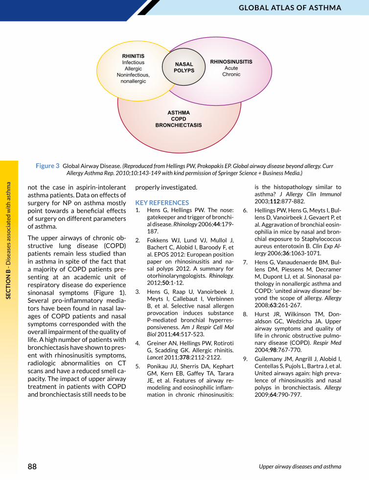
Figure 3 Global Airway Disease. (Reproduced from Hellings PW, Prokopakis EP. Global airway disease beyond allergy. Curr Allergy Asthma Rep. 2010;10:143-149 with kind permission of Springer Science + Business Media.)
not the case in aspirin-intolerant asthma patients. Data on effects of surgery for NP on asthma mostly point towards a beneficial effects of surgery on different parameters of asthma.
The upper airways of chronic ob-structive lung disease (COPD) patients remain less studied than in asthma in spite of the fact that a majority of COPD patients pre-senting at an academic unit of respiratory disease do experience sinonasal symptoms (Figure 1). Several pro-inflammatory media-tors have been found in nasal lav-ages of COPD patients and nasal symptoms corresponded with the overall impairment of the quality of life. A high number of patients with bronchiectasis have shown to pres-ent with rhinosinusitis symptoms, radiologic abnormalities on CT scans and have a reduced smell ca-pacity. The impact of upper airway treatment in patients with COPD and bronchiectasis still needs to be
properly investigated.
KEY REFERENCES1. Hens G, Hellings PW. The nose:
gatekeeper and trigger of bronchi-al disease. Rhinology 2006;44:179-187.
2. Fokkens WJ, Lund VJ, Mullol J, Bachert C, Alobid I, Baroody F, et al. EPOS 2012: European position paper on rhinosinusitis and na-sal polyps 2012. A summary for otorhinolaryngologists. Rhinology. 2012;50:1-12.
3. Hens G, Raap U, Vanoirbeek J, Meyts I, Callebaut I, Verbinnen B, et al. Selective nasal allergen provocation induces substance P-mediated bronchial hyperres-ponsiveness. Am J Respir Cell Mol Biol 2011;44:517-523.
4. Greiner AN, Hellings PW, Rotiroti G, Scadding GK. Allergic rhinitis. Lancet 2011;378:2112-2122.
5. Ponikau JU, Sherris DA, Kephart GM, Kern EB, Gaffey TA, Tarara JE, et al. Features of airway re-modeling and eosinophilic inflam-mation in chronic rhinosinusitis:
is the histopathology similar to asthma? J Allergy Clin Immunol 2003;112:877-882.
6. Hellings PW, Hens G, Meyts I, Bul-lens D, Vanoirbeek J, Gevaert P, et al. Aggravation of bronchial eosin-ophilia in mice by nasal and bron-chial exposure to Staphylococcus aureus enterotoxin B. Clin Exp Al-lergy 2006;36:1063-1071.
7. Hens G, Vanaudenaerde BM, Bul-lens DM, Piessens M, Decramer M, Dupont LJ, et al. Sinonasal pa-thology in nonallergic asthma and COPD: ‘united airway disease’ be-yond the scope of allergy. Allergy 2008;63:261-267.
8. Hurst JR, Wilkinson TM, Don-aldson GC, Wedzicha JA. Upper airway symptoms and quality of life in chronic obstructive pulmo-nary disease (COPD). Respir Med 2004;98:767-770.
9. Guilemany JM, Angrill J, Alobid I, Centellas S, Pujols L, Bartra J, et al. United airways again: high preva-lence of rhinosinusitis and nasal polyps in bronchiectasis. Allergy 2009;64:790-797.
ASTHMACOPD
BRONCHIECTASIS
NASALPOLYPS
RHINITISInfectiousAllergic
Noninfectious,nonallergic
RHINOSINUSITISAcute
Chronic

89
Global atlas oF asthmase
ct
ion
b - D
iseases associated
with
asthm
a
• Asthma and obesity are related chronic disease epidemics• Obesity modifies asthma, resulting in the obese-asthma
phenotype• The physiological changes in obese asthma include reduced
expiratory reserve volume and airway closure during tidal breathing
• Adipose tissue is inflamed leading to proinflammatory cytokine and adipokine production
• Airway inflammation is altered to a non-eosinophilic pattern• These changes may contribute to treatment resistance in obese
asthma• Management of asthma in obese patients requires intervention at
both the individual and societal levels• Effective public health interventions are urgently required
Asthma and obesity are linked global chronic disease epidemics. The prevalence of both diseases is high and shows considerable geo-graphic variation (Figure 1). Obesi-ty can potentiate the development and clinical severity of asthma. Like all chronic disease epidemics, asth-ma and obesity often begin in child-hood and several different chronic different diseases may occur in the same person. The approach to pre-vention and treatment of the asth-ma and obesity epidemics needs to be long-term and systematic.
Obesity modifies the clinical ex-pression of asthma, resulting in the obese-asthma phenotype (Table 1). Deposition of adipose tissue in the thoracic and abdominal re-gions leads to lung restriction and physiological changes such as re-duced expiratory reserve volume (the earliest change in static lung volumes) and airway closure dur-ing tidal breathing. This results in loss of the ‘physiological breathing space’, the gap between tidal and maximal expiratory airflow (Figure 2). In obesity, asthma symptoms are worse, and response to asthma treatment is impaired. Adipose tis-sue is inflamed with an infiltration of macrophages and mast cells, leading to proinflammatory cy-
ASTHMA AND OBESITY, THE TWIN EPIDEMICS3
Ke y m e ssag e s
Peter G. Gibson University of Newcastle
NSW, Australia
Asthma and obesity, the twin epidemics
tokine and adipokine production (Figure 3). This results in chronic low-grade systemic inflammation with elevated C-reactive protein levels and increased cardiovas-cular risk. In obese asthma, the changes in adipokines such as lep-tin are enhanced (Figure 4), and the pattern of airway inflammation is altered to a non-eosinophilic pat-tern, with elevated neutrophils in women with obese asthma. These changes may contribute to treat-ment resistance in obese asthma.
Obesity results from an imbalance between caloric intake and energy
expenditure. This includes eating excessive amounts of food that is high in saturated fat and reduc-ing physical activity levels. Both of these changes are increasingly prevalent in modern urbanized so-cieties, and identify the important social and political dimensions to obesity and its management. Con-sumption of a meal that is high in saturated fat leads to systemic in-flammation with elevated C-reac-tive protein in obese asthma. There are, in addition, changes to the asthmatic airway indicating acti-vation of innate immune responses with elevated gene expression for

90
Global atlas oF asthmase
ct
ion
b -
Dis
ease
s as
soci
ated
wit
h a
sth
ma
Asthma and obesity, the twin epidemics
b Prevalence of overweight (%)< 2020-39.9
40-59.9≥ 60
Data not availableNot applicable
*BMI ≥ 25 kg/m2
aProportion of population (%)
≥10.1
7.6-10.0
5.1-7.5
2.5-5.0
0-2.5
No standardised data available
Toll-like receptor 4 and elevated neutrophils, and activation of the pathways as depicted in Figure 3. The associated functional conse-quences include reduced broncho-dilator responsiveness.
Management of obese asthma re-quires intervention at both the in-dividual and societal levels. Weight loss leads to improved asthma and can even lead to resolution of asth-ma in some individuals. Weight loss can be achieved by caloric restriction and bariatic surgery. Increasing physical activity during weight loss can minimize the loss of lean body mass (skeletal mus-cle). The goals of weight reduction need to be clearly defined for in-dividuals, and can be to reverse obesity or to improve the asthma. Large amounts of weight loss are required to reverse obesity, how-ever only a modest weight loss of 10% body weight is sufficient to improve the medical complications of obesity, including asthma.
Effective public health interven-tions are urgently required at a so-cietal level to manage the obesity epidemic, and its adverse impact on asthma.
KEY REFERENCES1. Bousquet J, Khaltaev N, editors.
Global surveillance, prevention and control of chronic respira-tory diseases : a comprehensive approach. Geneva: WHO Press, 2007.
2. Lugogo NL, Kraft M. Dixon AE. Does obesity produce a distinct asthma phenotype? J Appl Physiol 2010;108:729-734.
3. Gibson PG. Obesity and Asthma. Ann Am Thorac Soc 2013;in press.
4. Lugogo N, Bappanad, Kraft M. Obesity, metabolic dysregu-lation, and oxidative stress in asthma. Biochem Biophys Acta 2011;1810:1120-1126.
5. Berthon BS, Macdonald-Wicks LK,
Figure 1 World map of the prevalence of asthma (panel a) and obesity (panel b). (Panel a reproduced from Masoli M, Fabian D, Holt S, et al. The global burden of
asthma: executive summary of the GINA Dissemination Committee report. Allergy 2004;59:469-8 with permission from Wiley-Blackwell. Panel b reproduced from World
Health Organisation, Global Health Observatory.)
TABLE 1
Characteristics of the obese asthma phenotype *
Worse asthma control
Decreased response to controller medication
Presence of comorbidities related to obesity
Presence of metabolic/immune derangements related to obesity
* Reproduced from Lugogo NL, Kraft M, Dixon AE, Does obesity produce a distinct asth-ma phenotype? J Appl Physiol 2010;108:729-734 with permission of The American Physiological Society.

91
Global atlas oF asthmase
ct
ion
b - D
iseases associated
with
asthm
a
Figure 3 Inflammatory pathways in obesity leading to altered systemic and pulmonary inflammatory responses in asthma. (Reprinted from Biochem Biophys Acta, 1810/11, Lugogo N, Bappanad, Kraft M. Obesity, metabolic dysregulation, and oxidative
stress in asthma, 1120-1126, Copyright 2011, with permission from Elsevier.)
Figure 4 Elevated leptin in obesity and asthma, and effects of gender.
(Reproduced from Berthon BS, Macdonald-Wicks LK, Gibson PG, et al. An investigation
of the association between dietary intake, disease severity and airway inflammation in asthma. Respirology 2013;18:447-454 with
permission from John Wiley and Sons, Inc.)
Asthma and obesity, the twin epidemics
Figure 2 Effects of obesity (solid lines) on airway physiology. Compared to normal (dotted lines), obesity leads to reduced static lung volumes (bars) and airflow limitation during tidal breathing in the expiratory flow volume curve, resulting in
loss of the ‘breathing space’, the gap between tidal flow and maximal expiratory flow. TLC - total lung volume; FRC - forced
residual capacity; RV - residual volume (Reproduced from Farah CS, Salome CM. Asthma and obesity: a known association but unknown mechanism. Respirology 2012;17:412-421 with
permission from John Wiley and Sons, Inc.)
Volume (L)
Obesity
PredictedRVFRCTLC
Flow
(L/m
in)
Gibson PG, Wood LG. An investi-gation of the association between dietary intake, disease severity and airway inflammation in asth-
ma. Respirology 2013;18:447-454.
6. Wood LG, Garg ML, Gibson PG. A high-fat challenge increases airway inflammation and im-
pairs bronchodilator recovery in asthma. J Allergy Clin Immunol 2011;127:1133-1140.
10000
9000
8000
7000
6000
5000
4000
3000
2000
1000
Healthy Control Asthma
Lept
in (p
g/m
l)
Female
Male

92
Global atlas oF asthmase
ct
ion
b -
Dis
ease
s as
soci
ated
wit
h a
sth
ma • Aspirin Exacerbated Respiratory Disease (AERD) is a distinct
phenotype of asthma with coexisting chronic rhinosinusitis, nasal polyps and hypersensitivity to aspirin and to other non-steroidal anti-inflammatory drugs
• AERD is characterized by an increased risk for uncontrolled upper and lower airway disease
• Patients with AERD require comprehensive and multidisciplinary diagnostic approach
• Management of asthma and rhinosinusitis in a patient with AERD is similar to other forms of asthma and rhinosinusitis
• Aspirin desensitization may be an effective treatment option for some AERD patients
DEFINITION AND CLINICAL CHARACTERISTICS OF AERDAspirin Exacerbated Respiratory Disease (AERD) is a distinct clini-cal syndrome observed in 5-10% of patients with asthma and char-acterized by history of acute dysp-nea usually accompanied by nasal symptoms (rhinorrhoea and/or nasal congestion) within two hours after ingestion of acetylsalisilic acid (ASA) (Figure 1). These patients suffer from chronic, usually severe rhinosinusitis with recurrent nasal polyps and do not tolerate other non-steroidal anti-inflammatory drugs (NSAIDs), which are strong cyclooxygenase-1 (COX-1) inhibi-tors. The syndrome has been previ-ously called “Aspirin-triad” or “As-pirin-Sensitive Asthma”. Patients with AERD are quite heterogene-ous with respect to asthma sever-ity, presence of atopic sensitization (up to 70% may be atopic) and gen-eral responsiveness to treatment. However, on average AERD is as-sociated with increased risk for severe asthma, frequent exacerba-tions and sudden death.
PATHOGENESIS OF AERD AND HYPERSENSITIVITY TO NSAIDSThe mechanism of hypersensitivity to ASA and NSAIDs in asthmatic patients is not immunological, but
is related to inhibition of COX-1, an enzyme that converts arachi-donic acid into prostaglandins, thromboxanes and prostacyclin. According to the “ prostaglandin/cyclooxygenase theory” proposed by Andrew Szczeklik inhibition of COX-1 by ASA or other NSAID, by depriving the system from prostaglandin E2 (PGE2) triggers activation of inflammatory cells (mast cells, eosinophils and plate-lets) with subsequent release of inflammatory mediators, including cysteinyl leukotrienes (Figure 2). Baseline abnormalities of arachi-donic acid metabolism (e.g. PGE2 deficiency and overproduction of leukotrienes), persistent viral in-
fections, Staphylococcus aureus en-terotoxins and underlying genetic predisposition may have important role in the pathogenesis of chronic eosinophilic inflammation typically present in the upper and lower air-way mucosa of AERD patients.
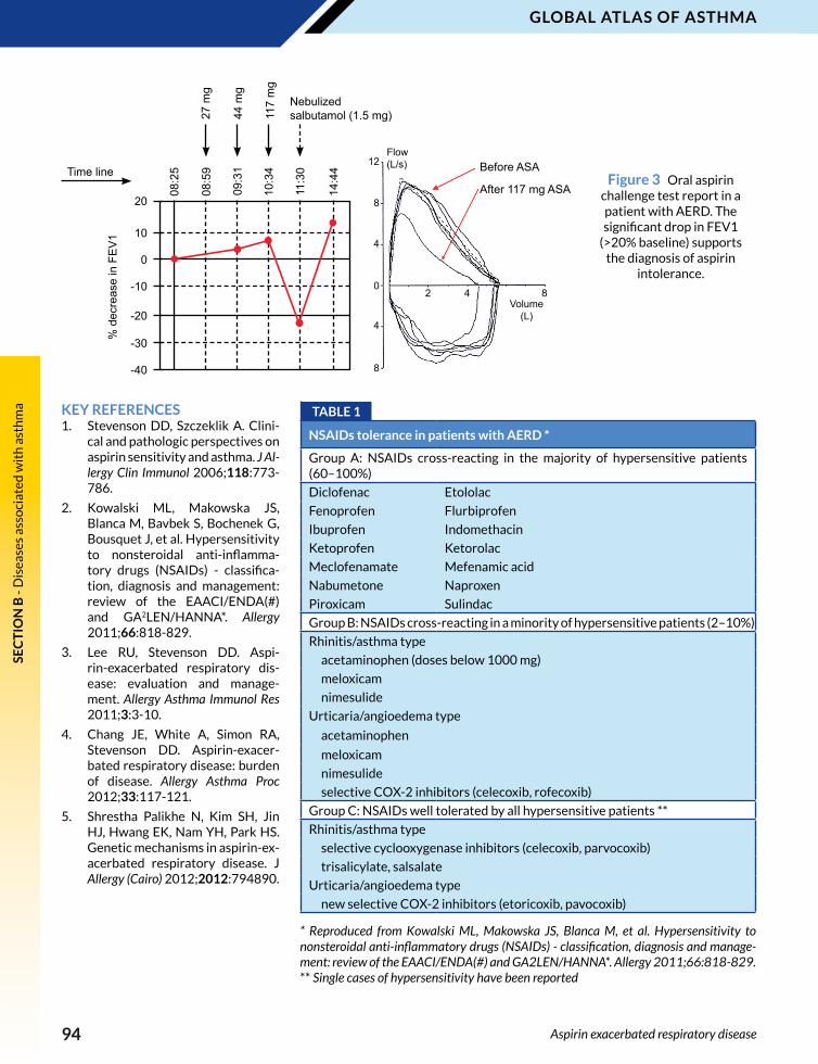
DIAGNOSIS OF NSAID HYPERSENSITIVITYIn the majority of patients the di-agnosis of ASA/NSAID hypersen-sitivity can be based on a history of respiratory symptoms induced by the ingestion of aspirin or oth-er NSAIDs. Confirmation by con-trolled aspirin challenge may be necessary in some patients. Oral aspirin provocation (Figure 3) is
ASPIRIN ExACERBATED RESPIRATORY DISEASE4
Ke y m e ssag e s
Marek L. Kowalski Medical University of Łódź
Poland
Sevim Bavbek Ankara University
Turkey
Aspirin exacerbated respiratory disease

93
Global atlas oF asthmase
ct
ion
b - D
iseases associated
with
asthm
a
Aspirin exacerbated respiratory disease
Figure 1 Clinical characteristics of Aspirin Exacerbated Respiratory Disease.
Figure 2 Pathomechanism of aspirin induced hypersensitivity reactions in AERD patients. (Reproduced and modified from Kowalski ML. Diagnosis of aspirin
sensitivity in aspirin exacerbated respiratory disease. In: Pawankar R, Holgate ST, Rosenwasser LJ, editors. Allergy frontiers: diagnosis and health economics. New York:
Springer, 2009; 349-372, with kind permission of Springer Science + Business Media.)
the gold standard for the diagnosis, but bronchial or nasal provocation with lysine-ASA may be valuable alternative diagnostic tools. Sever-
al in vitro cell activation tests have been evaluated, but none of them can be recommended for routine diagnosis.
Hypersensitivityto ASA
Chronic rhinosinusitis
with nasal polypsAsthma
• Cross- reactivity with COX-1 inhibitors
• General tolerance of COX-2 inhibitors
• Hyperplastic pansinusitis
• Recurrent nasal polyps• Hyposmia
• More severe than average
• More difficult to control• Increased death risk
Aspirin triad
ASA Arachidonic acid
Cell membrane phospholipids
15-LOX
15-HPETE
15-HETE
EoxinsLipoxins
COX-1
PLA
LTA4
LTC4
LTD4
LTE4
LTC4s
5-LOX
AsthmaRhinorrheaCongestion
UrticariaAngioedema
PGG2PGH2
PGE22
Eos Mast cell
Platelets
PGE2
EP-R
EP-R
COX-2
MANAGEMENT OF AERDCareful avoidance of ASA and oth-er NSAIDs, which are strong COX-1 inhibitors, is necessary to prevent severe asthma attacks. As alter-native to NSAIDs acetaminophen or preferential/selective COX-2 inhibitors, are recommended (Ta-ble 1). Management of asthma and rhinosinusitis in AERD is similar to other forms of asthma and rhinosi-nusitis and international treatment guidelines should be followed. Inhaled glucocorticosteroids in appropriate doses, often in combi-nation with long acting beta-2 ag-onists are effective in controlling asthmatic inflammation and symp-toms, but in some patients chronic treatment with oral prednisone may be necessary.
Addition of a leukotriene receptor antagonist such as montelukast to standard anti-inflammatory ther-apy may be effective in relieving symptoms and improving respira-tory function in some patients with AERD, but the degree of improve-ment is similar to ASA tolerant asthmatics. Topical nasal steroids are preferred for controlling symp-toms of rhinosinusitis and may slow down recurrence of nasal pol-yps. Surgical procedures (polypec-tomy, functional endoscopic sinus surgery or ethmoidectomy) are usually needed at certain stage of the disease.
The special approach for these pa-tients is ASA desensitization. The alleviation of chronic upper and lower airway symptoms, reduction in hospitalization and emergency room visits, and decreased need for nasal/sinus surgery is observed in desensitized patients. Howev-er, only a fraction of patients with AERD will benefit from aspirin de-sensitization and at present it is not possible to predict the responders.
94
Global atlas oF asthmase
ct
ion
b -
Dis
ease
s as
soci
ated
wit
h a
sth
ma
Figure 3 Oral aspirin challenge test report in a patient with AERD. The significant drop in FEV1
(>20% baseline) supports the diagnosis of aspirin
intolerance.
Aspirin exacerbated respiratory disease
TABLE 1
NSAIDs tolerance in patients with AERD *
Group A: NSAIDs cross-reacting in the majority of hypersensitive patients (60–100%)
Diclofenac Etololac
Fenoprofen Flurbiprofen
Ibuprofen Indomethacin
Ketoprofen Ketorolac
Meclofenamate Mefenamic acid
Nabumetone Naproxen
Piroxicam Sulindac
Group B: NSAIDs cross-reacting in a minority of hypersensitive patients (2–10%)
Rhinitis/asthma type
acetaminophen (doses below 1000 mg)
meloxicam
nimesulide
Urticaria/angioedema type
acetaminophen
meloxicam
nimesulide
selective COX-2 inhibitors (celecoxib, rofecoxib)
Group C: NSAIDs well tolerated by all hypersensitive patients **
Rhinitis/asthma type
selective cyclooxygenase inhibitors (celecoxib, parvocoxib)
trisalicylate, salsalate
Urticaria/angioedema type
new selective COX-2 inhibitors (etoricoxib, pavocoxib)
* Reproduced from Kowalski ML, Makowska JS, Blanca M, et al. Hypersensitivity to nonsteroidal anti-inflammatory drugs (NSAIDs) - classification, diagnosis and manage-ment: review of the EAACI/ENDA(#) and GA2LEN/HANNA*. Allergy 2011;66:818-829. ** Single cases of hypersensitivity have been reported
20
10
0
-10
-20
-30
-40
08:2
5
08:5
9
09:3
1
10:3
4
11:3
0
14:4
4
27 m
g
44 m
g
117
mg
Nebulized salbutamol (1.5 mg)
% d
ecre
ase
in F
EV
1
Time line Before ASA
After 117 mg ASA
12
8
4
0
4
8
2 4 8Volume
(L)
Flow(L/s)
KEY REFERENCES1. Stevenson DD, Szczeklik A. Clini-
cal and pathologic perspectives on aspirin sensitivity and asthma. J Al-lergy Clin Immunol 2006;118:773-786.
2. Kowalski ML, Makowska JS, Blanca M, Bavbek S, Bochenek G, Bousquet J, et al. Hypersensitivity to nonsteroidal anti-inflamma-tory drugs (NSAIDs) - classifica-tion, diagnosis and management: review of the EAACI/ENDA(#) and GA2LEN/HANNA*. Allergy 2011;66:818-829.
3. Lee RU, Stevenson DD. Aspi-rin-exacerbated respiratory dis-ease: evaluation and manage-ment. Allergy Asthma Immunol Res 2011;3:3-10.
4. Chang JE, White A, Simon RA, Stevenson DD. Aspirin-exacer-bated respiratory disease: burden of disease. Allergy Asthma Proc 2012;33:117-121.
5. Shrestha Palikhe N, Kim SH, Jin HJ, Hwang EK, Nam YH, Park HS. Genetic mechanisms in aspirin-ex-acerbated respiratory disease. J Allergy (Cairo) 2012;2012:794890.

95
Global atlas oF asthmase
ct
ion
b - D
iseases associated
with
asthm
a
• Gastro-esophageal reflux disease (GERD) is an increase of retrograde movement of gastric content into the esophagus
• Laryngopharyngeal reflux is reflux which reaches the larynx• Ten to 20% of the general adult population in western countries
and 5% of subjects in the Asia Pacific region suffer from GERD• Asthma and/or upper airway complaints or problems are
associated with GERD• Double-blind controlled studies demonstrate that treatment
of asymptomatic GERD in adults and children does not improve asthma
• Just as GERD may aggravate asthma, so too, could asthma or asthma therapy aggravate GERD
• Diagnosis of GERD in both adults and children is primarily suspected and made by a detailed history
• Treatment includes lifestyle changes and where necessary H2 blockers, proton pump inhibitors and prokinetics
Gastroesophageal reflux disease (GERD) is an increase of retrograde movement of gastric content into the esophagus. Laryngopharyngeal reflux is reflux which reaches the larynx. GERD is present when the frequency and duration of acid re-flux exceeds defined parameters, as quantified by a pH probe placed in the esophagus. Regardless of its more formal definition, it is a dis-ease in and of itself, often associat-ed with esophageal complications such as esophageal erosion and stricture and Barrett’s esophagus, the latter of which can lead to ad-enocarcinoma of the esophagus. Factors which contribute to or cause GERD are illustrated in Fig-ure 1.
Ten to 20% of the general adult population in western countries and 5% in the Asia Pacific region suffer from symptoms of GERD. The presence of GERD in some pediatric studies is between 2-8%. Typical symptoms particularly in adults, include esophageal burning and discomfort (heartburn) as well as regurgitation of gastric content into the posterior pharynx (wa-ter brash) (Table 1). Other symp-toms include belching, indigestion, nausea, vomiting, odynophagia, dysphagia, and halitosis. Throat
tightness, throat clearing, cough, chest tightness, postnasal drip, and hoarseness are all potential symp-toms of GERD, particularly with la-ryngeal pharyngeal reflux. Cough, associated with laryngopharyn-geal GERD, is usually described as originating in the laryngopharynx, whereas cough associated with asthma usually originates in the chest; however, this distinction is subjective as can be differentiat-ing the symptoms of cough from throat clearing. The same symp-toms can occur in children, how-
ever, recurrent regurgitation, with or without vomiting, weight loss or poor weight gain, irritability, and behavioral problems may occur.
Asthma and/or upper airway com-plaints or problems are associated with GERD. Epidemiologic studies demonstrate a variable prevalence in subjects with asthma of between 12 to 85%. The variability is largely dependent on the method used to define GERD. Conversely, asthma also appears to be more common in individuals with GERD. Two hy-
GASTRO-ESOPHAGEAL REFLUx DISEASE AND ASTHMA5
Ke y m e ssag e s
Richard F. Lockey University of South Florida
USA
Gastro-esophageal reflux disease and asthma

96
Global atlas oF asthmase
ct
ion
b -
Dis
ease
s as
soci
ated
wit
h a
sth
ma
trolled studies in both adults and children with asthma indicates that treating GERD does not increase asthma control but does decrease the use of albuterol; it benefited a subset of affected patients.
Just as GERD may aggravate asthma, so too, could asthma or asthma therapy aggravate GERD or GERD-associated symptoms. Beta agonists and theophylline re-duce esophageal sphincter tone, systemic glucocorticosteroids increase gastric acid production, and inhaled corticosteroids induce cough and cause chronic larynge-al irritation and hoarseness, the latter of which are also associat-ed with GERD. Asthma also is as-sociated with chronic cough and wheezing, both of which increase intra-abdominal pressure, which can theoretically result in pushing gastric contents up through the lower esophageal sphincter into the esophagus aggravating GERD.
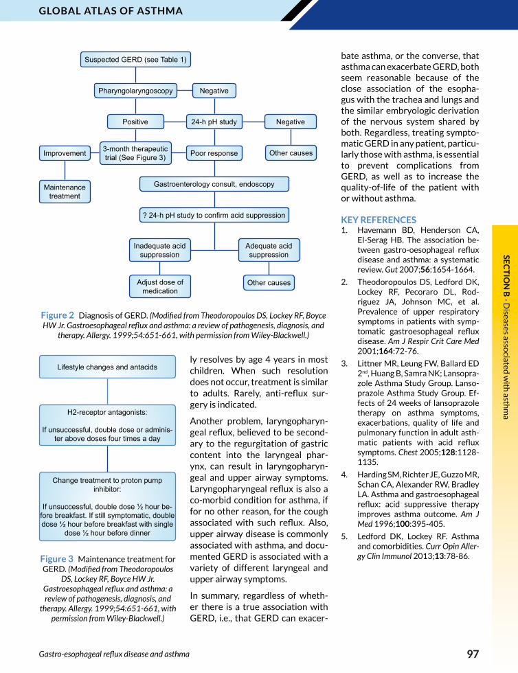
Diagnosis of GERD is primarily based on a detailed history since it is impossible to confirm the diag-nosis with a pH probe and/or en-doscopy in all individuals with this disease (Figure 2). When complica-tions are suspected, a gastroenter-ologist consultation is indicated.
Treatment (Figure 3) includes life-style changes, i.e., avoiding large meals, maintaining ideal weight, not eating meals three hours be-fore retiring, not lying down within two hours after meals, elevating the head of the bed with 6-inch blocks or using a foam wedge to el-evate the trunk and head. Avoiding acid-containing foods, carbonated beverages and fatty foods also may be beneficial. Medications include H2 blockers, proton pump inhib-itors and prokinetic agents, the latter for individuals with delayed gastric emptying. GERD common-
potheses are proposed to explain this association; asthma bronchos-pasm is attributed to aspiration or reflux of gastric contents into the trachea, whereas the second im-plicates vagal reflexes mediated through stimulation of esophageal mucosal receptors by a low pH and distention. Both mechanisms prob-ably contribute to asthma in vary-ing degrees.
Double-blind controlled stud-
ies demonstrate that treatment of asymptomatic GERD does not improve asthma in adults or chil-dren; however, other controlled studies show that GERD-treated subjects with asthma and sympto-matic GERD experience improved asthma quality-of-life and have a reduced number of asthma exacer-bations, while there are question-able effects on asthma symptoms, albuterol use, and pulmonary func-tion. A Cochrane review of con-
Gastro-esophageal reflux disease and asthma
TABLE 1
GERD Symptoms and Signs *
GastroesophagealHeartburn, chest/epigastric/cervical pain, water brash, belching, indigestion, nausea/vomiting/hematemesis
Respiratory Cough, wheeze, dyspnea, hemoptysis
LaryngealHoarseness, throat clearing, sighing dyspnea, irrita-tion, globus, voice changes, soreness
Nasal Congestion, itching, sneezing, soreness
Sinuses Headache, pressure, purulent discharge
Ears Otalgia
Teeth Loss of dental enamel
* Reproduced from Theodoropoulos DS, Lockey RF, Boyce HW Jr. Gastroesophageal re-flux and asthma: a review of pathogenesis, diagnosis, and therapy. Allergy. 1999;54:651-661, with permission from Wiley-Blackwell.
Figure 1 Factors which contribute to or cause GERD.A. Defective clearance of esoph-
ageal contents secondary to reduced salivary and esophageal submucosal gland secretion and ineffective peristalsis
B. Lower esophageal dysfunction with prolonged and inappropri-ate relaxations of the sphincter with reduction in basal lower esophageal sphincter pressure and tone
C. A hiatal hernia may compro-mise lower esophageal function causing gastric contents to be trapped above the diaphragm exacerbating reflux
D. Delay of gastric emptying may increase gastric contents available for reflux into the esophagus
E. Various illnesses, such as asthma, which are associated with chronic cough and expiratory straining during breathing, can increase intra-abdominal pressure, pushing gastric contents into the esophagus
D
E
A
BC
Pyloric Opening & Sphincter
Esophagus
Lower Esophageal Sphincter
Hiatal Hernia
Diaphragm
StomachDuodenum
97
Global atlas oF asthmase
ct
ion
b - D
iseases associated
with
asthm
a
Figure 2 Diagnosis of GERD. (Modified from Theodoropoulos DS, Lockey RF, Boyce HW Jr. Gastroesophageal reflux and asthma: a review of pathogenesis, diagnosis, and
therapy. Allergy. 1999;54:651-661, with permission from Wiley-Blackwell.)
Gastro-esophageal reflux disease and asthma
ly resolves by age 4 years in most children. When such resolution does not occur, treatment is similar to adults. Rarely, anti-reflux sur-gery is indicated.
Another problem, laryngopharyn-geal reflux, believed to be second-ary to the regurgitation of gastric content into the laryngeal phar-ynx, can result in laryngopharyn-geal and upper airway symptoms. Laryngopharyngeal reflux is also a co-morbid condition for asthma, if for no other reason, for the cough associated with such reflux. Also, upper airway disease is commonly associated with asthma, and docu-mented GERD is associated with a variety of different laryngeal and upper airway symptoms.
In summary, regardless of wheth-er there is a true association with GERD, i.e., that GERD can exacer-
Suspected GERD (see Table 1)
Pharyngolaryngoscopy Negative
Positive 24-h pH study Negative
Improvement 3-month therapeutic trial (See Figure 3) Poor response
Maintenance treatment
Other causes
Gastroenterology consult, endoscopy
? 24-h pH study to confirm acid suppression
Inadequate acid suppression
Adequate acid suppression
Adjust dose of medication
Other causes
Lifestyle changes and antacids
H2-receptor antagonists:
If unsuccessful, double dose or adminis-ter above doses four times a day
Change treatment to proton pump inhibitor:
If unsuccessful, double dose ½ hour be-
fore breakfast. If still symptomatic, double dose ½ hour before breakfast with single
dose ½ hour before dinner
bate asthma, or the converse, that asthma can exacerbate GERD, both seem reasonable because of the close association of the esopha-gus with the trachea and lungs and the similar embryologic derivation of the nervous system shared by both. Regardless, treating sympto-matic GERD in any patient, particu-larly those with asthma, is essential to prevent complications from GERD, as well as to increase the quality-of-life of the patient with or without asthma.
KEY REFERENCES1. Havemann BD, Henderson CA,
El-Serag HB. The association be-tween gastro-oesophageal reflux disease and asthma: a systematic review. Gut 2007;56:1654-1664.
2. Theodoropoulos DS, Ledford DK, Lockey RF, Pecoraro DL, Rod-riguez JA, Johnson MC, et al. Prevalence of upper respiratory symptoms in patients with symp-tomatic gastroesophageal reflux disease. Am J Respir Crit Care Med 2001;164:72-76.
3. Littner MR, Leung FW, Ballard ED 2nd, Huang B, Samra NK; Lansopra-zole Asthma Study Group. Lanso-prazole Asthma Study Group. Ef-fects of 24 weeks of lansoprazole therapy on asthma symptoms, exacerbations, quality of life and pulmonary function in adult asth-matic patients with acid reflux symptoms. Chest 2005;128:1128-1135.
4. Harding SM, Richter JE, Guzzo MR, Schan CA, Alexander RW, Bradley LA. Asthma and gastroesophageal reflux: acid suppressive therapy improves asthma outcome. Am J Med 1996;100:395-405.
5. Ledford DK, Lockey RF. Asthma and comorbidities. Curr Opin Aller-gy Clin Immunol 2013;13:78-86.Figure 3 Maintenance treatment for
GERD. (Modified from Theodoropoulos DS, Lockey RF, Boyce HW Jr.
Gastroesophageal reflux and asthma: a review of pathogenesis, diagnosis, and
therapy. Allergy. 1999;54:651-661, with permission from Wiley-Blackwell.)

98
Global atlas oF asthmase
ct
ion
b -
Dis
ease
s as
soci
ated
wit
h a
sth
ma • There is a conflict in the literature surrounding the asthma-related
risk of cardiovascular disease identified in large, longitudinal epidemiologic studies
• Relationships of asthma and cardiovascular disease seem to be stronger in women
• A common mechanism may contribute to allergies and atherosclerosis and systemic inflammation associated with asthma may adversely affect cardiovascular function
• Decreased pulmonary function, increased airway infection, and use of β2-agonists may increase the risk of cardiovascular disease
• Patterns of risks of myocardial infarction are similar between inhaled short-acting β2-agonists, long-acting β2-agonists and inhaled corticosteroids
Some studies report a significant association of asthma with cardio-vascular disease, but there is a con-flict in the literature surrounding the asthma-related risk of cardio-vascular disease identified in large, longitudinal epidemiologic studies.
Adult-onset asthma is associated with increased carotid atheroscle-rosis in women, and patients with bronchial hyperresponsiveness to methacholine demonstrated increased carotid intima–media thickness. Relationships between asthma and cardiovascular dis-ease in women seem to be stronger than those observed in men (Fig-ure 1). In general, allergic disease is more common in women after adolescence and it is thought that sex hormones modulate immune response. Estrogen is considered to increase humoral immunity. Be-ing female slightly increases the association of all cardiovascular diseases, mainly heart failure, but not angina, coronary disease and acute or old myocardial infarc-tion, with asthma. In contrast to females, males present with a pos-itive association between asthma and angina and coronary disease, but a negative association with acute or old myocardial infarction. An increase in age results in a pro-
gressive increase in the prevalence of the diagnosis of cardiovascular disease and hypertensive disease. Apparently, the smoking habit does not modify the prevalence of cardiovascular disease, compared to the general population.
A common mechanism may con-tribute to allergies and athero-sclerosis. IgE is itself potentially proatherogenic through actions on mast cells and platelets, although epidemiological studies indicate that atopy may be an independent protective factor against myocar-dial infarction. In any case, asth-ma and atherosclerosis occur on a
background of inflammation. Ani-mal studies have shown increased myocardial vulnerability in rabbits with systemic allergy and asthma. It has been suggested that airway allergen exposure results in im-paired vasodilatory response of the aorta in a murine model of pulmo-nary allergic response. This finding suggests that systemic inflamma-tion associated with asthma may adversely affect cardiovascular function. Actually, systemic in-flammation occurs in asthma, with an increase in circulating proin-flammatory cytokines, such as in-terleukin IL-6 and tumor necrosis factor-α and also in high-sensibility
CARDIOVASCULAR DISEASES AND ASTHMA6
Ke y m e ssag e s
Mario Cazzola University of Rome “Tor Vergata”
Italy
Cardiovascular diseases and asthma

99
Global atlas oF asthmase
ct
ion
b - D
iseases associated
with
asthm
a
Cardiovascular diseases and asthma
C-reactive protein, likely because of the systemic dissemination of lo-cal lung inflammation leading to an overspill effect (Figure 2). This sys-temic component could feed back into and perpetuate the original lo-cal reaction and lead to the devel-opment of distant local reactions.
However, the role of systemic in-
flammation in asthmatic patients is still unclear and, consequently, debated. Asthma is a long-term inflammatory status complicated by decreased pulmonary function, increased airway infection, and use of β2-agonists. These factors may increase the risk of cardiovascular disease. Patterns of risks of myo-cardial infarction are similar be-
Figure 1 The association between asthma and cardiovascular comorbidities in Italy. (This article was published in Respir Med, 106, Cazzola M, Calzetta L, Bettoncelli G, et al, Cardiovascular disease in asthma and COPD: a population-based retrospective
cross-sectional study, 249-56, Copyright Elsevier 2012.)
Figure 2 The inflammation in the lung ‘spills over’ into the systemic circulation to produce systemic effects, such as cardiovascular complications.
tween inhaled short-acting β2-ag-onists, long-acting β2-agonists and inhaled corticosteroids. It is likely that the initial presentation with symptoms evoking asthma (dysp-noea presumably) is, in a large pro-portion of cases, the appearance of ischaemic heart disease. None-theless, it is noteworthy that some epidemiological studies have been unable to register concrete associ-ation of asthma with acute or pre-vious myocardial infarction.
KEY REFERENCES1. Cazzola M, Calzetta L, Betton-
celli G, Cricelli C, Romeo F, Ma-tera MG, et al. Cardiovascular disease in asthma and COPD: a population-based retrospective cross-sectional study. Respir Med 2012;106:249-256.
2. Cazzola M, Calzetta L, Bettoncel-li G, Novelli L, Cricelli C, Rogliani P. Asthma and comorbid medical illness. Eur Respir J 2011;38:42-49.
3. Cazzola M, Segreti A, Calzetta L, Rogliani P. Comorbidities of asth-ma: current knowledge and future research needs. Curr Opin Pulm Med 2013;19:36-41.
4. Iribarren C, Tolstykh IV, Miller MK, Sobel E, Eisner MD. Adult asthma and risk of coronary heart disease, cerebrovascular disease, and heart failure: a prospective study of 2 matched cohorts. Am J Epidemiol 2012;176:1014-1024.
5. Khan UI, Rastogi D, Isasi CR, Coupey SM. Independent and synergistic associations of asthma and obesity with systemic inflam-mation in adolescents. J Asthma 2012;49:1044-1050.
6. Warnier MJ, Rutten FH, Kors JA, Lammers JW, de Boer A, Hoes AW, et al. Cardiac arrhythmias in adult patients with asthma. J Asthma 2012;49:942-946.
IL-6, TNF-α
CRP
IL-6
Atheromaplaque

100
Global atlas oF asthmase
ct
ion
b -
Dis
ease
s as
soci
ated
wit
h a
sth
ma • Foods can induce asthmatic reactions in patients with food allergy
• Patients with both asthma and food allergy are at risk of food-induced anaphylaxis
• Food allergy often precedes asthma and is a risk factor for its development
• Both being chronic allergic disorders, asthma and food allergy often go together
• Cross-reactive IgE of asthmatic patients with seasonal allergic rhinitis can cause subsequent food allergy
Food allergy (FA) is an adverse re-action to food caused by an over-reaction of the immune system that occurs each time a food is consumed. These reactions can be immune responses mediated by IgE antibodies, by immune cells, or by a combination of both. Food allergies are however most com-monly caused by IgE antibodies, and are characterized by acute onset of symptoms (usually within minutes to a few hours) following the ingestion of an implicated al-lergenic food. Symptoms can in-volve the skin, the gastrointestinal tract, the cardiovascular system including life-threatening ana-phylactic shock, and of relevance at this place, the respiratory tract including asthmatic symptoms. FA is estimated to affect 3-8% of chil-dren and 1-5% of adults, but con-siderable geographic differences exist also with respect to the type of foods involved. FA is often seen together with asthma, especially in infancy.
THE LINK BETWEEN FOOD ALLERGY AND ASTHMAFirstly, asthma can be one of the manifestations of an allergic reac-tion to food (Figure 1). Also, food additives, especially sulphites and monosodium glutamate, have been
reported to trigger asthma. Be-sides consumption of food, inhala-tion of cooking vapours of especial-ly fish, shellfish and eggs, is known to potentially cause asthmatic symptoms, and inhalation of wheat flour may cause occupational asth-ma in bakers.
Secondly, patients that have both asthma and food allergy have a higher risk to develop severe ana-phylactic reactions when exposed to the food they are allergic to (Fig-ure 2). Therefore, these patients need to be particularly cautious in avoiding the culprit foods.
Thirdly, sensitization (IgE) to food and clinical FA often precede the development of asthma (Figure 3). This sequence of appearance, often referred to as the “atopic
march”, points towards a common genetic predisposition for both dis-eases. A whole spectrum of gene polymorphisms has been implicat-ed in the development of asthma, illustrating the complex multi-fac-torial nature of the genetic predis-position of this disease. Far less is known about gene polymorphisms with relevance for FA. Recently a polymorphism in a gene coding for a protein involved in the integrity of the skin barrier was reported to be a risk factor for FA in patients that also have asthma.
Fourthly, food allergy not only precedes asthma but both chronic allergic disorders often also stick together (Figure 3). Although some food allergies like to milk and egg are outgrown by the majority of
FOOD ALLERGY AND ASTHMA7
Ke y m e ssag e s
Ronald van Ree Academic Medical Center
Netherlands
Antonella Muraro University of Padua
Italy
Food allergy and asthma

101
Global atlas oF asthmase
ct
ion
b - D
iseases associated
with
asthm
a
children before the age of five, most other common food allergies such as to peanut and tree nuts are usually persistent and are still present when asthma develops.
Lastly, asthma with seasonal al-lergic rhinitis (hay fever) induced by tree and/or grass pollen can also precede instead of follow the development of FA (Figure 4). Al-though most pollen allergies pres-ent as hay fever, some patients develop asthma as well. In many of those patients some years after the onset of their pollen allergy, also symptoms of FA start devel-oping. This phenomenon can be explained by pollen-induced IgE antibodies cross-reacting to foods. The most well-known example of
such cross-reactivity is observed in patients with birch pollen aller-gy. Their IgE antibodies cross-react with fruits like apple and cherry, with tree nuts like hazelnut and with some vegetables like carrot and celeriac. Symptoms of FA in such patients are almost always mild and limited to the oral cavity.
In conclusion, asthma is often ac-companied by FA but the basis of this co-morbidity is diverse.
KEY REFERENCES1. Burks AW, Tang M, Sicherer S, Mu-
raro A, Eigenmann PA, Ebisawa M, et al. ICON: food allergy. J Allergy Clin Immunol 2012;129:906-920.
2. Koplin JJ, Martin PE, Allen KJ. An update on epidemiology of ana-phylaxis in children and adults.
Curr Opin Allergy Clin Immunol 2011;11:492-496.
3. Illi S, von Mutius E, Lau S, Nickel R, Grüber C, Niggemann B. The nat-ural course of atopic dermatitis from birth to age 7 years and the association with asthma. J Allergy Clin Immunol 2004;113:925-931.
4. McAleer MA, Irvine AD. The mul-tifunctional role of filaggrin in allergic skin disease. J Allergy Clin Immunol 2013;131:280-291.
5. Vieths S, Scheurer S, Ballmer-We-ber B. Current understanding of cross-reactivity of food aller-gens and pollen. Ann N Y Acad Sci 2002;964:47-68.
6. Calvani M, Cardinale F, Martelli A, Muraro A, Pucci N, Savino F, et al. Risk factors for severe pediatric food anaphylaxis in Italy. Pediatr Allergy Immunol 2011;22:813-819.
Figure 1 Asthma can be one of the manifestations of an allergic reaction to food. FA - food allergy.
Figure 2 Patients that have both asthma (A) and food allergy have a higher risk to develop severe anaphylactic reactions
when exposed to the food they are allergic to.
Figure 3 Sensitization (IgE) to food and clinical FA often precede the development of asthma (the “atopic march”).
Figure 4 Asthma induced by tree and/or grass pollen can precede the development of FA (oral allergy syndrome).
Food allergy and asthma

102
Global atlas oF asthmase
ct
ion
b -
Dis
ease
s as
soci
ated
wit
h a
sth
ma • Atopic dermatitis (AD) is the most frequent inflammatory skin
disease and results from the complex interplay between defects in skin barrier function, environmental and infectious agents, and immune abnormalities
• Severe AD beginning early in life is a high-risk phenotype for the development of asthma
• Specific IgE is associated with food or environmental allergens, which may be relevant trigger factors for both AD and asthma
• The disturbed expression of the skin barrier protein filaggrin is linked to childhood AD and the subsequent development of asthma
• Urticaria is often a presenting feature of anaphylaxis, involving respiratory difficulty due to inspiratory stridor, expiratory wheeze or both. Acute urticaria by definition does not involve respiratory distress
• Bronchial hyperreactivity has been reported in some patients with chronic spontaneous and inducible patterns of urticaria
• Kinin-induced angioedema involving the airways in hereditary or acquired angioedema and angiotensin converting enzyme inhibitor-induced angioedema requires emergency treatment and may be fatal
Atopic dermatitis (AD) is a com-mon inflammatory skin disorder, characterized by pruritus, a chron-ic relapsing course, a distinctive distribution of eczematous skin lesions (Figure 1) and a personal or family history of atopic diseases in-cluding asthma. It results from the complex interplay between defects in skin barrier function, environ-mental and infectious agents, and immune abnormalities.
During the last decades a marked increase in the frequency of AD has been observed and it is now the most frequent inflammatory skin disease, with a childhood prev-alence of more than 10% in most European countries. The manifes-tation of AD in childhood is great-er in families with a higher income and a more privileged lifestyle. This
may be due to reduced incidence of infections in early childhood and reduced contact with agents that elicit Th1 associated cellular im-mune responses. Of note, differ-ences in prevalence of respiratory allergic diseases often do not par-allel prevalence of AD in larger ep-idemiologic studies, which points to independent risk and manifesta-
tion factors being critical for both atopic diseases.
AD often begins in early infan-cy. Severe AD beginning before 6 months of age is a high-risk phe-notype for the development of asthma, especially in boys. Further-more, AD is linked to food allergy and children with multiple severe food allergies are also at a higher
SKIN AND LUNG: ATOPIC DERMATITIS, URTICARIA AND
ASTHMA8
Ke y m e ssag e s
Thomas Werfel Hannover Medical School
Germany
Clive Grattan Norfolk & Norwich University
Hospital, UK
Skin and lung: atopic dermatitis, urticaria and asthma
Figure 1 Flexural dermatitis on the right arm and disseminated eczema on the trunk in a male adolescent patient
with atopic dermatitis.

103
Global atlas oF asthmase
ct
ion
b - D
iseases associated
with
asthm
a
risk of developing asthma.
Like in allergic asthma, there is an overexpression of Th2 cytokines in lymphatic organs, circulating T-cells and the acute phase of cu-taneous inflammation in many patients with AD. This is close-ly linked to the regulation of IgE, which is higher than normal in 80% of all patients. Specific IgE is com-monly associated with food or en-vironmental allergens, which may be relevant trigger factors both for AD, rhinitis and / or asthma in indi-vidual patients.
Studies of the gene of encoding the skin barrier protein filaggrin have shown the link between ear-ly childhood eczema and the sub-sequent development of asthma
which may, in part, be due to de-fective epidermal barrier function leading to increased allergen sensi-tization (Figure 2).
It appears that different disease mechanisms are important for dif-ferent subgroups of patients suf-fering from AD. Described genetic polymorphisms in AD involve me-diators of atopic inflammation on different chromosomes. Some, but not all of these may also play a role in respiratory atopy.
Besides the “allergic” variant of AD there is a non-allergic form which is found in 20% of diseases with the typical clinical appearance of the disease. In this respect, AD resem-bles asthma, which also has allergic and non-allergic variants.
Figure 2 Filaggrin mutations and atopic dermatitis and asthma. (Adapted from Irvine AD, McLean WH, Leung DY. Filaggrin mutations associated with skin and
allergic diseases. N Engl J Med 2011;365:1315-1327.)
Skin and lung: atopic dermatitis, urticaria and asthma
Asthma(overall risk)
Healthy skin
Allergen
Filaggrin-deficient skin
Allergen• Impaired skin barrier• Increased skin-surface pH• Increased allergen priming• Decreased hydration of
stratum corneum• Decreased resistance to
staphylococcus
Filaggrin mutation
Management of AD exacerbations is a therapeutic challenge, as it re-quires efficient short-term control of acute symptoms, without com-promising the overall management plan, which is aimed at long-term stabilization, flare prevention, and avoidance of side effects. Exacer-bation may sometimes uncover relevant provocation factors, for example food or inhalant allergy, or infection, which in turn may also lead to worsening of asthma (Fig-ure 3).
The prognosis for patients with AD is generally favourable, but patients with severe, widespread disease and concomitant asthma are likely to experience poorer out-comes.
Urticaria does not characteristical-ly involve the respiratory tract, but there are a few situations where there is overlap. The first is ana-phylaxis where urticaria involving wheals, angioedema or both may be the initial presentation of an acute systemic illness defined by respiratory difficulty, hypotension or both, with or without gastroin-testinal symptoms. Anaphylaxis is often due to an immediate hyper-sensitivity response to a food, drug or sting, but may be non-allergic. The boundaries between anaphy-laxis and acute urticaria may not be clear at the time, particularly when food allergy presents with general-ized urticaria. By definition, acute urticaria does not present with systemic symptoms, is continuous (daily or almost daily eruptions itchy skin or mucosal swellings) and lasts for up to 6 weeks, but usually resolves over 10 to 14 days.
Chronic urticaria is not associated with asthma and is hardly ever due to IgE-mediated allergies (except perhaps in very young children with undetected food allergies)
Asthma in the absenceof atopic dermatitis
Asthma in the presenceof atopic dermatitis
Odds ratio 1.5
No increased risk
Odds ratio, 3.3
Odds ratio 3.1
eczema/spongiosis
Langerhans cell activation and migration
dermal mononuclear
cells
Atopic dermatitis

104
Global atlas oF asthmase
ct
ion
b -
Dis
ease
s as
soci
ated
wit
h a
sth
ma
Figure 4 Kinin formation and breakdown relevant in urticaria. LMWK, low molecular weight kininogen; HMWK, high molecular weight kininogen;
kallidin, Lys-bradykinin; B2R, bradykinin receptor 2 – receptor for bradykinin; B1R, bradykinin receptor 1 – receptor for the active metabolite of bradykinin
(bradykinin 1-8 or des arginine bradykinin); ACE-angiotensin converting enzyme
Figure 3 Trigger factors of atopic dermatitis.
Skin and lung: atopic dermatitis, urticaria and asthma
Atopic dermatitis
Hormones
Food
Inhalant allergens
Bacterial colonization of the skin
Irritating substances
Climate
Psychological stress
Systemic infections
although it may occur as an appar-ently independent illness in atopic patients. Around 30% of patients with chronic spontaneous urticar-ia have functional autoantibodies that release histamine from skin mast cells and basophils, so it is surprising that the respiratory tract is not overtly affected. One study concluded that bronchial hyperresponsiveness is a common feature in patients with active chronic urticaria. Twenty six adults with chronic spontaneous urticar-ia were assessed with respiratory function tests and methacholine provocation. Two had asthma on baseline pulmonary function tests and twenty others (77%) showed bronchial hyperresponsiveness on methacholine challenge. Bronchial hyperresponsiveness has also been demonstrated in patients with cho-linergic urticaria and symptomatic dermographism.
Spontaneous and inducible urti-carias are believed to be caused by mast cell and basophil media-tor release (primarily histamine). By contrast, there is a small, but very important group of patients who present with angioedema without wheals due to kinin gen-eration. These include hereditary angioedema, acquired angioede-ma associated with lymphoprolif-erative disease or autoantibodies against C1 esterase inhibitor, and angiotensin converting enzyme in-hibitor (ACEI)-induced angioede-ma. The pathways involved with ACEI-induced angioedema are complicated (Figure 4). Kinin-in-duced angioedema often affects the respiratory tract from the lips to the larynx and may be fatal. The specific bradykinin 2 receptor re-ceptor antagonist, icatibant, offers a specific treatment for these pa-tients presenting acutely with res-piratory tract involvement.
KEY REFERENCES1. Akdis M. The cellular orchestra in
skin allergy; are differences to lung and nose relevant? Curr Opin Aller-gy Clin Immunol 2010;10:443-451.
2. Bieber T, Cork M, Reitamo S. At-opic dermatitis: a candidate for disease-modifying strategy. Aller-gy 2012;67:969-975.
3. Werfel T. The role of leukocytes, keratinocytes, and allergen-spe-cific IgE in the development of at-opic dermatitis. J Invest Dermatol 2009;129:1878-1891.
4. Asero R, Madonini E. Bronchial hyperresponsiveness is a common feature in patients with chronic urticaria. J Investig Allergol Clin Im-munol 2006;16:19-23.
5. Petelas K, Kontou-Fili K, Gratziou C. Bronchial hyperresponsiveness in patients with cholinergic urti-caria. Ann Allergy Asthma Immunol 2009;102:412-421.
6. Henz BM, Jeep S, Ziegert FS, Niemann J, Kunkel G. Dermal and bronchial hyperreactivity in urti-carial dermographism and urticaria factitia. Allergy 1996;51:171-175.
LMWK
Kallidin
HMWK
BRADYKININ
B2R
BRADYKININ (1-8)
tissue Kallikrein
B1R
BRADYKININ (1-7)
BRADYKININ (1-5)
Aminopeptidase P
Aminopeptidase PNeutral endopeptidase
ACE (kininase II)
Carboxypeptidase N (kininase 1)
ACE