genomic characterization of invasive lobular breast …
TRANSCRIPT

Genomic Characterization of Invasive Lobular Breast Carcinoma
Michael L. Gatza, Ph.D.
TCGA Breast Cancer AWG

2
Invasive Breast Carcinoma
webmd.com
Invasive Ductal Carcinoma (IDC) 50-80%
Mixed IDC.ILC 4-5%
Ductal
Lobular
Invasive Lobular Carcinoma (ILC) 10-15%

3
Pathology centrally re-reviewed (Andy Beck, Harvard)
817
490
127
88
112
Summary of Data Freeze

4
Identification of differentially expressed genes
Ductal Lobular
2 C
lass
SA
M F
DR
=0
N=6
63 g
enes
LumA Ductal
LumA Lobular
ATM network Immune signaling (multiple) MAPK signaling
MYC targets (multiple) E-cadherin stabilization
Normal
CDH1
Mike Gatza, UNC
Low High
mRNA Expression

5
Development of Integrated MAF
Matt Wilkerson (UNC), Lisle Mose (UNC) Giovanni Ciriello (MSKCC), Cyriac Kandoth (MSKCC) Mike McLellan (Wash U)
DNA-based MAF
Inte
grat
ed M
AF
DNA Exome sequencing
UNCeqR (mRNAseq / DNAseq)
ABRA (CDH1, TP53, GATA3, PTEN, RB1)
Integrated MAF
DNA-based MAF
Inte
grat
ed M
AF

6
Comparison of significant alterations: IDC vs. ILC
Giovanni Ciriello, MSKCC

7
Identifying IDC LumA and ILC LumA-specific alterations
Giovanni Ciriello, MSKCC
IDC ILC GATA3 (p=0.0002)
Low High
Protein Expression

8
PARADIGM analysis identifies IDC and ILC-associated signaling pathways
XBP1
MYC p53/DNA Damage Response
Immune Related
CDH1
Blue: ILC DOWN Red: ILC UP
Christina Yau, Buck Institute
IDC ILC mRNA
IDC ILC Protein
Low High
mRNA Expression
Low High
Protein Expression

9
Development of mRNA-based ILC classes
1 2 3
Cen
troi
d cl
assi
fier (
90ge
nes)
ConsensusClusterPlus to ID 3 ILC classes
TCGA ILC LumA (n=106)
Identified samples with positive sil. width
TCGA ILC LumA (n=89)
ClaNC developed centroid predictor
TCGA ILC LumA (n=89)
Mike Gatza, UNC

10
2 Class SAM identifies differentially expressed genes in ILC classes
Mike Gatza, UNC
988
gene
s (F
DR
=0)
Class1 C2 C3
N= 722 genes EGFR MET GLI1 FGF17 WNT6 AREG KIT KRT 14, 15, 17, 32, 81 KRK 1, 6-8 CLDN 8,10,11, 19 PTCH2 TP63 VIT ID4
N= 268 genes Immune-related genes: >100 LCK IFNG
Low High
mRNA Expression

11
ILC class mRNA / miRNA expression patterns correspond with IDC and Adjacent Normal
ILC IDC Norm
988
gene
s (F
DR
=0)
P<0.0001
miR
NA
(SA
Mse
q FD
R<0
.05)
Mike Gatza, UNC Reanne Bowlby, BC Cancer Agency
Low High
miRNA Expression
Low High
mRNA Expression ILC IDC Normal
Class 1 61 49 94
Class 2 39 167 0
Class 3 27 274 0

12
ILC Class1 corresponds with RPPA Reactive Subtype
ILC Class (n=127)
RPPA Subtype (n=70)
Reactive Non-reactive Missing data
Class1 Class2 Class3
Mike Gatza, UNC Gordon Mills, MDACC
P<0.0001
Annexin1 Caveolin1 Collagen IV Myh11 RMB15
RPP
A
Low High
Protein Expression

13
ILC Class1 tumors exhibit altered PDGFR/ STAT3 and FoxM1 signaling
FOXM1 sub-network
PAR
AGIG
M
RPP
A
PDGFR
FoxM1
FoxM1
pSRC Y527 pSTAT3 Y705
Christina Yau, Buck Institute Mike Gatza, UNC
Low High
Protein Expression

14
ILC class 2 defined by high immune signaling and proliferation
Mike Gatza, UNC
Low High
Signature Score
B-cell BCR B-cell (CS) CD8 CD8 (CS) T-cell (CS) LCK T-cell (TNBC) NK T-cell (Teschendo) MΦ TH1 (CS) MΦ CSF1 TCR TCGA ILC (n=127)
Class1 Class2 Class3
P=1.39e-10
Prol
ifera
tion
Scor
e (P
AM50
)
Class 1 Class 2 Class 3
15
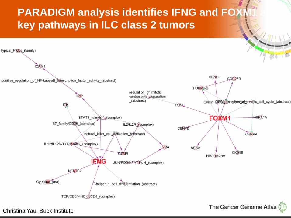
PARADIGM analysis identifies IFNG and FOXM1 as key pathways in ILC class 2 tumors
Christina Yau, Buck Institute
16
Summary
• Developed unique integrated MAF utilizing both DNA exome and mRNA sequencing
• ILC vs. IDC – FOXA1, CDH1 mutations associated with ILC – GATA3 mutation associated with IDC – Altered signaling: CDH1, Myc, p53/DNA damage, immune signaling – Identified differentially expressed miRNA and methylation
• ILC classes
– Class 1 associated with Reactive subtype – Class 2 immune component and highly proliferative

17
TCGA Breast Cancer Analysis Working Group Baylor College of Medicine Chad Creighton Xiaosong Wang
British Columbia Cancer Agency Andy Chu Elizabeth Chun Andy Mungall Gordon Robertson Dominik Stoll
Broad Institute Andrew Cherniack
Greater Poland Cancer Center Maciej Wiznerowicz
Harvard Medical School Terrence Wu Yonghong Xiao
Institute for Systems Biology Sheila Reynolds Ilya Shmulevich
Lawrence Berkeley National Laboratory Paul Spellman
Mayo Clinic Jim Ingle
The University of Texas MD Anderson Cancer Center Roel Verhaak Rehan Akbani Nancy Shih Gordon Mills
Memorial Sloan-Kettering Cancer Center Giovanni Ciriello Niki Schultz Ethan Cerami Arthur Goldberg Caitlin Byrne Anders Jacobsen Tari King Chris Sander
Nationwide Children’s Hospital Jay Bowen Julie Gastier-Foster
National Cancer Institute Chunhua Yan John Demchok Laura Dillon Margi Sheth Peter Good Jacqueline Palchik Heidi Sofia Kenna Shaw
University of California Santa Cruz Buck Institute Chris Benz David Haussler Christina Yau Sam Ng Ted Goldstein Kyle Ellrott Charlie Vaske Josh Stuart Jing Zhu
University of California, San Francisco Fred Waldman
University of Southern California Peter Laird Swapna Mahurkar Simeen Malik Dan Weisenberger Windber Research Institute Hai Hu Richard Mural
University of North Carolina Chuck Perou (co-chair) Katie Hoadley Mike Gatza Joel Parker Xiaping He Michael Iglesia Grace Silva Wei Zhao The Genome Institute at Washington University Matthew Ellis (co-chair) Li Ding Lucinda Fulton Daniel Koboldt Elaine Mardis