genetic engineering of Βeta-carotene production in …
TRANSCRIPT

GENETIC ENGINEERING OF ΒETA-CAROTENE PRODUCTION IN HONEYDEW
MELONS (CUCUMIS MELO L. INODORUS)
A Dissertation
by
YAN REN
Submitted to the Office of Graduate Studies of Texas A&M University
in partial fulfillment of the requirements for the degree of
DOCTOR OF PHILOSOPHY
December 2011
Major Subject: Horticulture

Genetic Engineering of Beta-Carotene Production in Honeydew Melons (Cucumis melo
L. inodorus)
Copyright 2011 Yan Ren

GENETIC ENGINEERING OF BETA-CAROTENE PRODUCTION IN HONEYDEW
MELONS (CUCUMIS MELO L. INODORUS)
A Dissertation
by
YAN REN
Submitted to the Office of Graduate Studies of Texas A&M University
in partial fulfillment of the requirements for the degree of
DOCTOR OF PHILOSOPHY
Approved by:
Co-Chairs of Committee, Kevin M. Crosby
Bhimanagouda S. Patil Committee Members, Jean H. Gould Keerti S. Rathore Head of Department, Leland S. Pierson III
December 2011
Major Subject: Horticulture

iii
ABSTRACT
Genetic Engineering of Beta-Carotene Production in Honeydew Melons (Cucumis Melo
L. inodorus). (December 2011)
Yan Ren, B.A.G., Shanghai Jiaotong University, China; M.A.G., Zhejiang University,
China
Co-Chairs of Advisory Committee: Dr. Kevin M. Crosby Dr. Bhimanagouda S. Patil
Genetic transformation is a useful tool to incorporate novel genes, potentially allowing
sexual incompatibility and interspecific barriers to be circumvented. The purpose of this
study was to improve β-carotene levels in melon fruits by transferring a phytoene
synthase (PSY) gene. At present, there are not sufficient regeneration and transformation
studies reported on two commercially important melon types - western shipper
cantaloupe and honeydew.
To establish a high efficiency shoot regeneration system, we evaluated three
types of explants in our elite breeding lines. A shoot tip with a hypocotyl and cotyledon
fragments, regenerated shoots whereas a shoot tip with a hypocotyl without cotyledon,
did not produce regenerants. Murashige & Skoog (MS) basal medium with 1 mg l-1
benzyladenine (BA), 0.26 mg l-1 abscisic acid (ABA) and 0.8 mg l-1 indole-3-acetic acid
(IAA) was the best for regeneration from cotyledon explants in cantaloupe ‗F39‘. MS
basal medium with 1 mg l-1 BA and 0.26 mg l-1 ABA was chosen for honeydew ‗150‘ to

iv
solve a curving-up problem of explants. Fifty to sixty percent of regenerants were found
to be polyploids.
To establish a reliable Agrobacterium-mediated transformation protocol,
kanamycin sensitivity as well as Timentin™ and Clavamox® were evaluated. Kanamycin
200 and 150 mg l-1 were chosen as the threshold levels for ‗F39‘ and ‗150‘ respectively.
No significant differences were found between Timentin™ and Clavamox® in ‗F39‘;
however, Clavamox® reduced the incidence of vitrification and increased the frequency
of shoot elongation in ‗150‘. A. tumefaciens strain EHA105, harboring pCNL56 carrying
nptII and gusA genes, was used to establish a transformation protocol. The
transformation efficiency was 0.3% from ‗F39‘ and 0.5% from ‗150‘.
We introduced a watermelon PSY-C gene under the control of a fruit-specific
promoter of a polygalacturonase gene into ‗150‘. All the transgenic plants were
tetraploids based on flow cytometry assays. Up to 32-fold of β-carotene was elevated in
the rind tissue of transgenic honeydew including phytoene increase. This is a very
promising result for a further investigation to increase β-carotene level in flesh tissue
using the PSY-C gene with an appropriate promoter.

v
ACKNOWLEDGEMENTS
I wish to thank Dr. Kevin Crosby for providing me with the opportunity to pursue a
Ph.D. degree and work on this wonderful project. Without his support and guidance, I
could not have done so much. I want to thank Dr. Bhimanagouda Patil for serving as my
co-adviser and letting me become a member of the Vegetable and Fruit Improvement
Center (VFIC). I would also like to thank Dr. Jean Gould for her input throughout my
project and publication, and Dr. Keerti Rathore for his help with my research. All of
them are valuable to my graduate career and success.
A special thank-you goes to Dr. Hajeen Bang for her devoted supervision and
assistance in my research and personal life. She taught me a lot and helped me beyond
measure. I appreciate the input and assistance from Drs. Kilsun Yoo and Eun Jin Lee for
the HPLC analysis work. I am grateful for technical advice and help from Drs. Ian Curtis,
Rebecca Grumet, Sung Hun Park, Jim Giovannoni, Li Li, Sylvain Marcel, Selvakumar
Veluchamy, Mehdi Kabbage, Ms. Marianne Arnold, Sue Hammar, and Jungeun Kim. I
want to express my appreciation to Dr. Justin Butcher for his help with the greenhouse
work. I also want to thank my fellows (Amit, Raj, Haejin, Jinhee, Priyanka, Kranthi,
Ram and Noa) for their kind friendships.
Finally, I wish to thank my father Xuchu Ren and my mother Jinyu Wang, for
their endless dedication to my life. Without their continual support and inspiration, my
goal of earning a Ph.D. in the United States would never come true. Thanks also go to

vi
my best friend, Jian Liu, who is always there to help and encourage me. All of them are
very important and irreplaceable in my life.

vii
NOMENCLATURE
ABA Abscisic acid
BA 6-Benzyladenine
HPLC High performance liquid chromatography
IAA Indole-3-acetic acid
MS Murashige and Skoog
NAA Naphthaleneacetic acid
nptII Neomycin phosphotransferase II
PCR Polymerase chain reaction
GUS β-Glucuronidase

viii
TABLE OF CONTENTS
Page
ABSTRACT……………………………………………………………….…………….iii
ACKNOWLEDGEMENTS.…………………………………………….….…………….v
NOMENCLATURE…………………………………………………………….……....vii
TABLE OF CONTENTS……………………………………………………….……...viii
LIST OF FIGURES…………………………………………………………….………...x
LIST OF TABLES…………………………………………………………….……….xiii
CHAPTER
I INTRODUCTION AND LITERATURE REVIEW………………………......1
Introduction…………………..……………….……..……………………1 Literature Review………...……………….……..……………………….3
II ESTABLISHMENT OF REGENERATION SYSTEMS FOR
CANTALOUPE AND CASABA MELONS….………………………....….23
Materials and Methods……..……………….……..……………………23 Results and Discussion………………..………………………….……..28 Conclusion………………………………..……………………….…….37
III SHOOT REGENERATION AND PLOIDY VARIATION IN
TISSUE CULTURE OF HONEYDEW MELON..………..…………..…….38
Materials and Methods…………………..……………………………..38 Results and Discussion…………………..……………………………..40 Conclusion……………………………… .…………………………47

ix
CHAPTER Page
IV ESTABLISHMENT OF TRANSFORMATION SYSTEMS FOR
CANTALOUPE LINE ‗F39‘ AND HONEYDEW LINE ‗150‘………..……48
Materials and Methods……………………………………….…………48 Results and Discussion………………………………………..……..….52 Conclusion…………………………………………………..…………65
V GENETIC ENGINEERING OF BETA-CAROTENE
PRODUCTION IN HONEYDEW..………………………………………….66
Materials and Methods………………..……………………………...…66 Results and Discussion…………..………………………………….…..70 Conclusion…………………………..…………………………………. 87
VI SUMMARY AND CONCLUSIONS………………………………………..88
REFERENCES…………………………………………………………………….……90
VITA…………………………………………………………………………………..110

x
LIST OF FIGURES
Page
Fig. 1 Carotenoid biosynthesis pathway in plants…………...…………….…….…..13
Fig. 2 Preparation of a shoot tip with hypocotyl and cotyledon (STHC) explant…………………………………………………....................25
Fig. 3 Effect of six different media (RM1-RM6) on the production of calli and shoot primordia from cotyledonary explants of three melon genotypes ‗141, ‗F39‖ and ‗TMS‘……………………………..….......29
Fig. 4 Callus production and shoot regeneration on the optimal medium from cotyledonary explants of three different melon genotypes after four weeks of culture………………………………..………30
Fig. 5 Multiple shoot regeneration, shoot elongation and rooting of melon ‗F39‘ on MS basal medium supplemented with various BA concentrations ………………………………………………………..…..31
Fig. 6 Shoot regeneration and development from shoot tip with hypocotyl and cotyledon fragments explants (A, C, E and F) as well as shoot tip with hypocotyls explants (B and D) of melon ‗F39‘ on six different media (RM1-M6)……………………………....35
Fig. 7 Comparison of shoot regeneration from shoot tip with hypocotyl and cotyledon explants (A) as well as shoot tip with hypocotyl explants (B) of melon ‗F39‘ on six different media (RM1-M6)……………...36
Fig. 8 Shoot regeneration from four media: M1 - M4……………………….………42
Fig. 9 Shoot elongation and rooting of melon ‗150‘ on MS basal medium supplemented with various BA oncentrations………………….……42
Fig. 10 The polyploidy percentage of plants regenerated from cotyledonary explants of honeydew ‗150‘………………..…………………..44
Fig. 11 Flow cytometry histograms of leave tissues from in vitro regenerated honeydew ‗150‘ plants …………………...……...…………...….44
Fig. 12 Morphological characteristics of polyploid regenerants, their fruits and seeds…………………………………………………………..……46

xi
Page
Fig. 13 Kanamycin sensitivity assay to determine selection thresholds using non-inoculated explants of melon ‗F39‘ and 150‘………….…………..53
Fig. 14 Effects of Clavamox® and Timentin™ on the regeneration of non-inoculated explants of honeydew ‗150‘ after 75 days of culture……..….54
Fig. 15 Transient GUS expression of explants inoculated with different Agrobacterium tumefaciens strains carrying pCNL56, 7 days after inoculation…………………………………………………………………….56
Fig. 16 Callus and shoot development from explants and GUS histochemical assay…………………..…………………………..…………...58
Fig. 17 PCR and southern blot analyses of putative transformants………..……….…60
Fig. 18 Morphological characteristics of transgenic melon plants…………….…….. 61
Fig. 19 Transient GUS expression of cotyledonary explants of ‗F39‘ and ‗150‘ inoculated with Agrobacterium tumefaciens strain EHA105 carrying two different plasmids……………………….…………....63
Fig. 20 Evaluation of the degree of GUS transient expression in cotyledonary explants of ‗F39‘……………………………………………….64
Fig. 21 T-DNA region of binary vector PSYC/pRD12………..………………….…..66
Fig. 22 PCR and southern blot analyses of putative transformants….………………..72
Fig. 23 Flow cytometry histograms of leaf tissues from honeydew ‗150‘ transgenic lines………………………………………………………………..74
Fig. 24 Morphologies of transgenic plants and their T1 progenies………….………..76
Fig. 25 Fruits from wild type (a), vector control (b) and transgenic plants (c-e) of honeydew ‗150‘…..…………………………….……………………78
Fig. 26 HPLC separation of carotenoids in rind tissue extractions from wild type (WT), vector control (VC) and transgenic line 042201 of honeydew ‗150‘.…………….……………………………………………...80

xii
Page
Fig. 27 HPLC separation of carotenoids in flesh tissue extractions from wild type (WT), vector control (VC) and transgenic line 042201 of honeydew ‗150‘….………………………………………………………...81
Fig. 28 HPLC separation of carotenoids in placenta tissue extractions from wild type (WT), vector control (VC) and transgenic line 042201 of honeydew ‗150‘…………………………………………………....82
Fig. 29 RT-PCR analysis of PSY-C transgene expression in transgenic honeydew ‗150‘………………………………………………………..….…..85
Fig. 30 PCR analysis to determine the segregation of PSY-C transgene in the T1 progeny of transgenic honeydew ‗150‘………………………….….86
Fig. 31 Seed germination of wild type and transgenic progeny of honeydew ‗150‘ on the selection medium containing 200 mg l-1 kanamycin…….……..87

xiii
LIST OF TABLES
Page
Table 1 Evaluation of six media on the regeneration of melon ‗141‘, ‗F39‘ and ‗TMS‘…………………….………………………………………26
Table 2 Scoring method used to estimate callus and shoot primordium induction from cotyledonary explants of melon ‗141‘, ‗F39‘ and ‗TMS‘….........................................................................................................27
Table 3 Regeneration of shoot primordial and shoots from shoot tip explants of ‗141‘, ‗F39‘ and ‗TMS‘…………………………………….…..33
Table 4 Evaluation of four media for shoot regeneration of honeydew ‗150‘…..…..39
Table 5 The frequency of initial shoot regeneration in the first 3 weeks on the different media and the polyploidy estimation of the regenerants…...….41
Table 6 Morphological characteristics of diploid, tetraploid and mixoploid regenerants of honeydew ‗150‘…………………………………..………...46
Table 7 Effects of Clavamox® and Timentin™ on shoot development and
elongation of non-inoculated explants of melon ‗F39‘ and ‗150‘…………..55
Table 8 Effect of light condition on GUS transient expression during the co-cultivation period in cotyledonary explants of ‗F39‘ and ‗150‘….…..….57
Table 9 Effects of different pre-culture periods on GUS transient expression in cotyledonary explants of ‗150‘………………………….……65
Table 10 HPLC analysis of phytoene and β-carotene concentrations in rind,
flesh and placenta tissues and the corresponding colors of transgenic honeydew ‗150‘….……………….…………………………..….83
Table 11 Segregation ratios of the PSY-C transgene in T1 progeny of transgenic honeydew ‗150‘…………………………………….…..………..86

1
CHAPTER I
INTRODUCTION AND LITERATURE REVIEW
Introduction
Melon (Cucumis melo L.) is a group of high-value crops including cantaloupe
(muskmelon), honeydew and casaba melon. This species has seven botanical variants:
cantalupensis, reticulatus, inodorus, flexuosus, conomon, chito and dudaim. Only
reticulatus and inodorus variants are commercially important in the United States.
Cantaloupe is one of the most-consumed melons in the USA (about 50 kg per person per
year (Flores 2005). Western-shipper cantaloupe (Cucumis melo var. reticulatus) refers to
the type of cantaloupes originally grown in western states and shipped throughout the
country. They usually have uniformly netted rinds, orange flesh and lack sutures.
Nowadays, they are already adapted and grown all over the United States (Boyhan et al.
2009). Honeydews (Cucumis melo var. inodorus) usually have green or cream-colored
rinds with pale-green flesh, and have been widely grown in the USA. New varieties of
honeydews with orange flesh, which were created from a cross between netted orange-
fleshed cantaloupe and non-netted green-fleshed honeydew, have been less popular in
melon production (Lester 2008; Hodges and Lester 2006). As a large producer and
consumer of melon, the United States has been involved in the improvement of melon
____________ This dissertation follows the style of Plant Cell, Tissue and Organ Culture.

2
quality to help increase melon profits and achieve more nutritional value for consumers.
Cantaloupe and orange-fleshed honeydew are good sources of β-carotene, potassium and
vitamin C. Pale green-fleshed honeydew lacks β-carotene but is rich in potassium,
vitamin C and folate (data from online USDA National Nutrient Database for Standard
Reference). In the past few years, melon breeders have worked on improving the
nutritional quality of melon. ―Consumers will get not only a better tasting, sweeter
melon, but one that can help boost their intake of β-carotene and vitamin C,‖ said Dr.
Gene Lester, who has been working at the USDA in Weslaco since 1983 (Flores, 2005).
Generally, β-carotene is the prevalent carotene in yellow, orange, and green leafy
fruits and vegetables, such as carrots, pumpkin, spinach, lettuce, pepper, sweet potatoes,
broccoli, muskmelon, citrus, oranges, and winter squash (USDA National Nutrient
Database for Standard Reference, Release 21). β-carotene is a natural food source of pro-
vitamin A which may contribute to the prevention of some diseases such as eye
problems, cardiovascular disease, and several cancers (Fraser et al. 2004). Heart disease
and cancer have been identified as the top two diseases causing death in Americans.
Therefore, consumption of foods rich in β -carotene is being recommended by the US
National Cancer Institute and the USDA.
Although β-carotene accounts for 90% of the carotenoids found in orange-
fleshed cantaloupe fruit (Karvouni et al. 1995), compared with other vegetables and
fruits, its β-carotene content (20.2 µg g-1 FW) is not considered to be very high.
Moreover, pale green-flesh honeydew‘s β-carotene content is very low (0.3 µg g-1 FW).
Conventional plant breeding has been very successful at increasing productivity but has

3
focused less on improving the levels of health-promoting phytochemicals such as
carotenoids. Genetic engineering (often termed genetic modification or genetic
manipulation) is a faster and targeted method to transfer gene(s) compared with
conventional breeding (Fraser et al. 2004). It is a useful procedure to incorporate novel
genes within and across plant species. Sexual incompatibility and interspecific barriers
to traditional plant breeding can therefore be circumvented. Several successful genetic
engineering attempts to enhance carotenoids have been reported in crop plants including
tomato (Fraser et al. 2001; 2002; Romer et al. 2000; Rosati et al. 2000; Ronen et al.
2000; Ralley et al. 2004; D'Ambrosio et al. 2004), carrot (reviewed from Fraser and
Bramley 2004), rice (Ye et al. 2000; Paine et al. 2005), canola (Shewmaker et al. 1999),
tobacco (Misawa et al. 1994; Ralley et al. 2004; Mann et al. 2000), lotus (Suzuki et al.
2007), Arabidposis seeds (Lindgren et al. 2003; Stalberg et al. 2003) and potato (Lopez
et al. 2008; Diretto et al. 2007; Gerjets and Sandmann 2006; Ducreux et al. 2005). This
study was undertaken to investigate the possibility of elevating β-carotene levels in
commercially valuable melons via genetic transformation.
Literature Review
Melon in vitro culture
In vitro melon regeneration protocols have been reported in the past 25 years. Several
factors impact regeneration efficiency, including genotype, explant type, and plant
growth regulators (reviewed by Nuňez-Palenius et al. 2008). The main regeneration

4
pathways are adventitious shoot organogenesis and somatic embryogenesis.
Adventitious shoot organogenesis has been used most often in tissue culture. The highest
regeneration frequency of melon cotyledon can reach 100% (Ficcadenti and Rotino
1995). In the last decade, somatic embryogenesis has become a popular method for in
vitro culture because it solved the problem of obtaining chimeric transformants
(Akasaka-Kennedy et al. 2004), and it is an efficient method for production of diploid
plants (Guis et al. 1997a). Akasaka-Kennedy et al. (2004) reported that an average of
116.7 and 130 of embryos were induced from each seed of Vedrantais and Earl‘s
Favourite Fuyu A melon, respectively. Two weeks after being transferred to liquid MS
medium without plant growth regulators, 75.9% and 23.3% of the embryos germinated
to produce plants with shoots and roots.
Genotype is the most important determinant of melon regeneration ability.
Galperin et al. (2003) screened 30 genotypes on 3 different media, including wild
landraces, breeding lines and commercial cultivars. Twenty four out of 30 genotypes
regenerated abnormal shoots while 5 genotypes had low frequency of regeneration. Only
one genotype ‗BU-21‘ showed a distinguished 100% regeneration frequency
accompanied by normal shoot development. Gray et al. (1993) used 51 commercial
varieties to test embryogenic regeneration frequency of cotyledons. The highest
responding varieties reached 100% regeneration efficiency with 20 embryos per explant,
while regeneration in the lowest responding varieties was only 5% with 0.1 embryos per
explant. This genotype-dependence phenomenon is normal for melon in vitro culture,

5
therefore establishing an efficient regeneration system for genetic transformation of a
specific melon cultivar is a necessary and important step.
Plant growth regulators have been used in melon tissue culture to optimize shoot
regeneration. Both auxins and cytokinins are known to be essential for bud/shoot
induction and the optimal auxin/cytokinin levels, critical for recovery of plants, are often
genotype specific. The auxin indole-3-acetic acid (IAA) and cytokinin 6-benzyladenine
(BA) are commonly used in organogenesis studies. The synthetic auxin 2,4-
dichlorophenoxyacetic acid has been widely used to induce somatic embryogenesis
(Akasaka-Kennedy et al. 2004; Gray et al. 1993; Guis et al. 1997a; Kintzios et al. 2004;
Kintzios and Taravira 1997; Oridate and Oosawa 1986). In addition, 6-(γ,γ-
dimethylallylamino)-purine (2iP), gibberellic acid (GA3), kinetin, thidiazuron and -
naphthaleneacetic acid (NAA) have been used in melon culture (Fang and Grumet 1990;
Ficcadenti and Rotino 1995; Guis et al. 2000; Souza et al. 2006; Yadav et al. 1996). The
effects of these growth regulators on melon regeneration differed depending on plant
genotype, explant type and culture conditions. Other media components also affect
melon regeneration efficiency. For example, abscisic acid (ABA) and sucrose have been
shown to enhance somatic embryogenesis in melon (Nakagawa et al. 2001). The anti-
gibberellin analogue, ancymidol, was reported to promote shoot regeneration from
cotyledonary explants of ‗Galia‘ melon in combination with a low concentration of BA
(0.44 µM), while the addition of GA3 to this medium reduced the regeneration frequency
12-fold after 13 d of treatment (Gaba et al. 1996).

6
Different explant types have been used in melon regeneration including mature
seed, cotyledon, hypocotyl, proximal zone of the hypocotyls, petiole, leaf, root,
protoplast and shoot tip. All these explants proved to be able to regenerate shoots
through either organogenesis or somatic embryogenesis. Cotyledon is the most-widely-
used explant type for melon regeneration and transformation. Explants were excised
from cotyledons at different ages such as unexpanded cotyledons on immature seeds
(Adelberg et al. 1994) and mature seeds (Ezura and Oosawa 1994), as well as expanded
cotyledons on seedlings. Age of the seedlings also varied from 2-day-old to 2-week-old
(Nunez-Palenius et al., 2008). Usually, physiologically younger tissues were preferred
for in vitro culture (Smith 2000). Molina and Nuez (1995) compared regeneration
abilities from leaf, cotyledon and hypocotyl explants in three melon populations. They
concluded that different types of explants had different regeneration capacities, which is
controlled by a common genetic system. In their research, cotyledon and leaf explants
had a similar regeneration frequency which was always higher than hypocotyl explants.
Shoot tip can be considered as an alternative explant type for melon regeneration
(Ezura et al. 1997b) and transformation for three reasons. First, the shoot apical
meristem is a potential target for direct gene transfer. Second, compared to plant
regeneration from protoplasts, callus cultures, or directly from the explants via
adventitious organogenesis or somatic embryogenesis, shoot apex has the potential to
maintain cultivar integrity by escaping from culture-induced mutations (Park et al.,
1998). Third, melon plants regenerated from cotyledons have a high tendency
(approximately 80%) towards tetraploidy in tissue culture and genetic transformation of

7
melon using cotyledonary explants (approximate 80%) (Guis et al. 2000; Nuňez-
Palenius et al. 2006 and 2008). On the other hand, plant regenerated from shoot
primordium had a much lower frequency of tetraploidy (4%) (Ezura et al. 1992 and
1997b). Shoot primordium culture might be critical for shoot apex transformation.
Tylicki et al. (2007) and Ogawa et al. (1997) indicated shoot primordia culture was an
efficient system for maintaining genetic stability and a good system for transformation in
melon.
Polyploidy is a common problem in melon in vitro culture. More than 80% of
melon plants regenerated from 2-day-old cotyledon explants were tetraploids, whereas
only 15% of the regenerated plants from leaf explants were tetraploids. Tetraploid melon
has some phenotypic changes including short internodes, smaller fruit size, flatness of
fruit and reduced productivity, which impede marketability due to the low fruit quality
(Guis et al. 2000). Compared with plants regenerated from somatic embryos and
adventitious shoots via nodes of elongated seedlings, those regenerated from shoot
primordia had a much lower frequency of tetraploidy: somatic embryo 31%, adventitious
shoots 30%, shoot primordial 4%; and the clonally propagated plants from axillary buds
didn‘t produce tetraploidy (Ezura et al. 1992), which indicated the explant source as an
important factor in reducing the number of tetraploid plants obtained from melon in vitro
culture. Ezura et al. (1997b) examined the ploidy levels of shoot primordium cells and
plants regenerated from the shoot tips. Both tissues were very stable in terms of ploidy,
and the culture time (from 1 to 6 years) did not affect ploidy level. But Kathal et al.
(1992) found that the polyploidy percentage of melon plants regenerated from leaf

8
explants increased as the time of in vitro culture increased. These opposing results
indicate that polyploidy in tissue culture could be induced by many unknown factors.
Further research on this phenomenon will be necessary to reduce this problem in melon
in vitro culture.
In Chapters II and III, we evaluated the responsiveness of different explant types
of our elite breeding lines on different regeneration media which have been reported to
be optimal for different melon genotypes. Then we calculated the percentage of
polyploidy in honeydew ‗150‘ regenerants, and observed the corresponding
morphologies.
Genetic transformation in melons
In the past 20 years, melon transformation research has been mainly focused on two C.
melo variants (cantalupensis and reticulatus) for disease resistance and longer shelf life.
The genes used for these purposes included cucumber mosaic virus (CMV), zucchini
yellow mosaic virus (ZYMV) and watermelon mosaic virus (WMV) coat protein genes
(Yoshioka et al. 1992; Fang and Grumet, 1993; Gonsalves et al. 1994; Clough and Ham,
1995; Fuchs et al. 1997; Yalçın-Mendi et al. 2004; Wu et al. 2009); and an antisense
ACC oxidase gene (Ayub et al. 1996; Clendennen et al. 1999; Ezura et al. 1997a; Guis et
al. 1997b, 2000; Shellie, 2001; Silva et al. 2004; Nuňez-Palenius et al. 2006; Hao et al.,
2011). Increasing sugar content is another target trait for genetic engineering of melon.
Two genes have been studied for regulating sucrose biosynthesis, which were an
antisense acid invertase gene (anti-MAI1) (Fan et al. 2007) and an antisense sucrose

9
phosphate synthase gene (Tian et al. 2010). Yalçın-Mendi et al (2004) reported an
Agrobacterium-mediated transfer of a ZYMV coat protein gene to a Turkey inodorus
variant melon, cultivar ―Kirkagac 637‖. They identified the frequency of gene escape
and fruit quality characteristics from two transgenic lines (Yalçın-Mendi et al. 2010).
Despite the development of genetic transformation protocols for melon reported
in the last two decades, transformation remains genotype-dependent and efficiencies are
relatively low (0-12.5%) (Nuňez-Palenius et al. 2006; 2008). To date, the most
successful methods for producing transgenic melon have been achieved in French (Fang
and Grumet 1990), Israeli (Nuňez-Palenius et al. 2006) and Asian germplasm (Dong et
al. 1991; Wu et al. 2009), which constitute a low percentage of the US consumer market.
Successful transformation protocols are needed for improvement of commercial
genotypes grown in the US, especially the western shipper cantaloupe (C. melo var.
reticulatus) and honeydew (C. melo var. inodorus).
A review of pertinent literature reveals only a few studies on the tissue culture or
transformation of western shipper and honeydew melons. At this writing, there were no
transformation studies available for honeydew melon; however, a number of studies on
shoot regeneration in culture have been reported (Ficcadenti and Rotino 1995; Keng and
Hoong 2005; Kintzios and Taravira 1997; Oridate et al. 1992; Orts et al. 1987). In
western shipper cantaloupe, only one microprojectile mediated transformation
(Gonsalves et al. 1994) and two Agrobacterium-mediated transformations (Clough and
Ham 1995; Fuchs et al. 1997) were reported.

10
A. tumefaciens-mediated transformation has become the method of choice for
melon transformation (Nuňez-Palenius et al. 2008). We opted to use Agrobacterium due
to reports of low copy gene transfers, which help to reduce the chances of multi-gene
triggered silencing in the transgenic plants. Unfortunately, previously reported
procedures provided insufficient detail for us to replicate. Though various A. tumefaciens
strains are available such as LBA4404, EHA105, ABI, C58B707, C58C1Rif® and
GV3111SE, the strain LBA4404 was used in nearly half of melon transformation reports
(Fang and Grumet 1990; Yoshioka et al. 1992; Fang and Grumet 1993; Vallés and Lasa
1994; Ayub et al. 1996; Bordas et al. 1997; Guis et al. 2000; Silva et al. 2004; Taler et al.
2004). In this study, we compared strains LBA4404 and EHA105 for their
transformation efficiency.
Based on the regeneration system established for cantaloupe ‗F39‘ and honeydew
‗150‘, we conducted kanamycin sensitivity assays to determine the concentration of the
antibiotic for selecting transformed shoots. Furthermore, the effects of two antibiotics,
Clavamox® and Timentin™, were compared on shoot regeneration. In the optimization of
producing transformed shoots, we have studied the effect of light condition during co-
cultivation. This has been neglected in designing transformation systems in melon but
has proven to be important in other plant species such as Phaseolus acutifolius and
Arabidopsis thaliana (Zambre et al. 2003). In addition, two strains of A. tumefaciens,
EHA105 (Hood et al. 1993) and LBA4404 (Hoekema et al. 1983), were tested to see
whether there were differences in their efficiency in producing transformed plants.
Melon regeneration and transformation are still considered to be difficult due to several

11
factors such as: strong genotype dependence, a high percentage of polyploidy, high rates
of ‗escapes‘ and aberrant shoot development (Castelblanque et al. 2008; Chovelon et al.
2008; Dong et al. 1991; Akasaka-Kennedy et al. 2004; Wu et al. 2009). The content of
Chapter IV describes establishment of a reliable genetic transformation system in these
two elite breeding lines with commercial qualities.
Carotenoid biosynthesis and regulation in plants
Carotenoids are pigments de novo synthesized by all photosynthetic organisms (higher
plants and algae) and many non-photosynthetic organisms (some bacteria and fungi)
(Bartley and Scolnik 1994). There are a series of gene expressions related to carotenoid
biosynthesis controlling carotenoid accumulation. These gene expressions occur in the
chloroplasts (photosynthetic tissues such as leaves) and in chromoplasts
(nonphotosynthetic plant tissues such as fruits and flowers). Carotenoids in plants are
isoprenoids formed from isopentenyl diphosphate (IPP) via the mevalonate-independent
(MVA-independent) pathway in the plastid (Li et al. 2006). Figure 1 shows that
Geranylgeranyl diphosphate (GGPP) deriving from IPP is the precursor of carotenoids.
Phytoene synthase (PSY) catalyzes the conversion of GGPP to phytoene which is the
first step in carotenoid biosynthesis. PSY enzyme, therefore, becomes the first key
enzyme in this pathway. Lycopene, imparting a red or red-orange color to some fruits
and vegetables, has dual roles in humans and plants as a free-radical scavenger (Collins
et al. 2006). It is derived from phytoene in a series of dehydrogenation reactions, which
is catalyzed by phytoene desaturase (PDS) and δ-carotene desaturase (ZDS). Lycopene is

12
then converted to β-carotene and α-carotene by lycopene β-cyclase (Lcy-b) and lycopene
ε-cyclase (Lcy-e), respectively (Fig. 1).
Although the carotenoid biosynthetic pathway in plants was elucidated in the
1950s and 1960s, the regulation of carotenoid biosynthesis at the gene and enzyme level
is still poorly understood. Cauliflower Or gene (Li et al. 2001) and the apricot (Ap)
tomato mutant have been identified, but no regulatory genes involved in carotenoid
formation have been isolated yet. Since carotenoids play a central role in plant
development and adaptation, their synthesis is consequently considered to be
coordinated with other developmental processes such as plastid formation, flowering and
fruit development. The partial knowledge of pathway regulation is responsible for the
practical difficulties of working with carotenoids and their biosynthetic enzymes. It is
believed that a single regulatory process is unlikely to control a branched pathway such
as that of carotenoid formation from isoprenoid precursors. In contrast, each branch
point is likely to be a control point and probably regulated on both transcriptional and
post transcriptional levels (Fraser and Bramley 2004).
Transcriptional Regulation
Plastid differentiation, the development of chromoplasts and de novo carotenoid
formation are processes which occur during tomato and pepper fruit ripening and flower
development in daffodil, tomato and marigold (Tagetes erecta). Carotenoid formation is
regulated by carotenoid genes, which have both up- and down-regulation of
transcription. For example, expression of PSY-1 and PDS is increased while LCY-B and

13
LCY-E mRNAs are decreased during tomato fruit ripening (Ronen et al. 1999; Fraser et
al. 1994). Enzyme activities in ripe fruit are also affected by these gene expression
IPP
DMAPP
GGPP
Phytoene
δ-Carotene
Lycopene
γ-Carotene
β-Carotene
Zeaxanthin
Ipi
Ggps
Psy
Pds
Zds
Lcy-b Cyc-B
δ-Carotene
α-Carotene
Lutein
Lcy-e (CtrL-e)
Cyc-B
CrtR-
b
Lcy-b (CtrL-
b)
CrtR-b (CrtR-e)
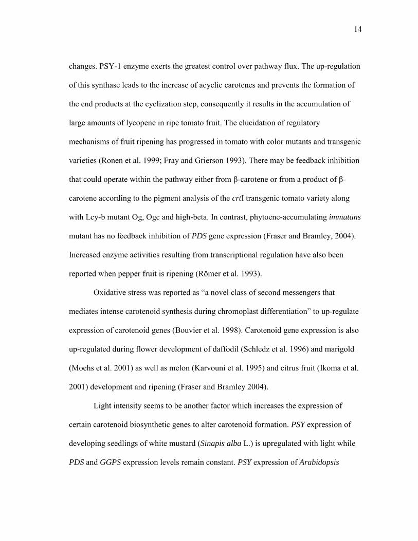
Fig. 1 Carotenoid biosynthesis pathway in plants (Li et al. 2006)
14
changes. PSY-1 enzyme exerts the greatest control over pathway flux. The up-regulation
of this synthase leads to the increase of acyclic carotenes and prevents the formation of
the end products at the cyclization step, consequently it results in the accumulation of
large amounts of lycopene in ripe tomato fruit. The elucidation of regulatory
mechanisms of fruit ripening has progressed in tomato with color mutants and transgenic
varieties (Ronen et al. 1999; Fray and Grierson 1993). There may be feedback inhibition
that could operate within the pathway either from β-carotene or from a product of β-
carotene according to the pigment analysis of the crtI transgenic tomato variety along
with Lcy-b mutant Og, Ogc and high-beta. In contrast, phytoene-accumulating immutans
mutant has no feedback inhibition of PDS gene expression (Fraser and Bramley, 2004).
Increased enzyme activities resulting from transcriptional regulation have also been
reported when pepper fruit is ripening (Römer et al. 1993).
Oxidative stress was reported as ―a novel class of second messengers that
mediates intense carotenoid synthesis during chromoplast differentiation‖ to up-regulate
expression of carotenoid genes (Bouvier et al. 1998). Carotenoid gene expression is also
up-regulated during flower development of daffodil (Schledz et al. 1996) and marigold
(Moehs et al. 2001) as well as melon (Karvouni et al. 1995) and citrus fruit (Ikoma et al.
2001) development and ripening (Fraser and Bramley 2004).
Light intensity seems to be another factor which increases the expression of
certain carotenoid biosynthetic genes to alter carotenoid formation. PSY expression of
developing seedlings of white mustard (Sinapis alba L.) is upregulated with light while
PDS and GGPS expression levels remain constant. PSY expression of Arabidopsis

15
thaliana also increased with both far-red and red light. PSY regulation is involved in
both light-labile and light-stable phytochromes (Von Lintig et al. 1997). A 5-fold
increase in the ratio between levels of LCY-B and LCY-E mRNAs in both Arabidopsis
thaliana and tomato leaves was observed when plants were shifted from low light to
strong light (Hirschberg, 2001).
Post-transcriptional Regulation
Fraser and Bramley (2004) mentioned that the key regulatory issue in the carotenoid
biosynthesis pathway is what mechanisms control the partitioning of precursors into the
branches of the pathway. The discovery of multigene families in the pathway has
supported the theory of metabolic channeling between each branch in the pathway. The
corresponding enzymes have been found in tomato (Fraser et al. 2000), pepper (Dogbo
et al. 1987) and daffodil (Lützow and Beyer 1988). The metabolic branches are believed
to be relatively independent from each other since there are relatively small changes of
isoprenoids in transgenic plants in spite of increased enzyme activities. However,
metabolite precursor pools such as GGPP pool can be diverted from one branch to the
next (Fray et al. 1995). The fact that constitutive expression of PSY-1 caused dwarfism
of tomato by redirecting metabolite availability shows metabolic cross talk does exist.
PSY and PDS were post-transcriptionally regulated in chromoplasts of daffodil
membrane association. In addition, light-induced membrane association and substrate
specificity are also believed to regulate carotenoid biosynthesis.

16
Carotenoid Sequestration Regulation
This is a common form of regulation but differs in the way of sequestering the end-
product carotenoids between chloroplasts and chromoplasts. Esterification of carotenoids
was considered to be the mechanism of sequestration regulation in pepper, tomato and
canola, and seems to be an effective method used by many flowers such as sunflower,
daffodil and marigold (Fraser and Bramley 2004).
Genetic engineering to enhance carotenoids in crop plants
Genetic engineering of carotenoids in crops for nutritional enhancement has been
studied for more than a decade. The first committed step in carotenoid biosynthesis is
catalyzed by phytoene synthase (PSY), which converts GGPP into phytoene
(Cunningham and Gantt 1998; Li et al. 2006). The overexpression of this gene,
combined with or without the downstream pathway genes (PDS and Lcy-b), have
enhanced carotenoids in crops such as tomato (Fraser et al. 2001; 2002), carrot
(reviewed by Fraser and Bramley 2004), canola (Shewmaker et al. 2004), rice (Ye et al.
1999: ‗Golden Rice‘; Paine et al. 2005: ‗Gold Rice 2‘), potato (Diretto et al. 2007;
Ducreux et al. 2005) and maize (Aluru et al. 2008). According to these reports, the total
carotenoids of the transgenic crops have been increased by 2 to 50 folds in different
species with 1.6 to 3600 folds in beta-carotene level, which resulted in the color changes
to yellow or orange. So far, ―Golden Rice‖ and ―Golden Potato‖ are the best
demonstrations for PSY gene function in carotenoid biosynthesis (Ye et al. 1999; Paine
et al. 2005; Diretto et al. 2007).

17
Tomato
Tomato has become the most intensively studied crop in carotenoid metabolism
engineering since its fruit and products represent an important source of carotenoids
among diets. Phytoene synthase (PSY) is the most important rate-limiting enzyme
manipulated by genetic engineering to regulate β-carotene levels in plants. The
bacterium Erwinia uredovora phytoene synthase (crtB) gene has been over-expressed in
tomato fruits. Total carotenoids of the transgenic tomatoes exhibited a 2- to 4-fold
increase over the control: 2.4-fold (n=3) in phytoene, 1.8-fold (n=6) in lycopene and 2.2-
fold (n=5) in β-carotene levels. Biochemical analysis showed that catalytically active
CRTB protein was plastid-located with 5- to 10-fold (n=4) higher levels (Fraser et al.
2002). However, the additional PSY reduced the regulatory influence of this step over
the pathway flux, which was suggested by Fraser and Bramley (2004).
Phytoene desaturation is another possible step that modifies carotenoid
biosynthesis. An Erwinia uredovora PDS gene (crtI) with the CaMV 35S promoter has
been transferred to tomato (CV. Ailsa Craig) plants (Römer et al. 2000). Total carotenoid
levels were not changed in the transgenic tomatoes, but the β-carotene content increased
about 3-fold (n=17), up to 45% of the total carotenoid content. The expression of the
endogenous carotenoid genes behind PDS (ZDS, LCY-B) was also increased, but the PSY
gene which is prior to PDS was decreased. Besides β-carotene, the content of other
downstream pathway carotenoids such as neoxanthin, antheraxanthin, lutein, and
zeaxanthin were increased. In contrast, the content of carotenoids prior to β-carotene such
as phytoene, lycopene, γ- carotene and δ-carotene decreased. As a consequence, the total
18
amount of carotenoids decreased. The enhancement of PDS activity did not lead to the
increase of lycopene. It was suggested that this result was due to crtI over-expression in
tomato, which reduced the content of compounds prior to phytoene desaturation and
induced endogenous lycopene cyclisation (Fraser and Bramley 2004). This phenotype of
modified carotenoids has been found to be stable and reproducible over at least four
generations.
Lycopene is a large precursor pool for β-carotene synthesis in ripe tomato fruit.
Over-expression of the Arabidopsis thaliana lycopene β-cyclase (β-Lcy) in tomato fruit
under the control of a fruit-specific promoter PDS has been achieved (Rosati et al.
2000), making lycopene convert into β-carotene. Transformants aimed at up-regulating
β-Lcy gene expression exhibited a 5-fold increase in β-carotene. Other carotenoids were
not changed in both fruits and leaf tissues, and endogenous carotenoid gene expression
was not significantly changed. In contrast, transformants aimed at down-regulating β-
Lcy gene showed up to 50% down-regulation of β-Lcy expression in ripe fruit, and
consequently their lycopene content was slightly increased.
Carrot
Fraser and Bramley (2004) reported that the Erwinia herbicola crt genes were over-
expressed in carrot, causing β-carotene levels to increase 2-5-fold in the root.

19
Canola
So far, the most dramatic increase in carotenoid levels was produced in transgenic
canola. An Erwinia uredovora phytoene synthase gene crtB was over-expressed in a
seed-specific manner. A 50-fold increase of carotenoids was found in the transgenic
plant seeds (Shewmaker et al. 1999). The seed-specific expression of crtB leads to
orange embryos in transgenic canola, predominantly containing α-carotene and β-
carotene.
Rice
Rice is a staple food in most developing countries, however, it lacks β-carotene (pro-
vitamin A). The first generation of the ‗Gold Rice‘ (Golden Rice 1) was reported (Ye et
al. 2000). Three biosynthetic genes were co-transformed via two vectors to enable the
metabolism from GGPP to β-carotene. The PSY and lycopene β-cyclase cDNAs from
daffodil (Narcissus pseudonarcissus) were expressed under the control of endosperm-
specific glutelin promoter, and the E. uredovora phytoene desaturase gene crtI under the
control of constitutive CaMV 35S promoter. As a result, total carotenoids accumulated
to 1.6 μg g-1 in endosperm (n=50). However, the limited production of β-carotene cannot
be a solution for the vitamin A deficiency. Paine et al. (2005) launched the genetic
engineering of a second generation of ‗Golden Rice‘ (Golden Rice 2) by replacing the
daffodil PSY with maize PSY, in combination with the E. uredovora phytoene desaturase
gene crtI. The total carotenoids were increased to a maximum of 37 μg g-1 with 86-89%

20
of β-carotene. In their study, PSY was found to still be the limiting step in the
accumulation of carotenoids.
Tobacco
The E. uredovora phytoene desaturase (crtI) overexpression in tobacco plants led to a
large amount of the CRTI proteins which correlates with the crtI mRNA levels produced
in the transformants. Carotenoid analysis of transgenic tobacco leaves suggested changes
in the composition of leaf carotenoids. β-carotene levels were increased and the level of
lutein was reduced, while the total amount of carotenoids was not significantly altered
(Misawa et al. 1994).
Potato
In recent years, genetic manipulation of carotenoids has been addressed in potato.
Ducreux et al. (2003) over-expressed E. uredovora phytoene synthase gene crtB in
potato tubers, which increased carotenoids from 5.6 μg g-1 to 35 μg g-1 in the potato
tubers, and their β-carotene reached to 11 μg g-1 compared to negligible amounts in the
controls. Later on, Diretto et al. (2007) reported ‗Golden Potato‘ with 114 μg g-1 of
carotenoids and 47 μg g-1 of β-carotene in the tubers by introducing three pathway genes
(crtB, crtI and crtY) from E. uredovora together. Lopez et al. (2008) incorporated a non-
pathway gene Or, isolated from an orange cauliflower mutant, into potato genome. This
transgene elevated carotenoids to 31 μg g-1 and β-carotene to 4-5 μg g
-1 in the potato
tubers.

21
Phytoene synthase gene (PSY)
A PSY gene family with at least two genes (PSY1 and PSY2) has been identified in C.
melo (Qin et al. 2011; Karvouni et al. 1995). Controversial results on melon PSY1
expression during fruit ripening have been reported. Qin et al. (2011) compared the gene
expressions between CmPSY1 and CmPSY2 in cantaloupe melon tissues. CmPSY1
expressed in most tissues (leaf, stem, flower and fruit) except the root where only
CmPSY2 was present. The highest expression of CmPSY1 in the fruits was at 40 DAP,
which was consistent with the previous report from Karvouni et al. (1995) that the
expression of melon PSY1 (MEL5) mRNA dramatically increased and reached its
highest level during the period when cantaloupe melon flesh color changed from green
to orange, which was assumed to coincide with the increase in carotenoid and beta-
carotene accumulation. However, Aggelis et al. (1997) examined four ripening-related
mRNAs including MEL5 identified by Karvouni et al. (1995), in seven varieties
exhibiting differences in their ripening behavior. MEL5 mRNA level was lower than
other ripening-related mRNAs (MEL1, MEL2 and MEL7), and its expression pattern
during ripening varied in different varieties. MEL5 decreased (from 100% to less than
20% of maximum signal) in an inodorus casaba melon ‗Marygold‘ (green-flesh, very
slow ripening); stayed unchanged in the cantalupensis melons ‗Delada‘ (green flesh,
long shelf-life), ‗Viva‘ (orange flesh, normal shelf-life) and ‗Alpha‘ (orange flesh,
normal ripening); increased till 40 days after anthesis (DPA) and decreased significantly
afterwards in cantalupensis melons ‗Tornado‘ and ‗Sirio‘ (both were orange flesh and
slow ripening); and continuously increased in cantalupensis melon ‗Topper‘ (orange

22
flesh, long shelf-life). These results suggested that PSY1 (MEL5) expression was not
necessarily correlated with melon fruit color change. It is possible that coordination of
multiple genes regulated carotenoid accumulation leading to the color change, or other
functional PSY gene(s) existed in melons like the PSY gene families in tomato, maize,
sorghum and rice (Fray and Grierson, 1993; Gallagher et al. 2004; Li et al. 2008).
Melon PSY1 (MEL5) was homologous to the PSY clones in tomato, pepper and
Arabidposis (Karvouni et al. 1995). Bang et al. (2006) identified a PSY gene family
(PSY-A, PSY-B and PSY-C) in red-fleshed watermelon and found that PSY-C had the
highest homology to tomato PSY1. Both nucleotide and amino acid sequences of PSY-C
(unpublished data) showed 94% similarity to C. melo PSY1 (GenBank: Z37543). It was
presumed that PSY-C may have a critical function in the watermelon carotenoid pathway
converting GGPP to phytoene as compared to PSY-A and PSY-B. Therefore, we have
selected this gene to study how carotenoid compositions in honeydew melon are
regulated by the transgene PSY-C; and moreover, to gain a novel genotype of honeydew.

23
CHAPTER II
ESTABLISHMENT OF REGENERATION SYSTEMS FOR
CANTALOUPE AND CASABA MELONS*
Materials and Methods
Plant material
Three melon breeding lines, ‗F39‘, ‗141‘ and ‗TMS‘ were initially selected to test
published melon regeneration protocols. ‗F39‘ and ‗141‘ are inbred lines of western
shipper cantaloupe (C. melo var. reticulatus), which produce high quality orange-fleshed
fruits with netted surface and have been inbred for over 10 generations at Texas AgriLife
Research Center, Weslaco, TX. In addition, ‗F39‘ has medium to high resistances to
multiple diseases. ‗TMS‘ is an elite white-fleshed casaba melon (C. melo var. inodorus)
with smooth surface.
Explant preparation
Seeds were surface sterilized in a 50% commercial bleach solution (3% sodium
hypochlorite) containing a drop of Tween-20 for 30 min and rinsed three times with
* Part of this chapter is reprinted with permission from ―Agrobacterium -mediated transformation and shoot regeneration in elite breeding lines of western shipper cantaloupe and honeydew melons (Cucumis
melo L.)" by Ren Y, Bang H, Curtis IS, Gould G, Patil BS, Crosby KM (2011) Plant Cell Tiss Organ Cult Online First™, 8 Sep 2011 , Copyrigtht 2011 by Springer.

24
sterile water. After soaking seeds in sterile water for 4-8 h, the seed coats were removed
to expose each embryo. De-coated embryos were sterilized in 70% ethanol for 30 s
followed by a 5% commercial bleach solution (0.3% sodium hypochlorite) containing a
drop of Tween-20 for 10 min. Embryos were rinsed three times with sterile water and
cultured in the dark on a germination medium, consisting of Murashige and Skoog (MS)
(1962) salts medium supplemented with 30 g l-1 sucrose and solidified by 5 g l-1 agar, pH
5.7-5.8. Three explants types were prepared for regeneration experiments as follows.
Cotyledonary explants
Seven days later, cotyledons were excised 2 mm from the cotyledonary nodes
(conjunction sites between cotyledons and hypocotyls). Each cotyledon was then cut into
6 equally-sized explants of approximately 3 mm 2 mm, and was placed adaxially on a
regeneration medium in a 100 mm ×15 mm Petri dish (12 explants/dish, 5
replicates/treatment).
Shoot tip with hypocotyl and cotyledon explant (STHC) vs. shoot tip with hypocotyl
explant (STH)
After cotyledonary explants were removed from the 7-day-old seedlings, the rest of the
tissue, a shoot tip connected with 2 mm-long proximal part of cotyledons and a 3 mm-
long hypocotyl (Fig. 2a and b), were longitudinally bisected into two halves (Fig. 2c and
d). This explant type was named shoot tip with hypocotyl and cotyledon (STHC).
Explant type shoot tip with hypocotyl (STH) referred to STHC without cotyledon

25
A C
B
Fig. 2 Preparation of a shoot tip with hypocotyl and cotyledon (STHC) explant. a A 7-day-old seedling removed from medium, b a STHC explant consisting of a shoot tip attached with 2 mm-long proximal part of cotyledons and a 3 mm-long hypocotyl, c bisected STHC explants, d enlarged look of a bisected STHC explant fragments attached (cotyledons were removed from STHC). Explants were then placed
into 100 mm × 15 mm Petri dishes (STHC: 6 explants/dish, 4 replicates/treatment; STH:
6 explants/dish, 2 replicates/treatment).
Shoot tip explant
2-3 mm-long shoot tips were isolated from 5-day-old seedlings and placed into 100×15
mm Petri dishes (6 explants/dish, 2 replicates/treatment). For bisected shoot apical
explants, three- to five-day pre-cultured intact shoot tips were longitudinally (from apex
to base) split into two asymmetrical tissues by a sterile blade, and the bisected shoot tips
were then placed back onto 100×15 mm Petri dishes (6 explants/dish, 4
replicates/treatment).
Cotyledon
fragment
Hypocotyl
Shoot tip
D

26
Shoot regenerations
Six regeneration media, RM1 (Ficcadenti and Rotino 1995), RM2 (Guis et al. 2000),
RM3 (Yadav et al. 1996), RM4 (Fang and Grumet 1990), RM5 (Souza et al. 2006), and
RM6 (Bordas et al. 1997), were evaluated with the cotyledonary explants of ‗141‘, ‗F39‘
and ‗TMS‘ (Table 1). Each medium represented the optimal composition that resulted in
the highest regeneration frequency in each study reported. All media were based on MS
salts supplemented with 30 g l-1 sucrose and various combinations of plant growth
regulators (BA, ABA, IAA, 2iP and kinetin). Media were solidified using agar 7 g l-1
except in RM3 where agar was replaced by 2.6 g l-1 Phytagel™.
Liquid shoot primoridum induction media, SPI1 and SPI2 (Table 1), were
reported to induce shoot primordium aggregates and shoot proliferation from C. melo
‗Prince‘, respectively (Ezura et al. 1997b). In our study, we evaluated shoot regeneration
of both intact and bisected shoot tip explants of ‗141‘, ‗F39‘ and ‗TMS‘ on the solidified
media SPI1 and SPI2 by adding 8 mg l-1 agar.
Table 1 Evaluation of six media on the regeneration of melon ‗141‘, ‗F39‘ and ‗TMS‘ Medium MS
vitamins Myo-inositol IAA BA 2iP Kinetin ABA AgNO3 CuSO45H2O NAA Ref.
RM1 - 0.63 0.26 Ficcadenti and Rotino 1995
RM2 - 0.20 0.20 Guis et al. 2000 RM3 + 0.80 1.00 0.26 5.40 Yadav et al. 1996
RM4 + 0.88 1.13 0.26 Fang and Grumet 1990
RM5 - 100 1.50 1.00 1.00 Souza et al. 2006 RM6 - 100 1.50 6.00 1.00 Bordas et al. 1997 SPI1 + 1.00 0.01 Ezura et al. 1997b SPI2 + 1.00 Ezura et al. 1997b Basal medium was Murashige and Skoog (1962) salts. Data is presented as mg l-1

27
Tissue cultures were placed in a room maintained at 25±2 ºC under cool white
fluorescent lights with 16 h light/8 h dark photoperiod and 60-80 µmol m-2 s-1 light
intensity. After 4 weeks, the induction of calli and shoot primordia was recorded and
scored from 0 to 100% (Table 2).
Table 2 Scoring method used to estimate callus and shoot primordium induction from cotyledonary explants of melon ‗141‘, ‗F39‘ and ‗TMS‘ Score Responding area of total cut surfaces of an explant (%)* 0 0.125 0.25 0.50 1
0 <12.5 12.6-25 26-50 51-100
* Cut surfaces produced calli and/or shoot primordia. See Fig. 2 Shoot elongation and rooting
Shoots or shoot primordia regenerated from cotyledonary explants were transferred to
shoot elongation medium, which consisted of MS basal medium supplemented with 30 g
l-1 sucrose, 8 g l-1 agar and BA at different concentrations (0, 0.01, 0.025, 0.05 and 0.1
mg l-1). Shoots that failed to root on elongation medium were transferred to a rooting
medium (MS basal medium supplemented with 30 g l-1 sucrose and 8 g l-1 agar).
Statistical analysis
Cotyledon regeneration experiments were conducted with two factors (genotype
medium) in a randomized complete block design (12 explants/dish; 5
replicates/treatment) and analyzed by two-way analysis of variance (ANOVA). Shoot

28
apex regeneration experiments were conducted with three factors (genotype medium
bisection) in a randomized complete block design (6 explants/dish; 2 or 3
replicates/treatment) and analyzed by generalized linear model. Cotyledonary-node
regeneration experiments were conducted with one-way ANOVA. Differences between
the means were performed using Duncan‘s Multiple Range Test where the 5%
probability level was considered significant. Kanamycin sensitivity was tested in a
randomized complete block design (4 explants/dish; 8 replicates/treatment) and analyzed
by one-way analysis of variance. Each dish was considered as a replicate in all the
experimental designs.
Results and Discussion
Plant regeneration
Cotyledonary explants
Six media, previously reported to be effective from other melon regeneration and/or
transformation protocols, were screened for our three different genotypes (Fig. 3). Calli
and shoot primordia were usually formed within 3-4 weeks on the initial regeneration
media. Shoots developed from shoot primordia approximately 2-3 weeks later.
Significant differences appeared in the frequency of explants producing calli and shoot
primordia on the various media, and an interaction between genotype and medium
composition was also observed (Table 3). Overall, media RM3, RM4 and RM1 were
best for shoot regeneration in ‗141‘, ‗F39‘ and ‗TMS‘, respectively (Fig. 4a, b and c).

29
0
10
20
30
40
50
60
70
80
90
100
‘141’ ‘F39’ ‘TMS'
RM1
RM2
RM3
RM4
RM5
RM6
Melon genotype
A
a b a b b ab a a a a b ab a ab b ab c c
Cal
lus
ind
uct
ion
freq
uen
cy (%
)
0
10
20
30
40
50
60
70
80
90
100
'141' 'F39' 'TMS'
RM1
RM2
RM3
RM4
RM5
RM6
Melon genotypes
bc c a ab ab c b b a a a ab a b ab b b c
B
Sho
ot p
rim
ord
ium
ind
uct
ion
fre
qu
en
cy (
%)
Fig. 3 Effect of six different media (RM1-RM6) on the production of calli and shoot primordia from cotyledonary explants of three melon genotypes ‗141, ‗F39‖ and ‗TMS‘.
a The frequency of explants producing calli, b the frequency of explants producing shoot primordia. Bars with same letters are not significantly different by Duncan‘s Multiple
Range Test at 5% probability level. Vertical bars show standard errors

30
RM3 and RM4 induced the highest frequency of both callus and shoot primordium
regeneration in ‗F39‘; however, RM4 performed better in terms of producing fewer
vitrified shoots (Fig. 4d and e).
A B C
D
E
Fig. 4 Callus production and shoot regeneration on the optimal medium from cotyledonary explants of three different melon genotypes after four weeks of culture. a ‗141‘ on RM3 [MS basal supplemented with 1mg l-1 BA, 0.26 mg l-1 ABA, 0.8 mg l-1 IAA and 5.4 mg l-1 AgNO3], b ‗F39‘ on RM4 [MS basal supplemented with 1.13 mg l
-1 BA, 0.26 mg l-1 ABA and 0.88 mg l-1 IAA], c ‗TMS‘ on RM1 [MS salt supplemented
with 0.63 mg l-1 BA and 0.26 mg l-1 ABA], d ‗F39‘ on RM4 (less vitrification), e ‗F39‘
on RM3 (more vitrification) (bars = 1cm)

31
Subsequently, we compared shoot primordium regeneration efficiency from each
region of the cotyledonary explants: proximal, middle and distal. No significant
differences were detected (data not shown), which was not in accord with the results
reported by Gonsalves et al. (1994). Their research showed that regeneration frequency
of the proximal side of the melon explant was significantly higher than that of the distal
region.
Regenerated shoots proliferated but did not elongate on MS basal medium
supplemented with BA levels higher than 0.1 mg l-1. A series of low BA concentrations,
0, 0.01, 0.025, 0.05 and 0.1 mg l-1, was examined on ‗F39‘ for shoot elongation. Low
0.0
2.0
4.0
6.0
8.0
10.0
0 0.01 0.025 0.05 0.1
BA concentration (mg l-1)
'F39'
Nu
mb
er o
f th
e m
ult
iple
sh
oo
ts
0 0.01 0.025 0.05 0.1
A
B
C
Fig. 5 Multiple shoot regeneration, shoot elongation and rooting of melon ‗F39‘ on MS basal medium supplemented with various BA concentrations. a Multiple shoot regeneration on BA 0, 0.01, 0.025, 0.05 and 0.1 mg l-1, b shoot elongation on BA 0, 0.01, 0.025, 0.05 and 0.1 mg l-1, c rooting on BA 0.1 mg l-1

32
levels of BA (0 and 0.01 mg l-1) treatments reduced the prevalence of multiple shoots
(Fig. 5a) and allowed shoots to elongate and produce roots (Fig. 5 b and c).
Shoot tip explants
Shoot primordium induction (SPI) media, SPI1 and SPI2, were compared between intact
shoot tips and bisected shoot tips of our three genotypes (Table 3). Shoot primordia
and/or shoots were formed within 4 weeks on both media. Overall, both media can
induce shoot primordia and shoots from both shoot tip explant types; however, SPI2 was
better for developing normal shoots. Genotypic differences were shown in shoot
primordium induction, where ‗141‘ responded best while ‗TMS‘ had the lowest
response. An interaction between medium composition and genotype was also observed
in shoot primordium and/or shoot induction. For ‗141‘, no significant differences were
found in media and explant types. SPI2 performed better than SPI1 to induce shoot
primordia and/or shoots in ‗F39‘. Bisection of shoot tips did not significantly affect
shoot regeneration in ‗141‘ and ‗F39‘ but in ‗TMS‘. Bisection of shoot tips exposes
shoot apical meristem to Agrobacterium to facilitate inoculation, which will improve the
Agrobacterium –mediated transformation efficiency (personal communication with Dr.
Jean Gould). Based on our result, bisected shoot tips could be considered as an explant
type for Agrobacterium-mediated transformation in ‗141‘ and ‗F39‘.

33
Table 3 Regeneration of shoot primordia and shoots from shoot tip explants of ‗141‘,
‗F39‘ and ‗TMS‘
Values are presented as means ± SE, where n=4. Means within the same columns within each genotype followed by same letters are not significantly different by Duncan‘s
Multiple Range Test at 5% probability level. The percentage data were transformed by an arcsin function before analysis to normalize the distribution. NS No significant difference. *** , **, * Significant at 0.1%, 1% and 5% probability level, respectively. Data were recorded after 4 weeks of culture Shoot tip with hypocotyl and cotyledon (STHC) explants vs. shoot tip with hypocotyls
(STH)
Preliminary tests showed that explant STHC of ‗F39‘ responded better than that of ‗141‘
and ‗TMS‘ on the six media (data not shown). Thus, ‗F39‘ was tested for shoot
regeneration from STHC and STH explants (Figs. 6 and 7). Two types of shoots
regenerated from STHC, primary shoots and adventitious shoots, which emerged from
the first week and the second week of the culture, respectively. Significant differences
Genotype Medium Shoot tip type
Shoot primordium induction percentage (%)
Shoot regeneration percentage (%)
Average number of shoots/explant
141 SPI1 Intact 91.7±8.3 60.0±27.3 1.5±1.1 Bisected 95.0±5.0 40.0±0 0.6±0.1 SPI2 Intact 70.2±13.1 63.1±20.2 2.0±0.3 Bisected 65.0±12.6 NS 55.0±9.6 NS 3.7±1.5 NS F39 SPI1 Intact 63.3±1.7 b 53.3±6.7 ab 1.2±0.3 Bisected 50.0±11.8 b 12.5±7.9 b 0.3±0.2 SPI2 Intact 100.0±0.0 a 66.7±8.3 a 1.6±0.2 Bisected 62.5±14.2 ab 58.3±10.8 a 1.8±0.7 NS TMS SPI1 Intact 29.8±13.1 76.2±9.5 a 1.1±0.1 a Bisected 19.7±8.5 20.1±3.4 b 0.3±0.1 b SPI2 Intact 39.3±10.7 61.9±4.8 a 0.8±0.2 ab Bisected 25.0±6.8 NS 16.9±8.3 b 0.3±0.1 b Genotype *** NS * Medium NS NS * Shoot tip type NS *** NS Genotype × Medium ** * NS Medium × Shoot tip type NS NS NS Genotype × Shoot tip type NS NS NS Genotype × Medium × Shoot tip type NS NS NS

34
were found in the frequencies of primary and adventitious shoot regenerations on
various media (Fig. 6a and c; Fig. 7a). Medium RM3 produced the most primary shoots
(83%) but the least adventitious shoots (8%) while medium RM4 induced the most
adventitious shoots (90%). This indicated that BA and IAA were necessary for
adventitious shoot induction but the addition of AgNO3 severely inhibited adventitious
shoot regeneration from this type of explant. Although less STHC explants produced
primary shoots on medium RM2 (46%), these primary shoots grew faster and
regenerated more axillary shoots than other media. Average length of the primary shoots
was more than 2.5 cm, and the average number of the axillary shoots grown on the
primary shoots was six (Fig. 6e-f). Axillary shoots were reported to be used as another
good explant type to maintain original ploidy in melon in vitro culture (Ezura et al.
1992). Therefore, RM2 and RM4 are considered to be a good medium for primary shoot
regeneration and adventitious shoot regeneration, respectively.
An interesting phenomenon was observed: very few primary and adventitious
shoots regenerated and could not normally grow when cotyledon fragments were
removed from STHC explants (Fig. 6b and d; Fig. 7b). Similar results were reported in
Vigna (Sen and Guha-Mukherjee 1998), melon (Curuk et al. 2002) and squash
(Ananthakrishnan et al. 2003). The hypocotyl explant with a fragment of cotyledon
attached could produce a high percentage of regenerated shoots while removal of
cotyledons caused either zero or a very low frequency of shoot regeneration. The
plausible reason could be the loss of certain signals and / or hormones existing in

35
0.0
10.0
20.0
30.0
40.0
50.0
60.0
70.0
80.0
90.0
100.0
RM1 RM2 RM3 RM4 RM5 RM6
Fre
qu
en
cy o
f ad
ven
titi
ou
s sh
oo
t re
gen
era
tio
n (%
)
Medium
0.0
10.0
20.0
30.0
40.0
50.0
60.0
70.0
80.0
90.0
100.0
RM1 RM2 RM3 RM4 RM5 RM6
Fre
qu
en
cy o
f pri
mar
y sh
oo
t re
gen
era
tio
n (%
)
Medium
0.0
10.0
20.0
30.0
40.0
50.0
60.0
70.0
80.0
90.0
100.0
RM1 RM2 RM3 RM4 RM5 RM6
Fre
qu
en
cy o
f pri
mar
y sh
oo
t re
gen
era
tio
n (%
)
Medium
0.0
10.0
20.0
30.0
40.0
50.0
60.0
70.0
80.0
90.0
100.0
RM1 RM2 RM3 RM4 RM5 RM6
Fre
qu
en
cy o
f ad
ven
titi
ou
s sh
oo
t re
gen
era
tio
n (%
)
Medium
ab
bc
a
bc
ab
c
bc
dcd
a
bbc
a a a a a a
a a a a a a
A
C
B
D
0.0
1.0
2.0
3.0
4.0
5.0
6.0
7.0
8.0
9.0
10.0
RM1 RM2 RM3 RM4 RM5 RM6
Nu
mb
er
of a
xilla
ry s
ho
ots
Medium
0.0
0.5
1.0
1.5
2.0
2.5
3.0
3.5
4.0
RM1 RM2 RM3 RM4 RM5 RM6
He
igh
t o
f pri
mar
y sh
oo
ts (c
m)
Medium
ab ab
a
ab ab
b
a
a
aa a
a
E F
Fig. 6 Shoot regeneration and development from shoot tip with hypocotyl and cotyledon fragments explants (A, C, E and F) as well as shoot tip with hypocotyl explants (B and D) of melon ‗F39‘ on six different media (RM1-RM6)

36
RM1 RM2 RM3
RM4 RM5 RM6
A
RM1 RM2 RM3
RM4 RM5 RM6
B
Fig. 7 Comparison of shoot regeneration from shoot tip with hypocotyl and cotyledon explants (A) as well as shoot tip with hypocotyl explants (B) of melon ‗F39‘ on six
different media (RM1-RM6)

37
cotyledons for shoot regeneration, which was also supported by other authors (Sen and
Guha-Mukherjee 1998; Curuk et al. 2002).
Conclusion
We tested shoot regeneration ability with three different explant types of our elite
breeding cantaloupe lines, ‗141‘ and ‗F39‘, as well as casaba melon line ‗TMS‘, on the
previously reported media. Each type of explant had a high efficiency of regeneration on
its optimal medium. Interaction between melon genotype and medium composition was
found in all the regeneration experiments. Medium RM4 is the best medium for shoot
regeneration from cotyledonary explants of ‗F39‘. Bisected shoot tip and bisected shoot
tip with hypocotyl and cotyledons are also good explant sources for regeneration and
transformation in ‗F39‘. Thus, our research proceeded to establish a transformation
protocol for cantaloupe ‗F39‘ based on the high efficiency shoot regeneration systems.

38
CHAPTER III
SHOOT REGENERATION AND PLOIDY VARIATION IN
TISSUE CULTURE OF HONEYDEW MELON
Materials and Methods
Plant material
Honeydew (C. melo var. inodorus) ‗150‘ is an elite inbred line with pale green flesh and
smooth rind surface, which has been inbred for more than 10 generations at Texas
AgriLife Research Center, Weslaco, TX.
Plant regeneration
Preliminary tests showed that the frequency of regeneration in ‗150‘ was very high on
the medium RM4 (MS basal+1.13 mg l-1 BA+0.26 mg l-1 ABA+0.88 mg l-1 IAA+30 g l-1
sucrose +8 g l-1 agar). A regeneration medium test was then conducted based on this
result. All four media were prepared by MS basal supplemented with 30 g l-1 sucrose, 8
g l-1 agar, and additions of different combinations of 1 mg l-1 BA, 0.26 mg l-1 ABA and
0.8 mg l-1 IAA (pH 5.8) (Table 4). Cotyledonary explants were prepared as for other
melons described in Chapter II.
Shoot elongation and rooting
The culture procedure was the same as the ―Shoot elongation and rooting‖ described in
Materials and Methods in Chapter II.

39
Table 4 Evaluation of four media for shoot regeneration of honeydew ‗150‘ Medium 1 mg l-1 BA 0.26 mg l-1 ABA 0.8 mg l-1 IAA M1 + - - M2 + + - M3 + - + M4 + + + Basal medium was Murashige and Skoog (1962) basal Ploidy determination
Ploidy level was determined by flow cytometry. Samples were prepared from the third
leaf below shoot apex of plants acclimatized in pots using a commercial kit CyStain PI
absolute P (PATTEC, Germany) following the manufacturer‘s instruction. Plant nuclei
were analyzed on a FASCSalibur (Becton Dickinson Immunocytometry System, San
Jose, CA) flow cytometer, equipped with a 15mW air-cooled argonlaser, using
CellQuest (Becton Dickinson) acquisition software. Propidium iodide fluorescence was
collected through a 585/42-nm bandpass filter. A minimum of 5,000 events, defined by a
region for single nuclei in a plot of propidium iodide area versus width, were measured
for each sample. Data analysis was performed in FlowJo (version 8.8.7, Treestar, Inc.,
Ashland, OR).
Statistical analysis
Regeneration was conducted with one factor (medium composition) in a randomized
complete block design (4 explants/dish; 6 replicates/treatment) and analyzed by one-way
analysis of variance. Each dish was considered as a replicate. Mean separations were

40
performed using Duncan‘s Multiple Range Test, and differences at the 5% probability
level were considered significant.
Results and Discussion
Medium optimization for shoot regeneration
Shoot regeneration has been previously tested on medium M4 which was an optimal
regeneration medium reported by Fang and Grumet (1990). The regeneration was highly
efficient on this medium (Ren et al. 2011). However, we observed that the cotyledonary
explants kept curving up away from the medium surface no matter which side of the
explant was in touch with the medium. This would cause the ‗escape‘ problem on the
selection medium for transformation (Ren et al. 2011). Therefore, efforts have been
made to solve this problem.
Based on the result of the addition of NAA into the best medium for multiple
shoot induction (MS + 8 mg l-1 BA), Keng and Hoong (2006) indicated that NAA was
not necessary for multiple shoot induction from the nodal segments of honeydew.
Ficcadenti and Rotino (1995) did a massive screening of multiple melon genotypes
including inodorus variants across MS and B5 medium supplemented with BA, ABA
and TDZ (thidiazuron). They reported that BA can produced shoots in honeydew, but the
combination of BA and ABA significantly increased the number of shoots regenerated
from cotyledonary explants regardless of genotypes. To solve the curving problem as
well as investigate the effects of plant growth regulators BA, ABA and IAA on shoot

41
regeneration from cotyledonary explants of our honeydew genotype, we conducted a
regeneration test with four different combinations of BA, ABA and IAA in MS basal
medium (Table 4).
On the media without IAA (M1 and M2), cotyledonary explants did not curve up
from the medium surface, which would help to increase the efficiency of kanamycin
selection to prevent false positive shoots from regenerating on selection medium. Media
with ABA (M2 and M4) had produced more shoots than media without ABA (M1 and
M3). This result was in agreement with Ficcadenti and Rotino (1995) that addition of
ABA remarkably induced more shoots regeneration from cotyledons. Furthermore,
addition of IAA increased formation of white friable callus which hampered shoot
regeneration and effective selection by kanamycin due to thickened explants (Table 5
and Fig. 8).
Table 5 The frequency of initial shoot regeneration in the first 3 weeks on the different media and the polyploidy estimation of the regenerants
Mediumz
No. of the explant tested
Percentage of responding explant
No. of shoots per responding explant
Percentage of white friable callus on explants
No. of the shoots tested for ploidy
Percentage of tetraploidy or mixoploidy
M1 24 75 b 2.4 b 62.5 bc 7 57.1 M2 24 95.8 a 15.1 a 33.3 c 12 50.0 M3 24 91.7 ab 5.9 b 95.8 a 5 60.0 M4 24 100 a 13.4 a 79.2 ab 8 50.0 z M1 (MS basal+1 mg l-1 BA), M2 (MS basal+1 mg l-1 BA+0.26 mg l-1 ABA), M3 (MS basal+1 mg l-1 BA+0.8 mg l-1 IAA) and M4 (MS basal+1 mg l-1 BA+0.26 mg l-1 ABA+0.8 mg l-1 IAA)

42
A B C D
Fig. 8 Shoot regeneration on four different media: M1 - M4. a M1 (MS basal+1 mg l-1 BA), b M2 (MS basal+1 mg l-1 BA+0.26 mg l-1 ABA), c M3 (MS basal+1 mg l-1 BA+0.8 mg l-1 IAA), d M4 (MS basal+1 mg l-1 BA+0.26 mg l-1 ABA+0.8 mg l-1 IAA) Shoot elongation and rooting
Like cantaloupe ‗F39‘, regenerated shoots of honeydew ‗150‘ on medium M4
also proliferated but did not elongate on MS basal medium supplemented with BA levels
higher than 0.1 mg l-1 (see Chapter II). The same series of low BA concentrations, 0,
0.01, 0.025, 0.05 and 0.1 mg l-1, was examined on ‗150‘ for shoot elongation.
Treatments below 0.1 mg l-1 of BA prohibited the regeneration of multiple shoots. BA
0.01 mg l-1 allowed shoots to elongate (Fig. 9) and produce roots in 2-3 weeks.
0.0
0.5
1.0
1.5
2.0
2.5
0 0.01 0.025 0.05 0.1
Nu
mb
er o
f th
e m
ult
iple
sh
oo
ts
BA concentration (mg l-1)
'150'A
Fig. 9 Shoot elongation and rooting of melon ‗150‘ on MS basal medium supplemented with various BA concentrations. a Multiple shoot regeneration on BA 0, 0.01, 0.025, 0.05 and 0.1 mg l-1, b shoot elongation on BA 0, 0.01, 0.025, 0.05 and 0.1 mg l-1

43
0 0.01 0.025 0.05 0.1
B
Fig. 9 Continued
Ploidy determination of regenerants
Polyploidy was a common problem of plants regenerated from cotyledon explants of
diploid melon from in vitro culture. After acclimatizing the regenerated plants in soils,
we randomly chose 32 plants to analyze their ploidy levels using flow cytometry. Fifty
to sixty percent of regenerants from each medium treatment were polyploid (Table 5).
Overall, only 15 (46.9%) regenerants identified as a diploid and the rest 17 (53.1%)
plants appeared to be a tetraploid or a mixoploid (diploid+tetraploid or
tetraploid+octoploid) (Fig. 10). A diploid histogram was identical to a seed-grown
diploid control plant having more than 50% of diploid cells (Fig. 11a-b). A tetraploid
was scored when having more than 50% of tetraploid cells (Fig. 11c). Mixoploid was
determined by the percentage of diploid, tetraploid and octoploid cells (Fig. 11d-e). The
4C peak in a diploid (Fig. 11a-b) probably represented the dividing 2C cells; the 8C
peaks in a tetraploid and a mixoploid (2C+4C) (Fig. 11c-d) were possibly from dividing
4C cells. To confirm the ploidy level of a mixoploid (4C+8C) sample, the cytometry was
reset to detect higher ploidy peaks (Fig. 11e). No 16C peak was found in this sample
(Fig. 11f).

44
46.9
28.1
21.9
3.1
diploid
tetraploid
diploid+tetraploid
tetraploid+octoploid
Fig. 10 The polyploidy percentage of plants regenerated from cotyledonary explants of honeydew ‗150‘
B CA
ED F
Fig. 11 Flow cytometry histograms of leaf tissues from in vitro regenerated honeydew ‗150‘ plants. a Diploid from seed-grown plant, b diploid, c tetraploid, d 2C+4C mixoploid, e 4C+8C mixoploid, f reset cytometry to detect 16C cells in the 4C+8C mixoploid

45
Higher polyploidy percentage in melon regenerants was reported by Guis et al.
(2000). Plants regenerated from 2-day-old cotyledon explants of Cucumis melo L. var.
Cantalupensis (cv. Védrantais) had more than 80% of tetraploids. Compared with plants
regenerated from somatic embryos and adventitious shoots, those regenerated from shoot
primordia had a much lower frequency of tetraploidy: somatic embryo 31%, adventitious
shoots 30%, and shoot primordia 4% (Ezura et al. 1992 and 1997b). Our results showed
that cotyledon tissue of honeydew melon also has a high tendency to induce polyploid
shoots during the tissue culture process. To maintain ploidy stability, shoot tip as another
explant type for honeydew regeneration needs to be investigated.
Morphology of polyploid regenerants
Extreme morphologic changes didn‘t occur in the polyploid plants except that thickened
leaves have been observed from some of them (Table 6 and Fig. 12). Nuňez-Palenius et
al. (2008) described many characteristics that appeared in tetraploid melon plants, such
as ―large male and hermaphrodite flowers, protruding stigmas, thickend and leathery
leaves, short internodes, flat fruits‖. In our study, large flowers and protruding stigmas
appeared on both tetraploid and diploid regenerated plants as well as diploid seed-grown
control plants, which may be caused by the environment. We found a high percentage of
empty-embryo seeds (80-90%) formed in the fruits from both diploid and polyploid
regenerated plants but not from the seed-grown control plant. The polyploid fruits‘
shape, size and total soluble solid (TSS) contents were similar to diploid fruits (Table 6
and Fig. 12b). Round-shaped seeds were observed in both diploid and polyploid

46
Table 6 Morphological characteristics of diploid, tetraploid and mixoploid regenerants of honeydew ‗150‘
Ploidy No. of the plants observed
No. of plants having thickened leaves
No. of plants having large flowers
No. of the fruits observed
No. of fruits having round seeds
TSS range (Brix) [means]
Diploid 15 0 7 13 2 7.5 - 13.5 [10.4]
Tetraploid 7 3 2 6 3 8.5 - 11.0 [9.9]
Mixoploid (2C+4C and 4C+8C)
8 1 0 8 3 6.5 - 13.0 [9.9]
CK (2n) 4n
CK (2n) seeds Round seedsCK (2n)4n 2n+4nA
B
C
Fig. 12 Morphological characteristics of polyploid regenerants, their fruits and seeds. a Regenerated plants growing in a greenhouse, b a fruit harvested from a diploid plant (left) and a tetraploid plant (right), c seeds harvested from a diploid plant (left) and regenerated plants (right). CK (2n) is diploid seed-grown plant as a control regenerated plants (Fig. 12c), which was possibly due to the tissue culture process and/or
environments. Normal fruit characteristics of polyploid honeydew may help their

47
acceptability in the market, not like other polyploid C. melo variants having reduced
productivity and impeded marketability due to the low fruit quality (Guis et al. 2000).
Conclusion
We solved the curving problem of cotyledonary explants during tissue culture by
removing IAA from the medium without reducing the frequency of regeneration. Thus,
an optimal regeneration system has been established for honeydew ‗150‘. Our research
proceeded to develop a transformation protocol for honeydew ‘150‘ based on this high
efficiency shoot regeneration system. In addition, we observed the ploidy level of the
regenerated plants and the morphologies of polyploid plants. Although 50-60% of
regenerated plants has an increased ploidy level, no extremely aberrant morphology was
found to impact the growth and development of polyploid plants. Further studies are
needed to maintain ploidy level in honeydew regeneration by using other types of
explants.

48
CHAPTER IV
ESTABLISHMENT OF TRANSFORMATION SYSTEMS FOR
CANTALOUPE LINE ‗F39‘ AND HONEYDEW LINE ‗150‘*
Materials and Methods
Plant materials and explant preparation
Genotypes and their cotyledonary explants were prepared by the same method described
in Chapter II.
Preparation of Agrobacterium tumefaciens
A.tumefaciens strains EHA105 and LBA4404 carrying pCNL56, harboring a CaMV 35S
promoter for constitutive gene expression of neomycin phosphotransferase II (nptII) and
gusA/intron genes (Li et al. 1992), as well as A.tumefaciens strain EHA105 carrying
pBI121, harboring a CaMV 35S promoter for constitutive gene expression of neomycin
phosphotransferase II (nptII) and gusA genes (Clontech), were both used in
transformation studies. A single colony of A. tumefaciens was inoculated in 3 ml of YEP
liquid medium supplemented with 50 mg l-1 kanamycin and 20 mg l-1 rifampicin and
* Part of this chapter is reprinted with permission from ―Agrobacterium -mediated transformation and shoot regeneration in elite breeding lines of western shipper cantaloupe and honeydew melons (Cucumis
melo L.)" by Ren Y, Bang H, Curtis IS, Gould G, Patil BS, Crosby KM (2011) Plant Cell Tiss Organ Cult Online First™, 8 Sep 2011 , Copyrigtht 2011 by Springer.

49
grown at 200 rpm at 28 ºC for 24 h. The 3 ml culture was added to 20 ml of YEP liquid
medium supplemented with the same concentrations of antibiotics and cultured for 3 to 5
h until an OD600 reached between 0.7 and 1.0. Bacterial cells were collected using
centrifugation at 4000 rpm for 10 min at room temperature and then re-suspended in
liquid RM4 medium supplemented with 100 µM acetosyringone.
Inoculation, co-cultivation and light test
Cotyledonary explants were pre-cultured on RM4 medium for 2 d and then inoculated by
immersion in the A. tumefaciens suspension for 10 min. They were removed from the
suspension, dried on sterile filter paper and then transferred to RM4 medium
supplemented with 100 µM acetosyringone (pH 5.5). The effect of light during co-
cultivation was compared by maintaining inoculated explants on co-cultivation medium
for 3 d in dark (22 ºC) or for 3 d in light (24 ºC, fluorescent desk lamp). Seven days after
inoculation, GUS transient expression in cotyledonary explants was assessed.
Antibiotic testing and plant regeneration
To determine sensitivity of cotyledonary explants to kanamycin, both cantaloupe ‗F39‘
and honeydew ‗150‘ genotypes were tested in RM4 medium with various kanamycin
concentrations: 0, 100, 125, 150, 175 and 200 mg l-1. Appropriate concentrations were
determined for each genotype. To eliminate A. tumefaciens overgrowth during selection,
the effects of Clavamox® (amoxicillin trihydrate/clavulanate potassium tablets, Pfizer
Animal Health) and Timentin™ (ticarcillin disodium/potassium clavulanate powders,

50
Duchefa Direct, St. Louis) on the regeneration of both genotypes were compared by
supplementing media with 250 mg l-1 Clavamox® or 300 mg l-1 Timentin™ throughout
the regeneration process (shoot regeneration, shoot elongation and rooting).
After co-cultivation at 22 ºC in the dark for 3 d, explants were transferred to
medium RM4 containing kanamycin and 250 mg l-1 Clavamox®, and sub-cultured every
14 d. Three to four weeks later, regenerated shoot primordium aggregates were excised
into small pieces (3 mm 3 mm) and transferred onto shoot elongation medium
supplemented with 50 mg l-1 kanamycin and 250 mg l-1 Clavamox®. Some elongated
shoots produced roots in 14 to 28 d on this medium. Large shoots that failed to produce
roots were transferred to rooting medium supplemented with 50 mg l-1 kanamycin as
described by Compton et al. (2004). When root systems were well developed, plants
were transferred to soil and acclimatized in the tissue culture room (24 ± 2 ºC, 16 h
light/8 h dark photoperiod). Plants were then transferred to a greenhouse where they
were allowed to grow to maturity.
Histochemical GUS assay, PCR and Southern blot analyses
To examine the expression of the gusA gene in putative transformed regenerants, tissues
were incubated in 5-bromo-4-chloro-3-indolyl glucuronide (X-gluc) staining solution at
37 ºC overnight followed by washing with 95% ethanol to remove chlorophyll (Jefferson
et al. 1987).
Genomic DNA was isolated from leaf tissues of wild type and putative
transformants by the method of Skroch and Nienhuis (1995). As an initial screen for

51
presence of the transferred gene, PCR primers were designed to amplify fragments from
nptII under the following conditions: forward and reverse primers were 5‘-CCC GGT
TCT TTT TGT CAA GAC CGA CCT-3‘ and 5‘-GTT TGC GCG CTA TAT TTT GTT
TTC TAT CGC-3‘, respectively. The PCR reaction mixture contained 50 ng of genomic
DNA, 1× PCR buffer, 200 M dNTP, 0.2 M forward and reverse primers and 1 l of
polymerase mix (Clontech Laboratories, Inc. Mountain View, CA, USA) in a total
volume of 50 l. The reaction consisted of an initial denaturation at 94 ºC for 3 min,
followed by 30 cycles of denaturation at 94 ºC for 30 s, annealing at 68 ºC for 30 s, and
elongation at 72 ºC for 2 min, and a final elongation at 72 ºC for 10 min. Amplified
products were visualized on 1% (w/v) agarose gels.
Twenty micrograms of genomic DNA was digested with EcoRI at 37 ºC
overnight and separated by electrophoresis on a 1% (w/v) agarose gel at 24 V overnight.
DNA fragments were then denatured and transferred onto an N+ Hybond nylon
membrane (Amersham Hybond™ -N, GE Healthcare Life Sciences). A gusA probe was
labeled using the PCR DIG Probe Synthesis Kit (Roche Applied Science, Mannheim,
Germany). The membrane was then hybridized with the DIG-labeled probe in DIG Easy
Hyb solution (Roche Applied Science, Mannheim, Germany) at 45 ºC overnight. DIG-
labeled nucleic acids were detected with CDP-Star (Roche Applied Science, Mannheim,
Germany) by exposing the membrane under a LAS-4000 Chemiluminescent Image
System (Fuji Film Life Science, USA). Probe labeling, hybridization, washing and
detection were conducted according to the manufacturer‘s instruction.

52
Statistical analysis
Differences between the means were identified using Duncan‘s Multiple Range Test
where the 5% probability level was considered significant. Kanamycin sensitivity was
tested in a randomized complete block design (4 explants/dish; 8 replicates/treatment)
and analyzed by one-way analysis of variance. Each dish was considered as a replicate in
all the experimental designs.
Results and Discussion
Selection for transformation
To determine the concentration of kanamycin for the selection of transformed shoots,
explants were cultured on regeneration media containing a range of antibiotic
concentrations (Fig. 13). In vitro shoot regeneration frequencies of both genotypes were
approximately 90% in medium RM4 without kanamycin. Some tissues produced calli
and shoot primordia on 100-150 mg l-1 kanamycin, but failed to develop normal shoots.
In ‗F39‘ and ‗150‘, shoot regeneration was completely inhibited on 200 and 150 mg l-1
kanamycin, respectively. These two concentrations were chosen as the selection
thresholds for those genotypes.
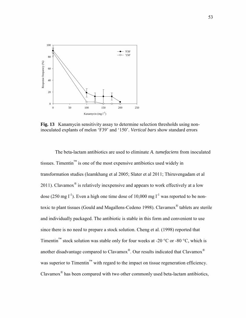
53
Kanamycin (mg l-1
)
0 50 100 150 200 250
Res
po
nse
fre
qu
ency
(%
)
0
20
40
60
80
100
'F39'
'150'
Fig. 13 Kanamycin sensitivity assay to determine selection thresholds using non-inoculated explants of melon ‗F39‘ and ‗150‘. Vertical bars show standard errors
The beta-lactam antibiotics are used to eliminate A. tumefaciens from inoculated
tissues. Timentin™ is one of the most expensive antibiotics used widely in
transformation studies (Ieamkhang et al 2005; Slater et al 2011; Thiruvengadam et al
2011). Clavamox® is relatively inexpensive and appears to work effectively at a low
dose (250 mg l-1). Even a high one time dose of 10,000 mg l-1 was reported to be non-
toxic to plant tissues (Gould and Magallens-Cedeno 1998). Clavamox® tablets are sterile
and individually packaged. The antibiotic is stable in this form and convenient to use
since there is no need to prepare a stock solution. Cheng et al. (1998) reported that
Timentin™ stock solution was stable only for four weeks at -20 °C or -80 °C, which is
another disadvantage compared to Clavamox®. Our results indicated that Clavamox®
was superior to Timentin™ with regard to the impact on tissue regeneration efficiency.
Clavamox® has been compared with two other commonly used beta-lactam antibiotics,

54
carbenicillin and cefotaxime, on the growth of A. tumefaciens strain LBA4404 and the
shoot regeneration of tomato. No significant differences were found (Hussain et al.
2008).
Clavamox® and Timentin™ were tested on non-inoculated explants of ‗F39‘ and
‗150‘ to examine possible adverse effects on plant regeneration. In ‗150‘, no significant
differences were detected during the early stages of shoot regeneration (first four weeks);
however, inclusion of Timentin™ resulted in vitrification and/or aberrant morphology in
70% of the shoots over time (Fig. 14 and Table 7). This vitrification severely reduced
shoot elongation frequency and ability to produce roots. In contrast, only 5% of the
shoots cultured on the medium containing Clavamox® became vitrified and/or abnormal.
This result was similar to results seen in the non-antibiotic treatments. For ‗F39‘,
Clavamox® and Timentin™ treatments failed to show any significant differences in shoot
regeneration and production of abnormal types, suggesting the effects of these
antibiotics could be genotype-dependent.
Fig. 14 Effects of Clavamox® and Timentin™ on the regeneration of non-inoculated explants of honeydew ‗150‘ after 75 days of culture. a & b Shoot growth and rooting on the regeneration medium supplemented with 250 mg l-1 Clavamox®, c with 300 mg l-1 Timentin™

55
Table 7 Effects of Clavamox® and Timentin™ on shoot development and elongation of non-inoculated explants of melon ‗F39‘ and ‗150‘
Genotype Antibiotic No. of explants
Regeneration frequency (%)
No. of shoots per explant
Percentage of elongated /rooted shoots (%)
Aberrant /vitrified shoot (%)
‗F39‘ Clavamox® 16 97.5 a 4.6 b 31.1 a 40.0 a Timentin™ 16 90.0 a 11.4 a 30.0 a 34.4 a ‗150‘ Clavamox® 40 62.5 a 6.8 a 40.0 a 5.0 b Timentin™ 40 56.3 a 8.2 a 5.0 b 70.0 a Means within the same columns within each genotype followed by different letters are significantly different by Duncan‘s Multiple Range Test at 5% probability level. The percentage data were transformed by an arcsin function before analysis to normalize the distribution
Like many other melon transformation reports (Dong et al. 1991; Akasaka-
Kennedy et al. 2004; Wu et al. 2009), the common problem of ‗escapes‘ was also
identified in our study. By reducing explant size (6 explants per cotyledon, 3 mm 2
mm) and increasing the volume of selection medium per Petri dish (45 ml), we were able
to reduce the curling of cotyledonary explants and the incidence of ‗escapes‘. A similar
result was obtained by Wu et al. (2009) in oriental melon transformation.
Inoculation and co-cultivation
Initially, A. tumefaciens strains LBA4404 and EHA105 were tested for their ability to
transform explants based on GUS histochemical staining. Both strains showed fairly
good inoculation efficiencies demonstrated by GUS assays (Fig. 15). EHA105 was used
in subsequent studies as it was reported to be more efficient in producing stable
transformants compared to LBA4404 (Galperin et al. 2003).

56
A B
Fig. 15 Transient GUS expression of explants inoculated with different Agrobacterium
tumefaciens strains carrying pCNL56, 7 days after inoculation. a strain EHA105, b strain LBA4404
Previous reports of melon transformation indicated optimal inoculation times of
20 sec to 30 min (Ayub et al. 1996; Guis et al. 2000; Vallés and Lasa 1994) depending
on the Agrobacterium strain used, concentration of the bacterial suspension, plant
species, explant size and thickness. We evaluated 10, 15 and 20 min inoculation times.
Three days after inoculation, the explants inoculated for 10 min remained healthy, but
the longer inoculation treatments resulted in the explants becoming brown or necrotic in
some cases (data not shown). Therefore, an inoculation period of 10 min was adopted.
Co-cultivation is one of the most important steps in a transformation procedure.
Factors during co-cultivation can enhance transformation efficiency, i.e., temperature
(Fullner and Nester 1996; Yasmin and Debener 2010; Sharma et al. 2011; Seo et al.
2011), lighting conditions (Zambre et al. 2003), co-cultivation period (Fang and Grumet
1990; Shilpa et al. 2010; Seo et al. 2011), addition of acetosyringone (Costa et al. 2006;
Afroz et al. 2010; Sharma et al. 2011) and antioxidants (Dan et al. 2009; Olhoft and
Somers 2001; Ostergaard and Yanofsky 2004; Toldi et al. 2002; Zheng et al. 2005;
Kumar et al. 2011; Dutt et al. 2011). Co-cultivation periods of 2 to 6 d were reported to
be optimal for melons (Galperin et al. 2003; Vallés and Lasa 1994; Akasaka-Kennedy et

57
al. 2004; Dong et al. 1991). Under our co-cultivation conditions at 22 ºC, A. tumefaciens
strain EHA105 began to overgrow the explant 3 d following the inoculation (data not
shown).
Zambre et al. (2003) reported a positive effect of light on gene transfer from A.
tumefaciens to callus explants of Phaseolus acutifolius (tepary bean) and root explants of
Arabidopsis thaliana. Our results showed there were no significant differences in
transformation rates between light and dark treatments (P 0.05) during co-cultivation
(Table 8). Plausible reasons for this result may be: 1) the light/dark influence on
Agrobacterium-mediated transformation may be genotype dependent; 2) the light/dark
influence may depend on explant types (callus and/or root); 3) the co-cultivation
temperatures of our light and dark treatments of 24 °C and 22 °C respectively, may have
influenced our results. Temperatures of 19-22 ºC have been reported to be critical for
high efficiency of Agrobacterium-mediated transformation while temperatures higher
than 22 ºC dramatically decreased this efficiency (Fullner and Nester 1996).
Table 8 Effect of light condition on GUS transient expression during the co-cultivation period in cotyledonary explants of ‗F39‘ and ‗150‘
Genotype Light condition No. of explants analyzed No. of explants stained blue GUS
positives (%) ‗F39‘ Light 80 18 22.8 a Dark 80 30 38.5 a ‗150‘ Light 80 74 92.5 a Dark 80 70 87.5 a Means within the same columns within each genotype followed by same letters are not significantly different by Duncan‘s Multiple Range Test at 5% probability level. The percentage
data were transformed by an arcsin function before analysis to normalize the distribution

58
GUS staining, PCR and Southern blot analyses
Cotyledonary explants were inoculated with A. tumefaciens strain EHA105. A total of
1075 explants of ‗F39' (from two experiments) and 1205 explants of ‗150‘ (from three
experiments) were used. GUS histochemical analysis was conducted throughout the
selection procedure to identify putative transformants, as well as non-transformed or
A B C
D
E F
Fig. 16 Callus and shoot development from explants and GUS histochemical assay. a & c a regenerated shoot of cantaloupe ‗F39‘ before (a) and after (c) GUS staining, b & d a cotyledonary explant with callus producing shoot primordia of honeydew ‗150‘ before
(b) and after (d) GUS staining, e an apical shoot of a GUS-positive plant of ‗150‘, f GUS expression in roots of a kanamycin-resistant plant of ‗150‘

59
chimeric tissues. Isolated pieces of leaf tissue from regenerating shoots were examined
for GUS activity (Figs. 16a-d). All GUS positive tissues were then analyzed further for
expression of the GUS gene in apical shoots (Fig. 16e) and roots (Fig. 16f). During large
scale GUS assays, we found a large proportion of chimeras in both genotypes (9 out of
12 samples of ‗F39‘ and 9 out of 13 samples of ‗150‘). Chimeric shoots exhibited blue
sectors of various sizes following GUS histochemical staining as reported in other crop
species (Moore 1995; Mollel et al. 2004; Kathiravan et al. 2006). Most chimeric shoots
did not survive kanamycin selection and died before or during rooting.
The binary plasmid pCNL56 harbors both nptII and gusA genes. All GUS-
positive plants that rooted on media containing kanamycin were selected for PCR
analysis to screen for the presence of nptII. Genomic DNA from all rooted GUS-
positive plants yielded a 1.2-kb fragment identical to the expected fragment amplified
from the nptII gene (Fig. 17a). Plants regenerated from non-inoculated explants failed to
produce a fragment. Most non-transformed shoots were killed by kanamycin selection
during regeneration, elongation and rooting. Although some shoots survived kanamycin
(150 and 200 mg l-1), many failed to root in the rooting medium containing 50 mg l-1
kanamycin. Based on PCR analysis, we found only one ‗F39‘ and two ‗150‘ regenerated
plants to be non-transgenic escapes.
Southern blot analysis was performed on all PCR-positive plants to identify
genomic integration of gusA (Fig. 17b). Transformation efficiency was estimated using
the total number of plants exhibiting genomic incorporation, divided by the total number
of inoculated explants. The transformation efficiency for ‗F39‘ ranged between 0.2%

60
and 0.4% (average efficiency was 0.3 ± 0.1%), and for ‗150‘ the range was between
0.2% and 0.8% (average was 0.5 ± 0.3%).
Fig. 17 PCR and southern blot analyses of putative transformants. a preliminary screen for detecting the presence of the nptII gene in putative transformants using PCR. Lane 1: 100-bp ladder; lane 2: pCNL56 (positive control); lane 3: wild type honeydew ‗150‘
(negative control); lanes 4-8: GUS-positive plants of honeydew ‗150‘; lane 9: wild type cantaloupe ‗F39‘ (negative control); lanes 10-12: GUS-positive plants of cantaloupe ‗F39‘, b integration of the gusA gene as detected by Southern blotting. Lane 1: pCNL56 (positive control); lane 2: wild type honeydew ‗150‘ (negative control); lanes 3-7: PCR-positive plants of honeydew ‗150‘; lane 8: wild type cantaloupe ‗F39‘ (negative control);
lanes 9-11: PCR-positive plants of cantaloupe ‗F39‘
1 2 3 4 5 6 7 8 9 10 11 12
A ‘F39’ ‘150’
1.2 kb
1 2 3 4 5 6 7 8 9 10 11
B
23
9.4 6.5
4.3
kb
‘F39’ ‘150’

61
Phenotypes of regenerated transformants
Abnormal morphological characteristics were observed in the transgenic melon plants
(Fig. 18a-g). The most severe phenotype was a lack of apical dominance (Fig. 18a)
H
A B
G
C D
E F I
Fig. 18 Morphological characteristics of transgenic melon plants. a transgenic honeydew line ‗090129‘ showing a lack of apical dominance, b wild type honeydew ‗150‘, c wild type cantaloupe ‗F39‘, d transgenic cantaloupe line ‗082417‘, e a leaf of wild type cantaloupe ‗F39‘, f a leaf of transgenic cantaloupe line ‗082417‘, g transgenic honeydew line ‗081105‘, h male flowers of wild type (left) and transgenic (right) honeydew ‗150‘, i perfect flowers of wild type (left) and transgenic (right) honeydew ‗150‘, j transgenic melon plants in the greenhouse

62
resulting in abnormal growth. Other aberrant morphologies included shorter (Fig. 18d)
and longer (Fig. 18g) internodal growth and irregular leaf shapes (Fig. 18f). Plants
exhibiting lack of apical dominance and shorter/longer internodes died prematurely in
the greenhouse. Morphologically normal transgenic plants produced both male (Fig. 18h)
and perfect (Fig. 18i) flowers.
Similar abnormalities reported in western shipper melon ‗Topmark‘ and other
melon types were suggested to be caused by a high kanamycin concentration (150 mg l-1)
in the selection medium (Gonsalves et al. 1994). In our study, kanamycin at 200 and 150
mg l-1 was used for in vitro selection of ‗F39‘ and ‗150‘ respectively, similar to the
levels reported by Gonsalves et al. (1994). However, in our study, abnormal in vitro
regenerated wild-type plants were also observed in the absence of kanamycin (data not
shown). This observation suggests that the tissue culture process and plant growth
regulator effects may induce aberrant morphology in regenerated melon shoots (Larkin
and Scowcroft 1981).
Pre-culture test with pBI121
We used EHA105 harboring the binary vector pCNL56 for the protocol development.
However, the gene of interest for our future study was cloned into the binary vector
pBI121 harbored by EHA105. By testing the protocol with this construct, we found that
GUS transient expression levels in both melon lines were much lower than those
inoculated with pCNL56 (Fig. 19a-b). Similar results were reported for Agrobacterium-
mediated transformation in finger millet (Sharma et al. 2010). To adjust our protocol for

63
CK pCNL56 pBI121 CK pCNL56 pBI121
A B
C
D
4h 2d1d
4h 2d1d
Fig 19 Transient GUS expression of cotyledonary explants of ‗F39‘ and ‗150‘
inoculated with Agrobacterium tumefaciens strain EHA105 carrying two different plasmids. Data was recorded 8 days after inoculation. a Plasmid pCNL56 vs. pBI121 in ‗F39‘, b plasmid pCNL56 vs. pBI121 in ‗150‘, c ‗F39‘ inoculated with pBI121 for 4 h, 1 d and 2 d, d ‗150‘ inoculated with pBI121 for 4 h, 1 d and 2 d
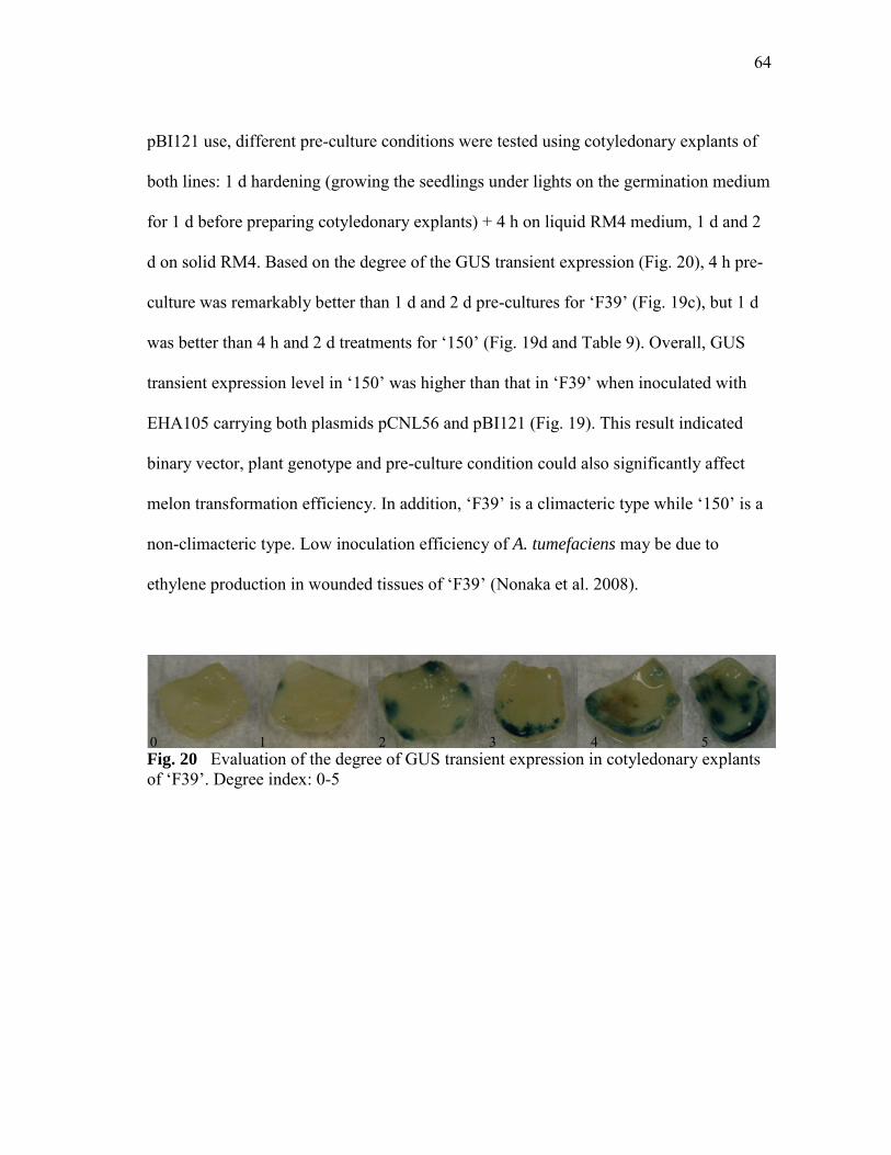
64
pBI121 use, different pre-culture conditions were tested using cotyledonary explants of
both lines: 1 d hardening (growing the seedlings under lights on the germination medium
for 1 d before preparing cotyledonary explants) + 4 h on liquid RM4 medium, 1 d and 2
d on solid RM4. Based on the degree of the GUS transient expression (Fig. 20), 4 h pre-
culture was remarkably better than 1 d and 2 d pre-cultures for ‗F39‘ (Fig. 19c), but 1 d
was better than 4 h and 2 d treatments for ‗150‘ (Fig. 19d and Table 9). Overall, GUS
transient expression level in ‗150‘ was higher than that in ‗F39‘ when inoculated with
EHA105 carrying both plasmids pCNL56 and pBI121 (Fig. 19). This result indicated
binary vector, plant genotype and pre-culture condition could also significantly affect
melon transformation efficiency. In addition, ‗F39‘ is a climacteric type while ‗150‘ is a
non-climacteric type. Low inoculation efficiency of A. tumefaciens may be due to
ethylene production in wounded tissues of ‗F39‘ (Nonaka et al. 2008).
Fig. 20 Evaluation of the degree of GUS transient expression in cotyledonary explants of ‗F39‘. Degree index: 0-5
1 2 5 4 3 0
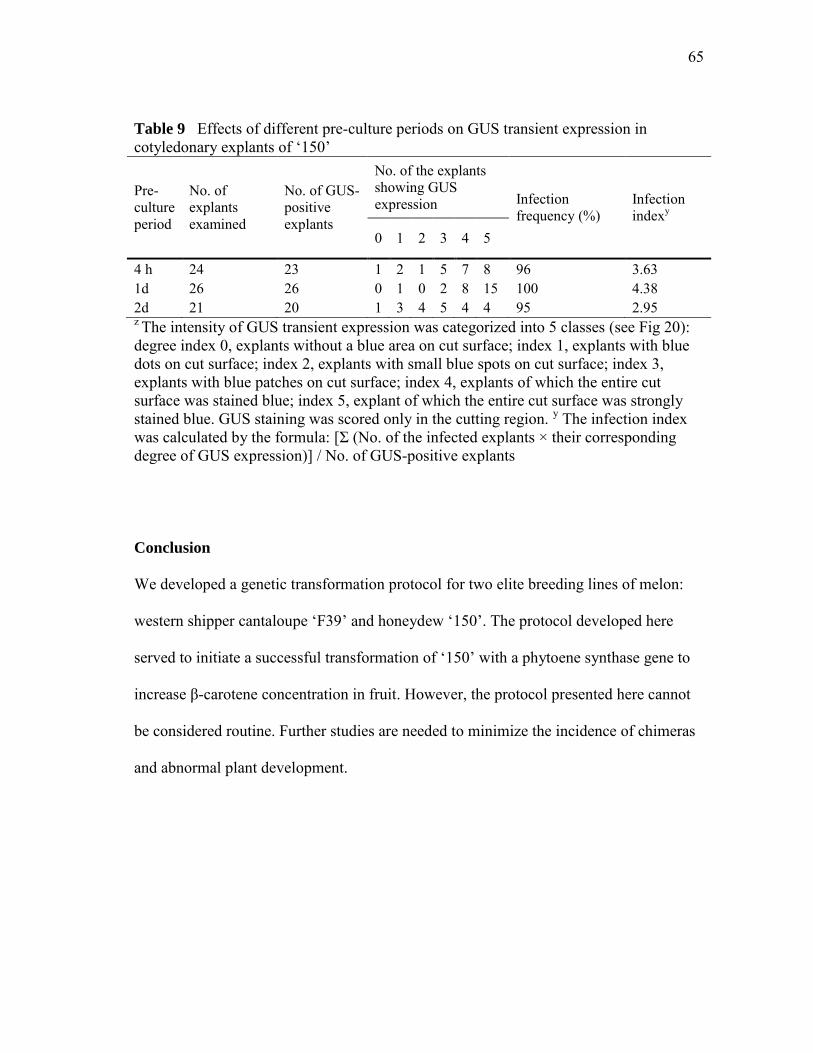
65
Table 9 Effects of different pre-culture periods on GUS transient expression in cotyledonary explants of ‗150‘
Pre-culture period
No. of explants examined
No. of GUS-positive explants
No. of the explants showing GUS expression Infection
frequency (%) Infection indexy
0 1 2 3 4 5
4 h 24 23 1 2 1 5 7 8 96 3.63 1d 26 26 0 1 0 2 8 15 100 4.38 2d 21 20 1 3 4 5 4 4 95 2.95 z The intensity of GUS transient expression was categorized into 5 classes (see Fig 20): degree index 0, explants without a blue area on cut surface; index 1, explants with blue dots on cut surface; index 2, explants with small blue spots on cut surface; index 3, explants with blue patches on cut surface; index 4, explants of which the entire cut surface was stained blue; index 5, explant of which the entire cut surface was strongly stained blue. GUS staining was scored only in the cutting region. y The infection index was calculated by the formula: [Σ (No. of the infected explants × their corresponding degree of GUS expression)] / No. of GUS-positive explants
Conclusion
We developed a genetic transformation protocol for two elite breeding lines of melon:
western shipper cantaloupe ‗F39‘ and honeydew ‗150‘. The protocol developed here
served to initiate a successful transformation of ‗150‘ with a phytoene synthase gene to
increase β-carotene concentration in fruit. However, the protocol presented here cannot
be considered routine. Further studies are needed to minimize the incidence of chimeras
and abnormal plant development.

66
CHAPTER V
GENETIC ENGINEERING OF BETA-CAROTENE PRODUCTION IN HONEYDEW
Materials and Methods
Agrobacterium inoculation and plant transformation
Agrobacterium tumefaciens strain EHA105 carrying a binary vector, PSYC/pRD12,
harboring an nptII and a PSY-C genes was used. We isolated a PSY-C gene from a
watermelon flesh and ligated into pRD12 having a 4.8-kb polygalacturonase (PG)
promoter and a 1.8-kb PG terminator isolated from ripe tomato fruits (Lau et al. 2009,
GenBank: FJ465170.1). The binary vector pRD12 was provided by Dr. Jim Giovannoni
at Cornell University (Fig. 21).
RBNos-
promoterPG-
terminator
LBPG-
promoter PSY-CNPT-IINosA
Fig. 21 T-DNA region of binary vector PSYC/pRD12 Ploidy determination
Ploidy level was determined by flow cytometry. Broccoli leaf sample was used as an
internal control. Samples were prepared from the third leaf below shoot apex of
acclimatized plants in pots using a commercial kit CyStain PI absolute P (PATTEC,
Germany). Plant nuclei were analyzed on a FASCSalibur (Becton Dickinson
Immunocytometry System, San Jose, CA) flow cytometer, equipped with a 15mW air-

67
cooled argonlaser, using CellQuest (Becton Dickinson) acquisition software. Propidium
iodide fluorescence was collected through a 585/42-nm bandpass filter. A minimum of
5,000 events, defined by a region for single nuclei in a plot of propidium iodide area
versus width, were measured for each sample. Data analysis was performed in FlowJo
(version 8.8.7, Treestar, Inc., Ashland, OR).
Phytoene and β-carotene extraction and analysis
Carotenoid profile was analyzed by HPLC. Rind, flesh and placental tissues were frozen
at -80 °C before analysis. Approximately 50 g of frozen tissue were pulverized with a
rubber mallet and mixed thoroughly while the tissue remained frozen. Two to three gram
slurries were ground to fine powders in a mortar and pestle with liquid nitrogen.
Powders were washed with acetone on filter paper until tissues became colorless.
Hexane (30 ml) was added to the acetone extracts and distilled water was added to
separate the hexane layer from the acetone/water layer. All carotenoids were collected in
the hexane layer and stored at -80 °C. Hexane extracts (15ml) were evaporated with a
stream of nitrogen in darkness and replenished with 1 ml acetone for HPLC analysis.
Carotenoids were analyzed using a Perkin Elmer HPLC system (Shelton, CT)
equipped with a Series 200 pump, autosampler, and diode array detector. An analytical
Spheriosorb ODS2 column (Waters, 4.6 250 mm, 5 µm) was used with a guard
cartridge. Solvents A and B were acetonitrile:water (9:1, v/v) and ethylacetate,
respectively, both containing 1% triethylamine. The solvent was programmed as an
isocratic condition (A:B, 7:3) for 40 min at a flow rate of 1 ml/min and flushed with

68
100% solvent A for 5 min. A 40 µl sample was injected. Chromatographic detection was
set at 280 nm and 475 nm, and spectral data were simultaneously collected from 200 to
700 nm. Phytoene (CaroteNature, Lupsingen, Switzerland) and β-carotene (Sigma-
Aldrich, St. Louis, USA) standards of predetermined concentration were used to
quantify phytoene and β-carotene in samples.
PCR and Southern blots
Genomic DNA was isolated from leaf tissue of wild type and putative transformants by
the method of Skroch and Nienhuis (1995). To determine if transgenes were integrated,
primers were designed to amplify fragments from PG promoter and PSY-C genes under
the following conditions. Primers PGF02 (forward) and PSYCR02 (reverse) were 5'-
TGA GAC GGG AGA AGA CAA GCC AGA CAA A-3' and 5'-CCG TTT TAC CAA
AGC CGC CTG TTT CAT-3', respectively. The PCR reaction mixture contained 50 ng
of genomic DNA, 1x PCR buffer, 200 M dNTP, 0.2 M forward and reverse primers
and 1 l Advantage2 polymerase mix (Clontech Laboratories, Inc. Mountain View, CA,
USA) in a total volume of 50 l. The reaction consisted of an initial denaturation at 94
ºC for 3 min, followed by 35 cycles of denaturation at 94 ºC for 30 s, annealing at 68 ºC
for 30 s, and elongation at 72 ºC for 2 min, and a final elongation at 72 ºC for 10 min.
Amplified products were visualized on 1% (w/v) agarose gels.
Twenty micrograms of genomic DNA was digested with HindIII and AvaII at 37
ºC overnight and then electrophoresed on a 1% (w/v) agarose gel at 24 V overnight,
respectively. DNA fragments were then transferred onto an N+ Hybond nylon membrane

69
(Amersham Hybond™) by alkaline transfer. A probe covering partial segments of the PG
promoter and PSY-C gene was labeled using PCR DIG probe synthesis kit (Roche
Applied Science, Mannheim, Germany). The membrane was then hybridized with the
DIG-labeled probe in DIG Easy Hyb solution at 45 ºC overnight. DIG-labeled nucleic
acids were then detected with CDP-Star (Roche Applied Science, Mannheim, Germany)
by exposing the membrane under a LAS-4000 Chemiluminescent Image System (Fuji
Film Life Science, USA). Probe labeling, hybridization, washing and detection were
conducted according to the manufacturer‘s instruction.
RNA extraction and reverse transcription polymerase chain reaction (RT-PCR)
Total RNA was isolated from rind, flesh and placenta tissues of wild type, vector control
and transgenic melon fruits with RNeasy® Plant Mini Kit (Qiagen, Valencia, CA, USA,
Catalog No. 74904) according to the manufacturer‘s instruction. Then RNA was
immediately converted into cDNA by priming with oligo (dT) using Advantage™ RT-
for-PCR Kit (BD Biosciences Clontech, Palo Alto, CA, USA, Catalog No. 639505).
To detect PSY-C transcript, primers were designed to amplify the PSY-C under
the following conditions. Forward and reverse primers were 5'-ACT GAG AAC CGC
CGG AGA ATT GGA TGT-3' and 5'-TGA AGG GCC AAG GAG AGA CCT TGC
ATA-3', respectively. The RT-PCR reaction mixture contained 50 ng of cDNA, 1x PCR
buffer, 200 M dNTP, 0.2 M forward and reverse primers and 1 l Advantage2
polymerase mix (Clontech Laboratories, Inc. Mountain View, CA, USA) in a total
volume of 50 l. The reaction consisted of an initial denaturation at 94 ºC for 3 min,

70
followed by 30 cycles of denaturation at 94 ºC for 30 s, annealing at 68 ºC for 30 s, and
elongation at 72 ºC for 2 min, and a final elongation at 72 ºC for 10 min. Amplified
products were visualized on 1% (w/v) agarose gels.
Segregation analysis
Segregation of the PSY-C transgene and its inheritance were analyzed by germinating
the seeds of T1 progenies of three transgenic lines, 042201, 071506_3 and 071506_4, on
the seed germination medium containing 200 mg l-1 kanamycin. DNA was extracted
from the kanamycin-survived seedlings, and then examined by PCR with primers
PGF02/PSYCR02 under the conditions described before. Amplified products were
visualized on 1% (w/v) agarose gels. Chi-square test was conducted to estimate the
segregation ratio.
Results and Discussion
Generation of transgenic plants
Cotyledonary explants were inoculated with A. tumefaciens strain EHA105 carrying
PSYC/pRD12. All regenerated plants that rooted on media containing 50 mg l-1
kanamycin were selected for PCR analysis to screen for the presence of the PSY-C.
Genomic DNA from all rooted kanamycin-resistant plants yielded a 500-bp fragment
identical to the expected fragment amplified from the PSY-C gene (Fig. 22a).

71
Genomic DNA from wild type and all PCR-positive plants (Fig. 22a) were
digested by restriction endonucleases HindIII and AvaII, respectively, and then
subjected to Southern blot analysis to determine the number of copies of the PSY-C gene
and identify where the transferred genes were incorporated into the plant‘s genome (Fig.
22b-c). Three sites of HindIII and two sites of AvaII were found on the plasmid (Fig.
22d). A 1.8-kb band appeared only in transgenic plants and plasmid control
PSYC/pRD12 digested with HindIII, and a 9.4-kb fragment showed only in transgenic
plants and plasmid control PSYC/pRD12 digested with AvaII. These two bands
confirmed that PSY-C has been integrated into melon genomic DNA (Fig. 22b-c).
Interestingly, an approximate 6.5-kb fragment was identified in transgenic plants (line
071506) digested with AvaII. It suggested that there may be an internal deletion
occurred in the promoter region because they were kanamycin resistant indicating the
region of nptII may be intact. Further investigations are needed to determine if there may
be any other reasons to result in unexpected size of T-DNA insertion. The largest band
appeared in line 042201 may indicate a different location of PSY-C integration in the
melon genome (Fig. 22c).
A total of 2804 explants of ‗150‘ (from 11 experiments) were used to regenerate
eight plants from four explants, becoming four transgenic lines 042201, 071503, 071506
and 071519. Lines 071503, 071506 and 071519 revealed a single copy of the PSY-C
integrated into the genome of each plant. Two copies of the PSY-C exhibited in the
genome of line 042201. A 6-kb band in all samples digested with HindIII and a 3-kb
band in all samples digested with AvaII were detected. Based on the sequence

72
500 bp
1 2 3 4 5 6 7 8 9 10A
1 2 3 4 5 6 7 8 9 10 11kb23.0
9.46.5
4.3
2.3
2.0
B
Fig. 22 PCR and southern blot analyses of putative transformants. a Preliminary screen for detecting the presence of the PSY-C gene in putative transformants using PCR analysis. Lane 1: 100-bp ladder; lane 2: PSYC/pRD12 (positive control); lane 3: wild type honeydew ‗150‘ (negative control); lanes 4-10: kanamycin-resistant plants of honeydew ‗150‘, b and c integration of the PSY-C gene as detected by Southern blot analysis of HindIII-digested (b) and AvaII-digested (c) genomic DNA from PCR-positive plants. Lane 1: digoxigenin-labeled DNA molecular weight marker II; lane 2: PSYC/pRD12 in a wild type honeydew ‗150‘ (positive control); lane 3: wild type honeydew ‗150‘ (negative control); lanes 4: line 042201; lane 5-7: line 071503 (071503_2, -71503_3 and 071503_4); lane 8-10: line 071506 (071506_2, 071506_3 and 071506_4); lane 11: line 071519, d restriction map of the T-DNA

73
RB LB
AvaIIHindIIIHindIII HindIII
PSYCprobe
AvaIID
1.8 kb
9.4 kb
1 2 3 4 5 6 7 8 9 10 11
kb
23.0
9.4
6.5
4.3
2.32.0
C
Fig. 22 Continued
information and similarity, it was assumed to be melon PSY (GenBank: Z37543) which
was 94% homologous to watermelon PSY-C.
Ploidy and morphology of transgenic plants and their fruit harvest
Flow cytometry was conducted to analyze ploidy level of transgenic melons using a
broccoli leaf tissue as an internal control. In the histograms (Fig. 23), peak ‗broccoli-G1‘
between two ‗melon‘ peaks represented diploid broccoli cells whose genome size is

74
similar to triploid melon genome size. All the transgenic plants had over 50% of
tetraploid cells (peak ―melon-2‖ on 400 PI-Area) while the diploid control plant had
more than 50% of diploid cells (peak ―melon-1‖ on 200 PI-Area). Thus, 100% of the
transgenic plants are tetraploids (Fig. 23b-e). The peak ‗melon-2‘ in diploid represented
the dividing melon diploid cells; the peaks on 800 PI-Area in tetraploid were from
dividing melon tetraploid cells; the peaks ‗broccoli-G2‘ on 600 PI-Area indicated
dividing diploid broccoli cells.
A
F
B C
E
Fig. 23 Flow cytometry histograms of leaf tissues from honeydew ‗150‘ transgenic
lines. a A diploid from seed-grown plant, b 042201, c 071503, d 071506, e 071519

75
Our previous study showed the percentage of polyploids regenerated from
cotyledonary explants of ‗150‘ through in vitro culture was 50-60% (see Chapter III).
This higher frequency of tetraploids that occurred in transformants was unexpected;
however, except line 071519 (Fig. 24c, left), other tetraploid transgenic honeydew plants
can still grow and develop normally, which was in agreement with our early observation
in non-transgenic honeydew ‗150‘ (Chapter III).
Irregular leaf shape happened in the early stage of line 042201 and 071503 (Fig.
24a), but it did not affect plant growth, flowering, fruiting and seeding. Plant size of line
071519 was smaller than those of other lines. This line grew slower on the elongation/
rooting medium containing 50 mg l-1 kanamycin and cannot produce normal healthy
roots compared to other transgenic plants. It had smaller but longer leaves and shorter
internodes (Fig. 24b-c) during all the life stages and did not produce fruit. Seeds of the
transgenic fruits were bigger and round compared to those of wild type, but they can
normally germinate and grow to plants (Fig. 24d-e). The different shape of the seeds was
also observed in non-transgenic tetraploid honeydew ‗150‘ regenerated from in vitro
culture (see in Chapter III). No abnormal morphologies have been found in the progeny
of line 042201 (Fig. 24b-c, right; Fig. 24e).

76
A B
C
E
D
Fig. 24 Morphologies of transgenic plants and their T1 progenies. a Leaves of wild type ‗150‘ (left) and transgenic line 042201(middle) and 071503 (right), b leaves of line 071519 (left) and line 042201‘s progeny (right) , c line 071519 (left) and line 042201‘s
progeny (right), d seeds of wild type ‗150‘ (left) and transgenic line 071506 (right), e progeny seedlings of line 042201
Fully mature fruits were harvested from non-transgenic and transgenic plants
during 34-41 days after pollination (DAP); however, the fruit harvested from transgenic
071506_4 was 42 DAP but had to be cut from vine prior to color break due to an
unhealthy condition. When harvested, the fruit rind was still creamy color but the area
around the blossom end began to turn orange (Fig. 25). To let it reach the same ripening
stage as other normally harvested fruits, the fruit was stored in a warm room (25±2 ºC
under cool white fluorescent lights with 16 h light/8 h dark photoperiod and 60-80 µmol

77
m-2 s-1 light intensity) for 2 weeks before being processed for HPLC analysis and mRNA
extraction.
Fruit colors and their related carotenoids
Fruit 042201 (34 DAP) had a orange-color rind and green-color flesh; fruit 071506_3
(35 DAP) had a pale orange-color rind and green-color flesh. After storing for two
weeks, the rind color of fruit 071506_4 still remained creamy but the orange area around
the blossom end extended a little. Wild type and vector control fruits exhibited cream-
color rinds and green-color flesh (Fig. 25).
The concentrations of phytoene and β-carotene in all the tissue samples
extracted from transgenic and non-transgenic melons were analyzed by HPLC (Figs. 26-
28). The peaks of both phytoene and β-carotene were detected in the rind tissue of
transgenic fruits; however, only trace volumes were found in the rind tissues of non-
transgenic fruits and the flesh tissues of all melons. Similar peaks of β-carotene were
shown in the placental tissues of transgenic and non-transgenic fruits.
Based on the calculation of HPLC data (Table 10), phytoene and β-carotene
levels were similar in the flesh and placental tissues between the transgenic and non-
transgenic fruits. However, the rind tissues of the transgenic fruits had remarkably
higher concentrations of phytoene and β-carotene than those of wild type and vector
control fruits. The highest concentrations existed in fruit 042201 rind tissue, which is in
accordance with the bright orange color. In this fruit rind tissue, the phytoene content
was approximately 2-fold higher than that of other transgenic fruits, and no phytoene

78
was detected in wild type and vector control fruits; the β-carotene dramatically increased
approximately 32 fold compared to wild type, It was also 2- to 3-fold higher than other
transgenic lines. Fruit 071506_3 and 071506_4 rind β-carotene contents were
approximately 11- and 9-time higher than wild type, respectively.
CA B
Fig. 25 Fruits from wild type (a), vector control (b) and transgenic plants (c-e) of honeydew ‗150‘. c 042201, d 071506_3, e 071506_4

79
D E Fig. 25 Continued

80
0.00
100.00
200.00
300.00
400.00
0 2 4 6 8 10 12 14 16 18 20 22 24 26 28 30 32 34 36 38
0.00
100.00
200.00
300.00
400.00
0 2 4 6 8 10 12 14 16 18 20 22 24 26 28 30 32 34 36 38
0.00
100.00
200.00
300.00
400.00
0 2 4 6 8 10 12 14 16 18 20 22 24 26 28 30 32 34 36 38
0.00
100.00
200.00
300.00
400.00
0 2 4 6 8 10 12 14 16 18 20 22 24 26 28 30 32 34 36 38
0.00
100.00
200.00
300.00
400.00
0 2 4 6 8 10 12 14 16 18 20 22 24 26 28 30 32 34 36 38
0.00
100.00
200.00
300.00
400.00
0 2 4 6 8 10 12 14 16 18 20 22 24 26 28 30 32 34 36 38
Retention time (min)
Res
po
nse
(mV
)β-carotene Phytoene
WT WT
042201042201
VCVC
Fig. 26 HPLC separation of carotenoids in rind tissue extractions from wild type (WT), vector control (VC) and transgenic line 042201 of honeydew ‗150‘

81
0.00
100.00
200.00
300.00
400.00
0 2 4 6 8 10 12 14 16 18 20 22 24 26 28 30 32 34 36 38
0.00
100.00
200.00
300.00
400.00
0 2 4 6 8 10 12 14 16 18 20 22 24 26 28 30 32 34 36 38
0.00
100.00
200.00
300.00
400.00
0 2 4 6 8 10 12 14 16 18 20 22 24 26 28 30 32 34 36 38
0.00
100.00
200.00
300.00
400.00
0 2 4 6 8 10 12 14 16 18 20 22 24 26 28 30 32 34 36 38
0.00
100.00
200.00
300.00
400.00
0 2 4 6 8 10 12 14 16 18 20 22 24 26 28 30 32 34 36 38
0.00
100.00
200.00
300.00
400.00
0 2 4 6 8 10 12 14 16 18 20 22 24 26 28 30 32 34 36 38
Retention time (min)
Res
po
nse
(mV
)
WT WT
042201042201
VCVC
β-carotene Phytoene
Fig. 27 HPLC separation of carotenoids in flesh tissue extractions from wild type (WT), vector control (VC) and transgenic line 042201 of honeydew ‗150‘

82
0.00
100.00
200.00
300.00
400.00
0 2 4 6 8 10 12 14 16 18 20 22 24 26 28 30 32 34 36 38
0.00
100.00
200.00
300.00
400.00
0 2 4 6 8 10 12 14 16 18 20 22 24 26 28 30 32 34 36 38
0.00
100.00
200.00
300.00
400.00
0 2 4 6 8 10 12 14 16 18 20 22 24 26 28 30 32 34 36 38
0.00
100.00
200.00
300.00
400.00
0 2 4 6 8 10 12 14 16 18 20 22 24 26 28 30 32 34 36 38
0.00
100.00
200.00
300.00
400.00
0 2 4 6 8 10 12 14 16 18 20 22 24 26 28 30 32 34 36 38
0.00
100.00
200.00
300.00
400.00
0 2 4 6 8 10 12 14 16 18 20 22 24 26 28 30 32 34 36 38
Retention time (min)
Re
spo
nse
(mV
)
WT WT
042201042201
VCVC
β-carotene Phytoene
Fig. 28 HPLC separation of carotenoids in placenta tissue extractions from wild type (WT), vector control (VC) and transgenic line 042201 of honeydew ‗150‘

83
Table 10 HPLC analysis of phytoene and β-carotene concentrations in rind, flesh and placenta tissues and the corresponding colors of transgenic honeydew ‗150‘
Tissue Genotype Phytoene (µg g-1 FW)
β-carotene (µg g-1 FW) Color
Rind Wide type - 0.480 Creamy Vector control - trace Creamy 042201 10.985 15.124 Orange 071506_3 4.364 5.614 Pale orange 071506_4 (whole) 4.005 4.389 Mixed color 071506_4 (around stem end) - 1.908 Creamy
071506_4 (around blossom end) 5.567 7.740 Pale orange
Flesh Wide type - 0.393 Green Vector control - 0.257 Green 042201 - trace Green 071506_3 - 0.729 Green 071506_4 - 0.891 Pale orange Placenta Wide type - 6.762 Orange Vector control - 5.547 Orange 042201 - 2.855 Orange 071506_3 - 3.598 Orange 071506_4 - 2.698 Orange Expression of PSY-C transgene
Total RNA was extracted from rind, flesh and placental tissues of wild type, vector
control and transgenic fruits, and then cDNA was immediately synthesized from the
RNA. PSY-C transgene transcription was examined using RT-PCR (Fig. 29). Transcript
was not detected in wild type and vector control but was detected in transgenic fruits.
Fruit 042201had stronger expression of PSY-C than others in the rind tissue, which is in
accordance with the dramatic increase of β-carotene level (Table 10). PSY-C expression
levels in the flesh and placental tissues were lower than those in the rind tissues. No
correlation was found between PSY-C expression level and carotenoid contents.

84
Phytoene is a direct product of phytoene synthase catalyzation in the carotenoid
biosynthesis pathway (Cunningham and Gantt 1998). Therefore, change of phytoene
levels in transgenic melons is an important indicator of PSY-C transgene functioning in
this study. Phytoene was detected only in the rind tissues (Fig. 26) but not flesh and
placenta of the transgenic fruits (Figs. 27-28 and Table 10); however, the RT-PCR
results indicated that the PSY-C transgene expressed in all the tissues. Thus, we assume
that post-transcriptional mechanisms of the PSY-C in the flesh and placental tissues were
inhibited. The 4.8-kb PG promoter used in this study contains four regulatory regions
which control temporal and spatial transcription of its ligated functional gene described
by Montgomery et al. (1993), Nicholass et al. (1995) and Lau et al. (2009). According to
these reports, the interaction of these regulatory regions directed ripening-specific
expression in outer pericarp only in ripe tomato fruit. In contrast, our results showed PG
promoter induced PSY-C expression in all the tissues (outer and inner pericarps). Further
studies will be needed to explain whether the PG promoter caused exclusive change of
β-carotene in the rind tissues. Another possibility of inhibited accumulation of β-
carotene in the flesh and placental tissues might be related to PSY-C tissue-specific
function. The accumulation of phytoene only happened in the rind but not the flesh and
placental tissues while PSY-C expression was detected in all the tissues. This
inexplicable result indicates that different mechanisms of carotenoid accumulation may
exist in different tissues of honeydew due to different metabolite precursor pools in
different tissues.

85
1 2 3 4 5 6 7 8 9 10 11 12 13 14 15
Rind Flesh Placenta
A
B
Fig. 29 RT-PCR analysis of PSY-C transgene expression in transgenic honeydew ‗150‘. a PSY-C transcript in different tissues of wild type, vector control and transgenic fruits (042201, 071506_3 and 071506_4), b 18S rRNA used as an internal control. Lane 1-5: rind tissue; lane 6-10: flesh tissue, lane 11-15: placental tissue Segregation and inheritance in the T1 progenies of the transformants
PCR analysis was used to detect the presence of PSY-C in the T1 progeny of each
transgenic fruit (Fig. 30). PCR results revealed single locus segregation in genotypes
042201 and 071506_3 (Table 11). However, transgenic genotype 071506_4 showed a
low possibility of Mendelian segregation probably due to the early harvest and abnormal
seed development.
To confirm the PCR results, we compared PCR-positive/negative individuals
with their corresponding kanamycin sensitive/resistant seedlings growing on the
selection medium containing 200 mg l-1 kanamycin. Like wild type (Fig. 31a),
kanamycin sensitive seedlings‘ root development was inhibited while kanaymcin
resistant seedlings developed normal healthy roots on the medium (Fig. 31b). Although
kanamycin sensitivitive seedlings elongated and cotyledons opened, their true leaves

86
were smaller than those on kanamycin resistant seedlings (Fig. 31c-d), and further
development was restrained. Eventually the true leaves failed to expand and were
bleached out resulting in the plants‘ death. Based on the comparison of results, most of
the kanamycin resistant seedlings were PCR positive and most of the kanamycin
sensitive seedlings were PCR negative. For the seedlings having controversial results,
we re-extracted DNA and re-did PCR analysis to assure the PSY-C presence.
500 bp
1 2 3 4 5 6 7 8 9 10 11
Fig. 30 PCR analysis to determine the segregation of PSY-C transgene in the T1 progeny of transgenic honeydew ‗150‘. Lane 1, 100-bp ladder; lane 2, PSYC/pRD12 (positive control); lane 3, wild type honeydew ‗150‘ (negative control); lanes 4-7, PCR-positive T1 progeny; lanes 8-11, PCR-negative T1 progeny Table 11 Segregation ratio of PSY-C transgene in T1 progeny of transgenic honeydew ‗150‘
Genotype Total number of seeds
PCR positive
PCR negative
Transgene segregation ratio
Probable PSY-C locus
χ2a P
042201 58 42 16 3:1 1 0.207 0.6491 071506_3 35 27 8 3:1 1 0.086 0.7693 071506_4 51 29 22 3:1 1 8.948 0.0027 a Chi-square value is calculated based on the hypothesis of 3:1 segregation ratio

87
A DCB
Fig. 31 Seed germination of wild type and transgenic progeny of honeydew ‗150‘ on
the selection medium containing 200 mg l-1 kanamycin. a-b Root development of wild type (a) and transgenic progeny (b), c-d cotyledon and true leaf development of wild type (c) and transgenic progeny (d)
Conclusion
We transferred a watermelon PSY-C gene with a fruit-specific PG promoter into our elite
honeydew breeding line ‗150‘. Two transgenic lines having normal morphologies have
been generated with up to 32-fold higher β-carotene in fruit rind tissue than wild type
honeydew. It was postulated that elevating β-carotene in the rind tissue may be related to
the fact that PG promoter was found to express only in outer pericarp in tomato fruit
depending on regulatory elements (Montgomery et al. 1993). This indicated that the
watermelon PSY-C gene may have a potential to increase β-carotene level in fruit flesh
tissue if a different promoter is used. Further studies will be needed to help understand
the mechanism of β-carotene accumulation in different tissues of melons as an essential
solution to improve carotenoid in melons.

88
CHAPTER VI
SUMMARY AND CONCLUSIONS
To genetically manipulate β-carotene level in melon fruits, two elite breeding lines,
western shipper cantaloupe ‗F39‘ and honeydew ‗150‘, were used to establish
regeneration and transformation systems. Six media were examined to evaluate the
capability of shoot regeneration from different explants types. Each explant type showed
a great potential to produce shoots. MS basal medium supplemented with 1 mg l-1 BA,
0.26 mg l-1 ABA and 0.8 mg l-1 IAA induced highest frequency of shoot regeneration
from cotyledonary explants in both ‗F39‘ and ‗150‘. Removal of IAA from this medium
solved a curving-up problem of cotyledonary explants in ‗150‘.
Our study then proceeded to establish an Agrobacterium-mediated
transformation protocol for both lines based on the optimal regeneration system. Several
factors have been tested to optimize the protocol, such as A. tumefaciens strains,
inoculation time, co-cultivation conditions, kanamycin concentrations and antibiotics. A.
tumefaciens strain EHA105, carrying pCNL56 containing nptII and gusA genes, was
selected to develop the protocol. Putative transformants were evaluated using GUS
histochemical assay, PCR and Southern blot analyses. Based on these parameters,
0.3±0.1% of the cotyledonary explants regenerated transgenic plants in ‗F39‘; and
0.5±0.3% of the cotyleondary explants produced transgenic plants in ‗150‘.
Whereafter, a watermelon PSY-C gene, under the control of a fruit-specific
promoter of PG gene, was transferred into honeydew ‗150‘. Putative transformants were

89
evaluated using PCR and Southern blot analyses. PSY-C transgene was expressed in all
the tissues of the transgenic fruits according to RT-PCR results. Changes of phytoene
and β-carotene concentrations were detected in the transgenic fruits using HPLC
analysis. Transgenic lines produced up to 32-fold higher β-carotene in their rind tissues
than the wild type.
In this study, we observed several problems in regeneration and transformation
of melons: 1) high percentage (50-60%) of polyploid shoots regenerated from
cotyledons; 2) chimeras and abnormal plants produced through transformation; 3) low
transformation efficiency; and 4) tissue-specific accumulation of β-carotene in
honeydew melons. Further studies are needed to reduce or solve these problems, and to
elevate β-carotene level in flesh tissue of melons.

90
REFERENCES
Afroz A, Chaudhry Z, Rashid U, Ali GM, Nazir F, Iqbal J, Khan MR (2010) Enhanced
resistance against bacterial wilt in transgenic tomato (Lycopersicon esculentum)
lines expressing the Xa21 gene. Plant Cell Tiss Organ Cult 104:227-237
Aggelis A, John I, Grierson D (1997) Analysis of physiological and molecular changes
in melon (Cucumis melo L.) varieties with different rates of ripening. J Exp Bot
48:769-778
Akasaka-Kennedy Y, Tomita K, Ezura H (2004) Efficient plant regeneration and
Agrobacterium-mediated transformation via somatic embryogenesis in melon
(Cucumis melo L.). Plant Sci 166:763-769
Aluru M, Xu Y, Guo R, Wang Z, Li S, White W, Wang K, Rodermel S (2008)
Generation of transgenic maize with enhanced provitamin A content. J Exp Bot
59:3551-3562
Ananthakrishnan G, Xia X, Elman C, Singer S, Paris HS, Gal-On A, Gaba V (2003)
Shoot production in squash (Cucurbita pepo) by in vitro organogenesis. Plant
Cell Rep 21:739-746
Ayub R, Guis M, Amor MB, Gillot L, Roustan JP, Latché A, Bouzayen M, Pech JC
(1996) Expression of ACC oxidase antisense gene inhibits ripening of cantaloupe
melon fruits. Nature Biotechnol 14:862-866

91
Bang H, Kim S, Leskovar DI, Davis AR, King SR (2006) Duplication of the phytoene
synthase gene in the carotenoid biosynthetic pathway of watermelon. HortSci
41:1007 (Abstract)
Bartley GE, Scolnik PA (1994) Molecular biology of carotenoid biosynthesis in plants.
Annu Rev Plant Physiol Plant Mol Biol 45:287-301
Bird CR, Smith CJS, Ray JA, Moureau P, Bevan MW, Bird AS, Huges S, Morris PC,
Grierson D, Schuch W (1988) The tomato polygalacturonase gene and ripening-
specific expression in transgenic plants. Plant Mol Biol 11:651-662
Bordas M, Montesinos C, Dabauza M, Salvador A, Roig LA, Serrano R, Moreno V
(1997) Transfer of the yeast salt tolerance gene HAL1 to Cucumis melo L.
cultivars and in vitro evaluation of salt tolerance. Trans Res 6:41-50
Bouvier F, Backhaus RA, Camara B (1998) Induction and control of chromoplast-
specific carotenoid genes by oxidative stress. J Biol Chem 273:30651-30659
Boyhan GE, Kelley WT, Granberry DM (2009) Cantaloupe and specialty melons. The
University of Georgia cooperative extention website.
http://www.caes.uga.edu/applications/publications/files/pdf/B%201179_2.PDF.
Accessed 24 Feb 24 2009
Castelblanque L, Marfa V, Claveria E, Martinez I, Perez-Grau L, Dolcet-Sanjuan R
(2008) Improving the genetic transformation efficiency of Cucumis melo subsp.
melo ‗Piel de Sapo‘ via Agrobacterium. In: Pitrat M (ed) Cucurbitaceae 2008,
Proc IXth EUCARPIA Mtg on Gene and Breed of Cucurbitaceae, INRA,
Avignon (France), 21-24 May 2008, pp 627-631

92
Cheng ZM, Schnurr JA, Kapaun JA (1998) Timentin as an alternative antibiotic for
suppression of Agrobacterium tumefaciens in genetic transformation. Plant Cell
Rep 17:646-649
Chovelon V, Restier V, Dogimont C, Aarrouf J (2008) Histological study of shoot
organogenesis in melon (Cucumis melo L.) after genetic transformation. In: Pitrat
M (ed) Cucurbitaceae 2008, Proc IXth EUCARPIA Mtg on Gene and Breed of
Cucurbitaceae, INRA, Avignon (France), 21-24 May 2008, pp 633-637
Clendennen S, Kellogg JA, Wolf KA, Matsumura W, Peters S, Vanwinkle JE, Copes B,
Pieper M, Kramer MG (1999) Genetic engineering of cantaloupe to reduce
ethylene biosynthesis and control ripening. In: Kanellis A, Chang C, Klee H,
Bleecker AB, Pech JC, Grierson D (ed) Biology and biotechnology of the plant
hormone ethylene, vol. II. Kluwer Academic Publishers, The Netherlands, pp
371-379
Clough GH, Hamm PB (1995) Coat protein transgenic resistance to watermelon mosaic
and zucchini yellows mosaic-virus in squash and cantaloupe. Plant Dis 79:1107-
1109
Collins JK, Perkins-Veazie P, Roberts W (2006) Lycopene: From plant to humans.
HortSci 41:1135-1144
Compton ME, Gray DJ, Gaba VP (2004) Genetic transformation of watermelon. In:
Curtis IS (ed) Transgenic crops of the world-essential protocols. Kluwer
Academic Publishers, The Netherlands, pp 425-433

93
Costa MS, Miguel C, Oliveira MM (2006) An improved selection strategy and the use of
acetosyringone in shoot induction medium increase almond transformation
efficiency by 100-fold. Plant Cell Tiss Organ Cult 85:205-209
Cunningham FX Jr, Gantt E (1998) Genes and enzymes of carotenoid biosynthesis.
Annu Rev Plant Physio Plant Mol Biol 49:557-583
Curuk S, Ananthakrishnan G, Singer S, Xia X, Elman C, Nestel D, Cetiner S, Gaba V
(2003) Regeneration in vitro from the hypocotyl of Cucumis species produces
almost exclusively diploid shoots, and does not require light. Hortsci 38:105-109
Curuk S, Elman C, Schlarman E, Sagee O, Shomer I, Cetiner S, Gray DJ, Gaba V (2002)
A novel pathway for rapid shoot regeneration from the proximal zone of the
hypocotyl of melon (Cucumis melo L.). In Vitro Cell Dev Biol Plant 38:260-267
Dan Y, Charles L, Armstrong, Dong J, Feng X, Fry JE, Keithly GE, Martinell BJ,
Roberts GA, Smith LA, Tan LJ, Duncan DR (2009) Lipoic acid — a unique plant
transformation enhancer. In Vitro Cell Dev Biol Plant 45:630-638
D'Ambrosio C, Giorio G, Marino I, Merendino A, Petrozza A, Salfi L, Stigliani AL,
Cellini F (2004) Virtually complete conversion of lycopene into beta-carotene in
fruits of tomato plants transformed with the tomato lycopene beta-cyclase (tlcy-
b) cDNA. Plant Sci 166:207-214
Diretto G, Al-Babili S, Tavazza R, Papacchioli V, Beyer P, Giuliano G (2007) Metabolic
engineering of potato carotenoid content through tuber-specific overexpression
of a bacterial mini-pathway. PLoS ONE 2:e350

94
Dogbo O, Bardat F, Laferriere A, Quennemet J, Brangeon J, Camara B (1987)
Metabolism of plastid terpenoids. I. Biosynthesis of phytoene in plastid stroma
isolated from higher plants. Plant Sci 49:89-101
Dong JZ, Yang MZ, Jia SR, Chua NH (1991) Transformation of melon (Cucumis melo
L.) and expression from the cauliflower mosaic virus-35s promoter in transgenic
melon plants. Biotechnol 9:858-863
Ducreux LJM, Morris WL, Hedley PE, Shepherd T, Davies HV, Millam S, Taylor MA
(2005) Metabolic engineering of high carotenoid potato tubers containing
enhanced levels of beta-carotene and lutein. J Exp Bot 56:81-89
Dutt M, Vasconcellos M, Grosser JW (2011) Effects of antioxidants on Agrobacterium-
mediated transformation and accelerated production of transgenic plants of
Mexican lime (Citrus aurantifolia Swingle). Plant Cell Tiss Organ Cult 107:79-
89
Ezura H, Hiroshi A, Keiko Y, Oosawa K (1992) Highly frequent appearance of
tetraploidy in regenerated plants, a universal phenomemon, in tissue cultures of
melon (Cucumis melo L.). Plant Sci 85:209-213
Ezura H, Hitomi A, Higashi K, Sato T, Kubota M (1997a) Introduction of ACC synthase
antisense gene to muskmelon (Cucumis melo L. var. reticulatus). In: Abak K,
Büyükalaca S (ed) First International Symposium on Cucurbits, Adana, Turkey,
Abstract
Ezura H, Kikuta I, Oosawa K (1997b). Long-term ploidy stability of shoot primordium
cultures and produced plants of melon. Plant Cell Tiss Organ Cult 48:31-35
95
Ezura H, Oosawa K (1994) Ploidy of somatic embryos and the ability to regenerate
plantlets in melon (Cucumis melo L.) Plant Cell Rep 14:107-111
Fan JD, He QW, Wang XF, Yu XY (2007) Antisense acid invertase (anti-MAI1) gene
alters soluble sugar composition and size in transgenic muskmelon fruits. Acta Hort
Sinica 34:677-682
Fang G, Grumet R (1990) Agrobacterium tumefaciens mediated transformation and
regeneration of muskmelon plants. Plant Cell Rep 9:160-164
Fang G, Grumet R (1993) Genetic-engineering of potyvirus resistance using constructs
derived from the zucchini yellow mosaicvirus coat protein gene. Mol Plant Microbe
Interact 6:358-367
Ficcadenti N, Rotino GL (1995) Genotype and medium affect shoot regeneration of
melon. Plant Cell Tiss Organ Cult 40:293-295
Flores A (2005) Kicking up nutrients in melons and income in growers' pockets. Agri
Res 53:20-21
Fraser PD, Bramley PM (2004) The biosynthesis and nutritional uses of carotenoids.
Progress Lipid Res 43:228-265
Fraser PD, Römer S, Kiano JW, Shipton CA, Mills PB, Drake R, Schuch W, Bramley
PM (2001) Elevation of carotenoids in tomato by genetic manipulation. J Sci Food
and Agri 81:822-827
Fraser PD, Römer S, Shipton CA, Mills PB, Kiano JW, Misawa N, Drake RG, Schuch
W, Bramley PM (2002) Evaluation of transgenic tomato plants expressing an

96
additional phytoene synthase in a fruit-specific manner. Proc Natl Acad Sci USA
99:1092-1097
Fraser PD, Truesdale MR, Bird CR, Schuch W, Bramley PM (1994) Carotenoid
biosynthesis during tomato fruit development (evidence for tissue-specific gene
expression). Plant Physiol 105:405-413
Fraser PD, Schuch W, Bramley PM (2000) Phytoene synthase from tomato
(Lycopersicon esculentum) chloroplasts — partial purification and biochemical
properties. Planta 211: 361-369
Fray RG, Grierson D (1993) Identification and genetic analysis of normal and mutant
phytoene synthase genes of tomato by sequencing, complementation and co-
suppression. Plant Mol Biol 22:589-602
Fuchs M, McFerson JR, Tricoli DM, McMaster JR, Deng RZ, Boeshore ML, Reynolds
JF, Russell PF, Quemada HD, Gonsalves D (1997) Cantaloupe line CZW-30
containing coat protein genes of cucumber mosaic virus, zucchini yellow mosaic
virus, and watermelon mosaic virus-2 is resistant to these three viruses in the
field. Mol Breed 3:279-290
Fullner KJ, Nester EW (1996) Temperature affects the T-DNA transfer machinery of
Agrobacterium tumefaciens. J Bacteriol 178:1498-1504
Gaba V, Elman C, Watad AA, Gray DJ (1996) Ancymidol hastens in vitro bud
development in melon. HortSci 31:1223-1224

97
Gallagher CE, Matthews PD, Li F, Wurtzel ET (2004) Gene duplication in the
carotenoid biosynthetic pathway preceded evolution of the grasses. Plant Physiol
135:1776-1783
Galperin M, Patlis L, Ovadia A, Wolf D, Zelcer A, Kenigsbuch D (2003) A melon
genotype with superior competence for regeneration and transformation. Plant
Breed 122:66-69
Gerjets T, Sandmann G (2006) Ketocarotenoid formation in transgenic potato. J Exp Bot
57:3639-3645
Gonsalves C, Xue B, Yepes M, Fuchs M, Ling KS, Namba S, Chee P, Slightom JL,
Gonsalves D (1994) Transferring cucumber mosaic virus-white leaf strain coat
protein gene into Cucumis melo L. and evaluating transgenic plants for protection
against infections. J Amer Soc Hort Sci 119:345-355
Gould JH, Magallens-Cedeno M (1998) Adaptation of cotton shoot apex culture to
Agrobacterium-mediated transformation. Plant Mol Biol Rep 16:1-10
Gray DJ, McColley DW, Compton ME (1993) High-frequency somatic embryogenesis
from quiescent seed cotyledons of Cucumis melo cultivars. J Amer Soc Hort Sci
118:425-432
Guis M, Amor MB, Latché A, Pech JC, Roustan JP (2000) A reliable system for the
transformation of cantaloupe charentais melon (Cucumis melo L. var.
cantalupensis) leading to a majority of diploid regenerants. Scientia Hort 84:91-
99

98
Guis M, Botondi R, BenAmor M, Ayub R, Bouzayen M, Pech JC, Latche A (1997b)
Ripening-associated biochemical traits of cantaloupe charentais melons
expressing an antisense ACC oxidase transgene. J Amer Soci Hort Sci 122:748-
751
Guis M, Latché A, Pech JC, Roustan JP (1997a) An efficient method for production of
diploid cantaloupe charentais melon (Cumcumis melo L. var cantalupensis) by
somatic embryogenesis. Scientia Hort 69:199-206
Hao J, Niu Y, Yang B, Gao F, Zhang L, Wang J, Hasi A (2011) Transformation of a
marker-free and vector-free antisense ACC oxidase gene cassette into melon via
the pollen-tube pathway. Biotechnol Lett 33:55-61
Hirschberg J (2001) Carotenoid biosynthesis in flowering plants (Physiology and
metabolism). Curr Opin Plant Biol 4:210-218
Hodges DM and Lester GE (2006) Comparisons between orange- and green-fleshed
non-netted and orange-fleshed netted muskmelons: antioxidant changes
following different harvest and storage periods. J Amer Soc Hort Sci 131:110-
117
Hoekema A, Hirsch PR, Hooykaas PJJ, Schilperoort RA (1983) A binary plant vector
strategy based on separation of vir- and T-region of the Agrobacterium
tumefaciens Ti-plasmid. Nature 303:179-180
Hood EE, Gelvin SB, Melchers, LS, Hoekema A (1993) New Agrobacterium helper
plasmids for gene transfer to plant. Transgenic Res 2:208-218

99
Hussain AF, Anfoka GH, Hassawi DS (2008) Transformation of tomato with TYLCV
gene silencing construct using optimized Agrobacterium-mediated protocol.
Biotechnol 7:537-543
Ieamkhang S, Chatchawankanphanich O (2005) Augmentin® as an alternative antibiotic
for growth suppression of Agrobacterium for tomato (Lycopersicon esculentum)
transformation. Plant Cell Tiss Organ Cult 82:213-220
Ikoma Y, Komatsu A, Kita M, Ogawa K, Omura M, Yano M, Moriguchi T (2001)
Expression of a phytoene synthase gene and characteristic carotenoid
accumulation during citrus fruit development. Physiol Plant 111:232-238
Jefferson RA, Kavanagh TA, Bevan MW (1987) GUS fusions: β-glucuonidase as a
sensitive and versatile gene fusion marker in higher plants. EMBO J 13:3901-
3907
Karvouni Z, John I, Taylor JE, Watson CF, Turner AJ, Grierson D (1995) Isolation and
characterisation of a melon cDNA clone encoding phytoene synthase. Plant Mol
Biol 27:1153-1162
Kathal R, Bhatnagar SP, Bhojwani SS (1992) Chromosome variations in the plants
regenerated from leaf explants of Cucumis melo L. cv. 'Pusa sharbati'. Caryologia
45:51-56
Kathiravan K, Vengedesan G, Singer S, Steinitz B, Paris HS, Gaba V (2006)
Adventitious regeneration in vitro occurs across a wide spectrum of squash
(Cucurbita pepo) genotypes. Plant Cell Tiss Organ Cult 85:285-295

100
Keng CL, Hoong LK (2006) In vitro plantlets regeneration from nodal segments of
muskmelon (Cucumis melo L.). Biotechnol 4:354-357
Kintzios S, Er. Stavropoulou, Skamneli S (2004) Accumulation of selected
macronutrients and carbohydrates in melon tissue cultures: association with
pathways of in vitro dedifferentiation and differentiation (organogenesis, somatic
embryogenesis). Plant Sci 167:655-664
Kintzios SE, Taravira N (1997) Effect of genotype and light intensity on somatic
embryogenesis and plant regeneration in melon (Cucumis melo L.). Plant Breed
116:359-362
Kumar V, Campbell LM, Rathore KS (2011) Rapid recovery- and characterization of
transformants following Agrobacterium-mediated T-DNA transfer to sorghum.
Plant Cell Tiss Organ Cult 104:137-146
Larkin PJ, Scowcroft WR (1981) Somaclonal variation - a novel source of variability
from cell cultures for plant improvement. Theor Appl Genet 60:197-214
Lau JM, Cooper NG, Robinson DL, Korban SS (2009) Sequence and in Silico
characterization of the tomato polygalacturonase (PG) promoter and terminator
regions. Plant Mol Biol Rep 27:250-56
Lester GE (2008) Antioxidant, sugar, mineral, and phytonutrient concentrations across
edible fruit tissues of orange-fleshed honeydew melon (Cucumis melo L.). J Agric
Food Chem 56:3694-3698
Li F, Vallabhaneni R,Wurtzel ET (2008) PSY3, a new member of the phytoene synthase
gene family conserved in the Poaceae and regulator of abioticstress-induced root

101
carotenogenesis. Plant Physiol 146:1333-1345
Li L, Paolillo DJ, Parthasarathy MV, DiMuzio EM, Garvin DF (2001) A novel gene
mutation that confers abnormal patterns of beta-carotene accumulation in
cauliflower (Brassica oleracea var. botrytis). Plant J 26:59-67
Li X, Liu C, Ritchie SW, Peng J, Gelvin SB, Hodges TK (1992) Factors influencing
Agrobacterium-mediated transient expression of gusA in rice. Plant Mol Biol
20:1037-1048
Li Z, Yao L, Yang Y, Li A (2006) Transgenic approach to improve quality traits of
melon fruit. Sci Hort 108:268-277
Lindgren LO, Stalberg KG, Hoglund AS (2003) Seed-specific overexpression of an
endogenous Arabidopsis phytoene synthase gene results in delayed germination
and increased levels of carotenoids, chlorophyll, and abscisic acid. Plant Physiol
132:779-785
Lopez AB, Van Eck J, Conlin BJ, Paolillo DJ, O'Neill J, Li L (2008) Effect of the
cauliflower Or transgene on carotenoid accumulation and chromoplast formation
in transgenic potato tubers. J Exp Bot 59:213-223
Lützow M, Beyer P (1988) The isopentenyl-diphosphate delta-isomerase and its relation
to the phytoene synthase complex in daffodil chromoplasts. Biochem Biophys
Acta 959:118-126
Mann V, Harker M, Pecker I, Hirschberg J (2000) Metabolic engineering of astaxanthin
production in tobacco flowers. Nat Biotechnol 18:888-892
Misawa N, Masamoto K, Hori T, Ohtani T, Böger P, Sandmann G (1994) Expression of

102
an Erwinia phytoene desaturase gene not only confers multiple resistance to
herbicides interfering with carotenoid biosynthesis but also alters xanthophyll
metabolism in transgenic plants. Plant J 6:481-489
Moehs CP, Tian L, Osteryoung KW, DellaPenna D (2001) Analysis of carotenoid
biosynthetic gene expression during marigold petal development. Plant Mol Biol
45:281-293
Molina RV, Nuez F (1995) Correlated response of in vitro regeneration capacity from
different source of explants in Cucumis Melo. Plant Cell Rep 15:129-132
Mollel MHN, Goyvaerts EMA (2004) Preliminary examination of factors affecting
Agrobacterium tumefaciens-mediated transformation of marula, Sclerocarya
birrea subsp. caffra (Anacardiaceae). Plant Cell Tiss Organ Cult 79:321-328
Montgomery J, Pollard V, Deikman J, Fischer RL (1993) Positive and negative
regulatory regions control the spatial distribution of polygalacturonase
transcription in tomato fruit pericarp. Plant Cell 5:1049-1062
Moore GA (1995) Phenotypic stability of transgenic citrus. HortSci 30:903-904
(Abstract)
Murashige T, Skoog F (1962) A revised medium for rapid growth and bio assays with
tobacco tissue cultures. Physiol Plant 15:473-497
Nakagawa H, Saijyo T, Yamauchi N, Shigyo M, Kako S, Ito A (2001) Effects of sugars
and abscisic acid on somatic embryogenesis from melon (Cucumis melo L.)
expanded cotyledon. Scientia Hort 90:85-92

103
Nicholass FJ, Smith CJS, Schuch W, Bird CR, Grierson D (1995) High levels of
ripening-specific reporter gene expression directed by tomato fruit
polygalacturonase gene-flanking regions. Plant Mol Biol 28:423-435
Nonaka S, Yuhashi K-I, Takada K, Sugaware M, Minamisawa K, Ezura H (2008)
Ethylene production in plants during transformation suppresses vir gene
expression in Agrobacterium tumefaciens. New Phytologist 178:647-656
Nuňez-Palenius HG, Cantliffe DJ, Huber DJ, Ciardi J, Klee HJ (2006) Transformation of
a muskmelon ‗Galia‘ hybrid parental line (Cucumis melo L. var. reticulatus Ser.)
with an antisense ACC oxidase gene. Plant Cell Rep 25:198-205
Nuňez-Palenius HG, Grumet R, Lester G, Cantliffe D (2008) Melon fruits: Genetic
diversity, physiology, and biotechnology features. Critical Rev Biotechnol 28:13-
55
Ogawa R, Ishikawa M, Niwata E, Oosawa K (1997) Cryopreservation of shoot
primordia cultures of melon using a slow prefreezing procedure. Plant Cell Tiss
Organ Cult 49:171-177
Olhoft PM, Somers DA (2001) L-Cysteine increases Agrobacterium-mediated T-DNA
delivery into soybean cotyledonary-node cells. Plant Cell Rep 20:706-711
Oridate T, Atsumi H, Ito S, Araki H (1992) Genetic differences in somatic
embryogenesis from seeds in melon (Cucumis melo L.). Plant Cell Tiss Organ
Cult 29:27-30
Oridate T, Oosawa K (1986) Somatic embryogenesis and plant regeneration from
suspension callus culture in melon. Jpn J Breed 36:424-428

104
Orts MC, Garciasogo B, Roche MV, Roig LA, Moreno V (1987) Morphogenetic
response of calli derived from primary explants of diverse cultivars of melon.
HortSci 22:666
Ostergaard L, Yanofsky M (2004) Establishing gene function by mutagenesis in
Arabidopsis thaliana. Plant J 39:682-696
Paine JA, Shipton CA, Chaggar S, Howells RM, Kennedy MJ, Vernon G, Wright SY,
Hinchliffe E, Adams JL, Silverstone AL, Drake R (2005) Improving the
nutritional value of Golden Rice through increased pro-vitamin A content. Nat
Biotechnol 23:482-487
Park SH, Kirubi D, Zapata C, Srivatanakul M, Ko TS, Bhaskaran S, Rose S, Smith RH
(1998) Monocot and dicot transformation using Agrobacterium tumefaciens and
the shoot apex. In: Celis JE (ed) Cell biology: a laboratory handbook, 2nd edn.
Academic Press, San Diego, pp 176-182
Qin X, Coku A, Inoue K, Tian L (2011) Expression, subcellular localization, and cis-
regulatory structure of duplicated phytoene synthase genes in melon (Cucumis
melo L.). Planta 234:737-748
Ralley L, Enfissi EM, Misawa N, Schuch W, Bramley PM, Fraser PD (2004) Metabolic
engineering of ketocarotenoid formation in higher plants. Plant J 39:477-486
Ren Y, Bang H, Curtis IS, Gould G, Patil BS, Crosby KM (2011) Agrobacterium-
mediated transformation and shoot regeneration in elite breeding lines of western
shipper cantaloupe and honeydew melons (Cucumis melo L.). Plant Cell Tiss
Organ Cult Online First™, 8 Sep 2011

105
Römer S, Fraser PD, Kiano JW, Shipton CA, Misawa N, Schuch W, Bramley PM (2000)
Elevation of the provitamin A content of transgenic tomato plants. Nat
Biotechnol 18:666-669
Römer S, Hugueney P, Bouvier F, Camara B, Kuntz M (1993) Expression of the genes
encoding the early carotenoid biosynthetic enzymes in Capsicum annuum.
Biochem Biophys Res Commun 196:1414-1421
Ronen G, Carmel-Goren L, Zamir D, Hirschberg J (2000) An alternative pathway to beta
-carotene formation in plant chromoplasts discovered by map-based cloning of
beta and old-gold color mutations in tomato. Proc Natl Acad Sci USA 97:11102-
11107
Ronen G, Cohen M, Zamir D, Hirschberg J (1999) Regulation of carotenoid biosynthesis
during tomato fruit development: expression of the gene for lycopene epsilon-
cyclase is down-regulated during ripening and is elevated in the mutant Delta.
Plant J 17:341-351
Rosati C, Aquilani R, Dharmapuri S, Pallara P, Marusic C, Tavazza R, Bouvier F,
Camara B, Giuliano G (2000) Metabolic engineering of beta-carotene and
lycopene content in tomato fruit. Plant J 24:413-419
Schledz M, Al-Babili S, Lintig JV, Haubruck H, Rabbani S, Kleinig H, Beyer P (1996)
Phytoene synthase from Narcissus pseudonarcissus: functional expression,
galactolipid requirement, topological distribution in chromoplasts and induction
during flowering. Plant J 10:781-792

106
Sen J, Guha-Mukherjee S (1998) In vitro induction of multiple shoots and plant
regeneration in Vigna. In Vitro Cell Dev Biol Plant 34:276-280
Seo MS, Takahashi S, Kadowaki K, Kawamukai M, Takahara M, Takamizo T (2011)
Expression of CoQ10-producing ddsA transgene by efficient Agrobacterium-
mediated transformation in Panicum meyerianum. Plant Cell Tiss Organ Cult
107:325-332
Sharma M, Kothari-Chajer A, Jagga-Chugh S, Kothari SL (2011) Factors influencing
Agrobacterium tumefaciens-mediated genetic transformation of Eleusine
coracana (L.) Gaertn. Plant Cell Tiss Organ Cult 105:93-104
Shellie KC (2001) Reduced ethylene concentration and postharvest quality of transgenic
netted melon (Cucumis melo L.) expressing S-adenosylmethionine hydrolase.
HortSci 36:467-467
Shewmaker CK, Sheehy JA, Daley M, Colburn S, Ke DY (1999) Seed-specific
overexpression of phytoene synthase: increase in carotenoids and other metabolic
effects. Plant J 20:401-412
Shilpa KS, Kumar VD, Sujatha M (2010) Agrobacterium-mediated genetic
transformation of safflower (Carthamus tinctorius L.). Plant Cell Tiss Organ Cult
103:387-401
Silva JA, da Costa TS, Lucchetta L, Marini LJ, Zanuzo MR, Nora L, Nora FR, Twyman
RM, Rombaldi CV (2004) Characterization of ripening behavior in transgenic
melons expressing an antisense 1-aminocyclopropane-1-carboxylate (ACC)
oxidase gene from apple. Postharvest Biol Technol 32:263-268

107
Skroch PW, Nienhuis J (1995) Qualitative and quantitative characterization of RAPD
variation among snap bean genotypes. Theor Appl Genet 91:1078-1085
Slater SMH, KellerW A, Scoles G (2011) Agrobacterium-mediated transformation of
Eruca sativa. Plant Cell Tiss Organ Cult 106:253-260
Smith RH (2000) Plant Tissue Culture Techniques and Experiments, 2nd edn. Academic
Press, San Diego, pp 60
Souza FVD, Garcia-Sogo B, da Silva Souza A, San-Juán P, Moreno V (2006)
Morphogenetic response of cotyledon and leaf explants of melon (Cucumis melo
L.) cv. Amarillo Oro. Brazilian Archives Biol Technol 49:21-27
Stalberg K, Lindgren O, Ek B, Hoglund AS (2003) Synthesis of ketocarotenoids in the
seed of Arabidopsis thaliana. Plant J 36:771-779
Suzuki S, Nishihara M, Nakatsuka T, Misawa N, Ogiwara I, Yamamura S (2007) Flower
color alteration in Lotus japonicus by modification of the carotenoid biosynthetic
pathway. Plant Cell Rep 26:951-959
Thiruvengadam M, Hsu W, Yang C (2011) Phosphomannose-isomerase as a selectable
marker to recover transgenic orchid plants (Oncidium Gower Ramsey). Plant Cell
Tiss Organ Cult 104:239-246
Toldi O, Tóth S, Pónyi T, Scott P (2002) An effective and reproducible transformation
protocol for the model resurrection plant Craterostigma plantagineum Hochst.
Plant Cell Rep 211:63-69

108
Tylicki A. Burza W. Kuraś M. Malepszy S (2007) Shoot meristem differentiation in
shoot primorida culture (SPC) obtained from root primordial culture (RPC) of
Solanum lycopersicoides Dun. Acta Physiol Plant 30:19-25
Vallés MP, Lasa JM (1994) Agrobacterium-mediated transformation of commercial
melon (Cucumis melo L., cv. Amarillo Oro). Plant Cell Rep 13:145-148
von Lintig J, Welsch R, Bonk M, Giuliano G, Batschauer A, Kleinig H (1997) Light-
dependent regulation of carotenoid biosynthesis occurs at the level of phytoene
synthase expression and is mediated by phytochrome in Sinapis alba and
Arabidopsis thaliana seedlings. Plant J12:625-634
Wu HW, Yu TA, Raja JAJ, Wang HC, Yeh SD (2009) Generation of transgenic oriental
melon resistant to zucchini yellow mosaic virus by an improved cotyledon-
cutting method. Plant Cell Rep 28:1053-1064
Yadav RC, Saleh MT, Grumet R (1996) High frequency shoot regeneration from leaf
explants of muskmelon. Plant Cell Tiss Organ Cult 45:207-214
Yalçın-Mendi NY, Ipek M, Serbest-Kobaner S, Curuk S, Aka Kacar Y, Cetiner S, Gaba
V, Grumet R (2004) Agrobacterium-mediated transformation of Kirkagac 637 a
recalcitrant melon (Cucumis melo) cultivar with ZYMV coat protein encoding
gene. Europ J Hort Sci 69:258-262
Yalçın-Mendi NY, Sari N, Akyildiz A, Solmaz I, Ünek C, Ozkaya O, Serçe S (2010)
Determination of gene escape and fruit quality characteristics in transgenic
melon. Turky Jour Agri Sci 34:135-143

109
Yasmin A, Debener T (2010) Transient gene expression in rose petals via
Agrobacterium infiltration. Plant Cell Tiss Organ Cult 102:245-250
Ye X, Al-Babili S, Kloti A, Zhang J, Lucca P, Beyer P, Potrykus I (2000) Engineering
the provitamin A (beta-carotene) biosynthetic pathway into (carotenoid-free) rice
endosperm. Sci 287:303-305
Yoshioka K, Hanada K, Nakazaki Y, Minobe Y, Yakuwa T, Oosawa K (1992)
Successful transfer of the cucumber mosaic virus coat protein gene to Cucumis
melo L. Japan J Breed 42:277-285
Zambre M, Terryn N, De Clercq J, De Buck S, Dillen W, Van Montagu M, Van Der
Straeten D, Angenon G (2003) Light strongly promotes gene transfer from
Agrobacterium tumefaciens to plant cells. Planta 216:580-586
Zheng Q, Ju B, Liang L, Xiao X (2005) Effects of antioxidants on the plant regeneration
and GUS expressive frequency of peanut (Arachis hypogaea) explants by
Agrobacterium tumefaciens. Plant Cell Tiss Organ Cult 811:83-89

110
VITA
Name: Yan Ren
Address: Dept. of Horticultural Sciences, Vegetable and Fruit Improvement
Center, 1500 Research Pkwy Suite A120, College Station, TX 77845
Email Address: [email protected]
Education: B.A.G., Horticulture, Shanghai Jiaotong University, 2003
M.A.G., Horticulture, Zhejiang University, 2006
Ph.D., Horticulture, Texas A&M University, 2011