generation and conduction of action potentials
TRANSCRIPT

Generation & Conduction of Action Potentials
Csilla Egri, KIN 306 Spring 2012
Speed 2 had action potential…but failed to deliver

Outline2
Synaptic transmission Gap junctions Chemical synapses
Neurotransmitters Neurotransmitter receptors
Synaptic integration Signal propagation
Electrotonic conduction Action potentials
Nerve conduction disorders

Synaptic transmission - introduction
3
http://kin450-neurophysiology.wikispaces.com/Synaptic+Transmission
Chemical synapseElectrical synapse
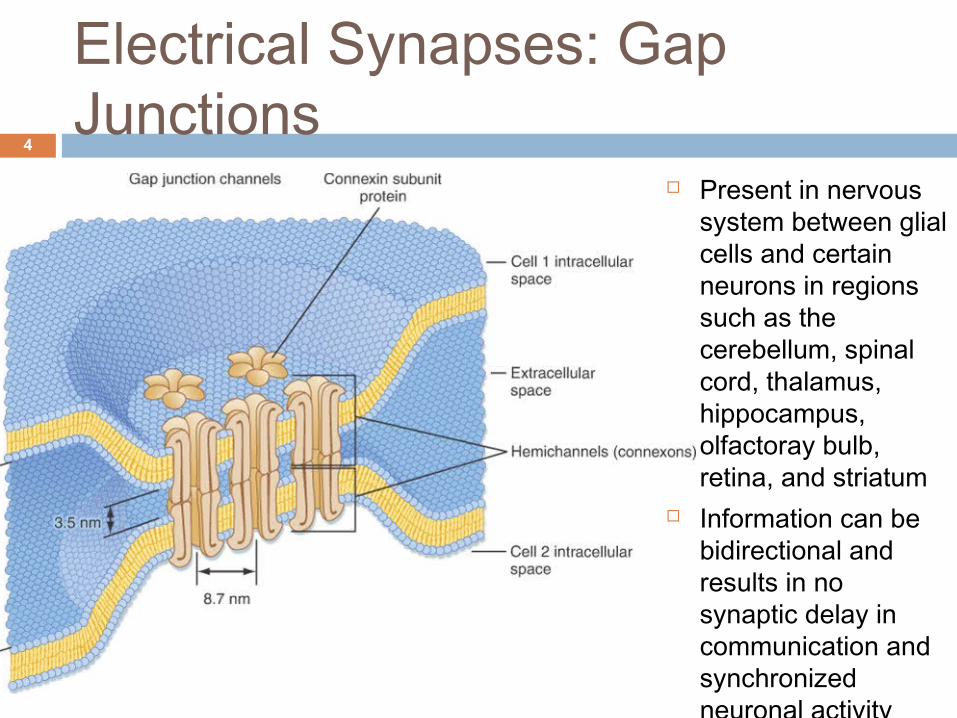
Electrical Synapses: Gap Junctions
4
Present in nervous system between glial cells and certain neurons in regions such as the cerebellum, spinal cord, thalamus, hippocampus, olfactoray bulb, retina, and striatum
Information can be bidirectional and results in no synaptic delay in communication and synchronized neuronal activity

5
Che
mic
al S
ynap
ses
B&
B F
igur
e 8
-2

Neurotransmitters: overview of major classes
6
Class/chemical Some Facts (B&L pg. 95-100)Acetylcholine (ACh)
Found in neuromuscular junctions and autonomic nervous system
Amines Can function as neurohormones/hormones.Norepinephrine (NE) Found diffusely thru CNS and also in sympathetic neurons.
Dopamine (DA) Associated with reward driven behaviour.Serotonin (5-HT) Thought to contribute to feelings of well-being and happiness.Amino Acids
Glutamate Major excitatory neurotransmitter of the CNS.GABA Major inhibitory neurotransmitter of the CNSGlycine Inhibitory in spinal cord, brainstem, retina and cerebellum.Purines
ATP Co-released from vesicles in almost all synaptic transmission.Peptides More than 100 types of active neuropeptides.Substance P Involved in pain transmission and effects smooth muscle cellsGases Not packaged into vesicles and not released via exocytosis; diffuses thru synaptic cleft.
Nitric Oxide (NO) Can also function as signal transduction molecule.

Neurotransmitter Receptors7
the response(s) elicited by a neurotransmitter is determined by receptor to which it binds
Two general families: Ionotropic receptors (ligand gated ion
channels) Directly linked to ion channels Activation results in post synaptic events that are
rapid in onset and decay Metabotropic receptors (G-protein coupled
receptors) Activation results in modification of enzyme or
membrane proteins Post synaptic events have slower onset and longer
duration

Ionotropic Receptors8
Two functional domains extracellular site binds
neurotransmitters membrane-spanning
domain forms ion channel
Examples: ionotropic glutamate
receptors ionotropic GABA
receptors nicotinic Ach receptor B&L Figure 6-12

Metabotropic Receptors9
Two functional domains: extracellular domain binds
neurotransmitters intracellular domain binds to
G-proteins G proteins couples
neurotransmitter binding to modulation of intracellular or membrane proteins
Examples: metabotropic glutamate
receptors muscarinic acetylcholine
(Ach) receptors adrenergic receptors B&L Figure 6-12

Postsynaptic Potentials (PSP)10
Binding of neurotransmitters to the post synaptic membrane can either be:
Excitatory (produces EPSP) Depolarizes membrane potential
Inhibitory (produces IPSP) Hyperpolarizes membrane potential
Both types of PSPs are graded potentials (as opposed to action potentials)

EPSPs and IPSPs11
time
Mem
bran
e po
tent
ial (
mV)
in
dend
rite
or s
oma
-70
EPSP IPSP

Post Synaptic Integration12
Most synaptic input occurs in dendrites or the cell body Amplitude of many graded PSPs from synaptic
inputs can sum, potentially triggering an action potential at the axon hillock = postsynaptic integration
Spatial Summation summation of simultaneous inputs at different
sites Temporal Summation
summation of consecutive inputs at same site

Spatial and Temporal Summation
13
B&B Figure 11-11

Signal Propagation: Electrotonic conduction
14
Amplitude of electrotonic signals decrease exponentially with membrane distance. This is due to: membrane resistance (rm)
Current leak across the membrane (membranes are poor insulators)
axoplasmic/axial resistance (ra) Impedance of current flow thru the cytoplasm
(cytoplasm is a poor conductor)

Electrotonic conduction: length constant
15
Signal decay is determined by the length constant (λ)
Distance over which signal decays to 37% of original size
Does a high ra increase ordecrease the length
constant?
How would you increase rm?
am rr /=λ
B&L Figure 5-3

Signal Propagation: Action Potentials (Review)
16
Transient, regenerative, constant amplitude signal that propagates long distances
Requires a high density of voltage-gated ion channels
Initiated when excitatory graded potentials arrive at axon hillock if Vm is above a threshold voltage (-55mV)
Different ionic currents determine each phase of AP
B&BFigure 7-1

Signal Propagation: Action Potentials (Review)
17
Threshold, amplitude, time course and duration depend on:
Gating properties and permeability's of ion channels (NaV, KV, CaV, ClV)
Intra/extracellular ionic concentration gradients
Membrane properties such as resistance, capacitance and cell geometry
Bean, J. The action potential in mammalian central neurons. Nature Reviews. 2007

Action Potentials: Ionic contributions
18
Membrane depolarization increases open probability of NaV channels Na conductance (gNa) increases and moves membrane potential towards ENa
NaV channels inactivate 1-2 ms after opening, reducing gNa
Slower to open voltage gated K+ channels activate & rapidly repolarize membrane
Brief undershoot, then Vm returns to resting value after K+ channels close
B&B Figure 7-4

Action Potentials: Ionic contributions
19
Absolute refractory period
Due to NaV channel inactivation
Prevents APs from travelling backwards along axon
Sets maximum AP frequency
Relative refractory period
Due to high gK Also involved in
determining AP frequency
B&B Figure 7-3
So now that you know about ionic contributions, what could cause a plateau phase?

Action Potential Propagation20
AP propogation also governed by membrane and cytoplasm resistance Larger diameter neurons conduct action potentials faster (decrease
ra) But there is a limit on mammalian neuron diameter
Mammals achieve increased conduction speed via myelination (increase rm)
B&B Figure 11-9
High density of ion channels
Little to no ion channels. Signal propagates passively (electrotonic conduction)
Signal regenerates and jumps from node to node

Nerve Conduction Disorders21
Demyelination Demyelinated axons
conduct action potentials slowly, unreliably, or not at all
Signal block leads to more serious consequences than decreased conduction velocity
B&B Figure 11-9

Nerve Conduction Disorders22
Demyelination A signal from one
demyelinated axon can excite an adjacent demyelinated axon (crosstalk), causing action potentials to be conducted in both directions in the adjacent axon.
B&B Figure 11-9

Nerve Conduction Disorders23
Multiple Sclerosis (MS) Most common CNS demyelination disorder Autoimmune disease that targets myelin directly or the oligodendrocyte cells Symptoms vary depending on location of demyelination,
but can include visual impairments, numbness or tingling, loss of balance, weakness, bowel and bladder problems, hearing loss
Remission and relapse common CNS inflammation, stress, or heat can exacerbate
symptoms
WebCT readings: Multiple Sclerosis

ObjectivesAfter this lecture you should be able to: Describe the process of synaptic transmission, beginning
from an action potential arriving at the pre synaptic membrane
Distinguish between ionotropic and metabotropic receptors Relate physical properties of neurons to speed of signal
conduction and signal decay List the differences between graded potentials and action
potentials Describe the phases and ionic components of a neuronal
action potential Understand how action potentials are propagated and how
demyelination affects signal propagation Relate these consequences to the CNS demyelination disorder; MS
24

25
1. What role would the length constant play in spatial summation of post synaptic potentials?
2. How is the intensity/strength of a stimulus coded for in a graded potential (electrotonic conduction) vs. an action potential?
3. How does myelination of an axon change the membrane resistance and membrane capacitance? How does this affect conduction speed?
Test your knowledge