generalized status epilepticus in a patient with acute
TRANSCRIPT
1993
□ CASE REPORT □
Generalized Status Epilepticus in a Patient with Acute-onsetType 1 Diabetes Mellitus Associated with Severe Kidney
Dysfunction: A Case Report and Literature Review
Nobumasa Ohara 1, Ryo Koda 2, Hirofumi Watanabe 2, Noriaki Iino 2, Kazumasa Ohashi 3,
Kenshi Terajima 4, Tetsutaro Ozawa 4, Yohei Ikeda 5, Hiroshi Sekiguchi 6,
Hitomi Ohashi 6 and Seigo Yamaguchi 6
Abstract
A 65-year-old Japanese man with advanced chronic kidney disease (CKD) developed acute-onset type 1
diabetes mellitus (T1D) that was associated with severe acute kidney injury and was manifested by general-
ized tonic-clonic status epilepticus. His seizures resolved without recurrence after correcting the diabetic ke-
toacidosis. Although hyperglycemia is an important cause of acute symptomatic seizure (ASS), patients with
ketotic hyperglycemia develop ASS less frequently. In this T1D case with CKD, severe hyperglycemia in
conjunction with other metabolic insults, such as uremia, hyponatremia, and hypocalcemia, probably pro-
voked his seizure despite the severe ketonemia.
Key words: type 1 diabetes mellitus, generalized tonic-clonic status epilepticus, diabetic ketoacidosis, acute
kidney injury, chronic kidney disease, electrolyte disturbance
(Intern Med 56: 1993-1999, 2017)(DOI: 10.2169/internalmedicine.56.8304)
Introduction
Type 1 diabetes mellitus (T1D) is characterized by the de-
struction of pancreatic beta cells and insulin-deficient hyper-
glycemia (1). The rate of beta-cell destruction varies from
patient to patient, and T1D is classified as fulminant, acute-
onset, or slowly progressive in Japan depending on the man-
ner of onset and progression (2). Acute-onset T1D is charac-
terized by diabetic ketosis or diabetic ketoacidosis (DKA)
within three months after the onset of hyperglycemic symp-
toms, with or without islet-related autoantibodies (3). Pa-
tients may exhibit acute kidney injury (AKI) and electrolyte
disturbances in association with severe dehydration when
they develop acute-onset T1D (4).
Acute symptomatic seizure (ASS) is a clinical seizure that
occurs in close temporal relationship with an acute central
nervous system (CNS) or systemic insult (5-7). Major in-
sults include stroke, traumatic brain injury, CNS infections,
and toxins and metabolic derangements, including hypo- or
hyperglycemia, uremia, and electrolyte disturbances. ASS
can involve either focal or generalized seizures, and patients
may present with status epilepticus (SE) (8).
Although hyperglycemia is an important metabolic insult
for ASS (5, 6, 9), patients with ketotic hyperglycemia less
frequently present with ASS than those with non-ketotic hy-
perglycemia (10-14). Nevertheless, in several case reports,
diabetic ketosis (15, 16) or DKA (17-21) was associated
1Department of Endocrinology and Metabolism, Uonuma Institute of Community Medicine, Niigata University Medical and Dental Hospital,
Japan, 2Department of Nephrology, Uonuma Institute of Community Medicine, Niigata University Medical and Dental Hospital, Japan, 3Depart-
ment of Respiratory Medicine, Uonuma Institute of Community Medicine, Niigata University Medical and Dental Hospital, Japan, 4Department
of Neurology, Uonuma Institute of Community Medicine, Niigata University Medical and Dental Hospital, Japan, 5Department of Radiology,
Uonuma Institute of Community Medicine, Niigata University Medical and Dental Hospital, Japan and 6Department of Emergency and Critical
Care Medicine, Uonuma Institute of Community Medicine, Niigata University Medical and Dental Hospital, Japan
Received for publication September 21, 2016; Accepted for publication December 12, 2016
Correspondence to Dr. Nobumasa Ohara, [email protected]
Intern Med 56: 1993-1999, 2017 DOI: 10.2169/internalmedicine.56.8304
1994
with ASS, in which the patients usually had coexistent dis-
orders that provoked seizure. However, few studies have in-
vestigated patients with T1D and DKA associated with ASS.
We herein report a rare case of a patient who exhibited
generalized SE upon the development of acute-onset T1D
and DKA associated with severe AKI and pre-existing ad-
vanced chronic kidney disease (CKD). In addition, we re-
view previously reported cases of DKA associated with
ASS.
Case Report
A 65-year-old Japanese man who presented with general-
ized tonic-clonic SE was admitted to our hospital in Febru-
ary 2016 with a diagnosis of DKA and severe kidney dys-
function. His family history revealed that his father and his
paternal uncle had type 2 diabetes mellitus. None of his
relatives had a convulsive disorder or mitochondrial disease.
The patient had smoked 10 cigarettes/day from 20 to 60
years of age, but had never consumed alcohol. The patient
had no history of diabetes mellitus, collagen disease, or a
CNS disorder, including head trauma, stroke, or encephalo-
pathy. The patient was found to have kidney dysfunction
[serum creatinine, 1.4 mg/dL; estimated glomerular filtration
rate (eGFR) (22), 44.9 mL/min/1.73 m2] and untreated hy-
pertension with negative urinalysis results for proteinuria,
microhematuria, pyuria, and glycosuria at 42 years of age.
The patient had experienced an uneventful clinical course
during conservative medical treatment for CKD, except for a
gradual increase in the serum creatinine level. Eight months
before admission, he was referred to the nephrology depart-
ment of our hospital (serum creatinine, 2.58 mg/dL; eGFR,
20.3 mL/min/1.73 m2). His height was 145 cm, weight 47
kg, and blood pressure 123/79 mmHg. There were no abnor-
mal physical findings suggesting congenital or other disor-
ders that caused his short stature. No dysacusis, muscle
pain, joint pain or swelling, alopecia, or discoid rash was
found. A urinalysis detected no protein, blood, pus, or glu-
cose in the urine. His casual plasma glucose (96 mg/dL),
glycated hemoglobin (HbA1c) (5.2%), blood lactic acid
(11.0 mg/dL, reference range: 4.5-14.4 mg/dL), and serum
levels of complement (CH50) (36 U/mL, reference range:
30-45 U/mL) and C-reactive protein (0.13 mg/dL) were nor-
mal. Anti-nuclear antibody was negative. Abdominal ultra-
sonography showed bilateral atrophic kidneys. The patient
continued medical treatment with oral valsartan (40 mg/
day), furosemide (20 mg/day), sodium bicarbonate (2 g/
day), febuxostat (10 mg/day), calcium polystyrene sulfonate
(5 g/day) and alfacalcidol (1 μg/day), and subcutaneous dar-
bepoetin alfa (30 μg/month) for CKD-related hypertension,
fluid retention, metabolic acidosis, hyperuricemia, hypocal-
cemia, and renal anemia, respectively. One month before ad-
mission, he weighed 47 kg, and his blood pressure was 128/
79 mmHg. Blood chemistry revealed the following: venous
pH, 7.359; bicarbonate, 23.6 mmol/L; red blood cells, 359×
104/μL; hemoglobin 11.8 g/dL; hematocrit, 34.2%; cre-
atinine, 2.54 mg/dL; eGFR, 20.7 mL/min/1.73 m2; urea ni-
trogen, 30.6 mg/dL; uric acid, 7.4 mg/dL; albumin, 4.1 g/
dL; sodium, 142 mEq/L; potassium, 3.3 mEq/L; chloride,
104 mEq/L; calcium, 7.2 mg/dL; phosphorus, 3.0 mg/dL;
and casual plasma glucose, 95 mg/dL. The patient developed
a cough and sore throat 7 days before admission. Three days
later, he became unusually thirsty and was polyuric. On the
day of admission, he developed a generalized tonic-clonic
seizure with disturbed consciousness and was brought by
ambulance to our hospital.
On arrival at the hospital, the patient was comatose and
presented with generalized tonic-clonic seizures that lasted
several minutes and frequently recurred. His seizures were
controlled with intravenous diazepam, but he rapidly devel-
oped respiratory depression, and he was placed on mechani-
cal ventilation. A physical examination revealed body
weight, body temperature, and blood pressure of 42 kg,
34.7℃, and 82/48 mmHg, respectively. His oral cavity was
markedly dry. No chest rales, heart murmurs, or peripheral
edema was detected. Electrocardiogram showed sinus tachy-
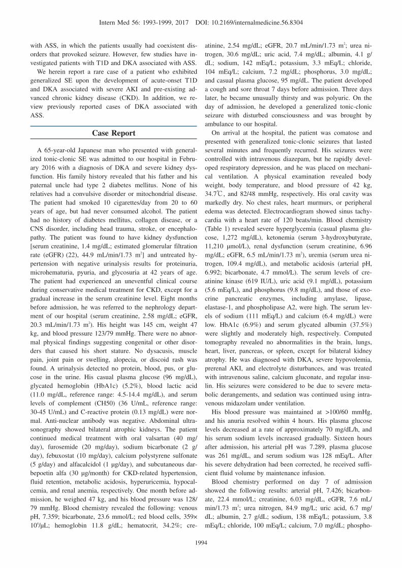
cardia with a heart rate of 120 beats/min. Blood chemistry
(Table 1) revealed severe hyperglycemia (casual plasma glu-
cose, 1,272 mg/dL), ketonemia (serum 3-hydroxybutyrate,
11,210 μmol/L), renal dysfunction (serum creatinine, 6.96
mg/dL; eGFR, 6.5 mL/min/1.73 m2), uremia (serum urea ni-
trogen, 109.4 mg/dL), and metabolic acidosis (arterial pH,
6.992; bicarbonate, 4.7 mmol/L). The serum levels of cre-
atinine kinase (619 IU/L), uric acid (9.1 mg/dL), potassium
(5.6 mEq/L), and phosphorus (9.8 mg/dL), and those of exo-
crine pancreatic enzymes, including amylase, lipase,
elastase-1, and phospholipase A2, were high. The serum lev-
els of sodium (111 mEq/L) and calcium (6.4 mg/dL) were
low. HbA1c (6.9%) and serum glycated albumin (37.5%)
were slightly and moderately high, respectively. Computed
tomography revealed no abnormalities in the brain, lungs,
heart, liver, pancreas, or spleen, except for bilateral kidney
atrophy. He was diagnosed with DKA, severe hypovolemia,
prerenal AKI, and electrolyte disturbances, and was treated
with intravenous saline, calcium gluconate, and regular insu-
lin. His seizures were considered to be due to severe meta-
bolic derangements, and sedation was continued using intra-
venous midazolam under ventilation.
His blood pressure was maintained at >100/60 mmHg,
and his anuria resolved within 4 hours. His plasma glucose
levels decreased at a rate of approximately 70 mg/dL/h, and
his serum sodium levels increased gradually. Sixteen hours
after admission, his arterial pH was 7.289, plasma glucose
was 261 mg/dL, and serum sodium was 128 mEq/L. After
his severe dehydration had been corrected, he received suffi-
cient fluid volume by maintenance infusion.
Blood chemistry performed on day 7 of admission
showed the following results: arterial pH, 7.426; bicarbon-
ate, 22.4 mmol/L; creatinine, 6.03 mg/dL, eGFR, 7.6 mL/
min/1.73 m2; urea nitrogen, 84.9 mg/L; uric acid, 6.7 mg/
dL; albumin, 2.7 g/dL; sodium, 138 mEq/L; potassium, 3.8
mEq/L; chloride, 100 mEq/L; calcium, 7.0 mg/dL; phospho-
Intern Med 56: 1993-1999, 2017 DOI: 10.2169/internalmedicine.56.8304
1995
Table 1. Laboratory Findings at the Time of Admission (February 2016).
Hematology
Red blood cells 363×104/μL (435-555)
Hemoglobin 12.0 g/dL (13.7-16.8)
Hematocrit 33.8 % (40.7-50.1)
White blood cells 13,300/μL (3,300-8,600)
Platelets 29.3×10⁴/μL (15.8-34.8)
Blood chemistry
Casual plasma glucose 1,272 mg/dL (70-139)
Glycated hemoglobin (HbA1c) 6.9 % (4.6-6.2)
Glycated albumin 37.5 % (11.6-16.4)
Acetoacetate 2,420 μmol/L (<55)
3-Hydroxybutyrate 11,210 μmol/L (<85)
Total protein 5.0 g/dL (6.6-8.1)
Albumin 3.3 g/dL (4.1-5.1)
Total cholesterol 143 mg/dL (130-220)
Triglycerides 45 mg/dL (50-130)
Aspartate aminotransferase 21 IU/L (13-30)
Alanine aminotransferase 19 IU/L (10-42)
Gamma-glutamyl transferase 16 IU/L (13-64)
Amylase 276 IU/L (44-132)
Lipase 249 U/L (17-57)
Elastase-1 1,363 ng/mL (72-432)
Phospholipase A2 1,684 ng/dL (130-400)
Creatine kinase 619 IU/L (59-248)
Urea nitrogen 109.4 mg/dL (8.0-18.4)
Creatinine 6.96 mg/dL (0.65-1.07)
eGFR* 6.5 mL/min/1.73 m2 (>90)
Uric acid 9.1 mg/dL (3.7-7.8)
Sodium 111 mEq/L (138-145)
Potassium 5.6 mEq/L (3.6-4.8)
Chloride 67 mEq/L (101-108)
Calcium 6.4 mg/dL (8.8-10.1)
Phosphorus 9.8 mg/dL (2.7-4.6)
Magnesium 1.89 mg/dL (1.7-2.3)
C-reactive protein 1.33 mg/dL (0-0.14)
Intact-parathyroid hormone 84 pg/mL (10-65)
Arterial blood gas analysis on artificial respiration
pH 6.992 (7.35-7.45)
Partial carbon dioxide pressure 19.3 mmHg (32-48)
Partial oxygen pressure 393.0 mmHg (83-108)
Bicarbonate 4.7 mmol/L (21-28)
The reference range for each parameter is shown in parentheses.
*Estimated glomerular filtration rate was calculated using the published Japanese equation as
follows (22): eGFR (mL/min/1.73 m2)=0.808×175×serum creatinine (mg/dL)-1.154×age-0.203.
rus, 3.4 mg/dL; and casual plasma glucose, 165 mg/dL. Be-
cause his metabolic status improved, sedation therapy was
terminated, and the patient was weaned from ventilatory
support on the same day. He recovered consciousness and
no longer presented any seizures or other neurological ab-
normalities.
The patient’s fasting serum C-peptide levels before and 5
minutes after intravenous glucagon loading were <0.2 ng/
mL and <0.2 ng/mL, respectively, indicating a diagnosis of
T1D. He tested negative for islet-related autoantibodies,
such as glutamic acid decarboxylase antibody (<5.0 U/mL),
islet cell antibody (<1.25 JDF units), insulinoma-associated
antigen-2 antibody (<0.4 U/mL), insulin antibody (<125.0
nU/mL), and zinc transporter-8 antibody (<10.0 U/mL). He
was also negative for anti-anterior pituitary, thyroid peroxi-
dase, thyroglobulin, thyroid-stimulating hormone receptor,
and anti-adrenal autoantibodies. Human leukocyte antigen
(HLA) typing showed A*24/26, B*40/(-), and C*08:01/(-)
class I genes and DRB1*08:03/15:01, DQB1*06:01/06:02,
and DQA1*01:02/01:03 class II genes.
The patient regained his appetite and was started on sub-
cutaneous insulin injection therapy on day 14 of admission.
A funduscopic examination detected no diabetic retinopathy.
An electroencephalographic examination performed on day

Intern Med 56: 1993-1999, 2017 DOI: 10.2169/internalmedicine.56.8304
1996
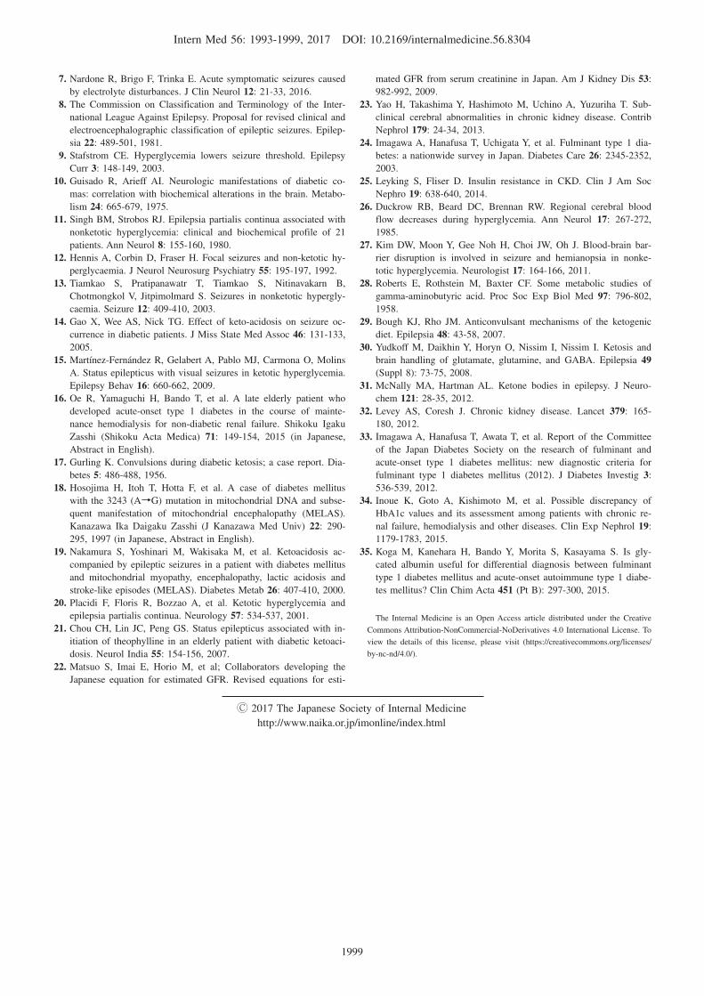
Figure. Brain magnetic resonance image (day 20 after admission in February 2016). Magnetic reso-nance imaging showed scattered hyperintense signals in the bilateral cerebral white matter on both T2-weighted (B) and fluid-attenuated inversion recovery (C) images, but no abnormal-intensity sig-nals were found on T1-weighted (A) or diffusion-weighted (D) images.
DC
BA
18 revealed no findings suggestive of an epileptogenic disor-
der. Magnetic resonance imaging (MRI) of the brain per-
formed on day 20 showed no abnormalities, except for scat-
tered hyperintense areas in the bilateral cerebral white mat-
ter on both T2-weighted and fluid-attenuated inversion re-
covery (FLAIR) images (Figure). No arterial lesions were
detected on magnetic resonance angiography.
The patient was discharged on day 46 of admission after
completing a diabetes mellitus self-management education
program. In August of the same year, he weighed 43.9 kg,
and blood chemistry showed the following results: venous
pH, 7.396; bicarbonate, 21.1 mmol/L; serum creatinine, 3.60
mg/dL; eGFR, 13.8 mL/min/1.73 m2; urea nitrogen, 58.1
mg/dL; uric acid, 5.3 mg/dL; albumin, 3.3 g/dL; sodium,
137 mEq/L; potassium, 3.9 mEq/L; chloride, 100 mEq/L;
calcium, 7.9 mg/dL; phosphorus, 4.9 mg/dL; casual plasma
glucose, 185 mg/dL; serum C-peptide, <0.2 ng/mL; HbA1c,
7.3%; and glycated albumin, 29.0%, under conventional
medical treatment for CKD and multiple daily insulin injec-
tion therapy (total of 26 units/day) for diabetes mellitus.
Brain MRI performed in August 2016 showed scattered hy-
perintense signals in the bilateral cerebral white matter on
both T2-weighted and FLAIR images, which remained un-
changed compared to those on MRI taken six months prior
(Figure) and indicated asymptomatic chronic ischemic
changes in relation to his aging and long-standing
CKD (23). His subsequent clinical course was stable without
recurrence of seizure.
Discussion
An elderly Japanese patient with advanced CKD abruptly
developed T1D and DKA that was accompanied by severe
AKI and manifested as generalized tonic-clonic SE. His sei-
zures resolved after correcting the DKA, electroencephalo-
graphic examinations showed no findings suggestive of epi-
leptogenic disorder, and brain MRI detected no remarkable
findings, except for chronic cerebral white matter ischemic
changes. The patient experienced no recurrence of seizures
during medical treatment for T1D and CKD. This is the first
reported case of proven T1D and DKA associated with ASS.
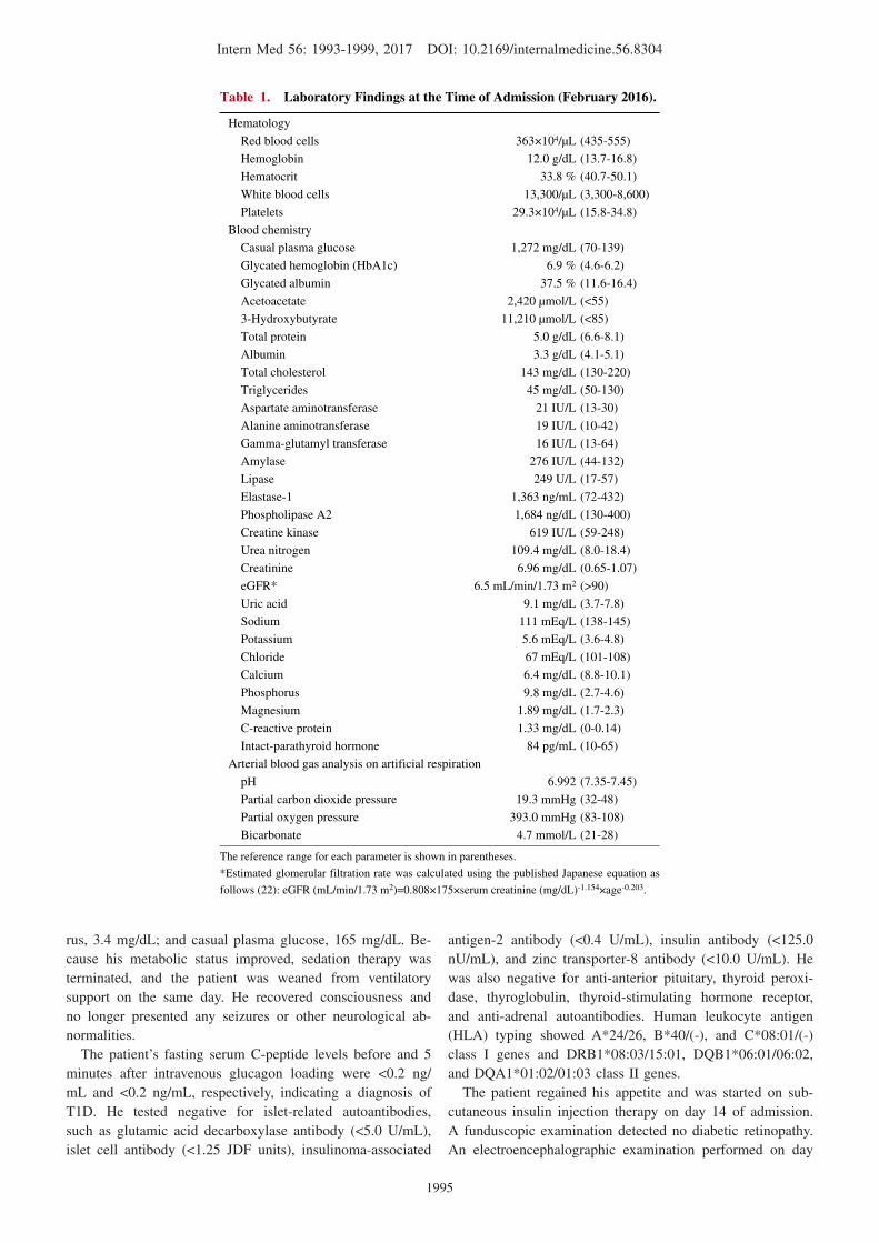
Table 2 summarizes the reported patients who exhibited
Intern Med 56: 1993-1999, 2017 DOI: 10.2169/internalmedicine.56.8304
1997
Tab
le 2
. Su
mmary
of R
epor
ted
Pat
ients
who
Exh
ibit
ed A
cute
Sym
ptom
atic
Sei
zure
in Ass
ocia
tion
wit
h D
iabe
tic
Ket
oacido
sis.
Cas
eA
ge
(yea
rs)
Sex
Type
of
seiz
ure
*
Eti
olo
gy o
f
dia
bet
es
mel
litu
s**
Know
n
dura
tion
of
dia
bet
es
mel
litu
s
(yea
rs)
HbA
1c
(%)
Pla
sma
glu
cose
(mg/d
L)
Ser
um
3-h
ydro
xybuty
r-
ate
(μm
ol/
L)
Art
eria
l
pH
Ser
um
crea
tinin
e
(mg/d
L)
Ser
um
ure
a
nit
rogen
(mg/d
L)
Mag
net
ic r
esonan
ce
imag
ing o
f th
e bra
in
Com
pli
cati
ng
dis
ord
ers
Ref
.
146
Fe-
mal
eG
ener
aliz
edU
ndet
erm
ined
0N
.D.
890
N.D
.N
.D.
N.D
.54
N.D
.
Ele
ctro
lyte
dis
turb
ance
(S
ever
e
hyper
nat
rem
ia)
(17)
249
Fe-
mal
eG
ener
aliz
edM
itoch
ondri
al15
10.7
1,2
31
1,4
10
7.1
21.3
49
N.D
.M
EL
AS
(18)
333
Fe-
mal
eF
oca
lM
itoch
ondri
al15
N.D
.540
1,9
90
6.8
8N
.D.
N.D
.
Hyper
inte
nse
are
as o
n
FL
AIR
im
ages
in t
he
bil
ater
al t
empora
l an
d
occ
ipit
al l
obes
(ab
norm
al
lact
ic a
cid a
ccum
ula
tion)
ME
LA
S(1
9)
444
Mal
eF
oca
lP
ancr
eati
c0
N.D
.506
N.D
.7.2
8N
.D.
N.D
.
Hypoin
tensi
ty o
n F
LA
IR
imag
es i
n t
he
left
subco
rtic
al p
rece
ntr
al
gyru
s (p
oss
ibly
cro
ssed
cere
bel
lar
dia
schis
is)
and
hyper
inte
nsi
ty i
n t
he
right
cere
bel
lar
hem
ispher
e
(poss
ibly
cer
ebra
l in
jury
seco
ndar
y t
o A
SS
)
Pre
vio
us
alco
hol
abuse
, ch
ronic
pan
crea
titi
s
(20)
580
Mal
eG
ener
aliz
ed
Type
220
14.0
608
N.D
.7.2
2N
.D.
N.D
.
Sli
m s
ubac
ute
subdura
l
hem
atom
as o
ver
the
bil
ater
al o
ccip
ital
lobes
Theo
phyll
ine
into
xic
atio
n, tr
aum
atic
bra
in i
nju
ry
(21)
659
Mal
eG
ener
aliz
ed
Type
10
6.9
1,2
72
11,2
10
6.9
97.0
109
Sca
tter
ed h
yper
inte
nse
area
s in
the
bil
ater
al
cere
bra
l w
hit
e m
atte
r on
T2-w
eighte
d a
nd F
LA
IR
imag
es (
chro
nic
isc
hem
ic
chan
ges
)
AK
I on a
dvan
ced
CK
D, el
ectr
oly
te
dis
turb
ance
(hyponat
rem
ia a
nd
hypoca
lcem
ia)
Pre
sent
case
*S
eizu
res
wer
e div
ided
into
foca
l or
gen
eral
ized
, ac
cord
ing t
o t
he
clin
ical
pre
senta
tion (
8).
**D
iabet
es m
elli
tus
was
cla
ssif
ied a
s ty
pe
1,
type
2,
ges
tati
onal
, or
“oth
er s
pec
ific
types
,” b
ased
on t
he
publi
shed
eti
olo
gic
al c
lass
ific
atio
n (
1).
“O
ther
spec
ific
types
” in
cluded
pan
crea
tic
or
mit
och
ondri
al d
iabet
es m
elli
tus.
The
etio
logy o
f dia
bet
es m
elli
tus
in C
ase
1 w
as u
ndet
erm
ined
bec
ause
of
a la
ck o
f in
form
atio
n r
egar
din
g e
ndogen
ous
insu
lin s
ecre
tory
cap
acit
y,
isle
t-re
late
d a
uto
anti
bodie
s, o
r a
requir
emen
t fo
r in
suli
n t
reat
men
t to
surv
ive
afte
r th
e
onse
t of
dia
bet
es m
elli
tus.
AK
I: a
cute
kid
ney
inju
ry,
AS
S:
acute
sym
pto
mat
ic s
eizu
re,
CK
D:
chro
nic
kid
ney
dis
ease
, F
LA
IR:
fluid
-att
enuat
ed i
nver
sion r
ecover
y,
HbA
1c:
gly
cate
d h
emoglo
bin
, M
EL
AS
: m
itoch
ondri
al m
yopat
hy e
nce
phal
opat
hy l
acti
c
acid
osi
s an
d s
troke-
like
epis
odes
, N
.D.:
not
des
crib
ed

Intern Med 56: 1993-1999, 2017 DOI: 10.2169/internalmedicine.56.8304
1998
ASS in association with DKA. These cases included adults
with various etiologies and durations of diabetes mellitus
who presented with focal or generalized seizures. The pa-
tients had a variety of coexisting CNS or systemic disorders
capable of precipitating seizures, including severe hyperna-
tremia (17), mitochondrial myopathy encephalopathy lactic
acidosis and stroke-like episodes ( MELAS ) syn-
drome (18, 19), and traumatic brain injury and theophylline
intoxication in relation to the management of chronic ob-
structive pulmonary disease (21). In the present case, CNS
insults, including traumatic brain injury and MELAS, were
ruled out by the clinical, laboratory, and imaging findings.
One of the notable findings at the onset of T1D in our
patient was the marked hyperglycemia and related metabolic
derangements. He had more severe hyperglycemia, electro-
lyte disturbances including hyponatremia, AKI, uremia, ke-
tonemia, and metabolic acidosis than typical cases of
T1D (24). Studies have shown that patients with CKD ex-
hibit insulin resistance (25), which may accelerate the rate
of elevation of blood glucose levels during the reduction of
endogenous insulin secretion at the development of T1D.
CKD also causes non-volatile acid accumulation as a result
of decreased renal excretion or disturbed acid-base homeo-
stasis. In our patient, the advanced CKD probably increased
the severity of hyperglycemia, ketonemia, and metabolic aci-
dosis by inducing insulin resistance and acid accumulation.
Simultaneously, his severe hyperglycemia and resulting os-
motic diuresis led to the observed marked hypovolemic hy-
ponatremia, prerenal AKI, and uremia.
The mechanisms underlying ASS when T1D occurred in
our patient remain unclear. However, hyperglycemia is
thought to decrease the seizure threshold (9), possibly
through decreased levels of gamma-aminobutyric acid
(GABA; the major CNS inhibitory neurotransmitter) (10-12),
reduced blood flow in an area of a previously silent cerebral
lesion (26), or disruption of the blood-brain barrier (27). In
contrast, several lines of evidence suggest that ketone bodies
have a protective effect against seizures through increased
GABA production or altered neuronal responsiveness to
GABA (28-31). These findings suggest that severe hypergly-
cemia in conjunction with other metabolic insults, such as
uremia and electrolyte disturbances, including acute hypovo-
lemic hyponatremia and chronic hypocalcemia, probably
provoked the seizures despite the patient exhibiting severe
ketonemia (Table 1).
CKD can be caused by a variety of renal disorders, and
major causes include diabetic nephropathy, hypertensive
nephrosclerosis, glomerulonephritis, and collagen diseases,
such as lupus nephritis (32). Our patient had no history of
diabetes mellitus before he was diagnosed with CKD, and
glomerulonephritis and collagen disease were ruled out
based on the clinical, urinary, and serological findings. A
kidney biopsy was not indicated because our patient already
had advanced CKD with atrophic kidneys when he was re-
ferred to our hospital; consequently, no histopathological di-
agnosis was obtained. However, the presence of untreated
hypertension at the diagnosis of kidney dysfunction suggests
that hypertensive nephrosclerosis was a causal factor for his
CKD.
Fulminant T1D is a subtype of T1D that results from the
extremely rapid and almost complete destruction of pancre-
atic beta cells (2). The published criteria for diagnosing this
disorder require near normal HbA1c values despite high
plasma glucose levels at disease onset, which reflects the
abrupt occurrence of hyperglycemia within a few days (33).
Our patient abruptly developed insulin-deficient hyperglyce-
mia and DKA with an HbA1c value of 6.9% and seemed to
develop fulminant T1D. However, because he had advanced
CKD and anemia treated with erythropoiesis-stimulating
agents, he probably showed a lower HbA1c value than one
truly reflecting the average blood glucose levels over the
preceding few months (34). Furthermore, his serum levels of
glycated albumin, another indicator reflecting the average
previous blood glucose levels, were substantially higher than
is usual in cases of fulminant T1D (35). Based on these
findings, we diagnosed him with acute-onset T1D.
In conclusion, we reported a patient who exhibited gener-
alized tonic-clonic SE upon development of acute-onset T1D
and DKA that was associated with severe AKI and advanced
CKD. Severe hyperglycemia in conjunction with uremia, hy-
ponatremia, and hypocalcemia probably provoked the sei-
zure, despite the patient exhibiting severe ketonemia. The
present case highlights the need for physicians to consider
that ASS can be associated with DKA, including that of
T1D onset, and may occur in the presence of other meta-
bolic derangements, such as uremia and electrolyte distur-
bances. In addition, ASS can occur as a predominant mani-
festation of T1D onset, particularly when patients have pre-
existing advanced kidney dysfunction.
The authors state that they have no Conflict of Interest (COI).
AcknowledgementThe authors thank the clinical laboratory technicians of
Uonuma Institute of Community Medicine, Niigata University
Medical and Dental Hospital, for their technical support.
References
1. American Diabetes Association. Diagnosis and classification of
diabetes mellitus. Diabetes Care 37 (Suppl 1): S81-S90, 2014.
2. Kawasaki E, Matsuura N, Eguchi K. Type 1 diabetes in Japan.
Diabetologia 49: 828-836, 2006.
3. Kawasaki E, Maruyama T, Imagawa A, et al. Diagnostic criteria
for acute-onset type 1 diabetes mellitus (2012): report of the Com-
mittee of Japan Diabetes Society on the research of fulminant and
acute-onset type 1 diabetes mellitus. J Diabetes Investig 5: 115-
118, 2014.
4. Kitabchi AE, Umpierrez GE, Miles JM, Fisher JN. Hyperglycemic
crises in adult patients with diabetes. Diabetes Care 32: 1335-
1343, 2009.
5. Beghi E, Carpio A, Forsgren L, et al. Recommendation for a defi-
nition of acute symptomatic seizure. Epilepsia 51: 671-675, 2010.
6. McLauchlan DJ, Powell R. Acute symptomatic seizures. Pract
Neurol 12: 154-165, 2012.
Intern Med 56: 1993-1999, 2017 DOI: 10.2169/internalmedicine.56.8304
1999
7. Nardone R, Brigo F, Trinka E. Acute symptomatic seizures caused
by electrolyte disturbances. J Clin Neurol 12: 21-33, 2016.
8. The Commission on Classification and Terminology of the Inter-
national League Against Epilepsy. Proposal for revised clinical and
electroencephalographic classification of epileptic seizures. Epilep-
sia 22: 489-501, 1981.
9. Stafstrom CE. Hyperglycemia lowers seizure threshold. Epilepsy
Curr 3: 148-149, 2003.
10. Guisado R, Arieff AI. Neurologic manifestations of diabetic co-
mas: correlation with biochemical alterations in the brain. Metabo-
lism 24: 665-679, 1975.
11. Singh BM, Strobos RJ. Epilepsia partialis continua associated with
nonketotic hyperglycemia: clinical and biochemical profile of 21
patients. Ann Neurol 8: 155-160, 1980.
12. Hennis A, Corbin D, Fraser H. Focal seizures and non-ketotic hy-
perglycaemia. J Neurol Neurosurg Psychiatry 55: 195-197, 1992.
13. Tiamkao S, Pratipanawatr T, Tiamkao S, Nitinavakarn B,
Chotmongkol V, Jitpimolmard S. Seizures in nonketotic hypergly-
caemia. Seizure 12: 409-410, 2003.
14. Gao X, Wee AS, Nick TG. Effect of keto-acidosis on seizure oc-
currence in diabetic patients. J Miss State Med Assoc 46: 131-133,
2005.
15. Martínez-Fernández R, Gelabert A, Pablo MJ, Carmona O, Molins
A. Status epilepticus with visual seizures in ketotic hyperglycemia.
Epilepsy Behav 16: 660-662, 2009.
16. Oe R, Yamaguchi H, Bando T, et al. A late elderly patient who
developed acute-onset type 1 diabetes in the course of mainte-
nance hemodialysis for non-diabetic renal failure. Shikoku Igaku
Zasshi (Shikoku Acta Medica) 71: 149-154, 2015 (in Japanese,
Abstract in English).
17. Gurling K. Convulsions during diabetic ketosis; a case report. Dia-
betes 5: 486-488, 1956.
18. Hosojima H, Itoh T, Hotta F, et al. A case of diabetes mellitus
with the 3243 (A→G) mutation in mitochondrial DNA and subse-
quent manifestation of mitochondrial encephalopathy (MELAS).
Kanazawa Ika Daigaku Zasshi (J Kanazawa Med Univ) 22: 290-
295, 1997 (in Japanese, Abstract in English).
19. Nakamura S, Yoshinari M, Wakisaka M, et al. Ketoacidosis ac-
companied by epileptic seizures in a patient with diabetes mellitus
and mitochondrial myopathy, encephalopathy, lactic acidosis and
stroke-like episodes (MELAS). Diabetes Metab 26: 407-410, 2000.
20. Placidi F, Floris R, Bozzao A, et al. Ketotic hyperglycemia and
epilepsia partialis continua. Neurology 57: 534-537, 2001.
21. Chou CH, Lin JC, Peng GS. Status epilepticus associated with in-
itiation of theophylline in an elderly patient with diabetic ketoaci-
dosis. Neurol India 55: 154-156, 2007.
22. Matsuo S, Imai E, Horio M, et al; Collaborators developing the
Japanese equation for estimated GFR. Revised equations for esti-
mated GFR from serum creatinine in Japan. Am J Kidney Dis 53:
982-992, 2009.
23. Yao H, Takashima Y, Hashimoto M, Uchino A, Yuzuriha T. Sub-
clinical cerebral abnormalities in chronic kidney disease. Contrib
Nephrol 179: 24-34, 2013.
24. Imagawa A, Hanafusa T, Uchigata Y, et al. Fulminant type 1 dia-
betes: a nationwide survey in Japan. Diabetes Care 26: 2345-2352,
2003.
25. Leyking S, Fliser D. Insulin resistance in CKD. Clin J Am Soc
Nephro 19: 638-640, 2014.
26. Duckrow RB, Beard DC, Brennan RW. Regional cerebral blood
flow decreases during hyperglycemia. Ann Neurol 17: 267-272,
1985.
27. Kim DW, Moon Y, Gee Noh H, Choi JW, Oh J. Blood-brain bar-
rier disruption is involved in seizure and hemianopsia in nonke-
totic hyperglycemia. Neurologist 17: 164-166, 2011.
28. Roberts E, Rothstein M, Baxter CF. Some metabolic studies of
gamma-aminobutyric acid. Proc Soc Exp Biol Med 97: 796-802,
1958.
29. Bough KJ, Rho JM. Anticonvulsant mechanisms of the ketogenic
diet. Epilepsia 48: 43-58, 2007.
30. Yudkoff M, Daikhin Y, Horyn O, Nissim I, Nissim I. Ketosis and
brain handling of glutamate, glutamine, and GABA. Epilepsia 49(Suppl 8): 73-75, 2008.
31. McNally MA, Hartman AL. Ketone bodies in epilepsy. J Neuro-
chem 121: 28-35, 2012.
32. Levey AS, Coresh J. Chronic kidney disease. Lancet 379: 165-
180, 2012.
33. Imagawa A, Hanafusa T, Awata T, et al. Report of the Committee
of the Japan Diabetes Society on the research of fulminant and
acute-onset type 1 diabetes mellitus: new diagnostic criteria for
fulminant type 1 diabetes mellitus (2012). J Diabetes Investig 3:
536-539, 2012.
34. Inoue K, Goto A, Kishimoto M, et al. Possible discrepancy of
HbA1c values and its assessment among patients with chronic re-
nal failure, hemodialysis and other diseases. Clin Exp Nephrol 19:
1179-1783, 2015.
35. Koga M, Kanehara H, Bando Y, Morita S, Kasayama S. Is gly-
cated albumin useful for differential diagnosis between fulminant
type 1 diabetes mellitus and acute-onset autoimmune type 1 diabe-
tes mellitus? Clin Chim Acta 451 (Pt B): 297-300, 2015.
The Internal Medicine is an Open Access article distributed under the Creative
Commons Attribution-NonCommercial-NoDerivatives 4.0 International License. To
view the details of this license, please visit (https://creativecommons.org/licenses/
by-nc-nd/4.0/).
Ⓒ 2017 The Japanese Society of Internal Medicine
http://www.naika.or.jp/imonline/index.html