general surgery grand rounds - university of colorado ... · 27 subdural hematoma zhemorrhage under...
TRANSCRIPT
1
General Surgery Grand General Surgery Grand RoundsRounds
General Management and Resuscitation in Acute Brain Injury
July 2007
Robert Neumann MD, PhDUniversity of Colorado
Department of Neurosurgery Assistant Professor,
Director UCHSC NICU

2
Severe Brain Injury: Learning Severe Brain Injury: Learning ObjectivesObjectives
EpidemiologyClassifications based on severity and typeClinical presentationsGeneral outcome predictorsOn Site Hospital TXGeneral conclusionsFuture directionsQuestions
3
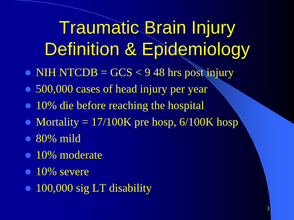
Traumatic Brain Injury Traumatic Brain Injury Definition & EpidemiologyDefinition & Epidemiology
NIH NTCDB = GCS < 9 48 hrs post injury500,000 cases of head injury per year10% die before reaching the hospitalMortality = 17/100K pre hosp, 6/100K hosp80% mild10% moderate10% severe100,000 sig LT disability

4
TBI: Epidemiology (cont)TBI: Epidemiology (cont)
Mechanism: MVA > Falls > Firearm > other assaultHigh risk: young, male, low income, unmarried, ethnic minority, city dweller, sub abuse, prev TBIMale to female incidence ratio 2.8:2, mortality ratio 3.4:1 Age: occurring most commonly age < 25 with a bi-modal distribution rising again at age > 65Cause of death in 45-50% of all trauma40-80% association with sig systemic trauma (thoracoabdominal, ortho,)Alcohol: implicated in 50-70%

5
Classifying Brain InjuryClassifying Brain Injury
Based on GCSMild: GCS 13-15Moderate: GCS 9-12Severe: GCS 8 or less

6
Glasgow Coma ScaleGlasgow Coma Scale
Eye Voice Motor 6 ----------- ------------- obeys
5 ----------- oriented localizes to pain4 spontaneous confused withdraw to pain3 To speech inappropriate flexor2 to pain incomprehensible extensor1 none none none

7
TBI Exam FindingsTBI Exam Findings
Depressed level of consciousness not clearly due to EtOH, drugs, metabolic, cardio-pulm abnormalities etc.Focal neurologic findings ( pupillary changes, false localizing, not all have mass lesion eg., DAI)Penetrating skull injury or depressed fractureRaccoon eyes, Battle’s sign, CSF rhinorreha an/or otorreha

8
TBI Presentation (conTBI Presentation (con’’t)t)SZDecorticate, deceribrate posturingCushing’s Triad:
- Inc BP W/ widened PP, and proportionallygreater inc in SBP over that of DBP
- Bradycardia (SR)- Irregular Breathing- Usually heralds brain damage/herniation sec/2 inc ICP/poor perfusion.
9
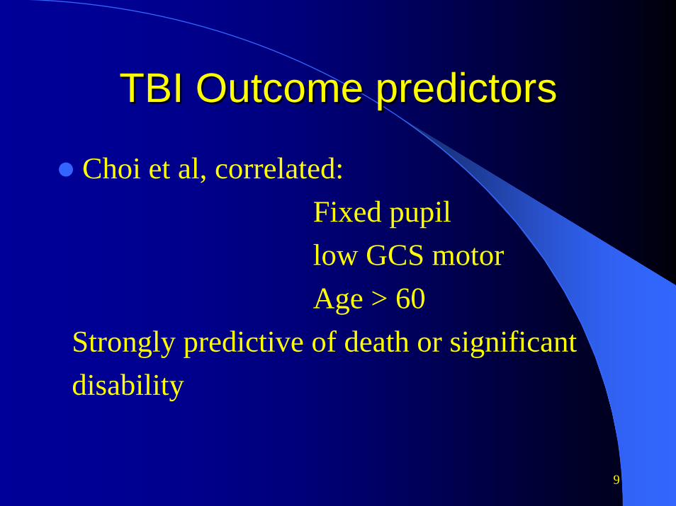
TBI Outcome predictorsTBI Outcome predictors
Choi et al, correlated:Fixed pupillow GCS motorAge > 60
Strongly predictive of death or significantdisability

10
Types of Traumatic Brain InjuryTypes of Traumatic Brain Injury
Forces = compressive, tensile (stretch), shearing (slide)Skull fracturesConcussionDiffuse axonal injuryContusionSubarachnoid hemorrhage (SAH)Subdural hematoma (SDH)Intraparenchymal hemorrhage (ICH)Epidural hematoma (EDH)Intraventricular hemorrhage (IVH)
11
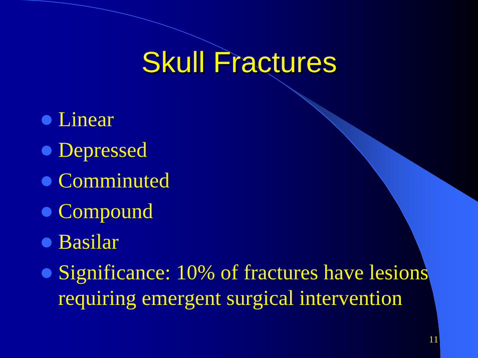
Skull FracturesSkull Fractures
LinearDepressedComminutedCompoundBasilarSignificance: 10% of fractures have lesions requiring emergent surgical intervention

12Linear Depressed Comminuted Skull Fx
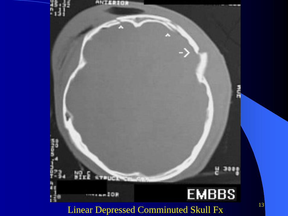
13Linear Depressed Comminuted Skull Fx

14
ConcussionConcussion
Reversible transitory neurologic deficit. Associated with rotational shear stress.Considered mild form of DAIThree grades; mild, moderate and severe, proportional to retrograde and anterograde amnesia
15
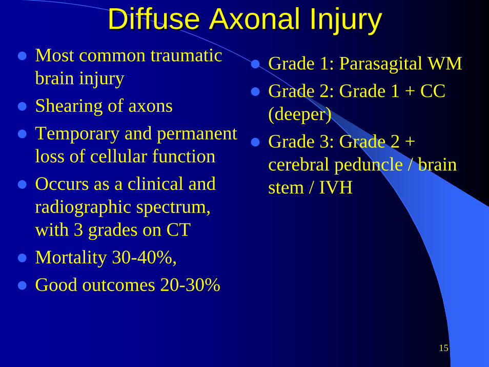
Diffuse Axonal InjuryDiffuse Axonal InjuryMost common traumatic brain injuryShearing of axonsTemporary and permanent loss of cellular functionOccurs as a clinical and radiographic spectrum, with 3 grades on CTMortality 30-40%, Good outcomes 20-30%
Grade 1: Parasagital WM Grade 2: Grade 1 + CC (deeper)Grade 3: Grade 2 + cerebral peduncle / brain stem / IVH

16
Diffuse Axonal Injury (conDiffuse Axonal Injury (con’’t)t)
Imaging:CT: petechial hemorrhage (hyperdense) along gray white junctions, symetric or asymetric edema, intraventricular / cisternal hemorrhage
MRI: hemorrhage as above, high signal on T2

17
Diffuse Axonal Injury (conDiffuse Axonal Injury (con’’t)t)
Overall PrognosisRelated to:
CT imagingControl of intracranial pressurePost resuscitation GCS

18
Diffuse Axonal Injury (conDiffuse Axonal Injury (con’’t)t)
CT Brain Grade 2 DAI T2 MRI Brain Grade 2 DAI

19
Cerebral ContusionCerebral ContusionSecond most common brain injuryCoup = small-moderate direct impactCountercoup = high energy with translational dissipation of energyEssentially a bruise on the brain with hemorrhage from torn pial vessels, evolution of localized edema Location: temporal > frontal > parasagittal, > occipital (convexities)20% will have delayed hemorrhage

20
Cerebral Contusion (conCerebral Contusion (con’’t)t)
CT Brain showing left frontal contusi

21
Cerebral Contusion (conCerebral Contusion (con’’t)t)
Day 1 Day 2 Day 14
22
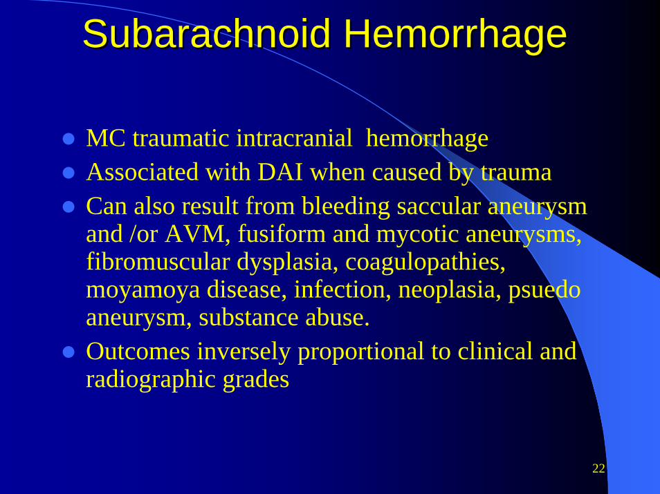
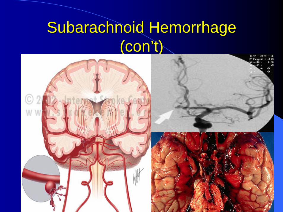
Subarachnoid HemorrhageSubarachnoid Hemorrhage
MC traumatic intracranial hemorrhageAssociated with DAI when caused by traumaCan also result from bleeding saccular aneurysm and /or AVM, fusiform and mycotic aneurysms, fibromuscular dysplasia, coagulopathies, moyamoya disease, infection, neoplasia, psuedo aneurysm, substance abuse. Outcomes inversely proportional to clinical and radiographic grades
23
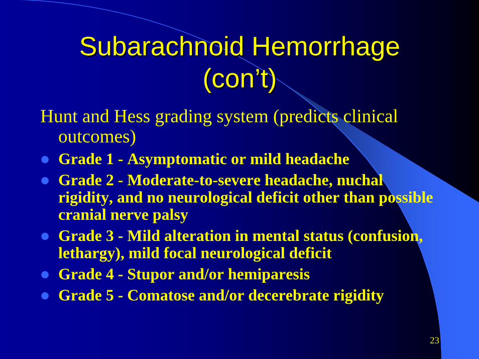
Subarachnoid Hemorrhage Subarachnoid Hemorrhage (con(con’’t)t)
Hunt and Hess grading system (predicts clinical outcomes)Grade 1 - Asymptomatic or mild headache Grade 2 - Moderate-to-severe headache, nuchal rigidity, and no neurological deficit other than possible cranial nerve palsy Grade 3 - Mild alteration in mental status (confusion, lethargy), mild focal neurological deficit Grade 4 - Stupor and/or hemiparesis Grade 5 - Comatose and/or decerebrate rigidity

24
Subarachnoid Hemorrhage Subarachnoid Hemorrhage (con(con’’t)t)
Fisher scale: Radiographic Grade (predicts degree of vasospazm)Grade 1 - No blood detected ( LP + Xantho) Grade 2 - Diffuse deposition of subarachnoid blood, no clots, and no layers of blood greater than 1-3 mm Grade 3 - Localized clots and/or vertical layers of blood 3 mm or greater in thickness Grade 4 – Diffuse, or no subarachnoid blood, with intracerebral or intraventricular clots are present
25
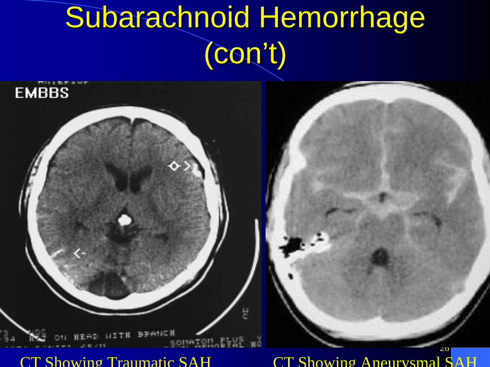
Subarachnoid Hemorrhage Subarachnoid Hemorrhage (con(con’’t)t)
26
Subarachnoid Hemorrhage Subarachnoid Hemorrhage (con(con’’t)t)
CT Showing Traumatic SAH CT Showing Aneurysmal SAH

27
Subdural HematomaSubdural HematomaHemorrhage under the duraCaused by torn bridging veins and /or bleeding contusions (pial vessels)Occur in 10-35% of severe head injuries10-50% are associated with skull fracturesCortical atrophy is RF for occurrence, but Pt’s with normal brain vol at higher risk for LT disabilityAlso associated with coagulopathiesOften associated with other brain injuriesAcute, sub acute and chronic (density on CT) depending on the age of SDH
28
Subdural Hematoma (conSubdural Hematoma (con’’t)t)
Prognosis:Mortality is 40-70%Complete recovery in 8%Severe disability in 74-84%
Imaging:CT scan: cresentric (concaved) shaped hematoma crosses sutures but not dural insertions, mass effect with shift, edema

29
Subdural Hematoma (conSubdural Hematoma (con’’t)t)
CT Showing Acute SHD CT Showing Acute on Chronic SDH
30
Intraparenchymal HemorrhageIntraparenchymal HemorrhageHemorrhage in the substance of the brain also called intracerebral hemorrhage (ICH)Associated with HTN, anerysmal SAH, amyloid, cerebral contusions and DAIOccur most commonly in frontal and gray white junctions of brain when caused by trauma, vs vascular ICH’s due to HTN, which occur in distinct vascular distributionsCommonly expand (re-bleed) within first 24 hoursOften cause localized edema and elevated intracranial pressureOutcomes inversely proportional to size of ICH, and Pt age
31
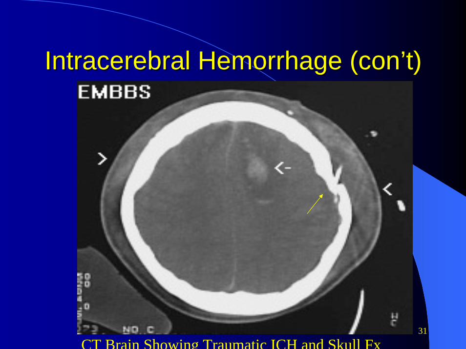
Intracerebral Hemorrhage (conIntracerebral Hemorrhage (con’’t)t)
CT Brain Showing Traumatic ICH and Skull Fx
32
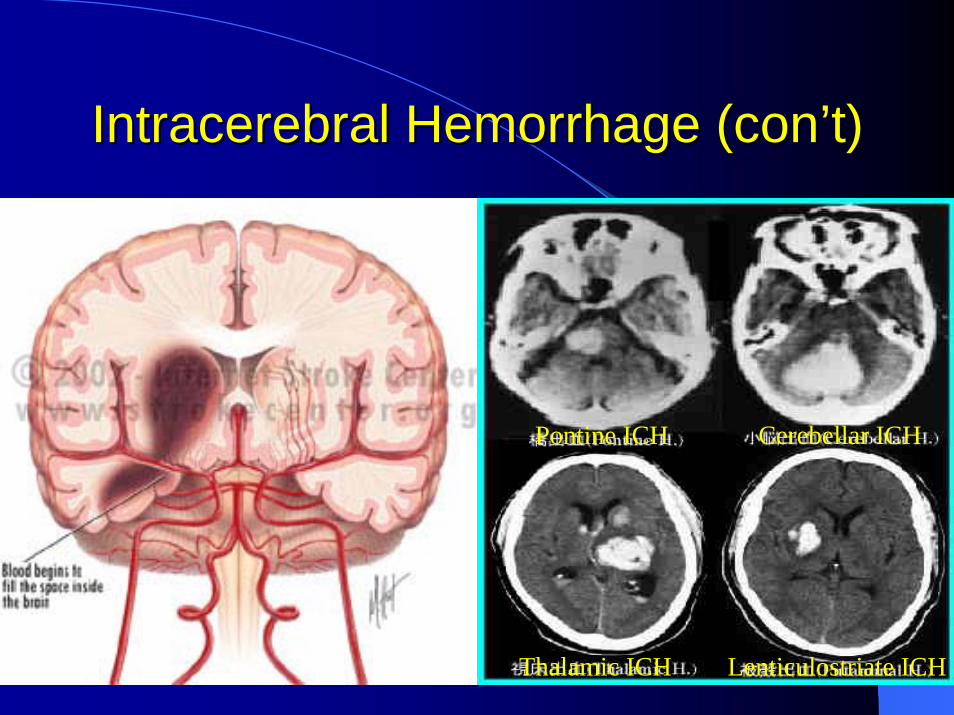
Intracerebral Hemorrhage (conIntracerebral Hemorrhage (con’’t)t)
Pontine ICH Cerebellar ICH
Thalamic ICH Lenticulostriate ICH

33
Epidural HematomaEpidural Hematoma
Hemorrhage between inner table of skull and duraOften caused by severed meningeal artery or torn large venous sinusObeys suture lines (coronal, lambdoid)Occur in 3-5% of head injuriesPeak incidence 10-30 years oldRare in those <2yrs or > 60yrs85-90% are associated with skull fractures

34
Epidural Hematoma (conEpidural Hematoma (con’’t)t)Often present with lucid intervalCommonly occur with other brain lesionsMortality and Morbidity 5% - 20% Higher rates are associated with the following: – Advanced age – Intradural lesions – Temporal location – Increased hematoma volume – Rapid clinical progression – Pupillary abnormalities – Increased intracranial pressure (ICP) – Lower Glasgow coma scale (GCS)
In the US: EDH occurs in 1-2% of all head trauma cases and in about 10% of patients who present with traumatic coma. CT appearance: convex hyperdensity, swirl sign, obeying suture lines
35
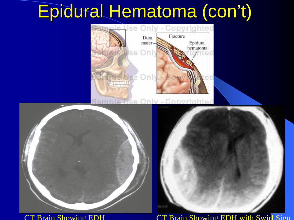
Epidural Hematoma (conEpidural Hematoma (con’’t)t)
CT Brain Showing EDH CT Brain Showing EDH with Swirl Sign
36
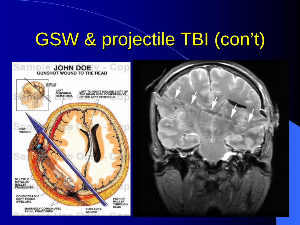
GSW & projectile TBIGSW & projectile TBI
Missile vs nonmissleEnergy dissipation = ½ progectile mass x velocity2 (velocity and blast proximity) Associated with all forms of traumatic hemorrhageOutcomes related to velocity, location of entry and exit, post resuscitation GCS
37
GSW & projectile TBI (conGSW & projectile TBI (con’’t)t)

38
““Type AType A”” Neuro Pt Neuro Pt = Over= Over--Achiever Achiever
39
On Site Hospital TX/RXOn Site Hospital TX/RXAirway and breathing assessment in the awake and directable neurological patientGCS 10-15, maintains Sao2 96-100%, NOT rapidly deteriorating neurologically, non-agitated/combative, and cardio-pulmonary status is stable:1) Nasal Cannula, Non Re-breather2) Bag and Mask Valve, as a bridge in some cases3) Serial Neuro exams and Chest auscultation4) CXR, and serial ABG’s5) “Big 3” BAT R/O
40
On Site Hospital TX/RX (conOn Site Hospital TX/RX (con’’t)t)Tracheal Intubation Criteria in the Neuro Pt:1) Depressed LOC GCS < 8-92) Rapidly deteriorating neurologic exam (minutes-Hrs)3) Immediate need for HV w/ target PCO2 30-364) Severe maxillofacial trauma or airway edema, w/ impending
loss of airway protection &/or patency5) Need for pharmacological sedation, SZ control &/or paralysis6) Cardiovascular instability ( MI, CHF, Sepsis, shock ) 7) Primary pulmonary instability (Edema, Asp, Apnea, Stridor)
a) In all cases, intubation should be based on emergent clinical criteria, rather than lab values and/or radiographic studies. When in doubt, elect to intubate the acute Neuro Pt., early and/or prior to any transport.

41
On Site Hospital TX/RX (conOn Site Hospital TX/RX (con’’t)t)Difficult Airways: clinical presentations/syndromes in the Neuro Pt:Potential or proven cervical spine injury, also RA, Down’sBasilar skull FxMaxillofacial trauma &/or burnsReceding chin (micrognathia)Prominent incisors (buck teeth)Short plethoric &/or muscular neckMorbid obesity, acromegaly, sclerodermaPrev Trac, head/neck surgx &/or radiationPregnant, Illeus/SBO/full stomach

42
On Site Hospital TX/RX (conOn Site Hospital TX/RX (con’’t)t)Tx Hypertension in the Neuro Pt with SAH, ICH, EDH, Trauma, & some strokes, ie, Pt’s having intracranial mass effect/inc ICP:
1) Control ICP2) Intubate/Sedate3) Control any SZ4) Nipride gtt: 1-10 mic/kg/min (SE’s: cardiopulm shunt, CN-thio
tox ?? Inc ICP)5) Esmolol gtt: 25-200mcg/kg/min (SE’s: bradycardia, heart
block, bronchospasm, unopposed alpha)6) Labatolol: 2.5-20 mg q 1 hr PRN (SE’s: bradycardia, heart
block, unopposed alpha)7) Hydralazine: 2.5-20 mg IV q 1hr PRN (SE’s: reflex tachy, >
AAA8) Nicardapine : (SE’s: Inc Myocardial O2 demand)
43
On Site Hospital TX/RX (conOn Site Hospital TX/RX (con’’t)t)Hypotension in the Neuro Pt:Causes: Cardiogenic = Hypovolemic > Acidosis >Neurogenic > Vasogenic (sepsis/anaphylaxis) Dx:IBP monitoring, 12 lead EKG, Trop-I, Blood gas, CXR, CVP or PAP monitoring, Blood Cultures, +/- BAT R/OTx:1) Fluid Challenge in face of inc ICP W/ 500 cc 5% Albumen, or blood (repleats intravasc vol and inc oncotic pressure)2) Dopamine gtt : 5-20mic/kg min titrate SIBP 100-1203) Neosyephrine gtt: 0.5-5mic/kg/min4) Vasopressin gtt: 0.01-0.1 u/min

44
On Site Hospital TX/RX (conOn Site Hospital TX/RX (con’’t)t)Control of Presumed Elevated ICP:(Trauma, SAH, ICH, SDH, EDH, Large stroke, Tumor)
1) Head of bed ~ 30 deg (inc JV outflow)2) Intubate (PCO2 30-36, PEEP ~ 5), ET fastened w/o IJ
occlusion, keep head forward & neck straight after intubation
3) Light- heavy sedation inversely proportional to GCS4) Avoid IJ Location for central venous catheter
(Subclavian) 5) Mannitol 1.5 G/kg IV, check Na++/serum osmo
afterwards if time permits, (+/-) Hypertonic saline 6) Lasix 10-20 mg IV ½ hr after Mannitol, check K+7) Minimal- Euvolemic total fluids (MIVF + Rx + TF) ~
40-100cc/hr8) Ventricular drainage &/or ICP monitor9) Early Crainectomy
45
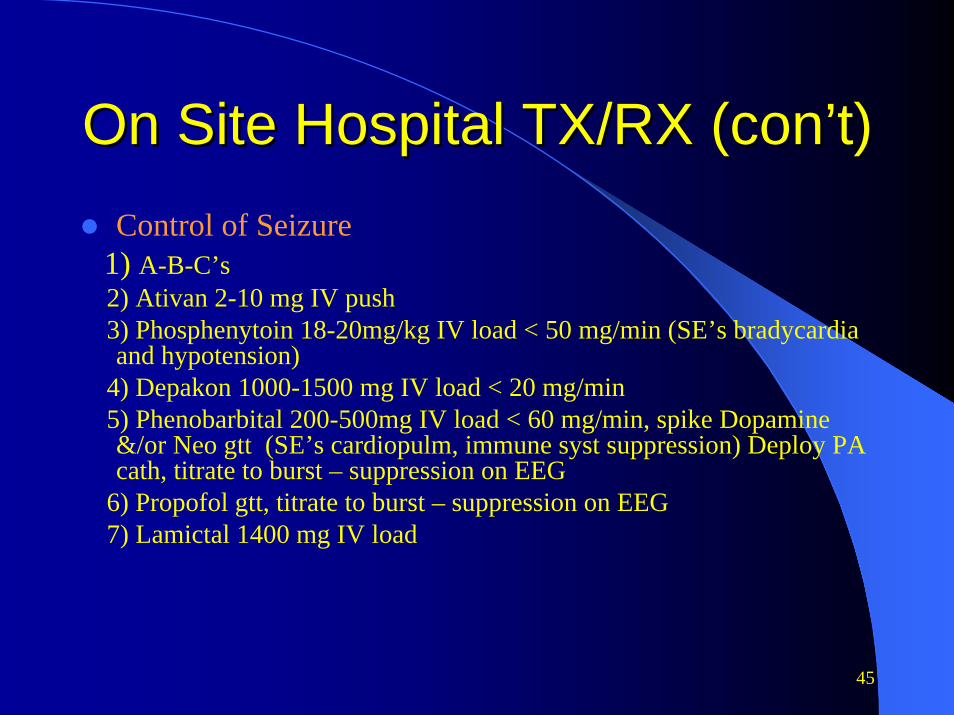
On Site Hospital TX/RX (conOn Site Hospital TX/RX (con’’t)t)Control of Seizure
1) A-B-C’s2) Ativan 2-10 mg IV push3) Phosphenytoin 18-20mg/kg IV load < 50 mg/min (SE’s bradycardia and hypotension)
4) Depakon 1000-1500 mg IV load < 20 mg/min5) Phenobarbital 200-500mg IV load < 60 mg/min, spike Dopamine &/or Neo gtt (SE’s cardiopulm, immune syst suppression) Deploy PA cath, titrate to burst – suppression on EEG
6) Propofol gtt, titrate to burst – suppression on EEG7) Lamictal 1400 mg IV load
46
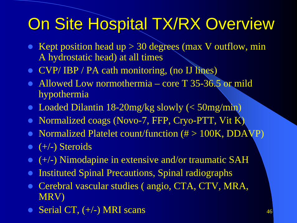
On Site Hospital TX/RX OverviewOn Site Hospital TX/RX OverviewKept position head up > 30 degrees (max V outflow, min A hydrostatic head) at all timesCVP/ IBP / PA cath monitoring, (no IJ lines)Allowed Low normothermia – core T 35-36.5 or mild hypothermiaLoaded Dilantin 18-20mg/kg slowly (< 50mg/min)Normalized coags (Novo-7, FFP, Cryo-PTT, Vit K)Normalized Platelet count/function (# > 100K, DDAVP)(+/-) Steroids(+/-) Nimodapine in extensive and/or traumatic SAHInstituted Spinal Precautions, Spinal radiographsCerebral vascular studies ( angio, CTA, CTV, MRA, MRV)Serial CT, (+/-) MRI scans

47
On Site Hospital TX/RX OverviewOn Site Hospital TX/RX OverviewIntubated, sedated, (Narcs, Benzos, Propofol, Presidex) +/-paralytic gtt (1-0/4 train of 4) or Barbs /24 hr EEGMaintained MAP 80 -100, Pressors (dopa, neo, vaso) or Anti-HTN Rx, with CVP or PA catheter monitoring in many Pt’s Given Mannitol 1.5g/kg load, can be followed by Lasix (synergy), albumen & blood, HCT 30-33, Euvolemia-Hypovolemia in acute phase, if toleratedTargeted PcO2 30-36, minimum PEEP / PS if possible, PaO2> 80-90Placed ICP monitor, goal ICP < 20, with CSF drainage via EVD if necessary and CPP > 60-70 in pt’s with ICP sustained > 20PbTO2, and Brain Temp monitoring (Licox) target PbTO2 > 20(+/-) Lumbar drainage, (+/-) ENT consult for some CSF leaksStarted Abx prophylaxis esp: pneumocephaly, CSF leaks, EVD(+/-) Emergent surgical evacuation and possible crainectomy

48
Timing of CraniotomyTiming of Craniotomy
“Four hour rule” Seelig et al 1981 N Engl J Med: 82 patients with acute subdural, operation in < 4 hours = 30% mortality, if > 4 hours 90% mortality
“Six hour rule” Citow 2001
Operation in < 6 hours = 30% mortality, if > 6 hours, 95% mortality
49
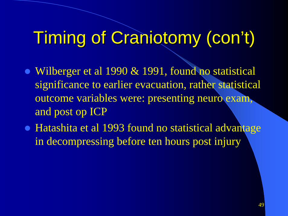
Timing of Craniotomy (conTiming of Craniotomy (con’’t)t)
Wilberger et al 1990 & 1991, found no statistical significance to earlier evacuation, rather statistical outcome variables were: presenting neuro exam, and post op ICPHatashita et al 1993 found no statistical advantage in decompressing before ten hours post injury
50
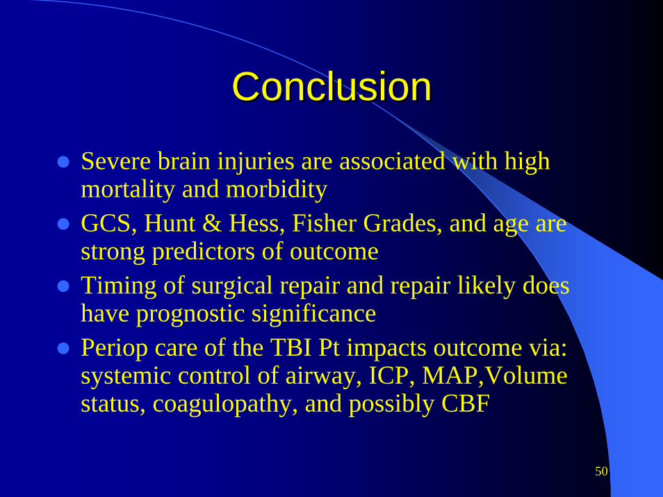
ConclusionConclusion
Severe brain injuries are associated with high mortality and morbidityGCS, Hunt & Hess, Fisher Grades, and age are strong predictors of outcomeTiming of surgical repair and repair likely does have prognostic significancePeriop care of the TBI Pt impacts outcome via: systemic control of airway, ICP, MAP,Volume status, coagulopathy, and possibly CBF

51
Future DirectionsFuture Directions
Invasive brain tissue oxygenation monitoringInvasive CBF monitoringInvasive brain T monitoringInvasive brain Dialysis Catheter monitoringMinimization of early apoptosis via control of excitatory AA (glutamate, aspartate), control of NMDA receptor agonists,….? control of presynaptic endogenous opioid peptides
52
QuestionsQuestions
53
Severe Spinal Injury Severe Spinal Injury EpidemiologyEpidemiology
54
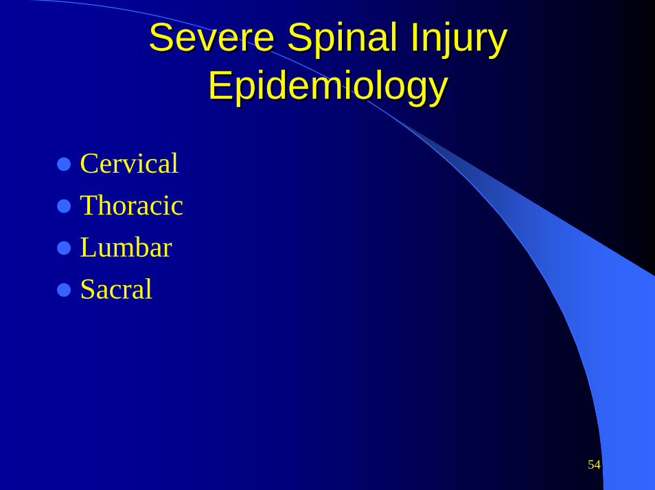
Severe Spinal Injury Severe Spinal Injury EpidemiologyEpidemiology
CervicalThoracicLumbarSacral

55
SCI R/OSCI R/O
All victims with significant mechanism of traumaTrauma Pt’s with LOCMinor trauma Pt’s with neck or back pain, or sensory motor, or vasomotor findings on PESCI may mask other injuries

56
InstabilityInstabilitySegmental instability is a loss of spinal motion segment stiffness, such that force application to that motion segment produces greater displacement(s) than would be seen in the normal structure, resulting in a painful condition, the potential for progressive deformity and neurologic structures at risk.
John W. Frymoyer

57
Instability and TreatmentInstability and Treatment
Acute SCI with potential for healing to stabilityAcute SCI with low potential for healing to stabilityChronic (glacial) Etiology– Osseous– Ligamentous
Risk/benefit

58

59
Spinal BiomechanicsSpinal Biomechanics
Cartesian SystemTwo motion types– Translations– Rotations
CouplingKinematics vsbiomechanics

60
Upper Cervical AnatomyUpper Cervical Anatomy
61
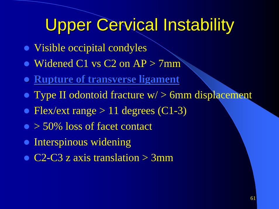
Upper Cervical InstabilityUpper Cervical InstabilityVisible occipital condylesWidened C1 vs C2 on AP > 7mmRupture of transverse ligamentRupture of transverse ligamentType II odontoid fracture w/ > 6mm displacementFlex/ext range > 11 degrees (C1-3)> 50% loss of facet contactInterspinous wideningC2-C3 z axis translation > 3mm
62
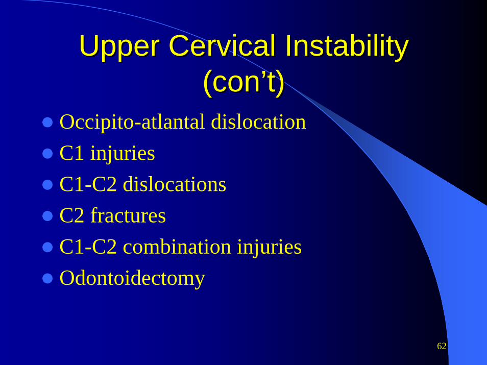
Upper Cervical Instability Upper Cervical Instability ((concon’’tt))
Occipito-atlantal dislocationC1 injuriesC1-C2 dislocationsC2 fracturesC1-C2 combination injuriesOdontoidectomy

63
OccipitoOccipito--atlantalatlantal DislocationDislocation
High mortality at sceneVery unstable (immediate halo fixation)Floating condylesPower’s ratioCT vs MRI

64

65
OccipitoOccipito--atlantalatlantal Dislocation Dislocation TreatmentTreatment
NO TRACTION!Backboard immobilizationImmediate haloMove pt to OR with haloUrgent internal fixation

66
OccipitoOccipito--atlantalatlantal DislocationDislocation
67
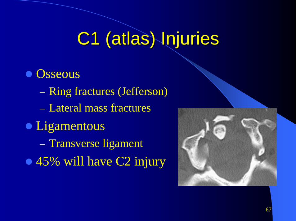
C1 (atlas) InjuriesC1 (atlas) Injuries
Osseous– Ring fractures (Jefferson)– Lateral mass fractures
Ligamentous– Transverse ligament
45% will have C2 injury
68
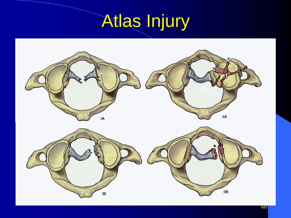
Atlas InjuryAtlas Injury

69
Atlas InjuryAtlas InjuryDiagnosisDiagnosis
C spine plain film with odontoid view, showing overlap of C1 on C2 on AP > 7mmCT with 1 mm resolution and 3d reconstruction, showing predental space > 3mmMRI for transverse ligament

70

71

72
Atlas InjuryAtlas InjuryTreatmentTreatment
Dependent on transverse ligament and potential for healing to stabilityRing fractures - external immobilizationTransverse ligament incompetence– Osseous basis - possible nonsurgical
management– Pure ligamentous injury - surgery

73
C1C1--C2 DislocationsC2 Dislocations
Transverse ligament injuryRotatory subluxation

74
Transverse Ligament Transverse Ligament DisruptionDisruption
75
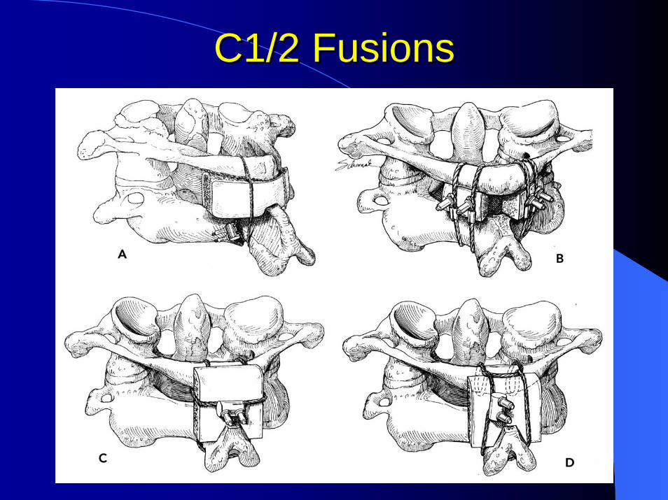
C1/2 FusionsC1/2 Fusions
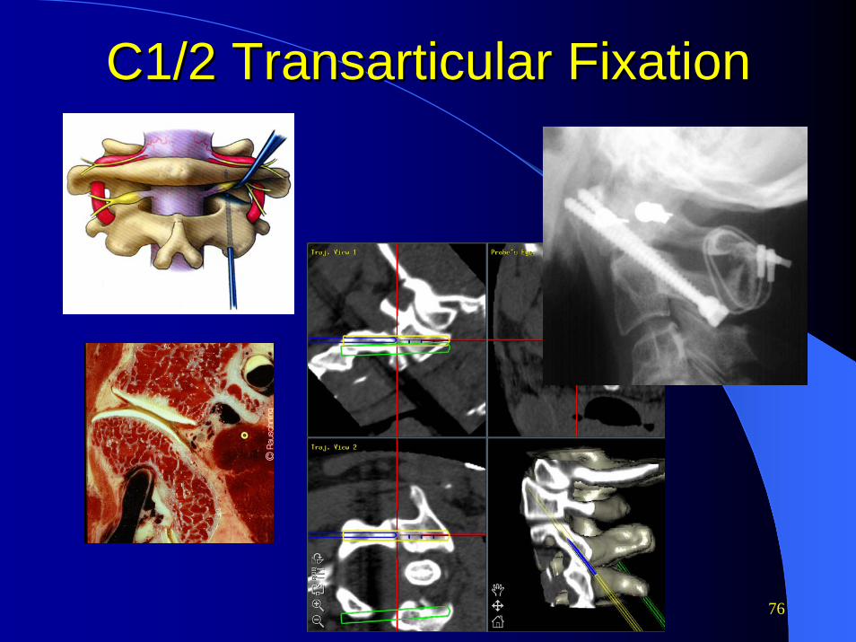
76
C1/2 Transarticular FixationC1/2 Transarticular Fixation

77
C1/2 C1/2 RotatoryRotatory SubluxationSubluxation

78
C1/2 Rotatory Subluxation
Transverse ligament disrupted
Normal transverse ligament
External reduction
ORIFExternal immobilization
Reducible Irreducible
79
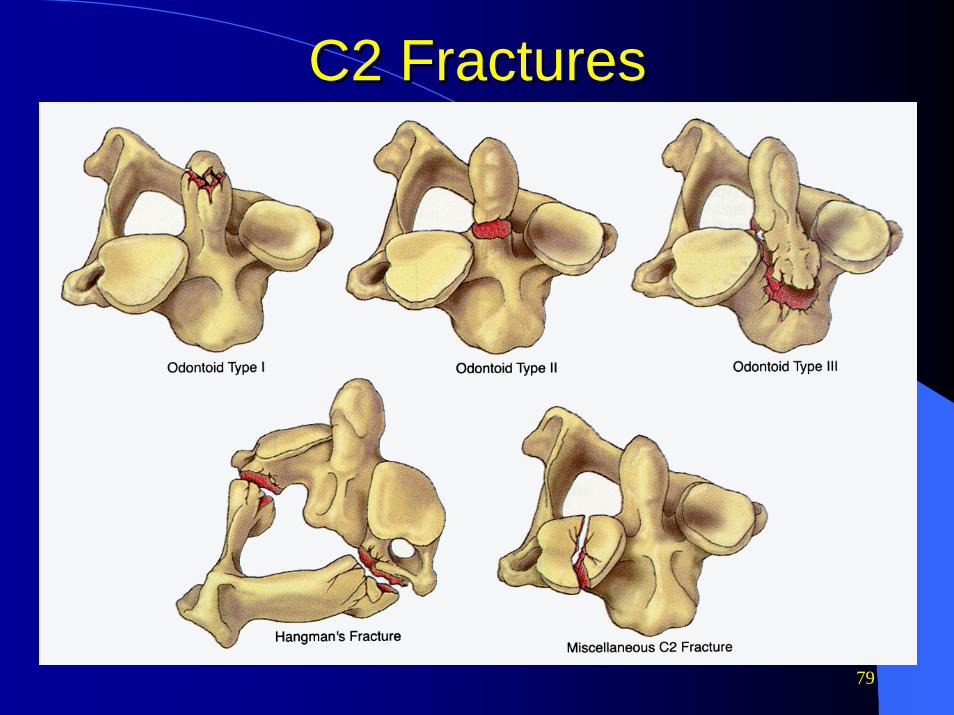
C2 FracturesC2 Fractures

80
Odontoid Fractures
Type I Type II Type III
<6mm displacement
>6mm displacement or
comminuted
Halo HaloORIF

81
Odontoid Fracture by Odontoid Fracture by DisplacementDisplacement
82
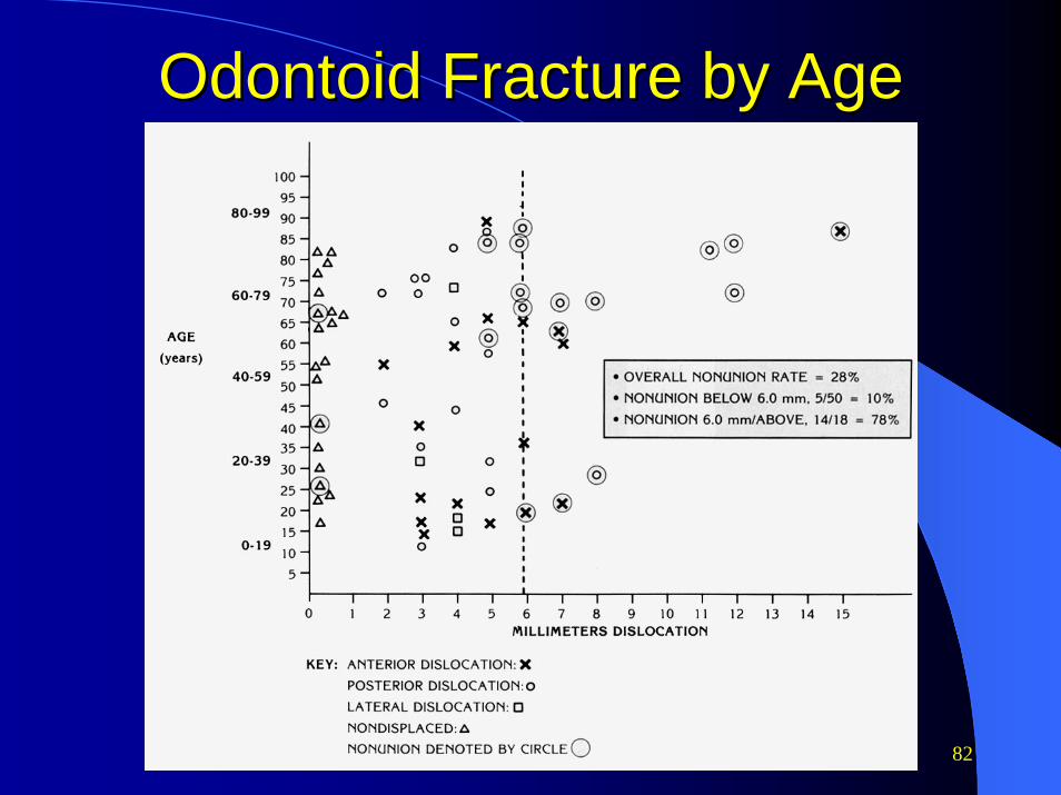
Odontoid Fracture by AgeOdontoid Fracture by Age

83
Odontoid FractureOdontoid Fracture

84

85
Hangman’s Fracture
Type I•Non-displaced•Minimally displaced
Type II•Angulated > 11 deg•Sublux > 4mm
Type III•Disrupted C2/3 facets
Irreducible or recurrent subluxation
Reducible
Rigid brace ORIFHalo

86
Complex Upper Cervical Complex Upper Cervical FracturesFractures
87
Operative InterventionOperative Intervention
Poor immobilization or recurrent deformity/malalignmentNonunion after nonsurgical treatmentLigamentous injuryAbove criteria for odontoid and Hangman’s fractures

88
Transoral OdontoidectomyTransoral Odontoidectomy

89
Transoral OdontoidectomyTransoral OdontoidectomyStabilityStability
90
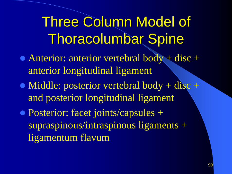
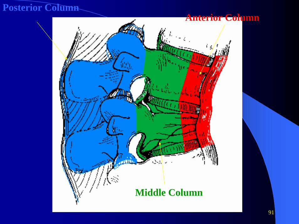
Three Column Model of Three Column Model of ThoracolumbarThoracolumbar Spine Spine
Anterior: anterior vertebral body + disc + anterior longitudinal ligamentMiddle: posterior vertebral body + disc + and posterior longitudinal ligamentPosterior: facet joints/capsules + supraspinous/intraspinous ligaments + ligamentum flavum
91
Posterior Column
Middle Column
Anterior Column
92
Hospital Care of Acute Hospital Care of Acute Non Traumatic Ischemic Non Traumatic Ischemic
StrokeStroke
93
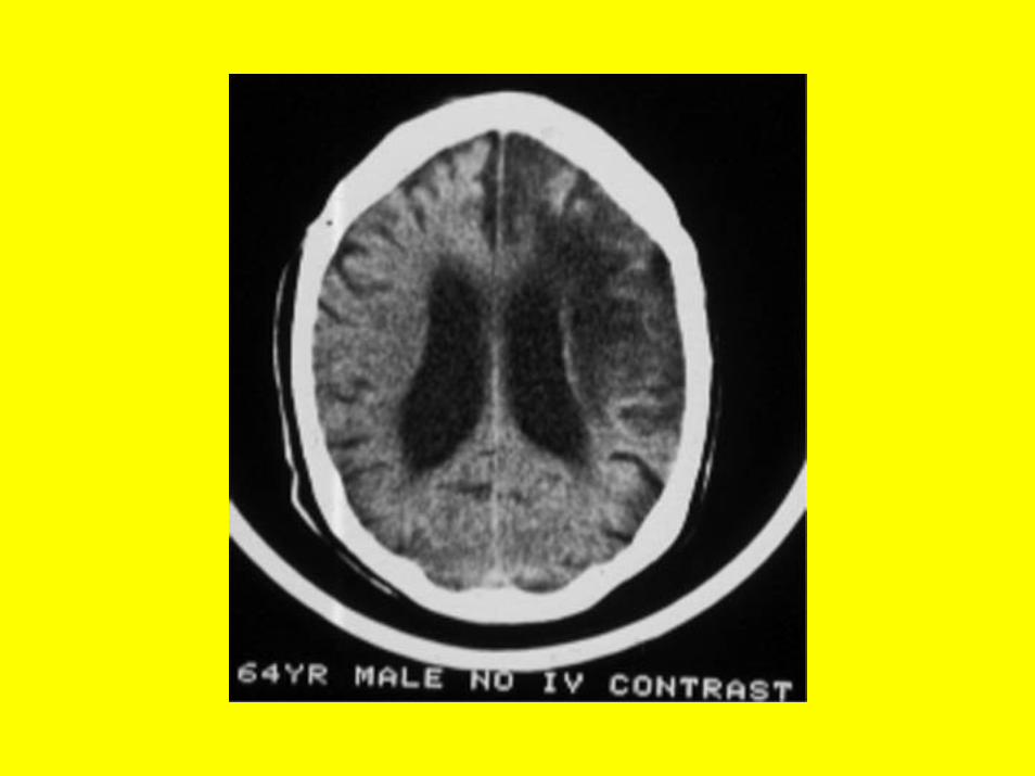
DefinitionsDefinitions
Stroke: Any vascular injury to the brainIschemic stroke is a persistent clinical deficit at 24 hours. A TIA lasts less than 24 hours and clears completely.This distinction is a continuum with damage proportional to the severity and duration of ischemia.
94
Definitions (conDefinitions (con’’t)t)
80% are ischemic20% are hemorrhagic (SAH, IPH, IVH)


98

99
Stroke in the United StatesStroke in the United States
750,000 new strokes a year4 million stroke survivors#1 cause of major neurologic disability#3 cause of death

100
Stroke risk factorsStroke risk factorsHTN 6XDiabetes 3XAsymptomatic bruit 3XRheumatic Atrial fib 17XParoxsysmal Atrial fib 6XLipids 2X
Smoking 2XPrior CVA/TIA 10XObesity 1.5XAge increases 10X/ 20 years of age

101
Ischemic stroke S/Ischemic stroke S/SxSx
102
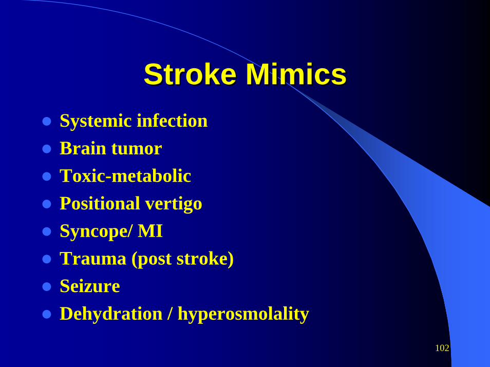
Stroke MimicsStroke MimicsSystemic infectionBrain tumorToxic-metabolic Positional vertigoSyncope/ MITrauma (post stroke)SeizureDehydration / hyperosmolality

103
Brief exam for strokeBrief exam for strokeGrimace (CN 7)Repeat a sentence (aphasia)Hold arms up with eyes closed (pronator drift)
104
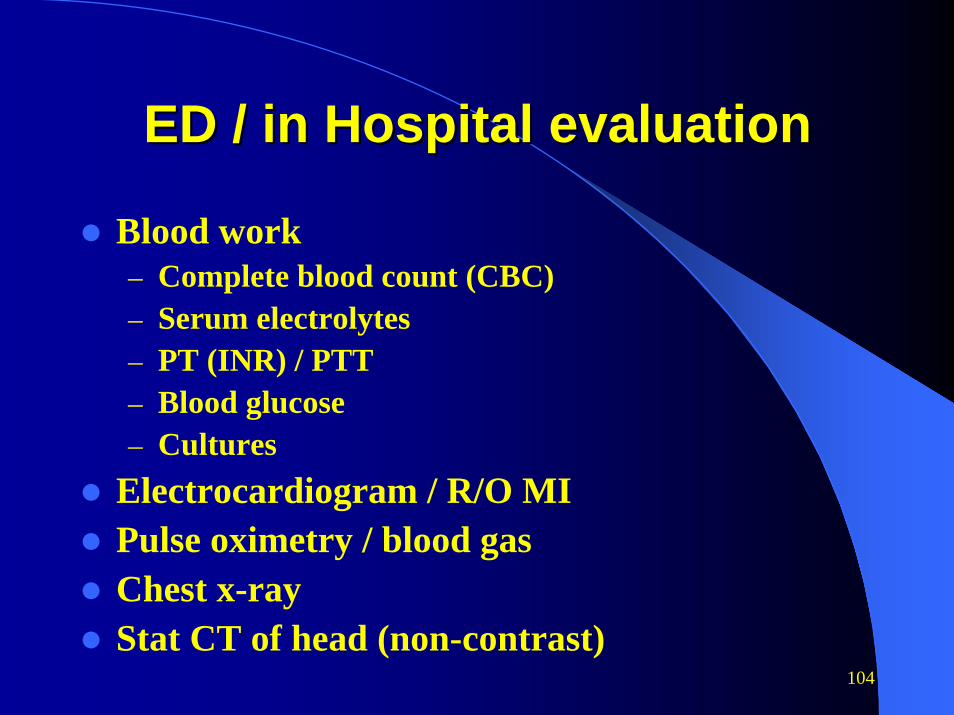
ED / in Hospital evaluationED / in Hospital evaluation
Blood work– Complete blood count (CBC)– Serum electrolytes– PT (INR) / PTT– Blood glucose– Cultures
Electrocardiogram / R/O MIPulse oximetry / blood gasChest x-rayStat CT of head (non-contrast)
105
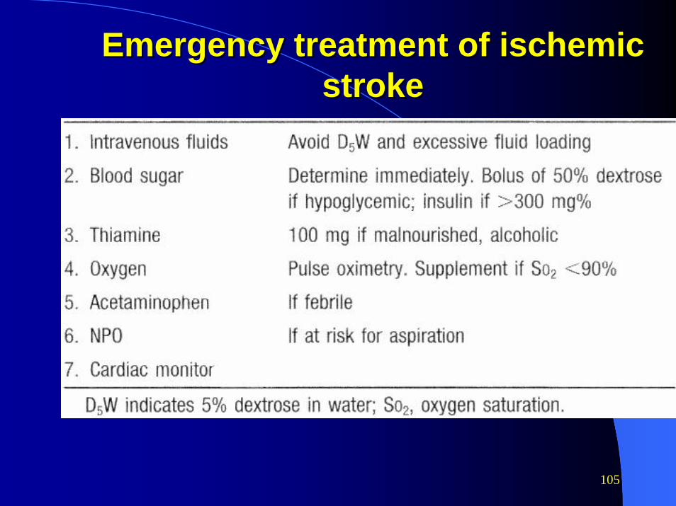
Emergency treatment of ischemic Emergency treatment of ischemic strokestroke

106
Blood pressure and acute Blood pressure and acute ischemic strokeischemic stroke
Transient and volatile elevation in blood pressure is commonUsually lasts several days after the strokePatients blood pressure may be very sensitive to medications

107
Blood pressure and Blood pressure and acuteacuteischemicischemic stroke (constroke (con’’t)t)
No benefit to aggressively reducing blood pressure in acute ischemic stroke, in other words, allow the pt to be HTNAHA: treat only if MAP > 130 or SBP > 220, DBP > 140
108
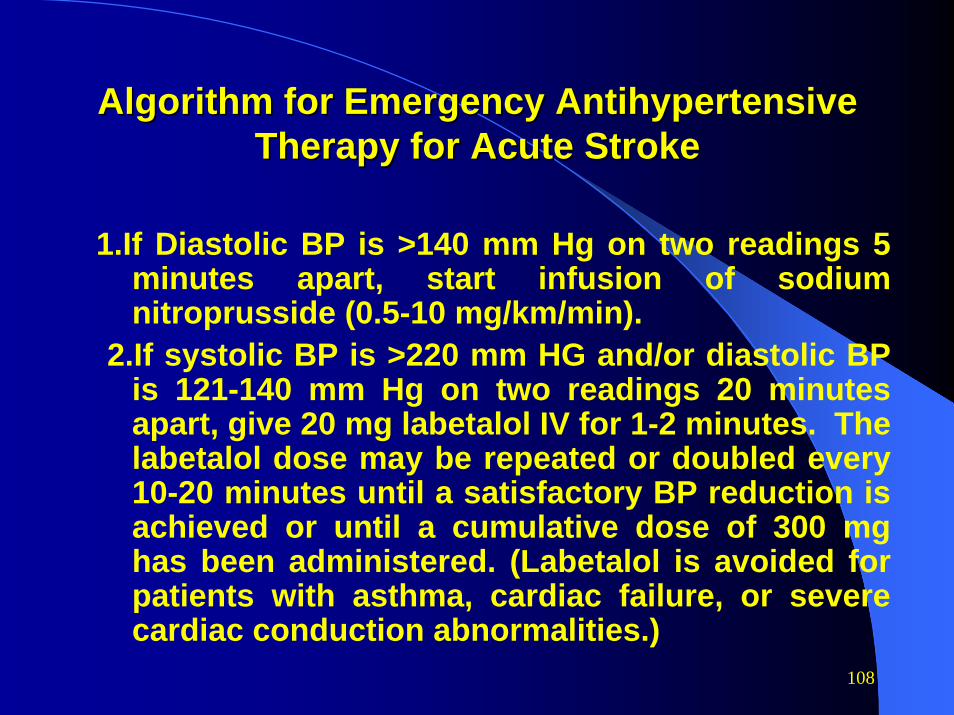
Algorithm for Emergency Antihypertensive Algorithm for Emergency Antihypertensive Therapy for Acute StrokeTherapy for Acute Stroke
1.If Diastolic BP is >140 mm Hg on two readings 5 minutes apart, start infusion of sodium nitroprusside (0.5-10 mg/km/min).
2.If systolic BP is >220 mm HG and/or diastolic BP is 121-140 mm Hg on two readings 20 minutes apart, give 20 mg labetalol IV for 1-2 minutes. The labetalol dose may be repeated or doubled every 10-20 minutes until a satisfactory BP reduction is achieved or until a cumulative dose of 300 mg has been administered. (Labetalol is avoided for patients with asthma, cardiac failure, or severe cardiac conduction abnormalities.)
109
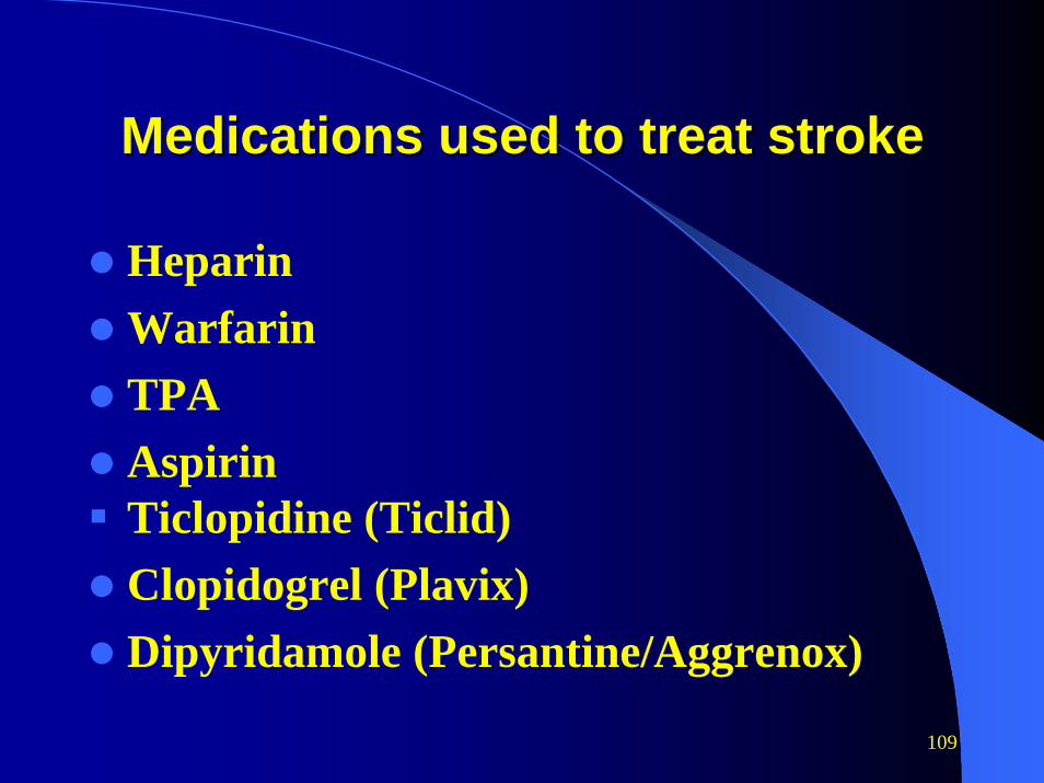
Medications used to treat strokeMedications used to treat stroke
HeparinWarfarinTPAAspirin Ticlopidine (Ticlid)Clopidogrel (Plavix)Dipyridamole (Persantine/Aggrenox)

110
Acute use of heparin and Acute use of heparin and StrokeStroke
Given IV without bolusControversialLittle evidence for benefit in most patients with completed stroke outside of pt’s with known A-fib/transmurial thrombus/ sig carodit and post circ stenosisNot recommended in new AHA guidelines
111
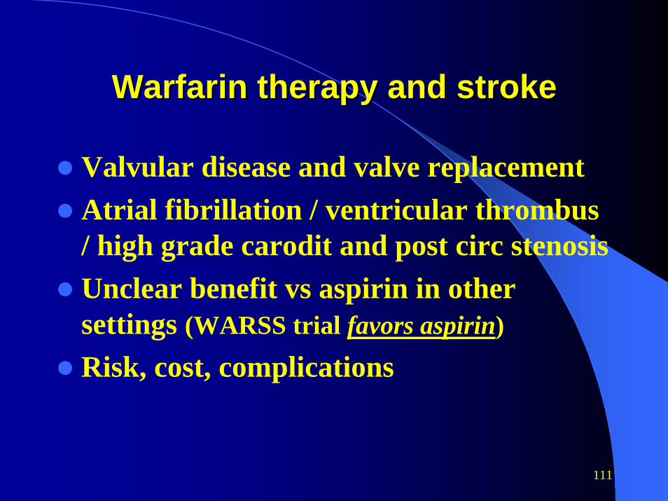
Warfarin therapy and strokeWarfarin therapy and stroke
Valvular disease and valve replacementAtrial fibrillation / ventricular thrombus / high grade carodit and post circ stenosisUnclear benefit vs aspirin in other settings (WARSS trial favors aspirin)
Risk, cost, complications

112
Systemic thrombolytic Systemic thrombolytic treatmenttreatment
Intravenous tissue plasminogen activator (TPA)– 3 hour window to treat from onset of
symptoms– Many contraindications– Risk of hemorrhage
113
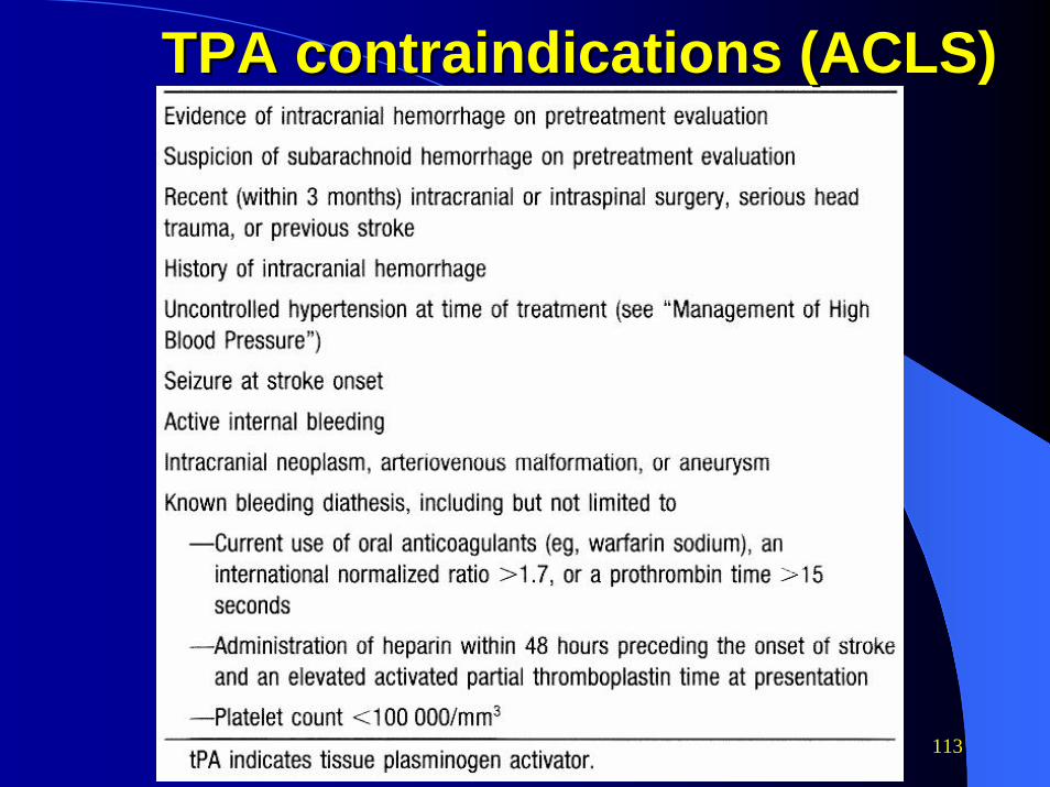
TPA contraindications (ACLS)TPA contraindications (ACLS)

114
TPA complicationsTPA complicationsNo difference in mortality at 3 months between the r-TPA and placebo– 17% TPA – 21% placebo
Higher incidence of symptomatic hemorrhage in the r-TPA group – 10X hemorrhage rate (6% with TPA)– 3% of TPA patients died from hemorrhage
115
TPA Results TPA Results
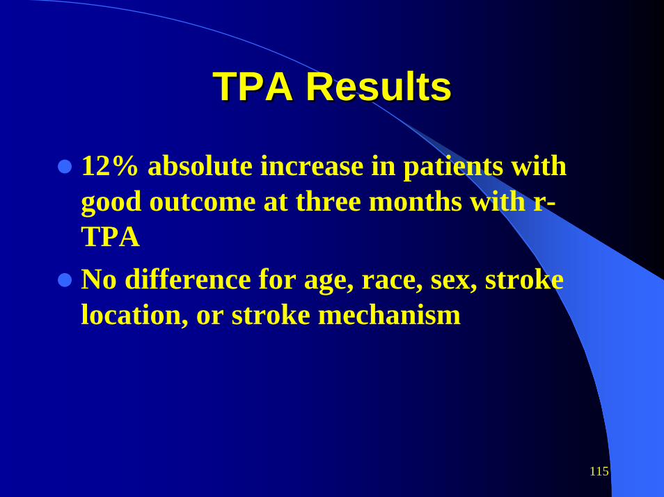
12% absolute increase in patients with good outcome at three months with r-TPA No difference for age, race, sex, stroke location, or stroke mechanism
116
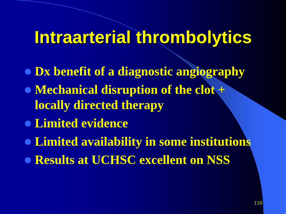
Intraarterial thrombolyticsIntraarterial thrombolytics
Dx benefit of a diagnostic angiographyMechanical disruption of the clot + locally directed therapyLimited evidenceLimited availability in some institutionsResults at UCHSC excellent on NSS

118
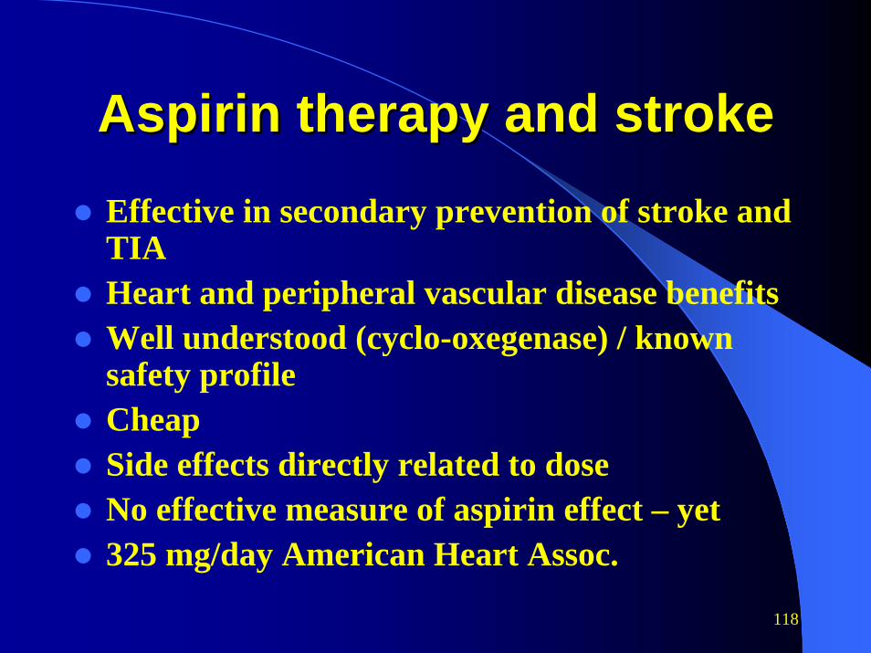
Aspirin therapy and strokeAspirin therapy and strokeEffective in secondary prevention of stroke and TIAHeart and peripheral vascular disease benefitsWell understood (cyclo-oxegenase) / known safety profileCheapSide effects directly related to doseNo effective measure of aspirin effect – yet325 mg/day American Heart Assoc.

119
Clopidogrel &Ticlopidine Clopidogrel &Ticlopidine Inhibit the platelet ADP pathway Clopidogrel better tolerated than ticlopidineMarginally more effective than aspirin$

120
Common complications of Common complications of strokestroke
SeizuresAspirationDVT/ pulmonary embolismAppendage dislocation

121
SummarySummary
New approach to stroke Time is criticalSimple interventions – glucose control, moderate IVF, temperature, blood pressureR/O stroke mimics prior to use of thrombolytics