general morphology of the oral cavity of the nile ... · pdf filethe literature (for a review...
TRANSCRIPT

INTRODUCTION
The morphology and microscopic anatomy of thereptilian oral cavity has received much attention inthe literature (for a review see Luppa 1977), withmost studies concentrating on the description andlocation of glandular tissue, taste receptors and
epithelial specialisation of the region. Attention hasalso been given to the embryological and evolu-tionary development of these specialisations. Sim-ilarly, most studies on the oral cavity of crocodilianshave concentrated on specific morphological fea-tures of this region.
Röse (1893) reported the presence of glandular tis-sue situated in pits between the teeth of the maxilla(glandulae palatinae) in Crocodylus porosus anddescribed the embryological development of theseglands. Woerdeman (1920) reviewed earlier litera-ture (ca. 1888 to 1914) on the subject and empha-sised discrepancies amongst the authors regardingthe presence or absence of oral glands in Croco-dylia. Farenholz (1937) reported two areas in thepalate in which glands occur, viz., median palatine
281
Onderstepoort Journal of Veterinary Research, 70:281–297 (2003)
General morphology of the oral cavity ofthe Nile crocodile, Crocodylus niloticus(Laurenti, 1768). I. Palate and gingivae
J.F. PUTTERILL1* and J.T. SOLEY2
ABSTRACT
PUTTERILL, J.F. & SOLEY, J.T. 2003. General morphology of the oral cavity of the Nile crocodile,Crocodylus niloticus (Laurenti, 1768). I. Palate and gingivae. Onderstepoort Journal of VeterinaryResearch, 70:281–297
The heads of nine 2.5 to 3-year-old Nile crocodiles (Crocodylus niloticus) were obtained from a com-mercial farm where crocodiles are raised for their skins and meat. The animals from which thesespecimens originated were clinically healthy at the time they were slaughtered. A detailed descrip-tion of the macroscopic and microscopic features of the palate and gingivae of the Nile crocodile ispresented and the results are compared with published information on this species and otherCrocodylia. The histological features are supplemented by information supplied by scanning electronmicroscopy. Macroscopic features of interest are the small conical process situated at the base ofthe first two incisors of the maxilla, the distribution of cobbled units on the palate, and the broad den-tary shelf forming the rostral aspect of the mandible. Histologically the palate and gingivae did notdiffer significantly from each other and both regions showed a presence of Pacinian-type corpuscles.Two types of sensory structures (taste receptors and pressure receptors) were identified in theregions examined, both involving modification of the epithelium and the underlying connective tis-sue.
Keywords: Crocodylus niloticus, histology, morphology, Nile crocodile, oral cavity, scanning elec-tron microscopy
* Author to whom correspondence is to be directed1 Electron Microscopy Unit, Onderstepoort Veterinary Institute,
Private Bag X05, Onderstepoort, 0110 South Africa. E-mail:[email protected]
2 Department of Anatomy and Physiology, Faculty of VeterinaryScience, University of Pretoria, Private Bag X04, Onderste-poort, 0110 South Africa
Accepted for publication 12 May 2003—Editor
glands found only in Caiman spp. and small glandsat the median aspect of the maxillary teeth, found inCaiman spp. and Alligator mississippiensis. How-ever, Taguchi (1920) found glandular tissue “in thesubmucosa of the caudal part of the palate and theoral surface of the velum”. Kochva (1978) exten-sively described glandular tissue in reptiles, but onlyfleetingly refers to the Crocodylia.
Bath (1905, 1906) described the histology of tastereceptors in the oral cavity, pharynx and oesopha-gus of Crocodilus niloticus (sic.) and Alligator missis-sipiensis (sic.), finding no clear distinction betweenthose seen in these species and those of higher ani-mals. Luppa (1977), who generalised his descrip-tion of the histological composition of the reptilianoral cavity, stated that “taste buds were scatteredthroughout the oral epithelium in reptiles and that inLacerta they were most numerous laterally and onthe palatal folds.” Hulanicka (1913) investigated theinnervation of the tongue, palate and the skin ofCrocodylus niloticus and Alligator lucius and de-scribed five different nerve endings in the regionsstudied. Alligator lucius represents A. mississippi-ensis (F.W. Huchzermeyer, personal communica-tion 2002).
Fuchs (1908, cited by Barge 1937) postulated theformation of the secondary palate in the Crocodyliaand compared this formation to other reptiles, con-cluding that the secondary palate of crocodiles wasunique amongst the reptiles. Barge (1937) describedthe embryological development and phylogeny ofthe secondary palate in crocodiles. Ferguson (1979)investigated the developmental mechanisms in nor-mal and abnormal palate formation in the Americanalligator (A. mississippiensis) and concluded thatthe Crocodylia showed characteristics which werepart mammalian and part reptilian, a unique combi-nation which made them a useful model to studypalatogenesis.
Dentition in Crocodylia has also received muchattention in the literature. Of note is the paper byPoole (1961) who described tooth replacement inC. niloticus, the studies by Westergaard & Ferguson(1986, 1987) who described the development ofdentition in hatchling and juvenile A. mississippien-sis, and the article by Kieser, Klapsidis, Law & Mari-on (1993) who examined heterodonty and patternsof tooth replacement in C. niloticus. Edmund (1962,1969) also made a major contribution to studies ondentition in the Reptilia, including the Crocodylia,describing the sequence and rate of tooth replace-ment in these reptiles.
Although detailed descriptions of specific compo-nents of the crocodilian oral cavity have been pre-sented, only a few studies have reported on thegeneral histological features of this region. Reese(1913) studied the histology of the enteron of the“Florida alligator”, which included the oral cavity.Reese’s description, however, concerned histologi-cal differences between hibernating and feeding,captive animals. Taguchi (1920) compared similarregions of the oral cavity to those examined byReese (1913) in three species of Crocodylia, name-ly, Alligator sinensis, Krokodilus porosus and Krok-odilus vulgaris. The latter is believed to representthe Nile crocodile, Crocodylus niloticus (see http://www.flmnh.ufl.edu/natsci/herpetology/turtcroclist/chklst2.htm). Throughout this paper, and pertainingonly to Taguchi (1920), “Krokodilus” is referred to as“Crocodylus” and “K. vulgaris” as “C. niloticus”. InChiasson’s (1962) publication on the anatomy ofthe alligator, components of the oral cavity (palateand tongue) are briefly mentioned without any fur-ther detail being given. Similarly Parsons & Cameron(1977), who examined the relief of the gastro-intes-tinal tract of the Reptilia, including the Crocodylia,only start their description from the oesophagusand do not describe the morphology of the oral andpharyngeal cavities.
In view of the paucity of information concerning thegeneral histological features of this part of the upperdigestive tract, this paper presents a general topo-graphical description of the oral cavity as well as themacroscopic and microscopic features of the palateand gingivae of the Nile crocodile, Crocodylus niloti-cus (Laurenti, 1768) and compares the results withpublished information on this species and otherCrocodylia. The histological features are supple-mented by information supplied by scanning electronmicroscopy (SEM). Morphological features of thetongue will be presented in another paper.
MATERIALS AND METHODS
Experimental animals
The heads of nine 2.5 to 3-year-old Nile crocodileswere obtained from a commercial farm where croc-odiles are raised for their skins and meat. Thelengths of the animals sampled ranged from 1.2–1.5 m and they were clinically healthy at the timethey were slaughtered. The animals were killed byshooting them in the brain at close range using a.22 calibre rifle. After the carcasses had beenskinned and eviscerated the heads were removed
282
Oral cavity of Nile crocodile, Crocodylus niloticus (Laurenti, 1768). I
and immersion-fixed in a large volume of 10 %phosphate-buffered formalin in plastic buckets for aminimum period of 48 h. Care was taken to excludeair from the oral cavity by wedging a small block ofwood in the angle of the mouth prior to immersionin the fixative. Samples from the palate and gingi-vae were taken from the heads and processed forlight microscopy (LM) and scanning electronmicroscopy (SEM) according to the proceduresdetailed below.
Topography
Prior to sampling, all nine heads were utilised for adescription of the gross anatomical features andtopographical relationships of the structures in theoral cavity. Macrophotographs were recorded digital-ly using a Nikon Coolpix 995 (Nikon, Tokyo, Japan)digital camera or on 35 mm film using a Chinon X-7(Chinon, Tokyo, Japan) single lens reflex camera,respectively. The oral cavities of these heads werealso examined and micrographed using a stereo-microscope (Wild M-400 Photomakroskop, Heer-brugg, Switzerland) to obtain higher magnificationmicrographs of specific topographical features.
A dried skull from a 5-year-old (approximate age)specimen was used to confirm the position andnaming of teeth in the maxilla and mandible as wellas to provide supporting evidence for the anatomi-cal description. Teeth were named and numberedaccording to Kieser et al. (1993).
Light microscopy
Samples of the gingiva from the mandible were re-moved from the various regions indicated in Fig. 1and were based on the position of the incisor andcanine teeth. The portion of gingiva caudal to theindicated regions, i.e. the region involving the molarteeth, was too firmly attached to the underlying boneto permit suitable samples to be taken. The mucosaof the palate was also sampled according to thedental arrangement of the teeth, i.e. from regions I1 to I 5, C 1 to C 5 and M 1 to M 8 as shown in Fig.2. As the gingiva of the maxilla appeared macro-scopically to be continuous with the palate, thesespecimens were removed together with the sam-ples of the palate. A similar set of specimens (adja-cent tissue) from all the indicated regions of themandible and palate was taken at the same time forSEM examination.
Samples for LM were dehydrated through 70, 80,96 and 2X 100 % ethanol and further processedthrough 50 : 50 ethanol : xylol, 2X xylol and 2X par-
affin wax (60–120 min per step) using a Shandonmodel 2LE Automatic Tissue Processor (Shandon,Pittsburgh, PA, USA). Tissue samples were finallyembedded manually into paraffin wax in brassmoulds. Sections were cut at 4–6 µm, stained withhaematoxylin and eosin (H&E) (Luna 1968) or per-iodic acid-Schiff (PAS) (Pearse 1985) and viewedand micrographed using a Reichert Polyvar (Reich-ert, Austria) compound light microscope fitted witha differential interference contrast (DIC) prism.
Scanning electron microscopy
The samples of the gingivae and palate obtained asindicated above and which had been fixed in 10 %phosphate-buffered formalin for a minimum of 48 hwere subsequently rinsed for several hours in waterto remove traces of phosphate buffer. These sam-ples were routinely dehydrated through an ascend-ing ethanol series (50, 70, 90, 95 and 3X 100 %—60 min per step) and critical point dried from 100%ethanol through liquid-CO2 in a Polaron CriticalPoint Drier (Polaron, Watford, England). The sam-ples were then mounted onto brass or aluminiumviewing stubs (to expose the epithelial surface) witha conductive paste (carbon dag) and sputter coatedwith gold using a Balzers 020 Sputter Coater (Bal-zers Union, Liechtenstein). Specimens were viewedand photographed using a Hitachi S-2500 scanningelectron microscope (Hitachi, Tokyo, Japan) oper-ated at 8 kV.
RESULTS
Macroscopic features
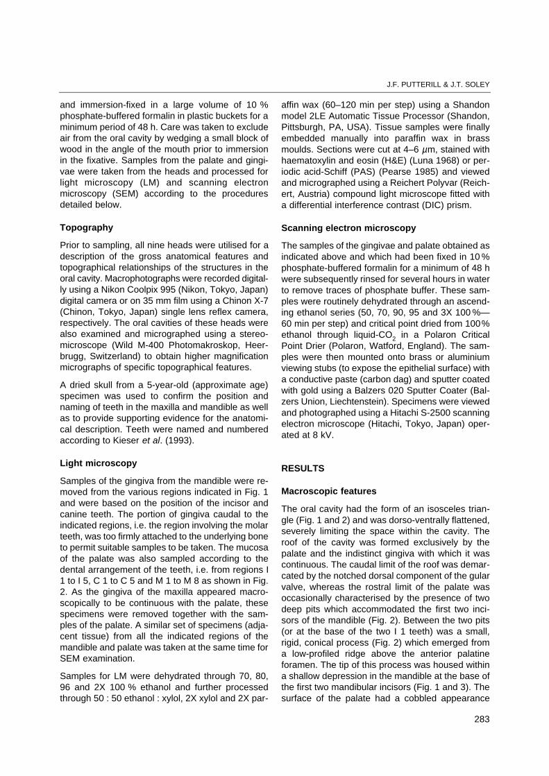
The oral cavity had the form of an isosceles trian-gle (Fig. 1 and 2) and was dorso-ventrally flattened,severely limiting the space within the cavity. Theroof of the cavity was formed exclusively by thepalate and the indistinct gingiva with which it wascontinuous. The caudal limit of the roof was demar-cated by the notched dorsal component of the gularvalve, whereas the rostral limit of the palate wasoccasionally characterised by the presence of twodeep pits which accommodated the first two inci-sors of the mandible (Fig. 2). Between the two pits(or at the base of the two I 1 teeth) was a small,rigid, conical process (Fig. 2) which emerged froma low-profiled ridge above the anterior palatineforamen. The tip of this process was housed withina shallow depression in the mandible at the base ofthe first two mandibular incisors (Fig. 1 and 3). Thesurface of the palate had a cobbled appearance
283
J.F. PUTTERILL & J.T. SOLEY

284
Oral cavity of Nile crocodile, Crocodylus niloticus (Laurenti, 1768). I

(Fig. 2) due to the presence of numerous, raised,cobble-like structures. The cobbles on the rostraltwo-thirds of the palate were large, whereas thoseoccupying the caudo-lateral aspects of the palatewere smaller, had a lower profile, but were denselyarranged. Between the latter two regions werepaired elliptical areas, devoid of cobbles, and whichmerged medially along the midline of the palate(Fig. 2). These smooth areas corresponded to thepositioning of the left and right posterior palatineforaminae which were formed by the caudal edgesof the maxillary, the lateral edges of the palatine, asmall region of the rostral edge of the pterygoid and
the medial edge of the transpalatine bones. Alongthe midline of the palate were a series of closelypositioned cobbles forming a clearly defined medi-an ridge. This ridge extended from the conical pro-cess mentioned above to the base of the dorsal foldof the gular valve. However, the part of the ridgedividing the two smooth elliptical areas above theposterior palatine foraminae was less distinct innature. The base of the palate adjacent to the dor-sal fold of the gular valve displayed a variable num-ber of transverse mucosal folds which closely fol-lowed the contours of the dorsal fold across itsentire breadth (Fig. 2).
285
J.F. PUTTERILL & J.T. SOLEY
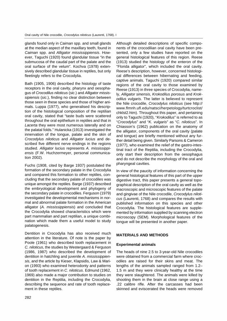
FIG. 1 Macrophotograph of the mandible with the tongue in situ showing the dental arrangement of the incisor (I 1–3), canine(C 1–5) and molar (M 1–7) teeth, demarcated by the stippled lines. The sampling sites for histology of the gingivae (D andE) are also indicated. The rostral dentary shelf is indicated in the region above the symphysis of the dentary bones. Theblack arrowhead indicates the position of the shallow depression which houses the small, rigid, conically formed process,situated at the base of the I 1 teeth, seen in Fig. 2. The glottis (GT) and laryngeal mound (LM) are shown in situ on thefloor of the pharyngeal cavity and the ventral fold (VF) of the gular valve is seen separating the ventral aspects of the oraland pharyngeal cavities. Formalin fixed specimen. X 0.75
FIG. 2 Macrophotograph of the maxilla and palate showing the dental arrangement of the incisor (I 1–5), canine (C 1–5) and molar(M 1–8) teeth, demarcated by the stippled lines. The smooth area of the palate, demarcated by the black arrows, lies abovethe left and right posterior palatine foraminae. The small, rigid, conical process, situated at the base of the I 1 teeth, is indi-cated by the arrowhead. Also note the smooth zone of mucosa forming the gingiva adjacent to the teeth (asterisks) whichstretches from approximately I 5 to M 8 on both sides of the maxilla. The common opening of the internal nares (IN) is seenon the roof of the pharyngeal cavity as well as the dorsal fold (DF) of the gular valve which separates the dorsal aspectsof the oral and pharyngeal cavities. Samples of the mucosa of the palate and gingiva were removed from regions A, B andC as indicated on the photograph. Formalin fixed specimen. X 0.75
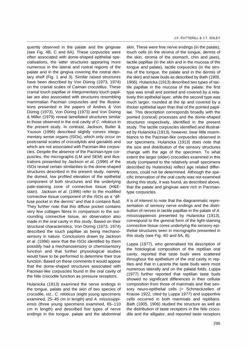
FIG. 3 Macrophotograph of the rostral portion of the mandible(I 1 to C 5). The gingiva is seen to have a slightly cob-bled appearance towards the rostral tip above thedentary symphysis. The black arrow indicates a shal-low depression which houses the small, rigid, conical-ly-shaped maxillary process seen in Fig. 2. The blockedarrow shows a hole made by the hook in the abattoirwhen the carcass was suspended during the eviscer-ation process. I 1 = Incisor 1; C 1, C 5 = Canine 1 and5. Formalin-fixed specimen. X 1.2

The gingiva of the maxilla was continuous with thepalate and could practically be considered to bepart of it (Fig. 2). A relatively wide (4–5 mm), clear-ly demarcated zone of smooth mucosa (possiblyrepresenting the palatal aspect of the gingiva) sep-arated the cobbled portion of the palate from themaxillary teeth, from approximately C 3 to M 8.From approximately C 2 rostrally, the surface of thegingiva also had a cobbled appearance similar tothat of the palate and the boundary between the lat-ter and the gingiva was not clearly defined. Theteeth of the maxilla reflected the dental formula de-scribed by Kieser et al. (1993) and were carried inthe premaxillary and maxillary bones. In the occlud-ed mouth, the teeth of the maxilla were accommo-dated in grooves to the outside of the mandible,between the teeth of the lower jaw. The tips of theteeth of the mandible were accommodated in pitssituated between the teeth of the maxilla, with theexception of C I, which was accommodated in amaxillary notch and remained visible when the jawswere closed.
The floor of the oral cavity was formed by the tongueand a wide, rostral mucosal plate continuous withthe gingiva (Fig. 1 and 3). This plate representedthe mucosa-covered surface of the widened rostraltips of the paired dentary bones of the mandiblewhere they met at the dentary symphysis. This plateextended from the rostrally-positioned first two inci-sors to a point approximately midway between C 1and C 2 (Fig. 1 and 3). The relatively long tonguewas roughly triangular in shape, being much broad-er caudally than at its tip (Fig. 1). It occupied thegreater part of the floor of the oral cavity (apart fromthe rostral plate over the symphysis of the dentarybones) and was bordered peripherally by a loose,highly folded, continuous, fibrous membrane (Fig. 1and 3).
The gingiva of the mandible was more clearlydefined than that of the maxilla, having a low pro-filed, cobbled appearance from approximately C 3rostrally. There was close attachment of the gingi-va to the mandibular (dentary and splenial) bones,especially in the region of C 3 (or C 4) to M 7 (orM 8). The rostral tip of the dentary bones formed abroad shelf or plateau (the rostral dentary shelf)which was divided medially by the dentary symph-ysis. In this region the gingiva had a slightly spongytexture, although the surface also had a cobbledappearance (Fig. 1 and 3). The teeth of the mandiblewere carried in the paired dentary bones and alsoreflected the dental formula described by Kieser etal. (1993) (see Fig. 1).
From M 4 (or M 5) to M 7 (or M 8) the dentary boneand the medially situated splenial bone were inclose association, although the mandibular teethwere clearly housed in the dentary bone.
Light microscopy
The palate
Sections of the palate stained with H&E revealed akeratinised stratified squamous epithelium of vari-able thickness in all the regions examined. Thestratum basale was composed of a single layer ofcuboidal to columnar cells resting on a basementmembrane. The basement membrane was mostobvious in PAS-stained sections and varied inprominence from conspicuous to barely visible. Thenuclei of the basal layer of cells were pale, vesicu-lar and round to oval in shape (Fig. 4A). Whereoval, the nuclei were oriented vertically to the sur-face of the epithelium.
The stratum spinosum consisted of 3–6 layers ofcells. The cells adjacent to the stratum basale werecuboidal in shape, while the more superficial cellswere horizontally flattened. All the cells of this layerdisplayed the characteristic inter-linking cytoplas-mic bridges connecting the individual components.The nuclei of these cells resembled those of thestratum basale. A thin (3–4 layers) stratum granu-losum was present above the stratum spinosum.Cells in this layer were spindle shaped or flattenedand oriented horizontally. The nuclei were pycnotic,flattened and oriented in the same plane as thecells, while the cytoplasm was filled with stronglybasophilic-staining keratohyaline granules (Fig.4A). The stratum corneum varied in thickness andwas composed of a number of compressed layersof cells in which no nuclei were apparent (Fig. 4A).In some areas, particularly towards the gingiva ofthe teeth and in convoluted regions of the epitheli-um, a stratum disjunctum consisting of a looselayer of keratinised cells was present (Fig. 4B).
The epithelium was supported by a thick layer ofirregular dense connective tissue with prominentbundles of variably oriented collagen fibres beingthe most prominent feature (Fig. 4A, B, D and E).Sandwiched between the deeper regions of theirregular dense supporting connective tissue andthe periostium of the palatine bones was a well-developed plexus of blood vessels, lymphatics andnerves (medullated and non-medullated). Deeplysituated striated muscle bundles were noted only inthe region of the posterior palatine foraminae,
286
Oral cavity of Nile crocodile, Crocodylus niloticus (Laurenti, 1768). I

287
J.F. PUTTERILL & J.T. SOLEY
stretching from the posterior third of the palate tothe base of the dorsal gular fold. No other muscu-lar tissue was observed. Immediately beneath thebasement membrane was a thin layer of fine con-nective tissue which in places displayed a vacuo-lated, spongy appearance. This region demonstrat-ed a rich capillary blood supply which was intimatelyassociated with the overlying epithelium. No glan-dular tissue was ever observed in any of the speci-mens during histological examination of the palate.Lymphocytic aggregations were also not apparentin these sections.
Melanocytes were observed in the connective tis-sue a short distance beneath the stratum basale,but never within the epithelial layer. The cells weretypically dendritic in nature and displayed large num-bers of brown to black melanin granules (melan-osomes). The melanocytes were concentratedaround the capillary plexus beneath the epitheliumand also around the larger blood vessels and nervesmore deeply positioned within the connective tissuestroma. In some areas the melanocytes formed adiffuse but definite layer beneath the epithelium.The presence of melanin varied amongst individualspecimens examined and in some cases it wasfound to be entirely absent.
Mast cells occurred either singly or in small groupsthroughout the connective tissue layer with concen-trations of five or more cells sometimes beingobserved. The mast cells were large, round andoften observed in the vicinity of blood vessels. Thepale, round to oval vesicular nucleus was centrallypositioned within the cytoplasmic mass which dis-played small fine, evenly distributed basophilic gran-ules. Prominent Pacinian-like corpuscles were ran-domly scattered throughout the connective tissuelayer a short distance beneath the epithelium (Fig.4B and C). These structures typically consisted of avariable number of connective tissue lamellae sur-rounding an inner core representing the terminalportion of the innervating nerve. The corpuscle wassurrounded by a prominent, dense connective tis-sue capsule (Fig. 4C) and large medullated nerveswere observed in the vicinity of the corpuscles.
Three types of surface specialisations were observedin the sections studied. The first type comprisedsmall pointed elevations of the epithelial lining sup-ported by a core of fine connective tissue (Fig. 4A).In some instances these elevations presented as aseries of small localised projections giving the sur-face of the palate a scalloped appearance. Thesestructures probably represented the epithelial folds
observed by SEM (see below). The remaining twotypes of specialised structures were characterisedby modification of both the epithelium and theunderlying connective tissue. Both structures (Fig.4D and E) displayed a localised thickening of theepithelium due mainly to an increase in the numberof layers of the stratum spinosum. The keratinisedlayer in the region of the epithelial thickening wasgenerally thinner than that of the adjacent tissue.The localised epithelial thickenings were most com-monly found in the form of an elevated, dome-shaped structure due primarily to the presence of adiffuse, ellipsoid or conical-shaped mass of looselyarranged connective tissue situated immediatelybeneath the epithelium (Fig. 4D). These regionswere more lightly stained (H&E-stain) than the sur-rounding connective tissue (due to a reduction insize and number of the collagen bundles) andcaused localised protrusion of the overlying epithe-lium into the mouth cavity. The morphological fea-tures of the specialised regions varied. In someinstances the diffuse connective tissue core con-tained a basophilic cell-rich mass situated immedi-ately adjacent to the basal lamina. In other regions,the connective tissue core displayed a paucity ofcells, possibly due to the plane of section. Associ-ated with the modified regions of connective tissuewere Pacinian-like corpuscles which were eitherfound in or adjacent to this zone. Nerve tissue fea-tured prominently within the modified connectivetissue and large medullated nerves and blood ves-sels were observed entering/leaving at the base ofthe connective tissue core. The dome-shaped spe-cialisations were distributed throughout the palatebut appeared to be more numerous on the rostralaspect up to the rostral border of the posterior pala-tine foraminae (see Fig. 2).
A small number of localised epithelial thickeningsappeared flattened in contrast to the dome-shapedstructures and were either positioned level with theadjoining epithelial surface or slightly raised aboveit. The floor of these epithelial specialisations juttedinto the underlying connective tissue layer. Epithe-lial cells towards the middle of the specialisationadopted a vertical orientation, forming a large ellip-tical structure reminiscent of a taste bud (Fig. 4Eand F). Some of the vertically inclined cells re-vealed dense, somewhat elongated nuclei, particu-larly towards the periphery of the elliptical structure,and were similar in appearance to the supportingcells of the mammalian taste-bud. Similarly inclinedcells with more vesicular nuclei were seen amongthe supporting cells and may have representedneuro-epithelial cells. A modified connective tissue

288
Oral cavity of Nile crocodile, Crocodylus niloticus (Laurenti, 1768). I

core similar to that seen beneath the dome-shapedstructures was also evident but did not appear to bespecifically associated with Pacinian-like corpus-cles. Attendant medullated nerves, however, weremuch in evidence. The taste receptors describedabove appeared to be concentrated on the morelateral aspects of the palate although they wereoccasionally encountered towards the midline.
SEM examination confirmed the cobbled appear-ance of the palate seen macroscopically. It shouldbe noted, however, that individual variation existedin the specimens examined regarding the promi-nence of the cobbling. Each clearly demarcatedcobbled unit displayed a centrally positioned dome-shaped structure or papilla surrounded by anexpanse of loosely attached surface epithelial cells.Desquamation of these cells was particularly obvi-ous at the perimeter of the dome-shaped structure(Fig. 5B). In much of the palate (roughly correspon-ding to the surface in contact with the dorsum of thetongue) the epithelial surface surrounding the papil-lae was thrown into a number of conspicuous foldswhich branched and anastomosed (Fig. 5A). Thefolds displayed a rostro-caudal or slightly obliquealignment. However, towards the periphery of thepalate bordering the gingivae and the dorsal fold ofthe gular valve, as well as in the smooth region ofthe palate overlaying the posterior palatine forami-nae, the cobbled units displayed a featureless sur-face around the domed papillae.
Some of the domed papillae revealed a small cen-trally positioned depression and radiating grooves
(Fig. 5C). These structures appeared to occur morecommonly in the rostro-lateral regions of the palate.All regions of the palate were characterised by dis-tinct desquamation of the superficial cells of theepithelium. This phenomenon was possibly accen-tuated by the critical point drying (CPD) processused for SEM sample preparation. Cracking of theepithelial layer was evident in some specimensexamined (see Fig. 5C) and was also attributed tothe CPD process. Higher magnification of the epi-thelial surface using SEM imaging revealed the typi-cal polygonal outline of the individual cells, althoughthe borders were not always clearly demarcated.The keratinised surfaces had a coarse, mattedappearance (Fig. 5D).
The gingivae
The composition and structure of the gingival mu-cosa was similar in general appearance to that ofthe palate, although some variations in structurewere apparent. The epithelial surface was moreundulating than that of the palate, with occasionalelevated structures protruding from the surface(Fig. 6A, C and D). The epithelium itself was thin-ner than that of the palate, with the stratum corneumand stratum disjunctum forming the most prominentlayers. The stratum spinosum was extremely thinand only obvious in regions of localised thickening.Below the basement membrane was a thin layer ofvacuolated, spongy connective tissue which wascontinuous with a thick layer of irregular dense con-nective tissue.
289
J.F. PUTTERILL & J.T. SOLEY
FIG. 4 Histological features of the palate
A A pointed elevation of the epithelial lining of the palate supported by subepithelial connective tissue (CT). Stratumbasale (1); stratum spinosum (2); thin stratum granulosum (asterisk); superficial stratum corneum (3). H&E-stain. Bar= 100 µm
B Randomly scattered Pacinian-like corpuscles (arrowhead) were located in the subepithelial connective tissue and oftenassociated with epithelial specialisations. Note signs of desquamation of the superficial layers of the stratum corneum(arrow). H&E-stain. Bar = 100 µm
C Typical Pacinian-like corpuscle displaying concentric lamellae around an inner core (arrowhead). Note the thick, fibrousconnective tissue capsule surrounding the corpuscle. H&E-stain. Bar = 50 µm
D Light micrograph of an elevated, dome-shaped specialisation commonly found in the rostral and lateral regions of thepalate showing a pale connective tissue mass (outlined area) below the epithelium. Note the localised thickening of theepithelium and the dark, sub-epithelial cellular mass (arrowhead) H&E-stain. Bar = 500 µm
E A flattened epithelial specialisation typically found in the mid-, lateral and posterior regions of the palate. Note thedense, dark cellular mass (arrowhead) in the deeper connective tissue associated with these receptors and thearrangement of the epithelial cells to form a structure similar to that of a taste bud (rectangle). H&E-stain. Bar = 250 µm
F Higher magnification of the epithelial specialisation shown in the rectangle in Fig. 4E. Note the translucent circularstructure (arrow) filled with fine fibrillar material. It was not possible to determine the function of this type of corpuscle,but it was thought that they might be associated with taste or osmoreception. H&E-stain. Bar = 100 µm

290
Oral cavity of Nile crocodile, Crocodylus niloticus (Laurenti, 1768). I

The occurrence, appearance and organisation ofmast cells, melanocytes, vascular and nerve plex-uses was similar to that seen in the palate. Surfacespecialisations similar to those seen in the palatewere evident in the gingiva, namely, the small,pointed epithelial elevations and the larger special-ised structures displaying modification of the epi-thelium and the underlying connective tissue. Thesestructures were particularly obvious at the rostralaspect of the mandible although they were foundthroughout the gingivae. The small epithelial pro-jections probably represent the conical processesseen by SEM (see Fig. 6D). The raised, dome-shaped structures with a thickened epitheliumwere, as in the palate, associated with Pacinian-likecorpuscles situated in the vicinity of the modifiedconnective tissue core. However, the Pacinian-likecorpuscles appeared to be more abundant in thegingivae, with three to four sometimes being asso-ciated with each specialisation. The thickened,non-elevated epithelial specialisations typically alsodisplayed structures resembling taste buds. The“taste bud” was generally situated in the centre ofthe thickened epithelial lining, although pairs of“taste buds” were sometimes observed (Fig. 6Band E).
SEM of the gingiva revealed a series of raised,dome-shaped structures each of which was sur-rounded by two concentric rows of smaller, raisedconical projections (Fig. 6D). These structural unitsappeared most concentrated on the shelf above thedentary symphysis (rostral dentary shelf) of themandible and showed smaller concentrations at thebase of each tooth (see Fig. 6C), from I 1 to C 5,but progressively reduced in numbers caudally onthe lingual surfaces of the dentary and splenialbones. The gingiva of the maxilla also displayed a
reduced number of these structural units. Situatedbetween some of these units were small, flattenedand slightly depressed, circular areas, often dis-playing a centrally situated pore (Fig. 6D). Pairs ofclosely associated pores were also occasionallyseen (Fig. 6E). Higher magnification of the flatteneddiscs sometimes showed a mass of fimbriae pro-truding from the pore (Fig. 6E and F). The poresare believed to represent the opening on the sur-face of the underlying “taste buds” and were some-times difficult to observe by SEM due to occlusionof the pore by cellular debris. The flattened areasdid not occur constantly between the domed unitsdescribed above and also did not appear to bearranged in any sequence or pattern, but theiroccurrence was most common on the rostral den-tary shelf. On occasion they also occurred isolatedfrom any other epithelial specialisations, althoughthey displayed similar morphological features.
Desquamation of the surface cells was much in evi-dence and the surface features of the cells weresimilar to those seen in the palate (see Fig. 5D).
Examination of fresh specimens from the palateand gingiva showed that the cobbled unitsdescribed above did not display epithelial folding asprominently as formalin-fixed or critical point driedspecimens. This phenomenon was presumed to beassociated with the shrinking effect of fixation andthe processing of the tissue samples for SEMobservation.
DISCUSSION
Meaningful gross morphological descriptions of theoral cavity of the Crocodylia are not available in theliterature and it appears that only specific speciali-
291
J.F. PUTTERILL & J.T. SOLEY
FIG. 5 SEM features of the palate
A SEM photograph of a polygonal-shaped cobbled unit (arrowheads) typical of the surface of the palate. Note the cen-trally positioned dome-shaped structure (papilla—circled) and the grooved appearance of the surrounding epithelialsurface. Bar = 250 µm
B Higher magnification of the slightly convex, circular structure circled in Fig. 5A, the surface of which appears feature-less. Note the epithelial desquamation (possibly accentuated by SEM processing) around the perimeter of the papilla(arrowheads). Bar = 50 µm
C A convexly raised papilla with a central depression (asterisk) with radiating grooves (black arrowheads). These papil-lae were observed in the rostro-lateral regions of the palate. Light desquamation is also apparent (white arrowheads).The block arrows indicate artefacts (cracking of epithelium) probably caused during SEM processing (critical point dry-ing). Bar = 100 µm
D Higher magnification of the typical cell-surface features seen throughout the palate. The arrows indicate the boundariesof a characteristic polygonal-shaped cell. Note the complex pattern of microridges on the cell surface. Bar = 2 µm

292
Oral cavity of Nile crocodile, Crocodylus niloticus (Laurenti, 1768). I

sations and structures (glands, dentition, the devel-opment and structure of the palate and osteology)have been described. Although illustrated in a num-ber of papers, the cobbled appearance of the epi-thelium of the palate and parts of the gingivae havenot drawn any comment by the authors. Certainly,the small, rigid, conically shaped process (Fig. 2)which emerged from a low-profiled ridge above theanterior palatine foramen has not been described.The true structure and function of this process isunknown and was not specifically examined in thisstudy, but may well prove an interesting topic forfurther investigation. Similarly, the description of theclearly defined median ridge, comprising a series ofclosely positioned cobbles along the midline of thepalate, appears also to have drawn no attentionfrom previous authors. The presence of a broad den-tary shelf forming the rostral aspect of the mandible(see Fig. 1A and 3), and which is richly suppliedwith sensory structures, is likewise not specificallymentioned in the literature.
In his generalised description of the reptilian oralcavity, Luppa (1977) noted that within the oral cav-ity of the Reptilia, the epithelium showed consider-able regional and specific variation and that withina single species, compound squamous epithelium,ciliated epithelium, goblet cells and simple non-cili-ated columnar epithelium may be found. This studyrevealed that the epithelium of the oral cavity (in-cluding the surface of the tongue) varied little instructure except for the lining of the oral aspect ofthe dorsal and ventral folds of the gular valve.
Throughout the oral cavity the epithelium was alightly keratinised stratified squamous epitheliumwhich showed slight localised variation in thick-ness.
A relatively thin stratified squamous epitheliumlined all aspects of the palate. However, towardsthe base of the dorsal fold and on the oral surfaceof the ventral fold of the gular valve, there was asharp transition from the lightly keratinised epitheli-um to a thick, non-keratinised stratified squamousepithelium with prominent epithelial and connectivetissue papillae (personal observation). Throughoutthe palate, the epithelium was supported by a thicklayer of irregular dense connective tissue at thebase of which, adjacent to the periostium of the pal-atal bones, were well-developed plexuses of bloodvessels, lymphatic vessels and nerves. Adjacent tothe basement membrane was a layer of melano-cytes. Variation in density was apparent amongstspecimens examined and in some cases no mela-nin appeared to be present. This region is similarlydescribed by Taguchi (1920) who also mentions ascattered presence of “pigmented cells”, presum-ably melanin containing cells.
The general composition of the epithelium of thegingivae appeared very similar to that of the palate,although the gingivae had a more undulating sur-face. The epithelium itself was slightly thinner thanthat of the palate, with the stratum corneum andstratum disjunctum forming the most prominent lay-ers, particularly in the immediate vicinity of theteeth.
293
J.F. PUTTERILL & J.T. SOLEY
FIG. 6 Morphological features of the gingivae
A Light micrograph of a raised sensory unit commonly found in the rostral region of the mandible showing a pale con-nective tissue mass (outlined area) below the epithelium. Two Pacinian-like corpuscles (arrowheads) are closely asso-ciated with the base of the modified region of connective tissue. H&E stain. Bar = 500 µm
B Light micrograph of paired structures (arrowheads) resembling taste buds within a flattened epithelial specialisation.This type of specialisation was generally found in the rostral and mid-lateral gingivae of the mandible. Note the paleconnective tissue mass (outlined area) similar to that shown in Fig. 6A below the specialisation. A diffuse, basophiliccell-rich mass (asterisk) is observed within the connective tissue. H&E stain. Bar = 500 µm
C Stereomicrograph of epithelial specialisations (arrows) in the rostral region of the gingiva of the mandible. The spe-cialisations were normally situated close to teeth (white arrowhead). Fresh specimen. Bar = 500 µm
D SEM photograph of a group of epithelial specialisations, similarly situated to those shown in Fig. 6C. Each dome-shaped papilla (shown in sagittal section in Fig. 6A) is in turn surrounded by rosettes of conical epithelial projections.Note the flattened circular area (arrowhead) in the centre of the group of papillae, indicating the flattened type of epithe-lial specialisation seen in sagittal section in Fig. 6B. Bar = 500 µm
E SEM photograph of paired pores (arrows) associated with the taste buds contained within flattened epithelial speciali-sations similar to those shown in Fig. 6B and 6D. Bar = 10 µm
F Higher magnification SEM photograph of one of the sensory pores seen in Fig. 6E showing exposed fimbriae. Bar =5 µm

The occurrence of glands and the presence of tastereceptors (sensory neuro-epithelial cells, [Luppa1977] or “Schmeckzellen” of Krause [1922, cited byLuppa 1977]) appear to dominate descriptionsamongst authors who have examined the histologyor morphology of the oral cavity of crocodiles. Koch-va (1978) extensively describes glandular tissue inreptiles, but only fleetingly refers to the Crocodylia(Caiman spp., A. mississippiensis and C. niloticus)noting only that “A cursory examination of someslides of Crocodylus niloticus reveals no sublingualglands”. In an earlier study Woerdeman (1920)observed that the oral cavity of reptiles was highlyglandular, but that crocodiles appeared to be anexception and that various authors had reportedthe absence of glandular tissue. Woerdeman (1920)also reviewed earlier literature (ca. 1888 to 1914) onthe subject and emphasised discrepancies amongstthe authors regarding the presence or absence oforal glands in Crocodylia. Gaupp (1888, cited byWoerdeman 1920) described the presence of smallGlandulae linguales but concluded that Glandulaesublinguales and Glandulae palatinae were absent.Stannius (no reference, cited by Woerdeman 1920)stated that crocodiles did not have any salivaryglands. Gegenbaur (1901, cited by Woerdeman1920) reported the absence of labial glands in croc-odiles. Schimkewitsch (1910, cited by Woerdeman1920) however, describes medial and lateral glan-dular groups in the palate of crocodiles. These Glan-dulae palatinae, according to Schimkewitsch (1910),were the equivalent of the intermaxillary glands inamphibians.
Farenholz (1937) reported two areas in the palatein which glands occur, viz., median palatine glands,found only in Caiman and small glands at the medi-an aspect of the maxillary teeth, found in Caimanspp. and A. mississippiensis. Woerdeman (1920),while investigating tooth development of Crocodylus,found that the development of glands, previouslydescribed by Röse (1893), was closely associatedwith the development of the dental system. Theseglands were observed to open into pits situatedbetween the maxillary teeth and into which fit thetips of the mandibular teeth. The glands are medi-ally situated in the pits and are surrounded by softconnective tissue and covered by a stratified squa-mous epithelium. The region between the maxillaryteeth (i.e., the pits into which the tips of the man-dibular teeth fit) was not examined during this studyand it is thus not possible to comment on the pres-ence or absence of any glandular tissue in thisregion. However, Taguchi (1920) found glandular
tissue “in the submucosa of the caudal part of thepalate and the oral surface of the velum” in all threespecies he examined and described the glands asbeing branched tubulo-alveolar mucous glands.This statement indicates that Taguchi found twoclear zones of glands, albeit in close proximity toeach other. This investigation clearly indicated thatthere was no glandular tissue in the palate “proper”and that glandular tissue (as described by Taguchi1920) only occurred on the oral surface of the dor-sal fold of the gular valve (personal observation).
Although dentition was only superficially examinedin the Nile crocodile during this study, it becameimportant when sampling methods of the palateand gingivae of the lower and upper jaws wereconsidered. Teeth were therefore named and num-bered according to Kieser et al. (1993), who con-cluded that the Nile crocodile was heterodont andhad five premaxillary incisor, five canine and six ormore post canine (molar) teeth in the maxilla. Thedental arrangement in the mandible was three pre-mandibular incisor, five canine and six or more postcanine (molar) teeth. The teeth of the mandiblewere accommodated in the paired dentary boneswhich united at the rostral, elongated dentary sym-physis (see Iordansky 1973 for osteology of thecrocodilian skull). Each tooth emerged from its ownalveolus in the dentary bone. The caudal region ofthe splenial bone, situated medially to the dentarybone, was in close association to molar teeth M 4to M 7 (or M 8), but did not form part of the accom-modation of the teeth in the jaw. This is in contrastto the findings of Chiasson (1962) who examinedthe alligator and stated that the dentary bone“bears the first 14–15 teeth in individual alveoli oneach side, the remaining 5–6 teeth being held in acommon groove between the dentary and splenialbones.” The teeth of the maxilla were similarlyaccommodated in individual alveoli in the premaxil-la and maxillary bones, which also formed themajor portion of the palate. Chiasson (1962) statedthat in the alligator there were “15 to 16 maxillaryteeth on each side. The first few of these are heldin individual alveolar sockets but the posteriorseries are set side by side in a common groove.”
Pressure receptors are noted by Pooley & Gans(1976) to occur “between the teeth and (in) thejaws” of the Nile crocodile and that they function togauge the intensity of a bite. They do not, however,give any histological description of these receptors,but do state that similar receptors are found in mam-mals, including humans. This investigation revealedthat lamellated, Pacinian-like corpuscles were fre-
294
Oral cavity of Nile crocodile, Crocodylus niloticus (Laurenti, 1768). I
quently observed in the palate and the gingivae(see Fig. 4B, C and 6A). These corpuscles wereoften associated with dome-shaped epithelial spe-cialisations, the latter structures appearing morenumerous in the lateral and rostral regions of thepalate and in the gingiva covering the rostral den-tary shelf (Fig. 1 and 3). Similar raised structureshave been described by Von Düring (1973, 1974)on the cranial scales of Caiman crocodilus. Thesecranial touch papillae or integumentary touch papil-lae are also associated with structures resemblingmammalian Pacinian corpuscles and the illustra-tions presented in the papers of Andres & VonDüring (1973), Von Düring (1973) and Von Düring& Miller (1979) reveal lamellated structures similarto those observed in the oral cavity of C. niloticus inthe present study. In contrast, Jackson, Butler &Youson (1996) described slightly convex integu-mentary sense organs (ISOs), which only occur onpostcranial scales of crocodylids and gavialids andwhich are not associated with Pacinian-like corpus-cles. Despite the absence of the Pacinian-type cor-puscles, the micrographs (LM and SEM) and illus-trations presented by Jackson et al. (1996) of theISOs reveal certain similarities to the dome-shapedstructures described in the present study, namely,the domed, low profiled elevation of the epithelialcomponent of both structures and the underlyingpale-staining zone of connective tissue (H&E-stain). Jackson et al. (1996) refer to the modifiedconnective tissue component of the ISOs as a “dif-fuse pocket in the dermis” and that it contains fluid.They further note that this diffuse pocket containsvery few collagen fibres in comparison to the sur-rounding connective tissue, an observation alsomade in the oral cavity in this study. Based on theirstructural characteristics, Von Düring (1973, 1974)described the touch papillae as being mechano-sensory in nature. Conclusions drawn by Jacksonet al. (1996) were that the ISOs identified by thempossibly had a mechanosensory or chemosensoryfunction and that further physiological studieswould have to be performed to determine their truefunction. Based on these comments it would appearthat the dome-shaped structures associated withPacinian-like corpuscles found in the oral cavity ofthe Nile crocodile function as pressure receptors.
Hulanicka (1913) examined the nerve endings inthe tongue, palate and the skin of two species ofcrocodile, viz., C. niloticus (eight young specimensexamined, 25–45 cm in length) and A. mississippi-ensis (three young specimens examined, 65–110cm in length) and described five types of nerveendings in the tongue, palate and the abdominal
skin. These were free nerve endings (in the palate),touch cells (in the stroma of the tongue, dermis ofthe skin, stroma of the stomach, chin and jaws),tactile papillae (in the skin and in the mucosa of thetongue and palate), tactile corpuscles (in the stro-ma of the tongue, the palate and in the dermis ofthe skin) and taste buds as described by Bath (1905,1906). Hulanicka (1913) described two types of tac-tile papillae in the mucosa of the palate; the firsttype was small and pointed and covered by a rela-tively thin epithelial layer, while the second type wasmuch larger, rounded at the tip and covered by athicker epithelial layer than that of the pointed papil-lae. This description corresponds broadly with thepointed (conical) processes and the dome-shapedstructures respectively, identified in the presentstudy. The tactile corpuscles identified and illustrat-ed by Hulanicka (1913), however, bear little resem-blance to the Pacinian-like corpuscles observed inour specimens. Hulanicka (1913) does note thatthe size and distribution of the sensory structureschange with the age of the specimen. To whatextent the larger (older) crocodiles examined in thisstudy (compared to the relatively small specimensdescribed by Hulanicka) reflect age related differ-ences, could not be determined. Although the spe-cific innervation of the oral cavity was not examinedduring this study, it was found, as described above,that the palate and gingivae were rich in Pacinian-type corpuscles.
It is of interest to note that the diagrammatic repre-sentation of sensory nerve endings and the distri-bution of nerves in tactile papillae in the palate of A.mississippiensis presented by Hulanicka (1913),correspond to the general form of the light-stainingconnective tissue cores underlying the sensory epi-thelial structures seen in micrographs presented inthis study (see Fig. 4D and 6A, B).
Luppa (1977), who generalised his description ofthe histological composition of the reptilian oralcavity, reported that taste buds were scatteredthroughout the epithelium of the oral cavity in rep-tiles and that in Lacerta the taste buds were mostnumerous laterally and on the palatal folds. Luppa(1977) further reported that reptilian taste budsshowed no significant differences in their cellularcomposition from those of mammals and that sen-sory neuro-epithelial cells (= Schmeckzellen ofKrause 1922, cited by Luppa 1977) and supportivecells occurred in both mammals and reptilians.Bath (1905, 1906) studied the structure as well asthe distribution of taste receptors in the Nile croco-dile and the alligator, and reported taste receptors
295
J.F. PUTTERILL & J.T. SOLEY

towards the back of the oral cavity and in the pha-ryngeal cavity and upper region of the oesophagusof C. niloticus. Taguchi (1920) identified small num-bers of taste buds in the palate of C. niloticus andC. porosus but not in A. sinensis. Sensory struc-tures observed during this study in the epithelium ofthe palate and gingivae, and presumed to be tastebuds, were of similar morphology to those describedby Bath (1905, 1906) and Taguchi (1920). Hulanicka(1913), however, disputed some of the findings ofBath (1905, 1906) regarding the structure of tastereceptors in the species he examined, specificallythe association between support cells and the nervefibres innervating the taste bud. In this study thetaste buds displayed typical longitudinally orientedsupportive and neuro-epithelial cells and wereobserved to be associated with medullated nerveconcentrations situated in the connective tissue atthe base of the taste receptors. In addition, thisstudy graphically illustrated by SEM the cuticularprocesses of the neuro-epithelial cells where theyemerged through the taste pore (see Fig. 6E andF).
Although they occurred throughout the palate andgingivae, taste receptors were less common thanthe ubiquitous pressure receptors. However, thepresence of both types of sensory receptors in thepalate and gingivae points to the important func-tional role played by both components of the oralcavity in monitoring taste and pressure.
ACKNOWLEDGEMENTS
The authors are indebted to Mr and Mrs Kuhlmannof Izintaba Croco Farm, De Wildt, NorthwestProvince, South Africa, who supplied the materialused in this study. The authors also gratefullyacknowledge the skilful assistance of the histologylaboratories of the Pathology Division of the Onder-stepoort Veterinary Institute and the PathologySection, Department of Paraclinical Sciences, Fac-ulty of Veterinary Science, University of Pretoria.The advice and guidance given by Dr F.W. Huch-zermeyer is gratefully acknowledged.
REFERENCES
ANDRES, K.H. & VON DÜRING, M. 1973. Morphology of cuta-neous receptors, in Handbook of sensory physiology, editedby H. Autrum, R. Jung, W.R. Loewenstein, D.M. MacKay &H.L. Teuber. Berlin: Springer Verlag.
BARGE, J.A.J. 1937. Mundhöhlendach und Gaumen, in Hand-buch der vergleichenden Anatomie der Wirbeltiere, editedby L. Bolk, E. Göppert, E. Kallius & W. Lubosch. Berlin:Urban & Schwarzenberg.
BATH, W. 1905. Über das Vorkommen von Geschmacks-organen in der Mundhöhle von Crocodilus niloticus Laur.(Abstract, Zoologischer Anzeiger, 29:352–353).
BATH, W. 1906. Die Geschmacksorgane der Vögel und Kroko-dile. Archiv für Biontologie Gesellschaft NaturforschenderFreunde zu Berlin, 1:1–47.
CHIASSON, R.B. 1962. Laboratory anatomy of the alligator.Dubuque, Iowa: WM.C. Brown Company Publishers.
EDMUND, A.G. 1962. Sequence and rate of tooth replacementin the crocodilia. Contributions. Life Sciences Division, RoyalOntario Museum, Toronto, 56:1–42.
EDMUND, A.G. 1969. Dentition, in Biology of the reptilia, mor-phology C, edited by C. Gans, A.A. Bellairs & T.S. Parsons.London: Academic Press.
FARENHOLZ, C. 1937. Drüsen der Mundhöhle, in Handbuchder vergleichenden Anatomie der Wirbeltiere, edited by L.Bolk, E. Göppert, E. Kallius & W. Lubosch. Berlin: Urban &Schwarzenberg.
FERGUSON, M.W.J. 1979. The American alligator (Alligatormississippiensis): A new model for investigating develop-mental mechanisms in normal and abnormal palate forma-tion. Medical Hypotheses, 5:1079–1090.
http://www.flmnh.ufl.edu/natsci/herpetology/turtcroclist/chklst2.htm. Crocodile Specialist Group. Reference to Krocodilus vul-garis Cuvier 1807 (=Crocodylus niloticus Laurenti, 1768).
HULANICKA, R. 1913. Recherches sur les terminaisons ner-veuses dans la langue, le palais, et la peau du crocodile.Archives de Zoologie Expérimentale et Générale, 53:1–14.
IORDANSKY, N.N. 1973. The skull of the crocodilia, in Biologyof the reptilia, morphology D, edited by C. Gans & T. Par-sons. London: Academic Press: 4:201–262.
JACKSON, K., BUTLER, D.G. & YOUSON, J.H. 1996. Mor-phology and ultrastructure of possible integumentary senseorgans in the estuarine crocodile (Crocodylus porosus).Journal of Morphology, 229:315–324.
KIESER, J.A., KLAPSIDIS, C., LAW, L. & MARION, M. 1993.Heterodonty and patterns of tooth replacement in Croco-dylus niloticus. Journal of Morphology, 218:195–201.
KOCHVA, E. 1978. Oral glands of the reptilia, in Biology of thereptilia, physiology B, edited by C. Gans & T.S. Parsons.London: Academic Press: 8:43–161.
LUNA, L.G. (Ed.) 1968. Manual of histologic staining methods ofthe Armed Forces Institute of Pathology, 3rd ed. AmericanRegistry of Pathology. New York: McGraw-Hill Book Com-pany.
LUPPA, H. 1977. Histology of the digestive tract, in Biology ofthe reptilia, morphology E, edited by C. Gans & T.S. Par-sons. London: Academic Press.
PARSONS, T.S. & CAMERON, J.E. 1977. Internal relief of thedigestive tract, in Biology of the reptilia, morphology E, edit-ed by C. Gans & T.S. Parsons. London: Academic Press.
PEARSE, A.G.E. 1985. Histochemistry. Theoretical and applied.Analytical technology, 4th ed. 2, Edinburgh: Churchill Living-ston.
POOLE, D.F.G. 1961. Notes on tooth replacement in the Nilecrocodile, Crocodylus niloticus. Proceedings of the Zoologi-cal Society of London, 136:131–160.
POOLEY, A.C. & GANS, C. 1976. The Nile crocodile. ScientificAmerican, 234:114–124.
REESE, A.M. 1913. The histology of the enteron of the Floridaalligator. Anatomical Record, 7:105–129.
296
Oral cavity of Nile crocodile, Crocodylus niloticus (Laurenti, 1768). I

RÖSE, C. 1893. Über die Nasendrüse und die Gaumendrüsenvon Crocodilus porosus. Anatomischer Anzeiger, 8:745–751.
TAGUCHI, H. 1920. Beiträge zur Kenntnis über die feinereStruktur der Eingeweideorgane der Krokodile. Mitteilungenaus der Medizinischen Fakultät der Kaiserlichen Universitätzu Tokyo, 25:119–188
VON DÜRING, M. 1973. The ultrastructure of lamellatedmechanoreceptors in the skin of reptiles. Zeitschrift für Ana-tomie und Entwicklungsgeschichte, 143:81–94.
VON DÜRING, M. 1974. The ultrastructure of cutaneous recep-tors in the skin of Caiman crocodilus. Abhandlungen Rhei-nisch-Westfaelische Akademie der Wissenschaften, 53:123–134.
VON DÜRING, M. & MILLER, M.R. 1979. Sensory nerve end-ings of the skin and deeper structures, in Biology of the rep-tilia, neurology A, edited by C. Gans, R.G. Northcutt & P.Ulinski. London: Academic Press.
WESTERGAARD, B. & FERGUSON, M.W.J. 1986. Develop-ment of dentition in Alligator mississippiensis: early devel-opment of the lower jaw. Journal of Zoology, 210:575–597.
WESTERGAARD, B. & FERGUSON, M.W.J. 1987. Develop-ment of dentition in Alligator mississippiensis: later develop-ment in the lower jaws of hatchlings and young juveniles.Journal of Zoology, 212:191–222.
WOERDEMAN, M.W. 1920. Über die Gaumendrüsen der Kroko-dile. Anatomische Anzeiger, 53:345–352.
297
J.F. PUTTERILL & J.T. SOLEY