gastrointestinal stromal...
TRANSCRIPT

Gastrointestinal Stromal Tumor….(GIST)
Marwan Hassan Moussa(Alexandria Medical School)
Margaret Mawuena Gavor(University of Ghana Medical School)
January 2008

History
53yr old male
lorry truck driver
Pmhx of HCV
Incidental discovery of periampullary mass on MRI during HCV work up.
On enquiry; sporadic epigastric pain + right sided abdominal pain… 1/10

Examination
No jaundice
No palpable abdominal mass
No NVD

Endoscopic Ultrasound: periampullary mass
• A round, hypoechoic and heterogenous mass with well demarcated borders at the ampullary region
3.8 cm X 4 cm
BIDMC PACS

Endoscopic ultrasound:mass compressing SMV
Mass adjacent to SMVwith compression
mass mass
BIDMC PACS

Endoscopic Ultrasound:periportal lymphnodes
• Peri-portal lymph nodes
BIDMC PACS
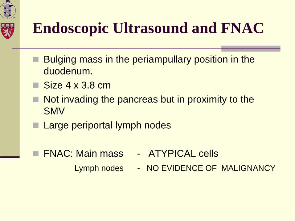
Endoscopic Ultrasound and FNAC
Bulging mass in the periampullary position in the duodenum.
Size 4 x 3.8 cm
Not invading the pancreas but in proximity to the SMV
Large periportal lymph nodes
FNAC: Main mass - ATYPICAL cells
Lymph nodes - NO EVIDENCE OF MALIGNANCY
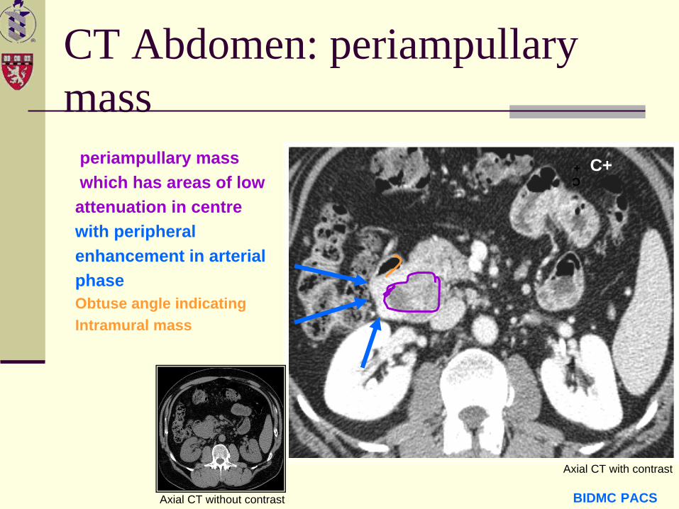
CT Abdomen: periampullary mass
periampullary masswhich has areas of lowattenuation in centrewith peripheral enhancement in arterial phaseObtuse angle indicatingIntramural mass
C+ C+
BIDMC PACS
Axial CT with contrast
Axial CT without contrast
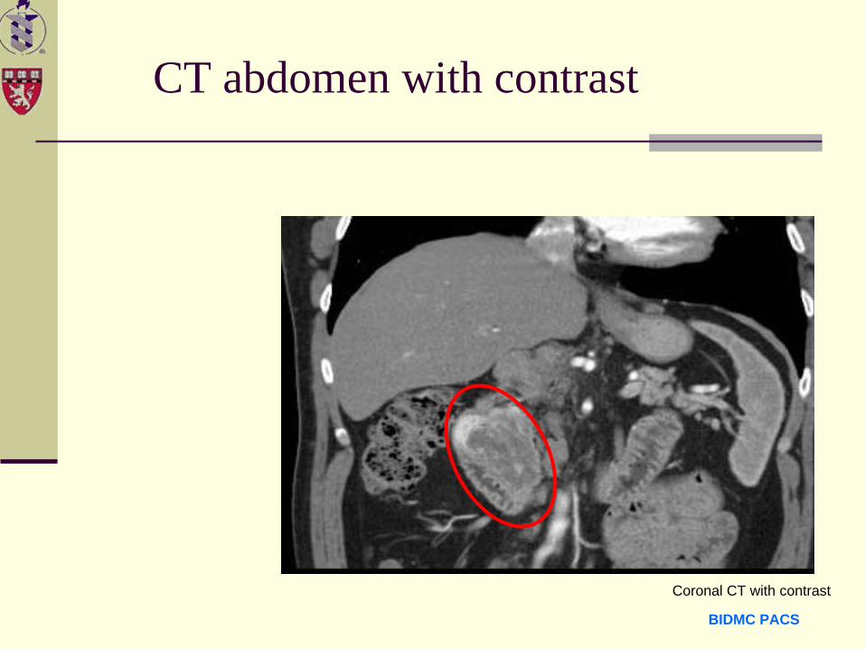
CT abdomen with contrast
Coronal CT with contrast
BIDMC PACS
PV
BIDMC PACS
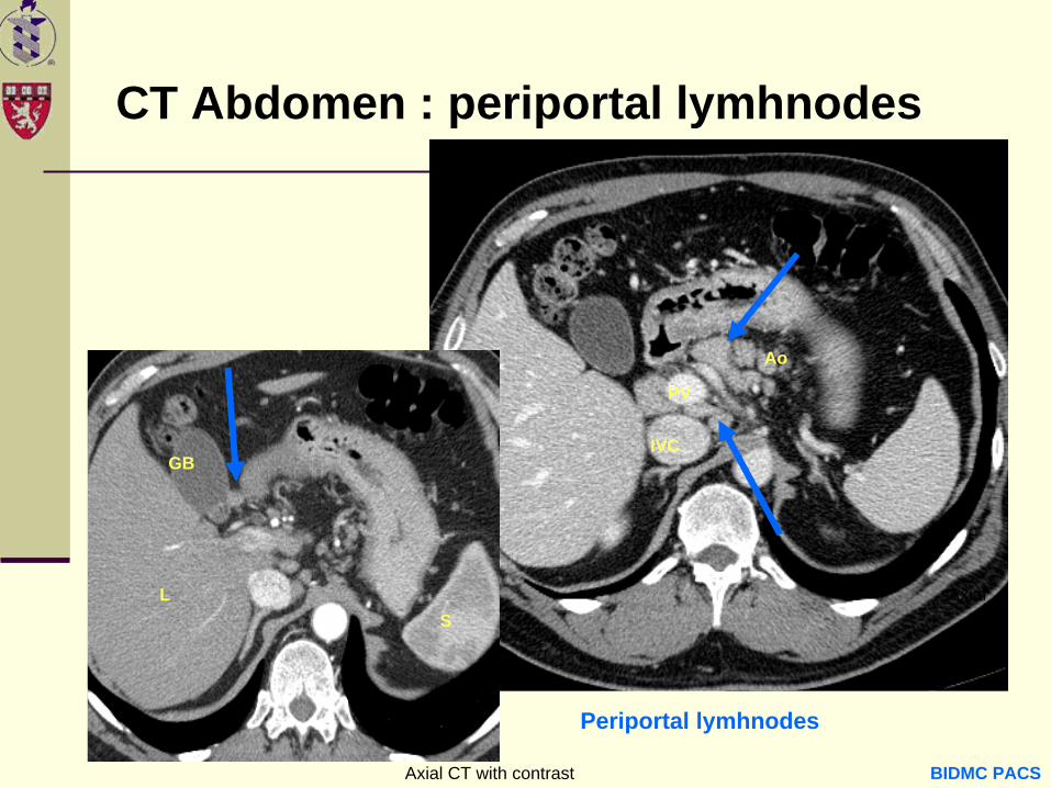
Periportal lymhnodes
GB
LS
IVC
Ao
Axial
CT Abdomen : periportal lymhnodes
Axial CT with contrast
CT findings
Large exophytic mass arising from the region of the neck of the pancreas.
Predominantly arterially enhancing with large areas of necrosis.
Multiple locally enlarged frankly pathologic appearing lymph nodes.
No frank metastases were identified.

Differential Diagnosis
GIST of duodenum
Non-functioning endocrine tumor
Lymphoma (focal)
Duodenal villous adenocarcinoma
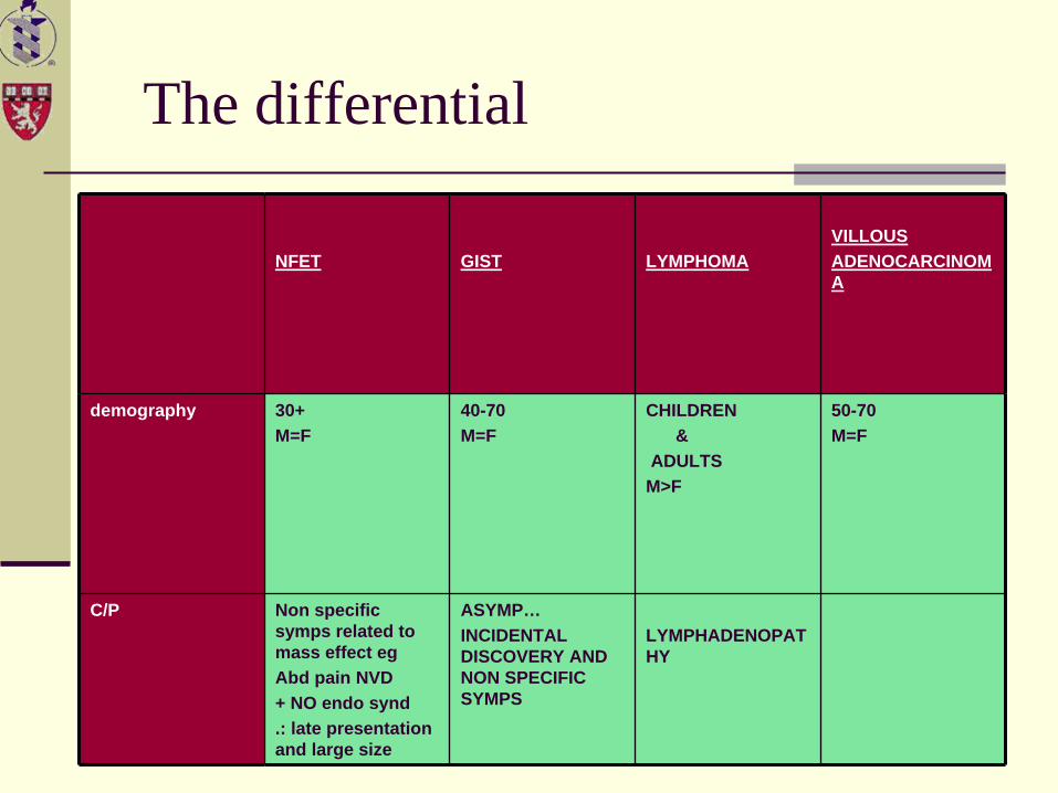
The differential
NFET GIST LYMPHOMAVILLOUSADENOCARCINOM A
demography 30+M=F
40-70M=F
CHILDREN &
ADULTSM>F
50-70M=F
C/P Non specific symps related to mass effect egAbd pain NVD + NO endo synd.: late presentation and large size
ASYMP…INCIDENTAL DISCOVERY AND NON SPECIFIC SYMPS
LYMPHADENOPAT HY
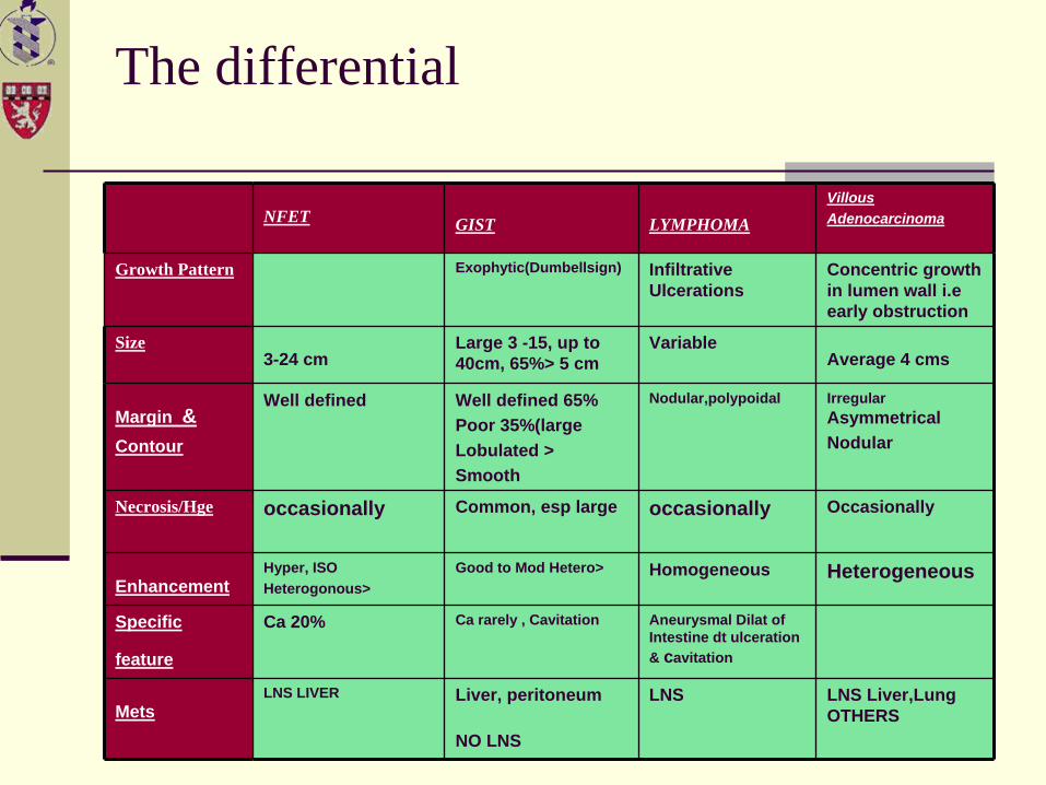
The differential
NFET GIST LYMPHOMAVillousAdenocarcinoma
Growth Pattern Exophytic(Dumbellsign) Infiltrative Ulcerations
Concentric growth in lumen wall i.e early obstruction
Size 3-24 cm
Large 3 -15, up to 40cm, 65%> 5 cm
VariableAverage 4 cms
Margin &Contour
Well defined Well defined 65%Poor 35%(large Lobulated >Smooth
Nodular,polypoidal Irregular AsymmetricalNodular
Necrosis/Hge occasionally Common, esp large occasionally Occasionally
EnhancementHyper, ISOHeterogonous>
Good to Mod Hetero> Homogeneous Heterogeneous
Specific
feature
Ca 20% Ca rarely , Cavitation Aneurysmal Dilat of Intestine dt ulceration & cavitation
MetsLNS LIVER Liver, peritoneum
NO LNS
LNS LNS Liver,Lung OTHERS
Management & Complication
Patient underwent a Whipple’s procedure and tumor was completely resected.
Pathology report
significant for GIST in the duodenum
all other tissues (pancreas, LNS, Gall bladder) came in with normal findings.
Patient developed a fistula as a complication of the procedure which resolved over the course of 2 months.

Gist: background & pathophysiology
GIST is a rare tumor of the GI about 3% of all tumors.
Accounts for 80% of mesenchymal tumors
Interstitial cells of Cajal of the intestine are argued to be the precursors of the GIST or at least shares a common precursor cell.

GIST: background & pathophysiology
GIST expresses CD117 antigen ,also known as C- KIT protein membrane receptor.
On mutation the Tyrosine kinase component of this antigen is unchecked leading to unchecked growth of cells and tumor development.
Most common sites are
Stomach 60-70%,
Small intestine 20-30%,
Anorectal 10%
Esophagus 1%
GIST: Demography
Demography
Age: Shows unimodal peak incidence in
age groups between 40-70yrs.
Sex: Almost equal incidence.
Race: No predilection.
GIST: Morbidity & Mortality
Patients with primary disease (no mets) show a median disease specific survival of 60 months(5 yrs)
Patients with Mets show 10-24 months ,and patients with recurrence show 12 months of survival.
RECURRENCE IS TYPICAL ,40% some suggest 91% on long term follow up.
GIST: clinical presentation
Up to 75% of GISTs are discovered when they are less than 4 cm in diameter and are either Asymptomatic or associated with Nonspecific symptoms
frequently diagnosed incidentally
Symptoms are vague, nonspecific abdominal pain or discomfort.
Patients also describe early satiety or a sensation of abdominal fullness. Rarely, an abdominal mass is palpable.
GISTs may also produce symptoms secondary to obstruction or hemorrhage
GISTs occur with a higher than expected frequency in patients with type 1 neurofibromatosis.
GIST:Differential Diagnosis
Differential diagnosis:
Gastrointestinal carcinoid
Adenocarcinoma
Gastric carcinoma
Liposarcoma
Others to be Considered:
Angiosarcoma
Inflammatory fibroid polyp
Inflammatory myofibroblastic tumor (pseudotumor, fibrosarcoma)
ntra-abdominal fibromatosis
Kaposi sarcoma
Lipoma
Lymphoma, abdominal
Melanoma, metastatic
Schwannoma, GI
GIST: Menu of tests
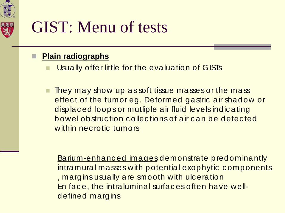
Plain radiographs
Usually offer little for the evaluation of GISTs
They may show up as soft tissue masses or the mass effect of the tumor eg. Deformed gastric air shadow or displaced loops or mutliple air fluid levels indicating bowel obstruction collections of air can be detected within necrotic tumors
Barium-enhanced images demonstrate predominantly intramural masses with potential exophytic components , margins usually are smooth with ulcerationEn face, the intraluminal surfaces often have well- defined margins

XRAY AND BARIUM IMAGES OF COMPANION PATIENTS WITH GIST
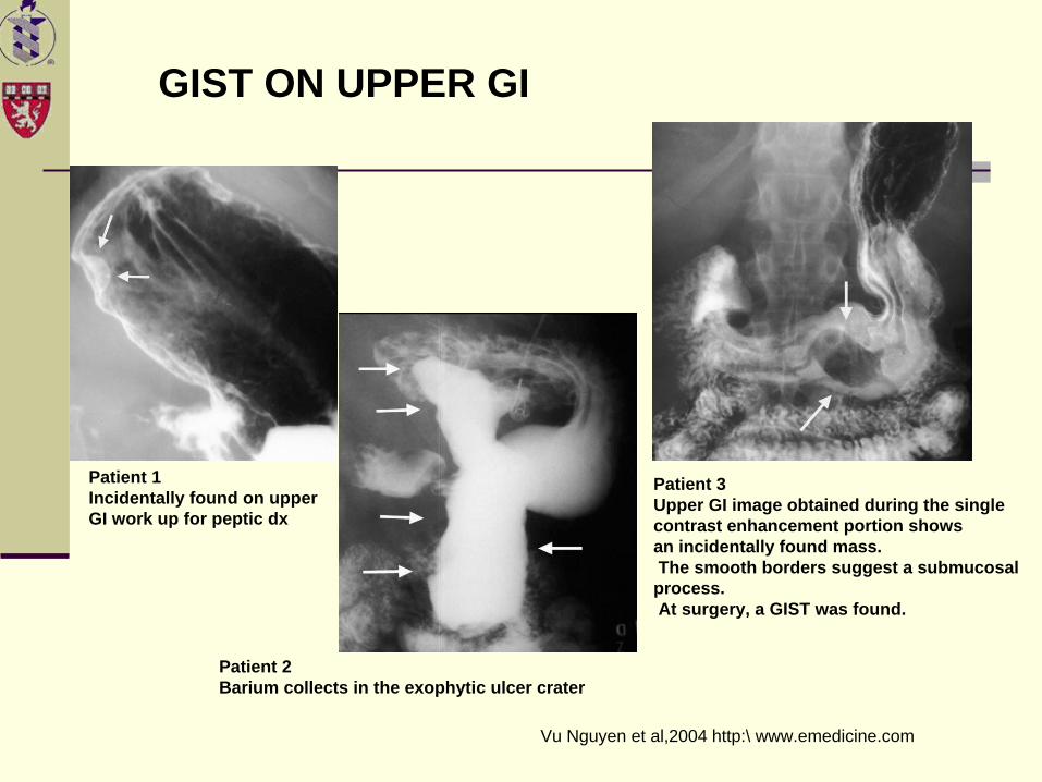
.. Patient 1Incidentally found on upperGI work up for peptic dx
Patient 2Barium collects in the exophytic ulcer crater
Patient 3Upper GI image obtained during the single contrast enhancement portion showsan incidentally found mass.The smooth borders suggest a submucosal
process.At surgery, a GIST was found.
GIST ON UPPER GI
Vu Nguyen et al,2004 http:\ www.emedicine.com
GIST:Ultrasound
Ultrasound
larger GISTs appear as complex masses with cystic and solid components
Endoscopic ultrsound:
hypoechoic masses that are contiguous with the fourth hypoechoic layer of the GI wall, which corresponds to the muscularis propria
Characteristics associated with malignancy include tumor size greater than 4 cm, an irregular extraluminal border, echogenic foci, and cystic spaces
GIST: CT Scan
CT is ideal in defining the endoluminal and exophytic extent of tumor.
Smaller GISTs appear as smooth, sharply defined intramural masses with homogenous attenuation.
Larger GISTs with necrosis appear as heterogeneous masses with enhancing borders of variable thickness and irregular central areas of fluid, air, or oral contrast attenuation that reflect necrosis
Occasionally, dense focal calcifications
Overlying mucosal ulcerations and extension into nearby structures may be present.

GIST: CT Scan
Metastasis in GIST are mainly hepatic and peritonealGIST DOES NOT METASTASIZE TO LNS Lymphnode metastasis is uncommon and is a characteristic radiologic finding of GISTCT is sensitive for the detection of metastatic liver lesions.Liver lesions can be hypervascular or appear as cystic multilocular lesions with fluid-fluid levels
CT IMAGES OF COMPANION PATIENTS WITH GIST

Companion patient 4: GIST in Fundus of stomach showing dumbell sign
Vu Nguyen et al,2004 http:\ www.emedicine.comAxial CT of the abdomen with contrast

Companion patient 5: GIST in SI
Vu Nguyen et al,2004 http:\ www.emedicine.comAxial CT of the abdomen

GIST: (Mets in liver pre / post Gleevec)
Vu Nguyen et al,2004 http:\ www.emedicine.com
Pre Gleevec
POST Gleevec
Axial CT of the abdomen with contrast
Axial CT of the abdomen with contrast

GIST: CT Scan
GISTs appear as sharply delineated, heterogeneous masses with cystic and necrotic areas.
The masses tend to be isointense relative to skeletal muscle on T1-weighted images and hyperintense on T2-weighted images

GIST: benign vs malignant
Unfortunately, no standard exists for their classification.
Many criteria such as number of mitotic figures, size, presence of necrosis and hemorrhage among others.
Size is the most important and most reliable
Tumor <5 cm is described as having low malignancy potential
Tumor >5 cm is described as being of high malignancy potential.

GIST: Treatment
Surgery remains the definitive treatment of choice.
Only effective, specific, nonsurgical therapy for GISTs is imatinib mesylate (gleevec)
Radiation and Chemo have yielded poor results.

In Conclusion
Key points to Remember:
GIST is RARE….Demo:40-70 yrs/M=FPatho: CD117 +VE (C-KIT) in more than 90%C/P:Non specific .Vague Abd pain,NVD rarely obstRadio: CT best modality proven.
Smaller GISTs appear as smooth, sharply defined intramural masses with homogenous attenuation
Larger GISTs with necrosis appear as heterogeneous masses with enhancing borders of variable thickness and irregular central areas of fluid
Bottom line: Exophytic mass, heterogenous ,hyperenhanecing Liver Mets with NO LNS is very suggestive of GIST
In Conclusion
Role of Radiology in GIST
It is more of a suggestive tool rather than a cut off measure .
Determines potential malignancy via size
In most literature CT is thought to be the best modality available (However some advocate that MRI is the gold standard?)
In Conclusion
Worthy of note
Daignosis of GIST may be complemented by immunohistochemical studies of FNAC samples.
However not all CD117+ cells are GIST.

References:
Gastrointestinal stromal tumors of the Duodenum:CT and Barium Findings
Mye-Cheol kim et al AJR August 2004
Case Report :Villous Adenocarcinoma of the Duodenum invading the ampulla of Vater . G.Catania et al HPB surgery 1996
Gastrointestinal Stromal tumors:Clinical, radiologic and pathologic features .Kumaresan Sandrasegraran et al AJR 2005
Textbook of Gastrointestinal Radiology. Gore , Levine and Laufer 1994
Gastrointestinal Stromal Tumors Michael AJ Sawyer et al emedicine.com 2006
Gastrointestinal Stromal Tumors - Leiomyoma/Leiomyosarcoma Vu Nguyen et al emedicine.com 2004

Acknowledgements
Dr. Maryellen Sun for her reference to this case
Dr. Raptopolis for guiding us through the case
Maria Levantakis for always being there for us
Dr.Lieberman for her undying support
And all attendings, fellows and residents who provided us with all the resources we needed and left no question unanswered Aaron,Arti,Alice and Justin…..

Thank you