gastric mucosal changes and ghrelin expression and...
TRANSCRIPT

Journal of Surgery 2018; 6(2): 36-42
http://www.sciencepublishinggroup.com/j/js
doi: 10.11648/j.js.20180602.12
ISSN: 2330-0914 (Print); ISSN: 2330-0930 (Online)
Gastric Mucosal Changes and Ghrelin Expression and Their Relation to Weight Reduction After Sleeve Gastrectomy
Sabry Ahmed Mahmoud1, Ahmed Fadaly Hussein
1, *, Waleed Omar
1, Emad Abdallah
1,
Wagdi Elkashef2, Mohamed Anwar
1, Sameh Emile
1
1General Surgery Department, Mansoura University Hospitals, Mansoura University, Mansoura, Egypt 2Pathology Department, Mansoura Faculty of Medicine, Mansoura University, Mansoura, Egypt
Email address:
*Corresponding author
To cite this article: Sabry Ahmed Mahmoud, Ahmed Fadaly Hussein, Waleed Omar, Emad Abdallah, Wagdi Elkashef, Mohamed Anwar, Sameh Emile. Gastric
Mucosal Changes and Ghrelin Expression and Their Relation to Weight Reduction After Sleeve Gastrectomy. Journal of Surgery.
Vol. 6, No. 2, 2018, pp. 36-42. doi: 10.11648/j.js.20180602.12
Received: February 6, 2018; Accepted: February 24, 2018; Published: March 19, 2018
Abstract: Background: Weight loss after laparoscopic sleeve gastrectomy (LSG) is usually attributed to the volume
restrictive effect of the procedure in addition to specific hormonal changes. Objective: The present study aimed to investigate
the changes in plasma ghrelin levels, the number of ghrelin producing cells and histopathological changes in the remaining
pouch after LSG. Methods: The present study included 27 patients with morbid obesity. The plasma ghrelin levels were
measured before and six months after LSG and the change in their levels was assessed in relation to body mass index (BMI)
after LSG. Immunohistochemical staining of cellular ghrelin was used to evaluate the number and distribution of ghrelin
producing cells in the resected specimen and the mucosal changes in the remaining gastric pouch after LSG were assessed at 6
months postoperatively. Results: The mean age of patients was 33.9± 21.9 years. At six months after LSG, BMI decreased
from 52.6± 12.8 to 40.8± 7.2 Kg/m2 (p= 0.0001). The plasma ghrelin level decreased significantly from 564.1± 15 to 434.7±
22.6 at six months after LSG. There was strong positive correlation between BMI after LSG and plasma ghrelin level (R=
0.906, p= <0.0001). A significant improvement in the inflammatory parameters was noticed by histopathologic examination.
The mean ghrelin positive cell per specimen decreased significantly from 25.37± 3.5 to 13.7 ± 2.12. Conclusion: There was
good positive correlation between weight loss and lowering of plasma ghrelin level, also complete removal of the fundus was
associated with more weight loss.
Keywords: Sleeve Gastrectomy, Mucosal Changes, Ghrelin, Weight Loss
1. Introduction
Obesity is a pan-endemic health problem in both
developed and developing countries that can be associated
with a high incidence of complications and decrease in life
expectancy, especially among younger adults. Obesity is
classified into classes according to the body mass index
(BMI) with morbid obesity defined as BMI ≥ 40 kg/m2 or a
BMI ≥ 35 kg/m2 in patients with at least one associated
comorbidity [1].
While conservative measures may succeed in treatment of
obesity in some patients, bariatric surgery remains a viable
option for the treatment of morbid obesity, resulting long-
lasting weight loss, improved quality of life, and resolution
of obesity- related comorbidities [2]. With the application of
laparoscopy to bariatric surgery a major expansion in the
number of laparoscopic bariatric procedures has been noted
worldwide [3].
Laparoscopic sleeve gastrectomy (LSG) is one of the most
popular bariatric procedures performed for morbid obesity

Journal of Surgery 2018; 6(2): 36-42 37
[4]. The mechanism of weight loss after sleeve gastrectomy
is attributed to a combination of volume restriction, creation
of a high pressure system, and the induction of a favorable
hormonal change [5].
A decline in appetite after LSG has been observed,
presumably due, in part, to ghrelin cell removal. Ghrelin is an
orexigenic hormone produced primarily by cells in the
oxyntic glands of the stomach mainly in the gastric fundus as
it has been demonstrated that the expression of Ghrelin
mRNA expression in the fundus is three fold higher than in
the pre-pyloric area and the same pattern was also seen in
ghrelin cell distribution [6].
Ghrelin hormone plays a physiologic role in the regulation
of food intake as its plasma levels rise during food
deprivation and decline after food ingestion [7]. In addition,
Ghrelin is involved in many biological processes ranging
from appetite regulation and growth hormone release to gut
motility [8]. The changes in plasma ghrelin levels after
different bariatric procedures vary according to the
procedure. While plasma ghrelin levels tend to decrease after
gastric bypass and after LSG, they tend to rise after gastric
banding in a way similar to dieting [9].
The present study aimed to evaluate the changes in plasma
ghrelin hormone at six months after LSG, correlating these
changes with weight loss. The study also aimed to investigate
the distribution of ghrelin producing cells in different regions
of the stomach and the change in their number after LSG and
to evaluate the histopathological changes after sleeve
gastrectomy since the evaluation of these changes can
improve the understanding of the local mechanism and the
outcome of LSG as Vrabie et al. implied [10].
2. Patients and Methods
This is a prospective cohort study on patients with morbid
obesity who underwent LSG in the General Surgery
Department of Mansoura University Hospitals in the period
of May 2015 to May 2017. Ethical approval of the study was
obtained from the Institutional Review Board (IRB) of
Mansoura Faculty of Medicine.
All patients with morbid obesity with BMI >40 kg/m2 or
BMI >35 kg/m2 with at least one obesity-related comorbidity
who failed conservative treatments were included to the
study.
We excluded patients who underwent previous bariatric
surgery, patients unfit for anesthesia as patients, patients with
gastric pathologies as peptic ulcer or neoplasm, patients with
secondary obesity due to endocrine disorders, patients with
psychological problems or patients unwilling to comply with
postoperative diet regimen.
Patients were asked about the onset and duration of obesity
and any obesity-related comorbidities such as diabetes
mellitus, hypertension, sleep apnea, and joint pain. Patients
were also asked about previous treatments for morbid obesity
including diet regimens, weight loss medications, exercise
programs, interventional therapies as intragastric ballon, and
any previous surgical procedures for morbid obesity or other
abdominal conditions. Detailed dietary history was taken
from all patients with regards to type and frequency of meals,
eating snacks in between meals, drinking of water or
beverages, level of appetite, and feeling of satiety after each
meal.
General examination was conducted to all patients.
Patients’ weight and height were recorded and BMI was then
calculated using the following equation: BMI= Weight in
kilograms/ (Height in meters)2. Abdominal examination was
performed to exclude presence of any abdominal masses,
ascites, and ventral or groin hernias.
Routine preoperative laboratory investigations including
complete blood count, liver and kidney functions tests,
prothrombin time, and random blood glucose were done to
confirm anesthetic fitness. Endocrine and metabolic panels
including serum lipid profile, thyroid function tests, and
fasting and postprandial blood sugar levels were done to
exclude secondary causes of obesity. Plasma ghrelin levels
were measured in all patients within 48 hours before the
procedure.
Abdominal ultrasonography was performed in all patients
to exclude any associated abdominal pathologies. The cardiac
function was evaluated by electrocardiography and
echocardiography in select patients and pulmonary function
tests were done to assess respiratory functions and chest
condition.
Informed consent for the study was taken from all patients.
Liquid diet was prescribed for 24 hours before the procedure
with fasting for at least six hours before the onset of
anesthesia.
Preoperative measures against thromboembolism were
taken in the form of: wearing elastic stocking 24 hours before
the operation, subcutaneous administration of low molecular
weight heparin (Enoxaprin, 40 IU) at the night of surgery.
Prophylactic antibiotics (cefotaxime 1 gm) were administered
intravenously on induction of anesthesia.
All procedures were done under general anesthesia with
end tracheal intubation. Patients were placed in the supine
position being appropriately secured to the operating table
with padding of all pressure points. The operating surgeon
stood between the legs of the patient and the two assistants
stood on each side of the patient.
After scrubbing of the abdomen with povidone iodine,
peumoperitoneum (15-17 mmHg) was established by
insertion of veress needle above the umbilicus and
insufflation of CO2. Trocars were inserted under direct vision
as follows: a 5-mm trocar is inserted in the subxiphoid area
for the liver retractor, another 5-mm trocar is placed in the
left upper quadrant at the anterior axillary line just below the
12th
rib for the assistant, additional 12-mm trocars were
placed in the right upper quadrant, epigastrium, left upper
quadrant, and right paramedian regions for inserting staplers.
Upon identification of the pylorus by visualizing the
prepyloric vein of Mayo, the greater curvature of the stomach
was devascularized using an advanced vessel-sealing device
(Harmonic scalpelTM
or LigasureTM
) starting at 6 cm away
from the pylorus. Devascularization continued proximally

38 Sabry Ahmed Mahmoud et al.: Gastric Mucosal Changes and Ghrelin Expression and Their Relation to Weight
Reduction After Sleeve Gastrectomy
onto the fundus until the left crus of the diaphragmatic hiatus
was clearly identified. All the short gastric vessels were
divided along the way. After devascularizaion was
completed, the hiatus was assessed for hernias and if large
hiatal hernia was encountered, it was repaired using standard
laparoscopic techniques.
A 36 French bougie was then inserted transorally by the
anesthesiologist, advanced under direct vision to the pylorus,
and then positioned against the lesser curvature. The gastric
transection was then performed using sequential applications
of 60-mm linear staplers beginning at 6 cm proximal to the
pylorus. As the gastric transection proceeds, the height of the
staples may need to be adjusted according to the thickness of
the tissue. We used green stapler at pylorus, then gold
staplers at middle of stomach, then blue staplers at fundus.
After the stomach was transected, an intraoperative leak
test was performed by injection of methylene blue in the
gastric sleeve. The resected portion of the stomach was
extracted through the right upper quadrant trocar then the
trocar sites were closed and an intra-abdominal drain was
inserted.
Patients were monitored in the regular ward with regards
vital signs, output of abdominal drain, and postoperative
complications. Intravenous fluids were administered for 24
hours postoperatively and one gram of cefotaxime was
administered intravenously every 12 hours. Analgesia was
achieved using non-steroidal anti-inflammatory drugs
(NSAIDs) or Nalufin amp. A second dose of low molecular
weight heparin was given subcutaneously 8 hours after the
operation then was repeated every 24 hours. Early
ambulation was advised in all patients. Resumption of oral
feeding was scheduled in every patient. Once assured to be
stable, started oral fluids, with no complaints, patients were
discharged home within 48 hours of the procedure.
The sleeve gastrectomy specimens were fixed in 4%
formaldehyde and embedded in paraffin, and 4 mm thick
sections from each specimen were stained with hematoxylin
and eosin for histopathological examination. The biopsy
specimens were examined in a blind manner by a pathologist
to indicate:
1- Presence of lymphoid follicles (Scored 0: absent, 1:
mild, 2: moderate, 3: Marked). 2- Interstitial inflammation
(Scored 0: absent, 1: mild, 2: moderate, 3: Marked).
Ghrelin immunoreactivity was determined on sections
after microwave pretreatment (10 min in 0.01 M citric acid
solution) and overnight incubation at 4°C with human
polyclonal antibody (Chongqing biopsies Co., Ltd.,
Chongqing, China) diluted 1:100. Diaminobenzidine–
hydrogen peroxide was used as chromogen, and the sections
were counterstained with diluted hematoxylin.
Ghrelin positive cells were counted in three (hot spots) in
areas containing mucosa in full thickness. The total number
was divided by 3 to obtain mean Ghrelin positive cell per
specimen.
Patients were asked to visit the outpatient clinic at one and
two weeks after sleeve gastrectomy for assessment of general
condition, wound status, and for removal of stitches, then
every month for six months.
At Six months after the procedure, body weight was
recorded and the amount and percentage of weight loss was
calculated and compared with the preoperative value. BMI
was calculated to estimate the reduction in BMI after surgery.
A blood sample was withdrawn and plasma ghrelin levels
were measured and compared to the preoperative value. In
addition, all patients underwent gastroscopy to evaluate the
condition of mucosa of the remaining pouch and take three
tissue samples from the remaining part of the stomach.
Tissue samples were examined pathologically with the
same techniques described earlier to assess postoperative
changes in ghrelin gene expression, mucosal cells
distribution and to demonstrate any histopathological
changes.
Data were analyzed using SPSS program version 23 (IBM
corp., Bristol, UK). Continuous variables were expressed as
mean and standard deviation (SD) or median and normal
range. Categorical variables were expressed as number and
percentages. Student t-test was used for processing of
quantitative data whereas Fisher's exact test was used for
processing of qualitative data. The correlation between BMI
and plasma ghrelin levels was measured using Pearson
correlation test. P value less than 0.05 was considered
significant.
3. Results
The present study included 27 patients with morbid obesity
who underwent LSG. Patients were all females except one
male. The mean age of patients was 33.9± 21.9 years,
ranging from 21 to 63 years.
Twenty-four patients had obesity-related comorbidities:
nine patients had type II DM, five had arterial hypertension,
ten had dyslipidemia, and four had joint pain. In addition, 12
patients had fatty liver and two had gall bladder stones in
abdominal ultrasonography.
The mean preoperative body weight was 137± 3.5 (range,
108- 164) Kg. The mean preoperative height of patients was
162.4 ± 21.2 (range, 148- 183) cm. The mean preoperative
BMI was 52.6± 12.8 (range, 38.8- 66.6) Kg/m2.
The mean operating time was 104.2± 14.1 (range, 80-135)
minutes. The mean hospital stay was 2.3± 0.4 (range, 2-4)
days. There was no conversion to open surgery in any
patient. Intraperitoneal drains were placed in 21 (77.7%)
patients.
No major complications or mortality were recorded. Minor
complications were recorded in 12 patients, wound infection
in 3 patients, chest infection in 5 patients, and vomiting in 4
patients. Simultaneous cholecystectomy was conducted with
LSG in two patients.
At six months postoperatively, significant weight loss was
noted as the mean preoperative body weight dropped from
137± 3.5 to 107± 2.12 Kg (p< 0.0001). The preoperative
BMI decreased from 52.6± 12.8 to 40.8± 7.2 Kg/m2 (p=
0.0001). The mean percentage of excess weight loss was
41.7± 11.1 (range, 29.3- 64.4) (table 1).
Journal of Surgery 2018; 6(2): 36-42 39
Table 1. Weight loss after sleeve gastrectomy.
Variable Preoperative Postoperative P value
Body weight 137± 3.5 107± 2.12 < 0.0001
BMI 52.6± 12.8 40.8± 7.2 <0.0001
%EWL ------- 41.7± 11.1 ------
Improvement in obesity associated comorbidities was
observed in 6 (66.7%) of nine diabetic patients, three (60%)
of five patients with hypertension, and one (25%) of four
patients with joint pain (table 2).
Table 2. Improvement in co-morbidities loss after sleeve gastrectomy.
Variable Preoperative (%) Postoperative (%) P value
D. M 9 (33.3) 3 (11.1) 0.1
Hypertension 5 (18.5) 2 (7.4) 0.42
Joint pain 4 (14.8) 3 (11.1) 1
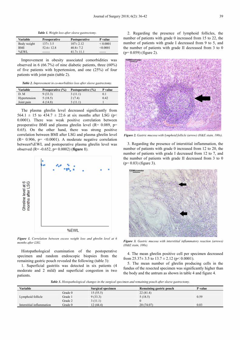
The plasma ghrelin level decreased significantly from
564.1 ± 15 to 434.7 ± 22.6 at six months after LSG (p<
0.0001). There was weak positive correlation between
preoperative BMI and plasma ghrelin level (R= 0.089, p=
0.65). On the other hand, there was strong positive
correlation between BMI after LSG and plasma ghrelin level
(R= 0.906, p= <0.0001). A moderate negative correlation
between%EWL and postoperative plasma ghrelin level was
observed (R= -0.652, p= 0.0002) (figure 1).
Figure 1. Correlation between excess weight loss and ghrelin level at 6
months after LSG.
Histopathological examination of the postoperative
specimen and random endoscopic biopsies from the
remaining gastric pouch revealed the following (table 3):
1. Superficial gastritis was detected in six patients (4
moderate and 2 mild) and superficial congestion in two
patients.
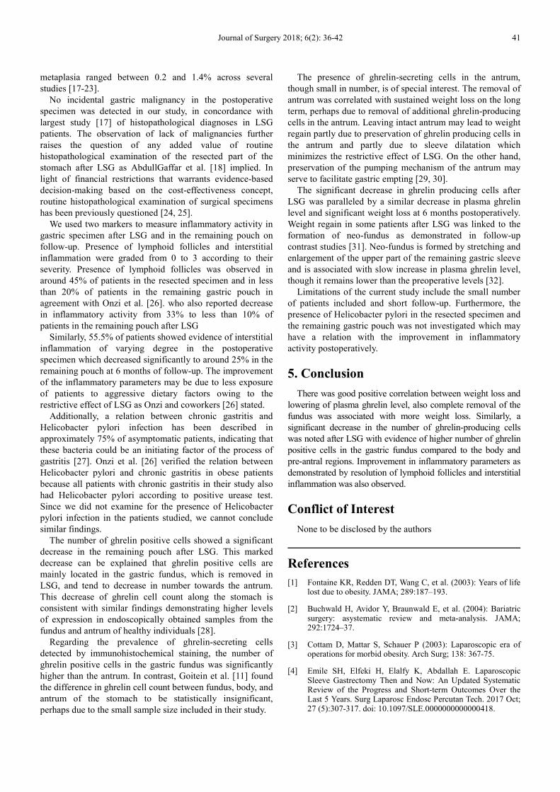
2. Regarding the presence of lymphoid follicles, the
number of patients with grade 0 increased from 15 to 22, the
number of patients with grade I decreased from 9 to 5, and
the number of patients with grade II decreased from 3 to 0
(p= 0.059) (figure 2).
Figure 2. Gastric mucosa with lymphoid follicle (arrow) (H&E stain, 100x).
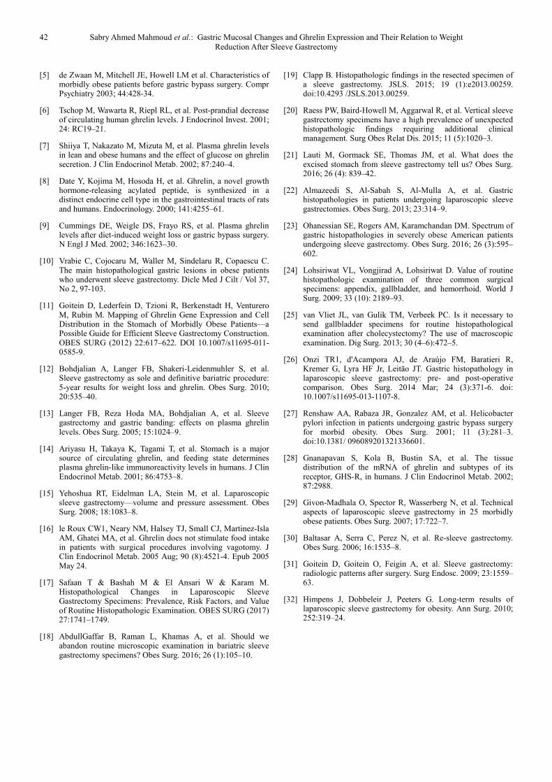
3. Regarding the presence of interstitial inflammation, the
number of patients with grade 0 increased from 12 to 20, the
number of patients with grade I decreased from 12 to 7, and
the number of patients with grade II decreased from 3 to 0
(p= 0.03) (figure 3).
Figure 3. Gastric mucosa with interstitial inflammatory reaction (arrows)
(H&E stain, 100x).
4. The mean ghrelin positive cell per specimen decreased
from 25.37± 3.5 to 13.7 ± 2.12 (p< 0.0001).
5. The mean number of ghrelin producing cells in the
fundus of the resected specimen was significantly higher than
the body and the antrum as shown in table 4 and figure 4.
Table 3. Histopathological changes in the surgical specimen and remaining pouch after sleeve gastrectomy.
Variable Surgical specimen Remaining gastric pouch P value
Lymphoid follicle
Grade 0 15 (55.5) 22 (81.4)
0.59 Grade 1 9 (33.3) 5 (18.5)
Grade 2 3 (11.1) 0
Interstitial inflammation Grade 0 12 (44.4) 20 (74.07) 0.03

40 Sabry Ahmed Mahmoud et al.: Gastric Mucosal Changes and Ghrelin Expression and Their Relation to Weight
Reduction After Sleeve Gastrectomy
Variable Surgical specimen Remaining gastric pouch P value
Grade 1 12 (44.4) 7 (25.9)
Grade 2 3 (11.1) 0
Table 4. Immunohistochemical distribution of ghrelin producing cells in the regions of the resected specimen.
Variable The fundus The body P value The antrum P value
Ghrelin producing cells (mean± SD) 15 ± 3.7 7.1± 3.1 <0.0001 3.3± 1.8 <0.0001
Figure 4. Gastric mucosa with positive reaction to Ghrelin
immunohistochemistry (Brown staining, arrows) (Ghrelin
immunohistochemical staining, 400x).
4. Discussion
Twenty-seven patients with morbid obesity were admitted
and underwent LSG in this prospective observational study.
Almost all patients were females reflecting the female
predominance of obesity as documented in the literature. The
majority of patients were of middle age in concordance with
the average age of patients undergoing bariatric surgery for
morbid obesity [4].
Significant weight loss at 6 months after LSG was
recorded with a significant decline in the preoperative BMI.
The average percentage of excess weight loss was around
40%. Improvement in associated co-morbidities was
observed yet was not statistically significant, perhaps due to
the short duration of follow-up.
We studied plasma ghrelin levels before and after LSG,
correlating the changes in these levels with weight loss after
the procedure. A significant decrease in plasma ghrelin levels
at 6 months after LSG was recorded which was associated
with significant weight loss as demonstrated by decline in
BMI. In concordance, other researchers found total plasma
ghrelin levels decrease significantly at 3 months after LSG,
with simultaneous significant weight loss [11].
Plasma ghrelin levels have been shown to decrease after
sleeve gastrectomy and its low levels can be maintained up to
five years after surgery [12, 13]. Since ghrelin is mainly
produced by oxyntic cells in the stomach in addition to other
parts of the gastrointestinal tract, it is logical that plasma
ghrelin levels decline after resecting around 75% of the
stomach volume in LSG [14].
We identified a weak correlation between plasma ghrelin
levels and BMI before surgery, nevertheless this positive
correlation became stronger at 6 months after LSG and this
correlation was statistically significant. Similarly, the
percentage of excess weight loss at 6 months had moderately
strong negative correlation with plasma ghrelin implying that
the drop in plasma ghrelin levels was associated with higher
percentage of weight loss. In discordance, Goitein et al. [11]
found no distinct correlation between plasma ghrelin levels
and weight loss which they explained that plasma ghrelin is
only one of multiple factors contributing to the success of
LSG and that weight loss per se is not responsible for ghrelin
level change [15].
The significant strong correlation we observed between
plasma ghrelin and BMI after surgery can be attributed to
different patient characteristics compared to the study of
Goitein et al. [11] where the BMI of our patients was
obviously higher than their study. In addition, we made the
correlation at six months after LSG, whereas the other
investigators made the same correlation at shorter period of
follow-up (three months postoperatively).
Ghrelin hormone has been recognized to contribute to the
incidence of morbid obesity through different mechanisms.
Ghrelin is one of the orexigenic peptide transmitters know to
stimulate appetite and induce a positive energy balance
leading to weight gain. It is implicated in regulating mealtime
hunger and meal initiation. Since ghrelin hormone is
considered a signal of starvation, it can induce sustained
adiposity as long as its levels are elevated [16].
Histopathological examination of the resected part of the
stomach after LSG revealed abnormal pathologic changes in
55.5% of patients in the present study. This implies that
around 45% of patients had normal histopathology of the
stomach preoperatively which is lower than other studies [17-
19] that reported normal histopathology in 50-54% of
patients. However, similar to our findings other investigators
reported normal histopathology of the stomach in 35.2 and
46.3% of patients [20, 21].
Abnormal histopathological findings in the present study
involved chronic gastritis, either active or inactive which is in
line with Safaan et al. [17] who reported chronic gastritis to
be the most common abnormal histopathological findings in
the resected part of the stomach after LSG. Nonetheless, the
study by Safaan and colleagues [17] documented other
important pathological findings as intestinal metaplasia,
GIST, leiomyoma, lipoma, and pancreatic heterotropia,
however, these abnormal findings were found in a very low
incidence of patients. In fact, the incidence of GIST ranged
between 0.2 and 1% and the incidence of intestinal
Journal of Surgery 2018; 6(2): 36-42 41
metaplasia ranged between 0.2 and 1.4% across several
studies [17-23].
No incidental gastric malignancy in the postoperative
specimen was detected in our study, in concordance with
largest study [17] of histopathological diagnoses in LSG
patients. The observation of lack of malignancies further
raises the question of any added value of routine
histopathological examination of the resected part of the
stomach after LSG as AbdullGaffar et al. [18] implied. In
light of financial restrictions that warrants evidence-based
decision-making based on the cost-effectiveness concept,
routine histopathological examination of surgical specimens
has been previously questioned [24, 25].
We used two markers to measure inflammatory activity in
gastric specimen after LSG and in the remaining pouch on
follow-up. Presence of lymphoid follicles and interstitial
inflammation were graded from 0 to 3 according to their
severity. Presence of lymphoid follicles was observed in
around 45% of patients in the resected specimen and in less
than 20% of patients in the remaining gastric pouch in
agreement with Onzi et al. [26]. who also reported decrease
in inflammatory activity from 33% to less than 10% of
patients in the remaining pouch after LSG
Similarly, 55.5% of patients showed evidence of interstitial
inflammation of varying degree in the postoperative
specimen which decreased significantly to around 25% in the
remaining pouch at 6 months of follow-up. The improvement
of the inflammatory parameters may be due to less exposure
of patients to aggressive dietary factors owing to the
restrictive effect of LSG as Onzi and coworkers [26] stated.
Additionally, a relation between chronic gastritis and
Helicobacter pylori infection has been described in
approximately 75% of asymptomatic patients, indicating that
these bacteria could be an initiating factor of the process of
gastritis [27]. Onzi et al. [26] verified the relation between
Helicobacter pylori and chronic gastritis in obese patients
because all patients with chronic gastritis in their study also
had Helicobacter pylori according to positive urease test.
Since we did not examine for the presence of Helicobacter
pylori infection in the patients studied, we cannot conclude
similar findings.
The number of ghrelin positive cells showed a significant
decrease in the remaining pouch after LSG. This marked
decrease can be explained that ghrelin positive cells are
mainly located in the gastric fundus, which is removed in
LSG, and tend to decrease in number towards the antrum.
This decrease of ghrelin cell count along the stomach is
consistent with similar findings demonstrating higher levels
of expression in endoscopically obtained samples from the
fundus and antrum of healthy individuals [28].
Regarding the prevalence of ghrelin-secreting cells
detected by immunohistochemical staining, the number of
ghrelin positive cells in the gastric fundus was significantly
higher than the antrum. In contrast, Goitein et al. [11] found
the difference in ghrelin cell count between fundus, body, and
antrum of the stomach to be statistically insignificant,
perhaps due to the small sample size included in their study.
The presence of ghrelin-secreting cells in the antrum,
though small in number, is of special interest. The removal of
antrum was correlated with sustained weight loss on the long
term, perhaps due to removal of additional ghrelin-producing
cells in the antrum. Leaving intact antrum may lead to weight
regain partly due to preservation of ghrelin producing cells in
the antrum and partly due to sleeve dilatation which
minimizes the restrictive effect of LSG. On the other hand,
preservation of the pumping mechanism of the antrum may
serve to facilitate gastric empting [29, 30].
The significant decrease in ghrelin producing cells after
LSG was paralleled by a similar decrease in plasma ghrelin
level and significant weight loss at 6 months postoperatively.
Weight regain in some patients after LSG was linked to the
formation of neo-fundus as demonstrated in follow-up
contrast studies [31]. Neo-fundus is formed by stretching and
enlargement of the upper part of the remaining gastric sleeve
and is associated with slow increase in plasma ghrelin level,
though it remains lower than the preoperative levels [32].
Limitations of the current study include the small number
of patients included and short follow-up. Furthermore, the
presence of Helicobacter pylori in the resected specimen and
the remaining gastric pouch was not investigated which may
have a relation with the improvement in inflammatory
activity postoperatively.
5. Conclusion
There was good positive correlation between weight loss and
lowering of plasma ghrelin level, also complete removal of the
fundus was associated with more weight loss. Similarly, a
significant decrease in the number of ghrelin-producing cells
was noted after LSG with evidence of higher number of ghrelin
positive cells in the gastric fundus compared to the body and
pre-antral regions. Improvement in inflammatory parameters as
demonstrated by resolution of lymphoid follicles and interstitial
inflammation was also observed.
Conflict of Interest
None to be disclosed by the authors
References
[1] Fontaine KR, Redden DT, Wang C, et al. (2003): Years of life lost due to obesity. JAMA; 289:187–193.
[2] Buchwald H, Avidor Y, Braunwald E, et al. (2004): Bariatric surgery: asystematic review and meta-analysis. JAMA; 292:1724–37.
[3] Cottam D, Mattar S, Schauer P (2003): Laparoscopic era of operations for morbid obesity. Arch Surg; 138: 367-75.
[4] Emile SH, Elfeki H, Elalfy K, Abdallah E. Laparoscopic Sleeve Gastrectomy Then and Now: An Updated Systematic Review of the Progress and Short-term Outcomes Over the Last 5 Years. Surg Laparosc Endosc Percutan Tech. 2017 Oct; 27 (5):307-317. doi: 10.1097/SLE.0000000000000418.
42 Sabry Ahmed Mahmoud et al.: Gastric Mucosal Changes and Ghrelin Expression and Their Relation to Weight
Reduction After Sleeve Gastrectomy
[5] de Zwaan M, Mitchell JE, Howell LM et al. Characteristics of morbidly obese patients before gastric bypass surgery. Compr Psychiatry 2003; 44:428-34.
[6] Tschop M, Wawarta R, Riepl RL, et al. Post-prandial decrease of circulating human ghrelin levels. J Endocrinol Invest. 2001; 24: RC19–21.
[7] Shiiya T, Nakazato M, Mizuta M, et al. Plasma ghrelin levels in lean and obese humans and the effect of glucose on ghrelin secretion. J Clin Endocrinol Metab. 2002; 87:240–4.
[8] Date Y, Kojima M, Hosoda H, et al. Ghrelin, a novel growth hormone-releasing acylated peptide, is synthesized in a distinct endocrine cell type in the gastrointestinal tracts of rats and humans. Endocrinology. 2000; 141:4255–61.
[9] Cummings DE, Weigle DS, Frayo RS, et al. Plasma ghrelin levels after diet-induced weight loss or gastric bypass surgery. N Engl J Med. 2002; 346:1623–30.
[10] Vrabie C, Cojocaru M, Waller M, Sindelaru R, Copaescu C. The main histopathological gastric lesions in obese patients who underwent sleeve gastrectomy. Dicle Med J Cilt / Vol 37, No 2, 97-103.
[11] Goitein D, Lederfein D, Tzioni R, Berkenstadt H, Venturero M, Rubin M. Mapping of Ghrelin Gene Expression and Cell Distribution in the Stomach of Morbidly Obese Patients—a Possible Guide for Efficient Sleeve Gastrectomy Construction. OBES SURG (2012) 22:617–622. DOI 10.1007/s11695-011-0585-9.
[12] Bohdjalian A, Langer FB, Shakeri-Leidenmuhler S, et al. Sleeve gastrectomy as sole and definitive bariatric procedure: 5-year results for weight loss and ghrelin. Obes Surg. 2010; 20:535–40.
[13] Langer FB, Reza Hoda MA, Bohdjalian A, et al. Sleeve gastrectomy and gastric banding: effects on plasma ghrelin levels. Obes Surg. 2005; 15:1024–9.
[14] Ariyasu H, Takaya K, Tagami T, et al. Stomach is a major source of circulating ghrelin, and feeding state determines plasma ghrelin-like immunoreactivity levels in humans. J Clin Endocrinol Metab. 2001; 86:4753–8.
[15] Yehoshua RT, Eidelman LA, Stein M, et al. Laparoscopic sleeve gastrectomy—volume and pressure assessment. Obes Surg. 2008; 18:1083–8.
[16] le Roux CW1, Neary NM, Halsey TJ, Small CJ, Martinez-Isla AM, Ghatei MA, et al. Ghrelin does not stimulate food intake in patients with surgical procedures involving vagotomy. J Clin Endocrinol Metab. 2005 Aug; 90 (8):4521-4. Epub 2005 May 24.
[17] Safaan T & Bashah M & El Ansari W & Karam M. Histopathological Changes in Laparoscopic Sleeve Gastrectomy Specimens: Prevalence, Risk Factors, and Value of Routine Histopathologic Examination. OBES SURG (2017) 27:1741–1749.
[18] AbdullGaffar B, Raman L, Khamas A, et al. Should we abandon routine microscopic examination in bariatric sleeve gastrectomy specimens? Obes Surg. 2016; 26 (1):105–10.
[19] Clapp B. Histopathologic findings in the resected specimen of a sleeve gastrectomy. JSLS. 2015; 19 (1):e2013.00259. doi:10.4293 /JSLS.2013.00259.
[20] Raess PW, Baird-Howell M, Aggarwal R, et al. Vertical sleeve gastrectomy specimens have a high prevalence of unexpected histopathologic findings requiring additional clinical management. Surg Obes Relat Dis. 2015; 11 (5):1020–3.
[21] Lauti M, Gormack SE, Thomas JM, et al. What does the excised stomach from sleeve gastrectomy tell us? Obes Surg. 2016; 26 (4): 839–42.
[22] Almazeedi S, Al-Sabah S, Al-Mulla A, et al. Gastric histopathologies in patients undergoing laparoscopic sleeve gastrectomies. Obes Surg. 2013; 23:314–9.
[23] Ohanessian SE, Rogers AM, Karamchandan DM. Spectrum of gastric histopathologies in severely obese American patients undergoing sleeve gastrectomy. Obes Surg. 2016; 26 (3):595–602.
[24] Lohsiriwat VL, Vongjirad A, Lohsiriwat D. Value of routine histopathologic examination of three common surgical specimens: appendix, gallbladder, and hemorrhoid. World J Surg. 2009; 33 (10): 2189–93.
[25] van Vliet JL, van Gulik TM, Verbeek PC. Is it necessary to send gallbladder specimens for routine histopathological examination after cholecystectomy? The use of macroscopic examination. Dig Surg. 2013; 30 (4–6):472–5.
[26] Onzi TR1, d'Acampora AJ, de Araújo FM, Baratieri R, Kremer G, Lyra HF Jr, Leitão JT. Gastric histopathology in laparoscopic sleeve gastrectomy: pre- and post-operative comparison. Obes Surg. 2014 Mar; 24 (3):371-6. doi: 10.1007/s11695-013-1107-8.
[27] Renshaw AA, Rabaza JR, Gonzalez AM, et al. Helicobacter pylori infection in patients undergoing gastric bypass surgery for morbid obesity. Obes Surg. 2001; 11 (3):281–3. doi:10.1381/ 096089201321336601.
[28] Gnanapavan S, Kola B, Bustin SA, et al. The tissue distribution of the mRNA of ghrelin and subtypes of its receptor, GHS-R, in humans. J Clin Endocrinol Metab. 2002; 87:2988.
[29] Givon-Madhala O, Spector R, Wasserberg N, et al. Technical aspects of laparoscopic sleeve gastrectomy in 25 morbidly obese patients. Obes Surg. 2007; 17:722–7.
[30] Baltasar A, Serra C, Perez N, et al. Re-sleeve gastrectomy. Obes Surg. 2006; 16:1535–8.
[31] Goitein D, Goitein O, Feigin A, et al. Sleeve gastrectomy: radiologic patterns after surgery. Surg Endosc. 2009; 23:1559–63.
[32] Himpens J, Dobbeleir J, Peeters G. Long-term results of laparoscopic sleeve gastrectomy for obesity. Ann Surg. 2010; 252:319–24.