gastric electrical stimulation. studies in patients with ... · intractable nausea and vomiting....
TRANSCRIPT

Department of Internal Medicine Institute of Medicine at Sahlgrenska Academy
Sahlgrenska University Hospital University of Gothenburg
Göteborg, Sweden
Gastric electrical stimulation. Studies in patients with
intractable nausea and vomiting.
Stina Andersson
Göteborg 2010

2
Gastric electrical stimulation. Studies in patients with intractable nausea and vomiting. Copyright© Stina Andersson [email protected] ISBN: 978-91-628-8037-8 Published by Department of Internal Medicine The Sahlgrenska Academy at University of Gothenburg, Sweden Printed in Sweden 2010 Intellecta infolog Göteborg

3
ABSTRACT
Gastric electrical stimulation. Studies in patients with intractable nausea and vomiting.
Stina Andersson
Institute of Medicine, Sahlgrenska Academy and Sahlgrenska University Hospital University of Gothenburg, Göteborg, Sweden
The most severe cases of gastroparesis are refractory to drugs. In the 1990s gastric electrical stimulation (GES) was introduced for treatment of nausea and vomiting in patients with diabetic and idiopathic gastroparesis. The electrodes and neurostimulator are usually placed with laparoscopy. The mechanism behind the effect of GES is unknown, but seems not to be correlated to improvement in gastric emptying.
The aims were to develop a simple method to implant gastric electrodes percutaneously with a minimal invasive technique, to test the clinical feasibility of temporary percutaneous GES (TPGES) and to evaluate TPGES for selection for permanent GES. Furthermore, to investigate whether patients with non-approved indications for GES, e.g. post-surgical gastroparesis, functional dyspepsia and chronic intestinal pseudo-obstruction (CIP), also can benefit from this treatment. Eventually, to evaluate if increased gastric accommodation is a mechanism behind the positive effect of GES on the symptoms nausea and vomiting.
A new type of gastric electrode was constructed, for percutaneous insertion into the muscular layer of the stomach under endoscopic guidance. In initial studies in pigs and in patients, the percutaneous electrodes could easily be anchored in the gastric submucosa. The gastric EMG could be recorded in animals and patients and temporary GES decreased patients´ symptoms indicating a proper electrode position.
Totally 30 patients with drug-refractory symptoms of severe nausea and vomiting, but non-approved indications for GES (13 patients with delayed gastric emptying, 17 normal gastric emptying), were enrolled for TPGES. The mean implantation time was 14 min. and the electrodes were in place for up to 60 days. The first patients received open stimulation and the following were randomized to crossover double-blind stimulation, ON/OFF, when appropriate. Twenty out of 22 responders to TPGES received permanent GES. At last follow-up (mean 21 months) 90% were still responders.
In a separate study focusing on GES for the non-approved indication CIP, patients had a reduction of symptoms comparable to that obtained in patients with diabetic gastroparesis.
To assess gastric accommodation healthy volunteers and patients with temporary or permanent GES underwent a slow caloric drinking satiety test. The patients had significantly lower drinking capacity than the healthy subjects. There was no significant difference in drinking capacity before and during GES, or during stimulation ON vs. OFF. Improvement of symptoms during GES did not correlate with change in drinking capacity.
Conclusions: Temporary electrodes for GES can easily be implanted percutaneously under endoscopic guidance. Most patients responded to TPGES with reduction of nausea and vomiting. The method of TPGES can be used to select patients for permanent GES. Patients with chronic intestinal pseudo-obstruction, post-surgical gastroparesis and patients with severe nausea but normal gastric emptying can also benefit from treatment with GES. The improvement in symptoms of nausea and vomiting is not primarily due to improvement in gastric emptying or an increased gastric accommodation capacity.
Key words: gastric electrical stimulation, gastroparesis, gastroscopy, vomiting, gastric accommodation, drinking test, dyspepsia

4
LIST OF PAPERS
This thesis is based on the following papers, which will be referred to in the text by their Roman numerals: I. Percutaneous implantation of gastric electrodes – a novel technique
applied in animals and in patients. Elfvin A, Andersson S, Abrahamsson H, Edebo A, Simrén M, Lönroth H. Neurogastroenterol Motil 2007;19:103-109 II. Temporary percutaneous gastric electrical stimulation (GES). A novel
technique tested in patients with non-established indications for GES. Andersson S, Ringström G, Elfvin A, Simrén M, Lönroth H, Abrahamsson H Submitted for publication. III. Gastric electrical stimulation for intractable vomiting in patients with
chronic intestinal pseudoobstruction. Andersson S, Lönroth H, Simrén M, Ringström G, Elfvin A, Abrahamsson H. Neurogastroenterol Motil 2006;18:823-830 IV. A slow caloric drinking satiety test in patients with temporary and
permanent gastric electrical stimulation. Andersson S, Elfvin A, Ringström G, Lönroth H, Abrahamsson H, Simrén M. Eur J Gastroenterol Hepatol. 2010;18. Epub ahead of print.

5
ABBREVIATIONS
CIP chronic intestinal pseudo-obstruction CNS central nervous system EGG electrogastrography EMG electromyography FDA United States Food and Drug Administration GES gastric electrical stimulation GET gastric emptying test GI gastrointestinal GP gastroparesis ICC interstitial cells of Cajal IQR interquartal range kcal kilocalories MMC migrating motor complex PEG percutaneous endoscopic gastrostomy PS post-surgical SD standard deviation TPGES temporary percutaneous gastric electrical stimulation TSS total symptom score VAS visual analogue scale WNT weekly nausea time (hours) WVF weekly vomiting frequency
6
CONTENTS
Page INTRODUCTION 8 Gastric function 8 Gastric wall 8 Gastric innervation 9 Electrical control of gastric motility 9 Vomiting mechanisms 10
Gastrointestinal motility disorders and nausea 10 Important motility disorders 11 Functional disorders 12 Gastric electrical stimulation (GES) 13 Background 13
Device 13 Effect on symptoms with GES 14 Objective parameters 14 Responders/non-responders 14 Mechanisms of gastric electrical stimulation 14 Effect on slow waves 15 Gastric emptying 15 Gastric accommodation 15 Central mechanisms 15 AIM OF THE PRESENT STUDIES 16 METHODS AND MATERIALS 17 Subjects 17 Gastric electrical stimulation 19 Temporary percutaneous GES 19 Permanent GES 21 Symptom assessment 22 Questionnaires 22 Responder criteria 22 Gastric function 22 Gastric emptying test 22 Manometry 23 Slow caloric satiety drinking test 23 Statistical methods 24

7
Page RESULTS 25 Temporary percutaneous GES 25 Animal studies 25 Implantation technique in patients 25 Lead observations and complications 27 Monopolar stimulation 28 Effect on symptoms 28 Outcome of TPGES vs. diagnosis and gastric emptying 29 Permanent GES 30 Symptoms 30 Upper GI symptoms after 12 months 31 Aspects on symptom relapses 32 Surgical results: Laparoscopy success 32 Hospital care 32 HbA1c 33 Device and surgery related observations 33 Slow caloric drinking satiety test 33 Healthy volunteers and untreated patients 33 Temporary percutaneous GES 35 Permanent GES 35 DISCUSSION 37 Temporary percutaneous GES 37 Novel principle 37 Implantation technique 37 Clinical feasibility 38 Open or blinded TPGES? 40 Permanent GES 40 New indications 40 Non-responder aspects 41 Stimulation aspects 42 Slow caloric satiety drinking test and application in patients with GES 43 Mechanisms of GES 44 SUMMARY AND CONCLUSIONS 45 ACKNOWLEDGEMENTS 46 REFERENCES 47 PAPERS I-IV
8
INTRODUCTION Vomiting is a defence mechanism of the body and can arise from a variety of reasons. Chronic delayed vomiting (one hour or more after eating) is indicative of gastric obstruction or gastric motility disorder such as gastroparesis. Such disorders can be very difficult to manage pharmacologically. Since vomiting can lead to volume and electrolyte depletion, acid-base imbalance and malnutrition, other means to relieve the vomiting are needed in severe cases.
This theses address the use of gastric electric stimulation to minimize nausea and vomiting in patients with common gastrointestinal disorders, such as gastroparesis and functional dyspepsia, as well as more uncommon motility disorders like chronic intestinal pseudo-obstruction. The first part of the introduction will focus on gastric function in health, and on gastric disorders in which motor functions are abnormal. The second part will discuss gastric electrical stimulation and the possible mechanisms behind the reported beneficial effect. GASTRIC FUNCTION The primary function of the stomach is to accommodate and store ingested food, and to initiate the digestion process by grinding and mixing the food with digestive juices. The stomach gradually releases its content to the duodenum, which has a much smaller volume capacity. After a meal the stomach volume is about 1.5 l in adults. In some patients with functional dyspepsia, gastric accommodation is impaired and the volume capacity less 1, which contribute to symptoms of early satiety, bloating, epigastric pain and nausea. After a meal, the stomach requires up to 2 hours to empty half of its content 2-4. Delayed gastric emptying can lead to symptoms like early satiety, nausea and vomiting. Gastric wall The shape and colour of the gastric mucosa are important landmarks during gastroscopy and interventions like placement of percutaneous endoscopic gastrostomy (PEG) tubes. On the luminal side the gastric wall forms mucosal folds, which are varyingly deep in the different regions of the stomach and are longitudinal in the gastric body. Four layers constitute the gastric wall. The mucosa lines the gastric lumen. The submucosa contains collagen and elastin fibers that make up a dense connective tissue skeleton. The muscularis propria is composed of three muscle layers, the inner circular, the outer longitudinal and the specialized oblique muscle layer. The serosa is a transparent layer and a continuation of the visceral peritoneum.

9
Gastric innervation The enteric nervous system of the stomach involved in motility regulation is mainly found between the muscle layers in the gastric wall. The myenteric ganglia have functional autonomy but also receive input from both the sympathetic and parasympathetic nerves. The main function of the gastric sympathetic innervation is to inhibit motility. Parasympathetic innervation is provided via the right and left vagus nerves. The vagus consists of up to 90 % afferent fibers from the viscera and is, consequently, mainly a sensory nerve. One major function is to regulate gastric motility e.g. via vago-vagal reflexes. During fasting, the stomach has a basal tonus, which is regulated via excitatory cholinergic input. On meal ingestion, the vagal excitatory activity increases and the vago-vagal reflexes are triggered. Via inhibitory nitrergic efferents, relaxation of the gastric body and fundus is induced to accommodate the stomach to the meal. Electrical control of gastric motility By altering smooth muscle membrane potential, control of gastrointestinal motility can be accomplished. Two types of cells are central in the function of gastric motility: smooth muscle cells and interstitial cells of Cajal (ICC). Smooth muscle cells are characterised by phasic and tonic contractions under control of autonomic and enteric neurons. ICC serves as rhythm generators and interface between nerves and smooth muscle 5. Via electrical coupling, ICC regulate smooth muscle membrane potential 6. Ca2+ entry via voltage sensitive Ca2+ channels trigger smooth muscle contraction 7.
Gastric myoelectrical control activity (the slow wave) is a rhythmic and omnipresent electrical pattern, that occur at a frequency of three cycles per minute. Gastric slow waves are generated by the ICC 6 and thought to originate at a site along the greater curvature in the proximal to middle corpus. The slow waves alone usually do not lead to contractions; these are related to spike potentials superimposed on the most depolarizing portion of the slow waves. Therefore spikes, and consequently contractions, are phase-locked to slow waves with the maximal frequency of three cycles per minute.
During fasting, a cyclical motor pattern migrates from the stomach to the distal ileum. This migrating motor complex (MMC) consists of three different phases. Phase I has no contractile activity; phase II has irregular contractile activity and phase III has intense contractions at maximum frequency (3/min in the stomach; 12/min in the duodenum). Phase III clears the stomach and small intestine from indigestible particles as well as bacteria. The total MMC cycle lasts 90-120 min. When food is ingested upper GI motility switches to a postprandial pattern. The MMC is suppressed and the vagus-mediated tonic contractile activity during fasting changes: a relaxation of the proximal stomach occurs, enabling gastric meal volume to increase without a rise in pressure 8. This receptive relaxation allows the stomach to function as a reservoir for food immediately after swallowing. Adaptive relaxation or gastric accommodation is the long-lasting relaxation during gastric filling.

10
Vomiting mechanisms During nausea gastric tone is reduced and the gastric peristalsis is decreased or absent. In contrast, the tone of duodenum and proximal jejunum is increased. Vomiting occurs as a consequence of forceful and sustained contractions of the abdominal muscles and diaphragm, at the same time as the cardia is elevated and open and pylorus contracted 9.
Vomiting involves a set of complex and repeated activities, which indicate control by a “vomiting centre” in the CNS. It has been suggested that a vomiting centre is located in the dorsal portion of the medulla and that vomiting can be induced by electrical stimulation of this area (animal studies) 10. However, the “vomiting centre” is probably associated with other medullary centres controlling respiration and salivation and should not be regarded as an anatomic entity 11. Afferent neural inputs to the vomiting centre are transmitted from the periphery via the vagus and sympathetic nerves 11 and from the vestibular organs. Furthermore, studies have demonstrated a chemoreceptor trigger zone in area postrema at the fourth ventricle, responsive to chemical stimuli only, by which nausea and the vomiting process can be elicited. GASTROINTESTINAL MOTILITY DISORDERS AND NAUSEA The spectra of symptoms in GI motility disorders include chronic nausea, frequent vomiting, abdominal pain, abdominal bloating, early satiety, postprandial fullness and belching. These symptoms may lead to lack of appetite as well as inability to keep food. In diabetic patients changes in blood sugar levels may occur and become difficult to manage. In severe cases the symptoms lead to weight loss, dehydration, electrolyte imbalances and malnutrition. As a consequence feeding via percutaneous endoscopic gastrostomy (PEG), jejunostomy (PEJ) or intravenous supply may be required in severe cases.
To exclude organic disease and determine a diagnosis, upper GI endoscopy and small bowel X-ray should be performed, as well as basic laboratory blood tests. Tests of gastric motor function may include gastric emptying test and antroduodenojejunal manometry. Electrogastrography (EGG) 12, ultrasound 13, and gastric barostat have so far mainly been performed in research settings. Treatment for these disorders includes an adjusted diet, and in diabetic patients a good blood sugar control. Antiemetic and prokinetic drugs may be helpful in some patients, but other do not respond to these drugs.
Gastroparesis is defined as persistently and severely delayed gastric emptying in the absence of mechanical obstruction 14. Delayed gastric emptying can be caused by a variety of conditions where the most common are diabetes mellitus and post-surgical 15. In many cases no underlying cause is found, and are therefore classified as idiopathic gastroparesis. Less frequent disorders accompanied by
11
gastroparesis are acid-peptic diseases, gastric ulcer and renal/liver failure, as well as several other disorders that are further described below. Delayed gastric emptying does not always lead to severe symptoms. In fact, symptoms often correlate poorly with the degree of the delayed gastric emptying. Important motility disorders Diabetic gastroparesis Gastroparesis is seen in up to 50% of diabetic patients 16. It involves mainly solid meals and is often associated with the presence of autonomic neuropathy. Motor abnormalities that have been documented are abnormal intragastric distribution of food, antral hypomotility, pyloric hypermotility and electrical dysrhythmias. Hyperglycemia contributes to delayed emptying 17, 18, which in itself may impair glycemic control. Some diabetic patients may instead suffer from accelerated emptying due to the neuropathy (most often in type 2 diabetes) or hypoglycemia. In addition to the symptoms mentioned above constipation, diarrhoea and dysphagia may also be present. GI symptoms in patients with diabetic gastroparesis are not stable over time, an episode of severe symptoms may be followed by a relatively symptom-free period. Idiopathic gastroparesis In this group no primary abnormality can be found. In some cases, an acute onset and the presence of viral antibodies suggest involvement of acute gastrointestinal infections in the pathogenesis. Spontaneous improvement has been reported 19. Post-surgical gastroparesis After vagus damage, receptive relaxation is impaired leading to accelerated early phase of liquid emptying. However, the later phases of liquid and solid emptying are prolonged 20. The antral component of the MMC becomes suppressed, which is often present in patients with symptomatic post-vagotomy gastroparesis. These patients are often refractory to prokinetic drugs. It should be noted that in patients with gastroparesis caused by fundoplication with vagal damage, the patient cannot vomit but only perceive severe nausea. Chronic intestinal pseudo-obstruction (CIP) This disorder is characterized by symptoms mimicking intestinal obstruction in the absence of an occluding lesion of the intestinal lumen. Abdominal pain, distension, vomiting and reflux are common. The patients have abnormalities of MMC and the ineffective intestinal propulsion leads to bacterial overgrowth and diarrhoea, or in some cases constipation 21. The intestine is dilated, which can be verified with plain X-ray. About 30% of the patients have delayed gastric emptying 22.
Primary CIP is categorized into myopathies and neuropathies. Myopathies are characterized by muscle cell degeneration and fibrosis of the muscularis propria. In neuropathies there is usually neuronal degeneration or inflammation of the myenteric plexus. Intestinal neuropathies and myopathies may remain

12
asymptomatic or moderately symptomatic (those cases are not classified as CIP), or can progress and lead to definite symptoms mimicking obstruction. CIP can also be secondary to systemic illness like scleroderma and amyloidosis, or to myotonic dystrophy and Parkinson´s disease.
Drugs have inconsistent efficacy in this disorder. Parenteral nutrition or feeding tube may be necessary to avoid malnutrition. In selected cases of most severe CIP, intestinal resections and ultimately intestinal transplantation can be considered. Functional disorders Functional dyspepsia Functional dyspepsia is defined as symptoms of early satiety, postprandial fullness, epigastric pain and nausea, in absence of organic disease that explains the symptoms. Up to 40% of the patients have impaired accommodation of the proximal stomach 1. Furthermore, abnormal intragastric distribution of food has been reported. Delayed gastric emptying is present in up to 30% of the patients. In case of persistent and severely delayed emptying, the symptoms should be classified as gastroparesis. However, the distinction between functional dyspepsia with delayed gastric emptying and idiopathic gastroparesis has not yet been clearly defined. Cyclical vomiting syndrome This disorder is characterized by recurring attacks of severe vomiting, sometimes at regular intervals. The attacks may last for up to 10 days. Headache, abdominal pain and fever may accompany vomiting 23. The patients are asymptomatic between the attacks. EGG abnormalities and rapid as well as delayed gastric emptying have been reported 24, 25. Drugs like propranolol, erythromycin, sumatriptan and ondansetron may be effective. Eating disorders Patients with anorexia nervosa often have delayed gastric emptying for solids and slow colonic transit time when the patient is in an anorectic phase 26. However, the gastric motility returns to normal after re-feeding. Irritable bowel syndrome (IBS) Apart from classical symptoms of abdominal pain, diarrhoea and/or constipation, about 20% of the patients with IBS also suffer from nausea 27, 28.

13
GASTRIC ELECTRICAL STIMULATION (GES) Background Electrical stimulation of the stomach was first reported in 1963, as treatment of postoperative ileus 29. The hypothesis behind gastric electrical stimulation was to pace the stomach to improve gastric emptying and thereby decrease gastric symptoms like nausea and vomiting. The first human studies of electrical stimulation as treatment of gastric motor dysfunction employed electrical frequencies similar to the physiological electrical activity frequency 30-33. Such low-frequency GES aims to entrain slow waves. However, these early studies produced conflicting results as to the efficacy of enhancing gastric motility or not. A few more recent studies have assessed the effect of low-frequency gastric pacing in patients with gastroparesis 34-36.
In the 1990s Familioni and co-workers demonstrated superiority of high-frequency gastric electrical stimulation (i.e. several times the physiological rate) over low-frequency stimulation (similar to the basal rate) in the canine stomach 37. Several frequencies were tested, however, stimulation at 4 times the physiologic rate of the canine stomach elicited the largest motility index. In the following case study of a pilot patient with severe diabetic gastroparesis, a stimulation signal of 12 cycles/min was chosen 38. Furthermore, different stimulation sites were tested. Electrodes in the pacemaker area in proximal corpus caused discomfort, while electrodes in proximal antrum and distal corpus were well tolerated. During stimulation gastric emptying improved in this pilot patient and a marked decrease in gastrointestinal symptoms was seen. Two international multi-centre studies followed, where the parameters of high-frequency low-energy GES were applied 39,
40. The pulse parameters used allows excitation of neural structures in the tissue, but does not excite the smooth muscle cells. Device The stimulation system used in these early multi-centre studies was the same as used nowadays. It consists of an implantable pulse generator, two leads and a programming system with an external programmer. Initially the two leads were implanted surgically by laparotomy, but in 1995 Lönroth introduced laparoscopy for lead implantation 40, 41. After anchoring the electrodes in the gastric wall, the leads are connected to the pulse generator, which is placed in a subcutaneous pocket. Standard stimulation is applied at 5 mA, 12 double-impulses per minute.
In 2000 the Enterra system (Medtronic) received a Humanitarian Use Device status by United States Food and Drug Administration (FDA), for patients with severe idiopathic or diabetic gastroparesis for whom no effective therapy is available 42.

14
Effect on symptoms with GES Several studies have demonstrated that GES has a beneficial effect on symptoms in patients with diabetic or idiopathic gastroparesis 39, 40, 43-45. A few studies have been performed in post-surgical gastroparesis suggesting that also these patients benefit from GES 46, 47. The main effect on symptoms has been decreased nausea and vomiting, but also decreased total symptom score (i.e. bloating, early satiety, fullness, nausea, pain, vomiting) 39, 43, 48. Moreover, patients who respond to GES have reported improved quality of life 39, 45, 49. Objective parameters GES has shown to improve nutritional status in terms of increased weight 40, 50 and less need of feeding tubes 36, 50. In diabetic patients, reduced HbA1c has been reported 51, 52. Furthermore, GES was found to reduce health care costs 53, 54. Responders/non-responders Although effective in most patients with severe diabetic or idiopathic gastroparesis, some patients have no effect of GES. In an early multi-centre study, including 33 gastroparetic patients, the non-responder rate was 13% 39. In one study, though not published as a full paper, 35% of the idiopathic patients were non-responders, compared to 10% of the diabetic patients 55. Another study, including mostly idiopathic patients, reported up to 50% non-responders 56. In general, diabetic patients have had better outcome than patients with idiopathic gastroparesis. Thus, the etiology and the severity of the gastric symptoms seem, so far, to be more appropriate criteria for consideration of GES than the degree of gastroparesis.
GES is a relatively expensive treatment and the patient undergoes an invasive implantation with potential adverse events. Therefore, it is important to lower the non-responder rate. It has been reported that some non-responders to the above mentioned standard stimulation may respond to an increase in stimulation strength and frequency 57. However, the relatively high non-responder rate implies that a feasible temporary test, to predict responders to GES, would be desirable. MECHANISMS OF GASTRIC ELECTRICAL STIMULATION Effect on slow waves High-energy, low-frequency gastric stimulation can entrain and pace the gastric myoelectrical activity 36. The presently used low-energy high-frequency GES (5 mA, 12 double-impulses per min) which improves gastrointestinal symptoms, has not been shown to affect the gastric myoelectrical activity in terms of improving frequency of gastric electrical rhythm or entraining gastric slow waves, as measured by EGG in patients with gastroparesis 58.

15
Gastric emptying Though many studies have demonstrated symptom improvement with low-energy high-frequency GES, reduction of symptoms has not been consistently correlated with improvement in the gastric emptying time. Some studies have reported an improvement in the gastric emptying time during GES 36, 45, 59, but in other studies no improvement in gastric emptying has been shown 40, 50, 51. In one multi-center study, gastric emptying was normalized in about 50% of the patients after GES. However, no correlation between improvement in symptoms and gastric emptying was seen 39. Several factors may account for these conflicting results. In diabetic patients an improvement in glycemic control has been observed 51, 52, which may contribute to an increased gastric emptying rate. In idiopathic gastroparesis spontaneous recovery has been reported 19. Thus, the effect of GES does not seem to correlate with an improvement in gastric emptying. Gastric accommodation Electrical afferent stimulation of small nerve branches from the stomach elicit gastric relaxation similar to the vago-vagal accommodation reflex 60, 61, and can also increase the gastric volume through nonvagal sympathoadrenergic mechanisms 62. Clinical GES is believed to act by excitation of nerve structures and autonomic nerve afferents to CNS. It has been proposed that GES may exert a therapeutic effect on symptoms in gastroparesis by increasing the gastric accommodation capacity. One study used a gastric barostat and showed decreased sensitivity to gastric distention and enhanced gastric accommodation to a meal in 8 patients with severe idiopathic gastroparesis who had received GES 63. A slow caloric satiety drinking test has been proposed as a non-invasive way to measure gastric accommodation capacity 64. In a study of 9 patients receiving GES, evaluated by a caloric satiety drinking test, an improvement in satiety score and an increased liquid test meal volume was reported 65. However, no full publication has appeared on these potential effects of GES on gastric accommodation. Central mechanisms It has been proposed that the effect of GES is mediated by vagal afferents, but few human studies have been performed to answer this question. In animal studies, the anti-emetic effect of GES was prevented by vagotomy 66. Furthermore, GES had excitatory effects on neurons of the nucleus of the tractus solitarius in rats 67. However, GES has been shown to improve symptoms also in patients with post-surgical gastroparesis, of whom some had a vagal disruption 46, 47. Thus, there are indications that GES activate central mechanisms. However, this area needs to be further explored.
16
AIMS The incomplete understanding of mechanisms behind the beneficial effect of GES, as well as the fact that some patients are non-responders to GES raised the following questions: 1. Is it possible to implant electrodes in the gastric wall with a percutaneous
minimal invasive technique? Can this new technique contribute to selection of responders to permanent GES? (I, II)
2. Can patients with non-approved indications to GES, e.g. post-surgical
gastroparesis, chronic intestinal pseudo-obstruction and functional dyspepsia, also benefit from treatment with GES? (II, III)
3. Can non-responders to the standard stimulation parameters respond to
stimulation with increased impulse current? (II) 4. Is the outcome of permanent GES in patients with chronic intestinal pseudo-
obstruction and vomiting comparable to that obtained in patients with an approved indication to GES, i.e. diabetic gastroparesis? (III)
5. Is increased gastric accommodation a mechanism behind the positive effect of
GES on the symptoms nausea and vomiting? If so, can it be measured by a slow caloric satiety drinking test used to measure gastric accommodation capacity in patients with GES? (IV)

17
METHODS AND MATERIALS SUBJECTS Animals Three female pigs (body weight 25-30 kg) were implanted with temporary percutaneous gastric electrodes. (I) Healthy controls 36 healthy adults performed a slow caloric satiety drinking test. (IV) Patients (I-IV) The patients had been referred to our unit 1995-2007, because of severe drug refractory nausea and/or vomiting for more than one year. All patients had been refractory to the dopamine antagonists metoclopramide or domperidone. Erythromycine had been tested in all patients with delayed gastric emptying. Several patients had also tried cisapride, tegaserod or other antiemetics than dopamine antagonists, however, with no effect on symptoms. Investigations before GES included gastroscopy, to exclude outlet obstruction, and a gastric emptying test. Manometry was performed in most patients. Exclusion criteria included drug abuse, pregnancy and psychogenic vomiting. In addition the patients fulfilled the following criteria: Diabetic or idiopathic gastroparesis: delayed gastric emptying Post-surgical gastroparesis: delayed gastric emptying after previous upper gastro-
intestinal surgery Post-surgical vomiting: normal gastric emptying, symptom start after upper
gastro-intestinal surgery Severe functional dyspepsia: normal gastric emptying, normal gastroscopy, other
diseases excluded Chronic intestinal pseudo-obstruction: symptoms compatible with CIP, radiologically
demonstrated small bowel dilatation, delayed small bowel transit but demonstrable transit to the colon, marked small bowel manometry abnormalities.
Other diagnoses: delayed gastric emptying/
and/or small bowel manometry abnormalities / pathological findings from small bowel full thickness biopsy

18
COMMENTS: Diabetic and idiopathic gastroparesis were considered approved indications for GES, based on multi-centre studies 39, 40, and according to approval by FDA (United States Food and Drug Administration, Department of Health and Human Services) 42. Other diagnoses were considered non-approved indications for GES. Paper I) In the initial part of this study, three pigs were implanted with percutaneous gastric electrodes during gastroscopy. After implantation gastric electromyography (EMG) was performed. In the second part, three patients (post-surgical intestinal pseudo-obstruction [n=1] and severe functional dyspepsia [n=2]) were implanted with percutaneous leads and received gastric electrical stimulation by an external neurostimulator. The electrodes were kept in place for 7-9 days. Electrode function was tested and symptoms before and during stimulation were assessed with symptom questionnaires. Paper II) Thirty patients, 29 with non-approved diagnoses for GES were implanted with temporary percutaneous leads, in order to receive gastric electrical stimulation by an external neurostimulator. The first 3 patients described in detail in paper I were included. Fourteen patients received open stimulation. Thirteen patients went through a double-blinded cross-over procedure, randomized to stimulation either ON or OFF for the first period (12-14 days) and the opposite mode (OFF or ON) for the second period (12-14 days). Non-responders to the stimulation in either group (open or blinded stimulation) were offered an immediate extra period of open stimulation, with increased stimulation current, for 2-3 weeks. In three patients the blinded ON/OFF test had to be interrupted and the patients could not be evaluated by temporary percutaneous GES. Responders to temporary percutaneous GES received permanent GES and were followed for a mean of 21 (range 6-48) months. Paper III) Four CIP patients, two of them selected from temporary GES, received permanent GES and were compared to 11 diabetic patients with permanent GES. Laparoscopy was the surgical first line option. Symptoms before and during stimulation were assessed. Days of hospital care due to vomiting before and after GES were compared. For the diabetic patients HbA1c before implantation were compared to the values at last visit. Paper IV) 36 healthy adults performed a slow caloric satiety drinking test. Maximum caloric intake and symptoms after the test were assessed. Patients receiving temporary or permanent GES (8 and 19 patients, respectively) in the period 2003-2006 underwent the satiety drinking test. At temporary percutaneous GES the patients underwent the slow caloric satiety drinking test at the end of each stimulation period (blinded crossover ON/OFF stimulation). The 19 patients receiving permanent GES, six of them having undergone temporary percutaneous GES, performed the drinking test at baseline and at 6 and 12 months follow-up.

19
GASTRIC ELECTRICAL STIMULATION Temporary percutaneous GES (I, II, IV) Lead electrodes The lead was insulated and had a diameter of 1.2 mm. At the distal end, an uninsulated cylindrical part of the lead (platinum-iridium), 2.4 mm long, diameter 1.2 mm, served as electrode. The tip of the lead was T-shaped with a pair of flexible tines, to anchor the lead in the submucosa (Fig. 1). The lead used in the initial animal experiments had a construction principally similar to the leads used in patients, but the diameter of the lead was 2.0 mm.
Figure 1. Schematic illustration of the gastric wall layers and the location of the temporary percutaneous lead at insertion. The T-shaped tines of the lead allow anchoring in the gastric submucosa (s). After retraction of the plastic cannula the uninsulated electrode will be in contact with the muscle layer (m). Surgical technique The implantation of electrodes was performed under gastroscopic guidance. After transabdominal identification of the gastroscopy light, local anesthesia was administered and abdominal skin incisions were made. A plastic cannula with an inner needle was introduced through the abdominal wall into the gastric wall until the tip of the needle was tenting, but not passing through, the gastric mucosa. Saline was injected into the submucosal layer, thus creating a liquid filled space between the mucosal and muscular layers. The lead was then inserted through the cannula into the submucosa (Fig. 1). The two electrodes were placed in the wall at the border between corpus and antrum. Any mucosal perforation would have been detected via the gastroscopic supervision. The gastroscope was then withdrawn, the leads connected to the impulse generator and electrode impedance measured.

20
Comments: Experience from PEG placement was a basis for the percutaneous implantation procedure. In practice, the first steps are similar to those of PEG placement, however, the mucosa should not be penetrated. The lead implanter utilizes the structure of the submucosa and mucosa; due to the elasticity of the mucosa, the implanter can recognize when the cannula has penetrated the muscular layer but not the mucosa.
The specially designed electrode has a key role in the success of the implantation procedure. Since the electrode anchor itself, penetration of the mucosa is avoided and sutures are not needed. Thus, the invasiveness is minimal.
Possible complications are likely the same as during PEG placement; bleeding, skin infection and bowel perforation. In case of complicated anatomy, ultrasound can be performed to identify the gastric wall. Evaluation of electrode function The distance between the two electrodes in the gastric wall was measured by fluoroscopy using an external metal probe on the abdominal skin as reference. The positions of the electrodes were checked during the implantation procedure, the day after implantation, at follow-up visits and at the end of the stimulation period.
In the animal experiments and in the two first patients electromyographic recording was made. In the patients the leads were disconnected from the impulse generator for about 30 minutes, and connected to the recording device. A bioamplifier (AD Instruments, Oxfordshire, England) connected to an amplifier and an AD converter (Powerlab, AD Instruments) was used. The signal was filtered at 50 Hz and 0.03 Hz. After the recording the leads were reconnected to the impulse generator. The EMG curve was smoothed before printing to eliminate cardiac and other signals at 0.5 Hz or higher. Stimulation techniques The stimulation rate was 14 Hz, though continuously switching from ON mode (duration 0.1 sec) to OFF mode (duration 5 sec). From this followed that each stimuli consisted of two discrete impulses, the pulse width was 330 µs and the interpulse interval 70 ms. Subsequently, the stimulation was 12 double impulses per minute, applied for 24h/day. The standard stimulation current was set to 5-7 mA, based on measured impedance and appropriate adjustment of the stimulation voltage.
In patients with no or inconclusive effect of GES during the standard stimulation 5-7 mA, increased stimulation at 8-10 mA was applied.
Comments: Due to minor fluctuations of the impedance, which is normal in human tissue, the current can differ somewhat over time. Therefore, the stimulation parameter mA cannot be an exact setting.
It has been reported that patients with permanent GES, who has not responded optimally to the standard stimulation 5 mA, may respond to increased stimulation 8-10 mA 57. In paper II, non-responders to temporary percutaneous GES were offered increased stimulation before final evaluation.
21
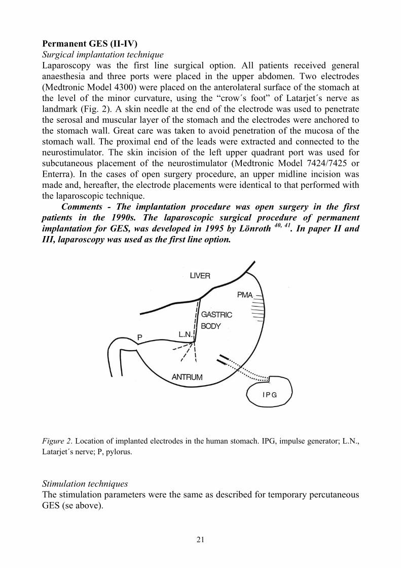
Permanent GES (II-IV) Surgical implantation technique Laparoscopy was the first line surgical option. All patients received general anaesthesia and three ports were placed in the upper abdomen. Two electrodes (Medtronic Model 4300) were placed on the anterolateral surface of the stomach at the level of the minor curvature, using the “crow´s foot” of Latarjet´s nerve as landmark (Fig. 2). A skin needle at the end of the electrode was used to penetrate the serosal and muscular layer of the stomach and the electrodes were anchored to the stomach wall. Great care was taken to avoid penetration of the mucosa of the stomach wall. The proximal end of the leads were extracted and connected to the neurostimulator. The skin incision of the left upper quadrant port was used for subcutaneous placement of the neurostimulator (Medtronic Model 7424/7425 or Enterra). In the cases of open surgery procedure, an upper midline incision was made and, hereafter, the electrode placements were identical to that performed with the laparoscopic technique.
Comments - The implantation procedure was open surgery in the first patients in the 1990s. The laparoscopic surgical procedure of permanent implantation for GES, was developed in 1995 by Lönroth 40, 41. In paper II and III, laparoscopy was used as the first line option.
Figure 2. Location of implanted electrodes in the human stomach. IPG, impulse generator; L.N., Latarjet´s nerve; P, pylorus. Stimulation techniques The stimulation parameters were the same as described for temporary percutaneous GES (se above).

22
SYMPTOM ASSESSMENT Questionnaires (I-IV) The patients completed daily symptoms in a GI symptom questionnaire. This was performed during the ON and OFF periods at temporary stimulation or during a two-week period at baseline and at 3, 6, 12, 24, etc. months after permanent implantation. Symptoms asked for included vomiting frequency, nausea time (hours) and severity of early satiety, bloating, epigastric pain, belching, postprandial fullness (severity score 0-3 were 0 = no symptoms, 1 = mild symptoms, 2 = moderate symptoms and 3 = severe symptoms). Responder criteria (II, IV) The criteria to be classified as a responder to GES were: a decrease of the main referral symptom; weekly vomiting frequency (WVF) or weekly nausea time (WNT), by ≥50% for a) open temporary percutaneous GES vs. baseline, or b) stimulation ON vs. stimulation OFF during the blinded TPGES test, or c) at 6 months follow-up vs. baseline for permanent GES.
The other criterion was the patient’s preference for the period of open permanent stimulation vs. baseline, or stimulation ON vs. stimulation OFF during the blinded test. The preference criterion could assess the overall effect of temporary percutaneous GES, including not only the intensity of nausea/vomiting but also other predominant symptoms.
Comments: The responder criteria for vomiting was based on a multi-centre study where 70% of the diabetic patients had a reduction of at least 50% of the vomiting frequency 39. Patients with post-surgical gastroparesis due to fundoplication cannot vomit. Therefore severe drug refractory nausea in gastroparetic patients has now also been adopted as indication for GES 46. GASTRIC FUNCTION Gastric emptying test (I-IV) Emptying of radiopaque markers (ROMs) or solid scintigraphic emptying was performed at baseline.
a) Twenty spheric ROMs (diameter 4 mm, density 1.27 g/mm3), were given with a meal and emptying was followed with fluoroscopy hourly until all ROMs were emptied or for a maximum of 8 hrs. The mean retention at fluoroscopy at 4, 5 and 6 hours after the meal was calculated. Upper reference values based on 131 control subjects were for men 25% and for women 65% 68, 69. With repeated fluoroscopy and estimation of the emptying of ROMs from the stomach and accumulation of ROMs in the small bowel and colon evaluation of small bowel transit could be made.

23
b) A standardized scintigraphic method was used. Imaging with gamma camera was performed at 0, 60 and 120 min after ingestion of the meal. Upper reference limits at 120 min based on 160 control subjects were 50% retention for men, 65% for premenopausal women and 54% for postmenopausal women 3.
Comments: Many patients with gastric dysmotility, also have small bowel pathology. The method of ROMs can measure both gastric emptying time and small bowel transit time, and was therefore used as the first line option at our laboratory. All patients included also in international multi-centre studies underwent the scintigraphic test (n=6). Manometry (I-III) Antroduodenojejunal manometry was performed 70, 71. The investigation was performed for five hours during fasting followed by 60 min after a test meal with a total energy content of 500 kcal. For pressure recording an assembly was used, with a central lumen for guide wire and eight water-perfused lumens with recording points in the antrum, the descending part of the duodenum, the distal duodenum close to the ligament of Treitz and in the proximal jejunum. The assembly was connected to pressure transducers and recordings made with a polygraph (PC Polygraph, Synectics, Stockholm, Sweden).
Comments: Manometry was performed in most patients to evaluate gastroduodenojejunal motility. Particularly in patients with normal gastric emptying, but with symptoms of dysmotility, manometry gives important information of the gastroduodenal function. In patients with chronic intestinal pseudo-obstruction, manometry is one of the investigations that verify the contractile abnormality in these cases. Slow caloric satiety drinking test (IV) A peristaltic pump filled one of two beakers with a nutrient drink at a rate of 15 ml/min (1.5 kcal/ml) and the patients were requested to ingest the drink at the filling rate and to cease meal intake when maximum satiety was reached. The amount of ingested kcal at maximum satiety was recorded. Thirty minutes after stopping the severity of bloating, fullness, nausea and pain were assessed using a visual analogue scale (VAS) with 100 mm lines and the words ‘unnoticeable’ and ‘unbearable’ at each end. A composite symptom score was defined as the sum of the four 100 mm VAS for each symptom (i.e. maximum 400).
Comments: Most studies of gastric accommodation have been performed using a gastric barostat, however, the procedure is cumbersome for the patients. A slow caloric drinking test has been proposed as a non-invasive and easy way to measure gastric accommodation capacity 64.
24
STATISTICAL METHODS Due to relatively low number of study participants and non-gaussian distribution non-parametric methods were used: Wilcoxon signed rank test for comparisons within groups between baseline and follow-up (II, III, IV) and Mann-Whitney-U test for comparisons between groups (IV). Spearman´s rank correlation was used for assessing correlations between two different parameters (IV). Results are presented as median, interquartal range (IQR) and range (II), mean and range (III) or mean and SD (IV). In general, significance was accepted at the 5% level.

25
RESULTS TEMPORARY PERCUTANEOUS GES Animal studies (I) In the anesthetized pig, injection of saline and insertion of the percutaneous electrodes into the space between the mucosal and the muscle layers of the stomach was performed under gastroscopic supervision. No perforation of the gastric mucosa occurred.
A spontaneous electric activity was recorded in all animals. The slow waves had a frequency of about 3 cycles/min. The amplitudes of the electrical waves were about 1.5-2.0 mV. In several sequences the individual gastric slow waves were followed by a superimposed short lasting potential wave (duration 3-5 sec) with the character of plateau potential.
At the end of the experiments, after about 1-1.5 hour of recording, the animals developed bradygastria with a decrease in the frequency of myoelectric activity to about 2 slow waves/min. However, the shape of the individual slow waves was unchanged during the period with bradygastria in each experiment. After the recording period the electrodes were removed by pulling.
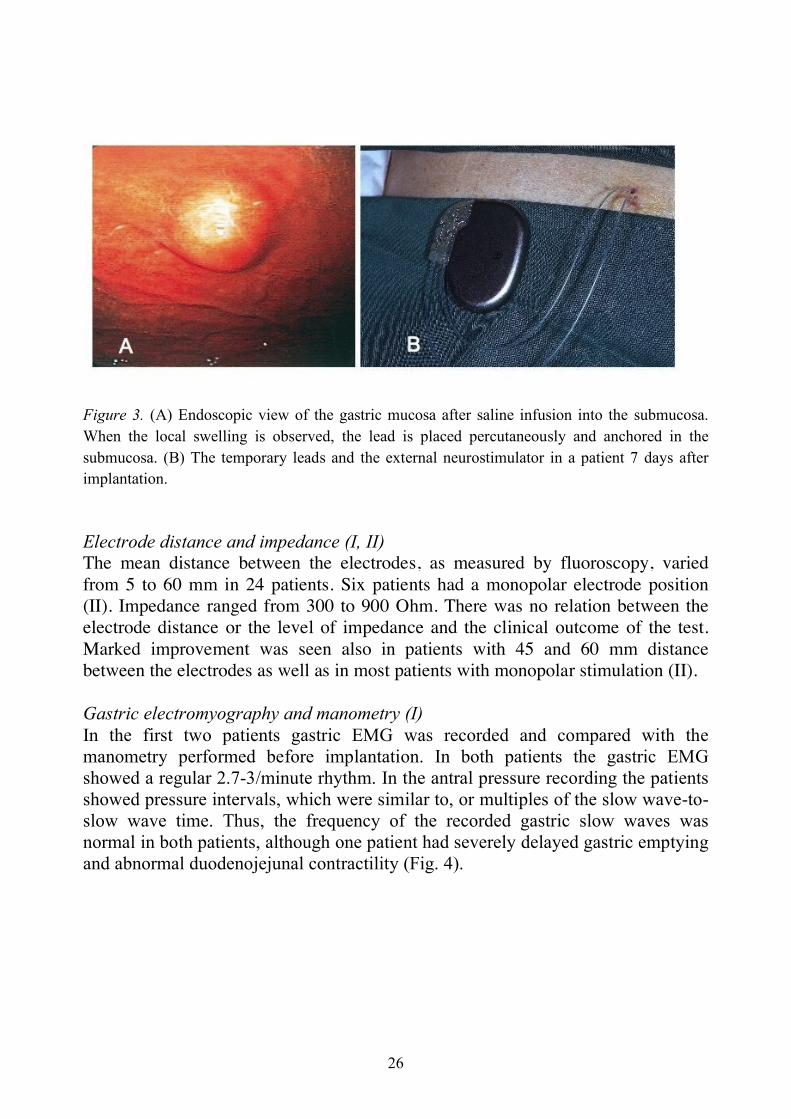
Eventually, autopsy was performed and a small lesion was noticed in the gastric wall at the site of electrode implantation but no other damage. The skin of the animals displayed only two approximately 5 mm long cuts after the intervention but no hematoma. Implantation technique in patients (I, II, IV) Lead implantation procedure The abdominal skin was disinfected before the gastroscopic procedure. In all patients the gastroscope light could be identified through the abdominal wall when the stomach was insufflated. The cannula was inserted into the stomach wall and was easily identified via the gastroscope. After saline infusion, local swelling of the mucosa was observed. No mucosal perforation occurred during the implantation (Fig. 3).
Implantation time from the start of gastroscopy (including skin anaesthesia and incisions) to the end of implantation of the two electrodes varied from 8 to 22 minutes (mean 14 minutes) in the 30 patients (II).
26
Figure 3. (A) Endoscopic view of the gastric mucosa after saline infusion into the submucosa. When the local swelling is observed, the lead is placed percutaneously and anchored in the submucosa. (B) The temporary leads and the external neurostimulator in a patient 7 days after implantation. Electrode distance and impedance (I, II) The mean distance between the electrodes, as measured by fluoroscopy, varied from 5 to 60 mm in 24 patients. Six patients had a monopolar electrode position (II). Impedance ranged from 300 to 900 Ohm. There was no relation between the electrode distance or the level of impedance and the clinical outcome of the test. Marked improvement was seen also in patients with 45 and 60 mm distance between the electrodes as well as in most patients with monopolar stimulation (II). Gastric electromyography and manometry (I) In the first two patients gastric EMG was recorded and compared with the manometry performed before implantation. In both patients the gastric EMG showed a regular 2.7-3/minute rhythm. In the antral pressure recording the patients showed pressure intervals, which were similar to, or multiples of the slow wave-to-slow wave time. Thus, the frequency of the recorded gastric slow waves was normal in both patients, although one patient had severely delayed gastric emptying and abnormal duodenojejunal contractility (Fig. 4).

27
Figure 4. Gastric electromyography performed with percutaneous leads and antroduodenaljejunal manometry in a patient with chronic intestinal pseudo-obstruction. Manometry showed almost complete absence of contractility in duodenum and jejunum trough out the 6 h recording, but some sequences with antral pressure waves (recording points A1-A3, 15 mm apart). Gastric EMG showed a continuous 3 min-1 and the distally moving antral pressure waves, when present, had an interval of 20 s or multiples thereof. Lead observations and complications (I, II) The gastric electrodes and the impulse generator, taped to the abdominal skin, were kept in place for 7-9 days in the first three patients (I) and 7-60 (mean 26) days in all 30 patients (II). At the end of the temporary stimulation test the leads could easily be pulled out without any complication. In most patients this was performed after anaesthesia (pethidine 25-50 mg i.v.).
Four patients could sense the stimulation as a discomfort, aching or cramping. However, the stimulation current could be adjusted to a level so that the patient could easily ignore the discomfort. One patient had a slight skin infection at the implantation site six weeks after implantation. However, the infection responded rapidly to oral antibiotics. One patient had an abdominal wall hematoma, which was due to a previously unknown bleeding disease.

28
Monopolar stimulation (II) In 6 patients, where one temporary electrode was verified to be in the gastric wall, and one electrode outside the gastric wall but still with the impedance within the range of 300-900 Ohm, the effect of monopolar GES instead of the conventional bipolar stimulation could be evaluated. Five out of six patients (83%) were responders to monopolar stimulation while 17 out of 21 patients (81%) were responders to bipolar stimulation. Thus, there was no significant difference in the response rate when bipolar and monopolar stimulation was compared. Effect on symptoms (I, II) Open stimulation (II) Twenty patients had their final evaluation from results of open stimulation, 13 with standard stimulation (5 mA) and 7 with high stimulation (8-10 mA) (Fig. 5). 16 of the 20 patients were considered responders; 15 had a decrease in severity of nausea and/or vomiting, and one patient had marked reduction in regurgitations (responder according to the preference criterion). Four patients having their final evaluation with open stimulation were non-responders to temporary percutaneous GES.
Figure 5. Vomiting and nausea for 20 patients at baseline and during open temporary percutaneous GES. Extreme values and outliers are not shown. Median weekly vomiting frequency (WVF) decreased from 5.8 (IQR 0-18; range 0-129) to 0 (IQR 0-4; range 0-52) and median weekly nausea time (WNT) (hours) decreased from 21.5 (IQR 13-40; range 0-112) to 6.0 (IQR 0-25; range 0-112). Blinded ON-OFF stimulation (II) Seven patients were evaluated under blinded conditions only, with stimulation ON or OFF for periods of 10-14 days in a randomised order (Fig. 6). From this blinded test 6 patients were responders; 5 with respect to decreased frequency of nausea and/or vomiting, and 1 patient in respect to marked reduction of retching (according to the preference criterion).

29
Figure 6. Vomiting and nausea for seven patients during blinded cross-over ON/OFF temporary percutaneous GES. At stimulation ON compared to OFF the median weekly vomiting frequency (WVF) was 2.0 (IQR 0-22; range 0-34) vs. 10.0 (IQR 1-36; range 0-162) while weekly nausea time (WNT) (hours) was 4.0 (IQR 3-12; range 0-33) vs. 12.0 (IQR 6-34; range 0-66). Outcome of TPGES vs. diagnosis and gastric emptying All 30 patients enrolled for temporary percutaneous GES underwent a gastric emptying test at baseline. 13 out of 30 patients had delayed gastric emptying. Table 1 summarizes gastric emptying, diagnosis and response to TPGES. Table 1. Outcome of temporary percutaneous GES in 27 patients with mainly non-approved indications for GES. Proportion of responders with decrease in nausea and/or vomiting related to gastric emptying and diagnosis. Three patients could not be evaluated by TPGES and are not shown in table: Ehler-Danlos’ gastroparesis (n=1), intestinal neuropathy (n=1), post-surgical gastroparesis (n=1). Functional dyspepsia was classified according to Rome II criteria. Gastric emptying / diagnosis Response rate Normal gastric emptying 12/16 (75%) Functional dyspepsia 6/9 Diabetes mellitus 2/2 Post-surgical nausea/vomiting 2/2 Malformation syndrome 1/1 Intestinal neuropathy 1/1 Intestinal ICC deficiency 0/1 Delayed gastric emptying 10/11 (91%) Post-surgical gastroparesis 7/8 Chronic intestinal pseudo-obstruction 2/2 Idiopathic gastroparesis 1/1
30
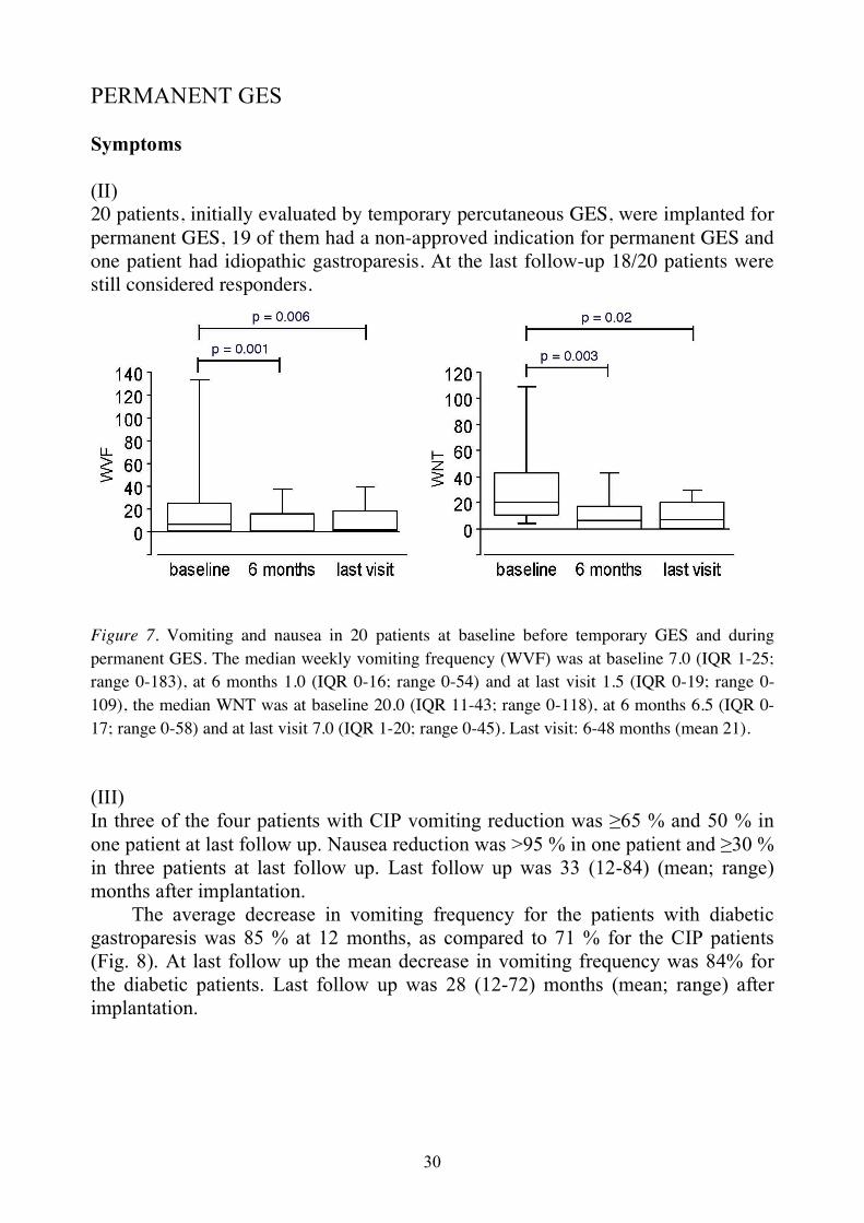
PERMANENT GES Symptoms (II) 20 patients, initially evaluated by temporary percutaneous GES, were implanted for permanent GES, 19 of them had a non-approved indication for permanent GES and one patient had idiopathic gastroparesis. At the last follow-up 18/20 patients were still considered responders.
Figure 7. Vomiting and nausea in 20 patients at baseline before temporary GES and during permanent GES. The median weekly vomiting frequency (WVF) was at baseline 7.0 (IQR 1-25; range 0-183), at 6 months 1.0 (IQR 0-16; range 0-54) and at last visit 1.5 (IQR 0-19; range 0-109), the median WNT was at baseline 20.0 (IQR 11-43; range 0-118), at 6 months 6.5 (IQR 0-17; range 0-58) and at last visit 7.0 (IQR 1-20; range 0-45). Last visit: 6-48 months (mean 21). (III) In three of the four patients with CIP vomiting reduction was ≥65 % and 50 % in one patient at last follow up. Nausea reduction was >95 % in one patient and ≥30 % in three patients at last follow up. Last follow up was 33 (12-84) (mean; range) months after implantation.
The average decrease in vomiting frequency for the patients with diabetic gastroparesis was 85 % at 12 months, as compared to 71 % for the CIP patients (Fig. 8). At last follow up the mean decrease in vomiting frequency was 84% for the diabetic patients. Last follow up was 28 (12-72) months (mean; range) after implantation.

31
Figure 8. Weekly vomiting frequency at baseline and during GES in patients with chronic intestinal pseudo-obstruction (n=4, solid lines) and diabetic gastroparesis (n=11, dotted lines). P<0.01. Upper GI symptoms after 12 months At the one-year follow-up for the 20 patients selected by TPGES, the referral symptoms nausea/vomiting were clearly decreased. The median WVF was 2.25 (IQR 0-14; range 0-132) and median WNT 11.5 (IQR 4-19; range 0-58). Compared to baseline decrease in vomiting and nausea were significant (respectively, p=0.01 and p=0.004). Other symptoms at the one-year follow-up are shown in Fig. 9. The decrease was significant for fullness and approached significance for bloating and epigastric pain.

32
Figure 9. Daily gastric symptoms at baseline and the one-year follow-up for patients selected for permanent GES by TPGES. Severity score 0-3 (0 = no symptoms, 3 = severe symptoms). Data presented as mean and SD. Aspects on symptom relapses Temporary relapses of vomiting were related to urinary infection in two patients and skin infection in two patients. The symptoms of vomiting rapidly improved when the infection was treated. In one patient relapse of vomiting was time related to primary hyperparathyroidism. Surgical results: Laparoscopy success (II, III) Of 35 patients, 33 could be implanted by laparoscopy (II, III). Markedly dilated small bowel was seen in all CIP patients, yet three out of four CIP patients could be implanted with laparoscopy (III). Hospital care (III) There was a significant decrease in days of hospital care due to nausea and vomiting (p=0.02). In the diabetic group the mean yearly hospital care due to vomiting decreased from 38 days the year before implantation to 12 days the year after implantation (p=0.07). In the CIP group the mean decrease was from 14 to 0 days. The mean yearly hospital care due to any reason (35 days) did not change after implantation.

33
HbA1c (III) There was a tendency towards improvement of HbA1c in the eleven diabetic patients. HbA1c decreased from 8.1 to 7.1 (p=0.09) (baseline vs. last control). Device and surgery related observations (II, III) Battery duration (III) All neurostimulator batteries showed OK at the last control. Time in use was up to 90 months (=7.5 years). One neurostimulator (Medtronic, model 7424) switched off due to external magnetic fields, and had to be exchanged to the magnet insensitive model 7425. Electrodes and neurostimulator (II, III) Dysfunction of one electrode was detected in one patient, and was corrected by reprogramming to monopolar gastric stimulation, with the neurostimulator as anode and the functioning lead as cathode (III). Standard bipolar stimulation in two patients was followed by epigastric pain. Monopolar stimulation with equivalent mA was well tolerated and nausea decreased (II).
In one case the neurostimulator had to be taken out because of skin erosion and a new stimulator was put in another pocket (III). This was due to marked reduction of subcutaneous tissue, later shown to be related to lung cancer. Surgical event and explantation (III) One patient with adhesions after previous peritoneal dialysis required laparotomy 12 hours after laparoscopy, to stop a post implantation bleeding. In one CIP patient the electrodes caused ileus and a short intestinal resection and explantation had to be performed 5 months after implant. Due to relapse of severe vomiting a second permanent implantation was done after 10 months. SLOW CALORIC DRINKING SATIETY TEST (IV) Healthy volunteers and untreated patients The test was well tolerated by the healthy controls (n=36), and the maximum drinking capacity was 1630±496 kcal (mean±SD). For the 19 patients that underwent permanent GES the maximum drinking capacity was 887±412 kcal at baseline, which was significantly lower than the drinking capacity of the healthy volunteers (p<0.001) (Fig. 10). 26% of the patients had a drinking capacity outside the total range in the healthy controls.

34
Figure 10. Drinking capacity in healthy volunteers and patients at baseline before permanent GES. Solid lines: mean drinking capacity. Dotted line: the cut off for the normal value, based on the total range in healthy volunteers. The healthy volunteers had less symptoms 30 min after maximum satiety than the patients; the composite score was 128±51 vs. 235±83 (p<0.001). In the patients there was no significant correlation between the drinking capacity at baseline and vomiting or nausea frequency at baseline (Fig. 11).
Figure 11. Drinking capacity at baseline was not correlated with symptom severity at baseline.

35
Temporary percutaneous GES Four out of 8 patients were responders to blinded temporary percutaneous GES. There was no significant change in drinking capacity during stimulation (stim ON) compared to the control period (stim OFF). The visual analogue scale (VAS) measurement for each symptom 30 min after maximum satiety and the composite score showed no difference during the ON vs. OFF periods. Permanent GES For the 19 patients having permanent GES there was no significant difference in drinking capacity at the 6 or 12 months control compared to baseline, and there was no significant effect on the visual analogue scale (VAS) measurement of each symptom or the composite score. There was no correlation between improvement in drinking capacity and decrease in vomiting or nausea (Fig. 12).
Figure 12. Change in drinking capacity (baseline to 6-months follow-up) was not correlated with improvement of frequency of vomiting or nausea time. There was no difference between responders (n=13) and non-responders (n=6) in change in drinking capacity at the 6-months follow-up compared to baseline (p=0.7) (Fig. 13).
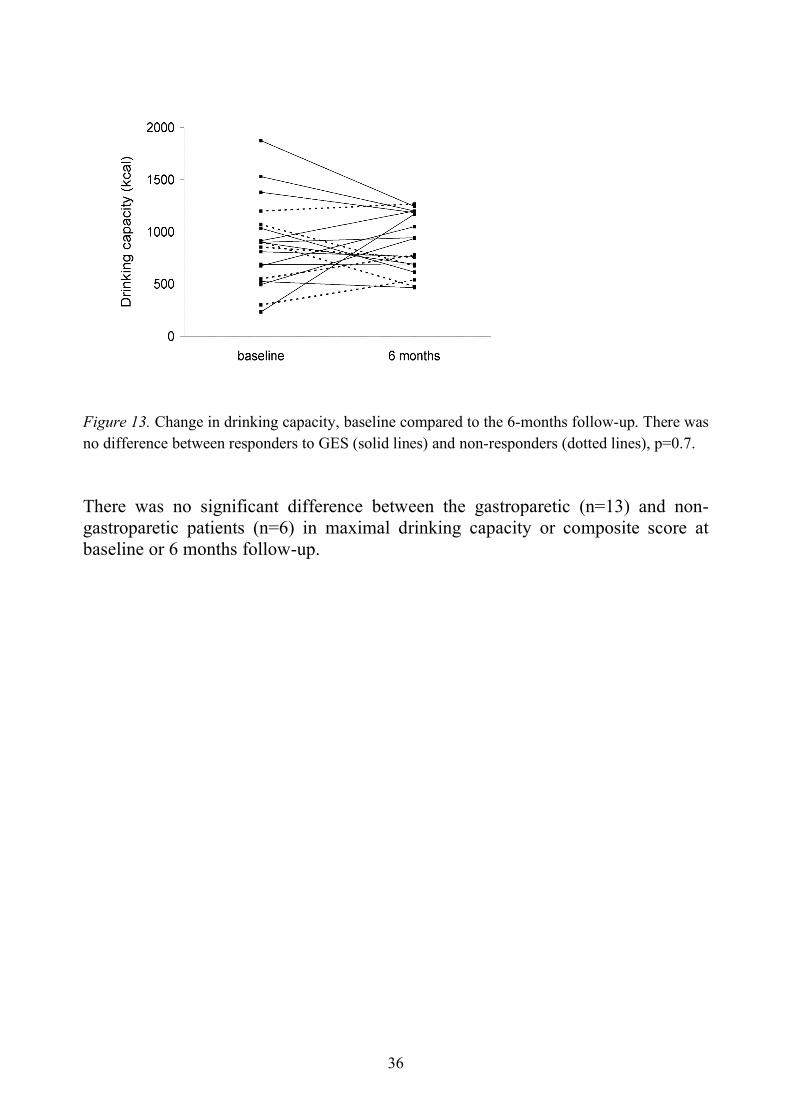
36
Figure 13. Change in drinking capacity, baseline compared to the 6-months follow-up. There was no difference between responders to GES (solid lines) and non-responders (dotted lines), p=0.7. There was no significant difference between the gastroparetic (n=13) and non-gastroparetic patients (n=6) in maximal drinking capacity or composite score at baseline or 6 months follow-up.

37
DISCUSSION
TEMPORARY PERCUTANEOUS GES Novel principle The gastroscopy-assisted implantation technique of percutaneous temporary GES (TPGES) described in this thesis, is a novel principle to implant lead electrodes to the stomach. It differs principally from the few other methods to perform temporary GES in humans that have been reported. The method applied in the first multi-centre study 40 demanded general anaesthesia and abdominal surgery to fixate the leads. Another more recent study published during the course of our study, suggested a gastroscopy-assisted method where the leads were placed via a stoma or orally 72. With this technique the electrodes could be kept in place for no more than 8 days in 78% of the patients in a blinded cross-over test 73. The orally placed leads remain functional for only about one week 72 and might be cumbersome for many patients. Leads placed via a stoma are functional for up to one month, but creation of a stoma means a higher risk for complications.
Implantation of TPGES is easy to perform in gastroenterological practice and do not require general anaesthesia. The procedure usually takes 10-20 minutes, and can be performed as an outpatient procedure. The patient is instructed to be careful while showering, but otherwise the device does not restrain daily life. The percutaneous leads can be left in place for at least 60 days with remained function. However, there does not seem to be a definite upper limit for the duration of TPGES. Due to the relatively comfortable conditions and the long duration of the temporary stimulation, a selection of responders from a test running for several weeks is possible. Thus, compared to previous temporary GES principles TPGES appears to be more comfortable for the patient, less demanding from a surgical facility point of view and can be performed over longer test periods. Implantation technique The first steps of TPGES have similarities with PEG placement. Gastroscopy is used to guide the insertion of the percutaneous electrodes. Due to the gastroscopic control and the elasticity of the mucosa, penetration by the tip of the cannula can be avoided when the electrodes are implanted. Another potential way to guide the insertion is by ultrasound.
Apart from endoscopic control at implant, a proper position of the TPGES leads was verified by other observations. a) During the animal studies, gastric EMG in the pigs showed a gastric electrical rhythm with a frequency of about 3 min-1, which is very similar to other reports of the gastric electrical rhythm in pigs 74. b) Gastric EMG performed in the two first patients showed a regular rhythm of about 3 min-1. The findings were supported by the manometry findings just prior to implantation, were the antral rhythm was similar to that observed in the EMG.
38
These recordings indicate that the electrodes had a proper position in the gastric wall for registration of EMG. Furthermore, the stable intra-individual frequency of slow waves before and during GES is in accordance with previous studies reporting that high-frequency low-energy GES does not entrain slow waves 58. c) The marked reduction of symptoms in the first two patients also indicated a correct electrode position from a treatment point of view. d) Fluoroscopy yielded information about electrode position after the lead insertion. Also in patients where the distance between the electrodes was verified to be relatively long (4-6 cm), the effect on symptoms was clear cut (II), which is at variance from the present concept that distance between leads should be about 1 cm 39. The present results suggest that the electrode distance is less important for the symptomatic outcome. However, further observations are needed.
An important aspect of the method of TPGES is the construction of the lead. The flexible tines at the tip enable the lead to anchor itself in the submucosa. Thus, the mucosa is kept intact, sutures are not needed and the invasiveness is minimal. However, in 20% of the patients one electrode was dislodged within one day after implantation (i.e. 10% of the implanted electrodes). Thus, method improvements should be considered for the various steps of the implant procedure: the lead construction, the puncture and retraction of the endoscope. It is notable that the effect of GES could be evaluated by monopolar stimulation in the patients where the dislodgement was verified within one day after the implantation. Moreover, the observations (II) suggest that a cutaneous electrode might be used as anode as an alternative to the conventional bipolar electrode arrangement. Clinical feasibility We have demonstrated the clinical feasibility of TPGES and shown that TPGES can be used to evaluate the effect of GES before permanent implantation. At present, there is no other available non-invasive test that can be applied for more than one week and capable to predict whether a patient will respond to GES. The simplicity of TPGES makes it applicable even in more complicated cases. The youngest patient was a two year old child, and the oldest was 81 years old having scars and complicated anatomy after previous surgery. Both patients were implanted with no complications and had a positive outcome of the TPGES. Thus, there seems to be no distinct age limitations for considerations of TPGES.
One main criteria for inclusion of the patients to TPGES was the severity and duration of the GI symptoms nausea and vomiting. The symptoms should be refractory to at least dopamine antagonist and, in case of delayed gastric emptying, also refractory to erythromycine. Patients with diabetic gastroparesis, previously shown to have a relatively high responder rate, were not considered for TPGES, as they were considered to be good candidates for permanent GES without prior responsiveness testing. There were no absolute contraindications with respect to the underlying gastrointestinal diagnosis. As shown in Table 1 (page 29) patients with nine different diagnoses, in addition to functional dyspepsia, were enrolled. This broad spectrum of diagnoses is partly due to the investigation program at our

39
laboratory for severely symptomatic patients. The program includes upper GI manometry, transit assessments and intestinal full-thickness biopsy when appropriate. Without such investigations patients with intestinal neuropathy would have been categorized as having idiopathic or functional disorders. Contraindications for TPGES are similar to PEG placement, and include untreated bleeding diseases.
Given the broad spectrum of GI disorders included for TPGES and the outcome of the TPGES tests (II) the logistics for TPGES and its present place in handling of patients with drug refractory nausea and/or vomiting at our laboratory is schematically outlined in Fig. 14.
Figure 14. Handling of patients with GI disorder and severe vomiting or nausea.

40
Open or blinded TPGES? For safety reasons the first four pilot patients were tested with open TPGES for 7-9 days only, without any technical or physical complication. The following patients were considered for the blinded crossover test. However, for patients living far away from the hospital it was impractical to participate in the blinded group, and these patients were included in the open TPGES group.
Totally, 81% of the patients completing the TPGES test were classified as responders and considered for permanent GES. All patients selected from blinded TPGES were considered responders also to permanent GES. Of the patients selected from open stimulation, two patients (14%) were non-responders to permanent GES. However, one out of the two non-responders was selected from only a one-week test with open TPGES. Thus, the available data suggest that the test should preferably be performed under blinded conditions. If a blinded test is not possible, we suggest that an open test should run for a minimum of 2 weeks. Moreover, a longer duration of the test is also important due to a possible carry-over effect that seems to be about 1-2 days (II). This is in line with another study, reporting a carry-over effect of 2-3 days 72. At present we cannot conclude that an open test is less reliable than a blinded test, provided that the test runs for a sufficiently long period of time. PERMANENT GES New indications After permanent implant 90% of the responders to TPGES had also a maintained respond to permanent GES at follow-ups after 6-48 months (II). This is a higher responder rate than in several other studies where no test stimulation was performed prior to the permanent implantation 43, 46, 56 and comparable to that reported for the hitherto best responding group, namely diabetic gastroparesis 39, 40.
Although we do not know how the non-implanted patients may have done with permanent GES it is reasonable to assume that the selection done with TPGES explains the high responder rate. This high response rate may seem surprising as we studied patients with mainly non-approved indications for GES, i.e. post-surgical gastroparesis and dyspepsia, functional dyspepsia, intestinal neuropathy, CIP, malformation syndrome and ICC deficiency. Furthermore, symptoms of vomiting and nausea were reduced irrespective of the gastric emptying was normal or delayed.
As we hypothesized, patients with severe symptoms of nausea and vomiting but normal gastric emptying, e.g. functional dyspepsia, had a beneficial effect of GES on symptoms (II). The results are supported by other recent studies of GES in patients with normal gastric emptying, reporting that patients with severe GI symptoms benefit from GES and that delayed gastric emptying is not a predictive
41
factor for a positive outcome 75-77. Since CIP is a disorder that usually is very difficult to manage, the outcome of
GES in these patients is notable. Three of the patients with CIP that were implanted with permanent GES (III) had hereditary CIP with a family history of very severe disease, and close relatives had died from intestinal failure. One patient with post-surgical CIP was implanted on vital indication. However, the four CIP patients were all responders to GES, and irrespective of whether the gastric emptying was delayed (n=2) or normal (n=2). The effect on nausea and vomiting was similar as for the patients with diabetic gastroparesis. Furthermore, another patient with myopathic CIP responded to TPGES (II), but was selected for multi-visceral intestinal transplantation due to severe lower bowel failure and nutritional problems and did not receive permanent GES. CIP has previously been an exclusion criteria in most other studies of GES and, at present, there are no other reports of GES in this disorder. Although the experience of GES in CIP is limited, the marked long-term effect in these few but very severe cases, clearly suggests that GES should be considered as a therapeutic alternative for intractable vomiting in CIP.
Moreover, most patients with post-surgical gastroparesis were responders to permanent GES (II). This is in line with other studies of GES for PS-GP 46, 47. Interestingly, since it has been suggested that the effect of GES might be mediated by vagal afferents, improvement of symptoms was seen also in patients with partial vagal disruption (II). The same observation has been reported by another research team 46. This could mean that the vagal disruption in these patients was not complete, or that the effect of GES is not mediated by vagal afferents only.
Earlier studies on GES in young patients have mainly included adolescents, but also two children (7 and 8 years old) 78, 79. The two years old child (paper II), tested with TPGES, seems to be the youngest patient implanted with permanent GES. Although even small children with severe vomiting can benefit from GES, from an ethical point of view direct implantation of permanent GES without a prior prediction of the outcome is questionable. Non-responder aspects As mentioned above a positive outcome of permanent GES was seen in most patients (82%) with diabetic gastroparesis having a permanent implant with (n=3) or without (n=8) a prior temporary test (III). The same was true for the patients with non-approved diagnoses who had been selected by TPGES to receive permanent GES (II). However, a maintained absence of response was seen in two patients with diabetic gastroparesis, as well as in two patients who had shown a positive response during the initial TPGES test. For patients with gastroparesis it has recently been suggested that depletion of gastric ICCs may contribute to a “non-response” to permanent GES 80. This explanation seems less likely for our two patients with non-approved indication for GES since they initially had responded to temporary GES, although one of them had only a one-week open TPGES test. An over-consumption of alcohol was disclosed in these two patients that cannot be excluded to be a negative factor for GES treatment. An analogue

42
observation was recently reported, namely that patients taking narcotic analgesics at the time of implantation had a poorer response compared to patients who were not 56. Furthermore, eating disorders and anxiety disorders may influence the outcome of GES 76. Since there is a relative high co-existence of psychiatric disorders in for example functional dyspepsia 81 and idiopathic gastroparesis 82, a careful exploration of the patient history is of great importance before implantation of permanent GES. Effort to exclude anxiety disorders and drug or alcohol abuse also seems necessary.
The mentioned negative predictive factors for GES response, e.g. abuse, should be taken into consideration early in the evaluation of patients with drug refractory nausea/vomiting, i.e. even before consideration of TPGES. With respect to ICC-depletion as a negative factor, it is worth noting that in one of the five non-responders to TPGES (non-gastroparetic) a full-thickness jejunal biopsy had shown ICC depletion (II). Although the proportion of non-responders to permanent GES is small in patients with diabetic gastroparesis future research is of interest to see if this small proportion could be further reduced with application of TPGES also for this group (c.f. Fig. 8, page 31). STIMULATION ASPECTS Stimulation current at 5 mA with impulses 300 µsec has been the most commonly used stimulation setting since the first report of high-frequency low-energy GES in humans 38. The clinical effect is believed to be due to stimulation of nerve endings in the gastric wall as the energy used is not sufficient to excite muscle structures. With the percutaneous lead electrodes we found that in most cases non-responders to TPGES with 5 mA responded to increased stimulation at 8-10 mA (paper II). This is in line with observations of permanent GES where non-responders had a better outcome after modification of the stimulation parameters, e.g. increased current 57, 83. Increased energy per pulse can be accomplished by either increasing the current (mA) or by extending the pulse width. At a given impulse strength, an increase in impulse frequency will increase the impulse frequency in the nerve fibers already activated. In contrast an increase of impulse strength (e.g. mA) will recruit more nerve fibers with higher stimulation threshold. In the present study an increase of mA was chosen as a mean for stimulation in a range of stimulation energy above the mentioned 5 mA setting used in earlier studies. It should be noted that normal fluctuations of the impedance in human tissue leads to some variation in the mA-value obtained with a given voltage setting. Our results with TPGES indicate that a current higher than 5 mA should probably be applied as the preferred TPGES stimulation to minimize the risk for ineffective stimulation. The optimal TPGES stimulation settings have yet to be determined. Anyhow, our results show that it is important to be aware of the risk of “understimulation” that can lead to

43
misclassification of a patient as non-responder. Interestingly, in the TPGES patients, monopolar GES was as effective as
bipolar stimulation with respect to proportion of responders. After permanent implant two patients perceived an abdominal discomfort with the conventional bipolar stimulation. This discomfort disappeared after reprogramming to monopolar settings.
In the whole group of 20 patients receiving permanent GES after the initial positive TPGES test 40% had their GES current increased within the first 6 months after implant. Although the effects of these changes were not analyzed in detail it is reasonable to assume that the increases of stimulation energy contributed to the highly significant clinical improvement noted at follow-ups (Fig. 7). SLOW CALORIC SATIETY DRINKING TEST AND APPLICATION IN PATIENTS WITH GES (IV) The slow caloric satiety drinking test has earlier been shown to correlate well with measurements of gastric accommodation using a gastric barostat, and to predict impaired accommodation in patients with functional dyspepsia 64. In paper IV healthy volunteers had a significantly higher drinking capacity than the patients with severe nausea and vomiting. The results of the healthy volunteers are similar to other studies using the same method 64, 84. It has been proposed that clinical GES may exert a therapeutic effect in gastroparesis by increasing the gastric accommodation capacity. However, we found no effect of GES on proximal gastric function as evaluated by the slow caloric satiety drinking test. This was the case in patients with approved as well as non-approved indications for GES, and irrespective of the effect of GES on symptoms, or whether the patients had normal or delayed gastric emptying. The results are at variance with some other reports. In one study, preliminarily reported, six out of totally nine gastroparetic patients had an improvement in satiety score and an increased liquid test meal volume during GES, which was independent of any effect on gastric emptying 65. Gastric barostat was used in another study on eight patients with idiopathic gastroparesis receiving GES. In that study, decreased sensitivity to gastric distension and enhanced gastric accommodation to a meal was reported 63. A recent study of 10 gastroparetic patients with GES, in which gastric barostat was used, reported an increase in mean volume and in the discomfort threshold 85. In that study five of seven patients who had an increase in gastric accommodation had ≥50% reduction of the total symptom score (TSS). However, two patients with increased accommodation had <50% reduction of TSS. Thus, at present, no definite correlation between the therapeutic effect of GES on nausea and changes in gastric accommodation has been demonstrated.

44
The drinking tests were originally developed as non-invasive investigations to assess upper digestive sensations. Most commonly, the drinking tests are performed in patients with functional dyspepsia and gastroparesis. However, several drug studies, in healthy volunteers 86-90 and functional dyspepsia 91, have used the slow caloric satiety test to evaluate the effect of drugs on postprandial symptoms and time/volume to maximum satiety.
The drinking tests are well tolerated, easy to perform and inexpensive. However, there are some limitations. Whereas a normal test indicates normal gastric function, an abnormal test is more difficult to interpret. Central or extra-gastric factors may play a role in the symptom generation and tolerance. In a study of patients with functional dyspepsia, the maximal ingested volume at a water load test was reduced in patients with co-existing depression 92. Furthermore, the maximal ingested volume has been positively correlated with quality of life in patients with functional dyspepsia 93, 94. To summarize, the drinking tests are interesting tools to assess important aspects on gastric functions. With respect to GES, its therapeutic effects on nausea and vomiting seem not to be linked to changes in the functions measured by these tests.
MECHANISMS OF GES GES does not seem to improve gastric accommodation capacity (IV), as discussed above. However, more research on the effect of GES on gastric function is warranted. Moreover, the positive effect of GES on nausea and vomiting also in patients with normal gastric emptying (II, III), corroborates the present common opinion that the effect of GES is not primarily due to increased gastric emptying rate 39, 95. An interesting question is the possible effect of GES on the central nervous system (CNS). As mentioned previously, it has been suggested that vagal afferents may mediate the impulses to CNS. However, human studies are few, and the findings are inconsistent also in animal studies. One animal study has shown that the effect of GES is prevented by vagotomy 66. In contrast another group reported that GES could modulate CNS also in vagotomized animals 96. One recent study on 10 patients receiving GES, reported decreased sympathovagal balance during GES, which would indicate increased vagal activity 85. The same study reported an increased thalamic activity in patients with GES compared to controls, using PET scan imaging. A contradictory finding, is the observation that patients with vagal disruption may respond favourably to GES (II) 46. To summarize, the mechanisms of the effect of GES are complicated and far from fully understood. More studies in this area are needed to discover the full usage of GES.
45
SUMMARY AND CONCLUSIONS 1. We have introduced temporary percutaneous GES as a novel principle to
implant gastric electrodes for electrical stimulation. The temporary electrodes can easily be implanted percutaneously into the stomach muscle wall during gastroscopic supervision. The first steps of the procedure are similar to PEG placement, but the gastric mucosa is kept intact.
2. Temporary percutaneous GES is minimally invasive and can be performed as
an outpatient procedure. The leads can be left in place for at least two months with remained function and under comfortable conditions, which makes a long test period for temporary GES possible.
3. Most patients with drug-refractory GI disorders and severe nausea or vomiting
responded to TPGES with reduction of symptoms, irrespective if the gastric emptying was delayed or normal. The method of TPGES can be used for patient selection for permanent GES. Most patients that are responders to TPGES also respond to permanent GES. There seems to be no age limitations for TPGES.
4. Non-responders to standard stimulation settings for GES, can respond to
stimulation with increased impulse strength. Though conventional gastric electrical stimulation is bipolar, patients with monopolar stimulation also responded favourable with improvement of symptoms.
5. Patients with chronic intestinal pseudo-obstruction, post-surgical gastroparesis
and patients with severe nausea but normal gastric emptying (e.g. functional dyspepsia and post-surgical vomiting) can also benefit from treatment with GES. The clinical effect of GES for these diagnoses is similar to the effect of GES in patients with approved indications for GES, i.e. diabetic and idiopathic gastroparesis. Permanent implantation can preferable be performed with laparoscopy.
6. As evaluated by a slow caloric satiety drinking test, GES has no or little effect
on proximal gastric function. The therapeutic effect of GES in GI disorders is not primarily due to improvement in gastric emptying or an increased gastric receptive capacity.

46
ACKNOWLEDGEMENTS I wish to express my sincere gratitude and appreciation to those who have made this thesis possible. I particularly want to thank: Hasse Abrahamsson, my supervisor, for introducing me into this exciting field and sharing his enormous knowledge. His enthusiasm, scientific brilliance and courage have been invaluable for this work. I am also very grateful for his confidence in me, and his never-ending support. Magnus Simrén, my co-supervisor, for his never-ending enthusiasm and encouraging support, and for all excellent and accurate ideas. Hans Lönroth, my co-supervisor, for skilful contributions to the studies and for his support during important phases of this work. Anders Elfvin, Gisela Ringström and Anders Edebo, co-authors of the papers included in this thesis, for their contributions. Anders Kilander and P-O Stotzer, former and present head of the Section of Gastroenterology, for their support during this time. All my colleagues at the Division of Gastroenterology at Sahlgrenska University Hospital. All nurses at the Gastrointestinal laboratory, for excellent assistance and for creating such a friendly atmosphere. All the patients and healthy volunteers that participated in the studies. My parents and my sister, for their endless support. My wonderful family, for their patience during this time.
47
REFERENCES 1. Tack J, Bisschops R, Sarnelli G. Pathophysiology and treatment of functional dyspepsia.
Gastroenterology 2004;127:1239-55. 2. Gryback P, Hermansson G, Lyrenas E, Beckman KW, Jacobsson H, Hellstrom PM.
Nationwide standardisation and evaluation of scintigraphic gastric emptying: reference values and comparisons between subgroups in a multicentre trial. Eur J Nucl Med 2000;27:647-55.
3. Thelin Schmidt P, Abrahamsson H, Dolk A, et al. Methods to assess gastric motility and sensation. Scand J Gastroenterol 2008;10:1-11.
4. Tougas G, Eaker EY, Abell TL, et al. Assessment of gastric emptying using a low fat meal: establishment of international control values. Am J Gastroenterol 2000;95:1456-62.
5. Huizinga JD. Gastrointestinal peristalsis: joint action of enteric nerves, smooth muscle, and interstitial cells of Cajal. Microsc Res Tech 1999;47:239-47.
6. Sanders KM, Koh SD, Ward SM. Interstitial cells of cajal as pacemakers in the gastrointestinal tract. Annu Rev Physiol 2006;68:307-43.
7. Sanders KM. Regulation of smooth muscle excitation and contraction. Neurogastroenterol Motil 2008;20 Suppl 1:39-53.
8. Azpiroz F, Malagelada JR. Vagally mediated gastric relaxation induced by intestinal nutrients in the dog. Am J Physiol 1986;251:G727-35.
9. Johnson HD, Laws JW. The cardia in swallowing, eructation, and vomiting. Lancet 1966;2:1268-73.
10. Borison HL, Borison R, McCarthy LE. Role of the area postrema in vomiting and related functions. Fed Proc 1984;43:2955-8.
11. Carpenter DO. Neural mechanisms of emesis. Can J Physiol Pharmacol 1990;68:230-6. 12. Parkman HP, Hasler WL, Barnett JL, Eaker EY. Electrogastrography: a document prepared
by the gastric section of the American Motility Society Clinical GI Motility Testing Task Force. Neurogastroenterol Motil 2003;15:89-102.
13. Gilja OH, Hausken T, Odegaard S, Berstad A. Ultrasonography and three-dimensional methods of the upper gastrointestinal tract. Eur J Gastroenterol Hepatol 2005;17:277-82.
14. Tack J. Gastric motor disorders. Best Pract Res Clin Gastroenterol 2007;21:633-44. 15. Horowitz M, Su YC, Rayner CK, Jones KL. Gastroparesis: prevalence, clinical significance
and treatment. Can J Gastroenterol 2001;15:805-13. 16. Kong MF, Horowitz M, Jones KL, Wishart JM, Harding PE. Natural history of diabetic
gastroparesis. Diabetes Care 1999;22:503-7. 17. Fraser RJ, Horowitz M, Maddox AF, Harding PE, Chatterton BE, Dent J. Hyperglycaemia
slows gastric emptying in type 1 (insulin-dependent) diabetes mellitus. Diabetologia 1990;33:675-80.
18. Schvarcz E, Palmer M, Aman J, Horowitz M, Stridsberg M, Berne C. Physiological hyperglycemia slows gastric emptying in normal subjects and patients with insulin-dependent diabetes mellitus. Gastroenterology 1997;113:60-6.
19. Bityutskiy LP, Soykan I, McCallum RW. Viral gastroparesis: a subgroup of idiopathic gastroparesis--clinical characteristics and long-term outcomes. Am J Gastroenterol 1997;92:1501-4.
20. Parkman HP, Hasler WL, Fisher RS. American Gastroenterological Association technical review on the diagnosis and treatment of gastroparesis. Gastroenterology 2004;127:1592-622.

48
21. Stanghellini V, Cogliandro RF, de Giorgio R, Barbara G, Salvioli B, Corinaldesi R. Chronic intestinal pseudo-obstruction: manifestations, natural history and management. Neurogastroenterol Motil 2007;19:440-52.
22. Schuffler M. Chronic Intestinal Pseudo-Obstruction. 7th ed. Sleisenger and Fordtran´s Gastrointestinal and Liver Disease. Philadelphia Saunders; 2002: 2140-2150.
23. Catto-Smith AG, Ranuh R. Abdominal migraine and cyclical vomiting. Semin Pediatr Surg 2003;12:254-8.
24. Namin F, Patel J, Lin Z, et al. Clinical, psychiatric and manometric profile of cyclic vomiting syndrome in adults and response to tricyclic therapy. Neurogastroenterol Motil 2007;19:196-202.
25. Chong SK. Electrogastrography in cyclic vomiting syndrome. Dig Dis Sci 1999;44:64S-73S.
26. Zipfel S, Sammet I, Rapps N, Herzog W, Herpertz S, Martens U. Gastrointestinal disturbances in eating disorders: clinical and neurobiological aspects. Auton Neurosci 2006;129:99-106.
27. Stanghellini V, Tosetti C, Barbara G, et al. Dyspeptic symptoms and gastric emptying in the irritable bowel syndrome. Am J Gastroenterol 2002;97:2738-43.
28. Caballero-Plasencia AM, Sofos-Kontoyannis S, Valenzuela-Barranco M, Martin-Ruiz JL, Casado-Caballero FJ, Lopez-Manas JG. Irritable bowel syndrome in patients with dyspepsia: a community-based study in southern Europe. Eur J Gastroenterol Hepatol 1999;11:517-22.
29. Bilgutay AM, Lillehei CW, Wingrove R, Griffen WO, Bonnabeau RC. Gastrointestinal Pacing: a New Concept in the Treatment of Ileus. Biomed Sci Instrum 1963;1:377-83.
30. Sarna SK, Daniel EE. Electrical stimulation of gastric electrical control activity. Am J Physiol 1973;225:125-31.
31. Sarna SK, Bowes KL, Daniel EE. Gastric pacemakers. Gastroenterology 1976;70:226-31. 32. Hocking MP, Vogel SB, Sninsky CA. Human gastric myoelectric activity and gastric
emptying following gastric surgery and with pacing. Gastroenterology 1992;103:1811-6. 33. Miedema BW, Sarr MG, Kelly KA. Pacing the human stomach. Surgery 1992;111:143-50. 34. Hocking MP. Postoperative gastroparesis and tachygastria--response to electric stimulation
and erythromycin. Surgery 1993;114:538-42. 35. Chen JD, Schirmer BD, McCallum RW. Serosal and cutaneous recordings of gastric
myoelectrical activity in patients with gastroparesis. Am J Physiol 1994;266:G90-8. 36. McCallum RW, Chen JD, Lin Z, Schirmer BD, Williams RD, Ross RA. Gastric pacing
improves emptying and symptoms in patients with gastroparesis. Gastroenterology 1998;114:456-61.
37. Familoni BO, Abell TL, Nemoto D, Voeller G, Johnson B. Efficacy of electrical stimulation at frequencies higher than basal rate in canine stomach. Dig Dis Sci 1997;42:892-7.
38. Familoni BO, Abell TL, Voeller G, Salem A, Gaber O. Electrical stimulation at a frequency higher than basal rate in human stomach. Dig Dis Sci 1997;42:885-91.
39. Abell T, McCallum R, Hocking M, et al. Gastric electrical stimulation for medically refractory gastroparesis. Gastroenterology 2003;125:421-8.
40. Abell TL, Van Cutsem E, Abrahamsson H, et al. Gastric electrical stimulation in intractable symptomatic gastroparesis. Digestion 2002;66:204-12.
41. Lonroth, Abrahamsson. Laparoscopic and open placement of electronic implants for Gastric Electrical Stimulation (GES): technique and results. Minim Invasive Ther Allied Technol 2004;13:336-9.
42. United States Food and Drug Administration. Device approvals and clearances: Enterra Therapy System. 2000. http://www.accessdata.fda.gov/scripts/cdrh/cfdocs/cftopic/pma/pma.cfm?num=H990014

49
43. Lin Z, Sarosiek I, Forster J, McCallum RW. Symptom responses, long-term outcomes and adverse events beyond 3 years of high-frequency gastric electrical stimulation for gastroparesis. Neurogastroenterol Motil 2006;18:18-27.
44. Mason RJ, Lipham J, Eckerling G, Schwartz A, Demeester TR. Gastric electrical stimulation: an alternative surgical therapy for patients with gastroparesis. Arch Surg 2005;140:841-8.
45. Anand C, Al-Juburi A, Familoni B, et al. Gastric electrical stimulation is safe and effective: a long-term study in patients with drug-refractory gastroparesis in three regional centers. Digestion 2007;75:83-9.
46. McCallum R, Lin Z, Wetzel P, Sarosiek I, Forster J. Clinical response to gastric electrical stimulation in patients with postsurgical gastroparesis. Clin Gastroenterol Hepatol 2005;3:49-54.
47. Oubre B, Luo J, Al-Juburi A, Voeller G, Familoni B, Abell TL. Pilot study on gastric electrical stimulation on surgery-associated gastroparesis: long-term outcome. South Med J 2005;98:693-7.
48. Abell T, Lou J, Tabbaa M, Batista O, Malinowski S, Al-Juburi A. Gastric electrical stimulation for gastroparesis improves nutritional parameters at short, intermediate, and long-term follow-up. JPEN J Parenter Enteral Nutr 2003;27:277-81.
49. Forster J, Sarosiek I, Lin Z, et al. Further experience with gastric stimulation to treat drug refractory gastroparesis. Am J Surg 2003;186:690-5.
50. Forster J, Sarosiek I, Delcore R, Lin Z, Raju GS, McCallum RW. Gastric pacing is a new surgical treatment for gastroparesis. Am J Surg 2001;182:676-81.
51. Lin Z, Forster J, Sarosiek I, McCallum RW. Treatment of diabetic gastroparesis by high-frequency gastric electrical stimulation. Diabetes Care 2004;27:1071-6.
52. van der Voort IR, Becker JC, Dietl KH, Konturek JW, Domschke W, Pohle T. Gastric electrical stimulation results in improved metabolic control in diabetic patients suffering from gastroparesis. Exp Clin Endocrinol Diabetes 2005;113:38-42.
53. Lin Z, McElhinney C, Sarosiek I, Forster J, McCallum R. Chronic gastric electrical stimulation for gastroparesis reduces the use of prokinetic and/or antiemetic medications and the need for hospitalizations. Dig Dis Sci 2005;50:1328-34.
54. Cutts TF, Luo J, Starkebaum W, Rashed H, Abell TL. Is gastric electrical stimulation superior to standard pharmacologic therapy in improving GI symptoms, healthcare resources, and long-term health care benefits? Neurogastroenterol Motil 2005;17:35-43.
55. Lin Z, Sarosiek I, Forster J, McCallum R. Predictors of symptom non-responders to high-frequency gastric electrical stimulation for refractory gastroparesis. Gastroenterology 2006;130:A246.
56. Maranki JL, Lytes V, Meilahn JE, et al. Predictive factors for clinical improvement with Enterra gastric electric stimulation treatment for refractory gastroparesis. Dig Dis Sci 2008;53:2072-8.
57. Abidi N, Starkebaum WL, Abell TL. An energy algorithm improves symptoms in some patients with gastroparesis and treated with gastric electrical stimulation. Neurogastroenterol Motil 2006;18:334-8.
58. Lin Z, Forster J, Sarosiek I, McCallum RW. Effect of high-frequency gastric electrical stimulation on gastric myoelectric activity in gastroparetic patients. Neurogastroenterol Motil 2004;16:205-12.
59. Brody F, Vaziri K, Saddler A, et al. Gastric electrical stimulation for gastroparesis. J Am Coll Surg 2008;207:533-8.
60. Abrahamsson H. Vagal relaxation of the stomach induced from the gastric antrum. Acta Physiol Scand 1973;89:406-14
61. Abrahamsson H, Jansson G. Vago-vagal gastric relaxation in the cat. Acta physiol scand 1973;88:289-95
50
62. Abrahamsson H. Reflex adrenergic inhibition of gastric motility elicited from the gastric antrum. Acta Physiol Scand 1974;90:14-24.
63. Tack JC, B. Van Cutsem, E. Ryden, J. Janssens, J. . The influence of gastric electrical stimulation on proximal gastric motor and sensory function in severe idiopathic gastroparesis. Gastroenterology 1999;116(Suppl2):A1090.
64. Tack J, Caenepeel P, Piessevaux H, Cuomo R, Janssens J. Assessment of meal induced gastric accommodation by a satiety drinking test in health and in severe functional dyspepsia. Gut 2003;52:1271-7.
65. Nowak TH, J. Morton, J. Ring, L. Effect of gastric electrical stimulation on satiety and meal volume in patients with gastroparesis. Gastroenterology 2003;124(Suppl1):A678.
66. Chen JD, Qian L, Ouyang H, Yin J. Gastric electrical stimulation with short pulses reduces vomiting but not dysrhythmias in dogs. Gastroenterology 2003;124:401-9.
67. Qin C, Sun Y, Chen JD, Foreman RD. Gastric electrical stimulation modulates neuronal activity in nucleus tractus solitarii in rats. Auton Neurosci 2005;119:1-8.
68. Stotzer PO, Fjalling M, Gretarsdottir J, Abrahamsson H. Assessment of gastric emptying: comparison of solid scintigraphic emptying and emptying of radiopaque markers in patients and healthy subjects. Dig Dis Sci 1999;44:729-34.
69. Strid H, Simren M, Stotzer PO, Abrahamsson H, Bjornsson ES. Delay in gastric emptying in patients with chronic renal failure. Scand J Gastroenterol 2004;39:516-20.
70. Bjornsson ES, Abrahamsson H. Interdigestive gastroduodenal manometry in humans. Indication of duodenal phase III as a retroperistaltic pump. Acta Physiol Scand 1995;153:221-30.
71. Bjornsson ES, Abrahamsson H. Contractile patterns in patients with severe chronic dyspepsia. Am J Gastroenterol 1999;94:54-64.
72. Ayinala S, Batista O, Goyal A, et al. Temporary gastric electrical stimulation with orally or PEG-placed electrodes in patients with drug refractory gastroparesis. Gastrointest Endosc 2005;61:455-61.
73. Abell T, Thompson J, Johnson W, Minocha A. Double blinded randomized study of temporary gastric electrical stimulation (GES): preliminary results of the Endostim Study (endoscopic stimulation temporarily implanted mucosally). Gastroenterology 2007;132 A113.
74. Tournadre JP, Allaouchiche B, Malbert CH, Chassard D. Metabolic acidosis and respiratory acidosis impair gastro-pyloric motility in anesthetized pigs. Anesth Analg 2000;90:74-9.
75. Gourcerol G, Leblanc I, Leroi AM, Denis P, Ducrotte P. Gastric electrical stimulation in medically refractory nausea and vomiting. Eur J Gastroenterol Hepatol 2007;19:29-35.
76. Reddymasu SC, Lin Z, Sarosiek I, Forster J, McCallum RW. Efficacy of Gastric Electrical Stimulation in Improving Functional Vomiting in Patients with Normal Gastric Emptying. Dig Dis Sci 2009;Epub.
77. Gourcerol G, Chaput U, LeBlanc I, et al. Gastric electrical stimulation in intractable nausea and vomiting: assessment of predictive factors of favorable outcomes. J Am Coll Surg 2009;209:215-21.
78. Islam S, Vick LR, Runnels MJ, Gosche JR, Abell T. Gastric electrical stimulation for children with intractable nausea and gastroparesis. J Pediatr Surg 2008;43:437-42.
79. Hyman P, Schropp K, Sarosiek I, et al. Feasibility and safety of gastric electrical stimulation for a child with intractable visceral pain and gastroparesis. J Pediatr Gastroenterol Nutr 2009;49:635-8.
80. Lin Z, Sarosiek I, Forster J, Damjanov I, Hou Q, McCallum RW. Association of the status of interstitial cells of Cajal and electrogastrogram parameters, gastric emptying and symptoms in patients with gastroparesis. Neurogastroenterol Motil 2010;22:56-61, e10.

51
81. Aro P, Talley NJ, Ronkainen J, et al. Anxiety is associated with uninvestigated and functional dyspepsia (Rome III criteria) in a Swedish population-based study. Gastroenterology 2009;137:94-100.
82. Soykan I, Sivri B, Sarosiek I, Kiernan B, McCallum RW. Demography, clinical characteristics, psychological and abuse profiles, treatment, and long-term follow-up of patients with gastroparesis. Dig Dis Sci 1998;43:2398-404.
83. Abrahamsson H, Andersson S, Ringström G, Simrén M, Lönroth H. Experience with change of stimulation settings in patients (pts) with gastric electrical stimulation (GES). Gastroenterology 2007;132 Suppl2:M1165.
84. Chial HJ, Camilleri C, Delgado-Aros S, et al. A nutrient drink test to assess maximum tolerated volume and postprandial symptoms: effects of gender, body mass index and age in health. Neurogastroenterol Motil 2002;14:249-53.
85. McCallum RW, Dusing RW, Sarosiek I, Cocjin J, Forster J, Lin Z. Mechanisms of symptomatic improvement after gastric electrical stimulation in gastroparetic patients. Neurogastroenterol Motil 2010;22:161-7, e50-1.
86. Bouras EP, Talley NJ, Camilleri M, et al. Effects of amitriptyline on gastric sensorimotor function and postprandial symptoms in healthy individuals: a randomized, double-blind, placebo-controlled trial. Am J Gastroenterol 2008;103:2043-50.
87. Choung RS, Talley NJ, Peterson J, et al. A double-blind, randomized, placebo-controlled trial of itopride (100 and 200 mg three times daily) on gastric motor and sensory function in healthy volunteers. Neurogastroenterol Motil 2007;19:180-7.
88. Choung RS, Cremonini F, Thapa P, Zinsmeister AR, Talley NJ. The effect of short-term, low-dose tricyclic and tetracyclic antidepressant treatment on satiation, postnutrient load gastrointestinal symptoms and gastric emptying: a double-blind, randomized, placebo-controlled trial. Neurogastroenterol Motil 2008;20:220-7.
89. Talley NJ, Camilleri M, Chitkara DK, et al. Effects of desipramine and escitalopram on postprandial symptoms induced by the nutrient drink test in healthy volunteers: a randomized, double-blind, placebo-controlled study. Digestion 2005;72:97-103.
90. Foxx-Orenstein A, Camilleri M, Stephens D, Burton D. Effect of a somatostatin analogue on gastric motor and sensory functions in healthy humans. Gut 2003;52:1555-61.
91. Talley NJ, Choung RS, Camilleri M, Dierkhising RA, Zinsmeister AR. Asimadoline, a kappa-opioid agonist, and satiation in functional dyspepsia. Aliment Pharmacol Ther 2008;27:1122-31.
92. Strid H, Norstrom M, Sjoberg J, et al. Impact of sex and psychological factors on the water loading test in functional dyspepsia. Scand J Gastroenterol 2001;36:725-30.
93. Jones MP, Hoffman S, Shah D, Patel K, Ebert CC. The water load test: observations from healthy controls and patients with functional dyspepsia. Am J Physiol Gastrointest Liver Physiol 2003;284:G896-904.
94. Jones MP, Roth LM, Crowell MD. Symptom reporting by functional dyspeptics during the water load test. Am J Gastroenterol 2005;100:1334-9.
95. Lin Z, Hou Q, Sarosiek I, Forster J, McCallum RW. Association between changes in symptoms and gastric emptying in gastroparetic patients treated with gastric electrical stimulation. Neurogastroenterol Motil 2008;20:464-70.
96. Gourcerol G, Gallas S, Mounien L, et al. Gastric electrical stimulation modulates hypothalamic corticotropin-releasing factor-producing neurons during post-operative ileus in rat. Neuroscience 2007;148:775-81.