neuromuscular electrical stimulation in … review... · cal uses of neuromuscular electrical...
TRANSCRIPT
INVITED REVIEW ABSTRACT: This review provides a comprehensive overview of the clini-cal uses of neuromuscular electrical stimulation (NMES) for functional andtherapeutic applications in subjects with spinal cord injury or stroke. Func-tional applications refer to the use of NMES to activate paralyzed muscles inprecise sequence and magnitude to directly accomplish functional tasks. Intherapeutic applications, NMES may lead to a specific effect that enhancesfunction, but does not directly provide function. The specific neuroprostheticor “functional” applications reviewed in this article include upper- and lower-limb motor movement for self-care tasks and mobility, respectively, bladderfunction, and respiratory control. Specific therapeutic applications includemotor relearning, reduction of hemiplegic shoulder pain, muscle strength-ening, prevention of muscle atrophy, prophylaxis of deep venous thrombo-sis, improvement of tissue oxygenation and peripheral hemodynamic func-tioning, and cardiopulmonary conditioning. Perspectives on futuredevelopments and clinical applications of NMES are presented.
Muscle Nerve 35: 562–590, 2007
NEUROMUSCULAR ELECTRICAL STIMULATIONIN NEUROREHABILITATION
LYNNE R. SHEFFLER, MD, and JOHN CHAE, MD
Cleveland Functional Electrical Stimulation Center, Case Western Reserve University,2500 MetroHealth Drive, Cleveland, Ohio 44109, USA
Accepted 4 January 2007
This article provides a comprehensive review of theclinical uses of neuromuscular electrical stimulation(NMES) in neurological rehabilitation. NMES refersto the electrical stimulation of an intact lower motorneuron (LMN) to activate paralyzed or paretic mus-cles. Clinical applications of NMES provide either afunctional or therapeutic benefit. Moe and Post207
introduced the term functional electrical stimula-tion (FES) to describe the use of NMES to activateparalyzed muscles in precise sequence and magni-tude so as to directly accomplish functional tasks. Inpresent-day applications, functional tasks may in-clude standing or ambulatory activities, upper-limb
performance of activities of daily living, and controlof respiration and bladder function. A neuropros-thesis is a device or system that provides FES. Accord-ingly, a neuroprosthetic effect is the enhancement offunctional activity that results when a neuroprosthe-sis is utilized. NMES is also used for therapeuticpurposes. NMES may lead to a specific effect thatenhances function but does not directly providefunction. One therapeutic effect is motor relearn-ing, which is defined as “the recovery of previouslylearned motor skills that have been lost followinglocalized damage to the central nervous system.”180
Evolving basic science and clinical studies on centralmotor neuroplasticity now support the role of activerepetitive-movement training of a paralyzed limb. Ifactive repetitive-movement training facilitates motorrelearning, then NMES-mediated repetitive-move-ment training may also facilitate motor relearning.Other examples of therapeutic applications includetreatment of hemiplegic shoulder pain, cardiovascu-lar conditioning, treatment of spasticity, and preven-tion of muscle atrophy, disuse osteoporosis, anddeep venous thrombosis (DVT).
This review focuses on the clinical uses of NMESfor functional and therapeutic applications in pa-tients with spinal cord injury or stroke. In order toprovide a foundation for the various clinical appli-cations, the neurophysiology of NMES and compo-
Available for Category 1 CME credit through the AANEM atwww.aanem.org.
Abbreviations: ANN, artificial neural network; DVT, deep venous thrombo-sis; ECU, external control unit; EMG, electromyography; FES, functional elec-trical stimulation; Fint, fatigue-intermediate; FR, fatigue-resistant; LMN, lowermotor neuron; LSU-RGO, Louisiana State University Reciprocating Gait Or-thosis; LTP, long-term potentiation; MHC, myosin heavy chain; MRI, magneticresonance imaging; NMES, neuromuscular electrical stimulation; PG/PS, pat-tern generator / pattern shaper; PID, proportional integral derivative; RF, ra-diofrequency; ROM, range of motion; SCI, spinal cord injury; TENS, transcu-taneous electrical nerve stimulation; UMN, upper motor neuronKey words: bladder function; functional electrical stimulation; motor relearn-ing; neuromuscular electrical stimulation; neuroprosthesis; rehabilitation; re-spiratory control; spinal cord injury; strokeCorrespondence to: L.R. Sheffler; e-mail: [email protected]
© 2007 Wiley Periodicals, Inc.Published online 13 February 2007 in Wiley InterScience (www.interscience.wiley.com). DOI 10.1002/mus.20758
562 Neuromuscular Electrical Stimulation MUSCLE & NERVE May 2007
nents of NMES systems are briefly reviewed. Thespecific neuroprosthetic or “functional” applicationsinclude upper- and lower-limb motor movement forself-care tasks and mobility, respectively, bladderfunction, and respiratory control. Specific therapeu-tic applications include poststroke motor relearningas well as the examples mentioned earlier. Lastly,perspectives on future developments and clinical ap-plications of NMES are presented.
NEUROPHYSIOLOGY OF NMES
NMES is initiated with the excitation of peripheralnervous tissue. The mathematical characterization ofneuronal action potential generation is largely predi-cated on the seminal work of scientists and neurophysi-ologists including Galvani,106 Lapicque175 andHodgkin and Huxley.130 More recently, McNeal200
mathematically defined the time course of events fol-lowing stimulus application to the propagation of theaction potential in a normal healthy myelinated nerve.The term “stimulus threshold” defines the lowest levelof electrical charge that generates an action potential.The “all or none” phenomenon of the action potentialproduced by natural physiologic means is identical tothe action potential induced by NMES.
Conduction of impulses in a nerve is influencedconsiderably by the nerve’s cable properties.Hodgkin and Rushton in 1946131 used extracellularelectrodes to measure applied current along lobsteraxons to describe the spread of current along nervefibers of uniform diameter composed of a centralconductor and insulating sheath. Nerve fiber recruit-ment and resultant force characteristics of musclecontraction are modulated by both stimulus pulsewidth277 and stimulus frequency.3 Other variablesinclude distance from the stimulating electrode andmembrane capacitance. The threshold for eliciting anerve fiber action potential is 100 to 1,000 times lessthan the threshold for muscle fiber stimulation.209
Thus, clinical NMES systems stimulate either thenerve directly or the motor point of the nerve prox-imal to the neuromuscular junction.
The nerve fiber recruitment properties elicitedby NMES differ from those elicited by normal phys-iologic means. An action potential produced by nor-mal physiologic mechanisms initially recruits thesmallest-diameter neurons prior to recruitment oflarger-diameter fibers, such as alpha motor neu-rons.127 Rushton248 was one of the first researchers toexamine the theoretical relationship between fiberdiameter and conduction velocity. Hodgkin132 pro-posed that the velocity of action potential propaga-tion should vary directly with the square root of the
fiber diameter. The Henneman size principle of vol-untary motor unit recruitment described this pro-gressive size-dependent recruitment of motorunits.128 Arbuthnott et al.9 examined in detail thisrelationship between fiber diameter and conductionvelocity in peripheral nerve. The nerve fiber recruit-ment pattern mediated by NMES follows the princi-ple of “reverse recruitment order” wherein the nervestimulus threshold is inversely proportional to thediameter of the neuron. Thus, large-diameter nervefibers, which innervate larger motor units, are re-cruited preferentially. Recent work by Lertmanoratand Durand183 proposes the clinical applicability of areshaping of the extracellular voltage that may allowthe reversal of the “reverse recruitment order” elic-ited by NMES.
NMES is dependent on an intact (alpha) LMN.Several studies document the therapeutic benefit ofelectrical stimulation on muscle-fiber regenerationin LMN denervation50,149,280; however, the clinicalapplication of NMES is presently limited to neuro-logic injuries involving the upper motor neuron(UMN) such as spinal cord injury (SCI), stroke,brain injury, multiple sclerosis, and cerebral palsy.NMES is delivered as a waveform of electrical cur-rent characterized by stimulus frequency, amplitude,and pulse width. The amplitude and pulse widthdetermine the number of muscle fibers that areactivated.209 Temporal summation is determined bythe rate at which stimulus pulses are applied tomuscle. The strength of the resultant muscle con-traction is modulated by adjustment of the stimulusparameters. The minimum stimulus frequency thatgenerates a fused muscle response is �12.5 Hz.Higher stimulus frequencies generate higher forcesbut result in muscle fiber fatigue and rapid decre-ment in contractile force. An optimal NMES systemutilizes the minimal stimulus frequency that pro-duces a fused response.26,173,200 Ideal stimulation fre-quencies range from 12–16 Hz for upper-limb appli-cations and 18–25 Hz for lower-limb applications(frequency range for NMES systems is 10–50 Hz).Greater muscle force generation is accomplished byeither increasing the pulse duration (typically 200�s) or stimulus amplitude to activate neurons at agreater distance from the activating electrode. Pa-rameters for safe stimulation for implanted NMESsystems have been established experimentally.209
The clinical application of NMES systems is com-plicated by the fact that the contractile force of muscleis highly nonlinear and variable over time. Muscleforce generation is also impacted by multiple factorsdistinct from the stimulation parameters of the NMESsystem. These factors include the inherent length–ten-
Neuromuscular Electrical Stimulation MUSCLE & NERVE May 2007 563
sion characteristics of the muscle, impact of the jointangle on changes in the tendon arm moment arm, andvolume conduction of the current that may recruitmuscles beyond the targeted muscle.115,153
Skeletal muscle contains “fast” and “slow” musclefibers that are distinguished on the basis of contrac-tion kinetics. These fiber types are generally catego-rized according to the specific myosin heavy chain(MHC) isoforms that they express.104,144 Histochem-ical analysis led to the original designations of typesI and II muscle fibers. Slow-twitch, oxidative type Ifibers generate lower forces, but are fatigue resistant;fast-twitch glycolytic type II fibers generate higherforces but fatigue more rapidly. Muscle fibers aretyped using histochemical staining for myosinATPase, MHC isoform identification, and biochem-ical identification of metabolic enzymes. MyosinATPase histochemistry energy metabolism distin-guishes the muscle fibers that comprise a motor unit,all of which exhibit similar contractile and fatiguecharacteristics. Motor units are thus classified basedon fiber type contractile characteristics as eitherslow-twitch (S) or fast-twitch (F). The F motor unitsare classified as either fast-twitch fatigue-resistant(FR), fast-twitch fatigue-intermediate (Fint), or fast-twitch fatigable (FF).256
An alteration in functional demands result inconversion of muscle fiber types via altered geneexpression, changes in the expression of contractileproteins and metabolic enzymes,35 and adaptationsin the cellular electrophysiologic properties (expres-sion or function of ion channels).171,312 Disuse mus-cle atrophy common in an UMN injury is character-ized by conversion of type I muscle fibers to type IIfibers.242 An ideal application of NMES allows pref-erential stimulation of fatigue-resistant type I fibers.However, NMES systems preferentially recruit type IIfibers due to lower stimulation thresholds. Chronicelectrical stimulation facilitates reversal of fiber typeconversion secondary to motor unit plasticity.224
This reversal of fiber type conversion may be relatedto the motor neuron firing patterns that controlexpression of contractile proteins and metabolic en-zymes in muscle fibers during electrostimulation.172
SYSTEM COMPONENTS
Most clinically available NMES systems fall into twobroad categories: transcutaneous (surface) and im-planted (percutaneous, epimysial, epineural, intran-eural, and cuff) systems. NMES systems are eithervoltage- or current-regulated. Despite the variablemotor response, voltage-regulated stimulation ismore common with transcutaneous NMES systems;
as impedance (resistance) increases due to elec-trode–skin interface changes, current is decreased.Current density as opposed to absolute voltage de-termines the potential for tissue injury. Due to lessvariability in resistance and the need for musclecontraction consistency and repeatability, constant-current applications are more common in implantedNMES systems.
Electrode type impacts on potential for tissueinjury and efficacy of intervention. The simplest elec-trode is the transcutaneous electrode that is appliedto the skin and stimulates directly over the periph-eral nerve or motor point. The motor point is themuscle location that exhibits the most robust con-traction at the lowest level of stimulation. All trans-cutaneous electrodes use external leads that connectto a stimulator. Two electrodes placed in either amonopolar or bipolar configuration are required toproduce an electrical current flow. The active elec-trode is placed directly over the peripheral nerve ormotor point; the indifferent electrode is placed ei-ther on fascia or a tendinous insertion (monopolar)or near the active electrode (bipolar). Bipolar stim-ulation creates a more localized electrical field,which may result in greater selectivity of muscles.115
Transcutaneous electrodes pose a risk of tissue in-jury, particularly in patients with concomitant sen-sory or cognitive deficits. Activation of cutaneouspain receptors, difficulties in positioning, poor selec-tivity, insecure fixation on moving limbs, skin irrita-tion, and cosmetic factors are common limitations oftranscutaneous electrodes. Although a constant-volt-age transcutaneous NMES system minimizes the riskof high current densities associated with tissue–elec-trode interface fluctuations, variability in musclestimulation and inconsistent functional response canresult. Transcutaneous electrodes are more com-monly used for therapeutic applications.22
The minimally invasive percutaneous intramus-cular electrode201 reduces the risk of tissue injury yetposes other safety issues, including the risk of dis-placement or breakage associated with anchoring ofthe external lead and electrode-related infectionand granuloma formation secondary to retainedelectrode fragments. The cumulative failure rate ofpercutaneous electrode varies between 56% and80%,160,202,261 which limits the use of a percutaneouselectrode to less than 3 months. Other researchershave demonstrated lower failure rates with the use ofultrafine percutaneous electrodes.124,255 All percuta-neous electrodes connect to lead wires that exit theskin and connect to the stimulator. The advantagesof the percutaneous electrode are the elimination ofskin resistance and cutaneous pain issues, greater
564 Neuromuscular Electrical Stimulation MUSCLE & NERVE May 2007
muscle selectivity, and lower stimulation currents.Percutaneous electrodes are particularly usefully inactivating small, deep muscles such as the intrinsicmuscles of the hand. For intramuscular electrodeapplications, safe stimulation parameters includecharge balanced, biphasic pulses with amplitudes of20 mA, and frequencies ranging from 10–50 Hz.209
Surgically implanted electrodes designed forlong-term use include epimysial, epineural, intran-eural, and helix (cuff) electrodes. These electrodesall require open surgical procedures. They connectto implanted lead wires and require the implanta-tion of a stimulator that receives power and com-mand instructions through a radiofrequency (RF)telemetry link to an external control unit (ECU).Epimysial electrodes are sutured directly to theepimysium or fascia of the target muscle.115,300 Theyare particularly useful for activation of broad, super-ficial, or thin muscles.152,287 Nerve-based electrodesare indicated depending on location and recruit-ment requirements of the target muscle.223 Epineu-ral electrodes are sutured to connective tissue di-rectly surrounding the nerve; intraneural electrodesthat penetrate to the intrafascicular bundles33,133,210
are presently limited to research applications. Directnerve stimulation is most commonly achieved via anerve cuff electrode which, by encompassing thenerve trunk, requires approximately one-tenth ofthe current necessary for intramuscular stimula-tion.145,211,273,276 Nerve cuff electrodes are safe andeffective.111,154,301 Complications of nerve cuff elec-trodes include mechanical irritation at the cuff–nerve interface and tissue growth with resultantnerve compression or blockage.211
There are a number of safety and biocompatibil-ity issues related to implanted electrodes utilized invarious NMES systems.4,176 Development of the idealelectrode requires minimizing issues of lead andelectrode breakage, variability in electrode place-ment, nerve–electrode interface complications, sur-gical invasiveness, and optimization of stimulusparameters to generate sufficient sustained, predict-able motor force. Although NMES systems are char-acterized by the peripheral nerve–electrode inter-face, research is increasingly focusing on newtechnologies such as harnessing cortical control sig-nals that may provide an enhanced means of inter-facing with a neuroprosthesis to facilitate functionalmovement.178
For neuroprostheses, there is the added require-ment of volitional control to carry out specific func-tional tasks. FES control system design presents asignificant challenge to correlate user intent to func-tional performance. An open-loop muscle activation
control pattern is characterized by a preset patternof neuromuscular stimulation. Most clinical FES sys-tems employ open-loop control with sensory feed-back limited to residual visual and proprioceptiveinput. A closed-loop control system allows for con-tinuous real-time modification of the stimulationpattern based on sensory feedback. Given the non-linear and temporal variability of contractile muscleforce, a closed-loop system modulated by sensor-derived feedback signals offers clear advantages.There are a number of potentially reliable sourcesof control signals including biomechanical sen-sors,49,64,236 tilt sensors,74 accelerometers,195,257,285 gy-roscopes,257,285 myoelectric control,296 artificial neu-ral networks (ANNs),98 pattern generator/patternshaper (PG/PS) controllers,240 biopotentials259 vianeural cuff recordings,232,275 a proportional integralderivative (PID) controller,237 and brain cortical ac-tivity.134 An optimal FES control system allows forconsistent and predictable response to external per-turbations including changing muscle loads and in-ternal time variations such as fatigue. Specific con-trol systems employed in available neuroprosthesessystems are presented later in this review.
A special case of an electrode/stimulator systemthat does not fit well into the traditional classificationscheme is the injectable microstimulator,10,302,303 whichis presently undergoing clinical trials for various appli-cations. The device is no more than 2 cm long andfunctions as stimulator, electrode, and receiver. Theindividually addressable microstimulator is injectedinto or near the target tissue via a minimally invasiveprocedure. The target tissue is within the muscle or softtissue contiguous to a specific nerve or motor point.The first-generation device was glass-encased with anexternal tantalum capacitor electrode. A second-gen-eration device is designed to be more durable and lesssusceptible to mechanical and electrostatic trauma.10
The device receives power and digital command datavia a single external RF coil and generates stimulationpulses of 0.2–30 mA at 4–512 �s pulse duration. Abattery-powered microstimulator is presently under de-velopment.
NEUROPROSTHESES
Upper-Limb Applications. Spinal Cord Injury. Neu-roprostheses can provide grasp and release functionfor individuals with a complete SCI at the cervicallevel to facilitate activities of daily living. The major-ity of upper-limb neuroprostheses are targeted forindividuals with C5 and C6 motor levels. Neuropros-theses can be applied to a limited extent to the C4motor level, but there are no clinically deployable
Neuromuscular Electrical Stimulation MUSCLE & NERVE May 2007 565
systems for this population to date. For individualswith C7 or C8 motor levels there are other treatmentoptions (such as tendon transfers) that can provideconsiderable enhancement of function.
All existing upper-limb neuroprostheses consist of astimulator that activates the muscles in the upper limb,an input transducer, and a control unit. The controlsignal for grasp is derived from retained voluntaryfunction. For example, a person with C5 completetetraplegia usually has the ability to retract and protractthe shoulder. A shoulder position transducer can de-tect this movement to generate a command signal forhand opening and closing. The user typically has con-trol over electrically stimulated gross hand grasp open-ing and closing, but does not have direct control overthe activation of each muscle, thus simplifying the con-trol task required by the user.
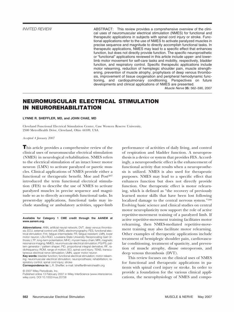
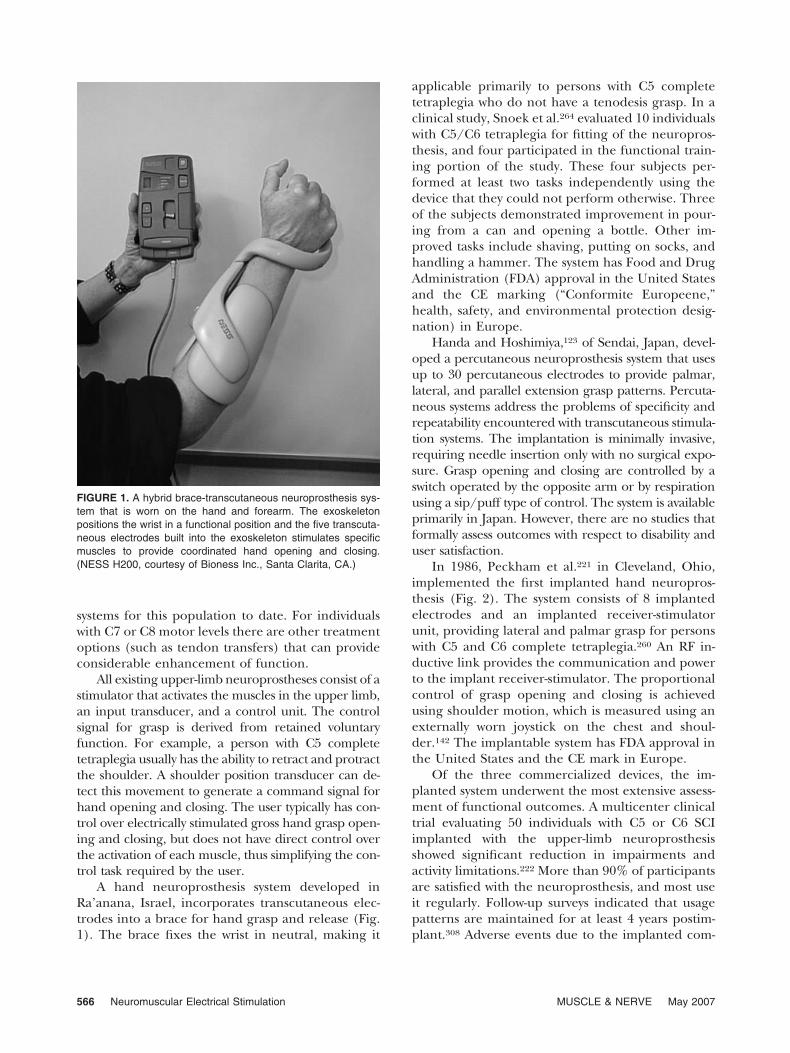
A hand neuroprosthesis system developed inRa’anana, Israel, incorporates transcutaneous elec-trodes into a brace for hand grasp and release (Fig.1). The brace fixes the wrist in neutral, making it
applicable primarily to persons with C5 completetetraplegia who do not have a tenodesis grasp. In aclinical study, Snoek et al.264 evaluated 10 individualswith C5/C6 tetraplegia for fitting of the neuropros-thesis, and four participated in the functional train-ing portion of the study. These four subjects per-formed at least two tasks independently using thedevice that they could not perform otherwise. Threeof the subjects demonstrated improvement in pour-ing from a can and opening a bottle. Other im-proved tasks include shaving, putting on socks, andhandling a hammer. The system has Food and DrugAdministration (FDA) approval in the United Statesand the CE marking (“Conformite Europeene,”health, safety, and environmental protection desig-nation) in Europe.
Handa and Hoshimiya,123 of Sendai, Japan, devel-oped a percutaneous neuroprosthesis system that usesup to 30 percutaneous electrodes to provide palmar,lateral, and parallel extension grasp patterns. Percuta-neous systems address the problems of specificity andrepeatability encountered with transcutaneous stimula-tion systems. The implantation is minimally invasive,requiring needle insertion only with no surgical expo-sure. Grasp opening and closing are controlled by aswitch operated by the opposite arm or by respirationusing a sip/puff type of control. The system is availableprimarily in Japan. However, there are no studies thatformally assess outcomes with respect to disability anduser satisfaction.
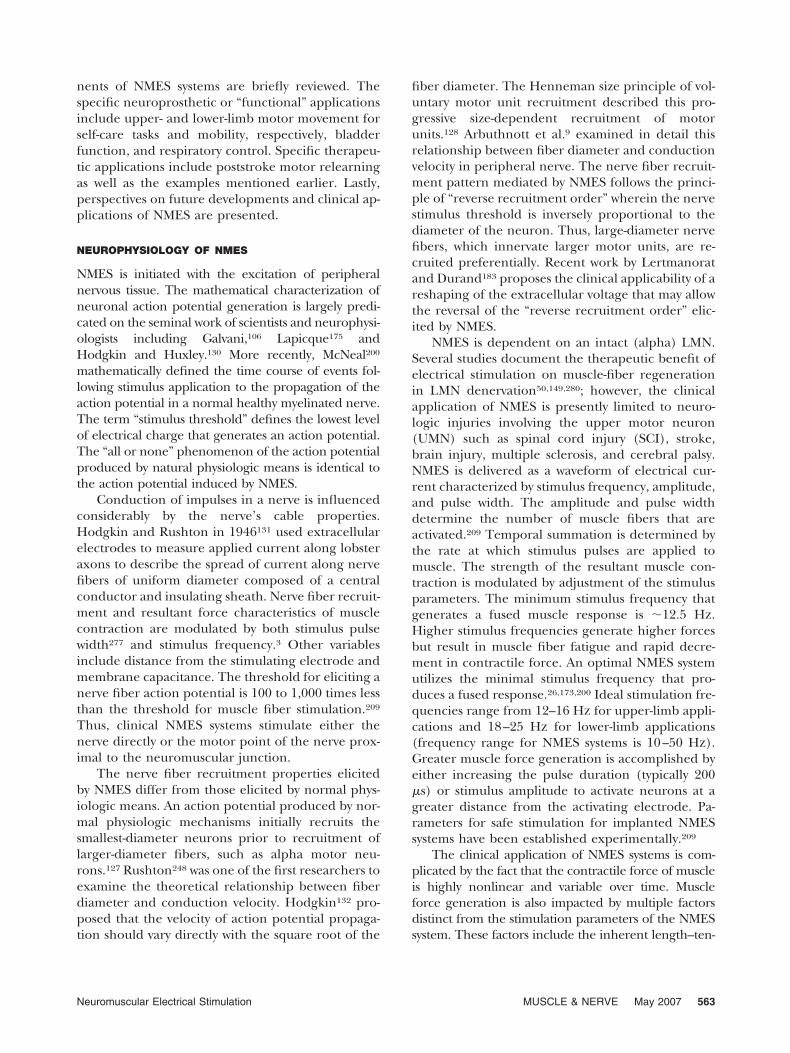
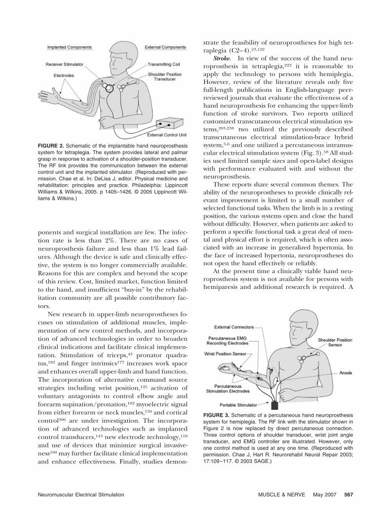
In 1986, Peckham et al.221 in Cleveland, Ohio,implemented the first implanted hand neuropros-thesis (Fig. 2). The system consists of 8 implantedelectrodes and an implanted receiver-stimulatorunit, providing lateral and palmar grasp for personswith C5 and C6 complete tetraplegia.260 An RF in-ductive link provides the communication and powerto the implant receiver-stimulator. The proportionalcontrol of grasp opening and closing is achievedusing shoulder motion, which is measured using anexternally worn joystick on the chest and shoul-der.142 The implantable system has FDA approval inthe United States and the CE mark in Europe.
Of the three commercialized devices, the im-planted system underwent the most extensive assess-ment of functional outcomes. A multicenter clinicaltrial evaluating 50 individuals with C5 or C6 SCIimplanted with the upper-limb neuroprosthesisshowed significant reduction in impairments andactivity limitations.222 More than 90% of participantsare satisfied with the neuroprosthesis, and most useit regularly. Follow-up surveys indicated that usagepatterns are maintained for at least 4 years postim-plant.308 Adverse events due to the implanted com-
FIGURE 1. A hybrid brace-transcutaneous neuroprosthesis sys-tem that is worn on the hand and forearm. The exoskeletonpositions the wrist in a functional position and the five transcuta-neous electrodes built into the exoskeleton stimulates specificmuscles to provide coordinated hand opening and closing.(NESS H200, courtesy of Bioness Inc., Santa Clarita, CA.)
566 Neuromuscular Electrical Stimulation MUSCLE & NERVE May 2007
ponents and surgical installation are few. The infec-tion rate is less than 2%. There are no cases ofneuroprosthesis failure and less than 1% lead fail-ures. Although the device is safe and clinically effec-tive, the system is no longer commercially available.Reasons for this are complex and beyond the scopeof this review. Cost, limited market, function limitedto the hand, and insufficient “buy-in” by the rehabil-itation community are all possible contributory fac-tors.
New research in upper-limb neuroprostheses fo-cuses on stimulation of additional muscles, imple-mentation of new control methods, and incorpora-tion of advanced technologies in order to broadenclinical indications and facilitate clinical implemen-tation. Stimulation of triceps,43 pronator quadra-tus,182 and finger intrinsics177 increases work spaceand enhances overall upper-limb and hand function.The incorporation of alternative command sourcestrategies including wrist position,125 activation ofvoluntary antagonists to control elbow angle andforearm supination/pronation,182 myoelectric signalfrom either forearm or neck muscles,159 and corticalcontrol306 are under investigation. The incorpora-tion of advanced technologies such as implantedcontrol transducers,143 new electrode technology,118
and use of devices that minimize surgical invasive-ness190 may further facilitate clinical implementationand enhance effectiveness. Finally, studies demon-
strate the feasibility of neuroprostheses for high tet-raplegia (C2–4).23,122
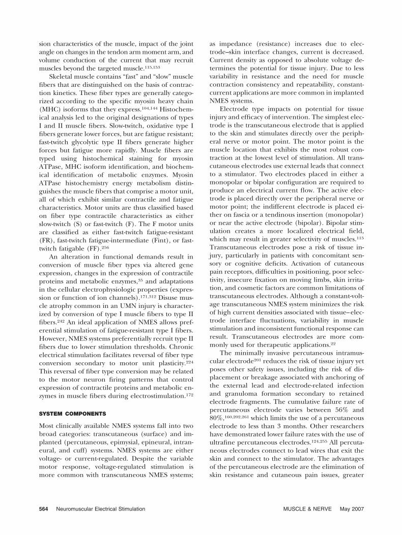
Stroke. In view of the success of the hand neu-roprosthesis in tetraplegia,222 it is reasonable toapply the technology to persons with hemiplegia.However, review of the literature reveals only fivefull-length publications in English-language peer-reviewed journals that evaluate the effectiveness of ahand neuroprosthesis for enhancing the upper-limbfunction of stroke survivors. Two reports utilizedcustomized transcutaneous electrical stimulation sys-tems,203,238 two utilized the previously describedtranscutaneous electrical stimulation-brace hybridsystem,5,6 and one utilized a percutaneous intramus-cular electrical stimulation system (Fig. 3).58 All stud-ies used limited sample sizes and open-label designswith performance evaluated with and without theneuroprosthesis.
These reports share several common themes. Theability of the neuroprostheses to provide clinically rel-evant improvement is limited to a small number ofselected functional tasks. When the limb is in a restingposition, the various systems open and close the handwithout difficulty. However, when patients are asked toperform a specific functional task a great deal of men-tal and physical effort is required, which is often asso-ciated with an increase in generalized hypertonia. Inthe face of increased hypertonia, neuroprostheses donot open the hand effectively or reliably.
At the present time a clinically viable hand neu-roprosthesis system is not available for persons withhemiparesis and additional research is required. A
FIGURE 3. Schematic of a percutaneous hand neuroprosthesissystem for hemiplegia. The RF link with the stimulator shown inFigure 2 is now replaced by direct percutaneous connection.Three control options of shoulder transducer, wrist joint angletransducer, and EMG controller are illustrated. However, onlyone control method is used at any one time. (Reproduced withpermission. Chae J, Hart R. Neurorehabil Neural Repair 2003;17:109–117. © 2003 SAGE.)
FIGURE 2. Schematic of the implantable hand neuroprosthesissystem for tetraplegia. The system provides lateral and palmargrasp in response to activation of a shoulder-position transducer.The RF link provides the communication between the externalcontrol unit and the implanted stimulator. (Reproduced with per-mission. Chae et al. In: DeLisa J, editor. Physical medicine andrehabilitation: principles and practice. Philadelphia: LippincottWilliams & Wilkins, 2005. p 1405–1426. © 2005 Lippincott Wil-liams & Wilkins.)
Neuromuscular Electrical Stimulation MUSCLE & NERVE May 2007 567
clinically deployable system must demonstrate theability to: (1) facilitate bilateral tasks, (2) provideproximal and distal control, (3) have sufficient min-iaturization to not interfere with ambulation, (4)utilize control paradigms that produce effortlessmovement of the impaired upper limb without com-promising the function of the intact limb, and (5)“turn off” overactive muscles as well as stimulateweak muscles.58
Lower-Limb Applications. Spinal Cord Injury. Mul-tichannel transcutaneous electrical stimulation sys-tems are successful in producing standing and step-ping for persons with complete SCI. These systemsare relatively simple, consisting of 2–6 channels ofcontinuous stimulation.116,140,168,310 Systems employ-ing percutaneous intramuscular electrodes allow formore complex movements196 and can provide sim-ple mobility and one-handed reaching tasks.162,286 Acochlear implant modified to stimulate motor neu-rons78 and a 12-channel system for activation of theL2-S2 motor roots86 have been used for exercise andstanding in a limited number of volunteers. Forlong-term clinical application, implanted systemssuch as these provide major advantages over trans-cutaneous and percutaneous systems including con-venience, cosmetic benefit, reliability, and repeat-ability.
The pioneering work in the application of neu-roprostheses for restoration of standing and walkingfor individuals with complete and incomplete SCIconducted in the 1970s and 1980s continues to beemployed in many laboratories and clinics aroundthe world.14,168 Standing is achieved by simulta-
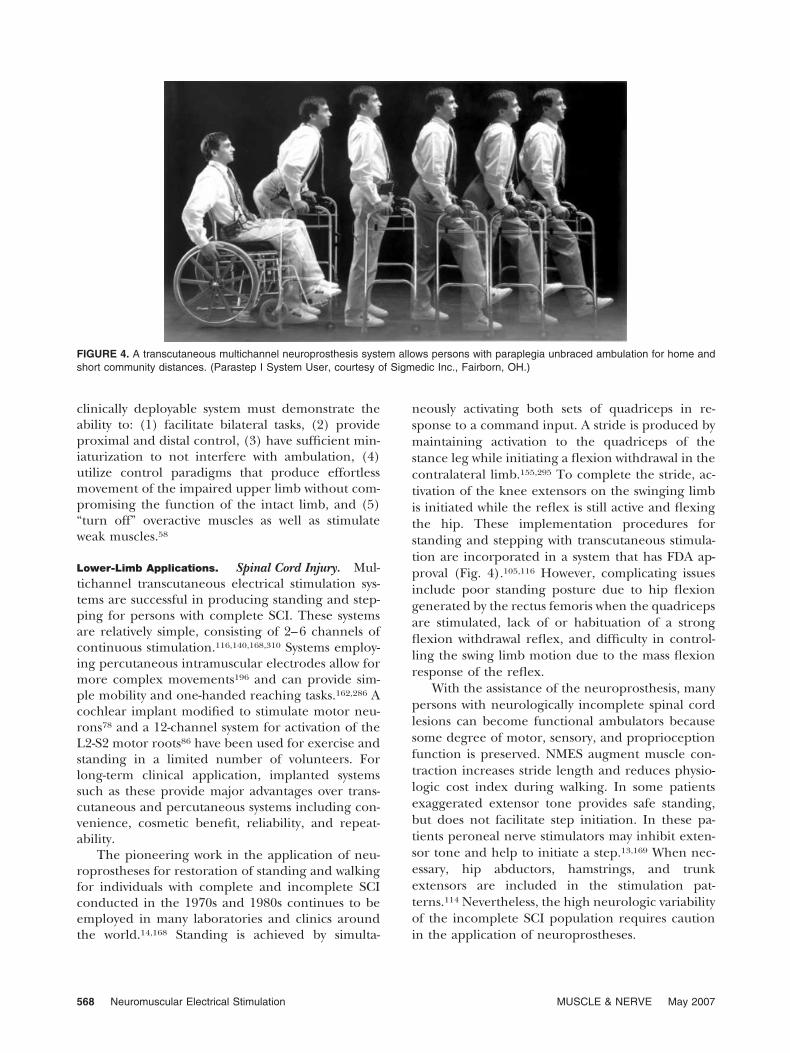
neously activating both sets of quadriceps in re-sponse to a command input. A stride is produced bymaintaining activation to the quadriceps of thestance leg while initiating a flexion withdrawal in thecontralateral limb.155,295 To complete the stride, ac-tivation of the knee extensors on the swinging limbis initiated while the reflex is still active and flexingthe hip. These implementation procedures forstanding and stepping with transcutaneous stimula-tion are incorporated in a system that has FDA ap-proval (Fig. 4).105,116 However, complicating issuesinclude poor standing posture due to hip flexiongenerated by the rectus femoris when the quadricepsare stimulated, lack of or habituation of a strongflexion withdrawal reflex, and difficulty in control-ling the swing limb motion due to the mass flexionresponse of the reflex.
With the assistance of the neuroprosthesis, manypersons with neurologically incomplete spinal cordlesions can become functional ambulators becausesome degree of motor, sensory, and proprioceptionfunction is preserved. NMES augment muscle con-traction increases stride length and reduces physio-logic cost index during walking. In some patientsexaggerated extensor tone provides safe standing,but does not facilitate step initiation. In these pa-tients peroneal nerve stimulators may inhibit exten-sor tone and help to initiate a step.13,169 When nec-essary, hip abductors, hamstrings, and trunkextensors are included in the stimulation pat-terns.114 Nevertheless, the high neurologic variabilityof the incomplete SCI population requires cautionin the application of neuroprostheses.
FIGURE 4. A transcutaneous multichannel neuroprosthesis system allows persons with paraplegia unbraced ambulation for home andshort community distances. (Parastep I System User, courtesy of Sigmedic Inc., Fairborn, OH.)
568 Neuromuscular Electrical Stimulation MUSCLE & NERVE May 2007
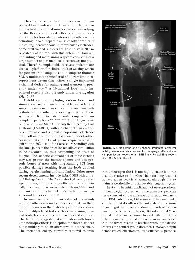
These approaches have implications for im-planted lower-limb systems. However, implanted sys-tems activate individual muscles rather than relyingon the flexion withdrawal reflex or extensive brac-ing. Complex lower-limb motions are synthesized byactivating up to 48 separate muscles with chronicallyindwelling percutaneous intramuscular electrodes.Some well-trained subjects are able to walk 300 mrepeatedly at 0.5 m/s with this system.163 However,implanting and maintaining a system consisting of alarge number of percutaneous electrodes is not prac-tical. Therefore, implantable receive-stimulators areused as a platform for clinical trials of walking systemfor persons with complete and incomplete thoracicSCI. A multicenter clinical trial of a lower-limb neu-roprosthesis system that utilizes a single implanted8-channel device for standing and transfers is pres-ently under way.77 A 16-channel lower limb im-planted system is also presently under investigation(Fig. 5).165
Hybrid systems employing various brace andstimulation components are reliable and relativelysimple to implement in clinical environments withorthotic and prosthetic fabricating capacity. Thesesystems are fitted to patients with complete or in-complete paraplegia.146,197,265,266 One design com-bines a Louisiana State University Reciprocating GaitOrthosis (LSU-RGO) with a 4-channel transcutane-ous stimulator and a flexible copolymer electrodecuff. Follow-up studies on RGO-based hybrid ortho-ses show that up to 41% of system recipients use it forgait101 and 66% use it for exercise.267 Standing withthe knee joints of the brace locked allows stimulationto be discontinued, thus postponing the onset offatigue. The orthotic components of these systemsmay also protect the insensate joints and osteopo-rotic bones of users with long-standing SCI frompossible damage resulting from the loads appliedduring weight-bearing and ambulation. Other morerecent developments include hybrid FES with a me-dial-linkage knee–ankle–foot orthosis,254 energy stor-age orthosis,90 more energy-efficient and cosmeti-cally accepted hip–knee–ankle orthosis,206,251 andimplantable multichannel FES with trunk–hip–knee–ankle foot orthosis.164
In summary, the inherent value of lower-limbneuroprosthesis systems for persons with SCI in theircurrent forms is in the ability to provide short-dura-tion mobility-related tasks, such as overcoming phys-ical obstacles or architectural barriers and exercise.The literature suggests that ambulation with lower-limb neuroprosthesis is an option for short distancesbut is unlikely to be an alternative to a wheelchair.The metabolic energy currently required to walk
with a neuroprosthesis is too high to make it a prac-tical alternative to the wheelchair for long-distancetransportation over level surfaces, although this re-mains a worthwhile and achievable long-term goal.
Stroke. The initial application of neuroprosthesesin hemiplegia focused on transcutaneous peronealnerve stimulation to treat ankle dorsiflexion weakness.In a 1961 publication, Lieberson et al.187 described astimulator that dorsiflexes the ankle during the swingphase of gait. In the only randomized study of transcu-taneous peroneal stimulation, Burridge et al.46 re-ported that stroke survivors treated with the deviceexhibit significantly greater increase in walking speedwith the device relative to baseline without the device,whereas the control group does not. However, despitedemonstrated effectiveness, transcutaneous peroneal
FIGURE 5. A radiograph of a 16-channel implanted lower-limbmobility neuroprosthesis system for paraplegia. (Reproducedwith permission. Kobetic et al. IEEE Trans Rehabil Eng 1999;7:390–398. © 1999 IEEE.)
Neuromuscular Electrical Stimulation MUSCLE & NERVE May 2007 569
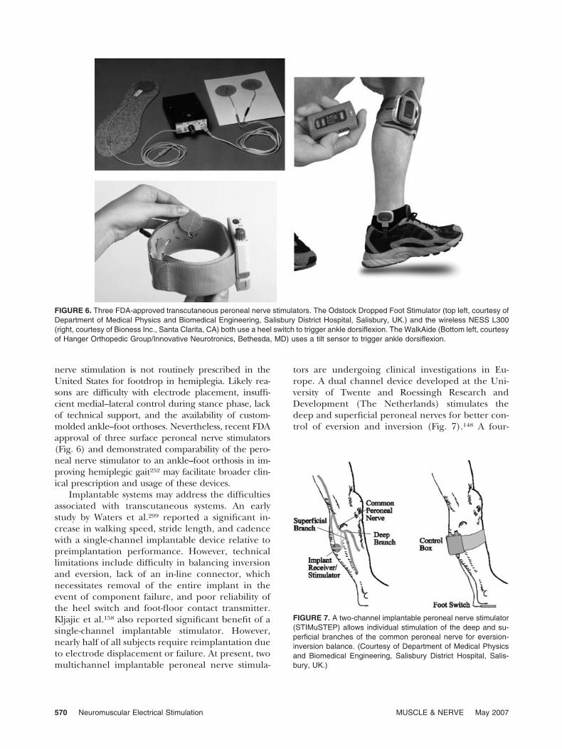
nerve stimulation is not routinely prescribed in theUnited States for footdrop in hemiplegia. Likely rea-sons are difficulty with electrode placement, insuffi-cient medial–lateral control during stance phase, lackof technical support, and the availability of custom-molded ankle–foot orthoses. Nevertheless, recent FDAapproval of three surface peroneal nerve stimulators(Fig. 6) and demonstrated comparability of the pero-neal nerve stimulator to an ankle–foot orthosis in im-proving hemiplegic gait252 may facilitate broader clin-ical prescription and usage of these devices.
Implantable systems may address the difficultiesassociated with transcutaneous systems. An earlystudy by Waters et al.299 reported a significant in-crease in walking speed, stride length, and cadencewith a single-channel implantable device relative topreimplantation performance. However, technicallimitations include difficulty in balancing inversionand eversion, lack of an in-line connector, whichnecessitates removal of the entire implant in theevent of component failure, and poor reliability ofthe heel switch and foot-floor contact transmitter.Kljajic et al.158 also reported significant benefit of asingle-channel implantable stimulator. However,nearly half of all subjects require reimplantation dueto electrode displacement or failure. At present, twomultichannel implantable peroneal nerve stimula-
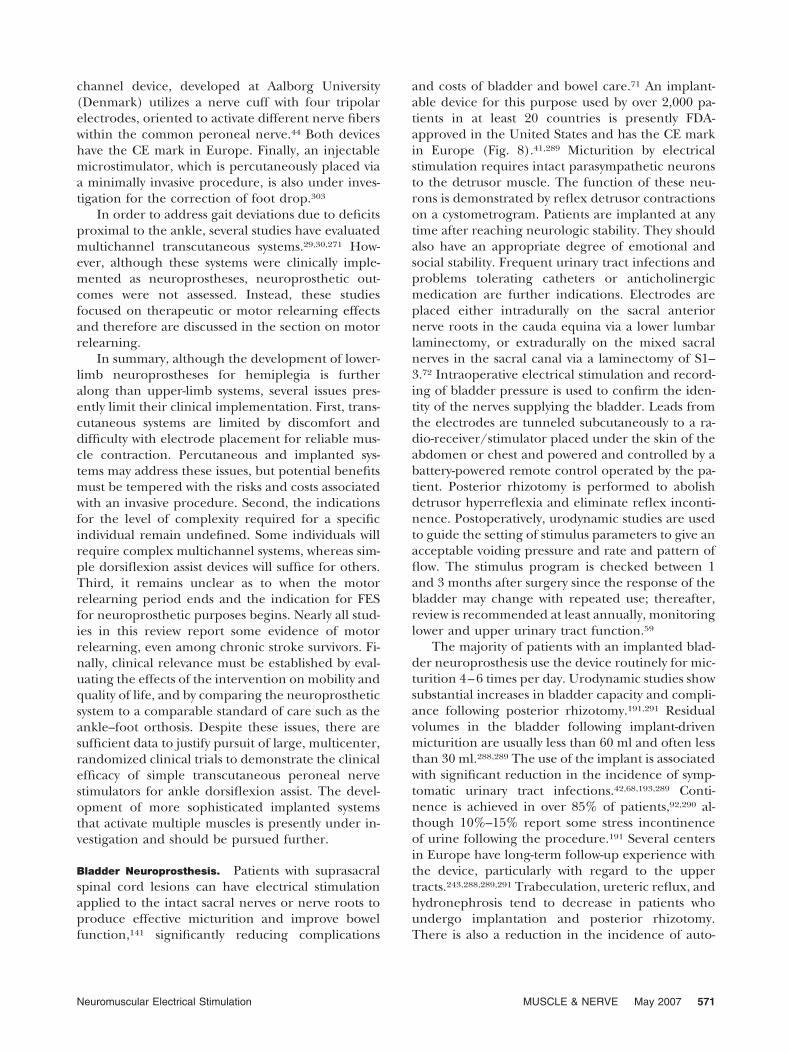
tors are undergoing clinical investigations in Eu-rope. A dual channel device developed at the Uni-versity of Twente and Roessingh Research andDevelopment (The Netherlands) stimulates thedeep and superficial peroneal nerves for better con-trol of eversion and inversion (Fig. 7).148 A four-
FIGURE 6. Three FDA-approved transcutaneous peroneal nerve stimulators. The Odstock Dropped Foot Stimulator (top left, courtesy ofDepartment of Medical Physics and Biomedical Engineering, Salisbury District Hospital, Salisbury, UK.) and the wireless NESS L300(right, courtesy of Bioness Inc., Santa Clarita, CA) both use a heel switch to trigger ankle dorsiflexion. The WalkAide (Bottom left, courtesyof Hanger Orthopedic Group/Innovative Neurotronics, Bethesda, MD) uses a tilt sensor to trigger ankle dorsiflexion.
FIGURE 7. A two-channel implantable peroneal nerve stimulator(STIMuSTEP) allows individual stimulation of the deep and su-perficial branches of the common peroneal nerve for eversion-inversion balance. (Courtesy of Department of Medical Physicsand Biomedical Engineering, Salisbury District Hospital, Salis-bury, UK.)
570 Neuromuscular Electrical Stimulation MUSCLE & NERVE May 2007
channel device, developed at Aalborg University(Denmark) utilizes a nerve cuff with four tripolarelectrodes, oriented to activate different nerve fiberswithin the common peroneal nerve.44 Both deviceshave the CE mark in Europe. Finally, an injectablemicrostimulator, which is percutaneously placed viaa minimally invasive procedure, is also under inves-tigation for the correction of foot drop.303
In order to address gait deviations due to deficitsproximal to the ankle, several studies have evaluatedmultichannel transcutaneous systems.29,30,271 How-ever, although these systems were clinically imple-mented as neuroprostheses, neuroprosthetic out-comes were not assessed. Instead, these studiesfocused on therapeutic or motor relearning effectsand therefore are discussed in the section on motorrelearning.
In summary, although the development of lower-limb neuroprostheses for hemiplegia is furtheralong than upper-limb systems, several issues pres-ently limit their clinical implementation. First, trans-cutaneous systems are limited by discomfort anddifficulty with electrode placement for reliable mus-cle contraction. Percutaneous and implanted sys-tems may address these issues, but potential benefitsmust be tempered with the risks and costs associatedwith an invasive procedure. Second, the indicationsfor the level of complexity required for a specificindividual remain undefined. Some individuals willrequire complex multichannel systems, whereas sim-ple dorsiflexion assist devices will suffice for others.Third, it remains unclear as to when the motorrelearning period ends and the indication for FESfor neuroprosthetic purposes begins. Nearly all stud-ies in this review report some evidence of motorrelearning, even among chronic stroke survivors. Fi-nally, clinical relevance must be established by eval-uating the effects of the intervention on mobility andquality of life, and by comparing the neuroprostheticsystem to a comparable standard of care such as theankle–foot orthosis. Despite these issues, there aresufficient data to justify pursuit of large, multicenter,randomized clinical trials to demonstrate the clinicalefficacy of simple transcutaneous peroneal nervestimulators for ankle dorsiflexion assist. The devel-opment of more sophisticated implanted systemsthat activate multiple muscles is presently under in-vestigation and should be pursued further.
Bladder Neuroprosthesis. Patients with suprasacralspinal cord lesions can have electrical stimulationapplied to the intact sacral nerves or nerve roots toproduce effective micturition and improve bowelfunction,141 significantly reducing complications
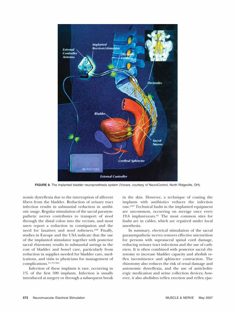
and costs of bladder and bowel care.71 An implant-able device for this purpose used by over 2,000 pa-tients in at least 20 countries is presently FDA-approved in the United States and has the CE markin Europe (Fig. 8).41,289 Micturition by electricalstimulation requires intact parasympathetic neuronsto the detrusor muscle. The function of these neu-rons is demonstrated by reflex detrusor contractionson a cystometrogram. Patients are implanted at anytime after reaching neurologic stability. They shouldalso have an appropriate degree of emotional andsocial stability. Frequent urinary tract infections andproblems tolerating catheters or anticholinergicmedication are further indications. Electrodes areplaced either intradurally on the sacral anteriornerve roots in the cauda equina via a lower lumbarlaminectomy, or extradurally on the mixed sacralnerves in the sacral canal via a laminectomy of S1–3.72 Intraoperative electrical stimulation and record-ing of bladder pressure is used to confirm the iden-tity of the nerves supplying the bladder. Leads fromthe electrodes are tunneled subcutaneously to a ra-dio-receiver/stimulator placed under the skin of theabdomen or chest and powered and controlled by abattery-powered remote control operated by the pa-tient. Posterior rhizotomy is performed to abolishdetrusor hyperreflexia and eliminate reflex inconti-nence. Postoperatively, urodynamic studies are usedto guide the setting of stimulus parameters to give anacceptable voiding pressure and rate and pattern offlow. The stimulus program is checked between 1and 3 months after surgery since the response of thebladder may change with repeated use; thereafter,review is recommended at least annually, monitoringlower and upper urinary tract function.59
The majority of patients with an implanted blad-der neuroprosthesis use the device routinely for mic-turition 4–6 times per day. Urodynamic studies showsubstantial increases in bladder capacity and compli-ance following posterior rhizotomy.191,291 Residualvolumes in the bladder following implant-drivenmicturition are usually less than 60 ml and often lessthan 30 ml.288,289 The use of the implant is associatedwith significant reduction in the incidence of symp-tomatic urinary tract infections.42,68,193,289 Conti-nence is achieved in over 85% of patients,92,290 al-though 10%–15% report some stress incontinenceof urine following the procedure.191 Several centersin Europe have long-term follow-up experience withthe device, particularly with regard to the uppertracts.243,288,289,291 Trabeculation, ureteric reflux, andhydronephrosis tend to decrease in patients whoundergo implantation and posterior rhizotomy.There is also a reduction in the incidence of auto-
Neuromuscular Electrical Stimulation MUSCLE & NERVE May 2007 571
nomic dysreflexia due to the interruption of afferentfibers from the bladder. Reduction of urinary tractinfection results in substantial reduction in antibi-otic usage. Regular stimulation of the sacral parasym-pathetic nerves contributes to transport of stoolthrough the distal colon into the rectum, and mostusers report a reduction in constipation and theneed for laxatives and stool softeners.192 Finally,studies in Europe and the USA indicate that the useof the implanted stimulator together with posteriorsacral rhizotomy results in substantial savings in thecost of bladder and bowel care, particularly fromreduction in supplies needed for bladder care, med-ications, and visits to physicians for management ofcomplications.71,73,304
Infection of these implants is rare, occurring in1% of the first 500 implants. Infection is usuallyintroduced at surgery or through a subsequent break
in the skin. However, a technique of coating theimplants with antibiotics reduces the infectionrate.247 Technical faults in the implanted equipmentare uncommon, occurring on average once every19.6 implant-years.41 The most common sites forfaults are in cables, which are repaired under localanesthesia.
In summary, electrical stimulation of the sacralparasympathetic nerves restores effective micturitionfor persons with suprasacral spinal cord damage,reducing urinary tract infections and the use of cath-eters. It is often combined with posterior sacral rhi-zotomy to increase bladder capacity and abolish re-flex incontinence and sphincter contraction. Therhizotomy also reduces the risk of renal damage andautonomic dysreflexia, and the use of anticholin-ergic medication and urine collection devices; how-ever, it also abolishes reflex erection and reflex ejac-
FIGURE 8. The implanted bladder neuroprosthesis system (Vocare, courtesy of NeuroControl, North Ridgeville, OH).
572 Neuromuscular Electrical Stimulation MUSCLE & NERVE May 2007
ulation, which may need to be provided byalternative techniques. Overall, these interventionsdramatically improve bladder and bowel function,reduce complications and costs, and increase qualityof life after spinal cord injury.
Although the implanted neuroprosthesis isclearly effective in providing urine storage and mic-turition, the need for a rhizotomy dampens the levelof enthusiasm among both clinicians and patients.In order to address this issue, various groups areinvestigating alternative means of reducing detrusorhyperactivity. Sensory neuromodulation via stimula-tion of the dorsal penile/clitoral nerve increasesbladder capacity among patients with SCI8 and mul-tiple sclerosis.249 However, the approach does notabolish reflex sphincter contractions. Thus, otherinvestigators focus on direct blockage of nerve im-pulses via high-frequency electrical stimulation. Al-though this approach is still at the level of animalexperimentation, its clinical implications reach farbeyond the bladder application24 and may includemanagement of the broader problems of spasticityand pain.25,151 Other forms of neuromodulation totreat uninhibited bladder contractions are reportedin the literature.241 However, these are generallyapplied to able-bodied populations and thus are notincluded in this review.
Respiratory Neuroprostheses. Phrenic nerve pacinghas been applied to more than 1,200 patients world-wide and is now a clinically accepted technique toprovide artificial ventilatory support in patients withtrauma, with respiratory failure secondary to cervicalSCI.108,109,112,136,137,284 There are several commer-cially available phrenic nerve pacing systems, buteach system has a similar configuration. The stimu-lating electrodes are implanted directly on eachphrenic nerve. Small wires tunneled subcutaneouslyconnect the electrodes to an RF receiver, which isimplanted in an easily accessible area over the ante-rior portion of the thorax. External antennas con-nect to the transmitter. The transmitter generates anRF signal, which is inductively coupled to the im-planted receiver. The signal is demodulated by thereceivers, which converts it to electrical signals, andthen delivered to the stimulating electrodes. Bilat-eral phrenic nerve stimulation results in descent ofeach diaphragm and decrease in intrathoracic pres-sure resulting in inspiration. Cessation of stimula-tion results in diaphragm relaxation, an increase inintrathoracic pressure, and exhalation.
All potential candidates must demonstrate intactphrenic nerves on nerve conduction studies. Theymust be free of significant lung disease or primary
muscle disease. Patients’ psychosocial conditions arealso important considerations. Before any technicalassessment a critical evaluation of the motivation ofboth the patient and family members is mandatory.The patient should also have a clear understandingof the potential benefits to be achieved.
Electrodes are positioned around the phrenicnerve in either the cervical region or within thethorax.108,110,112 Although the thoracic approach re-quires a thoracotomy, this is the preferred approach,as the cervical approach has the risk of not beingable to stimulate the entire nerve.100,293 An RF re-ceiver is positioned in a subcutaneous pocket on theanterior chest wall. Wires from the electrode arepassed through the 3rd or 4th intercostal space andconnected to the receiver. Postimplantation, the di-aphragm must be gradually reconditioned to im-prove strength and endurance.111 During the condi-tioning phase the patient must be monitored forsigns of fatigue, which is usually manifested by thepatient’s complaint of shortness of breath or reduc-tion in inspired volume.
Although a number of complications have beenreported since phrenic nerve pacing was first intro-duced, technical developments and patient experiencehave markedly reduced their incidence.85,107 Neverthe-less, all patients require a back-up mechanical ventila-tor in the event of pacemaker failure. Reported modesof failure include (1) low battery charge, (2) antennawire breakage, (3) iatrogenic injury to the phrenicnerve, (4) postimplantation adverse tissue reaction andscar tissue formation, (5) device infections, (6) collapseof the upper airway or obstructive apnea due to dia-phragm contraction without coincident contraction ofthe upper airway muscles, and (7) in children, reduc-tion in inspired volume due to paradoxical movementof the rib cage.
Phrenic nerve pacing is clearly an effective meansof providing ventilatory support with significant ad-vantages over mechanical ventilation.108,109,112 Un-fortunately, there are few recent analyses of modern-day success rates and incidence of side effects andcomplications. A long-term follow-up study of 14tetraplegic patients who use bilateral low-frequencystimulation reported successful use of the device foras long as 15 years, with a mean use of 7.6 years.107 Amore recent study of 64 patients (45 tetraplegic pa-tients) who underwent phrenic nerve pacing for amean of 2 years showed the incidence of electrodeand receiver failure as 3.1% and 5.9%, respectively,which is significantly lower than earlier reports. Atthis time there are no controlled studies relative tomechanical ventilators. However, it is possible thatphrenic nerve pacing improves life expectancy in
Neuromuscular Electrical Stimulation MUSCLE & NERVE May 2007 573
patients with tetraplegia. Carter et al.51 reported only63% survival at 9 years for patients on positive pres-sure ventilation. In contrast, all 12 tetraplegic pa-tients who completed the Yale phrenic nerve pacingprotocol were alive after 9 years.93
Phrenic pacing provides important health andlifestyle benefits relative to mechanical ventilation.However, many patients with ventilator-dependenttetraplegia cannot be offered phrenic nerve pacingdue to partial or complete injury of one of thephrenic nerves. Combined intercostal and unilateraldiaphragm pacing may be a useful therapeutic mo-dality in selected patients with only unilateralphrenic nerve function.84 Conventional placementof phrenic nerve electrodes carries the risk ofphrenic nerve injury and generally requires a thora-cotomy, which is a major surgical procedure withassociated risk, in-patient hospital stay, and highcost. Preliminary results suggest that intramusculardiaphragm pacing provide similar benefits as con-ventional phrenic nerve pacing without the need foran invasive surgical procedure and less risk ofphrenic nerve injury.83 The laparoscopy guided pro-cedure is performed on an outpatient basis and istherefore less costly. The development of fully im-plantable intramuscular diaphragm system will elim-inate the need for the application of devices on thebody surface and the risk of decoupling between thetransmitter and receiver.
NMES FOR MOTOR RELEARNING
Evolving basic and clinical studies on central motorneuroplasticity support the role of goal-oriented, ac-tive repetitive movement training of a paretic limb toenhance motor relearning. Asanuma and Keller12
demonstrated that electrical stimulation of the so-matosensory cortex alone or in conjunction withthalamic stimulation in an animal model induceslong-term potentiation (LTP) in the motor cortex.They hypothesized that proprioceptive and cutane-ous afferent impulses associated with repetitivemovements induce LTP in the motor cortex, whichthen modify the excitability of specific motor neu-rons and facilitate motor relearning.11 Consistentwith this hypothesis, nonhuman primate researchhas demonstrated that after local damage to themotor cortex, goal-oriented, active repetitive move-ment training of the paretic limb shapes subsequentfunctional reorganization in the adjacent intact cor-tex, and that the undamaged motor cortex plays animportant role in motor relearning.214 Specific typesof behavioral experiences that induce long-termplasticity in motor maps are repetitive movements
that entail the development of new motor skills. Thatis, the motor tasks are new and therefore “require”significant cognitive effort to complete.213 When an-imals are trained to perform new tasks such as re-trieving food pellets from a small well212,215,230 or arotating well,157 there is evidence of task-specific cor-tical reorganization. However, repetitive movementtasks that do not require new skill acquisition (i.e.,motor tasks that are already mastered and thereforeare easy to carry out and require minimal or nocognitive effort) are not associated with any signifi-cant changes in the motor cortex.157,230
If goal-oriented, repetitive movement therapy fa-cilitates motor relearning, it is possible that electricalstimulation–mediated goal-oriented repetitive move-ment therapy also facilitates motor relearning. Acuteadministration of electrical stimulation to a periph-eral nerve activates both sensory and motor struc-tures in the brain82,270 and reduces intracorticalinhibition.229,239 Functional magnetic resonance im-aging (fMRI) studies show activation of the con-tralateral somatosensory cortex and bilateral supple-mentary motor areas in response to NMES-mediatedwrist extension activity,120 as well as a dose–responserelationship between fMRI and NMES of the lower-limb muscles.262 These data suggest that repetitivemovement therapy mediated by NMES has the po-tential to facilitate motor relearning via corticalmechanisms.
It is also possible that electrical stimulation facil-itates motor relearning via spinal mechanisms. Rush-ton246 theorized that the corticospinal–anterior horncell synapse is a Hebb-type, modifiable synapse andthat the synapse can be modified by NMES. “Hebb’srule” proposed in 1949 by Donald Hebb126 states:“When an axon of cell A. . .excite[s] cell B and re-peatedly or persistently takes part in firing it, somegrowth process or metabolic change takes place inone or both cells so that A’s efficiency as one of thecells firing B is increased.” The synapse is thought tobe strengthened by the coincidence of presynapticand postsynaptic activities. Under normal circum-stances neural activity in the pyramidal tract easilydischarges the anterior horn cells and the strengthof the presumed Hebb-type pyramidal tract/anteriorhorn cell synapse is maintained by this traffic. How-ever, following brain injury, neural activity in thepyramidal tract is significantly reduced. Failure torestore this traffic leads to “decorrelation” of presyn-aptic and postsynaptic activities, which weakens thesynapse. Rushton suggested that NMES-mediated an-tidromic impulses provide an artificial means to syn-chronize presynaptic and postsynaptic activity in theaffected population of anterior horn cells. Accord-
574 Neuromuscular Electrical Stimulation MUSCLE & NERVE May 2007
ingly, he predicted that combining NMES with si-multaneous voluntary effort is an effective means offacilitating motor relearning.
This article limits the review of NMES effectivenessin enhancing motor relearning to the stroke popula-tion. Although there is evidence of the role of NMES infacilitating motor relearning in SCI, the breadth anddepth of this literature is limited.199 NMES can be usedby patients with hemiparesis who do not have enoughresidual movement to take part in volitional, activerepetitive movement therapy. Regardless of cortical orspinal mechanisms, the experimental and theoreticalconsiderations suggest that the necessary prerequisitesfor NMES-mediated motor relearning include repeti-tion, novelty of activity, concurrent volitional effort,and high functional content.
Three types of electrical stimulation are availablefor motor relearning: cyclic NMES, electromyogra-phy (EMG)/biofeedback-mediated NMES, and neu-roprostheses. Cyclic NMES activates paretic musclesat a set duty cycle for a preset time period. Thepatient is a passive participant and does not requirea cognitive investment, in the form of either initia-tion of muscle contraction, interpretation of afferentsignals, or functionality of motor task. The secondtype of NMES includes EMG or biofeedback-medi-ated electrical stimulation, which couples afferentfeedback to NMES-induced repetitive movementtherapy. These techniques may be applied to pa-tients who can partially activate a paretic muscle butare unable to generate sufficient muscle contractionfor adequate exercise or functional purposes.Whereas the patient is a passive participant whenusing cyclic NMES, EMG or biofeedback-mediatedNMES requires greater cognitive investment, whichmay result in greater therapeutic benefit. The thirdtype of NMES includes neuroprosthetic applicationsthat provide FES. In this strategy repetitive move-ment training is performed in the context of mean-ingful, functional behavioral tasks and has a theoret-ical advantage over both cyclic and EMG/biofeedback mediated NMES.
Upper-Limb Applications. There are four random-ized clinical trials investigating the efficacy of cyclicNMES in enhancing upper-limb motor relearn-ing.56,234,268,269,307 Note that the two studies by Sondeet al.268 refer to the same study with the 1998 publi-cation reporting end of treatment results and the2000 publication269 reporting 3-year follow-up re-sults. All four studies reported improved outcomesin motor impairment at the end of treatment, withmild to moderately impaired subjects benefitingmost. Among the three studies that provided fol-
low-up data, the two acute stroke studies reportedenduring effects,56,234 whereas the one chronic studydid not.269 All four studies evaluated activity limita-tion. However, only two of these reported improve-ments at the end of treatment,234,307 and the onestudy with follow-up data demonstrated no enduringeffect on activity limitation.234
In the most methodologically sound of the fourstudies, Powell et al.234 reported that isometric wristextension torques were significantly higher for thetreatment group at the end of treatment and at 32weeks. The grasp and grip subscores of the ActionResearch Arm Test were significantly higher for thetreatment group at the end of treatment, but not at32 weeks. A post-hoc subset analysis indicated thatthe intervention was most effective for those withresidual wrist extension torque at study entry.
The strengths of these studies rest on their ran-domized designs. However, numerous methodolog-ical limitations render the results difficult to inter-pret. Two of four studies were not blinded.268,269,307
Of the two blinded studies, only one was double-blinded.56 Three of four studies reported unequaltreatment intensity where the treatment group re-ceived NMES and “therapy,” while the control groupreceived only “therapy.”234,268,269,307 Of the two stud-ies with follow-up data, the one study with a signifi-cant drop-out rate did not use intent-to-treat analy-sis.56 Although methodological limitations preventformulation of definitive conclusions, these four ran-domized trials do suggest that cyclic NMES enhancesthe upper-limb motor relearning of stroke survivors.The effect appears to be more significant and endur-ing for acute stroke survivors and for those withmilder baseline impairments. The effect of cyclicNMES on activity limitations, however, remains un-certain.
There are six controlled trials using EMGbiofeedback, position, or EMG-triggered NMES forupper-limb motor relearning.34,54,55,102,156,166 All sixstudies demonstrated improved outcomes in motorimpairment at the end of treatment. In the one studywith follow-up data, the effect was enduring after 9months of follow-up.166 In the two studies that eval-uated activity limitation, improved outcomes werenoted.102,156 Finally, three studies reported evidenceof central mechanisms using neurophysiologic assayssuch as reaction time and fMRI.54,55,156
In the most recent of the six studies, Kimberly etal.156 evaluated 16 chronic stroke survivors in a dou-ble-blinded, randomized clinical trial. The treatmentgroup received 60 hours of NMES therapy over a3-week period applied to the extensor muscles of thehemiplegic forearm to facilitate hand opening. Half
Neuromuscular Electrical Stimulation MUSCLE & NERVE May 2007 575
of the 60 hours were devoted to EMG-triggeredNMES and the other half to cyclic NMES. The con-trol group received sham treatment, but participantswere asked to extend the finger in a repetitive man-ner. The EMG-triggered NMES group demonstratedsignificant improvements in measures of grasp andrelease of objects (box and block test and JebsenTaylor hand function), isometric finger extensionstrength, and self-rated activity limitation (motor ac-tivity log). In addition, using fMRI and a finger-tracking task, an index of cortical intensity in theipsilateral somatosensory cortex (relative to hemipa-retic limb) increased significantly from pretest toposttest following treatment. The participants receiv-ing sham treatments did not improve on any of theoutcome measures except isometric finger extensionstrength.
Unfortunately, as with the cyclic NMES studies,numerous methodological deficiencies limit the in-terpretation of results. Only two of six studies102,156
used blinded assessments. Five of the six studies didnot include follow-up evaluations.34,54,55,102,156 Theone study with long-term follow-up did not use arandomized design.166 Only one study was double-blinded.156 In all studies, demographic or baselinedifferences between groups were present or differ-ences could not be assessed. One of the two studiesthat reported improvements in activity limitationused a modified version of the self-care componentof the Functional Independence Measure with un-known psychometric properties.102 Finally, all stud-ies used small sample sizes. As with cyclic NMES,methodological limitations prevent formulation ofdefinitive conclusions regarding the effectiveness ofEMG or biofeedback-mediated NMES. Nevertheless,data suggest that such NMES reduced upper-limbmotor impairment and these changes, to at leastsome degree, translated into improvements in activ-ity limitations.
Finally, hand neuroprostheses may facilitate mo-tor relearning. Studies evaluating hand neuropros-theses for persons with hemiplegia were reviewedearlier.5,6,58,203,238 Although the primary objective ofthese earlier studies was to demonstrate a neuropros-thetic effect, nearly all reported some evidence ofimproved motor ability when the device was turnedoff. More recent studies used neuroprostheses tospecifically demonstrate a motor relearning effect.Alon et al.7 reported on 77 chronic stroke survivorstreated with a home-based training program usingthe previously described hybrid brace-NMES neuro-prosthesis. After 5 weeks of training, significant im-provements in motor impairment and activity limi-tations were noted relative to baseline. In the only
controlled trial of an upper-limb neuroprostheses asa motor relearning tool, Popovic et al.231 reportedthat performing intensive exercises with the assis-tance of a neuroprosthesis resulted in significantimprovement in upper-limb motor function in acutehemiplegia.
Lower-Limb Applications. A limited number of stud-ies have explored the potential motor relearningeffect of NMES in the lower limb. As noted earlier,Lieberson et al.187 described the first single-channeltranscutaneous peroneal nerve stimulator to provideankle dorsiflexion during the swing phase of gait.However, they also commented that, “On severaloccasions we observed, after training with the elec-trophysiologic brace [peroneal nerve stimulator]. . .patients acquire the ability of dorsiflexing thefoot by themselves.” Since then, numerous case se-ries using either implanted or transcutaneous sys-tems have described similar observations of im-proved ambulation function, more normal EMGmuscle activation patterns, emergence of EMG sig-nals in previously silent muscles, increased strengthof EMG activity, and decreased co-contraction ofantagonist muscles.48,158,272,278,279,283,299 The role thatvoluntary drive plays on motor relearning in repeti-tive electrical stimulation has been explored.150
However, to date there are no blinded randomizedclinical trials evaluating the motor relearning effectsof a peroneal nerve stimulator during ambulationtraining.
Merletti et al.204 demonstrated increased dorsi-flexion moments in hemiparetic subjects treatedwith cyclic peroneal nerve stimulation. Studies ofstroke patients treated with lower-limb NMES havedemonstrated enhanced walking ability, increasedmaximal isometric contraction of the ankle dorsi-flexors and plantarflexors, increased dorsiflexiontorque, increased agonist EMG activity, and de-creased EMG co-contraction ratios.184,309 EMG-trig-gered NMES of the lower limb is associated withincreases in voluntary EMG activity and mobility.99
FES combined with biofeedback is associated withimprovements in knee and ankle joint angles, ambu-lation velocity, symmetry in stance phase, and cycletime.69 Finally, Burridge et al.45 reported that afteran extended period of use of a transcutaneous per-oneal nerve stimulator, a subset of subjects no longerneeded the device due to improved ankle control,thought to be secondary to motor relearning.
Since gait deviation in hemiplegia is not limitedto ankle dysfunction, several studies have investi-gated multichannel transcutaneous stimulation sys-tems. Stanic et al.271 reported significant improve-
576 Neuromuscular Electrical Stimulation MUSCLE & NERVE May 2007
ments in qualitative and quantitative measures ofgait after training with a 6-channel transcutaneousneuroprosthesis system, which provided ankle dorsi-flexion, eversion, and plantarflexion, knee flexionand extension, and hip extension and abduction.Bogataj et al.30 reported similar findings with a6-channel NMES system, which provided ankle dor-siflexion and plantarflexion, knee extension andflexion, and hip extension. There was sufficient mo-tor relearning effect to allow all subjects to continuewith gait training without the neuroprosthesis. Bo-gataj et al.29 also reported a controlled trial of amultichannel transcutaneous neuroprosthesis sys-tem for hemiplegic gait. They reported significantlygreater improvements in gait performance and mo-tor function among participants treated with theneuroprosthesis for 3 weeks compared with thosetreated with conventional therapy.
As the number of electrodes increases, transcu-taneous systems become more difficult to implementclinically. Reduced muscle selectivity, poor reliabilityof stimulation, and pain of sensory stimulation fur-ther limit the practicality of multichannel transcuta-neous lower-limb systems. Accordingly, Daly et al.76
are investigating a multichannel percutaneous sys-tem to facilitate lower-limb motor relearning andmobility. In a single-blinded randomized clinicaltrial, chronic stroke survivors receiving percutaneousNMES treatments demonstrated significant improve-ments in gait components and knee flexion coordi-nation relative to controls who did not receiveNMES.75
Summary and Future Directions. Despite the numer-ous methodological limitations of controlled trials todate, the weight of the scientific evidence still sug-gests that NMES-mediated repetitive movement ther-apy reduces motor impairment in hemiplegia. Thereis some evidence that the effect is enduring andtranslates into clinically relevant improvements inhemiparetic arm-specific activity limitation. Al-though there are theoretical bases for expecting thatEMG-triggered NMES is more effective than cyclicNMES, there are no direct comparison studies dem-onstrating the superiority of one over the other.Similarly, there is limited experimental evidencethat neuroprostheses facilitate motor relearning.However, due to the high functional content, neu-roprostheses may have enhanced efficacy comparedto cyclic or EMG-mediated NMES in facilitating mo-tor relearning.
Future investigations should address issues ontwo fronts. First, the effect of NMES on motor re-learning and impact on clinical outcomes should be
confirmed by addressing the methodological limita-tions of prior studies. Future studies should be large,multicenter, randomized clinical trials, which shouldbe at least single-blinded. Investigators should care-fully define the subject population including theirstroke characteristics, identify potential confounds,and evaluate immediate and long-term outcomesusing valid and reliable outcome measures of motorimpairment, energy consumption (such as physio-logic cost index and oxygen consumption), activitylimitations, and quality of life. These trials shoulddirectly compare EMG-triggered NMES, cyclicNMES, and neuroprostheses to identify the mosteffective paradigm and the populations that willmost likely benefit from each approach. The secondfront for future investigations is refinement of stim-ulation technique to maximize patient complianceand clinical outcomes. Studies should be carried outin order to determine the optimal dose and prescrip-tive parameters. In order to increase cognitive invest-ment, systems that require initiation, maintenance,and termination of NMES, such as an EMG-con-trolled NMES system,57 should be considered. Fu-ture studies should also investigate more sophisti-cated proxies for cognitive intent such as corticalcontrol.177 Neuroprostheses that provide clear func-tional benefit to a broad range of stroke survivorsshould be developed in order to provide goal-ori-ented, repetitive movement therapy in the context offunctional and meaningful tasks. Finally, basic stud-ies should further investigate mechanisms in orderto optimize the treatment paradigm.
HEMIPLEGIC SHOULDER PAIN
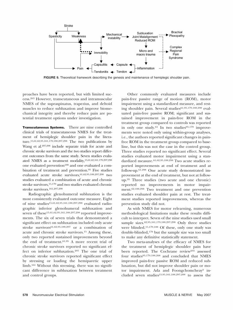
Shoulder pain is a common complication followingstroke.292 There are many possible causes of shoul-der pain in hemiparesis, including adhesive capsuli-tis, impingement syndrome, complex regional painsyndrome, brachial plexopathy, spasticity, and sub-luxation.21 Figure 9 shows a theoretical frameworkdescribing the genesis and maintenance of hemiple-gic shoulder pain. The general features of thisframework include initial spasticity and weaknessleading to mechanical instability and immobility ofthe glenohumeral joint. These conditions may causepain directly or they may place the capsule andextracapsular soft tissue at risk for micro- and macro-trauma, which then leads to inflammation, immobil-ity, and pain. In view of the importance of repetitiveand functional use of the limb for motor recovery,the immobility exacerbates the state of the alreadyparetic muscles (Fig. 9). The cycle repeats with wors-ening of the condition. Numerous treatment ap-
Neuromuscular Electrical Stimulation MUSCLE & NERVE May 2007 577
proaches have been reported, but with limited suc-cess.263 However, transcutaneous and intramuscularNMES of the supraspinatus, trapezius, and deltoidmuscles to reduce subluxation and improve biome-chanical integrity and thereby reduce pain are po-tential treatment options under investigation.
Transcutaneous Systems. There are nine controlledclinical trials of transcutaneous NMES for the treat-ment of hemiplegic shoulder pain in the litera-ture.15,61,62,95,161,179,188,297,298 The two publications byWang et al.297,298 include separate trials for acute andchronic stroke survivors and the two studies report differ-ent outcomes from the same study. Seven studies evalu-ated NMES as a treatment modality,15,61,62,161,179,297,298
one evaluated prevention188 and one evaluated a com-bination of treatment and prevention.95 Five studiesevaluated acute stroke survivors,61,62,95,188,297,298 twostudies evaluated a combination of acute and chronicstroke survivors,15,179 and two studies evaluated chronicstroke survivors.161,297,298
Radiographic glenohumeral subluxation is themost consistently evaluated outcome measure. Eightof nine studies15,61,62,95,161,188,297,298 evaluated radio-graphic inferior glenohumeral subluxation andseven of these15,61,62,95,161,188,297,298 reported improve-ments. The six of seven trials that demonstrated asignificant effect on subluxation included only acutestroke survivors61,62,95,188,297 or a combination ofacute and chronic stroke survivors.15 Among these,only two reported sustained improvements beyondthe end of treatment.61,95 A more recent trial ofchronic stroke survivors reported no significant ef-fect on inferior subluxation.297 The one trial ofchronic stroke survivors reported significant effectby stressing or loading the hemiparetic upperlimb.161 Without this stressing, there was no signifi-cant difference in subluxation between treatmentand control groups.
Other commonly evaluated measures includepain-free passive range of motion (ROM), motorimpairment using a standardized measure, and rest-ing shoulder pain. Several studies61,95,179,188,298 eval-uated pain-free passive ROM; significant and sus-tained improvement in pain-free ROM in thetreatment group compared to controls was reportedin only one study.61 In two studies95,179 improve-ments were noted only using within-group analyses,i.e., the authors reported significant changes in pain-free ROM in the treatment group compared to base-line, but this was not the case in the control group.Three studies reported no significant effect. Severalstudies evaluated motor impairment using a stan-dardized measure.61,62,95,188,298 Two acute studies re-ported improvements at end of treatment and atfollow-up.61,298 One acute study demonstrated im-provement at the end of treatment, but not at follow-up.95 Three studies (two acute and one chronic)reported no improvements in motor impair-ment.62,188,298 Two treatment and one preventionstudies evaluated shoulder pain at rest. The treat-ment studies reported improvements, whereas theprevention study did not.
As with NMES for motor relearning, numerousmethodological limitations make these results diffi-cult to interpret. Seven of the nine studies used smallsample sizes.62,95,161,179,188,297,298 Only three studieswere blinded.15,179,188 Of these, only one study wasdouble-blinded,179 but the sample size was too smallto make any definitive statistically statement.
Two meta-analyses of the efficacy of NMES forthe treatment of hemiplegic shoulder pain havebeen reported. The Cochrane review235 assessedfour studies95,179,188,268 and concluded that NMESimproved pain-free passive ROM and reduced sub-luxation, but did not improve shoulder pain or mo-tor impairment. Ada and Foongchomcheay2 in-cluded seven studies15,95,161,188,297,298 to assess the
FIGURE 9. Theoretical framework describing the genesis and maintenance of hemiplegic shoulder pain.
578 Neuromuscular Electrical Stimulation MUSCLE & NERVE May 2007

effects of NMES on shoulder subluxation and motorimpairment as a function of stroke acuity. They con-cluded that NMES reduced or prevented subluxa-tion and improved motor impairment in the acutephase, but not in the chronic phase. They also con-cluded that NMES did not improve passive lateralpain-free ROM in the acute phase, but that it mayimprove active pain-free ROM in the chronic phase.The differences in conclusions between the twometa-analyses are likely due to the differences ininclusion criteria used to accept specific studies inthe respective studies.
Intramuscular Systems. Despite the evidence fortherapeutic benefit, the clinical use of transcutane-ous NMES for shoulder subluxation and pain inhemiplegia is limited for several reasons. First, stim-ulation of cutaneous pain receptors cannot beavoided, resulting in stimulation-induced pain thatlimits tolerance and compliance. Second, activationof deep muscles cannot be achieved without stimu-lation of more superficial muscles. Third, stimulatedmuscle contraction cannot be titrated precisely. Fi-nally, clinical skill is required to place electrodes andadjust stimulation parameters to provide optimaland tolerable treatment. A potential solution is in-tramuscular NMES systems, which can be injected orpercutaneously placed into the target muscle. Thesesystems are less painful during stimulation, whichenhances patient compliance. Motor points do notneed to be located with each treatment session,which eases donning and doffing of the device. Sincethe electrodes are implanted, repeatability and reli-ability of stimulation are enhanced, which minimizesthe need for skilled care. And finally, due to the focalnature and reliability of intramuscular stimulation,the best muscles to stimulate can be identified andcurrent intensity on multiple channels can be ti-trated easily. Two intramuscular electrical stimula-tion systems are under investigation: an injectablesystem with an external antenna and a percutaneoussystem with an external stimulator.
The previously described injectable microstimu-lator is presently under investigation to treat hemi-plegic shoulder dysfunction. Preliminary resultsfrom a randomized clinical trial demonstrated re-duction in shoulder subluxation and an increase inthe thickness of the stimulated muscles.88 However,the effect on shoulder pain is still under investiga-tion. The stimulators are permanently implantedand, if shoulder subluxation or pain recurs, addi-tional treatments can be provided without an addi-tional invasive procedure. However, the system alsorequires a large antenna that must be worn, which
may interfere with daily activities and compromiseclinical acceptance.
The percutaneous system includes helical intra-muscular electrodes, which are percutaneous-placed, a “pager” size stimulator, which is worn on abelt, and a connector, which connects the electrodesto the stimulator (Fig. 10). Electrodes traverse theskin and remain across the skin for the duration oftreatment. Therefore, the system is at risk for infec-tion. The electrodes are removed by gentle tractionafter completion of treatment. A multicenter clinicaltrial demonstrated that the percutaneous system iseffective in reducing hemiplegic shoulder pain andimproving shoulder pain-related quality of life ofchronic stroke survivors for up to 12 months aftercompletion of treatment (Fig. 11).60,311 There wereno instances of electrode-related infections.
Summary. The use of transcutaneous NMES ap-pears to reduce shoulder subluxation and pain-freeROM and facilitate motor recovery, especiallyamong acute stroke survivors. However, additionalstudies that address the previously noted method-ological limitations are needed to address more de-finitively the question of clinical efficacy. Intramus-cular systems are still under investigation. Theinjectable system is undergoing clinical trials of ini-tial effectiveness for hemiplegic shoulder pain andthe percutaneous system is presently being com-pared to transcutaneous electrical nerve stimulation
FIGURE 10. A percutaneous intramuscular electrical stimulationsystem for treatment of hemiplegic shoulder pain. The pager-sizestimulator is connected to the implanted electrodes via a connec-tor that can be disconnected when not in use. (RestoreStIM,courtesy of NeuroControl, North Ridgeville, OH.)
Neuromuscular Electrical Stimulation MUSCLE & NERVE May 2007 579
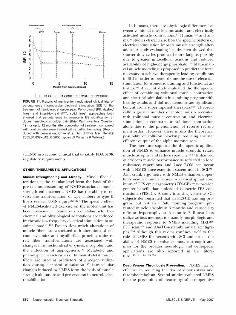
(TENS) in a second clinical trial to satisfy FDA 510Kregulatory requirements.
OTHER THERAPEUTIC APPLICATIONS
Muscle Strengthening and Atrophy. Muscle fiber al-terations at the cellular level form the basis of thepresent understanding of NMES-associated musclestrength enhancement. NMES has the ability to re-verse the transformation of type I fibers to type IIfibers seen in UMN injury.225,227 The specific effectof NMES-facilitated exercise on the motor unit hasbeen reviewed.91 Numerous skeletal-muscle bio-chemical and physiological adaptations are inducedby chronic low-frequency electrical stimulation in ananimal model.189 Fast to slow twitch alterations ofmuscle fibers are associated with alterations of cal-cium dynamics and myofibrillar proteins; white tored fiber transformations are associated withchanges in mitochondrial enzymes, myoglobin, andthe induction of angiogenesis.189 Metabolic andphenotypic characteristics of human skeletal musclefibers are used as predictors of glycogen utiliza-tion during electrical stimulation.117 Intracellularchanges induced by NMES form the basis of musclestrength alterations and preservation in neurologicalrehabilitation.
In humans, there are physiologic differences be-tween volitional muscle contraction and electricallyactivated muscle contractions.81 Human198 and ani-mal89 studies characterize how the specific pattern ofelectrical stimulation impacts muscle strength alter-ations. A study evaluating healthy men showed thatshorter duty cycles produced more fatigue, possiblydue to greater intracellular acidosis and reducedavailability of high-energy phosphate.198 Mathemati-cal muscle modeling is proposed to predict the forcenecessary to achieve therapeutic loading conditionsin SCI in order to better define the use of electricalstimulation for isometric training and functional ac-tivities.103 A recent study evaluated the therapeuticeffect of combining volitional muscle contractionand electrical stimulation in a training program withhealthy adults and did not demonstrate significantbenefit from superimposed therapies.220 Theoreti-cally, a greater number of motor units is recruitedwith volitional muscle contraction and electricalstimulation as compared to volitional contractionalone due to the phenomenon of reverse recruit-ment order. However, there is also the theoreticalpossibility of collision blocking, reducing the netefferent output of the alpha motoneuron.
The literature supports the therapeutic applica-tion of NMES to enhance muscle strength, retardmuscle atrophy, and reduce spasticity.16,87 Enhancedquadriceps muscle performance as reflected in loadresistance, repetitions, and knee ROM can occurwith a NMES knee-extension system used in SCI.244
Arm crank ergometry with NMES enhances upper-limb manual muscle scores in cervical spinal cordinjury.47 FES cycle ergometry (FES-CE) may providegreater benefit than unloaded isometric FES con-tractions (FES-IC). A study involving 26 acute SCIsubjects demonstrated that an FES-CE training pro-gram, but not an FES-IC training program, pre-vented muscle atrophy at 3 months and caused sig-nificant hypertrophy at 6 months.17 Researchersutilize various methods to quantify morphologic andtherapeutic response to NMES including MRI,216
PET scan,294 and 99mTC-sestamibi muscle scintigra-phy.226 Although this review confines itself to therole of NMES for persons with SCI and stroke, theability of NMES to enhance muscle strength andmass for the broader neurologic and orthopedicapplications are also reported in the litera-ture.119,121,174,185,274
Deep Venous Thrombosis Prevention. NMES may beeffective in reducing the risk of venous stasis andthromboembolism. Several studies evaluated NMESfor the prevention of neurosurgical postoperative
FIGURE 11. Results of multicenter randomized clinical trial ofpercutaneous intramuscular electrical stimulation (ES) for thetreatment of hemiplegic shoulder pain. Per-protocol (PP, dashedlines) and intent-to-treat (ITT, solid lines) approaches bothshowed that percutaneous intramuscular ES significantly re-duces hemiplegic shoulder pain (Brief Pain Inventory Question12) for up to 12 months after completion of treatment comparedwith controls who were treated with a cuffed hemisling. (Repro-duced with permission, Chae et al. Am J Phys Med Rehabil2005;84:832–842. © 2005 Lippincott Williams & Wilkins.)
580 Neuromuscular Electrical Stimulation MUSCLE & NERVE May 2007
thromboembolism32 and post-arthroplasty venouscomplications including DVT.208 Cardiovascular ef-fects, consistent with decreased lower-limb venousstasis, including increased stroke volume and cardiacoutput have been documented when lower-limbelectrical stimulation is used during surgery.96 Astudy of immobilized subjects showed that electricalstimulation of the foot and calf decreases venousstasis and reduces the risk of DVT.147 A review articleof the etiology, incidence, and prevention of DVT inacute SCI supported the use of electrical stimulationin combination with low-dose heparin for the pre-vention of thromboembolic disease.205
Bone Mass/Density. Physiological alterations ofbone associated with immobility in the neurologi-cally impaired patient are well documented. Follow-ing SCI, weight-bearing trabecular-rich sites such asthe distal femur and proximal tibia show the greatestdemineralization. Pathological fractures followingminor trauma are caused by reduced bone mass inassociation with modified bone matrix composi-tion.194 Animal studies demonstrate the preventionand reversal of osteoporosis with capacitively cou-pled electrical fields.37–40,186 Various forms of elec-trical stimulation affect growth, repair, and remod-eling of soft and hard tissues in animals.28
Several studies have described the effect of FESlower-limb cycling on bone density in SCI. A trendtoward increased bone lumbar spine bone density isseen in chronic SCI with neurogenic osteoporosis.19
In two studies, FES cycling exercises for 6 monthsenhanced the bone mineral density of the distalfemur and proximal tibia,20,63 although the effectwas not sustained with discontinuance of FES.63
However, other studies have failed to corroboratethese findings.94,181 A recent review article suggestedthat FES cycle ergometry helps prevent bone loss inwomen, specifically with acute spinal cord inju-ries.219
Tissue Oxygenation and Peripheral Hemodynamic Func-
tioning. NMES may improve tissue oxygenation andperipheral hemodynamic function. Eight weeks ofstimulation is associated with an increase in the un-loaded tissue oxygen level and a reduction in theischial region pressure, although total interface pres-sures does not change.31 Animal studies demonstrateincreased microvascular perfusion with transcutane-ous NMES.65–67 The ability of TENS to impact pe-ripheral vascular resistance and cause a transientlocal increase in blood flow is dependent on stimu-lation intensity.253
The impact of NMES on peripheral hemody-namic functioning is the focus of several stud-ies.18,52,228,250,313 In one study there was a linear in-crease in femoral arterial blood flow with increasingstimulation rates of TENS.313 However, a recentstudy evaluating peripheral vascular responses toNMES in chronic SCI showed improved muscle fibersize and fatigue without impact on femoral arterialdiameter or blood flow.250 After NMES-assistedtreadmill training for 3 months in chronic SCI, therewas an increase in systolic blood pressure at rest aswell as during exercise.52 In a study using arm crackergometry and NMES-induced leg contractions, anincrease in blood flow and decrease in venous pool-ing were associated with a reduced rate pressureproduct.228
Cardiorespiratory Conditioning. Enhancement ofcardiorespiratory fitness is associated with the use ofNMES in SCI. Exercise recommendations for SCIpresently include the use of NMES to facilitate exer-cise either via NMES leg cycle ergometry or NMES-assisted treadmill gait training.138 A study of peakand submaximal physiological responses in SCIshowed a significant increase in peak rate of oxygenconsumption.135 NMES leg cycle ergometry in-creases functional capacity in SCI subjects,70 al-though high peak NMES-assisted pedal forces con-tribute to low efficiency in paraplegic subjects.258
The rate of fatigue during NMES-assisted exercise isgreater than during volitional exercise.27
NMES-assisted ambulation training and arm er-gometry in SCI is associated with central cardiovas-cular adaptations including increased time to fa-tigue, peak power output, and peak rate of oxygenconsumption.139 Studies evaluating energy expendi-ture with NMES-assisted treadmill gait training andpartial body-weight support in SCI have shown im-provements in the rate of oxygen and energy con-sumption, which is consistent with enhanced meta-bolic and cardiovascular responses.53,79,80
CONCLUSIONS
The principal goal of rehabilitation management ofpersons with UMN paralysis is to maximize quality oflife. NMES systems bypass the injured central cir-cuitry to activate neural tissue and contract musclesto provide function to what is otherwise a nonfunc-tioning limb or structure. Recent advances in clinicalmedicine and biomedical engineering make theclinical implementation of NMES systems to en-hance the mobility and function of paralyzed personmore feasible. Hand neuroprosthesis systems can
Neuromuscular Electrical Stimulation MUSCLE & NERVE May 2007 581
significantly enhance the upper-limb function ofpersons with tetraplegia. The application of thistechnology for persons with hemiplegia is in its in-fancy and must await further technical and scientificdevelopments if it is to be applicable to the broaderstroke population. Several lower-limb systems withand without bracing are being investigated for thepurpose of functional transfers and standing, and toa lesser degree for ambulation for patients with para-plegia. While multichannel neuroprosthesis systemsfor hemiplegia are still under development, the foot-drop stimulator is ready for large-scale multicenterclinical trials. The bladder neuroprosthesis can pro-vide catheter-free micturition for persons with eitherparaplegia or tetraplegia. Phrenic and diaphrag-matic pacing systems can provide artificial ventila-tory support for patients with ventilator-dependenttetraplegia. NMES for motor relearning in hemiple-gia is a promising application of goal-oriented repet-itive movement therapy and is ready for large-scalemulticenter clinical trials. NMES for treatment ofshoulder subluxation and pain in hemiplegia hasyielded encouraging results and is ready for confir-matory large-scale multicenter clinical trials. Finally,NMES may be effective in strengthening muscles,preventing muscle atrophy, preventing DVT, im-proving tissue oxygenation and peripheral hemody-namic functioning, and facilitating cardiopulmonaryconditioning.
Although this review was limited to applicationsin the SCI and stroke populations, work with multi-ple sclerosis,282 traumatic brain injury,217 and cere-bral palsy129,218,233 is in its early stages and will un-doubtedly expand. Clinical practice is rarely limitedto a single intervention. Thus, with the developmentof pharmacological interventions,113 neuronal re-generation,36 and other innovations such as robotictherapy,97,170 mental imagery,1 virtual reality,167,245
and constraint-induced therapy,281,305 the future willlikely embrace combination therapies to treat themotor dysfunction of persons with central nervoussystem paralysis.
After decades of development, the clinical utilityof NMES systems is finally becoming realized. Bynecessity, scientists and clinicians must continue toexplore new ideas and improve upon the presentsystems. Components will be smaller, more durable,and more reliable. The issues of cosmesis and ease ofdonning and doffing will require systems to be fullyimplantable. Control issues will remain central, andthe implementation of cortical control will dictatethe nature of future generations of neuroprosthesissystems. Future developments will be directed byconsumers. In the present healthcare environment
where cost is an overwhelming factor in the devel-opment and implementation of new technology, theconsumer will become one of technology’s greatestadvocates. Finally, the usual drive toward greatercomplexity will be tempered by the practical issues ofclinical implementation where patient and clinicianacceptances are often a function of a tenuous bal-ance between the “burden and cost” associated withusing a system and the system’s impact on the user’squality of life.
Supported in part by grants R01HD49777 and R01HD044816from the National Institute for Child Health and Human Devel-opment.
REFERENCES
1. Abbruzzese G, Assini A, Buccolieri A, Marchese R,Trompetto C. Changes of intracortical inhibition duringmotor imagery in human subjects. Neurosci Lett 1999;263:113–116.
2. Ada L, Foongchomcheay A. Efficacy of electrical stimulationin preventing or reducing subluxation of the shoulder afterstroke: a meta-analysis. Aust J Physiother 2002;48:257–267.
3. Adrian E. The physical background of perception. Oxford:Clarendon Press; 1946.
4. Agnew WF, McCreery DB. Considerations for safety withchronically implanted nerve electrodes. Epilepsia 1990;31(Suppl 2):S27–32.
5. Alon G, Dar A, Katz-Behiri D, Wingarden H, Nathan R.Efficacy of a hybrid upper limb neuromuscular electricalstimulation system in lessening selected impairments anddysfunctions consequent to cerebral damage. J Neurol Re-habil 1998;2:73–80.
6. Alon G, McBride K, Ring H. Improving selected hand func-tions using a noninvasive neuroprosthesis in persons withchronic stroke. J Stroke Cerebrovasc Dis 2002;11:99–106.
7. Alon G, Sunnerhagen KS, Geurts AC, Ohry A. A home-based, self-administered stimulation program to improve se-lected hand functions of chronic stroke. Neurorehabilitation2003;18:215–225.
8. Amarenco G, Ismael SS, Even-Schneider A, Raibaut P, De-maille-Wlodyka S, Parratte B, et al. Urodynamic effect ofacute transcutaneous posterior tibial nerve stimulation inoveractive bladder. J Urol 2003;169:2210–2215.
9. Arbuthnott ER, Boyd IA, Kalu KU. Ultrastructural dimen-sions of myelinated peripheral nerve fibres in the cat andtheir relation to conduction velocity. J Physiol (Lond) 1980;308:125–157.
10. Arcos I, Davis R, Fey K, Mishler D, Sanderson D, Tanacs C, etal. Second-generation microstimulator. Artif Organs 2002;26:228–231.
11. Asanuma H, Keller A. Neurobiological basis of motor re-learning and memory. Conc Neurosci 1991;2:1–30.
12. Asanuma H, Keller A. Neuronal mechanisms of motor learn-ing in animals. Neuroreport 1991;2:217–224.
13. Bajd T, Kralj A, Stefancic M, Lavrac N. Use of functionalelectrical stimulation in the lower extremities of incompletespinal cord injured patients. Artif Organs 1999;23:403–409.
14. Bajd T, Kralj A, Turk R. Standing up of a healthy subject anda paraplegic patient. J Biomech 1982;15:1–10.
15. Baker LL, Parker K. Neuromuscular electrical stimulation ofthe muscles surrounding the shoulder. Phys Ther 1986;66:1930–1937.
16. Baker LL, Parker K, Sanderson D. Neuromuscular electricalstimulation for the head-injured patient. Phys Ther 1983;63:1967–1974.
582 Neuromuscular Electrical Stimulation MUSCLE & NERVE May 2007
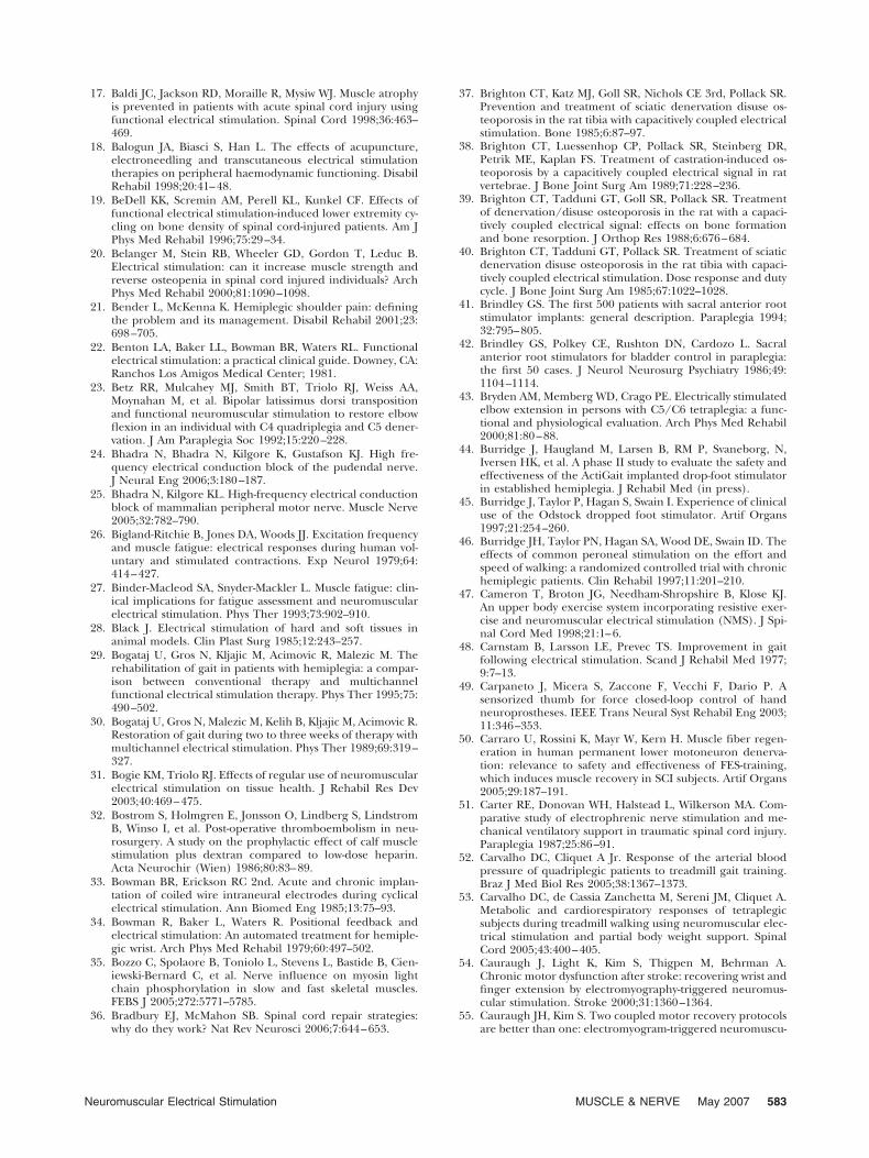
17. Baldi JC, Jackson RD, Moraille R, Mysiw WJ. Muscle atrophyis prevented in patients with acute spinal cord injury usingfunctional electrical stimulation. Spinal Cord 1998;36:463–469.
18. Balogun JA, Biasci S, Han L. The effects of acupuncture,electroneedling and transcutaneous electrical stimulationtherapies on peripheral haemodynamic functioning. DisabilRehabil 1998;20:41–48.
19. BeDell KK, Scremin AM, Perell KL, Kunkel CF. Effects offunctional electrical stimulation-induced lower extremity cy-cling on bone density of spinal cord-injured patients. Am JPhys Med Rehabil 1996;75:29–34.
20. Belanger M, Stein RB, Wheeler GD, Gordon T, Leduc B.Electrical stimulation: can it increase muscle strength andreverse osteopenia in spinal cord injured individuals? ArchPhys Med Rehabil 2000;81:1090–1098.
21. Bender L, McKenna K. Hemiplegic shoulder pain: definingthe problem and its management. Disabil Rehabil 2001;23:698–705.
22. Benton LA, Baker LL, Bowman BR, Waters RL. Functionalelectrical stimulation: a practical clinical guide. Downey, CA:Ranchos Los Amigos Medical Center; 1981.
23. Betz RR, Mulcahey MJ, Smith BT, Triolo RJ, Weiss AA,Moynahan M, et al. Bipolar latissimus dorsi transpositionand functional neuromuscular stimulation to restore elbowflexion in an individual with C4 quadriplegia and C5 dener-vation. J Am Paraplegia Soc 1992;15:220–228.
24. Bhadra N, Bhadra N, Kilgore K, Gustafson KJ. High fre-quency electrical conduction block of the pudendal nerve.J Neural Eng 2006;3:180–187.
25. Bhadra N, Kilgore KL. High-frequency electrical conductionblock of mammalian peripheral motor nerve. Muscle Nerve2005;32:782–790.
26. Bigland-Ritchie B, Jones DA, Woods JJ. Excitation frequencyand muscle fatigue: electrical responses during human vol-untary and stimulated contractions. Exp Neurol 1979;64:414–427.
27. Binder-Macleod SA, Snyder-Mackler L. Muscle fatigue: clin-ical implications for fatigue assessment and neuromuscularelectrical stimulation. Phys Ther 1993;73:902–910.
28. Black J. Electrical stimulation of hard and soft tissues inanimal models. Clin Plast Surg 1985;12:243–257.
29. Bogataj U, Gros N, Kljajic M, Acimovic R, Malezic M. Therehabilitation of gait in patients with hemiplegia: a compar-ison between conventional therapy and multichannelfunctional electrical stimulation therapy. Phys Ther 1995;75:490–502.
30. Bogataj U, Gros N, Malezic M, Kelih B, Kljajic M, Acimovic R.Restoration of gait during two to three weeks of therapy withmultichannel electrical stimulation. Phys Ther 1989;69:319–327.
31. Bogie KM, Triolo RJ. Effects of regular use of neuromuscularelectrical stimulation on tissue health. J Rehabil Res Dev2003;40:469–475.
32. Bostrom S, Holmgren E, Jonsson O, Lindberg S, LindstromB, Winso I, et al. Post-operative thromboembolism in neu-rosurgery. A study on the prophylactic effect of calf musclestimulation plus dextran compared to low-dose heparin.Acta Neurochir (Wien) 1986;80:83–89.
33. Bowman BR, Erickson RC 2nd. Acute and chronic implan-tation of coiled wire intraneural electrodes during cyclicalelectrical stimulation. Ann Biomed Eng 1985;13:75–93.
34. Bowman R, Baker L, Waters R. Positional feedback andelectrical stimulation: An automated treatment for hemiple-gic wrist. Arch Phys Med Rehabil 1979;60:497–502.
35. Bozzo C, Spolaore B, Toniolo L, Stevens L, Bastide B, Cien-iewski-Bernard C, et al. Nerve influence on myosin lightchain phosphorylation in slow and fast skeletal muscles.FEBS J 2005;272:5771–5785.
36. Bradbury EJ, McMahon SB. Spinal cord repair strategies:why do they work? Nat Rev Neurosci 2006;7:644–653.
37. Brighton CT, Katz MJ, Goll SR, Nichols CE 3rd, Pollack SR.Prevention and treatment of sciatic denervation disuse os-teoporosis in the rat tibia with capacitively coupled electricalstimulation. Bone 1985;6:87–97.
38. Brighton CT, Luessenhop CP, Pollack SR, Steinberg DR,Petrik ME, Kaplan FS. Treatment of castration-induced os-teoporosis by a capacitively coupled electrical signal in ratvertebrae. J Bone Joint Surg Am 1989;71:228–236.
39. Brighton CT, Tadduni GT, Goll SR, Pollack SR. Treatmentof denervation/disuse osteoporosis in the rat with a capaci-tively coupled electrical signal: effects on bone formationand bone resorption. J Orthop Res 1988;6:676–684.
40. Brighton CT, Tadduni GT, Pollack SR. Treatment of sciaticdenervation disuse osteoporosis in the rat tibia with capaci-tively coupled electrical stimulation. Dose response and dutycycle. J Bone Joint Surg Am 1985;67:1022–1028.
41. Brindley GS. The first 500 patients with sacral anterior rootstimulator implants: general description. Paraplegia 1994;32:795–805.
42. Brindley GS, Polkey CE, Rushton DN, Cardozo L. Sacralanterior root stimulators for bladder control in paraplegia:the first 50 cases. J Neurol Neurosurg Psychiatry 1986;49:1104–1114.
43. Bryden AM, Memberg WD, Crago PE. Electrically stimulatedelbow extension in persons with C5/C6 tetraplegia: a func-tional and physiological evaluation. Arch Phys Med Rehabil2000;81:80–88.
44. Burridge J, Haugland M, Larsen B, RM P, Svaneborg, N,Iversen HK, et al. A phase II study to evaluate the safety andeffectiveness of the ActiGait implanted drop-foot stimulatorin established hemiplegia. J Rehabil Med (in press).
45. Burridge J, Taylor P, Hagan S, Swain I. Experience of clinicaluse of the Odstock dropped foot stimulator. Artif Organs1997;21:254–260.
46. Burridge JH, Taylor PN, Hagan SA, Wood DE, Swain ID. Theeffects of common peroneal stimulation on the effort andspeed of walking: a randomized controlled trial with chronichemiplegic patients. Clin Rehabil 1997;11:201–210.
47. Cameron T, Broton JG, Needham-Shropshire B, Klose KJ.An upper body exercise system incorporating resistive exer-cise and neuromuscular electrical stimulation (NMS). J Spi-nal Cord Med 1998;21:1–6.
48. Carnstam B, Larsson LE, Prevec TS. Improvement in gaitfollowing electrical stimulation. Scand J Rehabil Med 1977;9:7–13.
49. Carpaneto J, Micera S, Zaccone F, Vecchi F, Dario P. Asensorized thumb for force closed-loop control of handneuroprostheses. IEEE Trans Neural Syst Rehabil Eng 2003;11:346–353.
50. Carraro U, Rossini K, Mayr W, Kern H. Muscle fiber regen-eration in human permanent lower motoneuron denerva-tion: relevance to safety and effectiveness of FES-training,which induces muscle recovery in SCI subjects. Artif Organs2005;29:187–191.
51. Carter RE, Donovan WH, Halstead L, Wilkerson MA. Com-parative study of electrophrenic nerve stimulation and me-chanical ventilatory support in traumatic spinal cord injury.Paraplegia 1987;25:86–91.
52. Carvalho DC, Cliquet A Jr. Response of the arterial bloodpressure of quadriplegic patients to treadmill gait training.Braz J Med Biol Res 2005;38:1367–1373.
53. Carvalho DC, de Cassia Zanchetta M, Sereni JM, Cliquet A.Metabolic and cardiorespiratory responses of tetraplegicsubjects during treadmill walking using neuromuscular elec-trical stimulation and partial body weight support. SpinalCord 2005;43:400–405.
54. Cauraugh J, Light K, Kim S, Thigpen M, Behrman A.Chronic motor dysfunction after stroke: recovering wrist andfinger extension by electromyography-triggered neuromus-cular stimulation. Stroke 2000;31:1360–1364.
55. Cauraugh JH, Kim S. Two coupled motor recovery protocolsare better than one: electromyogram-triggered neuromuscu-
Neuromuscular Electrical Stimulation MUSCLE & NERVE May 2007 583
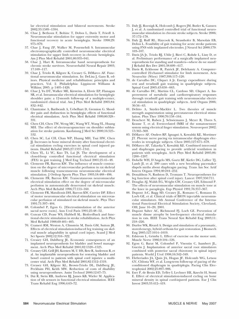
lar electrical stimulation and bilateral movements. Stroke2002;33:1589–1594.
56. Chae J, Bethoux F, Bohinc T, Dobos L, Davis T, Friedl A.Neuromuscular stimulation for upper extremity motor andfunctional recovery in acute hemiplegia. Stroke 1998;29:975–979.
57. Chae J, Fang ZP, Walker M, Pourmehdi S. Intramuscularelectromyographically controlled neuromuscular electricalstimulation for upper limb recovery in chronic hemiplegia.Am J Phys Med Rehabil 2001;80:935–941.
58. Chae J, Hart R. Intramuscular hand neuroprosthesis forchronic stroke survivors. Neurorehabil Neural Repair 2003;17:109–117.
59. Chae J, Triolo R, Kilgore K, Creasey G, DiMarco AF. Func-tional neuromuscular stimulation. In: DeLisa J, Gans B, ed-itors. Physical medicine and rehabilitation: principles andpractices, Vol. 2. Philadelphia: Lippincott Williams &Wilkins; 2005. p 1405–1426.
60. Chae J, Yu DT, Walker ME, Kirsteins A, Elovic EP, FlanaganSR, et al. Intramuscular electrical stimulation for hemiplegicshoulder pain: a 12-month follow-up of a multiple-center,randomized clinical trial. Am J Phys Med Rehabil 2005;84:832–842.
61. Chantraine A, Baribeault A, Uebelhart D, Gremion G. Shoul-der pain and dysfunction in hemiplegia: effects of functionalelectrical stimulation. Arch Phys Med Rehabil 1999;80:328–331.
62. Chen CH, Chen TW, Weng MC, Wang WT, Wang YL, HuangMH. The effect of electroacupuncture on shoulder sublux-ation for stroke patients. Kaohsiung J Med Sci 2000;16:525–532.
63. Chen SC, Lai CH, Chan WP, Huang MH, Tsai HW, ChenJJ. Increases in bone mineral density after functional electri-cal stimulation cycling exercises in spinal cord injured pa-tients. Disabil Rehabil 2005;27:1337–1341.
64. Chen YL, Li YC, Kuo TS, Lai JS. The development of aclosed-loop controlled functional electrical stimulation(FES) in gait training. J Med Eng Technol 2001;25:41–48.
65. Clemente FR, Barron KW. The influence of muscle contrac-tion on the degree of microvascular perfusion in rat skeletalmuscle following transcutaneous neuromuscular electricalstimulation. J Orthop Sports Phys Ther 1993;18:488–496.
66. Clemente FR, Barron KW. Transcutaneous neuromuscularelectrical stimulation effect on the degree of microvascularperfusion in autonomically denervated rat skeletal muscle.Arch Phys Med Rehabil 1996;77:155–160.
67. Clemente FR, Matulionis DH, Barron KW, Currier DP. Effectof motor neuromuscular electrical stimulation on microvas-cular perfusion of stimulated rat skeletal muscle. Phys Ther1991;71:397–404.
68. Colombel P, Egon G. [Electrostimulation of the anteriorsacral nerve roots]. Ann Urol Paris 1991;25:48–52.
69. Cozean CD, Pease WS, Hubbell SL. Biofeedback and func-tional electric stimulation in stroke rehabilitation. Arch PhysMed Rehabil 1988;69:401–405.
70. Crameri RM, Weston A, Climstein M, Davis GM, Sutton JR.Effects of electrical stimulation-induced leg training on skel-etal muscle adaptability in spinal cord injury. Scand J MedSci Sports 2002;12:316–322.
71. Creasey GH, Dahlberg JE. Economic consequences of animplanted neuroprosthesis for bladder and bowel manage-ment. Arch Phys Med Rehabil 2001;82:1520–1525.
72. Creasey GH, Grill JH, Korsten M, U HS, Betz R, Anderson R, etal. An implantable neuroprosthesis for restoring bladder andbowel control to patients with spinal cord injuries: a multi-center trial. Arch Phys Med Rehabil 2001;82:1512–1519.
73. Creasey GH, Kilgore KL, Brown-Triolo DL, Dahlberg JE,Peckham PH, Keith MW. Reduction of costs of disabilityusing neuroprostheses. Assist Technol 2000;12:67–75.
74. Dai R, Stein RB, Andrews BJ, James KB, Wieler M. Applica-tion of tilt sensors in functional electrical stimulation. IEEETrans Rehabil Eng 1996;4:63–72.
75. Daly JJ, Roenigk K, Holcomb J, Rogers JM, Butler K, GansenJ, et al. A randomized controlled trial of functional neuro-muscular stimulation in chronic stroke subjects. Stroke 2006;37:172–178.
76. Daly JJ, Ruff RL, Haycook K, Strasshofer B, Marsolais EB,Dobos L. Feasibility of gait training for acute stroke patientsusing FNS with implanted electrodes. J Neurol Sci 2000;179:103–107.
77. Davis JA Jr, Triolo RJ, Uhlir J, Bieri C, Rohde L, Lissy D, etal. Preliminary performance of a surgically implanted neu-roprosthesis for standing and transfers—where do we stand?J Rehabil Res Dev 2001;38:609–617.
78. Davis R, Eckhouse R, Patrick JF, Delehanty A. Computer-controlled 22-channel stimulator for limb movement. ActaNeurochir (Wien) 1987;39S:117–120.
79. de Carvalho DC, Cliquet A Jr. Energy expenditure duringrest and treadmill gait training in quadriplegic subjects.Spinal Cord 2005;43:658–663.
80. de Carvalho DC, Martins CL, Cardoso SD, Cliquet A. Im-provement of metabolic and cardiorespiratory responsesthrough treadmill gait training with neuromuscular electri-cal stimulation in quadriplegic subjects. Artif Organs 2006;30:56–63.
81. Delitto A, Snyder-Mackler L. Two theories of musclestrength augmentation using percutaneous electrical stimu-lation. Phys Ther 1990;70:158–164.
82. Deuchert M, Ruben J, Schwiemann J, Meyer R, Thees S,Krause T, et al. Event-related fMRI of the somatosensorysystem using electrical finger stimulation. Neuroreport 2002;13:365–369.
83. DiMarco AF, Onders RP, Ignagni A, Kowalski KE, MortimerJT. Phrenic nerve pacing via intramuscular diaphragm elec-trodes in tetraplegic subjects. Chest 2005;127:671–678.
84. DiMarco AF, Takaoka Y, Kowalski KE. Combined intercostaland diaphragm pacing to provide artificial ventilation inpatients with tetraplegia. Arch Phys Med Rehabil 2005;86:1200–1207.
85. Dobelle WH, D’Angelo MS, Goetz BF, Kiefer DG, Lallier TJ,Lamb JI, et al. 200 cases with a new breathing pacemakerdispels myths about diaphragm pacing. Trans Am Soc ArtifIntern Organs 1994;40:244–252.
86. Donaldson N, Rushton D, Tromans T. Neuroprostheses forleg function after spinal cord injury. Lancet 1997;350:711.
87. Douglas AJ, Walsh EG, Wright GW, Creasey GH, Edmond P.The effects of neuromuscular stimulation on muscle tone atthe knee in paraplegia. Exp Physiol 1991;76:357–367.
88. Dupont A-C, Bagg SD, Creasey JL, Romano C, Romano D,Loeb GE, et al. Clinical trials of BION injectable neuromus-cular stimulators. 6th Annual Conference of the Interna-tional Functional Electrical Stimulation Society, Cleveland,OH; June 16–20, 2001.
89. Dupont Salter AC, Richmond FJ, Loeb GE. Prevention ofmuscle disuse atrophy by low-frequency electrical stimula-tion in rats. IEEE Trans Neural Syst Rehabil Eng 2003;11:218–226.
90. Durfee WK, Rivard A. Design and simulation of a pneumatic,stored-energy, hybrid orthosis for gait restoration. J BiomechEng 2005;127:1014–1019.
91. Edstrom L, Grimby L. Effect of exercise on the motor unit.Muscle Nerve 1986;9:104–126.
92. Egon G, Barat M, Colombel P, Visentin C, Isambert JL,Guerin J. Implantation of anterior sacral root stimulatorscombined with posterior sacral rhizotomy in spinal injurypatients. World J Urol 1998;16:342–349.
93. Elefteriades JA, Quin JA, Hogan JF, Holcomb WG, LetsouGV, Chlosta WF, et al. Long-term follow-up of pacing of theconditioned diaphragm in quadriplegia. Pacing Clin Elec-trophysiol 2002;25:897–906.
94. Eser P, de Bruin ED, Telley I, Lechner HE, Knecht H, StussiE. Effect of electrical stimulation-induced cycling on bonemineral density in spinal cord-injured patients. Eur J ClinInvest 2003;33:412–419.
584 Neuromuscular Electrical Stimulation MUSCLE & NERVE May 2007
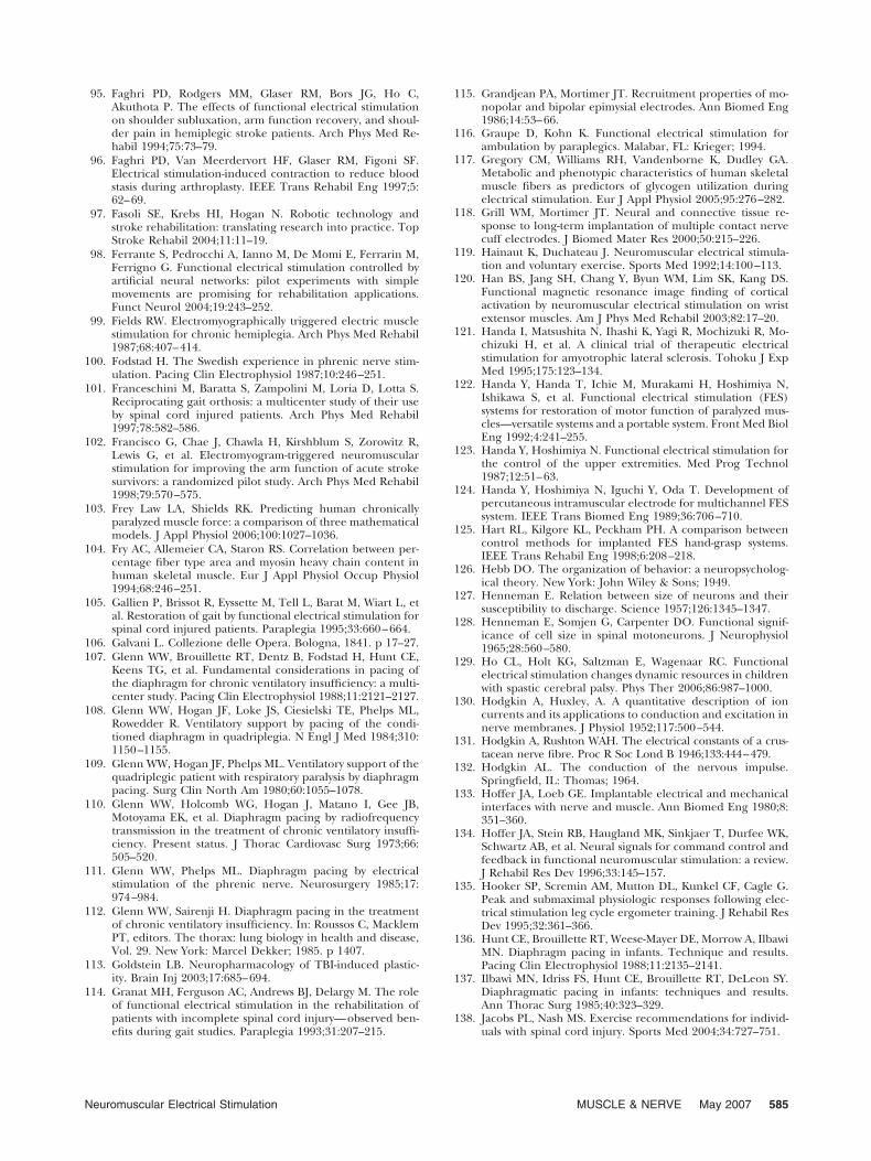
95. Faghri PD, Rodgers MM, Glaser RM, Bors JG, Ho C,Akuthota P. The effects of functional electrical stimulationon shoulder subluxation, arm function recovery, and shoul-der pain in hemiplegic stroke patients. Arch Phys Med Re-habil 1994;75:73–79.
96. Faghri PD, Van Meerdervort HF, Glaser RM, Figoni SF.Electrical stimulation-induced contraction to reduce bloodstasis during arthroplasty. IEEE Trans Rehabil Eng 1997;5:62–69.
97. Fasoli SE, Krebs HI, Hogan N. Robotic technology andstroke rehabilitation: translating research into practice. TopStroke Rehabil 2004;11:11–19.
98. Ferrante S, Pedrocchi A, Ianno M, De Momi E, Ferrarin M,Ferrigno G. Functional electrical stimulation controlled byartificial neural networks: pilot experiments with simplemovements are promising for rehabilitation applications.Funct Neurol 2004;19:243–252.
99. Fields RW. Electromyographically triggered electric musclestimulation for chronic hemiplegia. Arch Phys Med Rehabil1987;68:407–414.
100. Fodstad H. The Swedish experience in phrenic nerve stim-ulation. Pacing Clin Electrophysiol 1987;10:246–251.
101. Franceschini M, Baratta S, Zampolini M, Loria D, Lotta S.Reciprocating gait orthosis: a multicenter study of their useby spinal cord injured patients. Arch Phys Med Rehabil1997;78:582–586.
102. Francisco G, Chae J, Chawla H, Kirshblum S, Zorowitz R,Lewis G, et al. Electromyogram-triggered neuromuscularstimulation for improving the arm function of acute strokesurvivors: a randomized pilot study. Arch Phys Med Rehabil1998;79:570–575.
103. Frey Law LA, Shields RK. Predicting human chronicallyparalyzed muscle force: a comparison of three mathematicalmodels. J Appl Physiol 2006;100:1027–1036.
104. Fry AC, Allemeier CA, Staron RS. Correlation between per-centage fiber type area and myosin heavy chain content inhuman skeletal muscle. Eur J Appl Physiol Occup Physiol1994;68:246–251.
105. Gallien P, Brissot R, Eyssette M, Tell L, Barat M, Wiart L, etal. Restoration of gait by functional electrical stimulation forspinal cord injured patients. Paraplegia 1995;33:660–664.
106. Galvani L. Collezione delle Opera. Bologna, 1841. p 17–27.107. Glenn WW, Brouillette RT, Dentz B, Fodstad H, Hunt CE,
Keens TG, et al. Fundamental considerations in pacing ofthe diaphragm for chronic ventilatory insufficiency: a multi-center study. Pacing Clin Electrophysiol 1988;11:2121–2127.
108. Glenn WW, Hogan JF, Loke JS, Ciesielski TE, Phelps ML,Rowedder R. Ventilatory support by pacing of the condi-tioned diaphragm in quadriplegia. N Engl J Med 1984;310:1150–1155.
109. Glenn WW, Hogan JF, Phelps ML. Ventilatory support of thequadriplegic patient with respiratory paralysis by diaphragmpacing. Surg Clin North Am 1980;60:1055–1078.
110. Glenn WW, Holcomb WG, Hogan J, Matano I, Gee JB,Motoyama EK, et al. Diaphragm pacing by radiofrequencytransmission in the treatment of chronic ventilatory insuffi-ciency. Present status. J Thorac Cardiovasc Surg 1973;66:505–520.
111. Glenn WW, Phelps ML. Diaphragm pacing by electricalstimulation of the phrenic nerve. Neurosurgery 1985;17:974–984.
112. Glenn WW, Sairenji H. Diaphragm pacing in the treatmentof chronic ventilatory insufficiency. In: Roussos C, MacklemPT, editors. The thorax: lung biology in health and disease,Vol. 29. New York: Marcel Dekker; 1985. p 1407.
113. Goldstein LB. Neuropharmacology of TBI-induced plastic-ity. Brain Inj 2003;17:685–694.
114. Granat MH, Ferguson AC, Andrews BJ, Delargy M. The roleof functional electrical stimulation in the rehabilitation ofpatients with incomplete spinal cord injury—observed ben-efits during gait studies. Paraplegia 1993;31:207–215.
115. Grandjean PA, Mortimer JT. Recruitment properties of mo-nopolar and bipolar epimysial electrodes. Ann Biomed Eng1986;14:53–66.
116. Graupe D, Kohn K. Functional electrical stimulation forambulation by paraplegics. Malabar, FL: Krieger; 1994.
117. Gregory CM, Williams RH, Vandenborne K, Dudley GA.Metabolic and phenotypic characteristics of human skeletalmuscle fibers as predictors of glycogen utilization duringelectrical stimulation. Eur J Appl Physiol 2005;95:276–282.
118. Grill WM, Mortimer JT. Neural and connective tissue re-sponse to long-term implantation of multiple contact nervecuff electrodes. J Biomed Mater Res 2000;50:215–226.
119. Hainaut K, Duchateau J. Neuromuscular electrical stimula-tion and voluntary exercise. Sports Med 1992;14:100–113.
120. Han BS, Jang SH, Chang Y, Byun WM, Lim SK, Kang DS.Functional magnetic resonance image finding of corticalactivation by neuromuscular electrical stimulation on wristextensor muscles. Am J Phys Med Rehabil 2003;82:17–20.
121. Handa I, Matsushita N, Ihashi K, Yagi R, Mochizuki R, Mo-chizuki H, et al. A clinical trial of therapeutic electricalstimulation for amyotrophic lateral sclerosis. Tohoku J ExpMed 1995;175:123–134.
122. Handa Y, Handa T, Ichie M, Murakami H, Hoshimiya N,Ishikawa S, et al. Functional electrical stimulation (FES)systems for restoration of motor function of paralyzed mus-cles—versatile systems and a portable system. Front Med BiolEng 1992;4:241–255.
123. Handa Y, Hoshimiya N. Functional electrical stimulation forthe control of the upper extremities. Med Prog Technol1987;12:51–63.
124. Handa Y, Hoshimiya N, Iguchi Y, Oda T. Development ofpercutaneous intramuscular electrode for multichannel FESsystem. IEEE Trans Biomed Eng 1989;36:706–710.
125. Hart RL, Kilgore KL, Peckham PH. A comparison betweencontrol methods for implanted FES hand-grasp systems.IEEE Trans Rehabil Eng 1998;6:208–218.
126. Hebb DO. The organization of behavior: a neuropsycholog-ical theory. New York: John Wiley & Sons; 1949.
127. Henneman E. Relation between size of neurons and theirsusceptibility to discharge. Science 1957;126:1345–1347.
128. Henneman E, Somjen G, Carpenter DO. Functional signif-icance of cell size in spinal motoneurons. J Neurophysiol1965;28:560–580.
129. Ho CL, Holt KG, Saltzman E, Wagenaar RC. Functionalelectrical stimulation changes dynamic resources in childrenwith spastic cerebral palsy. Phys Ther 2006;86:987–1000.
130. Hodgkin A, Huxley, A. A quantitative description of ioncurrents and its applications to conduction and excitation innerve membranes. J Physiol 1952;117:500–544.
131. Hodgkin A, Rushton WAH. The electrical constants of a crus-tacean nerve fibre. Proc R Soc Lond B 1946;133:444–479.
132. Hodgkin AL. The conduction of the nervous impulse.Springfield, IL: Thomas; 1964.
133. Hoffer JA, Loeb GE. Implantable electrical and mechanicalinterfaces with nerve and muscle. Ann Biomed Eng 1980;8:351–360.
134. Hoffer JA, Stein RB, Haugland MK, Sinkjaer T, Durfee WK,Schwartz AB, et al. Neural signals for command control andfeedback in functional neuromuscular stimulation: a review.J Rehabil Res Dev 1996;33:145–157.
135. Hooker SP, Scremin AM, Mutton DL, Kunkel CF, Cagle G.Peak and submaximal physiologic responses following elec-trical stimulation leg cycle ergometer training. J Rehabil ResDev 1995;32:361–366.
136. Hunt CE, Brouillette RT, Weese-Mayer DE, Morrow A, IlbawiMN. Diaphragm pacing in infants. Technique and results.Pacing Clin Electrophysiol 1988;11:2135–2141.
137. Ilbawi MN, Idriss FS, Hunt CE, Brouillette RT, DeLeon SY.Diaphragmatic pacing in infants: techniques and results.Ann Thorac Surg 1985;40:323–329.
138. Jacobs PL, Nash MS. Exercise recommendations for individ-uals with spinal cord injury. Sports Med 2004;34:727–751.
Neuromuscular Electrical Stimulation MUSCLE & NERVE May 2007 585
139. Jacobs PL, Nash MS, Klose KJ, Guest RS, Needham-Shrop-shire BM, Green BA. Evaluation of a training program forpersons with SCI paraplegia using the Parastep 1 ambulationsystem: part 2. Effects on physiological responses to peakarm ergometry. Arch Phys Med Rehabil 1997;78:794–798.
140. Jaeger RJ, Yarkony GM, Smith RM. Standing the spinal cordinjured patient by electrical stimulation: refinement of aprotocol for clinical use. IEEE Trans Biomed Eng 1989;36:720–728.
141. Jezernik S, Craggs M, Grill WM, Creasey G, Rijkhoff NJ.Electrical stimulation for the treatment of bladder dysfunc-tion: current status and future possibilities. Neurol Res 2002;24:413–430.
142. Johnson MW, Peckham PH. Evaluation of shoulder move-ment as a command control source. IEEE Trans Biomed Eng1990;37:876–885.
143. Johnson MW, Peckham PH, Bhadra N, Kilgore KL, GazdikMM, Keith MW, et al. Implantable transducer for two-degreeof freedom joint angle sensing. IEEE Trans Rehabil Eng1999;7:349–359.
144. Jostarndt-Fogen K, Puntschart A, Hoppeler H, Billeter R.Fibre-type specific expression of fast and slow essential my-osin light chain mRNAs in trained human skeletal muscles.Acta Physiol Scand 1998;164:299–308.
145. Juch PJ, Minkels RF. The strap-electrode: a stimulating andrecording electrode for small nerves. Brain Res Bull 1989;22:917–918.
146. Kantor C, Andrews BJ, Marsolais EB, Solomonow M, LewRD, Ragnarrson KT. Report on a conference on motorprostheses for workplace mobility of paraplegic patients inNorth America. Paraplegia 1993;31:439–456.
147. Kaplan RE, Czyrny JJ, Fung TS, Unsworth JD, Hirsh J. Elec-trical foot stimulation and implications for the prevention ofvenous thromboembolic disease. Thromb Haemost 2002;88:200–204.
148. Kenney L, Bultstra G, Buschman R, Taylor P, Mann G, Her-mens H, et al. An implantable two channel drop foot stimula-tor: initial clinical results. Artif Organs 2002;26:267–270.
149. Kern H, Salmons S, Mayr W, Rossini K, Carraro U. Recoveryof long-term denervated human muscles induced by electri-cal stimulation. Muscle Nerve 2005;31:98–101.
150. Khaslavskaia S, Sinkjaer T. Motor cortex excitability follow-ing repetitive electrical stimulation of the common peronealnerve depends on the voluntary drive. Exp Brain Res 2005;162:497–502.
151. Kilgore KL, Bhadra N. Nerve conduction block utilisinghigh-frequency alternating current. Med Biol Eng Comput2004;42:394–406.
152. Kilgore KL, Peckham PH, Keith MW, Montague FW, HartRL, Gazdik MM, et al. Durability of implanted electrodesand leads in an upper-limb neuroprosthesis. J Rehabil ResDev 2003;40:457–468.
153. Kilgore KL, Peckham PH, Keith MW, Thrope GB. Electrodecharacterization for functional application to upper extrem-ity FNS. IEEE Trans Biomed Eng 1990;37:12–21.
154. Kim JH, Manuelidis EE, Glen WW, Kaneyuki T. Diaphragmpacing: histopathological changes in the phrenic nerve fol-lowing long-term electrical stimulation. J Thorac CardiovascSurg 1976;72:602–608.
155. Kim Y, Schmit BD, Youm Y. Stimulation parameter optimi-zation for functional electrical stimulation assisted gait inhuman spinal cord injury using response surface methodol-ogy. Clin Biomech (Bristol, Avon) 2006;21:485–494.
156. Kimberley TJ, Lewis SM, Auerbach EJ, Dorsey LL, LojovichJM, Carey JR. Electrical stimulation driving functional im-provements and cortical changes in subjects with stroke. ExpBrain Res 2004;154:450–460.
157. Kleim JA, Barbay S, Nudo RJ. Functional reorganization ofthe rat motor cortex following motor skill learning. J Neu-rophysiol 1998;80:3321–3325.
158. Kljajic M, Malezic M, Acimovic R, Vavken E, Stanic U, Pan-grsic B, et al. Gait evaluation in hemiparetic patients using
subcutaneous peroneal electrical stimulation. Scand J Reha-bil Med 1992;24:121–126.
159. Knutson JS, Hoyen HA, Kilgore KL, Peckham PH. Simulatedneuroprosthesis state activation and hand-position controlusing myoelectric signals from wrist muscles. J Rehabil ResDev 2004;41:461–472.
160. Knutson JS, Naples GG, Peckham PH, Keith WM. Electrodefracture rate and occurrences of infection and granulomaassociated with percutaneous intramuscular electrodes inupper-limb functional electrical stimulation application. JRehabil Res Dev 2003;39:671–684.
161. Kobayashi H, Onishi H, Ihashi K, Yagi R, Handa Y. Reduc-tion in subluxation and improved muscle function of thehemiplegic shoulder joint after therapeutic electrical stimu-lation. J Electromyogr Kinesiol 1999;9:327–336.
162. Kobetic R, Marsolais EB. Synthesis of paraplegic gait withmultichannel functional neuromuscular stimulation. IEEETrans Biomed Eng 1994;2:66–67.
163. Kobetic R, Marsolais EB, Samane P, Borges G. The next step:artificial walking. In: Rose J, Ganble JG, editors. Humanwalking. Baltimore: Williams & Wilkins; 1994. p 225–252.
164. Kobetic R, Marsolais EB, Triolo RJ, Davy DT, Gaudio R,Tashman S. Development of a hybrid gait orthosis: a casereport. J Spinal Cord Med 2003;26:254–258.
165. Kobetic R, Triolo RJ, Uhlir JP, Bieri C, Wibowo M, PolandoG, et al. Implanted functional electrical stimulation systemfor mobility in paraplegia: a follow-up case report. IEEETrans Rehabil Eng 1999;7:390–398.
166. Kraft GH, Fitts SS, Hammond MC. Techniques to improvefunction of the arm and hand in chronic hemiplegia. ArchPhys Med Rehabil 1992;73:220–227.
167. Krakauer JW. Motor learning: its relevance to stroke recoveryand neurorehabilitation. Curr Opin Neurol 2006;19:84–90.
168. Kralj A, Bajd T. Functional electrical stimulation: standingand walking after spinal cord injury. Boca Raton, FL: CRCPress; 1989.
169. Kralj A, Bajd T, Kvesic Z, Turk R. Electrical stimulation ofincomplete paraplegic patients/Proc 4th Annual RESNAConference, Washington, DC; 1981.
170. Krebs HI, Volpe BT, Aisen ML, Hogan N. Increasing pro-ductivity and quality of care: robot-aided neuro-rehabilita-tion. J Rehabil Res Dev 2000;37:639–652.
171. Kubis HP, Hanke N, Scheibe RJ, Meissner JD, Gros G. Ca2�transients activate calcineurin/NFATc1 and initiate fast-to-slow transformation in a primary skeletal muscle culture.Am J Physiol Cell Physiol 2003;285:C56–63.
172. Kubis HP, Scheibe RJ, Meissner JD, Hornung G, Gros G.Fast-to-slow transformation and nuclear import/export ki-netics of the transcription factor NFATc1 during electro-stimulation of rabbit muscle cells in culture. J Physiol(Lond) 2002;541:835–847.
173. Kugelberg E, Edstrom L. Differential histochemical effectsof muscle contractions on phosphorylase and glycogen invarious types of fibres: relation to fatigue. J Neurol Neuro-surg Psychiatry 1968;31:415–423.
174. Lake DA. Neuromuscular electrical stimulation. An overviewand its application in the treatment of sports injuries. SportsMed 1992;13:320–336.
175. Lapicque L. Recherches quantitatives sur l’excitation elec-trique des nerfs traitee comme une polarization. J PhysiolPathol Gen 1907;9:620–635.
176. Larsen JO, Thomsen M, Haugland M, Sinkjaer T. Degener-ation and regeneration in rabbit peripheral nerve with long-term nerve cuff electrode implant: a stereological study ofmyelinated and unmyelinated axons. Acta Neuropathol(Berl) 1998;96:365–378.
177. Lauer RT, Peckham PH, Kilgore KL. EEG-based control of ahand grasp neuroprosthesis. Neuroreport 1999;10:1767–1771.
178. Lauer RT, Peckham PH, Kilgore KL, Heetderks WJ. Appli-cations of cortical signals to neuroprosthetic control: a crit-ical review. IEEE Trans Rehabil Eng 2000;8:205–208.
586 Neuromuscular Electrical Stimulation MUSCLE & NERVE May 2007
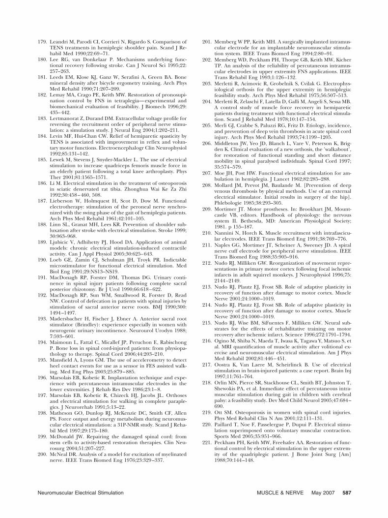
179. Leandri M, Parodi CI, Corrieri N, Rigardo S. Comparison ofTENS treatments in hemiplegic shoulder pain. Scand J Re-habil Med 1990;22:69–71.
180. Lee RG, van Donkelaar P. Mechanisms underlying func-tional recovery following stroke. Can J Neurol Sci 1995;22:257–263.
181. Leeds EM, Klose KJ, Ganz W, Serafini A, Green BA. Bonemineral density after bicycle ergometry training. Arch PhysMed Rehabil 1990;71:207–209.
182. Lemay MA, Crago PE, Keith MW. Restoration of pronosupi-nation control by FNS in tetraplegia—experimental andbiomechanical evaluation of feasibility. J Biomech 1996;29:435–442.
183. Lertmanorat Z, Durand DM. Extracellular voltage profile forreversing the recruitment order of peripheral nerve stimu-lation: a simulation study. J Neural Eng 2004;1:202–211.
184. Levin MF, Hui-Chan CW. Relief of hemiparetic spasticity byTENS is associated with improvement in reflex and volun-tary motor functions. Electroencephalogr Clin Neurophysiol1992;85:131–142.
185. Lewek M, Stevens J, Snyder-Mackler L. The use of electricalstimulation to increase quadriceps femoris muscle force inan elderly patient following a total knee arthroplasty. PhysTher 2001;81:1565–1571.
186. Li M. Electrical stimulation in the treatment of osteoporosisin sciatic denervated rat tibia. Zhonghua Wai Ke Za Zhi1992;30:458–460, 508.
187. Lieberson W, Holmquest H, Scot D, Dow M. Functionalelectrotherapy: stimulation of the peroneal nerve synchro-nized with the swing phase of the gait of hemiplegia patients.Arch Phys Med Rehabil 1961;42:101–105.
188. Linn SL, Granat MH, Lees KR. Prevention of shoulder sub-luxation after stroke with electrical stimulation. Stroke 1999;30:963–968.
189. Ljubicic V, Adhihetty PJ, Hood DA. Application of animalmodels: chronic electrical stimulation-induced contractileactivity. Can J Appl Physiol 2005;30:625–643.
190. Loeb GE, Zamin CJ, Schulman JH, Troyk PR. Indictablemicrostimulator for functional electrical stimulation. MedBiol Eng 1991;29:NS13–NS19.
191. MacDonagh RP, Forster DM, Thomas DG. Urinary conti-nence in spinal injury patients following complete sacralposterior rhizotomy. Br J Urol 1990;66:618–622.
192. MacDonagh RP, Sun WM, Smallwood R, Forster D, ReadNW. Control of defecation in patients with spinal injuries bystimulation of sacral anterior nerve roots. BMJ 1990;300:1494–1497.
193. Madersbacher H, Fischer J, Ebner A. Anterior sacral rootstimulator (Brindley): experience especially in women withneurogenic urinary incontinence. Neurourol Urodyn 1988;7:593–601.
194. Maimoun L, Fattal C, Micallef JP, Peruchon E, RabischongP. Bone loss in spinal cord-injured patients: from physiopa-thology to therapy. Spinal Cord 2006;44:203–210.
195. Mansfield A, Lyons GM. The use of accelerometry to detectheel contact events for use as a sensor in FES assisted walk-ing. Med Eng Phys 2003;25:879–885.
196. Marsolais EB, Kobetic R. Implantation technique and expe-rience with percutaneous intramuscular electrodes in thelower extremities. J Rehab Res Dev 1986;23:1–8.
197. Marsolais EB, Kobetic R, Chizeck HJ, Jacobs JL. Orthosesand electrical stimulation for walking in complete paraple-gics. J Neurorehab 1991;5:13–22.
198. Matheson GO, Dunlop RJ, McKenzie DC, Smith CF, AllenPS. Force output and energy metabolism during neuromus-cular electrical stimulation: a 31P-NMR study. Scand J Reha-bil Med 1997;29:175–180.
199. McDonald JW. Repairing the damaged spinal cord: fromstem cells to activity-based restoration therapies. Clin Neu-rosurg 2004;51:207–227.
200. McNeal DR. Analysis of a model for excitation of myelinatednerve. IEEE Trans Biomed Eng 1976;23:329–337.
201. Memberg W PP, Keith MH. A surgically implanted intramus-cular electrode for an implantable neuromuscular stimula-tion system. IEEE Trans Biomed Eng 1994;2:80–91.
202. Memberg WD, Peckham PH, Thorpe GB, Keith MW, KicherTP. An analysis of the reliability of percutaneous intramus-cular electrodes in upper extremity FNS applications. IEEETrans Rehabil Eng 1993;1:126–132.
203. Merletti R, Acimovic R, Grobelnik S, Cvilak G. Electrophys-iological orthosis for the upper extremity in hemiplegia:feasibility study. Arch Phys Med Rehabil 1975;56:507–513.
204. Merletti R, Zelaschi F, Latella D, Galli M, Angeli S, Sessa MB.A control study of muscle force recovery in hemipareticpatients during treatment with functional electrical stimula-tion. Scand J Rehabil Med 1978;10:147–154.
205. Merli GJ, Crabbe S, Paluzzi RG, Fritz D. Etiology, incidence,and prevention of deep vein thrombosis in acute spinal cordinjury. Arch Phys Med Rehabil 1993;74:1199–1205.
206. Middleton JW, Yeo JD, Blanch L, Vare V, Peterson K, Brig-den K. Clinical evaluation of a new orthosis, the ’walkabout’,for restoration of functional standing and short distancemobility in spinal paralysed individuals. Spinal Cord 1997;35:574–579.
207. Moe JH, Post HW. Functional electrical stimulation for am-bulation in hemiplegia. J Lancet 1962;82:285–288.
208. Mollard JM, Prevot JM, Baulande M. [Prevention of deepvenous thrombosis by physical methods. Use of an externalelectrical stimulator. Initial results in surgery of the hip].Phlebologie 1985;38:293–305.
209. Mortimer JT. Motor prostheses. In: Brookhart JM, Mount-castle VB, editors. Handbook of physiology: the nervoussystem II. Bethesda, MD: American Physiolgical Society;1981. p 155–187.
210. Nannini N, Horch K. Muscle recruitment with intrafascicu-lar electrodes. IEEE Trans Biomed Eng 1991;38:769–776.
211. Naples GG, Mortimer JT, Scheiner A, Sweeney JD. A spiralnerve cuff electrode for peripheral nerve stimulation. IEEETrans Biomed Eng 1988;35:905–916.
212. Nudo RJ, Milliken GW. Reorganization of movement repre-sentations in primary motor cortex following focal ischemicinfarcts in adult squirrel monkeys. J Neurophysiol 1996;75:2144–2149.
213. Nudo RJ, Plautz EJ, Frost SB. Role of adaptive plasticity inrecovery of function after damage to motor cortex. MuscleNerve 2001;24:1000–1019.
214. Nudo RJ, Plautz EJ, Frost SB. Role of adaptive plasticity inrecovery of function after damage to motor cortex. MuscleNerve 2001;24:1000–1019.
215. Nudo RJ, Wise BM, SiFuentes F, Milliken GW. Neural sub-strates for the effects of rehabilitative training on motorrecovery after ischemic infarct. Science 1996;272:1791–1794.
216. Ogino M, Shiba N, Maeda T, Iwasa K, Tagawa Y, Matsuo S, etal. MRI quantification of muscle activity after volitional ex-ercise and neuromuscular electrical stimulation. Am J PhysMed Rehabil 2002;81:446–451.
217. Oostra K, Van Laere M, Scheirlinck B. Use of electricalstimulation in brain-injured patients: a case report. Brain Inj1997;11:761–764.
218. Orlin MN, Pierce SR, Stackhouse CL, Smith BT, Johnston T,Shewokis PA, et al. Immediate effect of percutaneous intra-muscular stimulation during gait in children with cerebralpalsy: a feasibility study. Dev Med Child Neurol 2005;47:684–690.
219. Ott SM. Osteoporosis in women with spinal cord injuries.Phys Med Rehabil Clin N Am 2001;12:111–131.
220. Paillard T, Noe F, Passelergue P, Dupui P. Electrical stimu-lation superimposed onto voluntary muscular contraction.Sports Med 2005;35:951–966.
221. Peckham PH, Keith MW, Freehafer AA. Restoration of func-tional control by electrical stimulation in the upper extrem-ity of the quadriplegic patient. J Bone Joint Surg [Am]1988;70:144–148.
Neuromuscular Electrical Stimulation MUSCLE & NERVE May 2007 587
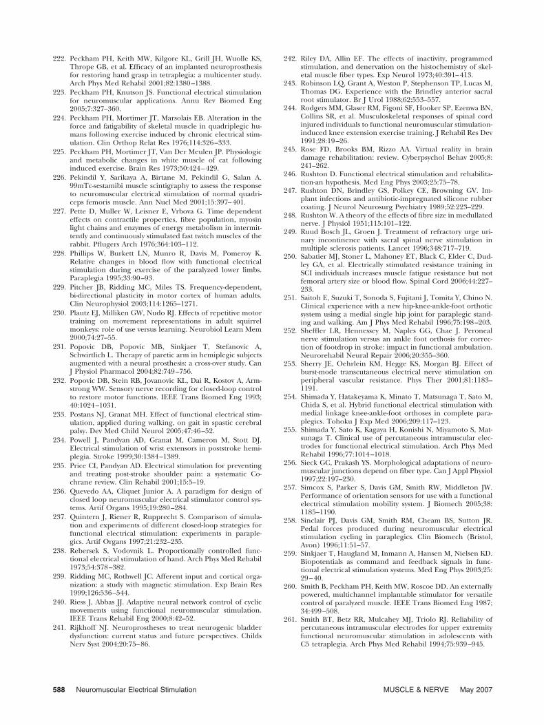
222. Peckham PH, Keith MW, Kilgore KL, Grill JH, Wuolle KS,Thrope GB, et al. Efficacy of an implanted neuroprosthesisfor restoring hand grasp in tetraplegia: a multicenter study.Arch Phys Med Rehabil 2001;82:1380–1388.
223. Peckham PH, Knutson JS. Functional electrical stimulationfor neuromuscular applications. Annu Rev Biomed Eng2005;7:327–360.
224. Peckham PH, Mortimer JT, Marsolais EB. Alteration in theforce and fatigability of skeletal muscle in quadriplegic hu-mans following exercise induced by chronic electrical stim-ulation. Clin Orthop Relat Res 1976;114:326–333.
225. Peckham PH, Mortimer JT, Van Der Meulen JP. Physiologicand metabolic changes in white muscle of cat followinginduced exercise. Brain Res 1973;50:424–429.
226. Pekindil Y, Sarikaya A, Birtane M, Pekindil G, Salan A.99mTc-sestamibi muscle scintigraphy to assess the responseto neuromuscular electrical stimulation of normal quadri-ceps femoris muscle. Ann Nucl Med 2001;15:397–401.
227. Pette D, Muller W, Leisner E, Vrbova G. Time dependenteffects on contractile properties, fibre population, myosinlight chains and enzymes of energy metabolism in intermit-tently and continuously stimulated fast twitch muscles of therabbit. Pflugers Arch 1976;364:103–112.
228. Phillips W, Burkett LN, Munro R, Davis M, Pomeroy K.Relative changes in blood flow with functional electricalstimulation during exercise of the paralyzed lower limbs.Paraplegia 1995;33:90–93.
229. Pitcher JB, Ridding MC, Miles TS. Frequency-dependent,bi-directional plasticity in motor cortex of human adults.Clin Neurophysiol 2003;114:1265–1271.
230. Plautz EJ, Milliken GW, Nudo RJ. Effects of repetitive motortraining on movement representations in adult squirrelmonkeys: role of use versus learning. Neurobiol Learn Mem2000;74:27–55.
231. Popovic DB, Popovic MB, Sinkjaer T, Stefanovic A,Schwirtlich L. Therapy of paretic arm in hemiplegic subjectsaugmented with a neural prosthesis: a cross-over study. CanJ Physiol Pharmacol 2004;82:749–756.
232. Popovic DB, Stein RB, Jovanovic KL, Dai R, Kostov A, Arm-strong WW. Sensory nerve recording for closed-loop controlto restore motor functions. IEEE Trans Biomed Eng 1993;40:1024–1031.
233. Postans NJ, Granat MH. Effect of functional electrical stim-ulation, applied during walking, on gait in spastic cerebralpalsy. Dev Med Child Neurol 2005;47:46–52.
234. Powell J, Pandyan AD, Granat M, Cameron M, Stott DJ.Electrical stimulation of wrist extensors in poststroke hemi-plegia. Stroke 1999;30:1384–1389.
235. Price CI, Pandyan AD. Electrical stimulation for preventingand treating post-stroke shoulder pain: a systematic Co-chrane review. Clin Rehabil 2001;15:5–19.
236. Quevedo AA, Cliquet Junior A. A paradigm for design ofclosed loop neuromuscular electrical stimulator control sys-tems. Artif Organs 1995;19:280–284.
237. Quintern J, Riener R, Rupprecht S. Comparison of simula-tion and experiments of different closed-loop strategies forfunctional electrical stimulation: experiments in paraple-gics. Artif Organs 1997;21:232–235.
238. Rebersek S, Vodovnik L. Proportionally controlled func-tional electrical stimulation of hand. Arch Phys Med Rehabil1973;54:378–382.
239. Ridding MC, Rothwell JC. Afferent input and cortical orga-nization: a study with magnetic stimulation. Exp Brain Res1999;126:536–544.
240. Riess J, Abbas JJ. Adaptive neural network control of cyclicmovements using functional neuromuscular stimulation.IEEE Trans Rehabil Eng 2000;8:42–52.
241. Rijkhoff NJ. Neuroprostheses to treat neurogenic bladderdysfunction: current status and future perspectives. ChildsNerv Syst 2004;20:75–86.
242. Riley DA, Allin EF. The effects of inactivity, programmedstimulation, and denervation on the histochemistry of skel-etal muscle fiber types. Exp Neurol 1973;40:391–413.
243. Robinson LQ, Grant A, Weston P, Stephenson TP, Lucas M,Thomas DG. Experience with the Brindley anterior sacralroot stimulator. Br J Urol 1988;62:553–557.
244. Rodgers MM, Glaser RM, Figoni SF, Hooker SP, Ezenwa BN,Collins SR, et al. Musculoskeletal responses of spinal cordinjured individuals to functional neuromuscular stimulation-induced knee extension exercise training. J Rehabil Res Dev1991;28:19–26.
245. Rose FD, Brooks BM, Rizzo AA. Virtual reality in braindamage rehabilitation: review. Cyberpsychol Behav 2005;8:241–262.
246. Rushton D. Functional electrical stimulation and rehabilita-tion-an hypothesis. Med Eng Phys 2003;25:75–78.
247. Rushton DN, Brindley GS, Polkey CE, Browning GV. Im-plant infections and antibiotic-impregnated silicone rubbercoating. J Neurol Neurosurg Psychiatry 1989;52:223–229.
248. Rushton W. A theory of the effects of fibre size in medullatednerve. J Physiol 1951;115:101–122.
249. Ruud Bosch JL, Groen J. Treatment of refractory urge uri-nary incontinence with sacral spinal nerve stimulation inmultiple sclerosis patients. Lancet 1996;348:717–719.
250. Sabatier MJ, Stoner L, Mahoney ET, Black C, Elder C, Dud-ley GA, et al. Electrically stimulated resistance training inSCI individuals increases muscle fatigue resistance but notfemoral artery size or blood flow. Spinal Cord 2006;44:227–233.
251. Saitoh E, Suzuki T, Sonoda S, Fujitani J, Tomita Y, Chino N.Clinical experience with a new hip-knee-ankle-foot orthoticsystem using a medial single hip joint for paraplegic stand-ing and walking. Am J Phys Med Rehabil 1996;75:198–203.
252. Sheffler LR, Hennessey M, Naples GG, Chae J. Peronealnerve stimulation versus an ankle foot orthosis for correc-tion of footdrop in stroke: impact in functional ambulation.Neurorehabil Neural Repair 2006;20:355–360.
253. Sherry JE, Oehrlein KM, Hegge KS, Morgan BJ. Effect ofburst-mode transcutaneous electrical nerve stimulation onperipheral vascular resistance. Phys Ther 2001;81:1183–1191.
254. Shimada Y, Hatakeyama K, Minato T, Matsunaga T, Sato M,Chida S, et al. Hybrid functional electrical stimulation withmedial linkage knee-ankle-foot orthoses in complete para-plegics. Tohoku J Exp Med 2006;209:117–123.
255. Shimada Y, Sato K, Kagaya H, Konishi N, Miyamoto S, Mat-sunaga T. Clinical use of percutaneous intramuscular elec-trodes for functional electrical stimulation. Arch Phys MedRehabil 1996;77:1014–1018.
256. Sieck GC, Prakash YS. Morphological adaptations of neuro-muscular junctions depend on fiber type. Can J Appl Physiol1997;22:197–230.
257. Simcox S, Parker S, Davis GM, Smith RW, Middleton JW.Performance of orientation sensors for use with a functionalelectrical stimulation mobility system. J Biomech 2005;38:1185–1190.
258. Sinclair PJ, Davis GM, Smith RM, Cheam BS, Sutton JR.Pedal forces produced during neuromuscular electricalstimulation cycling in paraplegics. Clin Biomech (Bristol,Avon) 1996;11:51–57.
259. Sinkjaer T, Haugland M, Inmann A, Hansen M, Nielsen KD.Biopotentials as command and feedback signals in func-tional electrical stimulation systems. Med Eng Phys 2003;25:29–40.
260. Smith B, Peckham PH, Keith MW, Roscoe DD. An externallypowered, multichannel implantable stimulator for versatilecontrol of paralyzed muscle. IEEE Trans Biomed Eng 1987;34:499–508.
261. Smith BT, Betz RR, Mulcahey MJ, Triolo RJ. Reliability ofpercutaneous intramuscular electrodes for upper extremityfunctional neuromuscular stimulation in adolescents withC5 tetraplegia. Arch Phys Med Rehabil 1994;75:939–945.
588 Neuromuscular Electrical Stimulation MUSCLE & NERVE May 2007
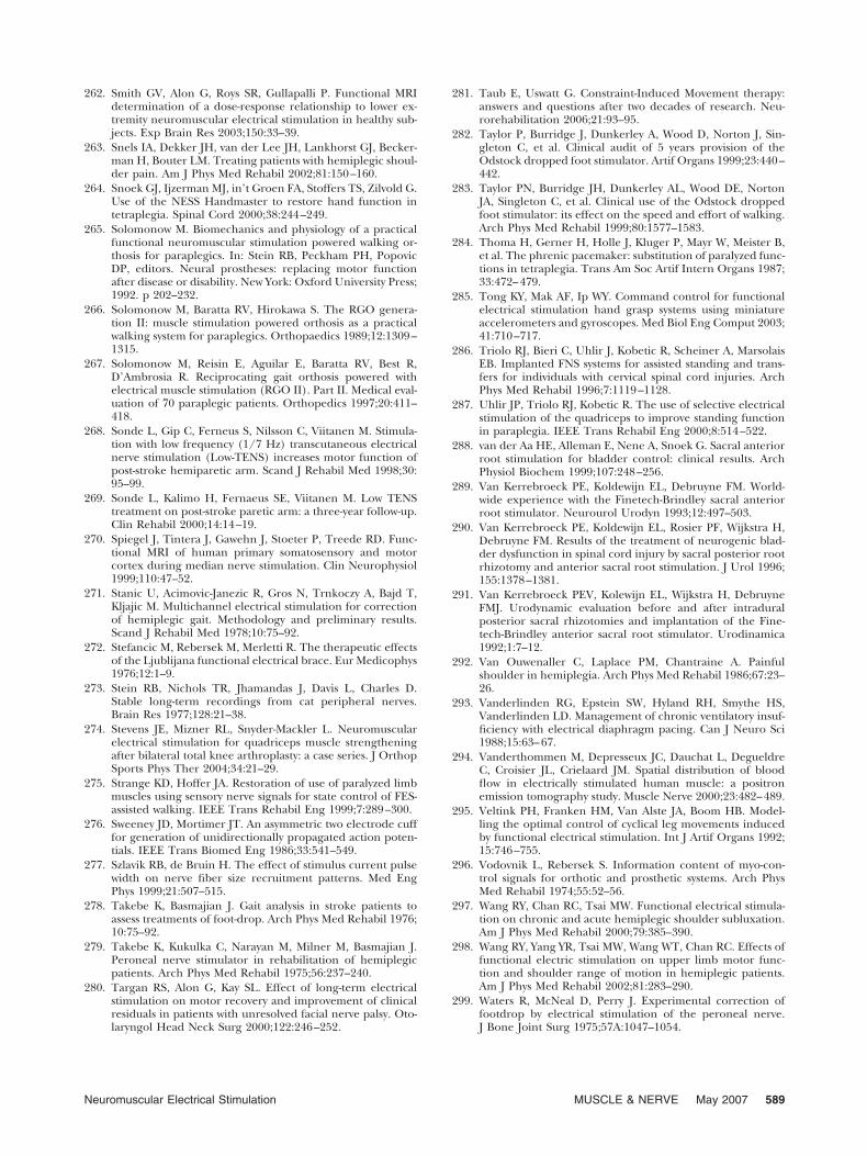
262. Smith GV, Alon G, Roys SR, Gullapalli P. Functional MRIdetermination of a dose-response relationship to lower ex-tremity neuromuscular electrical stimulation in healthy sub-jects. Exp Brain Res 2003;150:33–39.
263. Snels IA, Dekker JH, van der Lee JH, Lankhorst GJ, Becker-man H, Bouter LM. Treating patients with hemiplegic shoul-der pain. Am J Phys Med Rehabil 2002;81:150–160.
264. Snoek GJ, Ijzerman MJ, in’t Groen FA, Stoffers TS, Zilvold G.Use of the NESS Handmaster to restore hand function intetraplegia. Spinal Cord 2000;38:244–249.
265. Solomonow M. Biomechanics and physiology of a practicalfunctional neuromuscular stimulation powered walking or-thosis for paraplegics. In: Stein RB, Peckham PH, PopovicDP, editors. Neural prostheses: replacing motor functionafter disease or disability. New York: Oxford University Press;1992. p 202–232.
266. Solomonow M, Baratta RV, Hirokawa S. The RGO genera-tion II: muscle stimulation powered orthosis as a practicalwalking system for paraplegics. Orthopaedics 1989;12:1309–1315.
267. Solomonow M, Reisin E, Aguilar E, Baratta RV, Best R,D’Ambrosia R. Reciprocating gait orthosis powered withelectrical muscle stimulation (RGO II). Part II. Medical eval-uation of 70 paraplegic patients. Orthopedics 1997;20:411–418.
268. Sonde L, Gip C, Ferneus S, Nilsson C, Viitanen M. Stimula-tion with low frequency (1/7 Hz) transcutaneous electricalnerve stimulation (Low-TENS) increases motor function ofpost-stroke hemiparetic arm. Scand J Rehabil Med 1998;30:95–99.
269. Sonde L, Kalimo H, Fernaeus SE, Viitanen M. Low TENStreatment on post-stroke paretic arm: a three-year follow-up.Clin Rehabil 2000;14:14–19.
270. Spiegel J, Tintera J, Gawehn J, Stoeter P, Treede RD. Func-tional MRI of human primary somatosensory and motorcortex during median nerve stimulation. Clin Neurophysiol1999;110:47–52.
271. Stanic U, Acimovic-Janezic R, Gros N, Trnkoczy A, Bajd T,Kljajic M. Multichannel electrical stimulation for correctionof hemiplegic gait. Methodology and preliminary results.Scand J Rehabil Med 1978;10:75–92.
272. Stefancic M, Rebersek M, Merletti R. The therapeutic effectsof the Ljublijana functional electrical brace. Eur Medicophys1976;12:1–9.
273. Stein RB, Nichols TR, Jhamandas J, Davis L, Charles D.Stable long-term recordings from cat peripheral nerves.Brain Res 1977;128:21–38.
274. Stevens JE, Mizner RL, Snyder-Mackler L. Neuromuscularelectrical stimulation for quadriceps muscle strengtheningafter bilateral total knee arthroplasty: a case series. J OrthopSports Phys Ther 2004;34:21–29.
275. Strange KD, Hoffer JA. Restoration of use of paralyzed limbmuscles using sensory nerve signals for state control of FES-assisted walking. IEEE Trans Rehabil Eng 1999;7:289–300.
276. Sweeney JD, Mortimer JT. An asymmetric two electrode cufffor generation of unidirectionally propagated action poten-tials. IEEE Trans Biomed Eng 1986;33:541–549.
277. Szlavik RB, de Bruin H. The effect of stimulus current pulsewidth on nerve fiber size recruitment patterns. Med EngPhys 1999;21:507–515.
278. Takebe K, Basmajian J. Gait analysis in stroke patients toassess treatments of foot-drop. Arch Phys Med Rehabil 1976;10:75–92.
279. Takebe K, Kukulka C, Narayan M, Milner M, Basmajian J.Peroneal nerve stimulator in rehabilitation of hemiplegicpatients. Arch Phys Med Rehabil 1975;56:237–240.
280. Targan RS, Alon G, Kay SL. Effect of long-term electricalstimulation on motor recovery and improvement of clinicalresiduals in patients with unresolved facial nerve palsy. Oto-laryngol Head Neck Surg 2000;122:246–252.
281. Taub E, Uswatt G. Constraint-Induced Movement therapy:answers and questions after two decades of research. Neu-rorehabilitation 2006;21:93–95.
282. Taylor P, Burridge J, Dunkerley A, Wood D, Norton J, Sin-gleton C, et al. Clinical audit of 5 years provision of theOdstock dropped foot stimulator. Artif Organs 1999;23:440–442.
283. Taylor PN, Burridge JH, Dunkerley AL, Wood DE, NortonJA, Singleton C, et al. Clinical use of the Odstock droppedfoot stimulator: its effect on the speed and effort of walking.Arch Phys Med Rehabil 1999;80:1577–1583.
284. Thoma H, Gerner H, Holle J, Kluger P, Mayr W, Meister B,et al. The phrenic pacemaker: substitution of paralyzed func-tions in tetraplegia. Trans Am Soc Artif Intern Organs 1987;33:472–479.
285. Tong KY, Mak AF, Ip WY. Command control for functionalelectrical stimulation hand grasp systems using miniatureaccelerometers and gyroscopes. Med Biol Eng Comput 2003;41:710–717.
286. Triolo RJ, Bieri C, Uhlir J, Kobetic R, Scheiner A, MarsolaisEB. Implanted FNS systems for assisted standing and trans-fers for individuals with cervical spinal cord injuries. ArchPhys Med Rehabil 1996;7:1119–1128.
287. Uhlir JP, Triolo RJ, Kobetic R. The use of selective electricalstimulation of the quadriceps to improve standing functionin paraplegia. IEEE Trans Rehabil Eng 2000;8:514–522.
288. van der Aa HE, Alleman E, Nene A, Snoek G. Sacral anteriorroot stimulation for bladder control: clinical results. ArchPhysiol Biochem 1999;107:248–256.
289. Van Kerrebroeck PE, Koldewijn EL, Debruyne FM. World-wide experience with the Finetech-Brindley sacral anteriorroot stimulator. Neurourol Urodyn 1993;12:497–503.
290. Van Kerrebroeck PE, Koldewijn EL, Rosier PF, Wijkstra H,Debruyne FM. Results of the treatment of neurogenic blad-der dysfunction in spinal cord injury by sacral posterior rootrhizotomy and anterior sacral root stimulation. J Urol 1996;155:1378–1381.
291. Van Kerrebroeck PEV, Kolewijn EL, Wijkstra H, DebruyneFMJ. Urodynamic evaluation before and after intraduralposterior sacral rhizotomies and implantation of the Fine-tech-Brindley anterior sacral root stimulator. Urodinamica1992;1:7–12.
292. Van Ouwenaller C, Laplace PM, Chantraine A. Painfulshoulder in hemiplegia. Arch Phys Med Rehabil 1986;67:23–26.
293. Vanderlinden RG, Epstein SW, Hyland RH, Smythe HS,Vanderlinden LD. Management of chronic ventilatory insuf-ficiency with electrical diaphragm pacing. Can J Neuro Sci1988;15:63–67.
294. Vanderthommen M, Depresseux JC, Dauchat L, DegueldreC, Croisier JL, Crielaard JM. Spatial distribution of bloodflow in electrically stimulated human muscle: a positronemission tomography study. Muscle Nerve 2000;23:482–489.
295. Veltink PH, Franken HM, Van Alste JA, Boom HB. Model-ling the optimal control of cyclical leg movements inducedby functional electrical stimulation. Int J Artif Organs 1992;15:746–755.
296. Vodovnik L, Rebersek S. Information content of myo-con-trol signals for orthotic and prosthetic systems. Arch PhysMed Rehabil 1974;55:52–56.
297. Wang RY, Chan RC, Tsai MW. Functional electrical stimula-tion on chronic and acute hemiplegic shoulder subluxation.Am J Phys Med Rehabil 2000;79:385–390.
298. Wang RY, Yang YR, Tsai MW, Wang WT, Chan RC. Effects offunctional electric stimulation on upper limb motor func-tion and shoulder range of motion in hemiplegic patients.Am J Phys Med Rehabil 2002;81:283–290.
299. Waters R, McNeal D, Perry J. Experimental correction offootdrop by electrical stimulation of the peroneal nerve.J Bone Joint Surg 1975;57A:1047–1054.
Neuromuscular Electrical Stimulation MUSCLE & NERVE May 2007 589
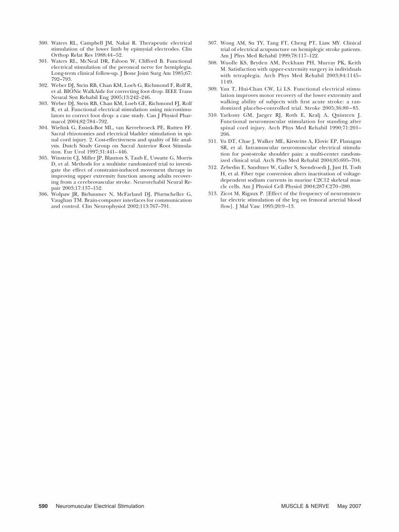
300. Waters RL, Campbell JM, Nakai R. Therapeutic electricalstimulation of the lower limb by epimysial electrodes. ClinOrthop Relat Res 1988:44–52.
301. Waters RL, McNeal DR, Faloon W, Clifford B. Functionalelectrical stimulation of the peroneal nerve for hemiplegia.Long-term clinical follow-up. J Bone Joint Surg Am 1985;67:792–793.
302. Weber DJ, Stein RB, Chan KM, Loeb G, Richmond F, Rolf R,et al. BIONic WalkAide for correcting foot drop. IEEE TransNeural Syst Rehabil Eng 2005;13:242–246.
303. Weber DJ, Stein RB, Chan KM, Loeb GE, Richmond FJ, RolfR, et al. Functional electrical stimulation using microstimu-lators to correct foot drop: a case study. Can J Physiol Phar-macol 2004;82:784–792.
304. Wielink G, Essink-Bot ML, van Kerrebroeck PE, Rutten FF.Sacral rhizotomies and electrical bladder stimulation in spi-nal cord injury. 2. Cost-effectiveness and quality of life anal-ysis. Dutch Study Group on Sacral Anterior Root Stimula-tion. Eur Urol 1997;31:441–446.
305. Winstein CJ, Miller JP, Blanton S, Taub E, Uswatte G, MorrisD, et al. Methods for a multisite randomized trial to investi-gate the effect of constraint-induced movement therapy inimproving upper extremity function among adults recover-ing from a cerebrovascular stroke. Neurorehabil Neural Re-pair 2003;17:137–152.
306. Wolpaw JR, Birbaumer N, McFarland DJ, Pfurtscheller G,Vaughan TM. Brain-computer interfaces for communicationand control. Clin Neurophysiol 2002;113:767–791.
307. Wong AM, Su TY, Tang FT, Cheng PT, Liaw MY. Clinicaltrial of electrical acupuncture on hemiplegic stroke patients.Am J Phys Med Rehabil 1999;78:117–122.
308. Wuolle KS, Bryden AM, Peckham PH, Murray PK, KeithM. Satisfaction with upper-extremity surgery in individualswith tetraplegia. Arch Phys Med Rehabil 2003;84:1145–1149.
309. Yan T, Hui-Chan CW, Li LS. Functional electrical stimu-lation improves motor recovery of the lower extremity andwalking ability of subjects with first acute stroke: a ran-domized placebo-controlled trial. Stroke 2005;36:80 – 85.
310. Yarkony GM, Jaeger RJ, Roth E, Kralj A, Quintern J.Functional neuromuscular stimulation for standing afterspinal cord injury. Arch Phys Med Rehabil 1990;71:201–206.
311. Yu DT, Chae J, Walker ME, Kirsteins A, Elovic EP, FlanaganSR, et al. Intramuscular neuromuscular electrical stimula-tion for post-stroke shoulder pain: a multi-center random-ized clinical trial. Arch Phys Med Rehabil 2004;85:695–704.
312. Zebedin E, Sandtner W, Galler S, Szendroedi J, Just H, TodtH, et al. Fiber type conversion alters inactivation of voltage-dependent sodium currents in murine C2C12 skeletal mus-cle cells. Am J Physiol Cell Physiol 2004;287:C270–280.
313. Zicot M, Rigaux P. [Effect of the frequency of neuromuscu-lar electric stimulation of the leg on femoral arterial bloodflow]. J Mal Vasc 1995;20:9–13.
590 Neuromuscular Electrical Stimulation MUSCLE & NERVE May 2007