g-protein coupled receptors: structure and function in
TRANSCRIPT
RSC Advances
REVIEW
Ope
n A
cces
s A
rtic
le. P
ublis
hed
on 0
1 O
ctob
er 2
020.
Dow
nloa
ded
on 1
1/21
/202
1 7:
31:3
9 A
M.
Thi
s ar
ticle
is li
cens
ed u
nder
a C
reat
ive
Com
mon
s A
ttrib
utio
n-N
onC
omm
erci
al 3
.0 U
npor
ted
Lic
ence
.
View Article OnlineView Journal | View Issue
G-Protein couple
aNottingham Trent University, 50 Shakespea
[email protected] and Integrated Bioengi
Jordanstown Campus, Newtownabbey, BT37cDe Montfort University, The Gateway, LeicedDepartment of Chemistry, University of BateDepartment of Chemistry – BMC, Uppsa
Uppsala, Sweden
Cite this: RSC Adv., 2020, 10, 36337
Received 20th July 2020Accepted 22nd September 2020
DOI: 10.1039/d0ra08003a
rsc.li/rsc-advances
This journal is © The Royal Society o
d receptors: structure andfunction in drug discovery
Chiemela S. Odoemelam, a Benita Percival, a Helen Wallis,a Ming-Wei Chang,b
Zeeshan Ahmad,c Dawn Scholey,a Emily Burton,a Ian H. Williams, d
Caroline Lynn Kamerlin e and Philippe B. Wilson *a
The G-protein coupled receptors (GPCRs) superfamily comprise similar proteins arranged into families or
classes thus making it one of the largest in the mammalian genome. GPCRs take part in many vital
physiological functions making them targets for numerous novel drugs. GPCRs share some distinctive
features, such as the seven transmembrane domains, they also differ in the number of conserved
residues in their transmembrane domain. Here we provide an introductory and accessible review
detailing the computational advances in GPCR pharmacology and drug discovery. An overview is
provided on family A-C GPCRs; their structural differences, GPCR signalling, allosteric binding and
cooperativity. The dielectric constant (relative permittivity) of proteins is also discussed in the context of
site-specific environmental effects.
Background
The G-protein coupled receptor (GPCR) superfamily consists ofstructurally similar proteins arranged into families (classes),and is one of the most abundant protein classes in themammalian genome.1–5 GPCRs undertake a plethora of essen-tial physiological functions and are targets for numerous noveldrugs.4,5 Their ligands are structurally heterogenous, includingnatural odorants, nucleotides, amines, peptides, proteins, andlipids.4 The conserved structure of GPCRs consists of sevenTMD of approximately 25–35 successive amino acid residuesthat express moderately high levels of hydrophobicity4 and arecharacterised by a-helices which span the plasma membrane.4
The primary function of GPCRs is the transduction of extra-cellular stimuli into intracellular signals.2 Currently, approxi-mately thirty to forty percent of marketed pharmaceuticalstarget GPCRs.1,6–10 Hence, there is enormous potential for thedevelopment of new drugs targeting these receptors.3 Examplesof drugs targeting GPCRs include histamine receptor blockers,opioid agonists, b-blockers and angiotensin receptor blockers.5
Computational biology methods are currently being employedto understand GPCRs as such drug targets.6,11,12 Breakthroughsin GPCR crystallography has facilitated novel discovery through
re St, Nottingham NG1 4FQ, UK. E-mail:
neering Centre, University of Ulster,
0QB, Northern Ireland, UK
ster, LE1 9BH, UK
h, Claverton Down, Bath, BA1 7AY, UK
la University, BMC Box 576, S-751 23
f Chemistry 2020
virtual screening as well as better off-target rationalisation.6
Recently, the Tikhonova group developed a computationalprotocol which combines concepts from statistical mechanicsand cheminformatics to explore the exibility of the bioaminereceptors as well as to identify the geometrical and physico-chemical properties which characterise the conformationalspace of the bioamine family.13 Multiple-microsecond timescalemolecular dynamics (MD) simulations have been used incapturing the process of several drugs binding to b1- and b2-adrenergic receptors.14 Molecular docking is one of the mostcommonly used methods in GPCR structure-based drug design(SBDD).14 Esguerra et al. developed GPCR-ModSim, a web-basedportal designed specically for the homology modelling andMD simulation of GPCRs.15
It was historically assumed that GPCRs exist in two confor-mations: active and inactive.16–18 The long-established extendedternary-complex model of GPCR-driven signalling was based onthis concept.16,19,20 This model suggested that the active GPCRconformation opted for by G-protein-coupled receptor kinases(GRKs), arrestins and G proteins is uniform.16 Nevertheless,biophysical investigations with a rened uorescent-labelledb2-adrenergic receptor (b2AR) demonstrated that a receptorcan exist in numerous conformations and that the conforma-tional equilibrium is inuenced both by the bound ligand andthe proximity to the related G protein.16
The human genome alone contains approximately 800GPCRs making it the largest family of membrane proteins.5,21
GPCRs have been classied based on structural and physio-logical features.4 Some systems of classication have groupedthese based on location of the ligand binding pocket, whilesome have utilised both the structural and physiological
RSC Adv., 2020, 10, 36337–36348 | 36337
RSC Advances Review
Ope
n A
cces
s A
rtic
le. P
ublis
hed
on 0
1 O
ctob
er 2
020.
Dow
nloa
ded
on 1
1/21
/202
1 7:
31:3
9 A
M.
Thi
s ar
ticle
is li
cens
ed u
nder
a C
reat
ive
Com
mon
s A
ttrib
utio
n-N
onC
omm
erci
al 3
.0 U
npor
ted
Lic
ence
.View Article Online
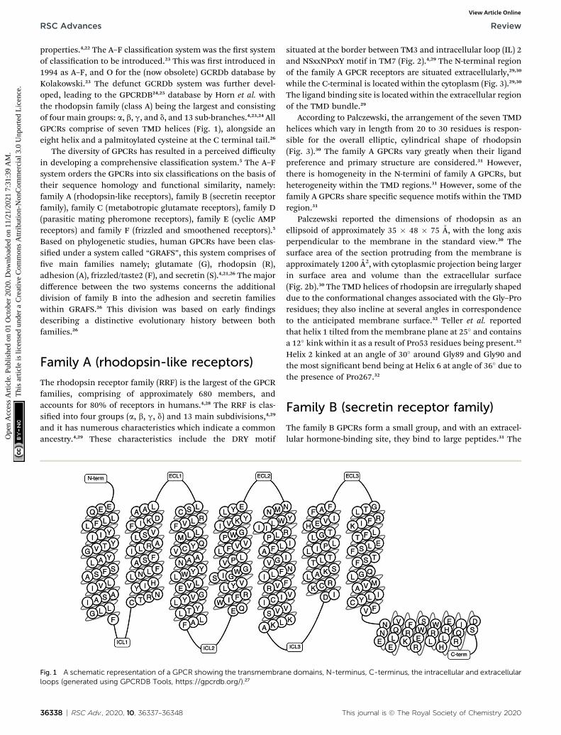
properties.4,22 The A–F classication system was the rst systemof classication to be introduced.23 This was rst introduced in1994 as A–F, and O for the (now obsolete) GCRDb database byKolakowski.23 The defunct GCRDb system was further devel-oped, leading to the GPCRDB24,25 database by Horn et al. withthe rhodopsin family (class A) being the largest and consistingof four main groups: a, b, g, and d, and 13 sub-branches.4,23,24 AllGPCRs comprise of seven TMD helices (Fig. 1), alongside aneight helix and a palmitoylated cysteine at the C terminal tail.26
The diversity of GPCRs has resulted in a perceived difficultyin developing a comprehensive classication system.5 The A–Fsystem orders the GPCRs into six classications on the basis oftheir sequence homology and functional similarity, namely:family A (rhodopsin-like receptors), family B (secretin receptorfamily), family C (metabotropic glutamate receptors), family D(parasitic mating pheromone receptors), family E (cyclic AMPreceptors) and family F (frizzled and smoothened receptors).5
Based on phylogenetic studies, human GPCRs have been clas-sied under a system called “GRAFS”, this system comprises ofve main families namely; glutamate (G), rhodopsin (R),adhesion (A), frizzled/taste2 (F), and secretin (S).4,21,26 The majordifference between the two systems concerns the additionaldivision of family B into the adhesion and secretin familieswithin GRAFS.26 This division was based on early ndingsdescribing a distinctive evolutionary history between bothfamilies.26
Family A (rhodopsin-like receptors)
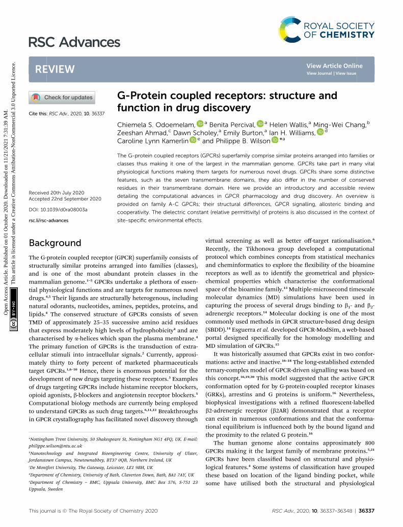
The rhodopsin receptor family (RRF) is the largest of the GPCRfamilies, comprising of approximately 680 members, andaccounts for 80% of receptors in humans.4,28 The RRF is clas-sied into four groups (a, b, g, d) and 13 main subdivisions,4,29
and it has numerous characteristics which indicate a commonancestry.4,29 These characteristics include the DRY motif
Fig. 1 A schematic representation of a GPCR showing the transmembranloops (generated using GPCRDB Tools, https://gpcrdb.org/).27
36338 | RSC Adv., 2020, 10, 36337–36348
situated at the border between TM3 and intracellular loop (IL) 2and NSxxNPxxY motif in TM7 (Fig. 2).4,29 The N-terminal regionof the family A GPCR receptors are situated extracellularly,29,30
while the C-terminal is located within the cytoplasm (Fig. 3).29,30
The ligand binding site is located within the extracellular regionof the TMD bundle.29
According to Palczewski, the arrangement of the seven TMDhelices which vary in length from 20 to 30 residues is respon-sible for the overall elliptic, cylindrical shape of rhodopsin(Fig. 3).30 The family A GPCRs vary greatly when their ligandpreference and primary structure are considered.31 However,there is homogeneity in the N-termini of family A GPCRs, butheterogeneity within the TMD regions.31 However, some of thefamily A GPCRs share specic sequence motifs within the TMDregion.31
Palczewski reported the dimensions of rhodopsin as anellipsoid of approximately 35 � 48 � 75 A, with the long axisperpendicular to the membrane in the standard view.30 Thesurface area of the section protruding from the membrane isapproximately 1200 A2, with cytoplasmic projection being largerin surface area and volume than the extracellular surface(Fig. 2b).30 The TMD helices of rhodopsin are irregularly shapeddue to the conformational changes associated with the Gly–Proresidues; they also incline at several angles in correspondenceto the anticipated membrane surface.32 Teller et al. reportedthat helix 1 tilted from the membrane plane at 25� and containsa 12� kink within it as a result of Pro53 residues being present.32
Helix 2 kinked at an angle of 30� around Gly89 and Gly90 andthe most signicant bend being at Helix 6 at angle of 36� due tothe presence of Pro267.32
Family B (secretin receptor family)
The family B GPCRs form a small group, and with an extracel-lular hormone-binding site, they bind to large peptides.31 The
e domains, N-terminus, C-terminus, the intracellular and extracellular
This journal is © The Royal Society of Chemistry 2020
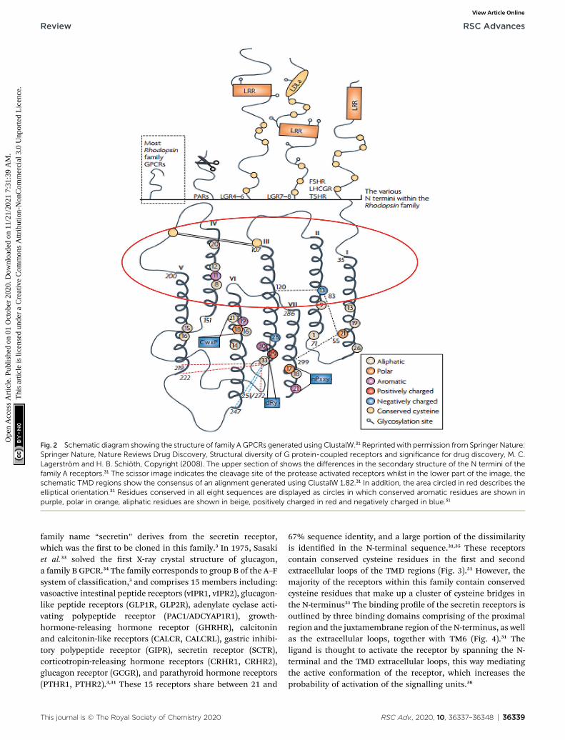
Fig. 2 Schematic diagram showing the structure of family A GPCRs generated using ClustalW.31 Reprintedwith permission from Springer Nature:Springer Nature, Nature Reviews Drug Discovery, Structural diversity of G protein-coupled receptors and significance for drug discovery, M. C.Lagerstrom and H. B. Schioth, Copyright (2008). The upper section of shows the differences in the secondary structure of the N termini of thefamily A receptors.31 The scissor image indicates the cleavage site of the protease activated receptors whilst in the lower part of the image, theschematic TMD regions show the consensus of an alignment generated using ClustalW 1.82.31 In addition, the area circled in red describes theelliptical orientation.31 Residues conserved in all eight sequences are displayed as circles in which conserved aromatic residues are shown inpurple, polar in orange, aliphatic residues are shown in beige, positively charged in red and negatively charged in blue.31
Review RSC Advances
Ope
n A
cces
s A
rtic
le. P
ublis
hed
on 0
1 O
ctob
er 2
020.
Dow
nloa
ded
on 1
1/21
/202
1 7:
31:3
9 A
M.
Thi
s ar
ticle
is li
cens
ed u
nder
a C
reat
ive
Com
mon
s A
ttrib
utio
n-N
onC
omm
erci
al 3
.0 U
npor
ted
Lic
ence
.View Article Online
family name “secretin” derives from the secretin receptor,which was the rst to be cloned in this family.3 In 1975, Sasakiet al.33 solved the rst X-ray crystal structure of glucagon,a family B GPCR.34 The family corresponds to group B of the A–Fsystem of classication,3 and comprises 15 members including:vasoactive intestinal peptide receptors (vIPR1, vIPR2), glucagon-like peptide receptors (GLP1R, GLP2R), adenylate cyclase acti-vating polypeptide receptor (PAC1/ADCYAP1R1), growth-hormone-releasing hormone receptor (GHRHR), calcitoninand calcitonin-like receptors (CALCR, CALCRL), gastric inhibi-tory polypeptide receptor (GIPR), secretin receptor (SCTR),corticotropin-releasing hormone receptors (CRHR1, CRHR2),glucagon receptor (GCGR), and parathyroid hormone receptors(PTHR1, PTHR2).3,31 These 15 receptors share between 21 and
This journal is © The Royal Society of Chemistry 2020
67% sequence identity, and a large portion of the dissimilarityis identied in the N-terminal sequence.31,35 These receptorscontain conserved cysteine residues in the rst and secondextracellular loops of the TMD regions (Fig. 3).31 However, themajority of the receptors within this family contain conservedcysteine residues that make up a cluster of cysteine bridges inthe N-terminus31 The binding prole of the secretin receptors isoutlined by three binding domains comprising of the proximalregion and the juxtamembrane region of the N-terminus, as wellas the extracellular loops, together with TM6 (Fig. 4).31 Theligand is thought to activate the receptor by spanning the N-terminal and the TMD extracellular loops, this way mediatingthe active conformation of the receptor, which increases theprobability of activation of the signalling units.36
RSC Adv., 2020, 10, 36337–36348 | 36339
Fig. 3 Illustration showing the modification of rhodopsin and its orientation in membranes.30 Reprinted with permission from Annual Reviews:Annual Reviews, Annual review of biochemistry, G protein–coupled receptor rhodopsin, K. Palczewski, Copyright (2006). (a) Two-dimensionalillustration of rhodopsin. The polypeptide of rhodopsin is seen to cross the membrane seven times with C-I, C-II, C-III comparable to thecytoplasmic loops and E-I, E-II, E-III to the extracellular loops. The yellow cylinders represent the transmembrane region (b) depicts the locationof the chromophore and the charges on the extracellular and cytoplasmic surface of rhodopsin. Red and blue colours represent negative andpositive charged residues respectively, while the location of the chromophore is revealed by deleting fragments of the transmembrane helices.30
RSC Advances Review
Ope
n A
cces
s A
rtic
le. P
ublis
hed
on 0
1 O
ctob
er 2
020.
Dow
nloa
ded
on 1
1/21
/202
1 7:
31:3
9 A
M.
Thi
s ar
ticle
is li
cens
ed u
nder
a C
reat
ive
Com
mon
s A
ttrib
utio
n-N
onC
omm
erci
al 3
.0 U
npor
ted
Lic
ence
.View Article Online
In addition to the presence of an extracellular N-terminaldomain (ECD) of 120–160 residues, three intracellular (IL) andextracellular (EL) loops interconnect seven TMD (TM1-TM7) of310–420 residues that are structurally similar and are thusmembers of the family B GPCR.37,38 According to Parthier et al.
36340 | RSC Adv., 2020, 10, 36337–36348
hormonal recognition in family B GPCRs is believed to followthe ‘two-domain’ binding mode, the N- and C-terminal regionsof the peptides interact with the J- and N-domains of thereceptors respectively, i.e. the C terminus of the peptide initiatesa peptide recognition with the ECD, thus allowing the peptide N
This journal is © The Royal Society of Chemistry 2020
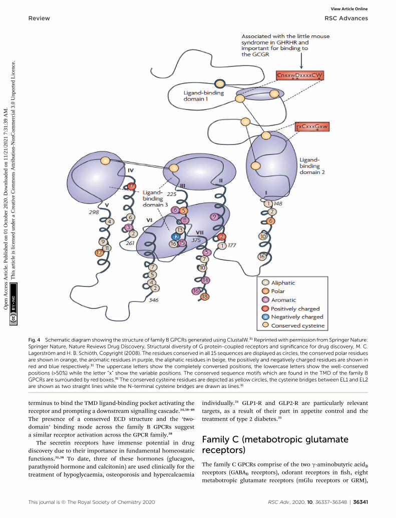
Fig. 4 Schematic diagram showing the structure of family B GPCRs generated using ClustalW.31 Reprintedwith permission from Springer Nature:Springer Nature, Nature Reviews Drug Discovery, Structural diversity of G protein-coupled receptors and significance for drug discovery, M. C.Lagerstrom and H. B. Schioth, Copyright (2008). The residues conserved in all 15 sequences are displayed as circles, the conserved polar residuesare shown in orange, the aromatic residues in purple, the aliphatic residues in beige, the positively and negatively charged residues are shown inred and blue respectively.31 The uppercase letters show the completely conversed positions, the lowercase letters show the well-conservedpositions (>50%) while the letter “x” show the variable positions. The conserved sequence motifs which are found in the TMD of the family BGPCRs are surrounded by red boxes.31 The conserved cysteine residues are depicted as yellow circles, the cysteine bridges between EL1 and EL2are shown as two straight lines while the N-terminal cysteine bridges are drawn as lines.31
Review RSC Advances
Ope
n A
cces
s A
rtic
le. P
ublis
hed
on 0
1 O
ctob
er 2
020.
Dow
nloa
ded
on 1
1/21
/202
1 7:
31:3
9 A
M.
Thi
s ar
ticle
is li
cens
ed u
nder
a C
reat
ive
Com
mon
s A
ttrib
utio
n-N
onC
omm
erci
al 3
.0 U
npor
ted
Lic
ence
.View Article Online
terminus to bind the TMD ligand-binding pocket activating thereceptor and prompting a downstream signalling cascade.34,38–40
The presence of a conserved ECD structure and the ‘two-domain’ binding mode across the family B GPCRs suggesta similar receptor activation across the GPCR family.38
The secretin receptors have immense potential in drugdiscovery due to their importance in fundamental homeostaticfunctions.31,38 To date, three of these hormones (glucagon,parathyroid hormone and calcitonin) are used clinically for thetreatment of hypoglycaemia, osteoporosis and hypercalcaemia
This journal is © The Royal Society of Chemistry 2020
individually.31 GLP1-R and GLP2-R are particularly relevanttargets, as a result of their part in appetite control and thetreatment of type 2 diabetes.31
Family C (metabotropic glutamatereceptors)
The family C GPCRs comprise of the two g-aminobutyric acidBreceptors (GABAB receptors), odorant receptors in sh, eightmetabotropic glutamate receptors (mGlu receptors or GRM),
RSC Adv., 2020, 10, 36337–36348 | 36341
RSC Advances Review
Ope
n A
cces
s A
rtic
le. P
ublis
hed
on 0
1 O
ctob
er 2
020.
Dow
nloa
ded
on 1
1/21
/202
1 7:
31:3
9 A
M.
Thi
s ar
ticle
is li
cens
ed u
nder
a C
reat
ive
Com
mon
s A
ttrib
utio
n-N
onC
omm
erci
al 3
.0 U
npor
ted
Lic
ence
.View Article Online
pheromone receptors, Ca2+-sensing receptors (CaS receptors orCASR), sweet and umami taste receptors (TAS1R1-3), GPCRClass C Group 6 Member A (GPRC6A) and seven orphanreceptors.3,4,31,41 The taste receptors in this GPCR family aretargeted by the taste additives used in the food industry.41 TheCaS, mGlu and GABAB receptors belong to a novel category ofdrug targets that are essential for considering conditions whichaffect the central nervous system and calcium homeostasis.42
Currently, family C GPCRs are targeted by two therapeutic drugsin the market. One is Cinacalcet,41–45 the rst GPCR allostericmodulator to be marketed, which targets the CaS receptor. Theother is Baclofen (now sold under the brand names Lioresal,Liofen, Gablofen, etc.), which is a GABAB agonist used in thetreatment of muscle spasms.41,42,44–46
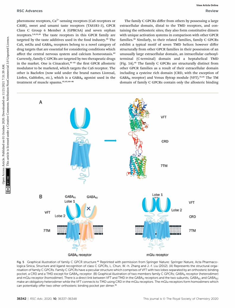
Fig. 5 Graphical illustration of family C GPCR structure.41 Reprinted witlogica Sinica, Structure and ligand recognition of class C GPCRs, L. Chunisation of family C GPCRs. Family C GPCRs have a peculiar structure whipocket, a CRD and a TMD except for GABAB receptor. (B) Graphical illustand mGlu receptor (homodimer). There is a direct link between VFT and Tmake an obligatory heterodimer while the VFT connects to TMD using CRcan potentially offer two other orthosteric binding pocket per dimer.41
36342 | RSC Adv., 2020, 10, 36337–36348
The family C GPCRs differ from others by possessing a largeextracellular domain, distal to the TMD receptors, and con-taining the orthosteric sites; they also form constitutive dimerswith unique activation systems in comparison with other GPCRfamilies.41 Similarly, to their related families, family C GPCRsexhibit a typical motif of seven TMD helices however differstructurally from other GPCR families in their possession of anunusually large extracellular domain, an intracellular carboxyl-terminal (C-terminal) domain and a heptahelical TMD(Fig. 5A).41 The family C GPCRs are structurally distinct fromother GPCR families as a result of their extracellular domainincluding a cysteine rich domain (CRD, with the exception ofGABAB receptor) and Venus ytrap module (VFT).31,41 The TMdomain of family C GPCRs contain only the allosteric binding
h permission from Springer Nature: Springer Nature, Acta Pharmaco-n, W.-h. Zhang and J.-f. Liu (2012). (A) Represents the structural orga-ch comprises of VFT with two lobes separated by an orthosteric bindingration of two members family C GPCRs; GABAB receptor (heterodimer)MD in the GABAB receptors and the two subunits, GABAB1 and GABAB2
D in the mGlu receptors. The mGlu receptors form homodimers which
This journal is © The Royal Society of Chemistry 2020
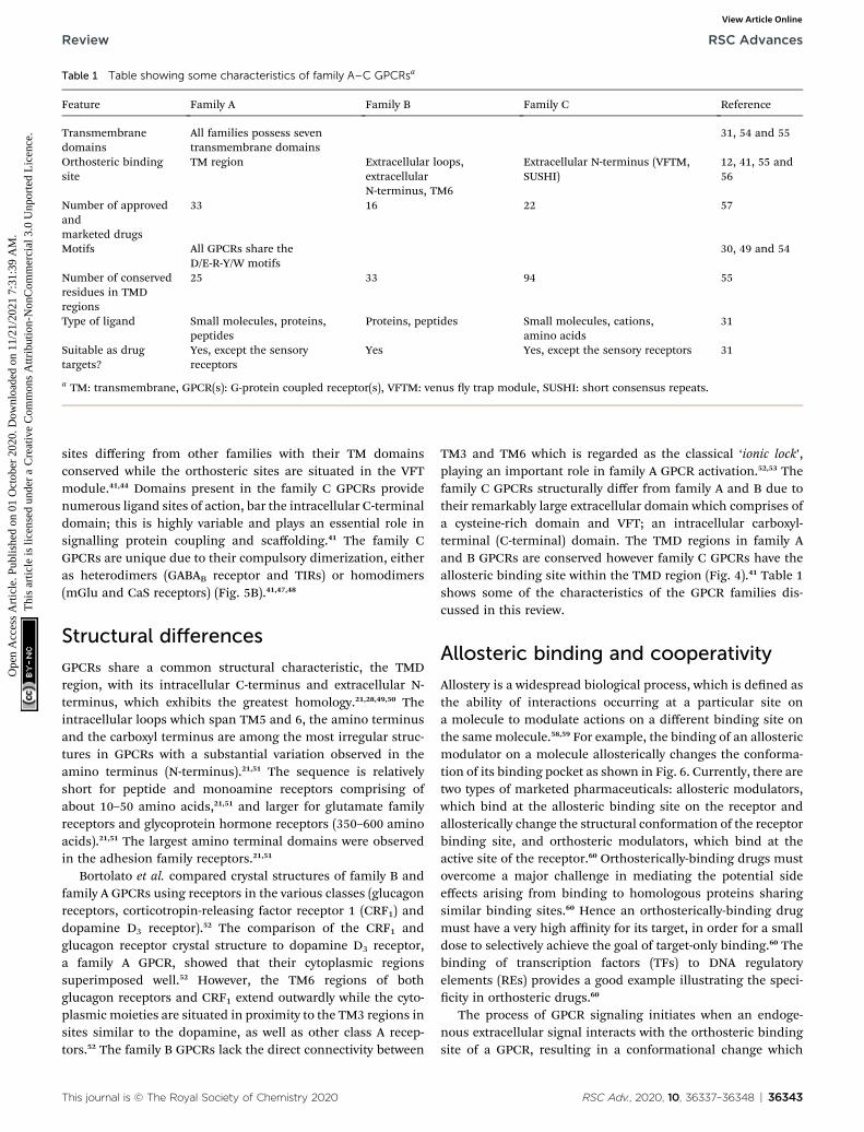
Table 1 Table showing some characteristics of family A–C GPCRsa
Feature Family A Family B Family C Reference
Transmembranedomains
All families possess seventransmembrane domains
31, 54 and 55
Orthosteric bindingsite
TM region Extracellular loops,extracellularN-terminus, TM6
Extracellular N-terminus (VFTM,SUSHI)
12, 41, 55 and56
Number of approvedandmarketed drugs
33 16 22 57
Motifs All GPCRs share theD/E-R-Y/W motifs
30, 49 and 54
Number of conservedresidues in TMDregions
25 33 94 55
Type of ligand Small molecules, proteins,peptides
Proteins, peptides Small molecules, cations,amino acids
31
Suitable as drugtargets?
Yes, except the sensoryreceptors
Yes Yes, except the sensory receptors 31
a TM: transmembrane, GPCR(s): G-protein coupled receptor(s), VFTM: venus y trap module, SUSHI: short consensus repeats.
Review RSC Advances
Ope
n A
cces
s A
rtic
le. P
ublis
hed
on 0
1 O
ctob
er 2
020.
Dow
nloa
ded
on 1
1/21
/202
1 7:
31:3
9 A
M.
Thi
s ar
ticle
is li
cens
ed u
nder
a C
reat
ive
Com
mon
s A
ttrib
utio
n-N
onC
omm
erci
al 3
.0 U
npor
ted
Lic
ence
.View Article Online
sites differing from other families with their TM domainsconserved while the orthosteric sites are situated in the VFTmodule.41,44 Domains present in the family C GPCRs providenumerous ligand sites of action, bar the intracellular C-terminaldomain; this is highly variable and plays an essential role insignalling protein coupling and scaffolding.41 The family CGPCRs are unique due to their compulsory dimerization, eitheras heterodimers (GABAB receptor and TIRs) or homodimers(mGlu and CaS receptors) (Fig. 5B).41,47,48
Structural differences
GPCRs share a common structural characteristic, the TMDregion, with its intracellular C-terminus and extracellular N-terminus, which exhibits the greatest homology.21,28,49,50 Theintracellular loops which span TM5 and 6, the amino terminusand the carboxyl terminus are among the most irregular struc-tures in GPCRs with a substantial variation observed in theamino terminus (N-terminus).21,51 The sequence is relativelyshort for peptide and monoamine receptors comprising ofabout 10–50 amino acids,21,51 and larger for glutamate familyreceptors and glycoprotein hormone receptors (350–600 aminoacids).21,51 The largest amino terminal domains were observedin the adhesion family receptors.21,51
Bortolato et al. compared crystal structures of family B andfamily A GPCRs using receptors in the various classes (glucagonreceptors, corticotropin-releasing factor receptor 1 (CRF1) anddopamine D3 receptor).52 The comparison of the CRF1 andglucagon receptor crystal structure to dopamine D3 receptor,a family A GPCR, showed that their cytoplasmic regionssuperimposed well.52 However, the TM6 regions of bothglucagon receptors and CRF1 extend outwardly while the cyto-plasmic moieties are situated in proximity to the TM3 regions insites similar to the dopamine, as well as other class A recep-tors.52 The family B GPCRs lack the direct connectivity between
This journal is © The Royal Society of Chemistry 2020
TM3 and TM6 which is regarded as the classical ‘ionic lock’,playing an important role in family A GPCR activation.52,53 Thefamily C GPCRs structurally differ from family A and B due totheir remarkably large extracellular domain which comprises ofa cysteine-rich domain and VFT; an intracellular carboxyl-terminal (C-terminal) domain. The TMD regions in family Aand B GPCRs are conserved however family C GPCRs have theallosteric binding site within the TMD region (Fig. 4).41 Table 1shows some of the characteristics of the GPCR families dis-cussed in this review.
Allosteric binding and cooperativity
Allostery is a widespread biological process, which is dened asthe ability of interactions occurring at a particular site ona molecule to modulate actions on a different binding site onthe same molecule.58,59 For example, the binding of an allostericmodulator on a molecule allosterically changes the conforma-tion of its binding pocket as shown in Fig. 6. Currently, there aretwo types of marketed pharmaceuticals: allosteric modulators,which bind at the allosteric binding site on the receptor andallosterically change the structural conformation of the receptorbinding site, and orthosteric modulators, which bind at theactive site of the receptor.60 Orthosterically-binding drugs mustovercome a major challenge in mediating the potential sideeffects arising from binding to homologous proteins sharingsimilar binding sites.60 Hence an orthosterically-binding drugmust have a very high affinity for its target, in order for a smalldose to selectively achieve the goal of target-only binding.60 Thebinding of transcription factors (TFs) to DNA regulatoryelements (REs) provides a good example illustrating the speci-city in orthosteric drugs.60
The process of GPCR signaling initiates when an endoge-nous extracellular signal interacts with the orthosteric bindingsite of a GPCR, resulting in a conformational change which
RSC Adv., 2020, 10, 36337–36348 | 36343
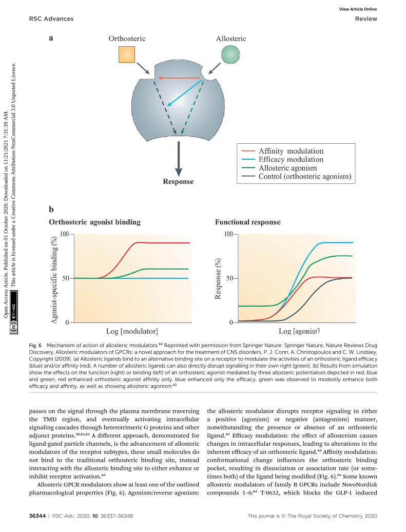
Fig. 6 Mechanism of action of allosteric modulators.63 Reprinted with permission from Springer Nature: Springer Nature, Nature Reviews DrugDiscovery, Allosteric modulators of GPCRs: a novel approach for the treatment of CNS disorders, P. J. Conn, A. Christopoulos and C. W. Lindsley,Copyright (2009). (a) Allosteric ligands bind to an alternative binding site on a receptor to modulate the activities of an orthosteric ligand efficacy(blue) and/or affinity (red). A number of allosteric ligands can also directly disrupt signalling in their own right (green). (b) Results from simulationshow the effects on the function (right) or binding (left) of an orthosteric agonist mediated by three allosteric potentiators depicted in red, blueand green; red enhanced orthosteric agonist affinity only, blue enhanced only the efficacy, green was observed to modestly enhance bothefficacy and affinity, as well as showing allosteric agonism.63
RSC Advances Review
Ope
n A
cces
s A
rtic
le. P
ublis
hed
on 0
1 O
ctob
er 2
020.
Dow
nloa
ded
on 1
1/21
/202
1 7:
31:3
9 A
M.
Thi
s ar
ticle
is li
cens
ed u
nder
a C
reat
ive
Com
mon
s A
ttrib
utio
n-N
onC
omm
erci
al 3
.0 U
npor
ted
Lic
ence
.View Article Online
passes on the signal through the plasma membrane traversingthe TMD region, and eventually activating intracellularsignaling cascades through heterotrimeric G proteins and otheradjunct proteins.58,61,62 A different approach, demonstrated forligand-gated particle channels, is the advancement of allostericmodulators of the receptor subtypes, these small molecules donot bind to the traditional orthosteric binding site, insteadinteracting with the allosteric binding site to either enhance orinhibit receptor activation.63
Allosteric GPCRmodulators show at least one of the outlinedpharmacological properties (Fig. 6). Agonism/reverse agonism:
36344 | RSC Adv., 2020, 10, 36337–36348
the allosteric modulator disrupts receptor signaling in eithera positive (agonism) or negative (antagonism) manner,notwithstanding the presence or absence of an orthostericligand.63 Efficacy modulation: the effect of allosterism causeschanges in intracellular responses, leading to alterations in theinherent efficacy of an orthosteric ligand.63 Affinity modulation:conformational change inuences the orthosteric bindingpocket, resulting in dissociation or association rate (or some-times both) of the ligand being modied (Fig. 6).63 Some knownallosteric modulators of family B GPCRs include NovoNordiskcompounds 1–6:63 T-0632, which blocks the GLP-1 induced
This journal is © The Royal Society of Chemistry 2020

Table 2 Some rules for the definition of dielectric constants in proteins
Denition Value Comments
Polar ¼ 3 large 3 ¼ large Protein sites are always polar near small radiiions.Nonpolar ¼ 3 small
3ðrÞ ¼ 332Q1Q2
rDG
3(r) > 10 oen 3(r) $ 40 The value of 3 is large for charge–chargeinteractions.
1� 1
3B¼ � aDG
166Q2
3B > 10 Proteins can provide as much solvation as waterfor ionised groups with small radii.
3ðrÞ ¼ �332Q1m2 cos q
r2DG
3 $ 4 For functionally important charge–dipoleinteractions, the value of 3 could be as small as4. Such a low value, however, requires relativelyxed dipoles with little energy forreorganisation.
Review RSC Advances
Ope
n A
cces
s A
rtic
le. P
ublis
hed
on 0
1 O
ctob
er 2
020.
Dow
nloa
ded
on 1
1/21
/202
1 7:
31:3
9 A
M.
Thi
s ar
ticle
is li
cens
ed u
nder
a C
reat
ive
Com
mon
s A
ttrib
utio
n-N
onC
omm
erci
al 3
.0 U
npor
ted
Lic
ence
.View Article Online
cAMP production63,64 (GLP 1 receptor); DMP696, which blocksthe CRF-stimulated adenylyl cyclase activity in cell lineexpressing CRF1 receptor;63,65 NBI 27914, which blocks the CRF1receptor;63,66 NBI 35965;63 antarlamin63 (CRF 1 receptor).63
Cooperativity is a thermodynamic term which has varyingmeanings in different biochemical contexts.67,68 It is used toexplain the complex interactions of identical ligands witha receptor at multiple binding sites.67 Cooperativity alsodescribes the thermodynamics of macromolecular conforma-tional transitions, which include nucleic acid helix–coil transi-tions and protein folding.67 Positive cooperativity is dened asthe increase of binding affinity at one site of a receptor whena ligand is bound elsewhere.69 A classic example of positivecooperativity is the binding of oxygen to haemoglobin; thebinding of one oxygen molecule to the ferrous iron of the hememolecule increases the affinity of deoxyhaemoglobin foroxygen.69 Negative cooperativity is observed when 2,3-bisphos-phoglycerate binds to an allosteric binding site of haemoglobinand the affinity for oxygen is reduced.67,69
GPCR signalling via G-proteins
G-proteins consist of several families of varied cellular proteinswhich perform several cellular functions, such as contractilityand angiogenesis, learning and memory.70,71 These proteinsbind to the guanine nucleotides (guanine diphosphate (GDP)and guanine triphosphate (GTP)) and also have inherentGTPase activity.71 They play a principal role in a many cellularprocesses, including protein synthesis and cell development,vesicular transport, and cytoskeleton assembly, in addition tosignal transduction.71 G-proteins are trimers comprising of twofunctional components: a beta-gamma dimer (35 and 8 kDa)which closely relates with the alpha subunit upon binding withGDP, and an alpha subunit (39–52 kDa) which is a catalyst forGTPase activity.72 Human G proteins are classied into twoclasses, namely small (monomeric), and heterotrimeric Gproteins.71,72
GPCRs are the largest superfamily of cell-surface receptorsinvolved in TMD signalling, usually transmitting signals intocells via their response to a range of extracellular stimuli, suchas glycoproteins, polypeptides and ions, and hence regulating
This journal is © The Royal Society of Chemistry 2020
a wide variety of physiological and developmental functions.73
The intracellular-signalling cascades activated by GPCRs havebeen proven to be remarkably complex.73,74 The binding ofa ligand to the GPCR binding site leads to a conformationalchange in the receptor, in turn promoting the binding of theheterotrimeric G proteins, consisting of Ga-GDP and Gbg-subunits, within the intracellular moiety of the receptor.74 Theexchange of GTP for GDP on the Ga-subunit results in thereversible dissociation of the G protein subunits, initiatinga downstream signalling via Ga-GTP and Gbg.73,74
Dielectric constant
The most effective way of correlating the structure and functionof macromolecules is through the examination of its electro-static energies.75 The intermolecular interactions present areaffected by the effective dielectric constant (relative permittivity,3r),76 which differs according to the size and composition of theprotein.77 The accuracy of the method of determination isimportant in understanding various biochemical interactionssuch as protein–ligand and protein–protein interactions, chargeseparation, ion channel selectivity and electron and protontransfer signal transduction and macromolecular assembly;77,78
these interactions are inuenced by the electrostatic potentialof the protein surface.77–79 The dielectric constant of dryproteins ranges from 2.5 to 3.5 obtained from direct measure-ment.78 The theoretical calculation of local dielectric constant oflone proteins based on their amino acid composition yielded anaverage of 2.7.80 The polarity of the residues which make up thestructural motifs within a protein have been shown to affect itsdielectric constant values, these ndings were based oncomputational studies based on continuum electrostatics andmolecular dynamics simulations.77,78
According to Warshel and Aqvist, the value of the dielectricconstant of proteins is dependent on the property used to deneit. They highlighted several possible ways of dening thedielectric constant in proteins, as outlined in Table 2,81 whereQ1 andQ2 are charges on ionisable groups separated by distancer, m is a group dipole moment (in units of electron Angstrom),DG is the electrostatic Gibbs free energy, �a is the effective radius
RSC Adv., 2020, 10, 36337–36348 | 36345
RSC Advances Review
Ope
n A
cces
s A
rtic
le. P
ublis
hed
on 0
1 O
ctob
er 2
020.
Dow
nloa
ded
on 1
1/21
/202
1 7:
31:3
9 A
M.
Thi
s ar
ticle
is li
cens
ed u
nder
a C
reat
ive
Com
mon
s A
ttrib
utio
n-N
onC
omm
erci
al 3
.0 U
npor
ted
Lic
ence
.View Article Online
of charge, and 3B is the effective dielectric constant associatedwith a given interaction.
Li et al. reported that the average dielectric constant insidea protein is relatively low, about 6–7, but this gure reachesabout 20–30 on the surface of the protein.82 The high averagelocal dielectric constant values are oen linked to the chargedresidues while the low values are assigned automatically to theregions comprised of mostly hydrophobic residues.82
According to Wilson et al. solvent effects on mechanisms ofreactions have been established, but its effect on kinetic isotopeeffects (KIEs) are rather well less comprehended.83 A change insolvent can alter the KIE indirectly by changing the transition-state (TS) structure. It can also affect KIE by affecting isotopi-cally sensitive vibrational frequencies directly, notwithstandingthe TS structure or identity of the rate-determining step.83 Wilsonet al. investigated the medium effects on KIE for SN2 methyltransfer using UFF or UAO cavity method within the polarizedcontinuum model (PCM) and a hybrid quantum mechanical/molecular mechanical (QM/MM) method.83 Their ndingsshowed that the majority of variation in the equilibrium isotopeeffects (EIE) occur within the same range of dielectric constants(1# 3# 10) as is considered to occur with enzyme active sites andproteins.83 There is a possibility that any reaction which involvesseparation, neutralisation or charge distribution within anenzyme active site could indicate variations in KIEs, betweena wildtype and mutant form of an enzyme, which originates asa result of changes in the local dielectric response within thediverse protein environment.83 The use of UFF or UAO cavitymethod within the polarized continuum model (PCM) anda hybrid QM/MM method to characterise ligand binding inGPCRs would further assist in understanding the interactionswhich occur in the both the active and inactive states of GPCRs,as well the changes which occur during the transition frominactive state to active state upon ligand activation.
Computational biology techniques inGPCR research
The rst major breakthrough in human GPCR structuralbiology took place in 2007 as the solving of the b2-adrenergicreceptor (b2AR with a diffusible ligand) using a modied lipidiccubic phase (LCP) produce to produce b2AR-TCL crystals whichdiffracted to a resolution of 2.2 A, the structure was furtherrened at a 2.4 A resolution.12 Presently 64 structures of uniqueGPCRs with varying resolutions have been solved using spec-troscopic methods such as uorescence, electron paramagneticresonance (EPR) and nuclear magnetic resonance (NMR) spec-troscopy and structural techniques such as cryogenic electronmicroscopy (cryo-EM), this provides opportunities in employingcomputational biology techniques such as molecular model-ling, and molecular docking in drug discovery research.84,85 Themilestones achieved in GPCR structural studies have providedinsights on the arrangements of the transmembranedomains,1–5,11,12 the location of the orthosteric,12,31,41 allo-steric,12,31,41 bitopic,12 as well as biased ligand binding sites,12
the homo- or hetero-oligomerization of receptors12 and the
36346 | RSC Adv., 2020, 10, 36337–36348
structural rearrangements associated with conformationalchanges upon GPCR activation and inactivation.12 This base ofstructural information on GPCRs is vital for SBDD,12,86 ligand-based drug design (LBDD),12 and integrated models whichcomplement drug discovery efforts.12
In 2012, Sosei Heptares published a detailed account on theuse of A2AR structure in identifying series of agents as potentialantagonists, this became the rst published GPCR SBDDdiscovery.87 In a research carried out by de Graaf et al. usingstructure based virtual screening (SBVS), they identied allo-steric modulators of two family B receptors namely; glucagonreceptor and glucagon-like peptide receptor.88 SBDDapproaches has also lead to the development of new agonists ofthe A3 adenosine receptor (A3AR).89
Conclusion and future prospects
GPCRs are multifaceted proteins which exist in varyingconformations, and that the conformational equilibrium ofthese group of receptors is inuenced both by the bound ligandand the proximity to the related G protein. Their structure ishighly conserved comprising of seven TMD. These receptorspossess different binding domains, namely; allosteric andorthosteric binding domains. The progress in GPCR structuralbiology has substantially accelerated our understanding ofGPCRs as potential drug targets using SBDD and LBDDapproaches. Further computational studies assessing nuclearquantum effects on ligand receptor binding, as well as hybridQM/MM and empirical valence bond theory in the mechanisticstudies of GPCRs would allow for further insight into theinteractions which occur in both the active and inactive states ofGPCRs, as well the changes which occur during the transitionfrom these states upon ligand activation. This review has aimedto provide an accessible and introductory perspective onadvances in GPCR-based drug discovery approaches; manyreviews on the topic highlighted herein are indeed highlydetailed and authoritative but may not provide as accessible anaccount for a less specialised or more general audience in thechemical sciences.
Conflicts of interest
There are no conicts to declare.
References
1 P. M. Dijkman, O. K. Castell, A. D. Goddard, J. C. Munoz-Garcia, C. de Graaf, M. I. Wallace and A. Watts, Nat.Commun., 2018, 9, 1710.
2 W. K. Kroeze, D. J. Sheffler and B. L. Roth, J. Cell Sci., 2003,116, 4867.
3 R. Fredriksson, M. C. Lagerstrom, L.-G. Lundin andH. B. Schioth, Mol. Pharmacol., 2003, 63, 1256.
4 H. B. Schioth and R. Fredriksson, Gen. Comp. Endocrinol.,2005, 142, 94–101.
5 E. Ghosh, P. Kumari, D. Jaiman and A. K. Shukla, Nat. Rev.Mol. Cell Biol., 2015, 16, 69–81.
This journal is © The Royal Society of Chemistry 2020
Review RSC Advances
Ope
n A
cces
s A
rtic
le. P
ublis
hed
on 0
1 O
ctob
er 2
020.
Dow
nloa
ded
on 1
1/21
/202
1 7:
31:3
9 A
M.
Thi
s ar
ticle
is li
cens
ed u
nder
a C
reat
ive
Com
mon
s A
ttrib
utio
n-N
onC
omm
erci
al 3
.0 U
npor
ted
Lic
ence
.View Article Online
6 A. S. Hauser, M. M. Attwood, M. Rask-Andersen,H. B. Schioth and D. E. Gloriam, Nat. Rev. Drug Discovery,2017, 16, 829–842.
7 X.-l. Tang, Y. Wang, D.-l. Li, J. Luo and M.-y. Liu, ActaPharmacol. Sin., 2012, 33, 363–371.
8 H. R. Kim, N. M. Duc and K. Y. Chung, Biomol. Ther., 2018,26, 101–108.
9 K. Sriram and P. A. Insel,Mol. Pharmacol., 2018, 93, 251–258.10 P. A. Insel, K. Sriram, M. W. Gorr, S. Z. Wiley, A. Michkov,
C. Salmeron and A. M. Chinn, Trends Pharmacol. Sci., 2019,40, 378–387.
11 K. A. Jacobson, S. Costanzi and S. Paoletta, Trends Pharmacol.Sci., 2014, 35, 658–663.
12 S. Basith, M. Cui, S. J. Y. Macalino, J. Park, N. A. B. Clavio,S. Kang and S. Choi, Front. Pharmacol., 2018, 9, 128.
13 A. Heifetz, G. F. X. Schertler, R. Seifert, C. G. Tate,P. M. Sexton, V. V. Gurevich, D. Fourmy, V. Cherezov,F. H. Marshall, R. I. Storer, I. Moraes, I. G. Tikhonova,C. S. Tautermann, P. Hunt, T. Ceska, S. Hodgson,M. J. Bodkin, S. Singh, R. J. Law and P. C. Biggin, Naunyn-Schmiedeberg's Arch. Pharmacol., 2015, 388, 883–903.
14 X. Yuan and Y. Xu, Int. J. Mol. Sci., 2018, 19, 2105.15 M. Esguerra, A. Siretskiy, X. Bello, J. Sallander and
H. Gutierrez-de-Teran, Nucleic Acids Res., 2016, 44, W455–W462.
16 V. V. Gurevich and E. V. Gurevich, Int. J. Mol. Sci., 2017, 18,2519.
17 P. S. H. Park, Curr. Med. Chem., 2012, 19, 1146–1154.18 D. Provasi, M. C. Artacho, A. Negri, J. C. Mobarec and
M. Filizola, PLoS Comput. Biol., 2011, 7, e1002193.19 P. Samama, S. Cotecchia, T. Costa and R. J. Leowitz, J. Biol.
Chem., 1993, 268, 4625–4636.20 S. M. de Munnik, M. J. Smit, R. Leurs and H. F. Vischer,
Front. Pharmacol., 2015, 6, 40.21 B. K. Kobilka, Biochim. Biophys. Acta, 2007, 1768, 794–807.22 J. Bockaert and J. Philippe Pin, EMBO J., 1999, 18, 1723–1729.23 M. N. Davies, A. Secker, A. A. Freitas, M. Mendao, J. Timmis
and D. R. Flower, Bioinformatics, 2007, 23, 3113–3118.24 F. Horn, E. Bettler, L. Oliveira, F. Campagne, F. E. Cohen and
G. Vriend, Nucleic Acids Res., 2003, 31, 294–297.25 F. Horn, J. Weare, M. W. Beukers, S. Horsch, A. Bairoch,
W. Chen, Ø. Edvardsen, F. Campagne and G. Vriend,Nucleic Acids Res., 1998, 26, 275–279.
26 G.-M. Hu, T.-L. Mai and C.-M. Chen, Sci. Rep., 2017, 7, 15495.27 G. Pandy-Szekeres, C. Munk, T. M. Tsonkov, S. Mordalski,
K. Harpsøe, A. S. Hauser, A. J. Bojarski and D. E. Gloriam,Nucleic Acids Res., 2018, 46, D440–D446.
28 D. M. Rosenbaum, S. G. F. Rasmussen and B. K. Kobilka,Nature, 2009, 459, 356–363.
29 S. B. Gacasan, D. L. Baker and A. L. Parrill, AIMS Biophys.,2017, 4, 491–527.
30 K. Palczewski, Annu. Rev. Biochem., 2006, 75, 743–767.31 M. C. Lagerstrom and H. B. Schioth, Nat. Rev. Drug Discovery,
2008, 7, 339–357.32 D. C. Teller, T. Okada, C. A. Behnke, K. Palczewski and
R. E. Stenkamp, Biochemistry, 2001, 40, 7761–7772.
This journal is © The Royal Society of Chemistry 2020
33 K. Sasaki, S. Dockerill, D. A. Adamiak, I. J. Tickle andT. Blundell, Nature, 1975, 257, 751–757.
34 C. Parthier, S. Reedtz-Runge, R. Rudolph and M. T. Stubbs,Trends Biochem. Sci., 2009, 34, 303–310.
35 M. Wheatley, D. Wootten, M. T. Conner, J. Simms,R. Kendrick, R. T. Logan, D. R. Poyner and J. Barwell, Br. J.Pharmacol., 2012, 165, 1688–1703.
36 C. R. R. Grace, M. H. Perrin, M. R. DiGruccio, C. L. Miller,J. E. Rivier, W. W. Vale and R. Riek, Proc. Natl. Acad. Sci. U.S. A., 2004, 101, 12836–12841.
37 V. Karageorgos, M. Venihaki, S. Sakellaris, M. Pardalos,G. Kontakis, M.-T. Matsoukas, A. Gravanis, A. Margiorisand G. Liapakis, Hormones, 2018, 17, 45–59.
38 C. de Graaf, G. Song, C. Cao, Q. Zhao, M.-W. Wang, B. Wuand R. C. Stevens, Trends Biochem. Sci., 2017, 42, 946–960.
39 F. Wu, L. Yang, K. Hang, M. Laursen, L. Wu, G. W. Han,Q. Ren, N. K. Roed, G. Lin, M. A. Hanson, H. Jiang,M.-W. Wang, S. Reedtz-Runge, G. Song and R. C. Stevens,Nat. Commun., 2020, 11, 1272.
40 K. Hollenstein, C. de Graaf, A. Bortolato, M.-W. Wang,F. H. Marshall and R. C. Stevens, Trends Pharmacol. Sci.,2014, 35, 12–22.
41 L. Chun, W.-h. Zhang and J.-f. Liu, Acta Pharmacol. Sin.,2012, 33, 312–323.
42 P. Rondard, C. Goudet, J. Kniazeff, J.-P. Pin and L. Prezeau,Neuropharmacology, 2011, 60, 82–92.
43 S. D. Hellyer, S. Albold, T. Wang, A. N. Y. Chen, L. T. May,K. Leach and K. J. Gregory, Mol. Pharmacol., 2018, 93, 504.
44 B.-O. Hans, P. Wellendorph and J. Anders, Curr. DrugTargets, 2007, 8, 169–184.
45 C. S. Tautermann, Bioorg. Med. Chem. Lett., 2014, 24, 4073–4079.
46 B. L. Roth, Nat. Struct. Mol. Biol., 2019, 26, 535–544.47 X. C. Zhang, J. Liu and D. Jiang, Protein Cell, 2014, 5, 492–
495.48 T. C. Møller, D. Moreno-Delgado, J.-P. Pin and J. Kniazeff,
Biophys. Rep., 2017, 3, 57–63.49 D. Zhang, Q. Zhao and B. Wu, Mol. Cells, 2015, 38, 836–842.50 M. Dong, C. Koole, D. Wootten, P. M. Sexton and L. J. Miller,
Br. J. Pharmacol., 2014, 171, 1085–1101.51 M. Orel, E. Padros and J. Manyosa, FEBS Journal, 2012, 279,
2357–2367.52 A. Bortolato, A. S. Dore, K. Hollenstein, B. G. Tehan,
J. S. Mason and F. H. Marshall, Br. J. Pharmacol., 2014,171, 3132–3145.
53 K. J. Culhane, Y. Liu, Y. Cai and E. C. Y. Yan, Front.Pharmacol., 2015, 6, 264.
54 P. Tewatia, N. Agrawal, M. Gaur and S. Sahi, Biochimie, 2014,101, 168–182.
55 B. Trzaskowski, D. Latek, S. Yuan, U. Ghoshdastider,A. Debinski and S. Filipek, Curr. Med. Chem., 2012, 19,1090–1109.
56 W. I. Weis and B. K. Kobilka, Annu. Rev. Biochem., 2018, 87,897–919.
57 D. Wacker, R. C. Stevens and B. L. Roth, Cell, 2017, 170, 414–427.
RSC Adv., 2020, 10, 36337–36348 | 36347
RSC Advances Review
Ope
n A
cces
s A
rtic
le. P
ublis
hed
on 0
1 O
ctob
er 2
020.
Dow
nloa
ded
on 1
1/21
/202
1 7:
31:3
9 A
M.
Thi
s ar
ticle
is li
cens
ed u
nder
a C
reat
ive
Com
mon
s A
ttrib
utio
n-N
onC
omm
erci
al 3
.0 U
npor
ted
Lic
ence
.View Article Online
58 P. R. Gentry, P. M. Sexton and A. Christopoulos, J. Biol.Chem., 2015, 290, 19478–19488.
59 H. N. Motlagh, J. O. Wrabl, J. Li and V. J. Hilser, Nature, 2014,508, 331–339.
60 R. Nussinov and T. Chung-Jung, Curr. Pharm. Des., 2012, 18,1311–1316.
61 N. Tuteja, Plant Signaling Behav., 2009, 4, 942–947.62 C. D. Hanlon and D. J. Andrew, J. Cell Sci., 2015, 128, 3533–
3542.63 P. J. Conn, A. Christopoulos and C. W. Lindsley, Nat. Rev.
Drug Discovery, 2009, 8, 41–54.64 E. C. Tibaduiza, C. Chen and M. Beinborn, J. Biol. Chem.,
2001, 276, 37787–37793.65 Y.-W. Li, L. Fitzgerald, H. Wong, S. Lelas, G. Zhang,
M. D. Lindner, T. Wallace, J. McElroy, N. J. Lodge,P. Gilligan and R. Zaczek, CNS Drug Rev., 2005, 11, 21–52.
66 T. Z. Baram, D. T. Chalmers, C. Chen, Y. Koutsoukos andE. B. De Souza, Brain Res., 1997, 770, 89–95.
67 J. R. Williamson, Nat. Chem. Biol., 2008, 4, 458–465.68 T. Lenaerts, J. Ferkinghoff-Borg, J. Schymkowitz and
F. Rousseau, BMC Syst. Biol., 2009, 3, 9.69 I. G. Denisov and S. G. Sligar, Arch. Biochem. Biophys., 2012,
519, 91–102.70 Y.-J. I. Jong, S. K. Harmon and K. L. O'Malley, Br. J.
Pharmacol., 2018, 175, 4026–4035.71 V. Zachariou, R. S. Duman and E. J. Nestler, in Basic
Neurochemistry, ed. S. T. Brady, G. J. Siegel, R. W. Albersand D. L. Price, Academic Press, New York,8th edn, 2012,pp. 411–422.
72 H. Schulman, in From Molecules to Networks, ed. J. H. Byrne,R. Heidelberger and M. N. Waxham, Academic Press,Boston, 3rd edn, 2014, pp. 119–148.
73 J. Doijen, T. Van Loy, B. Landuyt, W. Luyten, D. Schols andL. Schoofs, Biosens. Bioelectron., 2019, 137, 33–44.
36348 | RSC Adv., 2020, 10, 36337–36348
74 G. J. Augustine, Neuroscience, ed. D. Purves, G. Augustine, D.Fitzpatrick, L. Katz, A.-S. LaMantia, J. McNamara and M.Williams, Sinauer Associates, Sunderland MA, 3rd edn,2004.
75 A. Warshel and A. Papazyan, Curr. Opin. Struct. Biol., 1998, 8,211–217.
76 S. E. Braslavsky, Pure Appl. Chem., 2007, 79, 293–465.77 M. Amin and J. Kupper, ChemistryOpen, 2020, 9, 691–694.78 M. Amin and J. Kupper, 2020, arXiv e-prints,
arXiv:2001.07053.79 C. N. Schutz and A. Warshel, Proteins: Struct., Funct., Bioinf.,
2001, 44, 400–417.80 A. S. Alshami, J. Tang and B. Rasco, Food Bioprocess Technol.,
2017, 10, 1548–1561.81 A. Warshel and J. Aqvist, Annu. Rev. Biophys. Biophys. Chem.,
1991, 20, 267–298.82 L. Li, C. Li, Z. Zhang and E. Alexov, J. Chem. Theory Comput.,
2013, 9, 2126–2136.83 P. B. Wilson, P. J. Weaver, I. R. Greig and I. H. Williams, J.
Phys. Chem. B, 2015, 119, 802–809.84 M. Jaiteh, I. Rodrıguez-Espigares, J. Selent and J. Carlsson,
PLoS Comput. Biol., 2020, 16, e1007680.85 D. Hilger, M. Masureel and B. K. Kobilka, Nat. Struct. Mol.
Biol., 2018, 25, 4–12.86 P. Nakliang, R. Lazim, H. Chang and S. Choi, Biomolecules,
2020, 10, 631.87 M. Congreve, C. de Graaf, N. A. Swain and C. G. Tate, Cell,
2020, 181, 81–91.88 C. de Graaf, C. Rein, D. Piwnica, F. Giordanetto and
D. Rognan, ChemMedChem, 2011, 6, 2159–2169.89 A. Ciancetta and K. A. Jacobson, in Computational Methods
for GPCR Drug Discovery, ed. A. Heifetz, Springer New York,New York, NY, 2018, pp. 45–72.
This journal is © The Royal Society of Chemistry 2020