future directions for resuscitation research. iv. innovative advanced life support pharmacology
TRANSCRIPT

Resuscitation 33 (1996) 163-177
RESUSCITATION
Future directions for resuscitation research. IV. Innovative advanced life support pharmacology’ ,2
Charles Browna, Lars Wiklundb, Gad Bar-Joseph”, Brian Millerd, Nicholas Bircher”, Norman Paradisf, James Menegazzie, Martin von Plantag, George C. Kramer”,
Sven-Erik Gisvold’ “(Moderator), Columbus, OH, USA ‘(Co-moderator), Uppsala, Sweden
“Haifa. Israel ‘Denver, CO, USA
“Pittsburgh, PA, USA ‘New York, NY, USA
gBasel, Switzerland hGalveston, TX, USA ‘Trondheim, Norua)
Received 8 April 1996; revised 6 May 1996: accepted 6 May 1996
Abstract
The topics discussed in this session include a partial review of laboratory and clinical studies examining the effmts of adrenergic agonists on restoration of spontaneous circulation after cardiac arrest, the effects of varying doses of epiaephrine, and the effects of novel vasopressors, buffer agents (NaHCO,, THAM, ‘Carbicarb’) and anti-arrhythmics (lidocaine, bretylium, amiodarone) in refractory ventricular fibrillation. Novel therapeutic approaches include titrating electric countershocks against electrocardiographic power spectra and of preceding the first countershocks with single or multiple drug treatments. These approaches need to be investigated further in controlled animal and patient studies. Epidemiologic data from randomized clinical outcome studies can give clues, but cannot document pharmacologic mechanisms in the dynamically changing events during attempts to achieve restoration of spontaneous circulation from prolonged cardiac arrest. Also, rapid drug administration by the intraosseous route was compared with intratraoheal and intravenous (i.v.) drug administration.
Many studies on the above treatments have yielded conflicting results because of differences between healthy hearts of animals and sick hearts of patients, differences in arrest (no-flow) times and cardiopulmonary resuscitation (CPR) (Iow-flow) times, different pharmacokinetics, different dose/response requirements, and different timing of drug administration during low-flow CPR versus during spontaneous circulation. The need to stabilize normotension and prevent rearrest by titrated novel drug admiais;tration, once spontaneous circulation has been restored, requires research. Most of the above topics require some re-evaluation in clinically realistic animal models and in cardiac arrest patients, especially by titration of old and new drug treatments against variables that can be monitored continuously during resuscitation. Copyright 0 1996 Elsevier Science Ireland Ltd
Keywords: Adrenergic agonists; Anti-arrhythmia agents; Buffers; CarGopulmonary resuscitation; Clinical trials: Defibrillation; Electric countershock; Electrocardiography; Intraosseous medication; Ventricular fibrillation;
Edited by: Peter War, Uwe Ebmeyer and Laurence Katz, Safar Center for Resuscitation Research, University of Pittsburgh, PA, USA. ’ Address ali correspondence to: Peter Safar, M.D., Safar Center for Resuscitation Research, University of Pittsburgh. 3434 Fifth Avenue,
Pittsburgh, PA 15260, USA. Tel.: + 412 6246735; fax: + 412 6240943. ’ From a discussion at the International Resuscitation Research Conference of May 1994 at the University of Pittsburgh. For introduction and
other topics of this conference, see the journal Critical Care Medicine, supplement of February 1996.
0300-9572/96/$15.00 Copyright 0 1996 Elsevier Science Ireland Ltd. All rights reserved PrI SO3OO-9572~96)01017-9

164 C. Brown et al. /Resuscitation 33 (1996) 163-177
1. Perfusion pressures and blood flows
1.1. Brown
Crile and Dolley [l] and Redding and Pearson [2-51 demonstrated that epinephrine helps to maintain coro- nary and cerebral perfusion pressure during cardiopul- monary resuscitation (CPR). Subsequent investigators have tried to determine the minimal perfusion pressure and flow that is required during CPR to meet the metabolic demands of the heart and brain until the underlying cause(s) of the cardiac arrest can be resolved [6- 131. Data from these studies suggest that myocardial blood flow of at least 18 ml/min per 100 g, and cerebral blood flow (CBF) of at least 10 ml/min per 100 g, are required to meet these metabolic demands during ven- tricular fibrillation (VF). Myocardial blood flow and CBF in animal models of cardiac arrest and coronary perfusion pressure (CoPP) in humans are usually below these minimum levels during standard external CPR, especially after prolonged CPR and without the use of vasopressor agents [10,14- 161. An important, unre- solved issue is how to improve vital organ perfusion during external CPR.
1.2. Editor @afar)
A minimal CoPP of about 30 mmHg seems to be required for restoration of spontaneous circulation (ROSC). A brief period of hypertension following ROSC is beneficial to help overcome the cerebral no- reflow phenomenon [17,18]. However, the optimal CoPP and cerebral perfusion pressure (CPP) (mean arterial pressure [MAP] minus intracranial pressure [ICP]) during resuscitation remain to be determined.
1.3. Brown
Organ blood flow is determined by the driving pres- sure minus the back pressure divided by the vascular resistance of that organ. For myocardial blood flow, this is most commonly represented by the CoPP (i.e. aortic diastolic pressure minus the right atria1 diastolic pressure) divided by the myocardial vascular resistance [19]. Following prolonged cardiac arrest and resuscita- tion, aortic diastolic pressure becomes the main driving force for myocardial perfusion. Several studies in ani- mals and humans have shown that right atria1 ‘thoracic diastolic’ pressure is low during external CPR [10,14]. In addition, vascular resistance is probably quite low as well. This may be in part due to the profound vasodila- tion that occurs secondary to generation of tissue acidosis. When right atria1 pressure and vascular resis- tance are low, aortic diastolic pressure becomes the main driving force (and limiting factor) for myocardial perfusion during external CPR. Because aortic diastolic
pressure is approximately equal to the volume of fluid in the aorta divided by its capacitance, these two com- ponents of the equation can be examined as a means to increase the aortic diastolic pressure, and thus CoPP, during CPR.
Several studies [10,20,21] have shown that volume infusions not only increase aortic diastolic pressure, but also increase right atria1 pressure as well. These studies have shown, for the most part, no significant improve- ment in myocardial perfusion after volume perfusion in this setting. Another approach to improve aortic dias- tolic pressure has been to decrease vascular capacitance or increase peripheral vascular resistance. This has been the rationale for the use of a-adrenergic agonists during resuscitation. Through their a-agonist effects, they pro- mote peripheral vasoconstriction, and thus increase pe- ripheral vascular resistance, decrease arterial capacitance, and enhance aortic diastolic pressure and myocardial perfusion [22].
2. Adrenergic agonists
2.1. Brown
The overall results of our studies in a swine model of cardiac arrest and resuscitation (Appendix A, 1A) sug- gest that after a prolonged duration of no-flow (car- diopulmonary arrest), standard external CPR, followed by 0.02 mg/kg of epinephrine i.v., improves brain and heart perfusion minimally. On the other hand, epinephrine, in doses of 0.20 mg/kg iv., significantly increases perfusion to the myocardium, cerebral cortex and brainstem. Similar improvements in blood flow occur with norepinephrine in doses ranging from 0.12 mg/kg to 0.16 mg/kg [23]. In addition, very high doses of phenylephrine (10 mg/kg) increase myocardial blood flow to levels seen with epinephrine and nore- pinephrine, but are not as effective in promoting CBF. Methoxamine, in doses up to 10 mg/kg, is not as effective as epinephrine or norepinephrine [23]. In these studies and others [24], all drugs were given by the peripheral i.v. route. It is interesting to explore why some adrenergic agonists, such as epinephrine and norepinephrine, improve perfusion to the myocardium and brain during CPR, while other drugs, such as phenylephrine and methoxamine, are less effective.
This difference in a-adrenergic effect may be due to the differences in the ai- versus azagonist properties of these adrenergic agonists. The site of action of these drugs is the peripheral vasculature, which may help to explain differences in the effects of these agents. In the peripheral vascular wall, there are at least two adrener- gic receptors: the al-receptor located near the outer wall, and the a,-receptor which is located closer to the vascular lumen [23]. Stimulation of either c(,- or a,-re-

C. Brow et al. / Resuscitation 33 (I 996) 163- I77 I65
ceptors should cause peripheral vasoconstriction and an increase in aortic diastolic pressure. While n,-re- ceptors respond primarily to norepinephrine released from adrenergic nerve terminals, a,-receptors respond primarily to circulating catecholamines released from the adrenal medulla. Under conditions of extreme is- chemic hypoxia and acidosis, there may be a desensi- tization or down-regulation of x,-receptors. Post- synaptic r,-receptors may play a more important role in regulating vasomotor tone in the setting of pro- longed cardiac arrest. Methoxamine and phenyl- ephrine may be less effective in this setting because they exert primarily a,-receptor agonism. One must also note that methoxamine has some beta-blocking properties. In addition, it has been suggested that a fraction of the phenylephrine administered may be converted to epinephrine, accounting for its effects during CPR. Epinephrine and norepinephrine have both a,- and cr,-agonist properties. Epinephrine also has p-1 and P-2 agonist effects.
Theoretically, one could review the available clini- cal literature in which some of these adrenergic ago- nist agents have been compared (i.e. studies comparing ‘pure’ a,-agonists versus agents that con- tain both a,- and a,-agonist properties [25,26]) to ex- amine this hypothesis. Although these studies are difficult to interpret because of variable definitions for the outcome measures, ROSC rates appear to be bet- ter with the use of epinephrine, a mixed CI,- and CI~- agonist agent, compared with methoxamine [25]. This supports the notion that a2 agonism may be more important than x, activity, clinically.
The role of /I agonism is less well defined. Lindner 1371 compared the efficacy of I-mg doses of epinephrine versus a 1-mg dose of norepinephrine during cardiac arrest and external CPR on the ROSC and hospital discharge rate. In this study, there was a more than 2-fold increase in the ROSC rate in pa- tients who received norepinephrine. These results sug- gest that the pz effects (vasodilation) of epinephrine may be counterproductive.
Optimal dosing oj’ x-adrenergic agonists has not been established for cardiac arrest and resuscitation. Several studies in animals have shown that increasing doses of epinephrine improve the aortic diastolic pres- sure along with myocardial perfusion and resuscita- tion rates [28-301. Two human studies have shown similar improvements in arterial diastolic pressure and CoPP with increasing doses of epinephrine [31,32]. Recent retrospective uncontrolled case series in chil- dren suggest that after failure of two standard doses of epinephrine, higher doses of epinephrine may im- prove short-term resuscitation and hospital discharge rates [33]. More recent prospective controlled clinical trials in adults with out-of-hospital and in-hospital cardiac arrest, although showing an improvement in
ROSC rates [34-361, found no improvement in hospi- tal discharge rates with higher doses of epinephrine [34-381. Several studies suggest that epinephrine’s detrimental effect may stem from its /Y,-agonist prop- erties, which increase myocardial oxygen consumption and may worsen myocardial ischemia [39,40]. This appears to be supported in at least one large-scale clinical study which found increased rates of complex ventricular arrhythmias and rearrest in patients treated with high doses of epinephrine compared with those treated with standard doses of epinephrine [38]. Other studies have shown that epinephrine may cause ventilation/perfusion defects in rats [41] as well as myocardial necrosis [42,43]. Therefore, there may be many reasons why large doses of epinephrine and norepinephrine fail to improve survival rates.
Research is underway to develop new compounds that have specific X- and B-agonist characteristics. One area of investigation concerns halogenated cate- cholamines. Selective halogenation of the catechol ring has been suggested as a means to separate out the a,-. x2- and p-agonist effects of catecholamines. A new class of compounds, imidazaiines. have also been shown to act as peripheral vasoconstrictors in the setting of cardiac arrest 1441. Another promising vasopressor agent is angiotensin [45]. More pre-clini- cal studies delineating the x and ,@ effects of these compounds, their comparative efficacy and most ef- fective mode of administration, are needed.
Are casopressors needed at ail? A recent abstract reported on a study comparing no epinephrine to standard and high doses of epinephrine in cardiac arrest [46]. Although key confounding variables were not reported, it is interesting to note that the authors found no significant difference in the proportion of patients who achieved ROSC or hospital discharge among the three groups. This study raises questions about the use of vasopressors at all. and suggests that other factors, such as ‘metabolic’ issues. may have a significant role in resuscitation from cardiac arrest.
Endogenous Ienels of catecholamines attd metabolites. Plasma levels of epinephrine and norepinephrine dur- ing cardiac arrest and CPR may reach up to approxi- mately 50 times baseline values [47-Z]. If this is the case, why are these high endogenous catecholamine levels not effective in augmenting perfusion pressures during cardiac arrest and CPR? While the answer to this question is unknown, it supports the concept that endogenous metabolites may suppress or inhibit the effect of catecholamines during profound ischemia.
One such metabolite that has been proposed is adenosine. It is known that adenosine can inhibit the vasopressor effect of catecholamines [53,54]. The ef- fects of adenosine can be reversed by giving specific adenosine antagonists. A recent report examined the

166 C. Brown et al. 1 Resuscitation 33 (19%) 163-177
non-specific adenosine antagonist aminophylline dur- ing refractory bradyasystolic cardiac arrest. This case series suggests potential beneficial effects of aminophylline [55].
Route of drug administration. Intraarterial epin- ephrine [56-601 produces excellent hemodynamic re- sponses during CPR. It is much less effective when given by the peripheral i.v. route [23]. This raises the question of whether the catecholamines are metabo- lized by catechol-o-methyl transferase as they circu- late through the lungs following peripheral i.v. administration.
Finally, the area of post-resuscitation pharmacology has not been explored adequately. Specifically, how should we treat the patient who is resuscitated for prolonged cardiac arrest and has profound hypoten- sion? Few studies have addressed this issue.
2.2. Bar-Joseph
The American Heart Association [24,61] recom- mends administering epinephrine every 3-5 min. What is the scientific basis for this recommenda- tion? Is it based on the fact that the half-life of epinephrine in a normal circulation is only l-2 min? In our dog model of VF 10 min and external CPR, we used 0.1 mg/kg epinephrine repeated every 5 min and found no substantial increase in blood pressure with additional doses, after a marked re- sponse to the initial epinephrine dose [62]. I would like to ask whether the recommendation to repeat epinephrine every 3-5 min during CPR is based on any hard data. I believe that more research is needed to determine the ideal interval between epinephrine doses during CPR, especially when higher doses are used.
2.3. Miller
We have data on catecholamine support during the post-resuscitation period from a study of magne- sium sulfate given i.v. in patients with refractory car- diac arrest [63]. Some patients regained ROSC when they received MgSO,, but after ROSC they rapidly became hypotensive (systolic arterial pressure 45 mmHg) despite dopamine administration. When do- pamine was replaced with norepinephrine, the resusci- tation rate increased by 16%. Animal studies [64] have shown that the effect of magnesium to decrease aortic diastolic, aortic systolic, and coronary perfu- sion pressures during CPR lasts about 20 min. Simi- lar effects were observed in our clinical trials. Therefore, in future studies magnesium should be combined with a potent vasopressor, such as nore- pinephrine.
3. Anti-arrhythmic agents
3.1. Brown
Another class of compounds commonly used in cardiac arrest and resuscitation are anti-arrhythmic agents, including lidocaine and bretylium (Appendix A, 1A). One issue of discussion is the rationale be- hind the use of these drugs during VF cardiac arrest. One explanation that has been offered is that the drugs given during VF should make defibrillation eas- ier and avoid refibrillation. In part, to understand this concept, an understanding of how electrical coun- tershock may terminate VF is needed. While the definitive mechanism on how electrical countershock reverses VF has not been elucidated, it is interesting to hypothesize that electrical countershock, in part, converts VF by depolarizing the myocardial cells, thereby ‘opening up’ sodium channels 1651. Theoreti- cally, following electrical countershock, intracellular sodium concentration will increase, thus allowing spontaneous myocardial depolarization to occur. What we hope to achieve clinically through electrical countershock is complete myocardial depolarization followed by recovery of a single dominant pacemaker, thus restoring a normal heart rhythm. In this setting, what effect do agents like lidocaine and bretylium have on this process? Although lidocaine has many electrophysiological properties, one of its most promi- nent effects is that it is a sodium-channel blocker, which should make it more difficult for sodium to enter the myocardial cell during electrical counter- shock. On the other hand, bretylium, which increases the action potential duration, prevents potassium efflux from the myocardial cell. By preventing potas- sium from leaving the cell, the myocardium should require less energy for defibrillation.
The majority of animal studies to date have shown that lidocaine increases the defibrillation threshold and, as lidocaine concentrations increase, it becomes increasingly more difficult to successfully defibrillate [66-681. Although it is not known exactly how cur- rent i.v. dosing relates to myocardial tissue levels of lidocaine in this setting, it is possible that the admin- istration of lidocaine during cardiac arrest may pro- duce toxic levels [69]. It should also be noted that this explanation for the effect of lidocaine on the defibrillation threshold is not accepted universally. The interaction of anesthetic agents with the anti- arrhythmics has been suggested as an alternative ex- planation for the changes seen in defibrillation threshold [70]. Given these limitations, it is unclear why these anti-arrhythmics are used during CPR. In the only significant pre-clinical study, Redding [3], us- ing a canine model of VF cardiac arrest and resusci- tation, found no difference in outcome after

epinephrine alone versus epinephrine plus lidocaine. On the other hand, although bretylium seems to lower the energy requirements for defibrillation [71-731, an inter- esting factor that has been overlooked with bretylium in animal models of cardiac arrest is the high incidence of electromechanical dissociation and hypotension fol- lowing its use [74]. The Brain Resuscitation Clinical Trial group has also shown that hypotension after ROSC clearly correlates with poor neurological out- come [75]. Thus, the hypotensive effects of bretylium may overshadow its potential beneficial effect on the defibrillation threshold.
Although animal data suggest that lidocaine may make it more difficult to defibrillate, bretylium, which lowers the defibrillation threshold, may lead to post-re- suscitation hypotension. Limited clinical studies com- paring lidocaine and bretylium in patients show no difference in outcome between these two agents 176,771. In addition, no credible prospective trials have been conducted to date comparing these anti-arrhythmic agents to placebo. There is no proof of efficacy of these drugs given during cardiac arrest and CPR. Clearly. future studies are needed to delineate the role of these agents before and after conversion of VF.
3.2. Puradis
In light of the animal data, could you justify a prospective human study on these anti-arrhythmics? Could you go to a committee and say this drug should be given in a systematic manner knowing that it raises the electric energy required for defibrillation?
Weaver et al. [78] examined the effects of epine- phrine, lidocaine and bicarbonate in VF cardiac arrest. The results of this clinical study suggest an increased rate of ROSC with sodium bicarbonate alone.
3.4. Bur-Joseph
What is the inlluence of NaHCO, on sodium chan- nels and defibrillation threshold? Paradis says that some people feel that it is beneficial. Are resuscitation rates decreasing now that less NaHCO, is being admin- istered?
Is there experience with amiodarone as a potential anti-arrhythmic drug during cardiac arrest?
Amiodarone is a potassium-channel blocker like bretylium and, therefore, lowers the defibrillation threshold. There are only case reports of its use during CPR [79]. Patients who received amiodarone after
ROSC, when they rearrested, had apparently good results. Amiodarone also has potential peripheral va- sodilating effects that may counteract the vasoconstric- tive effect of epinephrine. Clearly, further study with amiodarone in refractory and recurrent VF is needed.
3.6. Gisvold
In our discussions, we should differentiate more clearly between the use of lidocaine during arrest and CPR versus its use after ROSC. It has been observed clinically that hypertrophic myocardium, which is difficult to defibrillate, is easier to convert to a supraventricular rhythm after administration of lidocaine. Experience has been similar during open heart surgery. Procaine may also be effective in this situation [80]. It would seem worthwhile also to test procaine in the post-arrest, post-ROSC setting.
3.7. Brown
Patients on cardiopulmonary bypass have continued coronary perfusion. These patients may more closely represent cardiac arrest patients following ROSC. If one gives judicious doses of norepinephrine or epinephrine after cardiac arrest to maintain arterial pressure, and NaHCO, and anti-arrhythmics to prevent rearrest, one is dealing with a situation quite different from the one we discussed before, namely cardiac arrest patients with no-flow or low-flow.
3.8. Wikhnd
We need more research on drug distribution during CPR.
4. VF signal analysis
4.1. Meneguzzi
I want to draw attention to a study from Milwaukee [81] concerning 1497 patients of witnessed arrest, pre- senting with VF. who first received the three routine countershocks recommended by the American Heart Association advanced cardiac life support (ACLS) al- gorithm [61]. After the first countershock, 12% had at least transient ROSC, while 34% achieved pulseless electric activity or asystole. After the second counter- shock, only an additional 4% achieved ROSC, while 16% more developed asystole. After the third counter- shock, the total ROSC rate had increased to 18% while 58% were in pulseless electric activity or asystole. One must question the rationale for routine administration of three shocks at first. Not all VF situations are the same.

168 C. Brown et al. / Resuscitation 33 (1996) 163-177
Because electric therapy is not innocuous, empiric shocking may not be as rational as shocking according to ECG power spectrum analysis [82-841. This area appears promising for research in the near future. The analogy of immediate countershock for all cases found in VF or pulseless VT is like trying to jumpstart a car that has no gasoline.
Our thinking is to first prime the myocardium, brain and other vital organs for countershock and reperfu- sion, using combination pharmacotherapy [85-871. We are exploring, in a pig model, the use of five drugs (epinephrine 0.2 mg/kg, aminosteroid U74389G 3 mg/ kg, lidocaine 1 mg/kg, bretylium 5 mg/kg, and propra- nolo1 1 mg/kg) given together i.v. at the start of CPR steps ABC, while delaying countershock by 2 min until these drugs are on board. We hypothesized that combi- nation pharmacotherapy and delayed countershock would produce higher rates of ROSC and better l-h survival compared to standard CPR-ACLS. After elec- trically induced VF of 8 min no-flow (simulating a clinical scenario of 1 min for collapse recognition, 1 min for dispatch, and 6 min for the mobile ICU to arrive), we gave CPR-basic life support (BLS) for 1 min. Then, group 1 received delayed countershocks 2 min after the entire drug combination; group 2 received only high- dose epinephrine; group 3 received the two anti-arrhyth- mics only; group 4 received propranolol only; group 5 received the aminosteroid only; and group 6 received delayed countershocks without any drugs. After the first countershock (in all at 11 min of VF), ROSC was achieved in 8/8 pigs in group 1, 7/8 pigs in group 2, 318 pigs in group 3, 318 pigs in group 4, 5/8 pigs in group 5 and 4/X pigs in group 6. l-h survival was achieved by 8/S in group 1, 518 in group 2, 218 in group 3, 218 in group 4, 318 in group 5 and 218 in group 6. Standard ACLS achieved ROSC in 3/8 and l-h survival in only l/S. We conclude that a drug first approach may be superior following prolonged cardiac arrest or VF. High-dose epinephrine and delayed countershock was found to have certain benefit in dogs [88]. In our study, a combination of drugs and delay in countershock produced superior ROSC rates and l-h survival.
4:2. Editor (Safar)
Certain dog models since 1960 have routinely in- cluded reperfusion with brief CPR plus epinephrine first, to achieve a vigorous VF ECG signal and an increase in artificial diastolic arterial pressure before applying countershocks; this seems to increase the chance for prompt ROSC [18,58].
4.3. Brown
A number of investigators have hypothesized that delaying countershock may be beneficial following a
prolonged cardiopulmonary arrest [89-911. In this clin- ical setting, reversing myocardial ischemia prior to countershock enhances resuscitation rates [89-911 and may avoid countershock-induced myocardial injury PO1 .
Our research has focused on titrating therapy based on the duration of ischemia [83]. In these studies, we collected the ECG signal during VF cardiac arrest in swine. The ECG signal was analyzed using fast Fourier transform analysis following digitization of the analog VF ECG signal [83]. The resulting power spectrum was used to calculate single-valued parameters of VF ampli- tude and frequency. These preliminary studies demon- strated that certain frequency parameters that can be derived from the power spectrum follow a repeatable and predictable time course during VF cardiac arrest. One such parameter, median frequency, can be used to estimate the duration of VF [83]. We believed that titrating therapy based on the duration of ischemia might be possible.
From this initial study, we extended the technique as a non-invasive tool to help guide therapy during CPR. A non-invasive monitor was developed for use in our laboratory, which tracked median frequency in real time during experimental CPR studies. Our initial experience with this monitor suggested that not only did median frequency follow a repeatable pattern following the onset of VF, but that it responded in a characteristic fashion to therapeutic interventions. When blood flow to the myocardium increased following a pharmacolog ical intervention, median frequency increased as well [92]. In this initial study, there was a reasonable correla- tion between myocardial blood flow and median fre- quency [92]. We then examined the relationship between median frequency and the ability to successfully coun- tershock VF into a perfusing rhythm. We found that a median frequency 2 9.14 Hz in swine had a sensitivity of 100% in predicting successful countershock with ROSC, and a median frequency below this value had a specificity of 92% in predicting unsuccessful counter- shocks. This form of non-invasive real-time ECG analy- sis during VF may provide a therapeutic modality for titrating therapy. Recent clinical studies suggest that this methodology may be applicable in the clinical setting of VF cardiac arrest [93].
4.4. von Planta
A high VF amplitude is known to predict successful defibrillation [94]. VF amplitude may be related to myocardial perfusion during CPR [95]. Methoxamine increases coronary perfusion pressure and survival after cardiac arrest in rats (Appendix A, 3A). Therefore, we proposed that methoxamine should increase VF ampli- tude during CPR. Surprisingly, in a rat model, CPR increased VF amplitude, but methoxamine had no ef-
C. Brown et al. / Resuscitation 33 (1996) 163 I77 169
feet, thus we were unable to use VF amplitude to monitor a response to therapy. These VF parameters should be examined vis-a-vis end-tidal CO,, which de- creases after epinephrine administration during CPR as total flow decreases, while perfusion pressures through heart and brain increase [96].
3.5. Brown
When reviewing work on ECG signal analysis during cardiac arrest and resuscitation, it is important to dis- tinguish between the native or original ECG signal and those ECG signals that may be corrupted secondary to interference. One such source of interference is the ‘noise’ generated by external CPR. Clearly, this CPR noise is one of increased amplitude and usually of low frequency (which is dependent on CPR rate). In most clinical cases that we have reviewed, the amplitude of the noise generated from CPR is much greater than the amplitude normally seen during VF. Therefore, if one is to make the determination that changes in myocardial perfusion correlate with changes in VF amplitude, one must be assured that the ECG signal is free from this noise, because CPR interference can contribute to a large part of the amplitude of the VF signal. On the other hand, CPR interference tends to shift the power spectrum of the VF signal to the left (‘downward’ bias), because most VF appears to occur at a dominant frequency of approximately 3-7 Hz. Therefore, to ob- tain optimal signals, CPR should be terminated briefly just prior to acquisition of the ECG signal. We have found no predictive value of the VF amplitude mea- surement in our clinical studies of the VF waveform for determining the success or failure of countershock ther- apy t931.
While this technique holds promise for guiding ther- apy during cardiac arrest and resuscitation, the re- suscitation community must come to some agreement on nomenclature and standardizing techniques, i.e. lead placement. in the acquisition and analysis of this data.
5. Buffer agents
5.1. Wiklund
Total blood flow generated by external CPR during cardiac arrest is approximately 5- 10% of normal [97]; organ blood flows generated are too low to guarantee the distribution of any drug to target organs. New and old methods should be re-examined to find the best method to promote blood flow during cardiac arrest (Appendix A, 4A). The old method of open-chest CPR generates at least 20% of normal blood flow [97]; its utility should be reconsidered.
When brain pH is monitored by rmclear magnetic resonance spectroscopy during cardiac arrest and exter- nal CPR, the decrease in pH can be to such low levels that it might cause coagulation within S-6 min. There is reason to believe that tissue pH is a crucial factor influencing recovery of brain and heart from cardiac arrest. When the heart stops beating in VF, there is rapid production of hydrogen ions, so that in myocar- dial venous blood sampks, the pH decreases from base line to 6.76 in about 10 min [98]. Tissue acidosis develops rapidly at the same time. During VF cardiac arrest and immediate external CPR, one sees signs of acidosis in peripheral blood by about 10 min [99]. Long before that there is tissue acidosis, at least in the heart and brain [ 1001.
Administration of NaHCO, during CPR increases arterial P,,, transiently. Because bicarbonate transport is limited a&oss cellular membranes, a transient wor- sening of acidemia is created. The opposite happens when we introduce Tris buffer (THAM), which binds hydrogen ions to the buffer and lowers CO,. In fact, CO, can be lowered so much that it can stop sponta- neous breathing. In isolated rat myocardial cells, there is an immediate decrease in pH when sodium bicarbon- ate is introduced into the system. The decrease in pH is dependent on the dose of bicarbonate. Therefore, the dose of sodium bicarbonate should be as small as possible during CPR to avoid increasing acidosis tran- siently.
Other buffers like THAM and Carbicarb theoreti- cally might be more beneficial during cardiac arrest since they do not induce this paradoxical (although transient) acidosis in the isolated cell system. MyOCdr-
dial contractility is reduced with use of bicarbonate [loll and improved by THAM [102]. A positive in- otropic effect of THAM is present even in the ischemic myocardium. Therefore, there are potentially beneficial effects of buffer therapy during cardiac arrest.
5.2. Bar-Joseph
I would like to critically review the animal models used in the experiments that have stirred up the bicar- bonate controversy and to challenge its basic hypothe- sis. The hypothesis which was brought about by Dr Weil’s group states that during the very low-flow state of CPR, the limiting factor in CO, elimination is tissue perfusion rather than alveolar venriIation. When sodium bicarbonate is administered, CO, will be pro- duced and, as it cannot be effectively eliminated, it accumulates in the tissues and penetrates the cells. This might aggravate intracehular ‘respiratory’ acidosis and therefore bicarbonate administration might worsen CPR outcome [ 103,104].
Let us review the experimental evidence of this hy- pothesis. The first issue is whether low tissue perfusion

170 C. Brown et al. 1 Resuscitation 33 (1996) 163-177
limits CO, elimination during CPR. The answer is probably yes, depending on the magnitude of perfusion reduction. In many animal models no epinephrine is used, the tissue perfusion pressures are extremely low, and as a result some increase in venous CO, is observed [101,103,105-1071. The veno-arterial PcO, gradient fur- ther increases due to arterial hypocarbia. Restoration of tissue perfusion is one of the mainstays of the control of acid-base balance during CPR, and failure to use epinephrine is an obvious deviation from AHA guidelines [6 11.
The second question is whether the administration of bicarbonate really increases venous CO, during CPR. The clinical reports are hard to interpret because they were not controlled [104,108]. We do not know when bicarbonate was given in the course of CPR. The amount of bicarbonate and timing of blood sampling were also not controlled. Results of animal studies depend heavily on the model used, including the dura- tion of cardiac arrest, the timing and dosage of bicar- bonate administration, and the timing of blood sampling. In a VF dog model using 0.1 mg/kg epinephrine during CPR, we have observed only a moderate increase in PVC0 after administration of I mmol/kg of sodium bicarbonate. PVC0 returned to pre-bicarbonate levels 5 min later [109f. If blood is sampled only 2 min after bicarbonate administration [101,105,106], the transitory nature of this increase in P vcoz can be missed.
The third issue is the hypothesized worsening of intracellular acidosis following bicarbonate administra- tion during CPR. This hypothesis is supposedly sup- ported by a single (often cited) in vivo study by Berenyi [llO]. The experimental protocol in dogs used in this study bears very little relevance to the clinical scenario. VF lasted only 4 min, by the end of which arterial pH was 7.37 and HCO, was 22 mmol/l. Bicarbonate ad- ministration was started immediately upon initiation of CPR as a continuous infusion of 0.5 mmol/kg per min (i.e. 5 mmol/kg per 10 min). Ventilation was main- tained at baseline level. After 20 min of CPR and administration of this massive bicarbonate infusion, arterial P,, was 64 mmHg, arterial pH was 7.8 1 and arterial HC& was 120 mmol/l! Cerebrospinal fluid- reflecting intracellular acid-base status-had a Pco2 of 81 mmHg and a pH of 7.24. I believe this model examined bicarbonate overdosing rather than its therapeutic use in CPR. No other animal study, to the best of my knowledge, has shown a decrease in intracellular pH related to bicarbonate administration [105,1~7,111,112].
The final and most significant issue is whether bicar- bonate administration has an adverse effect on the outcome of CPR. No controlled clinical trials have been performed. In retrospective, uncontrolled pub- lished clinical studies, the amount of bicarbonate ad-
ministered usually reflects the duration of CPR so that bicarbonate administration becomes an epiphenomenon of prolonged arrest and the CPR time interval [3,113- 1171. Animal experimental results again depend largely on the model, the CPR protocol, and the outcome parameters. Some have shown a clearcut benefit on outcome [3,109,116-l 191, whereas others have failed to show any benefit from bicarbonate administration [101,106,107,120,121]. A single study [105] found a lower ROSC rate in animals receiving bicarbonate, compared with controls. In our study comparing bicar- bonate, Carbicarb, THAM and NaCl in a VF lo-min dog model [109], the dogs that received bicarbonate had the highest rate of ROSC, and ROSC was achieved twice as fast, compared with controls.
In summary, I believe that the bicarbonate issue in CPR is not yet resolved. It is true that low tissue perfusion may limit CO, elimination and that PVC02 may transiently increase after bicarbonate administra- tion. However, venous and extracellular pH always increase. There is no evidence that intracellular acidosis worsens after bicarbonate administration during CPR. Several studies demonstrate a beneficial effect on out- come and I do not think there are any solid experimen- tal data to support the notion that sodium bicarbonate administration during CPR is detrimental. We should await the results of well-designed clinical trials before we decide on the role of bicarbonate therapy during cardiac arrest and CPR.
5.3. von Planta
A recent prospective clinical trial in Norway com- pared 245 tribonate-treated patients with 257 saline placebo controls [ 1221. After tribonate administration, 10% of the patients were discharged alive; and after saline administration, 14% were discharged alive. A logistic regression demonstrated that buffering failed to correlate with improved patient outcome.
5.4. Bircher
Despite evidence that control of pH is essential to cellular life [123], the treatment of ischemic acidosis [ 1241 during CPR remains highly controversial [125]. Experimentally, sodium bicarbonate (NaHCO,) is nei- ther helpful nor harmful after brief cardiac arrests [ 12 1,126]. Unwitnessed arrests, prolonged arrest inter- vals, and prolonged periods of CPR, however, occur frequently both in and out of the hospital [127], and tend to be associated with severe tissue acidosis and consequent acidemia [ 128- 1331. Previous human stud- ies have demonstrated that the immediate rate of resus- citation in persistent VF can be improved by NaHCO, 1781, as can the rate of hospital discharge [129]. Restor- ing an adequate spontaneous circulation is the best

C. Brown et al. Resuscitation 31 (I 9961 163 I77 ITI
means of correcting tissue acidosis, but whether or not NaHCO, facilitates this goal remains unclear [128].
Previously demonstrated beneficial effects of NaHCO, during experimental CPR include reduced energy required for defibrillation [116], reduced inci- dence of recurrent VF [ 1161, more frequent ROSC 131, increased 24-h survival [3] and improved neurological outcome [3,134]. No study has ever demonstrated wors- ened outcome with small doses of NaHCO, ( I 1 mmol/kg) when used along with epinephrine. When large doses of NaHCO, ( > 2 mmol/kg) are used after brief arrest times [ 1201, or epinephrine is omitted [ 1351, NaHCO, may impair resuscitation efforts.
Our hypothesis in our studies was that after 5, 10 or 15 min of untreated VF and with standardized CPR, titrated replenishment of bicarbonate ion with NaHCO, during and after CPR improves outcome as measured by ROSC, survival to 24 h, and neurological deficit scores (NDS) at 24 h, compared with a control group receiving no NaHCO, [ 118,119,136].
In our studies, epinephrine was administered into the right atrium in 0.1 mg/kg boluses [3,126,130] every 5 mitt, until ROSC or until 30 min had elapsed. In the NaHCO, groups, 1 mmol/kg (1 ml/kg) of 8.4% NaHCO, solution was given immediately after the first dose of epinephrine. NaHCO, proved a useful adjunct to defibrillation, CPR, and epinephrine in the treatment of VF after a prolonged arrest interval. This series of studies was designed to test the hypothesis that NaHCO, increases in efficacy with increasing arrest interval, and to define the therapeutic window [136] for NaHCO, with respect to arrest interval. Overall, ROSC was possible in 35/36 animals receiving NaHCO, com- pared with 24/36 in the control groups (P = 0.0013) [118,119]. With respect to the influence of arrest inter- val, ROSC was possible in 12/12 dogs after 5 min VF, 35140 dogs after 10 min VF, and in 12/20 dogs after 15 min VF arrest interval (P = 0.0125). Logistic regression on ROSC revealed significant effects of both NaHCO, (r = 0.3114, P = 0.0034) and arrest interval (Y = 0.3169, P = 0.0030) and a significant interaction (r = 0.4420, P = 0.0001). Logistic regression on 24-h survival also revealed significant effects of both NaHCO, (v = 0.3201. P = 0.0009) and arrest interval (u = 0.3315, P = 0.0006), and a significant interaction (r = 0.4100. P < 0.0005).
In the absence of epinephrine, hypertonic buffers limit resuscitability by reducing CoPP [135]. In our studies, NaHCO, improved CoPP by enhancing the pressor effect of epinephrine [3,116,128]. Consequently, both the proportion of dogs resuscitated as well as the speed with which this was accomplished were increased. In the ischemic acidosis of arrest and CPR, pH correc- tion alone would be expected to improve coupling of z,-adrenergic receptors [137,138].
Our data suggest that alveolar hypercarbia was re- solved within minutes of NaHCO, administration, both during CPR and after ROE, as occurs in humans [139]. The calculated HCO; values after 5 min ACLS suggest that exogenously administered bicarbonate ions (HCO,~ ) serve largely to replete HCO, concentration rather than being converted entirely to CO?. A large part of the hypercarbia observed after ROSC in the NaHCO, group (and all of that observed in the control group) represents washout of CO? accumulated in tis- sues during ischemia rather than that generated from exogenously administered HCO, .
In both myocardial and cerebral &hernia, the most important mechanism of intracellular pH regulation is the Na + -dependent Cl -~ -HCO; antiporter [123]. which couples influx of Na + and HCO; to an eftlux of H c and Cl -, i.e., NaHCO, is brought into the cell and HCl is extruded. This mechanism is twice as efficient as Na + --H + antiport, Na + --HCO,- symport and Na + - independent Cl-HCO; exchanger, as it neutralizes two protons for each sodium ion brought into the cell. As three of the four mechanisms of intracellular pH regulation are HCO,--dependent. reduction of the HCO,- concentration accentuates the role of the Na + H - antiporter and worsens sodium-dependent calcium loading [140]. Proton transport by both the Na + H + and Cl -HCO, antiport mechanisms may be in- creased by replenishment of extracellular HCO, [ 141.1421. Given the several HCO; -dependent mecha- nisms above, further study will be necessary to identify precisely which HCO, effects are specific to that ion and which are related to general buffering effects. Al- though intracellular acidosis is a putative mechanism of necrosis [143], it may also induce apoptosis [144,145].
In 1985, the AHA recommended use of NaHCO, only as a last resort during resuscitation, based on limited evidence of benefit. Three of the four studies demonstrating benefit [3,116,129,134], however, were not cited in the rationale. Reported and theoretical risks of NaHCO, therapy include shifts in the axy- hemoglobin dissociation curve, hypernatremia (with consequent hyperosmolarity), exacerbation of preexist- ing hypoventilation and mixed venous hypercarbia [ 1281; iatrogenic alkalemia; hemodynamic compromise and increased lactate production if hypoxia is deliber- ately not corrected; and inactivation of catecholamines. None of these were seen in our studies, as Yaff CO, was titrated to correct base deficit and adequate alveolar ventilation was maintained. The present AHA position [61] reflects the ongoing controversy, as NaHCO, is variably classified between probably efficacious and inappropriate, depending on the indication.
In our studies, neurologic impairment at 24 h after resuscitation was much less extensive in the NaHCO, group [118,119]. Decreases in intracellular pH in the brain during CPR are prevented by administration of

172 C. Brown et al. /Resuscitation 33 (1996) 163-177
NaHCO, and accelerated by infusion of lactic acid, compared with CPR alone [125]. Persistent ischemic acidosis of the brain delays mitochondrial recovery 11461 and produces free radical-mediated damage [147]. Severe depletion of intracellular HCO, also leads to failure of metabolic recovery in neurons [147]. Our results suggest faster return of neurological function and better long-term outcome with NaHCO, therapy. Our survival and neurological outcome data are consis- tent with those of others [3,109,116,134]. The most likely explanation for the absence of a histologic differ- ence between groups at 24 h is that the histopathologic lesions did not have adequate time to develop so as to allow discrimination by light microscopy [148,149].
Further study will be needed to confirm improve- ments in long-term neurological outcome. Our data strongly suggest that NaHCO, therapy as used in these studies is safe at any arrest (no-flow) interval and efficacious at arrest intervals of 10 min or greater. Clinical trials of NaHGO, therapy in cardiac arrest are both needed and justified to clarify the indications for this agent in human resuscitation.
6. Route of drug administration
6. I. Kramer
Being cardiovascular physiologists, primarily work- ing on fluid resuscitation for hemorrhagic shock, our group studied intraosseous fluid infusion for treatment of hypovolemia [150]. Intraosseous vascular access is now recommended for emergency fluid and drug deliv- ery in pediatric patients [151]. The intraosseous route should also be considered for vascular access in adults when standard access methods fail (Appendix A, 2A). Epinephrine and other drugs have been delivered suc- cessfully to adults by this route during CPR [152]. Because rapid treatment is critical to success in CPR and achieving vascular access can be difficult during cardiac arrest, the intraosseous route might be a more rapid means to deliver drugs into the circulation during CPR. There are commercially available intraosseous needles that can be used in adults for reaching the bone marrow in the sternum or tibia. However, the density and thickness of the adult tibia makes placement difficult and, in the sternum, there are concerns of hitting a great vessel or heart if the entire bone is pierced.
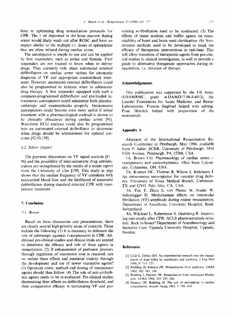
A better approach for intraosseous therapy could be an autoinjector to rapidly puncture tibia1 bone and automatically deliver drug into the marrow. Fig. 1 shows the positioning of a prototype intraosseous au- toinjector, designed to deliver 2 ml of drugs into the tibia1 bone marrow. Tests in animals and cadavers show that it takes about 5-10 s to place the injector in
position on the tibia and about 2 s for drug delivery. The power of a compressed spring generates momen- tum for rapid bone puncture and placement of a 16- gauge needle in less than 0.1 s and drug infusion of 2 ml in 2 s. The needle is retracted automatically into the device after drug delivery.
We have tested a prototype intraosseous autoinjector and compared it with peripheral i.v. drug therapy dur- ing CPR in swine [153]. The intraosseous autoinjector delivered a 2 mg/2 ml epinephrine dose into the tibia and was compared with a standard 1 mg/ml i.v. dose administered via a leg vein. Epinephrine doses were tagged with tracer radioisotopes. Carotid arterial sam- pling and gamma camera imaging showed equivalent circulation times and arterial concentrations (Fig. 1). The larger intraosseous dose required during CPR is consistent with a depot of approximately 1 ml in the marrow as seen with the gamma camera and is most likely due to poor marrow perfusion in this CPR model.
In several studies, we and others have found in- traosseous and i.v. drug delivery to be equivalent when the circulation is intact or an injection is followed by a flush [154,155]. The often used endotracheal route can only be used for some drugs and is problematic in that only a small and unpredictable portion of the dose is delivered to the circulation [156,157]. Direct compari- sons of the endotracheal route and the i.v. route should be performed with all the drugs used for ACLS. Clearly, there are many questions to keep researchers
B I 0.15, -O- IO (2 mL) Technetium
I :;;;;
tp” . 0 80 160240320400480560640~20~00
Number of Chest Compressions
Fig. 1. Top: A prototype intraosseous autoinjector designed to rapidly deliver 2 ml of drug into the vasculature via the bone marrow. Bottom: Representative experiment of CPR in swine. Intraosseous (IO) autoinjector delivery resulted in equivalent appearance times and arterial concentrations of tracers as did i.v. administration [153].
C. Brown et al. : Resuscitation 33 (1996) It‘d 177 173
busy in optimizing drug resuscitation protocols for CPR. The 1 ml deposited in the bone marrow during arrest would likely wash out after ROSC and have an impact similar to the multiple i.v. doses of epinephrine that are often infused during cardiac arrest.
The autoinjector is simple to use and can be applied by first responders, such as police and firemen. First responders are not trained to know when to deliver drugs. They currently only place automated external defibrillators on cardiac arrest victims for automatic diagnosis of VF and appropriate countershock treat- ment. However, automated external defibrillators could also be programmed to indicate when to administer drug therapy. A first responder equipped with such a computer-programmed defibrillator and pre-loaded in- traosseous autoinjectors could administer both pharma- cotherapy and countershocks properly. Intraosseous autoinjectors could become even more useful if initial treatment with a pharmacological cocktail is shown to be clinically efficacious during cardiac arrest [91]. Waveform EGG analyses could also be into an automated external defibrillator when drugs should be administered for come [92,93,158].
programmed to determine optimal out-
6.2. Editor (Sufiw)
The previous discussions on VF signal analysis [S 1~ 961 and the possibility of semi-automatic drug adminis- tration are strengthened by the results of a recent report from the University of Ulm [159]. This study in pigs shows that the median frequency of VF correlates with myocardial blood flow and the likelihood of successful defibrillation during standard external CPR with vaso- pressor treatment.
7. Conclusion
Based on these discussions and presentations, there are clearly several high-priority areas of research. These include the following: (1) It is necessary to delineate the role of adrenergic agonists (vasopressors) in CPR. Ad- ditional pre-clinical studies and clinical trials are needed to determine the efficacy and role of these agents in resuscitation. (2) If enhancement of perfusion pressure through regulation of vasomotor tone is required, can we isolate these effects and minimize toxicity through the development and use of newer vasoactive agents? (3) Optimum route, method and dosing of vasopressor agents should then follow. (4) The role of anti-arrhyth- mic agents needs to be re-evaluated. Pre-clinical studies determining their effects on defibrillation threshold, and their comparative efficacy in terminating VF and pre-
venting re-fibrillation need to be conducted. (5) The effects of tissue acidosis and buffer agents on resus- citability of heart and brain need clarification. (6) Non- invasive methods need to be developed to track the efficacy of therapeutic interventions in real-time. This will allow transition of therapeutic agents from pre-clin- ical studies to clinical investigation, as well as provide a guide to alternative therapeutic approaches during re- suscitation, i.e. titration of therapy.
Acknowledgements
This publication was supported by the US Army (USAMRMC, grant # DAMD17-94-J-40 13), the Laerdal Foundation for Acute Medicine, and Baxter Laboratories. Francie Siegfried helped with editing. Fran Mistrick helped with preparation of the manuscript.
Appendix A
Abstracts of the International Resuscitation Re- search Conference in Pittsburgh, May 1994, available from P. Safar, SCRR, University of Pittsburgh, 3434 Fifth Avenue, Pittsburgh, PA 15260, USA.
1A. Brown CG. Pharmacology of cardiac arrest --. vasopressors and antiarrhythmics. Ohio State Univer- sity, Columbus, OH, USA.
2A. Kramer GC, Thomas B, Wilson J. Jenkinson J. An intraosseous auto-injector for vascular drug deliv- ery. University of Texas Medical Branch, Galveston, TX and GVO. Palo Alto, CA, USA.
3A. Pan T, Zhou S, von Planta M, Studer W. Scheidegger D. Methoxamine effects on ventricular fibrillation (VF) amplitude during rodent resuscitation. Department of Anesthesia, University Hospital, Easel. Switzerland.
4A. Wiklund L. Rubertsson S, Gedeborg R. Improv- ing end-results after CPR: ACLS pharmaceuticals revis- ited. Back to basics? Department of Anesthesiology and Intensive Care, Uppsala University Hospital, Uppsala, Sweden.
References
[l] Crile G. Dolley DH. An experimental research into the resusci- tation of dogs killed by anesthetics and asphyxia. J Exp Med 1906; 8: 713-725.
[2] Redding JS, Pearson JW. Resuscitation from asphyxia. JAMA 1962; 182: 163-166.
[3] Redding J, Pearson JW. Resuscitation from ventricular fibrilla- tion. JAMA 1968; 203: 255-260.
[4] Pearson JW, Redding JS. The role of epinephrine in cardiac resuscitation. Anesth Analg 1963: 5, 599 606.

174 C. Brown et al. /Resuscitation 33 (1996) 163-177
[5] Pearson JW, Redding JS. Epinephrine in cardiac resuscitation. tromechanical dissociation in human beings. Ann Emerg Med Am Heart J 1963; 66: 210-214. 1988; 17: 443-449.
[6] Monroe RG, French G. Ventricular pressure-volume relation- ships and oxygen consumption in fibrillation and arrest. Circ Res 1960; 8: 260-266.
[27] Lindner KH, Ahnefeld FW, Grunert A. Epinephrine versus norepinephrine in prehospital ventricular fibrillation. Am J Cardiol 1991; 67: 427-428.
[7] Hashimoto K, Shigei T, Imai S et al. Oxygen consumption and coronary vascular tone in the isolated fibrillating dog heart. Am J Physiol 1960; 198: 965-970.
[8] Downey JM, Chagrasulis RW, Hemphill V. Quantitative study of intramyocardial compression in the fibrillating heart. Am J Physiol 1979; 237: HI91 -H196.
1281 Kosnik JW, Jackson RE, Keats S et al. Dose-related response of centrally administered epinephrine on the change in aortic diastolic pressure during closed-chest massage in dogs. Ann Emerg Med 1985; 14: 204-208.
[9] Gibbs CL, Papdoyannis DE, Drake AJ et al. Oxygen consump- tion of the nonworking and potassium chloride-arrest dog heart. Circ Res 1980; 47: 408-417.
[lo] Paradis NA, Martin GB, Rivers EP et al. Coronary perfusion pressure and return of spontaneous circulation in human car- diopulmonary resuscitation. JAMA 1990; 263: 1106- 1113.
[l l] Sundt TM, Sharbrough FW, Piepgras DG et al. Correlation of cerebral blood flow and electroencephalographic changes dur- ing carotid endarterectomy with results of surgery and hemody- namics of cerebral ischemia. Mayo Clin Proc 1981; 56: 533-543.
[29] Brown CG, Werman HA, Davis EA et al. The effects of graded doses of epinephrine on regional myocardial blood flow during cardiopulmonary resuscitation in swine. Circulation 1987; 75: 491-497.
[12] Branston NM, Strong ‘AS, Symon L. Extracellular potassium activity, evoked potential and tissue blood flow. J Neurol Sci 1977; 32: 305-321.
[30] Lindner KH, Ahnefeld FW, Bowdler IM. Comparison of dif- ferent doses of epinephrine on myocardial perfusion and resus- citation success during cardiopulmonary resuscitation in a pig model. Am J Emerg Med 1991; 9: 27-31.
[31] Gonzalez ER, Ornato JP, Garnett AR et al. Dose-dependent vasopressor response to epinephrine during CPR in human beings. Ann Emerg Med 1989; 18: 920-926.
[32] Paradis NA, Martin GB, Rosenberg J et al. The effect of standard- and high-dose epinephrine on coronary perfusion pressure during prolonged cardiopulmonary resuscitation. JAMA 1991; 265: 1139-1144.
[13] Niemann JT. Differences in cerebral and myocardial perfusion during closed-chest resuscitation. Ann Emerg Med 1984; 13: 849-853.
[33] Goetting MG, Paradis NA. High-dose epinephrine improves outcome from pediatric cardiac arrest. Ann Emerg Med 1991; 20: 22-26.
[14] Ditchey RV, Winkler JV, Rhodes CA. Relative lack of coro- nary blood flow during closed-chest resuscitation in dogs. Cir- culation 1982; 66: 291-302.
[34] Lindner KH, Ahnefeld FW, Prengel AW. Comparison of stan- dard and high-dose adrenaline in the resuscitation of asystole and electromechanical dissociation. Acta Anaesthesiol Stand 1991; 35: 253-256.
[15] Lee SK, Vaagenes P, Safar P et al. Effect of cardiac arrest time on cortical cerebral blood flow during subsequent standard external cardiopulmonary resuscitation in rabbits. Resuscitation 1989; 17: 105-117.
[16] Lee SK, Vaagenes P, Safar P et al. Effect of cardiac arrest time on the cortical cerebral blood flow generated by subsequent standard external CPR in rabbits. Ann Emerg Med 1984; 13: 385 (abstract).
[17] Stem F, Leonov Y, Safar P et al. Hypertension with or without hemodilution after cardiac arrest in dogs. Stroke 1990; 21: 1178-1184.
[35] Callaham M, Madsen CD, Barton CW, Saunders CE, Pointer J. A randomized clinical trial of high-dose epinephrine and norepinephrine vs standard-dose epinephrine in prehospital car- diac arrest. JAMA 1992; 268: 2667-2672.
[36] Abramson NS, Safar P, Sutton-Tyrrell K et al. Randomized clinical trial of escalating doses of high-dose epinephrine during cardiac resuscitation. Ann Emerg Med 1995; 25: 130 (abstract).
[37] Stiell IG, Hebert PC, Weitzman BN et al. High-dose epinephrine in adult cardiac arrest. N Engl J Med 1992; 327: 1045- 1050.
[18] Safar P. Prevention and therapy of postresuscitation neurologic dysfunction and injury. In: Paradis NA, Halperin HR, Nowak RM, editors. Cardiac arrest. The science and practice of resus- citation medicine, Ch. 49. Philadelphia: Williams and Wilkins, 1996: 8599887.
[19] Klocke FJ, Ellis AK. Control of coronary blood flow. Ann Rev Med 1980; 31: 489-508.
[38] Brown CG, Martin D, Pepe P et al. A comparison of standard- dose and high-dose epinephrine in cardiac arrest outside the hospital. N Engl J Med 1992; 327: 1051-1055.
[39] Ditchey RV, Lindenfeld J. Failure of epinephrine to improve the balance between myocardial oxygen supply and demand during closed-chest resuscitation in dogs. Circulation 1988; 78: 382-389.
[20] Ditchey RV, Lindenfeid J. Potential adverse effects of volume loading on perfusion of vital organs during closed-chest resusci- tation. Circulation 1984; 69: 181-189.
[21] Gentile NT, Martin GB, Appleton TJ et al. Effects of arterial and venous volume infusion on coronary perfusion pressure during canine CPR. Resuscitation 1991; 22: 55-63.
[22] Michael JR, Guerci AD, Koehler RC et al. Mechanisms by which epinephrine augments cerebral and myocardial perfusion during cardiopulmonary resuscitation in dogs. Circulation 1984; 69: 822-835.
[40] Klocke FJ, Kaiser GA, Ross J et al. Mechanism of increase of myocardial oxygen uptake produced by catecholamines. Am J Physiol 1965; 209: 913-918.
[23] Brown CG, Werman HA. Adrenergic agonists during car- diopulmonary resuscitation. Resuscitation 1990; 19: 1 - 16.
[24] Ornato JP. Use of adrenergic agonists during CPR in adults (review). Ann Emerg Med 1993; 22: 411-416.
[25] Olson DW, Thakur R, Stueven HA et al. Randomized study of epinephrine versus methoxamine in prehospital ventricular fibrillation. Ann Emerg Med 1989; 18: 250-253.
[26] Turner LM, Parsons M, Luetkemeyer RC et al. A comparison of epinephrine and methoxamine for resuscitation from elec-
[41] Tang W, Weil MH, Gazmuri RJ et al. Pulmonary ventilation/ perfusion defects induced by epinephrine during cardiopul- monary resuscitation. Circulation 1991; 84: 2101-2107.
[42] Todd GL, Baroldi G, Pieper GM et al. Experimental cate- cholamine-induced myocardial necrosis. I. Morphology, quan- tification and regional distribution of acute contract band lesion. J Mol Cell Cardiol 1985; 17: 317-338.
[43] Rona G. Catecholamine cardiotoxicity. J Mol Cell Cardiol 1985; 17: 291-306.
[44] Brown CG, Little C, Swart G et al. The effect of St-91 on hemodynamics during CPR. Ann Emerg Med 1994; 23: 923- 924 (abstract).
[45] Little CM, Brown CG. Angiotensin II improves myocardial blood flow in cardiac arrest. Resuscitation 1993; 26: 203-210.
[46] Woodhouse SP, Case C, Cox S et al. Trial of large dose adrenaline versus placebo in cardiac arrest. Resuscitation 1994; 26: 89 (abstract).

C. Brown et al. / Resuscitation 33 (I 996) 163 I77 1 75
[47] Little RA, Frayn KN, Randall PE et al. Plasma catecholamines in patients with acute myocardial infarction and in cardiac arrest. Q J Med 1985; 54: 133-140.
[48] Foley PJ. Tacker WA, Wortsman J et al. Plasma catecholamine and serum cortisol responses to experimental cardiac arrest in dogs. Am J Physiol 1987; 253: E283-E289.
[49] Kern KB, Elchisak MA, Sanders AB et al. Plasma cate- cholamines and resuscitation from prolonged cardiac arrest, Crit Care Med 1989; 17: 786-791.
[50] Schoffstall JM, Spivey WH, Davidheiser S et al. Endogenous and exogenous plasma catecholamine levels in cardiac arrest in swine. Resuscitation 1990; 19: 241-251.
[51] Pdradis NA, Rivers EP, Martin GB et al. The change in arterial epinephrine levels after standard and high dose epinephrine during CPR in humans. Crit Care Med 1990; 18: S221 (ab- stract).
[52] Wortsman J, Nowak RM, Martin GB et al. Plasma epinephrine levels in resuscitation with cardiopulmonary bypass. Crit Care Med 1990: 18: 1134&1137.
[53] Shen WK, Kurachi Y. Mechanisms of adenosine-mediated actions on cellular and clinical cardiac electrophysiology. Mayo Clin Proc 1995; 70: 274-291.
[54] Kern K, Garewal H, Sanders A et al. Depletion of myocardial adenosine triphosphate during prolonged untreated ventricular fibrillation: effect on defibrillation success. Resuscitation 1990; 20: 221-229.
[55] Viskin S. Aminophyllin for bradyasystolic cardiac arrest refrac- tory to atropine and epinephrine. Ann Intern Med 1993; 118: 279-281.
[56] Negovsky VA, Gurvitch AM, Zolotokrylina ES. Postresuscita- tion disease. Amsterdam: Elsevier, 1983.
[57] Kirimli B, Kampschulte S, Safar P. Resuscitation from cardiac arrest due to exsanguination. Surg Gynecol Obstet 1969: 129: x9-97.
[58] Vaagenes P, Cantadore R, Safar P et al. Amelioration of brain damage by lidoflazine after prolonged ventricular fibrillation cardiac arrest in dogs. Crit Care Med 1984; 12: 846-855.
[59] Ebmeyer U, Safar P, Katz L et al. Intracarotid-intraaortic (IA) epinephrine injection for CPR in cardiac arrest (CA) of rats. Resuscitation 1992; 24: 190 (abstract).
[60] Manning JE. Murphy CA, Batson DN et al. Aortic arch versus central venous epinephrine during CPR. Ann Emerg Med 1993; 22: 703.-708.
1611 American Heart Association Committee on Emergency Cardiac Care. Guidelines for cardiopulmonary resuscitation and emer- gency cardiac care. Adult advanced cardiac life support. JAMA 1992: 268: 2199-2241.
[62] Bar-Joseph G, Ben-Haim S, Caste1 T et al. Response to re- peated, equal, high doses of epinephrine during cardiopul- monary resuscitation in dogs. Crit Care Med 1993; 21: S271 (abstract).
(63) Miller D. Craddock L, Hoffenberg S et al. Pilot study of i.v. magnesium sulfate in refractory in-hospital cardiac arrest: safety data and recommendations for future studies. Resuscita- tion 1995; 30: 3- 15.
[64] Brown C, Griffith R, Miller B et al. The effect of intravenous magnesium administration on aortic, right atrial, and coronary perfusion pressures during CPR in swine. Resuscitation 1993; 26: 3-12.
[65] Tacker WA, Niebauer MJ, Babbs CF et al. The effect of newer antiarrhythmic drugs on defibrillation threshold. Crit Care Med 1980: 8: 177. 180.
1661 Babbs CF, Yim GKW, Whistler SJ et al. Elevation of ventricu- lar defibrillation threshold in dogs by antiarrhythmic drugs. Am Heart J 1979; 98: 345-3.50.
[67] Echt DS. Black JN, Barbey JT et al. Evaluation of antiarrhyth- mic drugs on defibrillation energy requirements in dogs. Circu- lation 1989; 79(5): I 106- 1117.
v31
F91
[701
1711
~721
[731
[741
[751
[761
1771
[781
1791
WI
WI
P31
L841
IS51
WI
v71
Dorian P, Fain ES, Davy J et al. Lidocaine causes a reversible, concentration-dependent increase in defibrillation energy re- quirements. JACC 1986; 9: 327-332. Chow MSS, Ronfeldt RA, Hamilton RA et al. Effect of external CPR on lidocaine pharmacokinetics in dogs. J Phar- macol Exp Ther 1983; 244: 531-537. Kerber RE, Pandian NG, Jensen SR et al. Effect of lidocaine and bretylium on energy requirements for transthoracic defibril- lation: experimental studies. JACC 1986; 7: 397.-405. Dorian P, Fain ES, Davy J et al. Effect of quinidine and bretylium on defibrillation energy requirements. Am Heart .I 1986; 112(l): 19-24. Koo CC, Allen JD, Pantridge JF. Lack of effect of bretylium tosylate on eiectrical ventricular defibrillation in a controlled study. Cardio Res 1984; 18: 762-767. Chow MSS, Kluger J, Lawrence R et al. The effect of lidocaine and bretylium on the defibrillation threshold during cardiac arrest and cardiopulmonary resuscitation. Proc Sot Exp Rio Med 1986; 182: 63-67. Euler DE, Zeman TW, Wallock ME et al. Deleterious effects of bretylium on hemodynamic recovery from ventricular fibrilla- tion. Am Heart J 1986; 112: 25-31. Spivey WH, Abramson NS, Safar P et al. (‘orrelation of blood pressure with mortality and neuroiogic recovery in comatose postresuscitation patients. Ann Emerg Med 1991: 20: 453 -454 (abstract). Haynes RE, Chinn TL, Copass MK et al. Comparison of bretylium tosylate and lidocaine in management of out of hospital ventricular fibrillation: a randomized clinical trial. Am J Cardiol 1981; 48: X3--356. Olson DW, Thompson BM, Darin JC et al. A randomized comparison study of bretylium tosylate and lidocaine in resusci- tation of patients from out-of-hospital ventricular fibrillation in a paramedic system. Ann Emerg Med 1984: 1X9): 807 -8 10. Weaver WD. Fahrenbruck CE, Johnson DD et al. Effect of epinephrine and lidocaine therapy on outcome after cardiac arrest due to ventricular fibrillation. Circulation 1990: X3: 2021-2034. Williams ML. Intravenous amiodarone during prolonged resus- citation from cardiac arrest. Ann Int Med 1989: t 10: 839 842. Sellevold OFM, Berg EM, Levang OW. Procaine is effective for minimizing postischemic ventricular fibrillation in cardiac surgery. Anesth Analg 1995: 81: 932 938. Hargarten KM, Stueven HA, Waite EM et al. Prehospital experience with defibrillation of coarse ventricular fibrillation: :I ten year review. Ann Emerg Med 1490: 19(2X 157. 162. Stewart AJ, Allen JD, Adgey AAJ. Frequency analysis of ventricular fibrillation and resuscitation success. Q J Med 1992; 85: 761 769. Brown CG, Dzwonczyk R, Werman HA et al. Estimating the duration of ventricular fibrillation. Ann Emerg Med 1989: 18: 1181 1185. Menegazzi JJ, Watanabe S, Klain M et a!. ECG spectrum analysis predicts return of spontaneous circulation following prolonged ventricular fibrillation. Prehosp Dihaster Med 1993: S(2): S61 (abstract). Menegazzi J. Seaberg D, Yealy D et al. Combined pharma- cotherapy with delayed countershock versus standard advanced cardiac life support following prolonged ventricular fibrillation. Acad Emerg Med 1994; l(2): Al9 (abstract). Seaberg D; Menegazzi J, Check B et al. Use nfcardiocerebral- protective drug cocktail prior to countershock following pro- longed ventricular fibrillation. Prehosp Disaster Med 19941 9(3): S52 (abstract). Menegazzi J, Davis E, Yealy D et al. An experimental al- gorithm vs standard ACLS in a swine model of out-of-hospital cardiac arrest. .Ann Emerg Med 1993, 27: _‘i? ?!‘I.

C. Brown et al. 1 Resuscitation 33 (1996) 163-177 176
WI
[891
t901
[911
~921
]931
t941
1951
[961
[971
[981
[991
w01
w11
Niemann JT, Cairns CB, Sharma J et al. Treatment of pro- longed ventricular fibrillation: immediate countershock versus high-dose epinephrine and CPR preceding countershock. Circu- lation 1992; 85(l): 281-287. Yakaitis RW, Ewy GA, Otto CW et al. Influence of time and therapy on ventricular fibrillation in dogs. Crit Care Med 1980; 8: 157-163. Niemann JT, Cairns CB, Sharma J et al. Treatment of pro- longed ventricular fibrillation: immediate countershock versus high-dose epinephrine and CPR preceding countershock. Circu- lation 1992; 85: 281-287. Menegazzi JJ, Davis EA, Yealy DM et al. An experimental algorithm versus standard ACLS in a swine model of out-of- hospital cardiac arrest. Ann Emerg Med 1993; 22: 235-239. Brown CG, Griffith RF, Van Ligten P et al. Median fre- quency-a new parameter for predicting defibrillation success rate. Ann Emerg Med 1991; 20: 787-789. Brown CG, Dzwonczyk R. Signal analysis of the human ECG during ventricular fibrillation (VF)-frequency and amplitude parameters as predictors of successful countershock. Ann Emerg Med 1996; 27: 184-188.’ Weaver WD, Cobb LA, Dennis D et al. Amplitude of ventricu- lar fibrillation waveform and outcome after cardiac arrest. Ann Intern Med 1985; 102: 53-55. ’ Stults KR, Brown DD, Kerber RE. Ventricular fibrillation amplitude predicts ability to defibrillate. Am J Emerg Med 1986; 4: 423. von Planta I, Wagner 0, von Planta M et al. Determinants of survival after rodent cardiac arrest: implications for therapy with adrenergic agents. Int J Cardiol 1993; 38: 235-245. Rubertsson S, Grenvik A, Wiklund L. Blood flow and perfu- sion pressure during open-chest versus closed-chest cardiopul- monary resuscitation in pigs. Crit Care Med 1995; 23: 715-725. Eleff SM, Schleien CL, Koehler RC et al. Brain bioenergetics during cardiopulmonary resuscitation in dogs. Anesthesiology 1992; 76: 77-84. Wiklund L, Jorfeldt L, Stjernstrom H et al. Gas exchanged as monitored in mixed venous and arterial blood during experi- mental cardiopulmonary resuscitation. Acta Anaesthesiol Stand 1992; 36: 427-435. Capparelli EV, Chow MSS, Kluger J et al. Differences in systemic and myocardial blood acid-base status during experi- mental cardiopulmonary resuscitation. Acta Anaesthesiol Stand 1992; 36: 427-435. Gazmuri RJ, von Planta M, Weil MH et al. Cardiac effects of carbon dioxide consuming and carbon dioxide-generating buffers during cardiopulmonary resuscitation. J Am Co11 Car- diol 1990; 15: 482-490.
[102] Effron MB, Guarnieri T, Fredriksen JW et al. Effect of tris (hydroxymethyl) aminomethane on ischemic myocardium. Am J Physiol 1978; 235(2): H167-H174.
[103] Gnmdler W, Weil MH, Rackow EC. Arteriovenous carbon dioxide and pH gradients during cardiac arrest. Circulation 1986; 74: 1071-1074.
[104] Weil MH, Rackow EC, Trevino R et al. Differences in acid- base state between venous and arterial blood during cardiopul- monary resuscitation. N Engl J Med 1986; 315: 153-156.
[105] Kette F, Weil MH, von Planta M et al. Buffer agents do not reverse intramyocardial acidosis during cardiac resuscitation. Circulation 1990; 81: 1660-1666.
[106] von Planta M, Gudipati C, Weil MH et al. Effects of tromethamine and sodium bicarbonate buffers during cardiac resuscitation. J Clin Pharmacol 1988; 28: 594-599.
[107] Sanders AB, Otto CW, Kern KB et al. Acid-base balance in a canine model of cardiac arrest. Ann Emerg Med 1988; 17: 667-671.
[108] Adrogue HJ, Rashad MN, Gorin AB et al. Assessing acid-base status in circulatory failure. Differences between arterial and central venous blood. N Engl J Med 1989; 320: 1312-1316.
[109] Bar-Joseph G, Ben-Ha& S, Caste1 T et al. Improved resus- citability with sodium bicarbonate and Carbicarb after 10 min- utes ventricular fibrillation in dogs. Crit Care Med 1993; 21: S271 (abstract).
[l lo] Berenyi KJ, Wolk M, Killip T. Cerebrospinal fluid acidosis complicating therapy of experimental cardiopulmonary arrest. Circulation 1975; 52: 319-324.
[l 1 l] Wiklund L, Soderberg D, Henneberg S et al. Kinetics of carbon dioxide during cardiopulmonary resuscitation. Crit Care Med 1986; 14: 1015-1022.
[112] Rosenberg JM, Martin GB, Paradis NA et al. The effect of CO, and non-CO, generating buffers on cerebral acidosis after cardiac arrest: A 3’P NMR study. Ann Emerg Med 1989; 18: 341-347.
[ll3] Roberts D, Landolfo K, Light RB et al. Early predictors of mortality for hospitalized patients suffering cardiopulmonary arrest. Chest 1990; 97: 413-419.
[I141 Weil MH, Ruiz CE, Sybil M et al. Acid-base determinants of survival after cardiopulmonary resuscitation. Crit Care Med 1985; 13: 888-892.
[115] Aufderheide TP, Martin DR, Olson DW et al. Prehospital bicarbonate use in cardiac arrest. A 3-year experience. Am J Emerg Med 1992; 10: 4-7.
u 171
11181
11191
[116] Kirimli B, Harris LC, Safar P. Evaluation of sodium bicarbon- ate and epinephrine in cardiopulmonary resuscitation. Anesth Analg 1969; 48: 649-658. Sanders AB, Kern KB, Fonken S et al. The role of bicarbonate and fluid loading in improving resuscitation from prolonged cardiac arrest with rapid manual chest compression CPR. Ann Emerg Med 1990; 19: l-7. Bircher NG. Sodium bicarbonate improves cardiac resuscitabil- ity, 24 hour survival and neurological outcome after ten min- utes of cardiac arrest in dogs. Anesthesiology 1991; 75: A246 (abstract). Vukmir RB, Bircher NG, Radovsky A et al. Sodium bicarbon- ate may improve outcome in dogs with brief or prolonged cardiac arrest. Crit Care Med 1995; 23: 515-522. Wiklund L, Ronquist G, Stjernstrom H et al. Effects of alkaline buffer administration on survival and myocardial energy metabolism in pigs subjected to ventricular fibrillation and closed-chest CPR. Acta Anaesthesiol Stand 1990; 34: 430-439. Guerci AD, Chandra N, Johnson E et al. Failure of sodium bicarbonate to improve resuscitation from ventricular fibrilla- tion in dogs. Circulation 1986; 74 (Suppl IV): 75-79. Dybvik T, Strand T, Steen PA. Buffer therapy during out-of- hospital cardiopulmonary resuscitation. Resuscitation 1995; 29: 89-95.
WOI
Kw
11221
~231
u241
11251
PW
WI
WI
Alberts B, Bray D, Lewis J et al, editors. Membrane transport of small molecules and the ionic basis of membrane excitability. In: The molecular biology of the cell, 3rd edn. New York: Garland Publishing, 1994: 518-519. Bircher NG. Acid-base physiology during advanced cardiac life support. Anesthesiol Clin North Am 1995; 13: 799-808. Eleff SM, Sugimoto H, Schaffner DH et al. Acidemia and brain pH during prolonged cardiopulmonary resuscitation in dogs. Stroke 1995; 26: 1028-1034. Bircher N, Safar P. Cerebral preservation during cardiopul- monary resuscitation. Crit Care Med 1985; 13: 185-190. Cummins RO, Graves JR. Clinical results of standard CPR: prehospital and inhospital. In: Kaye W, Bircher NG, editors. Cardiopulmonary resuscitation. New York: Churchill Living- stone, 1989: 87- 102. Bircher NG. Physiology and pharmacology of standard car- diopulmonary resuscitation. In: Kaye W, Bircher NG, editors.

C. Brown et al. I Resuscitation 33 (I 996) l&L 177 177
Cardiopulmonary resuscitation. New stone, 1989: 55-86.
[129] Edmonds-Seal J. Acid-base studies report of 64 cases. Acta Anaesthesiol 235-241,
York: Churchill Living- [I451
after cardiac arrest. A Stand 1966; 23 (Suppl): [I461
[130] Redding JS, Pearson JW. Metabolic acidosis: a factor in car- diac resuscitation. South Med J 1967; 60: 926932.
[I311 Chazan JA, Stenson R, Kurland GS. The acidosis of cardiac arrest. N Engl J Med 1968; 278: 360-364.
[I321 Henneman PL, Gruber JE, Marx JA. Development of acidosis in human beings during closed-chest and open-chest CPR. Ann Emerg Med 1988; 17: 672-675.
[133] Ornato JP, Gonzalez ER, Coyne MR et al. Arterial pH in out-of-hospital cardiac arrest: response time as a determinant of acidosis. Am J Emerg Med 1985; 3: 4988502.
[134] Ledingham IMCA, Norman JN. Acid-base studies in experi- mental cardiac arrest. Lancet 1962; ii: 967-969.
[135] Kette F. Weil MH, Gazmuri RJ. Buffer solutions may compro- mise cardiac resuscitation by reducing coronary perfusion pres- sure. JAMA 1991; 266: 2121-2126.
[ 1361 Bircher NG. Cardiopulmonary cerebral resuscitation: brain re- suscitation, mediators, glucose and anesthetics. Curr Opinion Anaesthesiol 1990; 3: 259-264.
[137] Marsh JD, Margolis TI, Kim D. Mechanisms of diminished contractile response to catecholamine during acidosis. Am J Physiol 1988; 254: H20-H27.
[138] Davies AG. Rapid desensitization and uncoupling of human /1-adrenergic receptors in an in vitro model of lactic acidosis. J Clin Endocrinol Metab 1984; 59: 398-405.
[139] Falk JL, Rackow EC, Weil MH. End-tidal carbon dioxide concentration during cardiopulmonary resuscitation. N Engl J Med 1988; 318: 607-611.
[I401 Haigney MCP, Lakatts EG, Stem MD et al. Sodium channel blockade reduces hypoxic sodium loading and sodium-depen- dent calcium loading. Circulation 1994; 90: 391-399.
[141] Weissberg PL, Little PJ, Cragoe EJ et al. The pH of sponta- neously beating cultured rat heart cells is regulated by an ATP-calmodulin-dependent Na +/H + antiport. Circ Res 1989; 64: 676685.
[I421 Montrose MH, Murer H. Kinetics of Na+/H+ exchange. In: Grinstein S, editor. Na + /H + exchange. Boca Raton, FL: CRC Press, 1988: 57775.
[143] Kalimo H, Rehncrona S, Soderfeldt B et al. Brain lactic acidosis and ischemic cell damage: 2. Histopathology. J Cereb Blood Flow Metab 1981: 1: 313-327.
[144] Barry MA, Eastman A. Endonuclease activation during apop- tosis: the role of cytosolic Ca’ + and pH. Biochem Biophys Res Commun 1992: 186: 782 -789.
v471
[1481
u491
]I501
[I511
I1521
]I531
u541
u551
U561
u571
[W
[I 591
Barry MA, Reynolds JE, Eastman A. Etoposide-induced apop- tosis in human HL-60 cells is associated with intracellular acidification. Cancer Res 1993; 53: 2349..2357. Hurn PD, Koehler RC, Norris SE et al. Dependence of cerebral energy phosphate and evoked potential recovery on end-is- chemic pH. Am J Physiol 1991; 290: H532-H541. Hurn PD, Koehler RC, Blizzard KK et al. Deferoxamine reduces early metabolic failure associated with severe cerebral ischemic acidosis in dogs. Stroke 1995; 26: 688 -6%. Plum F. What causes infarction in ischemic brain? Neurology 1983; 33: 222-233. Garcia JH, Liu KF. Ho KL. Neuronal necrosis after middle cerebral artery occlusion in Wistar rats progresses at different time intervals in caudoputamen and the cortex. Stroke 1995: 26: 636-643. Halvorsen L, Bay BK, Perron PR et al. Evaluation of an intraosseous infusion device for the resuscitation of hypov- olemic shock. J Trauma 1990; 30: 652--659. Fiser DH. lntraosseous infusion. N Engl J Met1 1990: 322: 157991581. Iserson KV. Intraosseous infusions in adults. .I Emerg Ned 1989; 7: 5877591. Jenkinson JP, Benedetto AR, Watson SC et al. lntraosseous small-volume fluid bolus injections enter central circulation during experimental cardiopulmonary resuscitation. Ann Emerg Med 1994; 23: A91 (abstract). Dubick MA, Pfeiffer JW, Clifford CB et al. Comparison of intraosseous and intravenous delivery of hypertonic saline/dex- tran in anesthetized. euvolemic pigs. Ann Emerg Med 1992; 21(5): 498.-503. Spivey WH, Lathers CM, Malone DR et al. Comparison of intraosseous, central. and peripheral routes of sodium bicar- bonate administration during CPR in pigs. Ann Emerg Med 1985; 14: 1135 -1140. Orlowski J, Poremberg D, Gallagher J et al. Comparison study of intraosseous, central intravenous, and peripheral intravenous infusion of emergency drugs. AJDC 1990: 144: 112. I 17. Schneider SM, Yealy DM, Michaelson EA et al. Endotracheal versus intravenous epinephrine in the prehospital treatment of cardiac arrest. Orig Res 1990; 5: 341 -348. Brown CG, Dzwonczyk R. Measurement ot’ the approximate elapsed time of ventricular fibrillation and monitoring the re- sponse of the heart to therapy. US Patent No. X077,667, 1991. Strohmenger H-LJ, Lindner KH, Prengel AW et al. Effects of epinephrine and vasopressin on median fibrillation frequency and defibrillation success in a procine model of cardiopul- monary resuscitation. Resuscitation 1996: 3 I: 65 73.