functional orthopaedics and neuro-muscular re-education clinical
TRANSCRIPT

Supporting Functional Motions of Healing
Functional Orthopaedics and Neuro-Muscular Re-EducationClinical Applications Manual and Home Exercise Program

dEvELOPERS NOTE
Whether your patients suffer from chronic shoulder pain or have recently undergone any form of a corrective surgical procedure to the shoulder, you can be assured the neuro-muscular communications within the shoulder system are to varying degrees out of balance and reorganized around compensatory biomechanics and therefore prone to supporting resultant micro-traumas and perpetuating pain if left unresolved.
The shoulder complex, while offering the most versatility of movement within the human body, requires a delicate balance to be maintained between its moving parts and the muscles that steer those parts. In the progressive onset of chronic pain or following a traumatic injury a common occurrence is for our bodies to protect against further pain. For most of us we are unable to stop participating in our daily activities to fully heal. Consequently to enable us to remain active, our bodies will create compensatory “solution” moves. These particular compensations, while successful in avoiding provocations of repetitive pain are by defi nition neither normal nor ideal and thus are destined to create secondary origins of pain either within multiple regions of the spine, the distal upper extremity, or back in the same shoulder at a later date.
As a former patient requiring a surgical shoulder procedure, a Physical Therapist of 12 + years and the developer of the UE Ranger please allow me to share with you how I believe this clinical rehabilitation tool can best support your efforts in restoring the movement health to the shoulder girdle complex and its supportive kinetic chain. The UE Ranger was designed to meet persons suffering from the multitude of shoulder pathologies, both chronic or post-operatively at their current and personal impairment level in a manner that is compatible with the biomechanics of the full upper extremity including its foundational shoulder girdle and its supportive sensory and motor nervous systems.
Please take the time to educate yourself on each of the following sections within this manual as the ability to alter the communications within the nervous system coordinating this dynamic and versatile region of our bodies is a subtle and progressive process. Becoming educated with the available therapeutic values based on movement health science and rehabilitation principles will expand your clinical knowledge and enable you to optimize the clinical capacities within this tool, leading to superior outcomes and increased referrals.
We at Rehab Innovations, Inc. wish you and your patients well in the most effi cient recovery of movement health.
dan Miller, PT, MS
Rehab Innovations, Inc. • www.ueranger.com

Functional Orthopaedics and Neuro-Muscular Re-Education Clinical Applications Manual and Home Exercise Program
TABLE OF CONTENTSPost-Operative and Chronic Pain Rehabilitation Protocol ................................................................................................................. 2 Rehabilitation Applications ............................................................................................................................................................. 2 Definitions Relevant to the UE Ranger Home Exercise Program ............................................................................................... 3 Components of the UE Ranger ....................................................................................................................................................... 4 Components of the Wall Mount Tracking Frame .......................................................................................................................... 5 Movement Relation Requirements of the Full Upper Extremity ................................................................................................. 6Post-Operative and Chronic Pain Protocol – Phase One – Passive Range of Motion (PROM) ..................................................... 7 Clinical Consideration #1 ................................................................................................................................................................ 8 Initiation and Progression of Forward Reaching and Elevations – PROM ................................................................................. 9 Frequency and Volume of Use .......................................................................................................................................................12 Initiation and Progression of External Rotation ............................................................................................................................13 Clinical Consideration #2 ...............................................................................................................................................................14 Reintegration Consideration #1 ......................................................................................................................................................14 Reintegration Consideration #2 ......................................................................................................................................................14 Use of Ice, Rest, Postural Awareness, and Respiration ................................................................................................................15Post-Operative and Chronic Pain Protocol – Phase Two – Active Assistive Range of Motion (AAROM) ..............................................16 Clinical Consideration #3 ...............................................................................................................................................................17 Neuro-Motor Re-Education: The Keystone In Resolving Pain and Restoring Healthy Biomechanics of the Shoulder Girdle .............................................................................................................................18 Reintegration Consideration #3 ......................................................................................................................................................19 Neuro-Motor Re-Education: Serratus Anterior Isolation .............................................................................................................20 Neuro-Motor Re-Education: Supraspinatus Isolation ..................................................................................................................22 Neuro-Motor Re-Education: Isolation of the External Rotators .................................................................................................24 Neuro-Motor Re-Education: Isolation of the Lower Trapezius With the Integration of the Rotator Cuff and Serratus Anterior Muscles ...............................................................................................................................................26 Initiation and Progression of Forward Reaching and Elevations – AAROM.............................................................................28 Closed Kinetic Chain – Standing Floor to Platform Supportive Progressions ...........................................................................29 Reintegration Consideration #4 ......................................................................................................................................................30 Reintegration Consideration #5 ......................................................................................................................................................32 Open Kinetic Chain – Hook-lying Position ..................................................................................................................................34 Closed Kinetic Chain Wall or Door Mounted Tracking Frame .................................................................................................. 36 Reintegration Consideration #6 ......................................................................................................................................................37 Reintegration Consideration #7 ......................................................................................................................................................38 Open Kinetic Chain External Rotation – Standing Position ........................................................................................................39 Open Kinetic Chain Elevation – Standing Position ......................................................................................................................40 Reintegration Consideration #8 ......................................................................................................................................................41 Closed Kinetic Chain – Seated Floor to Platform Supportive Progressions ..............................................................................42 Reintegration Consideration #9 ......................................................................................................................................................42 Closed Kinetic Chain Elevation – Side-lying Position .................................................................................................................43 Reintegration Consideration #10....................................................................................................................................................44Post-Operative and Chronic Pain Protocol – Flexibility ...................................................................................................................45Post-Operative and Chronic Pain Protocol – Manual Interventions ................................................................................................47Research Section ..................................................................................................................................................................................49
Prior to Applications - Conditions of Sale, Warranty, and LimitationsThe UE Ranger ™ is an upper extremity rehabilitative motion assistive device which is designed to compliment the natural motion of the involved upper extremity (UE). This capacity allows a patient to avoid unnatural and unnecessary forces throughout the full upper extremity. It is imperative that a patient be instructed by their rehabilitation professional in the proper biomechanics of the upper extremity to avoid compensations and a chance of injury from being promoted. Rehab Innovations, Inc. warrants that this product is free from manufacturing defects, is fit for the ordinary purposes for which this product is designed and conforms to the descriptions stated herein. Satisfactory results should be obtained if this product is used according to the foundational principles and application guidelines within this manual and with the directions and recommendations of the patient’s health care professional. Unintended consequences may result due to such factors as improper use or without consultation and guidance of a healthcare professional all of which are beyond the control of Rehab Innovations, Inc. or the seller, thus all such risks shall be assumed by the buyer. Under no circumstances shall the buyer be entitled to any remedy or damages. Remedies for incidental and consequential damages are specifically excluded.
Second Edition

�
POST OPERATivE ANd CHRONiC PAiN REHABiLiTATiON PROTOCOLintroductionThe UE Ranger Movement Health Device and Movement Health System were designed specifically to restore the physical capacities of the upper extremity (shoulder girdle [humerus, scapula, ribs, and clavicle], elbow, forearm, wrist, and hand) (illustration A) as a functional system.
The functional system means that the individual joints, nerves and muscles are inter-related in terms of movement productions and the coordination of purposeful efforts. As you will soon learn, the most successful recovery of the upper extremity function requires the clinical skills of a Rehabilitation Professional, (i.e. Physical Therapist, Occupational Therapist, Athletic Trainer, or Chiropractor: hereafter referred to as Rehabilitation Professional) as well as the patients participation in both clinical interventions and a specific home exercise program. The UE Ranger has been designed to support in specific detail each phase of recovery; which includes what is termed Passive Range of Motion (PROM), Neuro-muscular Re-education (NMR), and Active Assisted Range of Motion (AAROM). Additionally the UE Ranger is very supportive of functional strengthening and flexibility efforts. These phases along with their individual components will be discussed in order of progression and within the accepted principles of the Physical Rehabilitation Profession. Please note this manual is designed to maximize rehabilitation in the most efficient manner. Sharing this information with your patients will enable them to be an informed and an active participant in the process of their rehabilitation. This manual and the UE Ranger Movement Health System are not intended to replace the need of a Rehabilitation Professional, but rather to compliment the efforts of both you and your patient. In order to maximize rehabilitation, it is imperative that the patient follow the instructions of their Rehabilitation Professional and the guideline applications of this manual.
Rehabilitation ApplicationsPatients who are attempting to rehabilitate from a musculoskeletal pathology or a neurological insult that involves the upper extremity, including the shoulder girdle, elbow, forearm, and or wrist, predictably will require successful resolution of a number of physical impairments. These include, but are not limited to the following:
• Musculo-tendinous injury • Neurological injury• Muscle weakness• Muscle spasms• Limitations of joint motion• Soft tissue adhesions
s iLLUSTRATiON A
• Swelling • Fear • Pain• Compensations or dyskinetic movement patterns• Apprehensiveness

Rehab Innovations, Inc. • www.ueranger.com�
By supporting the natural movements and physiological healing processes of the upper extremity, the UE Ranger movement health device offers the most thorough and efficient resolution of the above physical impairments by facilitating and or enhancing:• A balanced state of the nervous system • The manual skills of the rehabilitation professional• The accepted principles of therapeutic exercise• The essential reintegration into the movement whole
The UE Ranger thus facilitates a Working Team between the Patient and the Rehabilitation Professional. By supporting and ultimately integrating the knowledge and skills of the Rehabilitation Professional, the patient can by application guidance be empowered to contribute a meaningful role in the expected successful outcome. To achieve the most efficient return on the efforts of all players including the expanded team of the referring Physician and Reimbursing Party it is recommended that the UE Ranger Movement Health Device be included as early as day one (within the allowances of the referring physician and rehabilitation professional) in the patients home exercise program. In the subsequent sections the benefits of this approach will be described in specific detail.
definitions Relevant to the UE Ranger Home Exercise ProgramHealthy Biomechanics – Movement proceeding in a most efficient manner and without undue stress on non-contractile structures to preserve the integrity and prevent injury of the musculo-skeletal system.
Awareness of Movement – The individual capacity to accurately perceive the coordinated joint contributions during functional movements involving healthy biomechanics.
Strength – The ability of the required muscles to generate an adequate force to support the intended movements.
Neuro-Motor Re-Education – Getting the right message(s) to the right muscle(s) is the first requirement of strengthening, and subsequent re-establishment of healthy biomechanics.
Endurance – The ability to perform the necessary repetitive muscular contractions required to support repetitive functional movements with healthy biomechanics.
Movement Coordination – The ability during a functional movement to sequence the appropriate muscle contractions at the most opportune time and with the most opportune intensities.
Soft Tissue Mobility – the ability of muscles, tendons, fat, blood vessels, nerves, and synovial tissues (tissues around joints) to allow necessary relational movements to support the advancement of a functional movement.
Substitution or Compensation – Using muscles and joint efforts beyond those normally designed to participate in the execution of healthy motions. Generally a sign of deficiency in one or more of the following:• Strength• Endurance • Motor control • Movement understanding• Soft tissue mobility
Fatigue – Point at which one loses the capacity to support healthy biomechanics and/or experiences pain with movement.

4
COMPONENTS OF THE UE RANGERThe UE Ranger is an Upper Extremity Movement Health Device designed to support the rehabilitation of a patients upper extremity function. The UE Ranger arrives ready to be implemented. Please familiarize yourself with the following components.
Custom Molded Hand Support designed to support in a relaxed posture either the left or the right hand of the involved upper extremity.
Securing Neoprene Hand Strap and velcro Attachments designed to comfortably support and secure the involved hand within the molded hand support. This strap should be secured in a comfortable position, yet snug enough for a patient’s hand to not slide on the molded hand support.
Proximal Multi-Plane Articulating Joint designed to support the natural relational motions of each joint of the upper extremity (shoulder girdle, elbow, forearm, and wrist) during open and closed chain kinetic functional applications.
Telescopic Supportive and Guidance Tubing with Adjusting Locking Collar designed to support multiple patient applications with considerations of healing stages, desired intensity levels, skill levels, and varying current upper extremity joint mobility measurements.
Non-involved Hand Support designed to support the guidance and force produced by the non-involved upper extremity in multiple applications.
Weight-Bearing detachable Articulating Base Plate with a Skid Resistant Rubber Pad on its undersurface designed to support closed kinetic chain functional applications.
distal Multi-Plane Articulating Joint designed to support the natural relational motions of each joint of the upper extremity (shoulder girdle, elbow, forearm, and wrist) during closed chain kinetic functional applications.
Base Plate Options:Open Kinetic Chain Techniques: Detach the base plate by simply pulling it and the distal articulating joint out of the telescopic tubing (fi gure 1).Closed Kinetic Chain Techniques: Reattach the base plate in a reverse manner. Be sure the base plate is fully secure before application (fi gure 2).
Securing Hand StrapCustom Molded
Hand Support
Proximal Multi-Plane Articulating Joint
Telescoping TubeAdjusting
Locking Collar(User-friendly and secure) Non-involved
Hand Support(Multiple position
comfort)
Weight-Bearing detachable Articulating Base Plate(with skid-resistant rubber pad)
distal Multi-Plane Articulating
Joint
s FiGURE 1 s FiGURE 2

5
s FiGURE 1 s FiGURE 2 s FiGURE 3
s FiGURE 4
COMPONENTS OF THE CLiNiCAL WALL MOUNT TRACKiNG FRAME
Adjustable Height Frame
Adjusting T-slot Bolt(User-friendly and secure)
Wall Mounted Track
(Mount the bottom edge two feet above
the fl oor)
Securely insert the base plate of the UE Ranger into the wall mount tracking frame by fi rst angling the top of the base plate up and under the top portion of the wooden frame as shown in (fi gure 1). Progressively guide the base plate up and under the top portion of the frame to a point where the bottom of the base plate clears the bottom portion of the frame, allowing the base plate to then be received and rest securely within the full frame as shown in (fi gures 2 and 3).
The Wall Mount Tracking Frame sold separately from the UE Ranger is designed to support specifi c and progressive closed kinematic chain neuro-motor re-education, strengthening, fl exibility, and endurance applications.
s FiGURE 5
Home Use Option:To support the patient’s Home Exercise Program a Door Mount version of the Wall Mount Tracking Frame is available. It can be securely fastened to a door and the UE Ranger inserted as shown in (fi gures 4 and 5).

6
MOvEMENT RELATiON REQUiREMENTS OF THE FULL UPPER EXTREMiTYDuring normal activities of everyday living, all joints of the upper extremity move in concert and in varying proportions with each other. The UE Ranger compliments and supports these motion relations, allowing a patient to rehabilitate normal functional movement of the full upper extremity. Illustrated below are each of the upper extremity components beyond that of the shoulder girdle that participates in natural motions of the shoulder. Limitations in any of these contributory joints can impair the health and function of the remaining components.
Wrist Radial deviation Forearm Supination
Wrist Ulnar deviation Forearm Pronation
Wrist Extension Elbow Flexion
Full Upper Extremity Function
Elbow ExtensionWrist Flexion

Rehab Innovations, Inc. • www.ueranger.com�
POST-OPERATivE ANd CHRONiC PAiN PROTOCOL • PHASE ONE
PASSivE RANGE OF MOTiON (PROM)
The success achieved in the PROM phase of rehabilitation is the foundation from which all further gains will be determined. By definition PROM means that your involved upper extremity (UE) is being supported and moved by the combination of the UE Ranger and your non-injured upper extremity, and its supportive kinetic chain. The goals of this phase of your rehabilitation are as follows.
PROM Goals1. Preserve the integrity of the surgical repair2. Resolution of pain and swelling3. Resolution of a balanced ANS, absent of the fight or flight drivers4. Restoration of resting tone of the full shoulder girdle’s musculature5. Restoration of primary or diaphragm supported respiration absent of neck and shoulder
bracing6. Preserve and enhance the integrity of the circulatory role of healing 7. Reduce the need of medications, eliminating their side effects, thus supporting restorative
sleep8. Prevent adhesions9. Resolve and prevent further compensations10. Restoration of patient supported Range of Motion to between approximately 90 and 110
degrees of elevation with the understanding of proper biomechanics to this point.
Foundational Scientific Principles Supporting the UE Ranger’s Application in the PROM Phase of Rehabilitation
To personally experience the science is an ideal way to appreciate the extent that subtleties matter when working with the nervous system and then to recognize how the UE Ranger can best facilitate and enhance your clinical interventions to most efficiently meet the goals of your patients.

8
Clinical Consideration #1 Step one: With either of your hands make a moderately tensed gripped hand and position yourself in a pronated forearm position as shown in (fi gure 1).
Step two:Prior to the next instruction the goal of this example is for you to feel the point at which your effort to move becomes impeded by your own body’s resistance.
Step three: Begin to slowly move your arm from the starting position as shown in (fi gure 1) into a movement of supination (fi gure 2). Stop at the exact point in which you begin to feel impedance to your movement or in other words when you feel like your muscles tighten in an effort to go further. Mentally remember both this point and what the effort felt like.
Step four: Open your hand and return to the starting position as shown in (fi gure 3). Now with your open hand repeat the same motion as you produced in step 3 as shown in (fi gure 4). Compare in your mind, both the feel of the motion and how far you go versus that with the gripped hand prior to feeling the impedance to your movement effort.
Results:
Most often persons report that they can go further with a noticeable ease of the effort to move.
Why does This Clinically Matter?As you read this section, keep in mind as a global reference the 10 goals to be achieved in this fi rst phase of rehabilitation. A gripped hand with respect to the upper extremity is a: 1. Distal effort which requires a proximal role or effort to stabilize. 2. Muscular co-contractions from the elbow distally, which essentially diminishes the ease of movement production
throughout the full kinetic chain.
A patient whom has recently undergone a surgical repair or who has been compensating for chronic pain related to an impaired Rotator Cuff musculo-tendonous unit will likely be utilizing a protective guard or substitutive motor pattern. During such a period of immobilization or avoidances of movement, there are potential detrimental effects on muscle strength, motor unit recruitment and fi brous connective tissue formation which can all impair motion and function. Thus it is an accepted principle that our bodies are intended to move, and need to in a manner to support healing and restore function.
s FiGURE 1 s FiGURE 2
s FiGURE 3 s FiGURE 4

9
Our fi rst priority is to do no harm to the underlying tissues. From the 2008 independent University of Kentucky research study, the UE Ranger utilizing the execution of movements to be described below, demonstrated thecapacity for a patient to self produce mobility of their involved shoulder without producing to a level of clinical concern, motor activity of any portion of the rotator cuff (see research section). With this assurance in mind, we can proceed to accomplish the other 9 PROM goals concurrently. Sustaining a relaxed state of either the surgically repaired tissue or tissue provoking chronic pain, while simultaneously producing mobility of the gleno-humeral joint in a dissociated manner (unlocked from the bound protective or compensatory state) allows the autonomic central nervous system or brain to let down it’s fi ght or fl ight guard and consequently we can produce for our patients a “window of relaxed opportunity” to reorganize their coordination of muscle activity away from the history of compensations and towards healthy biomechanical movement productions. Restoring a balanced state of motor tone enables oxygenated blood to deliver its nurturing ingredients to the active processes of tissue healing, while carrying off for discard the infl ammatory products responsible for the perpetuation of pain, motor inhibitions, susceptibilities of adhering down and subsequent reductions of mobility. Establishing this supportive state of natural physiological healing will diminish your patients requirements of anti-infl ammatory and or pain reduction medications, thus avoiding the negative side effects which often includes disruption of restorative sleep which further enhances a persons capacity to heal as well as supports their general well being and outlook.
initiation and Progression of Forward Reaching and Elevations – PROM(Following the set up adjustments, the executions of movement are written from the perspective of the user, as to support any copies you desire to provide for your patients.)
Set up adjustments
With the patient in a standing position, adjust the length of the UE Ranger to approximately the height of their elbow, as to duplicate the supported and resting position of their arm in its sling (fi gure 1). If a person is unable to stand simply duplicate this measurement and all further instructions/applications from a seated position (fi gure 2).
Place their involved hand in the molded support and comfortably secure it with the overlying strap (fi gure 3). At this point allow suffi cient time for their full upper extremity, shoulder girdle and neck to establish a sensation of security and relaxation (fi gure 4).
s FiGURE 1 s FiGURE 2
s FiGURE 3 s FiGURE 4
10
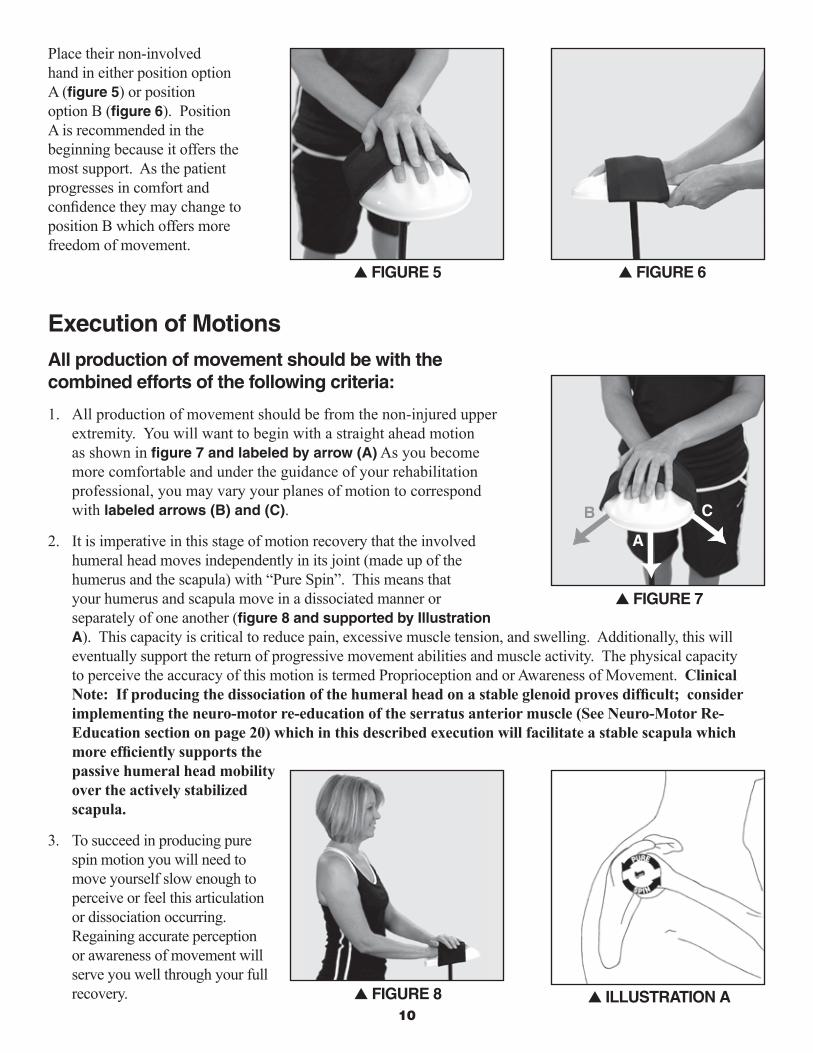
Place their non-involved hand in either position option A (fi gure 5) or position option B (fi gure 6). Position A is recommended in the beginning because it offers the most support. As the patient progresses in comfort and confi dence they may change to position B which offers more freedom of movement.
Execution of MotionsAll production of movement should be with the combined efforts of the following criteria:
1. All production of movement should be from the non-injured upper extremity. You will want to begin with a straight ahead motion as shown in fi gure 7 and labeled by arrow (A) As you become more comfortable and under the guidance of your rehabilitation professional, you may vary your planes of motion to correspond with labeled arrows (B) and (C).
2. It is imperative in this stage of motion recovery that the involved humeral head moves independently in its joint (made up of the humerus and the scapula) with “Pure Spin”. This means that your humerus and scapula move in a dissociated manner or separately of one another (fi gure 8 and supported by illustration A). This capacity is critical to reduce pain, excessive muscle tension, and swelling. Additionally, this will eventually support the return of progressive movement abilities and muscle activity. The physical capacity to perceive the accuracy of this motion is termed Proprioception and or Awareness of Movement. Clinical Note: If producing the dissociation of the humeral head on a stable glenoid proves diffi cult; consider implementing the neuro-motor re-education of the serratus anterior muscle (See Neuro-Motor Re-Education section on page 20) which in this described execution will facilitate a stable scapula which more effi ciently supports the passive humeral head mobility over the actively stabilized scapula.
3. To succeed in producing pure spin motion you will need to move yourself slow enough to perceive or feel this articulation or dissociation occurring. Regaining accurate perception or awareness of movement will serve you well through your full recovery.
s FiGURE 5
s FiGURE 8 s iLLUSTRATiON A
s FiGURE 6
s FiGURE 7
B
A
C

11
4. Always begin with a warm up using the base on or near the ground (fi gure 9). All warm-ups and any progressions in height should begin with partial strokes and gradually progress to full strokes.
Partial or Short strokes means that your forward motions are progressive and pain free. The forward motion is a blend of the contributory movements of the wrist, forearm, elbow, and shoulder – however, recall that all production of movement comes from the non-involved upper extremity. Avoid achieving full elbow extension at the expense of an elevated effort from your shoulder. Also, avoid moving the shoulder into extension (or the elbow past your side) upon the return of forward motion since this can potentially stress the front of the shoulder.
Full or Long strokes means that you have developed the capacity to move your elbow into full extension without the binding or straining of your shoulder. You will be taught by your rehabilitation professional that reaching full extension of your elbow requires a specifi c motion (supination) to occur in your forearm.
5. Perform up to 6-10 total strokes per height progression intervals. In the early stages only execute 1 to 3 height intervals (fi gure 10-12). As you progress in post-operative time, movement awareness, and endurance you will reach up to 3 to 5 height interval increases from your current beginning height and working towards the goal of approximately 90 to 110 degrees of elevation (fi gure 13-16).
s FiGURE 9
s FiGURE 10
s FiGURE 11 s FiGURE 12
s FiGURE 14 s FiGURE 15 s FiGURE 16
s FiGURE 13

1�
6. For heights in elevation above 70 degrees it is necessary to externally rotate your involved humerus and scapula (fi gure 17 and supported by illustration B). Observe how Position B of your non-involved hand can support this effort.
7. Always fi nish with a cool down, by working back down each of your height interval progressions until reaching your beginning level. During your cool down you can reduce your repetitions to 3-8, as well as shorten your strokes. Maintain pure spin motions and slow speed.
The following recommendations have been shown to be most effective, however for your personal needs, your rehabilitation professional may advise you differently.All patients who have succeeded with the UE Ranger have learned to stop themselves should they experience:1. Pain
2. An inability to either produce and or perceive pure spin and other correct biomechanics due to fatigue.
Frequency and volume of use• For pain relief: Perform as needed very gradual partial strokes to mid strokes, and proceed to full strokes for
1 to 3 sets, at 6-10 repetitions per set. For pain relief you will keep your height intervals from 1 to 3 and at a non-challenging fi nal height.
• For maintenance of current motion abilities: Up to 2 to 3 times per day utilizing the advised warm up and 1 to 3 height intervals with 6-10 repetitions per each non-challenging height interval.
• For progression of motion capacities: In addition to your daily maintenance, your rehabilitation professional may authorize you to challenge your current available motion. If allowed by your rehabilitation professional, it is recommended to challenge yourself after a warm-up reaching your current limit and then carefully work into a challenge. At your challenge height, perform 4 to 8 very light challenges and then proceed to work down your intervals for an appropriate cool down. This should be done no greater than 1 to 2 times per week with at least two days in between to allow your body to integrate the effort.
CAUTiON: NEvER CONTiNUE MOTiONS iF YOU ARE EXPERiENCiNG ANY PROGRESSiONS OF PAiN. ANY PAiN STEMMiNG FROM USE OF THE UE RANGER COULd BE RELATEd TO THE FOLLOWiNG REASONS:
Reasons for Pain:
1. Failure to adequately warm up and relax respective upper quadrant muscles
2. Not supporting a Pure Spin Motion
3. Failure to produce other correct biomechanics as movement involves the greater kinetic chain
4. Over extending your current physical capacities
s FiGURE 17 s iLLUSTRATiON B

1�
initiation and Progression of External RotationCAUTiON: FOLLOWiNG CERTAiN SURGiCAL PROCEdURES THiS MOTiON MAY NOT BE ALLOWEd BY YOUR SURGEON FOR UP TO 6 - 8 WEEKS FROM THE dATE OF YOUR SURGERY AS iT MAY STRAiN A PORTiON OF YOUR REPAiR. BEFORE PROCEEdiNG, BE SURE TO CLEAR THiS PARTiCULAR iNTROdUCTiON OF MOTiON WiTH YOUR REHABiLiTATiON PROFESSiONAL.
(Following the set up adjustments, the executions of movement are written from the perspective of the user, to support any copies you desire to provide for your patients.)
Set up adjustmentsFor this application remove the articulating base from the UE Ranger (fi gure 1). Depending on whether your patient has been advised to use a standard sling or one with a pillowed bolster you can have the patient rest their arm against their side or utilize a standard pillow to accommodate either situation as shown in (fi gures 2 and 3). At this point as in the previous section, prior to the execution of movement allow suffi cient time for their full upper extremity, shoulder girdle and neck to establish a sensation of security and relaxation.
All production of movement should be with the combined efforts of the following criteria:
• Following an adequate warm up as described above in the forward reaching section or that which is instructed to you by your rehabilitation professional
• The available pain free range of motion
• With the correct biomechanics, including pure spin, but with a different axis of rotation (fi gures 4 and 5 supported by illustration A)
s FiGURE 1 s FiGURE 2 s FiGURE 3
s FiGURE 4 s FiGURE 5 s iLLUSTRATiON A

14
• Attention and concentration is advised to insure you are producing actual shoulder rotations without mistaking either elbow or forearm substitutions
• The range of motion limit advised by your rehabilitation professional
You will want to perform up to 6 to 10 partial to full strokes. If advised by your rehabilitation professional you can perform 1 to 2 sets of this exercise per session. It is advised to end each session of external rotation motion support with a similar cool down as described in the previous section.
Clinical Consideration #2Consider implementing the above application processes into your patient’s pre-manual warm-ups. You may have heard the analogy of working smarter - not harder. This application can provide for your own hands and clinical mind, a relaxed patient and a supple shoulder joint from which you can apply your manual skills without having to take time quieting their guarded upper quadrant. The session will foster a post-treatment carryover and thus a real demonstration of clinical value. Clinical reminder: remember the gripped hand effects on the full kinetic chain and respiratory system in your consideration of the UBE as a warm up tool.
Reintegration Consideration #1From the rehabilitation professionals I have talked to, it is unanimous that most if not all patients are unable to perform pendulums correctly. Beyond the original intent of pendulums to potentially relax the shoulder, and in doing so potentially relieve pain, are the potential negative costs worth this effort?
Potential negative costs of performing pendulums:• Persons with lower back pain or orthostatic hypertension are going to either be physiologically on guard, and
therefore unable to relax and thus potentially walk away with an aggravated spine and reflexively guarded shoulder.
• While the postural alignment and execution of pendulums offers potential pain relief through the distraction forces on the gleno-humeral joint, what if these forces become higher than a grade one load on the joints soft tissues resulting in a reflexively guarded shoulder.
• From a consideration of function and the propensity in the early stages of AAROM and certainly AROM for persons to initiate nearly all forward movements with a compensatory shrug, would not quieting the shoulder on a consistent basis in a postural alignment meaningful to the whole body nervous system and resultant kinetic chain support be clinically superior to that of bending over and confusing the movement health healing processes. If this is meaningful to you and worth clinically executing with your patients, consider implementing it following your manual interventions as a means of securing or integrating the gains of mobility. In other words making the level of recovery for this session most meaningful to the nervous system versus off the plinth table and either out the door or to the lobby for static cryo-therapy.
Reintegration Consideration #2Commonly with any prolonged history of shoulder pain and resultant compensations there is to some degree muscular imbalances and resultant joint restrictions of the contralateral upper quadrant. Two positive opportunities exist within the patient’s current situation, with both supporting a greater outcome for the patient as a whole:1. As a means of rebalancing the shoulder’s neuro-motor system and subsequently resolving the pain pattern of the
contralateral shoulder girdle, guide your patient through the neuro-motor re-education section. Depending on the patients current involvements there may be a need to apply your manual skills to a mild degree, but worth it for the restoration of complementary biomechanics and an extra grateful patient.

15
2. The second opportunity is for the primarily involved shoulder’s neuro-motor system to be “tutored” by either the healthy or the secondarily involved shoulder complex. As the patient performs with their contralateral upper extremity, the full neuro-motor section and subsequent reintegration process, while the ipsilateral or primarily involved shoulder complex is provided a perceivable reference to guide its own neuro-motor re-education, to take place in a matter of 4 to 6 weeks. Using this lesson, the patient can combine motor imagery or mental practice executed by thinking through with the primarily involved extremity, a functional movement utilizing the learned perceived reference, creating a proprioceptive awareness of the intended movement to both support the recovery of the altered neuro-motor communications and progress towards integration with a meaningful and functional task. The use of motor imagery and mental practice has long been implemented by sports trainers and neuro-specialty rehabilitation professionals to both teach new motor skills and support the recovery following an injury.
USE OF iCE, REST, POSTURAL AWARENESS, ANd RESPiRATiONFollowing each session of UE Ranger Phase One exercise, it is imperative to rest and generally indicated to ice your shoulder (15 to 25 minutes). For optimal circulatory and motor relaxation support, position your upper extremity as shown in (fi gure 1).
HOME iCE RECiPE Place two parts water to one part rubbing alcohol in a Ziploc bag. Put this mixture in the freezer, which will produce a fl exible slush instead of a solid. This fl exibility allows the most contact area of your skin. Use two bags, one over the shoulder, and one in the axilla (arm pit). It is advised for safety and comfort to use some form of barrier (pillow case or t-shirt) between your skin and the ice.
When at rest, and separate from your icing times support your upper extremity at the elbow with pillows as previously shown in (fi gure 1). In this situation your sling should be off and efforts made to gradually move your elbow, forearm, and hand without moving your shoulder. This will be reviewed by your rehabilitation professional as a means of supporting circulation, preventing stiffening of these supportive joints, and alleviating some forms of pain related to reduced activity.
Respiration focused on using your diaphragm muscle, as demonstrated by (illustration A) (showing inhalation) and (illustration B) (showing exhalation), will support healthy mobility of your torso, which also infl uences shoulder function. Restricted mobility of your clavicle, your upper two ribs and their respective muscles can both compromise your full respiration as well as upper extremity function. Your brain perceives compression due to poor posture or muscle guarding due to pain or for protection as a threat and will respond with compensatory respiration. Imagine the drawing up effect of holding a helium balloon in your hand. These helium balloons provide you with a visualization cue that is designed to relax the traditionally over active muscles and restore the capacity to achieve relaxed comfortable postures and support full respiration.
s FiGURE 1
s iLLUSTRATiON Bs iLLUSTRATiON A

Rehab Innovations, Inc. • www.ueranger.com16
POST-OPERATivE ANd CHRONiC PAiN PROTOCOL • PHASE TWO
ACTivE ASSiSTivE RANGE OF MOTiON (AAROM)
By definition AAROM means that the involved upper extremity (UE) is now contributing to the motions produced. The degree to which the involved upper extremity successfully contributes to the production of motion will vary as a patient re-learns new motions and or as the person fatigues. In all situations the combination of the UE Ranger and the non-injured upper extremity and its full body supportive kinetic chain should be providing the necessary assistance of movement to insure the execution of healthy biomechanics.
AAROM Goals1. Preserve the integrity of the surgical repair
2. Maintain resolution of pain and swelling
3. Preserve the integrity of the circulatory system’s role of healing and prevent capsular adhesions and or myo-facial restrictions
4. Preserve primary or diaphragm supported respiration absent of neck and shoulder bracing
5. Preserve the capacity to achieve restorative sleep and minimize the need of medications with their resultant side effects
6. Facilitate neuro-motor re-education to support the introduction of balanced synergistic motor activation
7. Maintain resolution of a balanced ANS, absent of the fight or flight drivers evidenced in part by balanced resting tone when at rest
8. Restoration of patient produced Active Assistive Range of Motion within the capacity to produce proper biomechanics
9. Efforts will continue with your rehabilitation professional to gain further PROM
10. Integrate the shoulder girdle synergistic motor activity into the full body kinetic chain supportive system
Foundational Scientific Principles Supporting the UE Ranger’s Application in the AAROM Phase of Rehabilitation
To personally experience the science is an ideal way to appreciate the extent that subtleties matter when working with the nervous system and then to recognize how the UE Ranger can best facilitate and enhance your clinical interventions to most efficiently meet the goals of your patients.

Rehab Innovations, Inc. • www.ueranger.com1�
Clinical Consideration #3 Step one: With either of your hands make a moderately tensed gripped hand and position yourself with your arm by your side as shown in (fi gure 1).
Step two: Prior to the next instruction the goal of this example is for you to feel the point at which your effort to move becomes impeded by your own body’s resistance.
Step three:Begin to slowly raise your arm from the starting position as shown in (fi gure 1) into a movement of elevation (fi gure 2). Stop at the exact point in which you begin to feel impedance to your movement or in other words when you feel like your muscles tighten to go further. Mentally remember both this point and what the effort felt like.
Step four: Open your hand and return to the starting position as shown in (fi gure 3). Now with your open hand repeat the same motion as you produced in step 3 as shown in (fi gure 4). Compare in your mind both the feel of the motion and how far you go versus that with the gripped hand prior to feeling the impedance to your movement effort.
Results:Most often persons report that they can go further with a noticeable ease of the effort to move.
Why does This Clinically Matter? As you read this section, keep in mind as a global reference the 10 goals to be achieved in this second phase of rehabilitation.As mentioned in Clinical Consideration #1, a gripped hand with respect to the upper extremity is a: 1. Distal effort which requires a proximal role or effort to stabilize. 2. Muscular co-contraction from the elbow distally, which essentially diminishes the ease of movement production
throughout the full kinetic chain.A patient whom has recently undergone a surgical repair or who has been compensating for chronic pain related to an impaired rotator cuff musculo-tendonous unit will likely be utilizing a protective or substitutive motor pattern. The shoulder joint in the absence, for example of the supraspinatus muscle due to either a level of inhibition or general weakness will lack its communication and force couple role to guide the initiation and progression of varying functional movements. Thus prior to execution of volitional yet assisted motions of the involved upper extremity we need to, as indicated facilitate the contribution of the synergistic dynamic stabilizers and commonly weakened and even inhibited muscles.
s FiGURE 3 s FiGURE 4
s FiGURE 1 s FiGURE 2

18
Clinical Note: Considering the above clinical exercise of a gripped versus opened hand and the physiological responses we recognize the following two concerns:1. If a patient is asked to grip a rigid bar or pulley and has an insufficient rotator cuff to communicate the synergistic
responses, intensities, or endurances then a likely occurrence will be a reflexively substitutive and guarded shoulder.
2. Further if we ask a patient to grip a rigid bar or pulley and we combine the above scenario of an insufficient force couple with the impedance caused by the co-contraction effect or impedances of movement to the components of the poly-articular kinetic chain (fingers, wrist, forearm, elbow, and shoulder) we realize we will likely experience a limit to the patients use of their available PROM, followed by a likely occurrence of a reflexively substitutive and guarded shoulder.
From the 2008 University of Kentucky independent research study, the UE Ranger utilizing the executions of movements to be described below, demonstrated the capacity for a patient to self produce AAROM mobility of their involved shoulder in a progressively graded manner and without raising to a level of clinical concern, the motor activity of any portion of the rotator cuff (see research section). With this assurance in mind, we can proceed to accomplish the other 9 AAROM goals concurrently. Prior to the execution of any AAROM movement, please thoroughly read the following sections on neuro-motor re-education in its role to resolve pain and restore healthy biomechanics absent the compensations of the past. Then as indicated for your individual patient’s needs, apply the isolated neuro-motor facilitations by utilizing the UE Ranger as described for each of the dynamic stabilizers of the shoulder girdle.
Neuro-Motor Re-Education: The Keystone in Resolving Pain and Restoring Healthy Biomechanics of the Shoulder Girdle
“You mean I have to re-learn to walk all over again.” A statement I have heard so many times in working with patients with the needs of resolving patho-mechanics of gait, that I gave it enough thought to realize we never are really “taught” how to walk. In actuality walking is an innate ability that with no prior experience we experiment with our bodies through trial and error and the help of our parents until we learn how to crawl, stand, and finally walk. You may be wondering if this introduction was misplaced, asking yourself what does walking have to do with restoring healthy biomechanics and motor control of the shoulder girdle. My intention, for purposes of helping you and your patients achieve the greatest return for your efforts is by devoting our discussion to a system that can confuse us, frustrate us, amaze us, and certainly humble us, that of the nervous system. Within the title of this section and within the first sentence we see the words, re-educate and re-learn denoting prior to injury we must have known “how to – best move”. With an injury for example to our shoulder, our bodies become unable to fully execute a once mastered movement such as waving to a friend, reaching to the mid level shelves for a drinking glass, or even walking with a full body efficiency. How to – best move, now becomes dependent on what is available. Through the effects of various impairments such as pain avoidances, muscular spasms, swelling, joint restrictions, motor inhibition and actual weakness leads to both a state of unfamiliarity as well as vulnerability and finally to a further disorganized condition. Meanwhile life as we knew it tries to influence and restore normalcy, as the surrounding body is subconsciously recruited into developing compensations and before long these adaptations become invested in our nervous system as new motor pathways. Thus whether your patients have suffered from chronic shoulder pain or have recently undergone any form of a corrective surgical procedure to the shoulder, you can be assured the neuromuscular communications within the
19
shoulder system are to varying degrees out of balance and reorganized around compensatory biomechanics and therefore prone to supporting resultant micro-traumas and perpetuating pain if left unresolved. In the case of a post-operative patient, what must be remembered is that while the surgical procedure was likely successful in repairing/resolving for example an internal derangement (rotator cuff tear) and the resultant origin of pain, what remains a concern are the compensatory movement patterns which were established pre-surgically and capable of reproducing patho-mechanics sufficient to cause limits in healing and subsequent un-met expectations both short and long term. In order to most efficaciously serve our patients we must keep these above principles in mind as there is a broader presentation to clinically address with our treatment interventions versus considering only traditional PROM, and AAROM. An integral step in resolving compensatory patho-mechanics in either a post-surgical or chronic pain patient is to re-establish the neuro-motor communications of the surgically repaired, previously inhibited or weakened muscles. These weakened muscles which normally play a key role in healthy movements, have in a sense assumed a submissive role due to either post-operative induced inhibition or in the development of chronic compensatory movement patterns. In this context, the muscles assigned to make up for the deficits consequently dominate the neuro-muscular communications and ultimately become over worked, painful and contributory in either prolonging or even progressing the pathological state. Utilizing an analogy of that involving crisp communications similar to a harmonious orchestra within the neuro-motor system, we need to successfully restore the “voice” of the currently inhibited integral players, while quieting but not eliminating the contributions of the currently overpowering players. Utilizing the UE Ranger’s design and capacities enables you to meet each of your patients at their current impairment level and in a manner that is compatible with the biomechanics of the full upper extremity including its foundational shoulder girdle and its supportive sensory and motor nervous systems.
Reintegration Consideration #3
With respect to achieving the true restoration of this harmony, an analogy I share with my patients in our efforts to facilitate an inhibitory muscle in the midst of a dominating compensatory muscle or movement pattern is that of comparing a standard on/off light switch with that of a dimmer switch. In a standard switch you have an abrupt electrical connection producing an abrupt outcome. With a dimmer switch you have an ability to grade the electrical output and thus a subtle but effective outcome. In order to progressively recruit the motor units in a particular inhibited muscle, while progressively reducing the recruitment of motor units to a muscle involved in a substitution role one must subtly execute (dimmer switch) the prescribed movements versus an abrupt motion (standard on/off switch) absent of either the perception of proper execution or the sensation of a favorably altering motor activity. Below are the key principles which enable this analogy or graded dissemination of electrical activity to be restored. Recall that motor units are generally recruited in order of smallest to largest (fewest fibers to most fibers) as contraction increases.Among the key principles in recovering each of the subsequent muscles via this applications are:1. A relaxed state of both the involved shoulder girdle and ideally the ANS (Autonomic Nervous System), indicative
in this case by a relaxed mind and relaxed diaphragm supported respirations2. The presence of pain, while not ideal can still be considered an indication to pursue this form of intervention
as absence of any of these muscles can be a significant contribution to pain provocations both at rest and with movement
3. Allow the UE Ranger to completely support the weight of your arm as to avoid a reflexive compensatory response due to the brains recognition that any or all of the dynamic stabilizers are weak or inhibited
4. Sufficient joint mobility of intended movements5. Sufficient proprioception and understanding of the intended movement, as to enable an awareness of healthy
execution of movement versus perpetuating the patho-mechanics.

Rehab Innovations, Inc. • www.ueranger.com�0
Signs of failure to execute correctly and therefore unlikely for benefit to occur are:1. Holding of the breath2. Visibly compensating or bracing anywhere3. Provocation and or progression of pain, beyond that a person began the exercise with4. Absence of palpable facilitation of intended motor activity 5. Presence of palpable facilitation of un-intended motor activity
Commonly weakened or inhibited muscles in shoulder biomechanics:• Supraspinatus• Infraspinatus• Teres Minor• Serratus Anterior• Lower Trapezius • Respiratory Diaphragm
Commonly overworked or hypertonic muscles in shoulder biomechanics:• Upper Trapezius• Levator Scapulae• Scalenes• Deltoid• Pectoralis Minor• Pectoralis Major
Please take the time to educate yourself on each of the following exercises as the ability to alter the communications within the nervous system coordinating this dynamic and versatile region of our bodies is a subtle and progressive process. The intent of these exercises is to facilitate the motor activity of the weakened muscles initially in an isolated manner and absent the amplification of the chronically over active muscles and progressively integrating their designed role into the neuro-muscular control mechanisms relevant for daily living activities.
Neuro-Motor Re-Education:Serratus Anterior isolation
Goal: Facilitate amplification of the serratus anterior muscle, thus restoring a key neuro-motor communicator in the execution of proper biomechanics.Key Requirements:1. Pre-exercise systemic relaxation and proprioceptive awareness of the intended movement. 2. Proper adjustments and positioning of the UE Ranger and executions per below.3. The patient must allow the UE Ranger to support their arm’s weight; otherwise the hypertonic muscles
will both conflict with the proper alignment of the respective joint relations and consequently disrupt the intended precise message to the respective serratus anterior.
Unintended Result: Facilitating amplification of the commonly overworked or hypertonic muscles and resultant perpetuation of patho-mechanics of the shoulder girdle.

Rehab Innovations, Inc. • www.ueranger.com�1
In a standing position, as shown in (fi gure 1) adjust the UE Ranger so that the hand support suspends the weight of the persons involved arm when the elbow is bent to 85 degrees or a slight downward angle (this measurement is a rule of thumb). As you become familiar with the instructions below you will recognize it may require a slight adjustment higher or lower as to most effectively activate the serratus anterior, without facilitating an over powering pectoralis group, deltoid and or upper trapezius. Insert the patients hand into the support and place the base within the region of the front of their ipsilateral foot as to most comfortably support the weight of their involved arm as shown in (fi gure 2). Ask the patient with their non-involved hand to place one to two fi ngers against the cubital fossa as shown in (fi gure 3).
Instruct your patient to initiate movement into and within the sagittal to scaption planes as follows: 1. Produce a combined movement execution of slight supination and slight radial deviation of the forearm and
wrist respectfully and as shown in (fi gure 4). The patient should be instructed to focus a contact pressure of the hypo-thenar eminence into the hand support (fi gure 5 and supported by the concentric circles on the hand support as in a target) as well as given tactile cueing/feedback shown in (fi gure 6) by the therapist through the intended serratus anterior (right hand) and not the compensatory muscles in this case the anterior deltoid (left hand).
s FiGURE 1 s FiGURE 2 s FiGURE 3
s FiGURE 4 s FiGURE 5 s FiGURE 6

��
These combined efforts should produce a protraction of the scapula and drawing open the chest via the facilitation of the serratus anterior mms.• Note that the patients mind is to be thinking of shaking a persons hand
in front of them as shown in (fi gure 7). This commonly produced friendly gesture potentially can connect with previous motor planning sequences and is more likely to re-establish itself as a normal neuro-motor contributor.
Notice the overall excursion of movement is very minimal. The key is getting the right message to the right muscle.
Return to the resting position by your side and repeat the above execution up to the amount prescribed by your therapist, stopping for any of the following reasons:1. Onset of pain2. Fatigue in the form of inability to facilitate intended muscle activation3. Onset of compensatory efforts in the form of a shoulder shrug, holding your breath, or tensing your neck
and other unintended body parts
As a means of securing the intended benefi ts of this exercise and thus realizing a carryover of functional gain without the delayed onset of muscle soreness, it would be helpful to perform one to two sets of familiar motions (fi gures 8 and 9) of “Pure Spin” at low intensity AAROM or PROM depending on your level of fatigue or soreness. Clinical Note: Considering the execution of the above clinical exercise, recognize the opportunity to more effi ciently produce a pure spin or dissociated humeral head spinning on a now more stable glenoid fossa via the reactivation of the serratus anterior muscle.
Neuro-Motor Re-Education:Supraspinatus isolation
Goal: Facilitate amplifi cation of the supraspinatus muscle, thus restoring a key neuro-motor communicator in the execution of proper biomechanics.Key Requirements:1. Pre-exercise systemic relaxation and proprioceptive awareness of the intended movement. 2. Proper adjustments and positioning of the UE Ranger and executions per below.3. The patient must allow the UE Ranger to support their arm’s weight; otherwise the hypertonic muscles
will both confl ict with the proper alignment of the respective joint relations and consequently disrupt the intended precise message to the respective supraspinatus muscle.
Unintended Result: Facilitating amplifi cation of the commonly overworked or hypertonic muscles and resultant perpetuation of patho-mechanics of the shoulder girdle.
s FiGURE 7
s FiGURE 8 s FiGURE 9
Rehab Innovations, Inc. • www.ueranger.com��
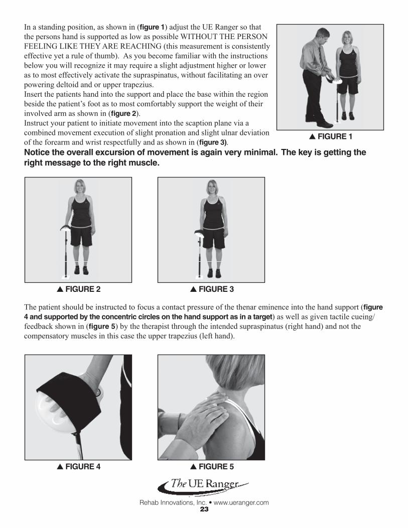
In a standing position, as shown in (fi gure 1) adjust the UE Ranger so that the persons hand is supported as low as possible WITHOUT THE PERSON FEELING LIKE THEY ARE REACHING (this measurement is consistently effective yet a rule of thumb). As you become familiar with the instructions below you will recognize it may require a slight adjustment higher or lower as to most effectively activate the supraspinatus, without facilitating an over powering deltoid and or upper trapezius.Insert the patients hand into the support and place the base within the region beside the patient’s foot as to most comfortably support the weight of their involved arm as shown in (fi gure 2).Instruct your patient to initiate movement into the scaption plane via a combined movement execution of slight pronation and slight ulnar deviation of the forearm and wrist respectfully and as shown in (fi gure 3). Notice the overall excursion of movement is again very minimal. The key is getting the right message to the right muscle.
The patient should be instructed to focus a contact pressure of the thenar eminence into the hand support (fi gure 4 and supported by the concentric circles on the hand support as in a target) as well as given tactile cueing/ feedback shown in (fi gure 5) by the therapist through the intended supraspinatus (right hand) and not the compensatory muscles in this case the upper trapezius (left hand).
s FiGURE 3
s FiGURE 4 s FiGURE 5
s FiGURE 1
s FiGURE 2

�4
This combined motion should create a tipping of the UE Ranger away from the body as shown in (fi gure 6). As the movement is initiated couple this effort by further activating the supraspinatus and fi nishing in an “Empty Can” position as shown in (fi gure 7).
Return to the resting position by your side and repeat the above execution up to the amount prescribed by your therapist, stopping for any of the following reasons:1. Onset of pain2. Fatigue in the form of inability to facilitate intended muscle activation3. Onset of compensatory efforts in the form of a shoulder shrug, holding your breath, or tensing your neck
and other unintended body parts
As a means of securing the intended benefi ts of this exercise and thus realizing a carryover of functional gain without the delayed onset of muscle soreness, it would be helpful to perform one to two sets of familiar motions (fi gures 8 and 9) of “Pure Spin” at low intensity AAROM or PROM depending on your level of fatigue or soreness.
Neuro-Motor Re-Education:isolation of the External Rotators
Goal: Facilitate amplifi cation of the external rotators (infraspinatus and teres minor muscles, thus restoring a key neuro-motor communicator in the execution of proper biomechanics.Key Requirements:1. Pre-exercise systemic relaxation and proprioceptive awareness of the intended movement. 2. Proper adjustments and positioning of the UE Ranger, and executions per below.3. The patient must allow the UE Ranger to support their arm’s weight; otherwise the hypertonic muscles
will both confl ict with the proper alignment of the respective joint relations and consequently disrupt the intended precise message to the respective External Rotators.
s FiGURE 7s FiGURE 6
s FiGURE 8 s FiGURE 9

�5
Unintended Result: Facilitating amplifi cation of the commonly overworked or hypertonic muscles and resultant perpetuation of patho-mechanics of the shoulder girdle.In a standing position, as shown in (fi gure 1) adjust the UE Ranger so that:1. the back edge of the base plate is lined up with the patient’s toes 2. base plate is approximately one foot away from the ipsilateral foot 3. overall height such that tilt of the guidance tubing positions your elbow
at approximately 80 degrees of fl exion4. and the hand supported with the palm facing down at approximately the height of the umbilicus
(This overall measurement is a rule of thumb). The key is to facilitate the gradual arc of motion outward while making sure, that at the most external excursion of this motion that the patients shoulder with respect to the commonly hypertonic muscles remain at or near a relaxed tone.
Instruct your patient to initiate movement into the transverse plane via a combined movement execution ofslight supination of the forearm and external rotation of the shoulder as shown in (fi gure 2). The patient should be instructed to focus a contact pressure of the hypo-thenar eminence into the hand support(fi gure 3 and supported by the concentric circles on the hand support as in a target) as well as given tactilecueing/feedback shown in (fi gure 4) by the therapist through the intended external rotators (right hand) and notthe compensatory muscles in this case the upper trapezius (left hand).
The patient in the early stages of restoring the tone of this muscle group should maintain a light contact of their elbow against their side. This will support the proper rotation of the shoulder joint and deter encouragement of the hypertonic muscles.
As a person becomes skilled with the basics of this exercise, instruct your patient to combine the setting of the scapula as learned in the neuro-motor re-education of the serratus anterior while supporting their humerus slightly away from their body and in the scaption plane as shown in (fi gure 5).This combined effort requires a higher level of coordination, by dissociating one joint movement from another an advancement of dexterity is made possible.Notice the overall excursion of movement remains very minimal. The key continues to be getting the right message to the right muscles. s FiGURE 5
s FiGURE 4s FiGURE 3
s FiGURE 1
s FiGURE 2

Rehab Innovations, Inc. • www.ueranger.com�6
Return to the starting or resting position and repeat the above execution up to the amount prescribed by your therapist, stopping for any of the following reasons:1. Onset of pain2. Fatigue in the form of inability to facilitate intended muscle activation3. Onset of compensatory efforts in the form of a shoulder shrug, holding your breath, or tensing your neck
and other unintended body parts
As a means of securing the intended benefi ts of this exercise and thus realizing a carryover of functional gain without the delayed onset of muscle soreness, it would be helpful to perform one to two sets of familiar motions (fi gures 6 and 7) of “Pure Spin” at low intensity AAROM or PROM depending on your level of fatigue or soreness.
Neuro-Motor Re-Education:isolation of the Lower Trapezius With the integration of the Rotator Cuff and Serratus Anterior Muscles
Goal: Facilitate amplifi cation of the lower trapezius muscle along with the rotator cuff and serratus anterior muscles, thus restoring a key synergistic neuro-motor communication in the execution of proper biomechanics.Key Requirements:1. Pre-exercise systemic relaxation and proprioceptive awareness of the intended movement 2. Proper adjustments and positioning of the UE Ranger and executions per below3. The patient must allow the UE Ranger to support their arm’s weight; otherwise the hypertonic muscles
will both confl ict with the proper alignment of the respective joint relations and consequently disrupt the intended precise message to the respective lower trapezius and complimentary muscles
Unintended Result: Facilitating amplifi cation of the commonly overworked or hypertonic muscles and resultant perpetuation of patho-mechanics of the shoulder girdle.
NOTE: As a clinical example, typically the lower trapezius muscle begins to contribute to elevations of the humerus via the combined movements of the scapula at humeral elevations from 50 to 70 degrees through end ranges.
s FiGURE 6 s FiGURE 7
��
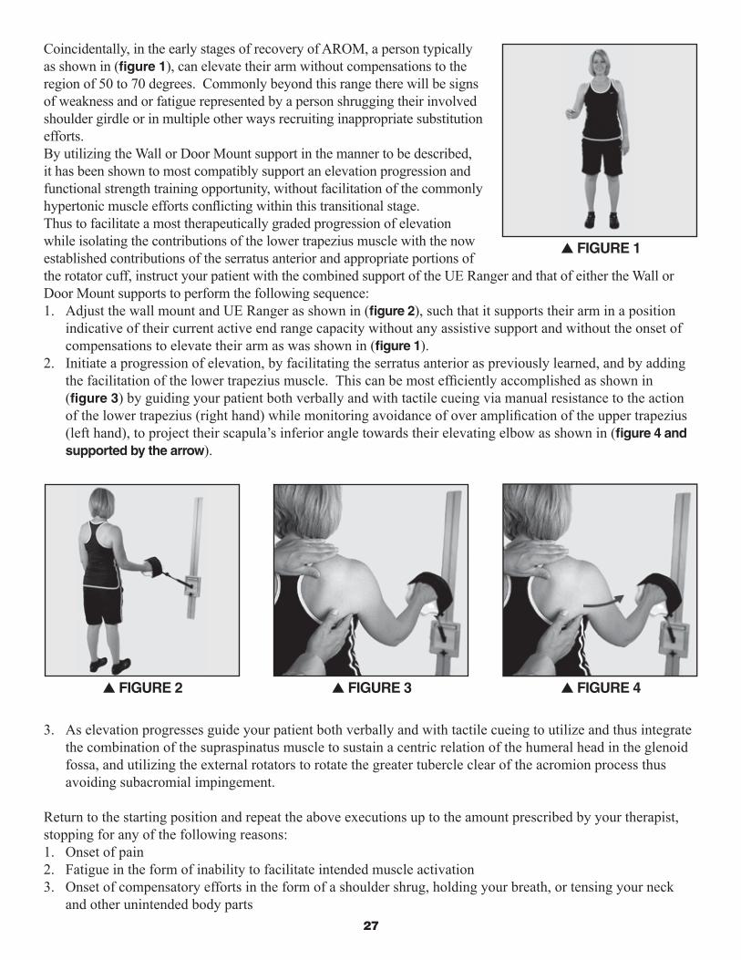
Coincidentally, in the early stages of recovery of AROM, a person typically as shown in (fi gure 1), can elevate their arm without compensations to the region of 50 to 70 degrees. Commonly beyond this range there will be signs of weakness and or fatigue represented by a person shrugging their involved shoulder girdle or in multiple other ways recruiting inappropriate substitution efforts. By utilizing the Wall or Door Mount support in the manner to be described, it has been shown to most compatibly support an elevation progression and functional strength training opportunity, without facilitation of the commonly hypertonic muscle efforts confl icting within this transitional stage.Thus to facilitate a most therapeutically graded progression of elevation while isolating the contributions of the lower trapezius muscle with the now established contributions of the serratus anterior and appropriate portions of the rotator cuff, instruct your patient with the combined support of the UE Ranger and that of either the Wall or Door Mount supports to perform the following sequence:1. Adjust the wall mount and UE Ranger as shown in (fi gure 2), such that it supports their arm in a position
indicative of their current active end range capacity without any assistive support and without the onset of compensations to elevate their arm as was shown in (fi gure 1).
2. Initiate a progression of elevation, by facilitating the serratus anterior as previously learned, and by adding the facilitation of the lower trapezius muscle. This can be most effi ciently accomplished as shown in (fi gure 3) by guiding your patient both verbally and with tactile cueing via manual resistance to the action of the lower trapezius (right hand) while monitoring avoidance of over amplifi cation of the upper trapezius (left hand), to project their scapula’s inferior angle towards their elevating elbow as shown in (fi gure 4 and supported by the arrow).
3. As elevation progresses guide your patient both verbally and with tactile cueing to utilize and thus integrate the combination of the supraspinatus muscle to sustain a centric relation of the humeral head in the glenoid fossa, and utilizing the external rotators to rotate the greater tubercle clear of the acromion process thus avoiding subacromial impingement.
Return to the starting position and repeat the above executions up to the amount prescribed by your therapist, stopping for any of the following reasons:1. Onset of pain2. Fatigue in the form of inability to facilitate intended muscle activation3. Onset of compensatory efforts in the form of a shoulder shrug, holding your breath, or tensing your neck
and other unintended body parts
s FiGURE 3 s FiGURE 4
s FiGURE 1
s FiGURE 2

Rehab Innovations, Inc. • www.ueranger.com�8
As a means of securing the intended benefi ts of this exercise and thus realizing a carryover of functional gain without the delayed onset of muscle soreness, it would be helpful to perform one to two sets of familiar motions (fi gures 5 and 6) of “Pure Spin” at low intensity AAROM or PROM depending on your level of fatigue or soreness.
s FiGURE 5 s FiGURE 6
initiation and Progression of Forward Reaching and Elevations - AAROM(Following the set up adjustments, the executions of movement are written from the perspective of the user, as to support any copies you desire to provide for your patients)
Set up adjustments With the patient in a standing position, adjust the length of the UE Ranger to approximately the height of their elbow, as to duplicate the prior supported and resting position of their arm in its sling (fi gure 1). If a person is unable to stand simply duplicate this measurement and all further instructions/applications from a seated position (fi gure 2).
Place their involved hand in the molded support and comfortably secure it with the overlying strap, (fi gure 3). At this point allow suffi cient time for their full upper extremity, shoulder girdle and neck to establish a sensation of security and relaxation (fi gure 4).
s FiGURE 1
s FiGURE 3s FiGURE 2 s FiGURE 4

�9
Execution of MotionsCAUTiON SHOULd BE GivEN TO THE AMOUNT OF EFFORT THAT iS GivEN FROM THE iNvOLvEd UPPER EXTREMiTY iN ALL OF THE FOLLOWiNG PROGRESSiONS. UNTiL YOU HAvE BEEN SPECiFiCALLY iNSTRUCTEd BY YOUR REHABiLiTATiON PROFESSiONAL ANd HAvE dEMONSTRATEd SAFE TECHNiQUE, dO NOT AdvANCE YOURSELF iN ANY OF THE FOLLOWiNG LEvELS.
NEvER CONTiNUE MOTiONS iF YOU ARE EXPERiENCiNG ANY PROGRESSiONS OF PAiN. SUCH PAiN PROvOCATiONS COULd BE RELATEd TO THE FOLLOWiNG REASONS:
1. Going too fast, thus not supporting dissociations of joint movements to be realized
2. Motor imbalance of the dynamic stabilizers and movers
3. Failure to produce correct biomechanics
4. Over extending your current physical capacities
• Always begin with a warm up, with the base placed on the ground or platform and the UE Ranger working height at a comfortable level.
• Progressions in variable planes of movement are described and illustrated below. The sequence of order is designed for optimal success and according to both the findings of the University of Kentucky’s research study and that of clinical observation. Recognize however that there exists, both variances in patient presentations and patient responses thus it is again imperative that you utilize the guidance of your rehabilitation professional. As you progress in degree of difficulty, your rehabilitation professional may encourage you to perform a specific sequence of these challenges and benefits. The progressions within each of these levels should only be initiated under the guidance of your rehabilitation professional.
• At the first sign of unhealthy biomechanics due to pain, fatigue, or poor coordination, the patient should either correct the biomechanics or return to a lower intensity and begin an appropriate cool down. It is recommended that your cool downs follow the guidelines as described in the cool down instructions of Phase One or that of your rehabilitation professional.
All production of movement should be with the combined efforts of the following criteria:
1. Within the current capable volitional effort of the involved upper extremity without provocation of pain and or compensations.
2. With the necessary supplemental support of the UE Ranger and the non-involved upper extremity.
Closed Kinetic Chain – Standing Floor to Platform Supportive ProgressionsCharacteristics:
• Foundationally the lowest level of intensity
• Foundationally the lowest level of difficulty
• Functionally meaningful and integrative into broader kinetic chain movements

Rehab Innovations, Inc. • www.ueranger.com�0
1. Production of movement should be from the involved upper extremity, and as needed with an intermittent involvement of the supplemental non-involved upper extremity. You will want to begin with a straight ahead motion as shown in (fi gure 1). As you become more comfortable and under the guidance of your rehabilitation professional, you may vary your planes of motion to correspond with both your tolerances and allowances.
2. It is imperative in this stage of volitional motion recovery to continue the production of the involved humeral head moving independently in its joint (made up of the humerus and the scapula) with “Pure Spin”. This means that your humerus and scapula move in a dissociated manner or separately of one another (fi gure 2 and supported by illustration A). This capacity at this stage is indicative of successful motor facilitation and subsequent support of healthy initial biomechanics. Additionally, this will eventually support the return of progressive movement abilities and supportive muscle activity.
Clinical Note: If producing the dissociation of the humeral head on a stable glenoid proves diffi cult; consider implementing the neuro-motor re-education of the serratus anterior muscle (See Neuro-Motor Re-Education section on page 20), which in this described execution will facilitate a stable scapula which more effi ciently supports the passive humeral head mobility over the actively stabilized scapula.
Additionally, the active role of the supraspinatus is extremely infl uential in commanding the balance of communications within the active muscles associated with this and many other progressive functional movements of the shoulder girdle.
Reintegration Consideration #4 When necessary for the rehabilitation specialist to assist you with the facilitation of these muscles attempt while your therapist is working with you to sense what the activation below the therapist’s fi ngers actually feels like and attempt to sustain and or reproduce this feel as you proceed through the session your therapist is guiding you through. In addition when performing your home exercises and or self warm ups and cool downs utilize the lessons learned in Reintegration Consideration #2 of the PROM section as you attempt to reintegrate the role of these dynamic infl uencers of the shoulder’s function. Remember also, that to succeed in producing pure spin motion you will need to move yourself slow enough to perceive or feel this actual articulation or dissociation occurring. Regaining accurate perception or awareness of movement will continue to serve you well through your full recovery.
s FiGURE 1
s FiGURE 2 s iLLUSTRATiON A
B
A
C

Rehab Innovations, Inc. • www.ueranger.com�1
Always begin with a warm up using the base on or near the ground (fi gure 3). All warm-ups and any progressions in height should begin with partial strokes and gradually progress to full strokes.
Partial or Short strokes means that your forward motions are progressive and pain free. The forward motion is a blend of the contributory movements of the involved shoulder, elbow forearm and wrist. Avoid achieving full elbow extension at the expense of an excessive effort from your shoulder. Also, avoid moving the shoulder into extension (or the elbow past your side) upon the return of forward motion since this can potentially stress the front of the shoulder.
Full or Long strokes means that you have developed the capacity to move your elbow into full extension without the binding or straining of your shoulder. You will be taught by your rehabilitation professional that reaching full extension of your elbow requires a specifi c motion (supination) to occur in your forearm.
Perform up to 6 -10 total strokes per height progression intervals. In the early stages only execute 1 to 3 height intervals (fi gures 4-6). As you progress in post-operative time, movement awareness, and endurance you will reach up to 3 to 5 height interval increases from your current beginning height and working towards the goal of approximately 90 to 110 degrees of elevation (fi gure 7-8).
s FiGURE 3
s FiGURE 4 s FiGURE 5 s FiGURE 6
s FiGURE 7 s FiGURE 8

Rehab Innovations, Inc. • www.ueranger.com��
For heights in elevation above 70 degrees it is necessary to externally rotate your involved humerus. Clinical Note: If producing the external rotation of the humeral head on a dynamically advancing scapula proves diffi cult; consider implementing the neuro-motor re-education of the external rotators along with the progressive role of the serratus anterior and the supraspinatus (fi gure 9 and supported by illustration B). Also observe and utilize if necessary how Position B of your non-involved hand can support this effort.
CAUTiON: FOLLOWiNG CERTAiN SURGiCAL PROCEdURES THiS MOTiON MAY NOT BE ALLOWEd BY YOUR SURGEON FOR UP TO 6 TO 8 WEEKS FROM THE dATE OF YOUR SURGERY AS iT MAY STRAiN A PORTiON OF YOUR REPAiR. BEFORE PROCEEdiNG, BE SURE TO CLEAR THiS PARTiCULAR iNTROdUCTiON/ PROGRESSiON OF MOTiON WiTH YOUR REHABiLiTATiON PROFESSiONAL.
Always fi nish with a cool down by working back down each of your height interval progressions until reaching your beginning level. During your cool down you can reduce your repetitions to 3-8, as well as shorten your strokes. Maintain pure dissociated motions and slow speed.
Reintegration Consideration #5As mentioned in the introductory section of neuro-motor re-education as it relates to the restoration of integrated biomechanics and as a means of securing the above facilitation of motor activity consider the following commonly found whole body compensation as well as apply the described execution of movement reintegration to resolve this potentially perpetuating provocation of shoulder micro-traumas. The scenario is with shoulder pain especially that requiring a period of sling immobilization, there often is a reduction of functional mobility both across the torso and pelvic girdle and into one or both of the lower extremities as related to the normally whole body investment in the act of ambulation. The following sequence has been successfully demonstrated in the clinic to both unload the heavily invested neuro-motor dyskinesia thus both enhancing the above efforts to restore dynamic control of the involved shoulder girdle, but also to contribute to the dissipation of the patho-mechanical compensatory gait. Depending on the investment in other regions of the body a broader evaluation and intervention process may be necessary to fully restore the complexities of gait.
s FiGURE 9 s iLLUSTRATiON B
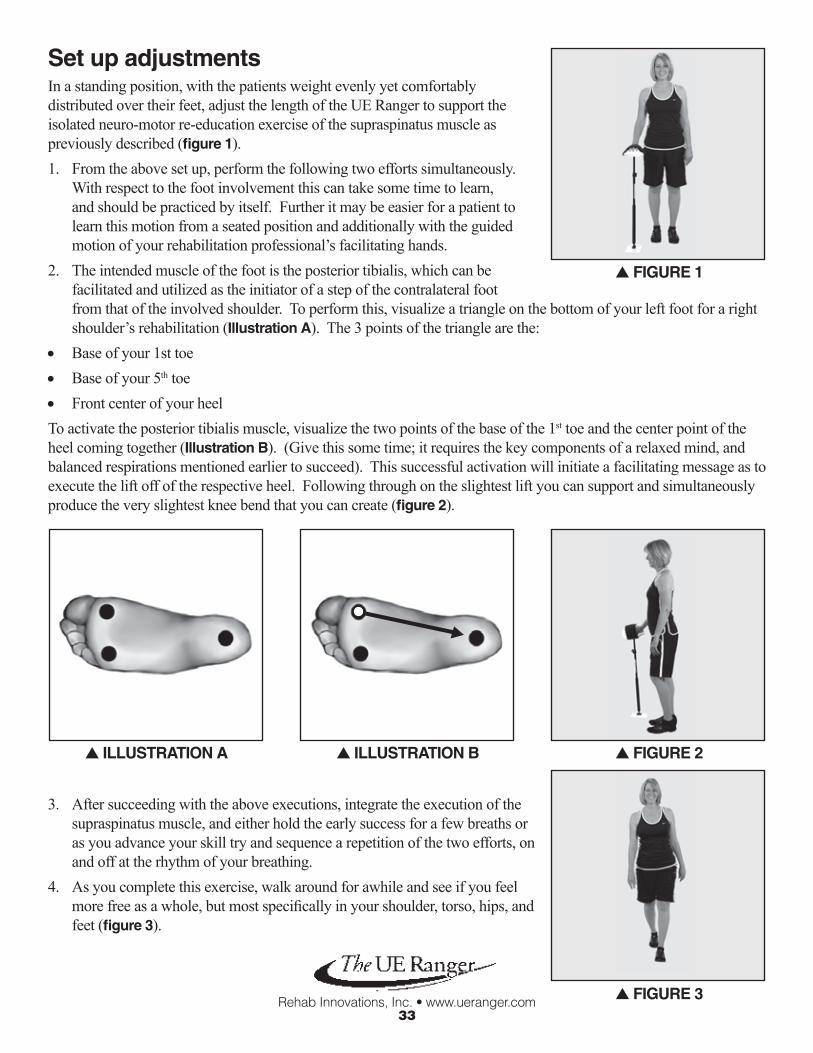
Rehab Innovations, Inc. • www.ueranger.com��
Set up adjustmentsIn a standing position, with the patients weight evenly yet comfortably distributed over their feet, adjust the length of the UE Ranger to support the isolated neuro-motor re-education exercise of the supraspinatus muscle as previously described (fi gure 1).1. From the above set up, perform the following two efforts simultaneously.
With respect to the foot involvement this can take some time to learn, and should be practiced by itself. Further it may be easier for a patient to learn this motion from a seated position and additionally with the guided motion of your rehabilitation professional’s facilitating hands.
2. The intended muscle of the foot is the posterior tibialis, which can be facilitated and utilized as the initiator of a step of the contralateral foot from that of the involved shoulder. To perform this, visualize a triangle on the bottom of your left foot for a right shoulder’s rehabilitation (illustration A). The 3 points of the triangle are the:
• Base of your 1st toe• Base of your 5th toe• Front center of your heelTo activate the posterior tibialis muscle, visualize the two points of the base of the 1st toe and the center point of the heel coming together (illustration B). (Give this some time; it requires the key components of a relaxed mind, and balanced respirations mentioned earlier to succeed). This successful activation will initiate a facilitating message as to execute the lift off of the respective heel. Following through on the slightest lift you can support and simultaneously produce the very slightest knee bend that you can create (fi gure 2).
3. After succeeding with the above executions, integrate the execution of the supraspinatus muscle, and either hold the early success for a few breaths or as you advance your skill try and sequence a repetition of the two efforts, on and off at the rhythm of your breathing.
4. As you complete this exercise, walk around for awhile and see if you feel more free as a whole, but most specifi cally in your shoulder, torso, hips, and feet (fi gure 3).
s FiGURE 1
s iLLUSTRATiON A s iLLUSTRATiON B s FiGURE 2
s FiGURE 3

Rehab Innovations, Inc. • www.ueranger.com�4
Open Kinetic Chain – Hook-lying PositionCharacteristics:
• Foundationally a mild to moderate level of intensity
• Foundationally a mild to moderate level of diffi culty
• Functionally meaningful in terms of capacity to enhance endurances of dynamic stabilizers
Set up adjustmentsWith the patient in a hook-lying position as shown in (fi gure 1)
1. Beginning with the UE Ranger in the fully closed position (also fi gure 1) elevate your involved upper extremity from a resting position to a vertical position or 90 degrees of fl exion (fi gure 2).
2. Due to the potential of an elevated activation of muscular contraction of the involved shoulder, extreme caution should be observed both in initiating this motion and returning to rest in the 0 to 90 degree range. In this respect, simply utilize the non-involved upper extremity as the primary mover in getting the involved upper extremity to and from 90 degrees.
3. The 90 degree position can be considered a “home base” position for both rest, and progression of movement in varying planes (fi gures 3-5). Equally important, as above in progressing away from the 90 degree base position, is the positioning of the UE Ranger guidance handle in relation to gravity. For maximum support you will want to position the guidance handle to optimally support the involved shoulder.
s FiGURE 2
s FiGURE 3 s FiGURE 4 s FiGURE 5
s FiGURE 1

Rehab Innovations, Inc. • www.ueranger.com�5
4. An excellent opportunity in this stage of AAROM and from this position is to restore the role of the rotator cuff as a force couple. This can proceed from approximately 70 degrees and progress as far as the patient is currently able to safely move as in (fi gures 6-8, and supported by illustrations A, B, and C). As the patient initiates this sequence as described above and demonstrated in fi gures 6-8, the required inferior glide produced by the force couple can potentially be more easily taught and successfully produced by the patient envisioning the action of a pole vault (supported by illustration d). The resultant action produced by this force couple is essentially an inferior glide of the humeral head into the inferior capsule with the patient potentially feeling an axillary stretch.
s FiGURE 6 s FiGURE 7
s iLLUSTRATiON C
s FiGURE 8
s iLLUSTRATiON A s iLLUSTRATiON B
s iLLUSTRATiON d
a) Subscapularisb) Infraspinatusc) Teres Minord) Supraspinatus
ab
c
d

Rehab Innovations, Inc. • www.ueranger.com�6
5. Lastly, another option from this position is to isolate gravity resisted strengthening of the serratus anterior muscle in the traditional scapular protraction or “scapular punch” motions demonstrated in (fi gures 9 and 10). However, by utilizing the support of the UE Ranger it offers another isolated facilitation opportunity to unload the invested neuro-motor dyskinesia thus both enhancing the efforts to restore dynamic control of the involved shoulder girdle, and in a positional alignment to support endurance training.
Closed Kinetic Chain Wall or door Mounted Tracking FrameCharacteristics:
• Foundationally a mild to moderate level of intensity and diffi culty
• Functionally meaningful and integrative into broader kinetic chain movements
• Functionally meaningful in terms of capacity to enhance endurances of dynamic stabilizers
indications:
• Execute if a person is challenged in the 70 to 110 degree range of AAROM with the base on the progression of height intervals
• Execute when a person can elevate their involved UE to 40 to 70 degrees with AROM and without compensations
• *** Precursor to Open Kinetic Chain Elevation – Standing Position
Set up adjustmentsSecurely insert the base of the UE Ranger in the tracking frame of the Clinical Wall Mount or Home Door Mount. Per indications, of compensatory onset of either AAROM or AROM, position the tracking frame and respective UE Ranger adjustments such that the person begins their execution of elevation approximately 10 degrees of gleno- humeral joint fl exion below that of where they have demonstrated the onset of either compensations and or fatigue. (If a person fatigues or begins to shrug or produce any other form of compensation at 55 degrees then adjust them to begin their execution of elevations at 45 degrees).
s FiGURE 9 s FiGURE 10

��
*** Within this setup the guidance tubing angle of the UE Ranger in relation to either the Wall Mount or Door Mount should be in the range to best offer both:
1. A mechanical support
2. The capacity to elevate their shoulder up to 20 degrees from their starting height of 45 degrees in the above example
Clinical Note: The 20 degrees of available range should be viewed as the training interval from which to execute the foundational principles of neuro-motor reintegration and biomechanics. Within this interval of rehabilitation, the execution of this exercise can incorporate the facilitation of the previously described lower trapezius muscles as well as integrate the full system of dynamic stabilizers according to the description of the just described force couple or in this case the inferior glide.
As indicated and instructed to you by your rehabilitation professional, you can progress yourself in the intensity level by raising the height of the tracking frame to the recommended level and appropriate challenge (fi gures 1-4).
Reintegration Consideration #6As mentioned in the introductory section of neuro-motor re-education as it relates to the restoration of integrated biomechanics and as a means of securing the above facilitation of motor activity, consider the commonly found whole body compensation of reaching to a mid to higher level shelf, as well as apply the described execution of movement reintegration to resolve this potentially perpetuating provocation of shoulder micro-traumas. Commonly with such a functional limitation we lose the supportive role of nearly the whole body beyond the shoulder. For whatever reason, many people forget that a normal execution of reaching to mid and higher levels involves the full kinetic chain clear to the toes if necessary.
Set up adjustmentsIn the process of performing the exercise as described above with the wall mount tracking frame, position yourself with the ipsilateral or same side lower extremity as the involved shoulder ahead of the contralateral lower extremity. 1. From this above set up, perform the following two efforts simultaneously. With respect to the foot involvement
this can take some time to learn, and should be practiced by itself. Further it may be easier for a patient to learn this motion from a seated position and additionally with the guided motion of your rehabilitation professional’s facilitating hands.
s FiGURE 2 s FiGURE 3 s FiGURE 4
s FiGURE 1

�8
2. The intended muscle of the foot is the posterior tibialis, which can be facilitated and utilized as the initiator of a step of the contralateral foot from that of the involved shoulder. To perform this, visualize a triangle on the bottom of your left foot for a right shoulder’s rehabilitation (illustration A). The 3 points of the triangle are the:• Base of your 1st toe• Base of your 5th toe• Front center of your heel
To activate the posterior tibialis muscle, visualize the two points of the base of the 1st toe and the center point of the heel coming together (illustration B). (Give this some time; it requires the key components of a relaxed mind, and balanced respirations mentioned earlier to succeed). This successful activation will initiate a facilitating message as to execute the lift off of the respective heel. Following through on the slightest lift you can support and simultaneously produce the very slightest knee bend that you can create (fi gure 1).
After succeeding with the above foot and knee progressions, integrate the executions of elevation described above with a simultaneous progression of body weight transfer onto the front right lower extremity for energy transfer and fulfi lling a whole body reach within the current capacities of the involved shoulder.
Reintegration Consideration #7Set up adjustmentsWithin the lower half of the 20 degree training interval described above, an option to consider is that classifi ed as rhythmic stabilization, utilizing:1. An external stimulation such as a
manual perturbation from proximal kinetic UE chain and progressively distally, while the patient stabilizes the static position of the full upper quadrant, absent compensations such as holding the breath. Also consider eyes closed and open for further proprioception challenges (fi gure 1).
2. Patient facilitated humeral head rhythmic movements on stable glenoid, such as ABC’s of varying intensities such as size and speed (fi gure 2).
s FiGURE 1s iLLUSTRATiON A s iLLUSTRATiON B
s FiGURE 1 s FiGURE 2

Rehab Innovations, Inc. • www.ueranger.com�9
3. Patient facilitated glenoid rhythmic movements on stable humeral head, such as the patient’s whole body turning in variable planes around the stable humerus (fi gures 3 and 4).
**Be sure to try and apply an integrative execution such as functional reaching following this intervention type as well as participating in an appropriate cool down. Also consider mental imagery of the exercises you just performed while resting with or without ice as indicated to you by your rehabilitation professional.
Open Kinetic Chain External Rotation – Standing PositionCharacteristics:
• Foundationally a mild to moderate level of intensity and diffi culty
• Functionally meaningful and integrative into broader kinetic chain movements
• Functionally meaningful in terms of capacity to enhance endurances of dynamic stabilizers
indications:
• Execute if a person is challenged in the above execution of AAROM elevation intervals
• Execute when a person can elevate their involved UE to 40 to 70 degrees with AROM and without compensations
• *** Precursor to Open Kinetic Chain Elevation – Standing Position
Set up adjustmentsBegin with the involved upper extremity supported in a position within the scaption plane (as described to you by your rehabilitation professional) and in a comfortable level of elevation (fi gure 1). It is advised to maintain some form of external support for the elbow to rest on (initially that can be your rehabilitation professionals hand which can allow this person to cue the appropriate movement) progressing to the tracking frame with a towel to cushion your support or comparable book shelf at home, and fi nally progressing further to a mild pressure against the wall. These progressive supports help tremendously to guide the very precise motion of external rotation. Attention and concentration is advised to insure you are producing actual shoulder rotations without mistaking either elbow or forearm replications. For maximum benefi t insure that your motion is opposing gravity.
1. Progress as needed to assist the involved upper extremity through the current available range of external rotation motion. It is recommended (within the safe limits of the involved shoulder muscles) to fully participate in the execution of this effort of motion.
s FiGURE 1
s FiGURE 3 s FiGURE 4

40
2. Within the safe limits of your capacity to support healthy biomechanics, progressively elevate your upper extremity (fi gures 2-8).
3. Gradually you can return to lower elevations and produce pure external rotation with or without support of the elbow at your side as a cool down.
4. Additionally you can perform the cool down progression as you learned in Phase One.
Open Kinetic Chain Elevation – Standing PositionCharacteristics:
• Foundationally a moderate to signifi cant level of intensity (as most challenging to not engage the upper trapezius and deltoid)
• Foundationally a mild to moderate level of diffi culty
• Therapeutically effective for end range elevation infl uences of neuro-motor endurances of tone balance of the dynamic stabilizers
• Therapeutically effective for end range elevation infl uences of soft tissue elasticity
• Functionally meaningful and integrative into broader kinetic chain movements
s FiGURE 5 s FiGURE 6 s FiGURE 7
s FiGURE 8
s FiGURE 4s FiGURE 2 s FiGURE 3
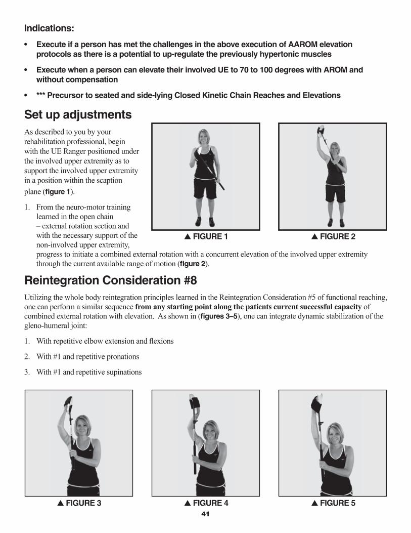
41
indications:
• Execute if a person has met the challenges in the above execution of AAROM elevation protocols as there is a potential to up-regulate the previously hypertonic muscles
• Execute when a person can elevate their involved UE to 70 to 100 degrees with AROM and without compensation
• *** Precursor to seated and side-lying Closed Kinetic Chain Reaches and Elevations
Set up adjustmentsAs described to you by your rehabilitation professional, begin with the UE Ranger positioned under the involved upper extremity as to support the involved upper extremity in a position within the scaption plane (fi gure 1).
1. From the neuro-motor training learned in the open chain – external rotation section and with the necessary support of the non-involved upper extremity, progress to initiate a combined external rotation with a concurrent elevation of the involved upper extremity through the current available range of motion (fi gure 2).
Reintegration Consideration #8Utilizing the whole body reintegration principles learned in the Reintegration Consideration #5 of functional reaching, one can perform a similar sequence from any starting point along the patients current successful capacity of combined external rotation with elevation. As shown in (fi gures 3–5), one can integrate dynamic stabilization of the gleno-humeral joint:
1. With repetitive elbow extension and fl exions
2. With #1 and repetitive pronations
3. With #1 and repetitive supinations
s FiGURE 2
s FiGURE 3 s FiGURE 4 s FiGURE 5
s FiGURE 1

Rehab Innovations, Inc. • www.ueranger.com4�
Closed Kinetic Chain – Seated Floor to Platform Supportive ProgressionsCharacteristics:
• Foundationally moderate level of intensity and difficulty, thus susceptible to engaging the previous compensatory patterns
• Functionally meaningful in terms of capacity to enhance endurances of dynamic stabilizers
• Functionally meaningful and integrative into broader kinetic chain movements
indications:
• Execute if a person has met the challenges in the above execution of AAROM elevation protocols as there is a potential to up-regulate the previously hypertonic muscles
• Execute when a person can from standing, elevate their involved UE to 70 to 100 degrees with AROM and without compensations
• *** Precursor to Side-lying Closed Kinetic Chain Reaches and Elevations
Set up adjustmentsAs previously discussed, begin with the UE Ranger adjusted to support the patient’s upper extremity in a position with the elbow being flexed to 85 to 90 degrees.
Clinical Note: In terms of upper extremity execution, all efforts are the same as the standing floor to platform supportive progressions, however in a seated position there is a considerable higher level of effort required from the shoulder girdle due to the legs being in a position of minimal contribution in terms of force production. Thus there is a susceptibility to recruit old compensatory patterns.
Reintegration Consideration #9Due to the common uses of the shoulder and distal upper extremity in the lower ranges of elevation while sitting, consider in (figures 1-5) along with mild progressions of elevation and multi-plane task oriented movements, to incorporate the progression of dynamic core muscles which can offer the dynamic replacement support which until now has been provided by the combined kinetic chain energy originating in the lower extremities and core muscles in the standing exercises.
Also, consider combining the previous discussion described in Reintegration Consideration #2 of utilizing benefits of mental imagery along with the support of the UE Ranger as one imagines meaningful tasks such as those below:
• Putting the car key in the ignition for right shoulder patients
• Operating the car radio for right shoulder patients
• Executing the computer keys or TV remote for either a left or right shoulder patient
• Playing cards or board games for either a left or right shoulder patient
• Eating and or reaching for a glass for either a left or right shoulder patient

Rehab Innovations, Inc. • www.ueranger.com4�
Closed Kinetic Chain Elevation – Side-lying Position Characteristics:
• Foundationally moderate level of intensity and diffi culty, thus susceptible to engaging the previous compensatory patterns
• Functionally meaningful in terms of capacity to enhance endurances of dynamic stabilizers
indications:
• Execute if a person has met the challenges in the above execution of AAROM elevation protocols as there is a potential to up-regulate the previously hypertonic muscles
• Execute when a person can from standing, produce a combined external rotation and elevation of their involved UE to 70 to 100 degrees with AROM and without compensations
s FiGURE 1 s FiGURE 2 s FiGURE 3
s FiGURE 4 s FiGURE 5

44
Set up adjustmentsBegin with the patient lying on their non-involved side, with the involved upper extremity supported in a position of comfort by the UE Ranger (fi gure 1).
Clinical Note: From this position and with the support of the UE Ranger, the patient can successfully work on the endurance of the muscles that support elevation with the integrative role of the combined gravity resisted external rotation. In this position the motion of elevation is in a gravity lessened infl uence, thus if the patient fi nds comfort in this postural setup, they can work on the endurance of elevations with a simultaneous investment in challenging the external rotators (fi gure 2).
Reintegration Consideration #10Similarly to the execution of movement considerations of Reintegration Consideration #7 your patient can perform from any starting point along the patient’s current successful capacity of combined external rotation with elevation, as shown in (fi gures 3–5), the dynamic stabilization integration of the gleno-humeral joint:
1. With repetitive elbow extension and fl exions
2. With #1 and repetitive pronations
3. With #1 and repetitive supinations
CAUTiON SHOULd BE OBSERvEd iN THiS POSiTiON AS THE PATiENT’S PROPRiOCEPTiON WiLL LiKELY BE CHALLENGEd iN THiS NEW ALiGNMENT.
The effort to succeed in maintaining dissociation during simultaneous progressions of external rotation and elevation is a challenge and worth the effort to succeed. Success will be demonstrated by the arm staying parallel to the fl oor as opposed to the elbow rising towards the ceiling. At fi rst this execution may require the manual assistance of your rehabilitation professional.
s FiGURE 1 s FiGURE 2
s FiGURE 3 s FiGURE 4 s FiGURE 5

45
POST-OPERATivE ANd CHRONiC PAiN PROTOCOL • FLEXiBiLiTY
For purposes of relaxing and or elongating specifi c soft tissues, including both contractile and non-contractile, the UE Ranger Movement Health System can be of great help both in the rehabilitation and maintenance of general movement health. Stretching should never be painful rather it should be very relaxed and without the holding of your breath, or any other form of bracing due to a pain avoidance or actual pain. If muscles, tendons or any other soft tissue are stretched to the point of pain, the body will respond with a protection mechanism called the ‘stretch refl ex’. This is the body’s safety measure to prevent injury from occurring to the involved soft tissue structures. To avoid the ‘stretch refl ex’, proceed very slowly until you sense the fi rst minimal level of stretch being applied. Never push yourself beyond what is comfortable, rather hold the elongated position static until either you tire from the positioning and effort, or your tissue elongates. If you experience a release of the sensation of the stretch, progress further within the guidelines of your rehabilitation professional.
Clinical Note: Executing Diaphragm Respiration promotes blood fl ow and increases the delivery of oxygen and nutrients to your muscles. Breathing slowly helps to relax your muscles, which makes stretching easier and more benefi cial. This will ensure that your stretching is safe, and that you gain the greatest possible benefi ts.
Set up AdjustmentsWithin the guidelines per above, position the UE Ranger in isolated end range positions or points at which a stretch is felt. Below are positions that support the stretching of specifi c muscles and other soft tissues.
THE dURATiON OF YOUR STRETCH SHOULd BE AT A MiNiMUM OF 30 TO 40 SECONdS, ANd 1 TO 3 REPETiTiONS. iT iS RECOMMENdEd TO PERFORM A WARM UP OF YOUR MUSCLE TiSSUES AS dESCRiBEd iN PHASES ONE ANd TWO OR WiTH ANOTHER OPTiON AS PRESCRiBEd BY YOUR REHABiLiTATiON PROFESSiONAL.
The stretching positions below are designed to support:• Relaxed tone of the intended muscles being stretched• Maximal safety• Functional relations
1. Wall or Door Mount Pectoral Stretch; Clavicular Head (fi gure 1)2. Wall or Door Mount Pectoral Stretch; Sternal Head (fi gure 2)3. Wall or Door Mount Lattismus Dorsi Stretch (fi gure 3)
s FiGURE 1 s FiGURE 2 s FiGURE 3
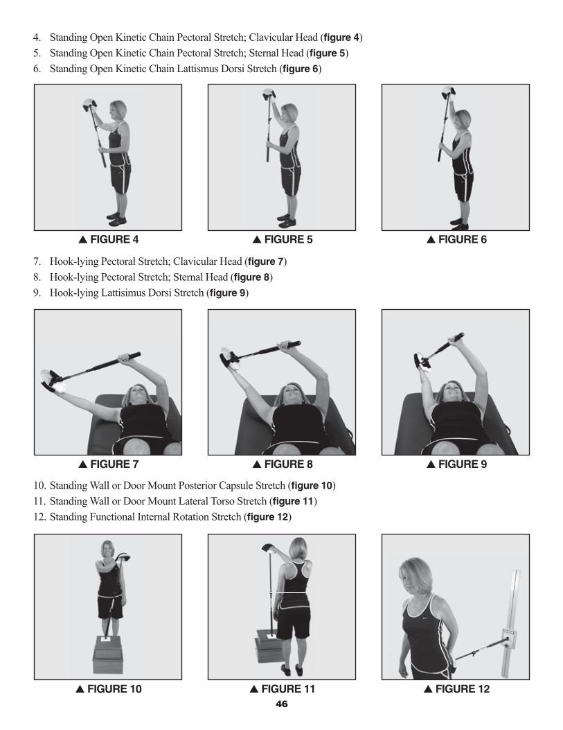
46
4. Standing Open Kinetic Chain Pectoral Stretch; Clavicular Head (fi gure 4)5. Standing Open Kinetic Chain Pectoral Stretch; Sternal Head (fi gure 5)6. Standing Open Kinetic Chain Lattismus Dorsi Stretch (fi gure 6)
7. Hook-lying Pectoral Stretch; Clavicular Head (fi gure 7) 8. Hook-lying Pectoral Stretch; Sternal Head (fi gure 8)9. Hook-lying Lattisimus Dorsi Stretch (fi gure 9)
10. Standing Wall or Door Mount Posterior Capsule Stretch (fi gure 10)11. Standing Wall or Door Mount Lateral Torso Stretch (fi gure 11)12. Standing Functional Internal Rotation Stretch (fi gure 12)
s FiGURE 4 s FiGURE 5 s FiGURE 6
s FiGURE 7 s FiGURE 8 s FiGURE 9
s FiGURE 10 s FiGURE 11 s FiGURE 12

4�
Following the executions of your stretches it is advised to perform at your current level of volitional movement capacity (PROM or AAROM) a functionally meaningful and compensatory free sequence of motions as to reintegrate any new mobility into the progressive recovery levels of movement health.
POST-OPERATivE ANd CHRONiC PAiN PROTOCOL • MANUAL iNTERvENTiONS
As a manual therapist, have you ever been in the middle of a therapeutic manual intervention and wished you had a third hand? Often this is the case as we feel what the body needs; we are at times unable to follow through as we have run out of a free hand to support that next infl uence. The Manual Therapist whom espouses the integration of movement health principles into their clinical efforts has just been given two free hands, to mobilize and guide, resist or assist, and facilitate and or inhibit. With the movement health principles supported by the UE Ranger Movement Health System, your patient becomes an active participant within their current capacities thus supporting the integration of your manual interventions with an Active Afferent Efferent Nervous System. The UE Ranger offers you and your patients the capacity to participate in:
• Positional passive static mobilizations
• Dynamic active mobilizations with local and or full kinematic chain integration
• Rhythmic stabilizations and other proprioceptive neuro-muscular facilitation techniques
• Functional active self stretching
• Functional positional manual cueing to facilitate and or inhibit
• Functional positional myofascial release
This manual is not intended to teach specifi c manual interventions; rather it is designed to offer clinical opportunities to blend with your preferred manual skills. The following are examples of such clinical capacities:
1. S/L UE Ranger supported STJ Passive Mobilizations at an appropriate current soft tissue restriction (fi gure 1)
2. S/L UE Ranger supported Neuro-muscular re-education of scapular ER via manual resistance at the inferior lateral angle, during active elevation (fi gure 2)
3. S/L UE Ranger supported isolated thoracic costal vertebral and intercostals joints passive mobilizations during active elevation (fi gure 3)
s FiGURE 1 s FiGURE 2 s FiGURE 3

Rehab Innovations, Inc. • www.ueranger.com48
4. S/L UE Ranger supported ACJ passive mobilizations during active elevation (fi gure 4)
5. H/L UE Ranger supported SCJ passive mobilizations into an inferior glide during active elevation (fi gure 5)
6. H/L UE Ranger supported clavicular passive mobilizations during a supported static position of elevation (fi gure 6)
7. H/L UE Ranger supported GHJ inferior posterior glide passive mobilizations during active elevation (fi gure 7)
8. H/L UE Ranger Pectoral Myofacial Release during a supported static position of elevation (fi gure 8)
9. Standing supported elevation; passively mobilizing the component movements of the STJ into external rotation and clavicle into a superior roll during active elevation (fi gure 9)
10. Standing Supported Elevation; passively mobilizing the component movements of the GHJ into a forward glide and the clavicle into a superior roll during the active assistive movements of combined ER, and elevation (fi gure 10).
Following the executions of your patient assisted manual interventions it is advised for your patient to perform at their current level of volitional movement capacity (PROM, or AAROM) a functionally meaningful and compensatory free sequence of motions as to reintegrate any new mobility into the progressive recovery levels of movement health.
s FiGURE 4 s FiGURE 5 s FiGURE 6
s FiGURE 7 s FiGURE 8 s FiGURE 9
s FiGURE 10
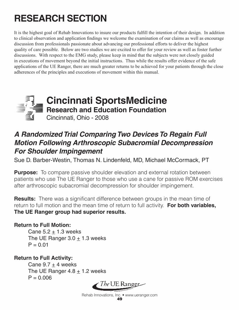
Rehab Innovations, Inc. • www.ueranger.com49
RESEARCH SECTiON
Cincinnati SportsMedicine Research and Education FoundationCincinnati, Ohio - 2008
A Randomized Trial Comparing Two Devices To Regain Full Motion Following Arthroscopic Subacromial Decompression For Shoulder ImpingementSue D. Barber-Westin, Thomas N. Lindenfeld, MD, Michael McCormack, PT
Purpose: To compare passive shoulder elevation and external rotation between patients who use The UE Ranger to those who use a cane for passive ROM exercises after arthroscopic subacromial decompression for shoulder impingement.
Results: There was a significant difference between groups in the mean time of return to full motion and the mean time of return to full activity. For both variables, The UE Ranger group had superior results.
Return to Full Motion: Cane 5.2 + 1.3 weeks The UE Ranger 3.0 + 1.3 weeks P = 0.01
Return to Full Activity: Cane 9.7 + 4 weeks The UE Ranger 4.8 + 1.2 weeks P = 0.006
It is the highest goal of Rehab Innovations to insure our products fulfill the intention of their design. In addition to clinical observation and application findings we welcome the examination of our claims as well as encourage discussion from professionals passionate about advancing our professional efforts to deliver the highest quality of care possible. Below are two studies we are excited to offer for your review as well as foster further discussions. With respect to the EMG study, please keep in mind that the subjects were not closely guided in executions of movement beyond the initial instructions. Thus while the results offer evidence of the safe applications of the UE Ranger, there are much greater returns to be achieved for your patients through the close adherences of the principles and executions of movement within this manual.

Rehab Innovations, Inc. • www.ueranger.com50
Comparing Muscle Activity during Post-Surgical Range Of Motion Exercises
University of Kentucky - 2008
TL Uhl, TM Muir, CD Melton - Division of Athletic TrainingAJ Nitz - Division of Physical TherapySD Mair - Division of Orthopedic Surgery
The UE Ranger demonstrating both its capacity to safely support early post-surgical mobility as well as a graded progression of muscular facilitation depending on its diverse and functional applications.
Pendulum UE Ranger supported passive shoulder elevation
Forward Bow UE Ranger supported active shoulder elevation
Rope and Pulley
UE Ranger supported humeral elevation
AFEWall WalkUE Ranger supported active vertical elevation
T-Bar
PROM AAROM AROM
EMG Levels (%RVC)
Rehab innovations, inc.8727 Ames Ave.Omaha, NE 68134Phone (402) 210-8406Fax (402) [email protected]