functional characterization of hypothetical proteins and
TRANSCRIPT

1
Functional characterization of
hypothetical proteins and their role
in radiation tolerance
of Deinococcus radiodurans
By
Anubrata Das
LIFE01200704001
Bhabha Atomic Research Centre
A thesis submitted to the
Board of Studies in Life Sciences
In partial fulfillment of requirements
For the Degree of
DOCTOR OF PHILOSOPHY
of
HOMI BHABHA NATIONAL INSTITUTE
October 2012

2

3
STATEMENT BY THE AUTHOR
This dissertation has been submitted in partial fulfillment of requirements for an
advanced degree at Homi Bhabha National Institute (HBNI) and is deposited in
the library to be made available to borrowers under rules of the HBNI.
Brief quotations from this dissertation are allowable without special permission,
provided that accurate acknowledgement of source is made. Requests for
permission for extended quotation from or reproduction of this manuscript in
whole or in part may be granted by the Competent Authority of HBNI when in
his or her judgment the proposed use of the material is in the interests of
scholarship. In all other instances, however, permission must be obtained from
the author.
(Anubrata Das)

4
DECLARATION
I, hereby declare that the investigation presented in the thesis has been
carried out by me. The work is original and has not been submitted
earlier as a whole or in part for a degree / diploma at this or any
other Institution / University.
( Anubrata Das)

5
Dedicated to
My Family

6
ACKNOWLEDGEMENTS
I feel highly privileged to express my heartfelt sincere gratitude to my esteemed
guide, Prof. H.S. Misra (HBNI), Head, Moleular Genetics Section, Molecular Biology
Division for providing me an opportunity to register for Ph. D. It is difficult to find the
words to express my gratitude and respect but I want to thank him for constantly
keeping me focussed and providing me with keen powers of observation. Indeed, I
am honored to be one of his students.
It is my great privilege to acknowledge Dr. S.F.D’souza, Associate Director (A), BMG
and Head, NA&BTD for his encouragement, moral support and valuable guidance
which inspired me to do my best. I am also grateful to Dr. (Mrs) J.K.Sainis, Raja
Ramanna Fellow and Dr. M.V.Hosur, Head, PCS, SSPD, members of my HBNI doctoral
committee for providing valuable suggestions for the completion of this work.
It has been my proud privilege and immense pleasure to extend my sincere gratitude
for Dr. K. B. Sainis, Director, Bio-Medical Group, BARC for his encouragement and
support in carrying out this work. With utter pleasure I acknowledge the role of
Dr.S.K.Apte, Dean, Life sciences, HBNI in guiding me to a career in research.
I wish to thank my lab colleagues Shri Y.S.Rajpurohit, Mrs. Swati Kota, Mrs. Nivedita
P. Khairnar, Shri C. Vijay Kumar, Mrs. Shruti Desai, Mrs. Vidya Kamble and Ms. Kruti
Mehta and Shri Sunil D. Dhotre for their constant support. I am also thankful to all my
erstwhile colleagues in Environmental Biotechnology Section, Dr. (Mrs) V.P.Joshi and
members of MBD and my project trainees for their support at different times.
I am deeply obliged to Mamoni and Bapi who undertook lot of hardships to provide a
comfortable life for me. This Ph.D would have been impossible without the steadfast
support and the sacrifice of Mantu and am grateful for being blessed with Teetul who
taught me to work with dedication. I also wish to thank Dr.P.Venubabu for standing
by me during times of difficulty.
September, 2012
MBD, BARC Anubrata Das

7
Table of Contents
SYNOPSIS ....................................................................................................................................... 8
List of figures……………………………………………………………………………………..29
List of Tables……………………………………………………………………………………..32
List of Abbreviations……….........................................................................................................33
Chapter 1 General Introduction and Review of Literature………………………………...….…..37
1.1 Radiation resistance and Deinococcus radiodurans……………………………….………....37
1.2 Repair Systems in Deinococcus radiodurans…………………………………………….…..43
1.3 Hypothetical Proteins and their Role in D. radiodurans……………………………….…..…54
1.4 Role of Bioinformatics in Studying Hypothetcial Proteins…………………………….…..…61
1.5 Aims and Objectives of this Study……………………………………………………………68
Chapter 2 Materials and Experimental Procedures……………………………………………....70
2.1. Materials……………………………………………………………………………………..70
2.2 Methods………………………………………………………………………….………......78
Chapter 3 Results……………………………………………………………………………….…98
3.1 Bioinformatics analysis of selected hypothetical proteins identified from the pool of DNA
binding proteins exhibiting ATP sensitive nucleolytic function ................................................... 99
3.2 Functional characterization of DRA0282 for its role in radiation resistance in bacteria ....... 121
3.3.Characterization of DR2417 for its predicted functions and its role in bacterial resistance
to radiation ............................................................................................................................... 144
Chapter 4 Discussion ................................................................................................................... 178
Chapter 5 Summary Conclusion and Future perspectives ........................................................... 187
Reference List .............................................................................................................................. 195
Appendix Copies of Research Articles

8
HOMI BHABHA NATIONAL INSTITUTE
PH. D. PROGRAMME
1. Name of the Student: Anubrata Das
2. Name of the Constituent Institution: Bhabha Atomic Research Centre
3. Enrolment No. : LIFE01200704001
4. Title of the Thesis: Functional characterization of hypothetical proteins and their role
in radiation tolerance of Deinococcus radiodurans
5. Board of Studies: Life Sciences

9
SYNOPSIS
Functional characterization of hypothetical proteins and their role in
radiation tolerance of Deinococcus radiodurans.
Maintaining genetic integrity is the key to survival. When organisms are exposed to
various stresses such as ionizing and non-ionizing radiation, numerous types of DNA
damage such as single strand breaks, double strand breaks, base modifications etc occur
by the direct deposition of radiation energy on DNA molecules and by the free radicals
generated in the aqueous environment. It is believed that organisms, which sustain high
doses of these stressors, have evolved processes to either protect the biomolecules from
the incident damage and/or have efficient repair mechanisms. D. radiodurans is amongst
those few organisms known to tolerate up to 15 kGy of radiation which produces more
than 200 double strand breaks and more than 3000 single strand breaks [1]. It achieves this
by a strong oxidative stress tolerance mechanism consisting of high quality antioxidant
enzymes and antioxidant metabolites like caretenoids[2,3], pyrroloquinoline quinone [4,5]
and a higher ratio of Mn2+
/Fe2+
, along with an efficient error free DNA strand break
repair mechanism [6]. In an effort to understand the extraordinary radiation resistance of
D. radiodurans, the genome of this bacterium was sequenced [14] and it shows two
classes of proteins 1) those that are homologous to known proteins and 2) those that
satisfy in silico criteria for being ORFs but have no known homologs, or even any
physical evidence. These ORFs are termed as ‘Orphans’ and the encoded proteins are
labelled as ‘hypothetical proteins’. Over the past decade around 60 proteins from D.

10
radiodurans have been characterized and 25% of these have been hypothetical proteins.
Some of these hypothetical proteins like DdrA, PprI have been shown to play an important
role in DNA repair [7]. These ‘hypothetical proteins’, assume importance because
majority of the known proteins in D. radiodurans are similar to their counterparts in
sensitive bacterium like E. coli and thus they do not necessarily explain the remarkable
capacity of D. radiodurans in reparing damaged DNA [8]. Earlier studies have shown that
D. radiodurans cells exposed to radiation produce numerous breaks in DNA, which this
organism could reassemble by two distinct mechanisms occurring in 2 phases. Phase I
normally occurs between the first 2.5 to 4.5 hrs after irradiation when the cells are grown
in complex medium after irradiation. If the cells are grown in nutritionally deficient
mineral medium the repair process has been shown to slow down. In this phase, by the
Extended Synthesis Dependent Strand Annealing (ESDSA) mechanism, double stranded
DNA ends are first extensively recessed to form single stranded DNA with 3’-OH
overhangs. These overhangs invade complementary template strands and fresh DNA
synthesis takes place [9]. In phase II RecA dependent homologous recombination pathway
then follows phase I to finally generate intact chromosomes.
The genesis of this study began with an observation of D. radiodurans cells undergoing
post irradiation recovery (PIR), after exposure to a 4kGy dose of radiation. When the cell
lysate of the cells, obtained 1hour after irradiation was fractionated by heparin column
followed by ion exchange chromatography, a fraction was obtained, which showed ATP
inhibited nucleolytic degradation of double stranded DNA [10]. At a 4kGy dose of
radiation the DNA inside the cell shatters into >100 DNA pieces and these pieces are
accurately reassembled back by a poorly known network of the proteins which execute
11
ESDSA [11]. Since, the above regulated nucleolytic function was seen in the first hour of
repair after irradiation, we got curious to find out the possible nucleases/kinases in this
fraction. To identify the possible nucleases/kinases, this fraction was analyzed by Fourier
Transform Ion Cyclotron Resonance-Mass Spectroscopy (FTICR-MS), but no known
nuclease or kinase was found. However 11 “hypothetical proteins” were also found and an
initial domain search showed that 6 (DR0041, DR0390, DR0461, DR1654, DR2417m,
DRA0282) of them had possible domains for DNA metabolic functions.
In this thesis we have attempted to predict the functions of four proteins from these six
hypothetical proteins for possible nuclease/kinase activity using bioinformatics and
functionally characterized two of these proteins DR2417 and DRA0282 both in vitro and
in vivo. Total work described in this thesis has been accomplished through following
objectives:
1. Theoretical prediction of functional domains in the above ORFs with the help of
bioinformatics tools and online databases.
2. In vitro characterization of recombinant DR2417m and DRA0282 for their predicted
functions and their effect on the response of transgenic cells to DNA damaging agents.
3. Molecular genetic studies to understand the role of DR2417 and DRA0282 in DNA
repair and radiation resistance of Deinococcus radiodurans.
The work carried out to address the above objectives, will be presented in this thesis as
following chapters:

12
Chapter 1 General Introduction and review of literature
Chapter 2 Materials and experimental procedures
Chapter 3 Results
3.1 Bioinformatics analysis of the ORFs encoding hypothetical protein identified from
the pool of DNA binding proteins exhibiting ATP sensitive nucleolytic function
3.2 Functional characterization of DRA0282 for its role in radiation resistance.
3.3 Characterization of DR2417 for its predicted functions and its role in bacterial
resistance to radiation
Chapter 4. Discussion
Chapter 5. Summary, conclusions and future perspective
Chapter 1. General Introduction and review of literature
This chapter describes the general information both on D. radiodurans, the various types
of damage to DNA and the repair systems available in D. radiodurans. It further discusses
the role of hypothetical proteins in its DNA repair and various bioinformatics approaches
available to predict their structure and function.

13
Chapter 2. Materials and Experimental Procedures
This chapter describes the details of materials used with their sources and common
experimental methods used in this study. Different techniques and approaches used in this
study will be described under the category of (1) Bioinformatics –use of available tools
BLAST, PSI-BLAST, ClustalX, Pfam database, Treeview, Swissmodel server, python etc.
(2) biochemical techniques including protein purification, Electrophoretic Mobility Shift
Assay, Sequencing gel, RNase assays (3) recombinant DNA technology and molecular
biology techniques including cloning, generation of deletion mutants and their
confirmation.
Chapter 3. Results
This chapter starts with a preamble describing the hypotheses and logical anticipation
from this study. The results obtained to test the hypotheses along with other new findings
during the course of this investigation will be summarized. Total results obtained from this
study have been presented in three distinct sections. Each starts with a brief introduction,
experimental procedures and results obtained from this specific study, followed by a brief
discussion on the findings specific to results included in this section.

14
3.1 Bioinformatics analysis of selected hypothetical proteins identified from the
pool of DNA binding proteins exhibiting ATP sensitive nucleolytic function
We have used online tools to find possible nuclease/kinase amongst the hypothetical
proteins. The features studied were (1) protein sequence analysis (2) genomic context (3)
subcellular location (4) peptide abundance in FTICR-MS data (5) theoretically modeled
structure (6) phylogenetic relationships. DRA0282 (84 peptides), DR0390 (45 peptides),
DR2417m (36 peptides), DR1654 (39 peptides) were abundant in the fraction amongst all
ORFs encoding hypothetical protein detected, while DR0041 (5 peptides) and DR0461 (4
peptides) were below the significance threshold and hence we focussed on these four
abundant proteins. Initial BLAST analysis showed DR2417m has a match with a β-CASP
family nuclease- Artemis, DRA0282 with a DNA binding repair protein –Ku 70/80,
DR0390 with DAK family kinases and DR1654 with DEAD box helicases. A critical
histidine residue of the DEA/X-DH motif was missing in DR1654 and hence, a further
study on this was postponed. Ku70/80 and Artemis proteins form the critical components
of Non Homologous End Joining (NHEJ) repair Pathway and also initial matches showed
that DR2417m could be a nuclease, and hence we selected DR2417m and DRA0282 for
further studies. The ORF encoding DR2417 was reported to have a genuine frameshift
mutation and hence the protein was annotated as DR2417m.
β-CASP family nucleases are involved in diverse functions like 3’ end processing of RNA
precursors, NHEJ repair, V(D)J recombination etc. These have several key conserved
motifs, motifs 1,2,3,4 of the catalytic metallo-β lactamse domain and motifs A, B, C of the
substrate specificity determining CASP domain. DR2417m and its β-CASP family

15
homologs obtained from a PSI-BLAST search were aligned in ClustalX, to look for
conserved motifs. Sequence alignments showed that DR2417m had all these conserved
motifs and an unusually arginine and proline rich C-terminus as per annotated sequence.
The distance tree of DR2417m with its homologs showed that DR2417m is closer to
Dgeo2150 from D.geothermalis but not its paralog DRA0069.
To find the function of DRA0282, we did a BLAST search with RefSeq database to find
putative homologs, however none were found. We then did a PSI-BLAST with DRA0282,
with the keywords like DNA, repair etc, as DRA0282 was found in a PIR fraction. Mostly
Ku homologues were obtained as the nearest matches and we aligned DRA0282 with
these sequences. The multiple sequence alignment showed that DRA0282 had an overall
homology with these proteins. Since DNA binding was a structural property, we built a 3-
D model of DRA0282 with the structure of Ku80 from Protein Data Bank (PDB code-
1JEY). A model with RMSD of 4.0 was built. We observed that DRA0282 had a
remarkable homology with Ku80. The N terminal von Willebrand A domain, which binds
nucleotides and the DNA binding β-barrel domain were seen. Interestingly, DRA0282
showed a closer match with eukaryotic Ku, than with prokaryotic Ku proteins
3.2 Functional characterization of DRA0282 for its role in radiation resistance in
bacteria.
Computational analyses suggested DRA0282 of D. radiodurans was homologous to
human Ku80. It was thus checked for its similarity to human Ku80 in in vitro activity and

16
its in vivo role in radiation resistance in both E. coli and D. radiodurans. The drA0282
ORF was cloned and expressed in E. coli. Recombinant DRA0282 was purified to
homogeneity and its interaction with DNA and protection of double stranded DNA from
exonuclease III degradation were checked. It preferred Mn2+
for its DNA binding activity,
and its ability to bind DNA was inhibited by ATP. Electrophoretic Mobility Shift Assay
experiments showed that it has the highest affinity (Kd=2.930.5 nM) with covalently
closed circular double stranded DNA followed by linear single stranded DNA (Kd =
60.229.1 nM) and linear double stranded DNA with (Kd=196.930.8 nM). These results
suggested that DRA0282 is indeed a DNA binding protein like human Ku80 while it was
unique in its substrate preference. [12]
Transgenic E. coli cells expressing DRA0282 showed enhanced resistance to UVC and
radiation in wild type background. The recA- E. coli JC1553 cells expressing DRA0282
did not show UVC and γ radiation protection. The RecA dependent protection of E. coli
cells expressing DRA0282, was found to be independent of RecA’s role through SOS
response as the E. coli DM49 (lexA) cells having a full-fledged homologous
recombination pathway but deficient in SOS response, continued showing UVC protection
in presence of DRA0282. These results suggested the possible role of DRA0282 in
processes requiring functional RecA at least in E. coli.
The role of DRA0282 was further confirmed in D. radiodurans. The drA0282 deletion
mutant of D. radiodurans was generated and radiation response of the mutant was
compared with wild type cells. The mutant showed no visible effect on radiation
treatment. This could be possible if DRA0282 has other functional homologues in D.
radiodurans. Earlier two proteins, DdrA and PprA were shown for having in vitro

17
functional similarities with DRA0282. Therefore, drA0282 gene was deleted from the
pprA deletion mutant of D. radiodurans. The drA0282pprA double mutant was evaluated
for radiation resistance and compared with single pprA and drA0282 mutant and wild
type. The double mutant did not add any effect to pprA single mutant suggesting that pprA
and drA0282 possible functions through common mechanisms in D. radiodurans.
These results suggested that DRA0282 is an important DNA repair protein similar to
human Ku80, which exhibits a role in heterlogous system that does not have its functional
homologues. On the other hand, D. radiodurans seems to have other proteins which
provide functional redundancy to drA0282 deletion or simply our screen was inadequate.
3.3. Characterization of DR2417 for its predicted functions and its role in
bacterial resistance to radiation
Earlier DR2417m (m stands for mutation) was predicted to be a member of the β-CASP
family of nucleases based on its N-terminal homologies and presence of defined motifs in
these proteins except for motif C [13]. The C-terminal of the annotated polypeptide is
unusually rich for arginine and proline amino acids, which is normally not observed in
genuine proteins and for these reasons it was earmarked as a hypothetical protein. Since,
the full length gene was cloned and a recombinant protein of the desired size was made as
well and since this protein was detected in a pool of DNA binding proteins, the possibility
of a wrong annotation of a frame shift in this ORF was looked into. Complete nucleotide
sequencing was carried out with the PCR amplified products from genomic DNA

18
templates. It was observed that the reported frame shift was wrongly annotated. This
information has been reported to the NCBI database and revised sequence of dr2417 has
been submitted vide accession number JQ432552 and subsequently we refer to the protein
as DR2417. In vitro functional characterization of this protein was also carried out.
Recombinant DR2417 was purified from E. coli BL21 and checked for its preferences to
different types of DNA and RNA substrates and other requirements. Purified DR2417 was
found to be a Mn2+
dependent nuclease. It acted both as a potent DNase and a weak
RNase. It was inhibited by ATP and GTP but not by AMP or GMP. At a gross level,
DR2417 degrades both ØX174 virion DNA (single stranded DNA) and ØX174 RFII DNA
(covalently closed double stranded DNA). It degrades PCR DNA (linear double stranded
DNA), when the ends of the PCR fragment have been phosphorylated by treatment with
polynucleotide kinase. To understand the mechanism, we tested it with defined
oligonucleotides. We found that it acts on double stranded DNA from 3’ end. It does not
degrade single stranded DNA. It has a strict requirement for a 5’ phosphate, without which
it does not act upon its substrate. Although DR2417, has a 40% overall identity with
RNaseJ from T.thermophilus and 100% conservation at the active site residues, it
nevertheless has a poor RNase activity. These characteristics make this enzyme distinct
from other members of β-CASP family proteins and offers the novelty in this enzyme.
The in vivo effect of DR2417 was studied both in a heterologous host E. coli and in D.
radiodurans. DR2417 was expressed both in recA- and wild type background of E. coli.
No significant change in cell survival upon UVC and radiation treatment was observed
in case of E. coli. Attempts to delete the DR2417 gene from the D. radiodurans genome
were always unsuccessful. We monitored the deletion process by checking for

19
amplification with primers flanking 500 bp upstream and downstream to the dr2417 ORF.
The homozygous knockout of dr2417 was never seen, however heterozygous clones
amplifying both 1.7 kb dr2417 and 0.8kb nptII marker were obtained. These heterozygous
clones generally grew poorly as compared to the wild type, and also were more sensitive
to U.V and radiation. Interestingly, we observed that this gene shows dose effect on
growth of this bacterium. The transformants were grown for several generations and at
each stage, the reduction in copy number of dr2417 was monitored. We observed a direct
correlation between copy number and growth inhibition. We did real time PCR to
quantify the genomic copies of dr2417 present in these clones and found that the
population with the least number of copies of dr2417 was also the most sensitive. This
suggested that deletion of dr2417 from D. radiodurans was deleterious for the survival of
this bacterium and thus dr2417 seems to be an essential gene.
Chapter 4. Discussion
In this chapter the findings on functional characterization of these two proteins in the
context of various hypotheses proposed for explaining the efficient DNA strand break
repair in this bacterium has been discussed. D. radiodurans has been extensively studied
for its ability to tolerate UVC, ionizing radiation, desiccation etc, which generate a huge
number of DSBs, such that it would debilitate many other organisms. Interestingly, this
bacterium is not tolerant to near UV (beyond 254nm). Early reports indicated novel DNA
repair mechanisms to be the key to survival, but sequencing of genomes showed that its
repair machinery is unremarkable, save a few oddities [14]. Newer studies now hint to an

20
efficient system which protects proteins from oxidative damage and may be the key to its
resistance [15]. But till date not much is known how after suffering a 10 kGy dose of γ
radiation, growth remains halted, damaged DNA and proteins are recycled, while 200
random fragments of DNA are accurately paired back, when evidence exists for increased
transposon activity, and all this with the same set of repair genes, found in other
radiosensitive bacteria ![16] Thus the role of ‘hypothetical proteins/predicted proteins’ or
proteins with ‘putative function’, becomes relevant, for e.g. at 3 hours PIR 30% of over-
expressed proteins are ‘hypothetical proteins’ [17]. The broad outlines of the repair
process involves regulated nucleolytic processing of double stranded DNA during early
period of post irradiation recovery, followed by the synthesis of >10 kb long single
stranded DNA and then an accurate patchwork assembly of old and new DNA [9].
Molecular mechanisms of nucleolytic processing from indiscriminate to productive DNA
processing and their regulation is not clearly understood. Initial work from our laboratory
has however, indicated the involvement of high energy nucleotide triphosphates in these
processes [10]. It was during these studies with various PIR fractions that ATP inhibited
nuclease activity was seen and DR2417 and DRA0282 were selected as interesting
candidates.
Initial bioinformatics showed that DR2417 has homology to Artemis, a β-CASP family
nuclease, and DRA0282 has structural similarity with Ku80, a double strand break sensing
protein. These proteins were then expressed in E. coli and the purified proteins were
characterized for their predicted functions. Both are found to be DNA interacting proteins
and DRA0282 showed protection of linear double stranded DNA from exonuclease

21
degradation but DRA0282 had higher affinity to linear single stranded DNA and
supercoiled double stranded DNA than linear double stranded DNA. The later observation
was different from human Ku80 type proteins. On the other hand, dr2417m was annotated
as a genuine frameshift in D. radiodurans genome. This protein was detected in various
studies as a functional component induced upon irradiation [10,18]. Also C-terminal was
unnaturally rich with proline and arginine, a normal anomaly of translating non-coding
genes. Complete nucleotide sequencing confirmed the wrong annotation of this ORF in
genome sequence. The polypeptide derived from new gene sequence was confirmed by
MALDI-MS analysis. The peptide mass fingerprints with both N-terminal and C-terminal
peptides were completely in agreement with the new sequence and differed grossly at C-
terminal of earlier annotation. Recombinant DR2417 was characterized as a novel β-
CASP family nuclease. Like other members of the β-CASP family, DR2417 needed a
5’phosphate end and was inhibited by ATP, but unique to it was the exonucleolytic
degradation from 3’ end, and specificity for double stranded DNA or junction substrates
and a weak RNase activity. The bioinformatics function prediction based on corrected
sequence indicated DR2417 to be an RNase, as the translated protein would have histidine
at motif C. All earlier studies had argued that this would make the protein RNA specific,
as histidine is seen in RNases and valine is seen in DNAses.[13] Based on the structural
differences between our 3-D model of DR2417 and the structure of RNase J, a well
characterized member of β-CASP family, we suspect that absence of the β sheets, β6 and
β7 in DR2417 which makes the channel for binding nucleic acids wide, thus fitting well to
DNA, but loosely fitting to RNA [19]. Also, the structure of RNase J shows that binding
to nucleic acids is by sugar phosphate backbone only. Thus, we assume that overall

22
structure and not a single residue determines substrate specificity. Recombinant
DRA0282, was characterized as a DNA binding protein which protected DNA ends from
ExoIII nuclease activity like Ku, but further studies showed it actually preferred single
stranded and covalently closed circular DNA over linear double stranded DNA unlike Ku.
The role of both these proteins in some common processes of DSB repair may be
speculated. It gets strong supports from the facts that both are abundantly present in same
pool of DNA binding proteins isolated from 1hour PIR sample and transcription of both
genes are concurrently induced at 1hour of post irradiation. Interestingly, DNA
interaction of both these proteins was inhibited with high-energy phosphates like ATP and
GTP. However, the exact mechanism(s) of ATP inhibition is not clear yet. Since
DRA0282 and DR2417 are structurally very close to human Ku80 and Artemis
respectively, the possibility of these proteins associated with Non Homologous End
Joining (NHEJ) repair of double strand breaks in this bacterium cannot be ruled out. Since
patch work assembly is seen during the repair process, the possibility of joining of 2
double stranded DNA molecule ends by a mechanism similar to NHEJ is an open question
and needs to be cleared up. NHEJ has been demonstrated in Bacillus subtilis and
Mycobacterium tuberculosis in prokaryotes [20]. Molecular genetic studies with these
proteins showed interesting phenotypes. DR2417 did not grossly affect the survival of E.
coli cells expressing this protein while E. coli expressing DRA0282 showed improved
resistance to UVC and γ radiation. D. radiodurans cells deleted for drA0282 showed no
discernable effect and found to contribute in pathways also supported by other proteins
with similar functions. On the other hand, an attempt to create a complete deletion mutant

23
of dr2417 failed and the partial knockouts, which were obtained, were debilitated. Thus
DR2417 seems to have an essential role in growth and survival of D. radiodurans, while
DRA0282 is needed possibly in an accessory function. Nevertheless, our findings hint to
the fact that in D. radiodurans DNA metabolic proteins may be the same as that of
radiosensitive organisms but they act in different combinations in unique pathways.
Chapter 5. Summary, Conclusion and Future Perspective
This chapter will summarize briefly the work presented in this thesis by highlighting the
potential observations that will include the answers to the hypotheses on the basis the
work that has been undertaken and unanswered questions / new hypotheses generated
during course of this study. The conclusion drawn from this study will be deciphered.
Along with several interesting findings reported in this thesis, two hypothetical proteins in
D. radiodurans genome have been assigned novel functions and one misled annotation has
been corrected during the course of this study. We appreciated from this study also that
structure –function prediction using bioinformatics tools guide well but require validation
by wet biology. While this study has answered some of the questions and hypotheses
raised both from experimental data of earlier work and bioinformatics analyses of certain
proteins, it has left several unanswered questions. The immediate ones which require
attention include (i) how these proteins work inside D. radiodurans cells, (ii) who are their
interacting partners under in vivo conditions, (iii) how does ATP/GTP influence the
interaction of these proteins with DNA and what is the functional significance of strong

24
binding of these proteins to supercoiled DNA. Also highlighted are the needs for more
accurate algorithms for prediction of function of the ever increasing number of
hypothetical proteins.
List of Publications
[A] Papers Published in Peer-reviewed International Journal
1. Das, AD and Misra, HS (2011). Characterization of DRA0282 from Deinococcus
radiodurans for its role in bacterial resistance to DNA damage.
Microbiology (SGM) 157: 2196-2205
2. Das, AD and Misra, HS (2012). DR2417m, a hypothetical protein characterized as a
novel β- CASP family nuclease in radiation resistant bacterium, Deinococcus
radiodurans.
Biochim. Biophys. Acta (General Subjects) 1820:1052-1061
3. Das, AD and Misra, HS (2013). Hypothetical proteins present during recovery phase of
radiation resistant bacterium Deinococcus radiodurans are under purifying selection.
J. Mol. Evol 77:31-42
[B] Abstracts Presented in National conferences
1. Das A. D. and Misra H. S. “Functional characterization of a hypothetical protein
from Deinococcus radiodurans and its role in bacterial resistance to DNA damage” in 34th
All India Cell Biology Conference and symposium on quantitative biology : from
molecules to cells, Abstract No. 64, p.142, December 4-6, 2010, Kolkata.

25
2. Das A. D. and Misra H. S. “A novel member of the beta CASP family of nuclease
in involved in the survival of Deinococcus radiodurans” in 52nd
Annual Conference of
Association of Microbiologists of India (AMI-2011), Abstract No. MM111, p392,
November 3-6, 2011, Punjab University, Chandigarh.
[C] Genbank Submission
A. D. Das and H. S. Misra (2012) Deinococcus radiodurans strain ATCC 13939
DR2417 gene. Accession No. JQ432552.
[D] Award and other recognition
A. D. Das received Dr. Rana Memorial Best Poster Award during 52nd
Annual
Conference of Association of Microbiologists of India (AMI-2011), held at Punjab
University, Chandigarh, November 3-6, 2011.
References
1. Battista JR (2000) Radiation resistance: the fragments that remain. Curr Biol 10: R204-
205.
2. Tian B, Sun Z, Xu Z, Shen S, Wang H, et al. (2008) Carotenoid 3',4'-desaturase is
involved in carotenoid biosynthesis in the radioresistant bacterium Deinococcus
radiodurans. Microbiology 154: 3697-3706.
3. Markillie LM, Varnum SM, Hradecky P, Wong KK (1999) Targeted mutagenesis by
duplication insertion in the radioresistant bacterium Deinococcus radiodurans: radiation
sensitivities of catalase (katA) and superoxide dismutase (sodA) mutants. J Bacteriol 181:
666-669.

26
4. Misra HS, Khairnar NP, Barik A, Indira Priyadarsini K, Mohan H, et al. (2004)
Pyrroloquinoline-quinone: a reactive oxygen species scavenger in bacteria. FEBS Lett
578: 26-30.
5. Rajpurohit YS, Gopalakrishnan R, Misra HS (2008) Involvement of a protein kinase
activity inducer in DNA double strand break repair and radioresistance of Deinococcus
radiodurans. J Bacteriol 190: 3948-3954.
6. Slade D, Lindner AB, Paul G, Radman M (2009) Recombination and replication in
DNA repair of heavily irradiated Deinococcus radiodurans. Cell 136: 1044-1055.
7. Harris DR, Tanaka M, Saveliev SV, Jolivet E, Earl AM, et al. (2004) Preserving
genome integrity: the DdrA protein of Deinococcus radiodurans R1. PLoS Biol 2: e304.
8. Makarova KS, Aravind L, Wolf YI, Tatusov RL, Minton KW, et al. (2001) Genome of
the extremely radiation-resistant bacterium Deinococcus radiodurans viewed from the
perspective of comparative genomics. Microbiol Mol Biol Rev 65: 44-79.
9. Zahradka K, Slade D, Bailone A, Sommer S, Averbeck D, et al. (2006) Reassembly of
shattered chromosomes in Deinococcus radiodurans. Nature 443: 569-573.
10. Kamble VA, Rajpurohit YS, Srivastava AK, Misra HS (2010) Increased synthesis of
signaling molecules coincides with reversible inhibition of nucleolytic activity during
postirradiation recovery of Deinococcus radiodurans. FEMS Microbiol Lett 303: 18-25.
11. Misra HS, Khairnar NP, Kota S, Shrivastava S, Joshi VP, et al. (2006) An exonuclease
I-sensitive DNA repair pathway in Deinococcus radiodurans: a major determinant of
radiation resistance. Mol Microbiol 59: 1308-1316.
12. Ristic D, Modesti M, Kanaar R, Wyman C (2003) Rad52 and Ku bind to different
DNA structures produced early in double-strand break repair. Nucleic Acids Res 31:
5229-5237.
13. Callebaut I, Moshous D, Mornon JP, de Villartay JP (2002) Metallo-beta-lactamase
fold within nucleic acids processing enzymes: the beta-CASP family. Nucleic Acids Res
30: 3592-3601.

27
14. White O, Eisen JA, Heidelberg JF, Hickey EK, Peterson JD, et al. (1999) Genome
sequence of the radioresistant bacterium Deinococcus radiodurans R1. Science 286: 1571-
1577.
15. Slade D, Radman M (2011) Oxidative stress resistance in Deinococcus radiodurans.
Microbiol Mol Biol Rev 75: 133-191.
16. Pasternak C, Ton-Hoang B, Coste G, Bailone A, Chandler M, et al. (2010) Irradiation-
induced Deinococcus radiodurans genome fragmentation triggers transposition of a single
resident insertion sequence. PLoS Genet 6: e1000799.
17. Liu Y, Zhou J, Omelchenko MV, Beliaev AS, Venkateswaran A, et al. (2003)
Transcriptome dynamics of Deinococcus radiodurans recovering from ionizing radiation.
Proc Natl Acad Sci U S A 100: 4191-4196.
18. Kota S, Misra HS (2008) Identification of a DNA processing complex from
Deinococcus radiodurans. Biochem Cell Biol 86: 448-458.
19. Li de la Sierra-Gallay I, Zig L, Jamalli A, Putzer H (2008) Structural insights into the
dual activity of RNase J. Nat Struct Mol Biol 15: 206-212.
20. Weller GR, Kysela B, Roy R, Tonkin LM, Scanlan E, et al. (2002) Identification of a
DNA nonhomologous end-joining complex in bacteria. Science 297: 1686-1689.
Publications:
a. Published
1. Das, AD and Misra, HS (2011). Characterization of DRA0282 from Deinococcus
radiodurans for its role in bacterial resistance to DNA damage. Microbiology (SGM)
157: 2196-2205

28
2. Das, AD and Misra, HS (2012). DR2417m, a hypothetical protein characterized as a
novel β- CASP family nuclease in radiation resistant bacterium, Deinococcus
radiodurans. Biochim Biophys Acta (General Subjects) 1820:1052-1061
3. Das, AD and Misra, HS (2013). Hypothetical proteins present during recovery phase of
radiation resistant bacterium Deinococcus radiodurans are under purifying selection.
J. Mol. Evol 77:31-42
Signature of Student:
Date:
Doctoral Committee:
S. No. Name Designation Signature Date
1. Dr. S.F D’Souza Chairman
2. Dr. H.S.Misra Guide
3. Dr.M.V.Hosur Member
4. Dr.(Mrs) J.K.sainis Member

29
List of figures
Figure
Number
Title of Figure Page
Number
1.1.1.1 Unrooted phylogenetic tree 39
1.2.1.1 DNA Repair Systems 43
1.2.1.2 DSB Repair Pathways 46
1.2.2.1 Dose profile of expression levels 53
1.3.1.1 Hypothetical protein vs genomes 56
1.3.2.1 Expression profile of hypothetical proteins 58
1.4.1.1 Bioinformatic approaches for protein function
determination
62
1.4.1.2 Sequence domain families in CATH 65
1.4.1.3 Evolutionary distance and gene order 67
3.1.2.1 BLAST search output 104
3.1.2.2 MSA of DR2417 107

30
3.1.2.3 Structure and phylogenetic tree of DR2417 109
3.1.2.4 MSA of DRA0282 112
3.1.2.5 Structure and phylogenetic tree of DRA0282 113
3.1.2.6 MSA of N-terminal of DR0390 115
3.1.2.7 Domain organization of DR0390 117
3.1.2.8 Domain organization of DR1654 119
3.2.2.1 Expression and purification of DRA0282 128
3.2.2.2 DNA binding assay of DRA0282 130
3.2.2.3 Determination of binding constant of DRA0282 132
3.2.2.4 ExoIII protection by DRA0282 134
3.2.2.5 Expression of DRA0282 in E. coli 136
3.2.2.6 Expression of DRA0282 in SOS deficient E. coli 138
3.2.2.7 Deletion of DRA0282 in D. radiodurans 140
3.2.2.8 Expression of drA0282 during PIR 142
3.3.2.1 Reannotation of dr2417 153
3.3.2.2 MALDI-TOF analysis of DR2417 154

31
3.3.2.3 Expression and purification of DR2417 156
3.3.2.4 Nucleolytic assay of DR2417 on DNA 158
3.3.2.5 Nucleolytic assay of DR2417 on RNA 161
3.3.2.6 Homology model of DR2417 162
3.3.2.7 Expression of dr2417 during PIR 164
3.3.2.8 DNA processing by DR2417 166
3.3.2.9 Assay of DR2417 on phosphorylated DNA 168
3.3.2.10 Deletion mutation of dr2417 170
3.3.2.11 Survival and radiation resistance of Δdr2417 171
3.3.2.12 Expression of DR2417 in E. coli 173
3.3.2.13 DNA-protein complex of DR2417 176

32
List of Tables
Table
Number
Title of Table Page Number
1.3.2.1 List of ORFans 59
1.4.1.1 List of databases 63
1.4.1.2 Relationship between folds 66
2.1.6.1 List of chemicals 72
2.1.6.2 List of antibiotics 74
2.2.1.1 List of bacterial strains 80
2.2.2.1 list of plasmids 88
3.1.2.1
BLAST output of ORFs encoding
hypothetical protein
105

33
List of Abbreviations
> greater than
1 kGy absorption 1 joule of ionizing radiation by 1 kg of matter
AMP Adenosine monophosphate
AP Apurinic/apyrimidinic
ATM Ataxia-telangiectasia
ATP Adenosine triphosphate
ATR ATM and Rad3 related
BIR Break induced repair
bp base pair
BrdU Bromodeoxyuridine
CCC Covalently closed circular DNA
DDBJ DNA databank of Japan
DEPC Diethylpyrocarbonate
DNA Deoxyribonucleic acid

34
dNTP Deoxyribonucleotides
DSB Double strand break
DSBR Double strand break repair
dsDNA Double stranded DNA
EDTA Ethylenediamine tetraacetic acid
EEO electroendoosmosis
EMBL European nucleotide sequence database
ESDSA Extended synthesis dependent stand annealing
FTICR-MS Fourier transform ion cyclotron resonance-mass spectroscopy
GMP Guanosine monophosphate
GTP Guanosine triphosphate
Kd dissociation constant –a equilibrium constant
MALDI-MS Matrix assisted lase desorption/ionization- Mass spectroscopy
MB Mega basepair
MSA Multiple Sequence Alignment
NCBI National Center for Biotechnology Information

35
NER Nucleotide exicision repair
NHEJ Non homologous end joining
nm nanometre
ORF Open reading frame
PAGE polyacrylamide gel electrophoresis
PCR Polymerase chain reaction
PDB Protein data bank
PIR Post irradiation recovery
PMSF phenylmethanesulfonylfluoride
PQQ Pyrroloquinoline-quinone
PSI-BLAST Position specific iterated-Basic local alignment search tool
RMSD Root mean square deviation
ROS Reactive oxygen species
RPM Rounds per minute
SDS sodium dodecyl sulphate
SDSA Synthesis dependent strand annealing

36
SOS save our soul- in this thesis it refers to SOS repair pathway
SSB Single strand break
ssDNA Single stranded DNA
TCA Tricholoroacetic acid
TYG Typtone-yeast extract-glucose medium for culture of D. radiodurans
UV Ultraviolet
UVC short wavelenght UV radiation between 250-270 nm

37
Chapter 1
General Introduction and Review of
Literature
Deinococcus radiodurans has been the most widely studied organism for understanding
the molecular mechanism of resistance to ionizing radiation and repair of DNA damaged
by ionizing radiation. Although in the last five decades lot information has been gained,
yet the exact details of this accurate repair process are sketchy. This work started from an
observation of a novel nuclease activity in D. radiodurans during the early hours of the
repair process. Herein we provide the basis of undertaking this study on an aspect of the
important initial phase of DNA repair in D. radiodurans.
1.1 Radiation Resistance and Deinococcus
radiodurans
D. radiodurans is a species among the estimated 11 million species which inhabit the
earth and its oceans [1]. It is a gram positive, red pigmented, nonspore forming bacterium
which belongs to a genus whose members have been isolated from diverse environments
like canned meat [2], desert soils[3], radiation contaminated soils and air 10 kms above

38
the earth’s surface[4]. It occurs in diads and tetrads with an average cell diameter of 1
µM. It is a mesophile and is dessication tolerant. Its 3.28 MB genome is GC rich (66%
GC content) and made up of 2 chromosomes and 2 plasmids. It is an organotrophic
organism and amino acids are the preffered carbon energy source. What sets it apart from
other organisms is its unusual resistance to ionizing radiation [5]. Ionizing radiation like
γ rays (1-10 MeV) and X-rays (100-10 KeV) have very high energies which can knock
off electrons from nearby atoms. When electrons get knocked off, bonds are broken and
atoms with unpaired electrons called radicals are generated, in a process called
Ionization. These radicals are highly reactive and spontaneously interact with double
bonds and break them. In biological systems the predominant moiety is water and these
radiation ionize the water molecules to generate very reactive radicals like OH., O2
- and
H2O2 collectively called as Reactive Oxygen Species (ROS). These ROS cause protein
carbonylation, lipid peroxidation and several damages to DNA including the formation of
abasic sites, single strand breaks (SSBs), base modifications and double strand breaks
(DSBs) [6]. Due to the systemic nature of the damage affecting several subcellular
components, most species are susceptible to ionizing radiation but several reports have
shown that a key factor to the survival from ionizing radiation is the ability to repair
DSBs in the damaged DNA [7]. In this milieu it is interesting to note that D. radiodurans
easily survives a dose of 15 kGy of γ radiation whereas most humans are killed by a dose
1000 times less. It shares this property with but a handful of organisms including
prokaryotes like other members of the Deinococcace family, a cyanobacterium
Chroococcidiopsis, Rubrobacter spp and several hyperthermophiles like Pyrococcus
furiosus and a few eukaryotes like the slime mold Dictyostelium discoideum, various

39
fungi like Filobasidium and the bdelloid rotifer Adineta vaga [5]. Ever since its
discovery in 1956, lot of efforts have been made to understand the reason for D.
radiodurans’s radioresistance as it is would help to understand the cellular ROS
protection systems, the DNA repair processes as well as find potential applications to
protect against the damaging effects of radiation. The origin of this phenomenal
resistance to ionizing radiation is mysterious. As the phylogenetic tree below shows (Fig
1.1.1.1) Deinococcus-Thermus belong to the same lineage, but while Deinococci are
resistant to ionizing radiation and desiccation, members of Thermus species are radiation
and desiccation sensitive but are thermotolerant.

40
Fig.1.1.1.1. An unrooted phylogenetic tree of Deinococcus spp. based on sigma 70
subunit [8]. Note that Deinococcus and Thermus are closely related and they both differ
from organisms of other phyla.
There are evidences of extensive ‘Horizontal Gene Transfer’ in this lineage which has
given its members their unique properties [9]. It is assumed that genes responsible for
radiation resistance have been imported into D. radiodurans during evolution but for the
purpose of desiccation tolerance. This is so because on Earth there are no known sources
of radiation which have or had levels of radiation upto which D. radiodurans easily
tolerates but there are many arid environments and mostly deinococci have been isolated
from these environments. Desiccation causes extensive double strand breaks in DNA like
radiation does and deinococci are desiccation tolerant too.
Till date no single factor has be pinpointed in deinococci or other radioresistant
organisms as the sole source of resistance. Indeed the present view is that several factors
coalesce to provide for this phenomenon [10]. At first, D. radiodurans has metabolic
mechanisms to quench ROS generation. It has fewer Fe-S cluster respiratory enzymes to
prevent ‘iron toxicity’. It induces the glyoxylate cycle in response to radiation, which
allows generation of reductants like NADH which can quench the free radicals formed. It
has 3 different alleles of superoxide dismutase, 3 alleles of catalase and 2 alleles of
peroxidase to scavenge free radicals and also the basal level of catalase activity is 15 fold
more than that of E. coli [5]. In rat mitochondria even 0.1 nM concentration of
superoxide radical (O2- ) inactivates Aconitase, a key Tricarboxylic Acid Cycle enzyme
[11]. Superoxide dismutase converts the superoxide radical to oxygen and hydrogen

41
peroxide and catalase further decomposes the toxic hydrogen peroxide to oxygen and
water. Peroxidases also assist in scavenging ROS by converting hydrogen peroxide to
water. D. radiodurans also has pyrroloquinoline-quinone (PQQ) coenzyme which
efficiently scavenges ROS [12]. PQQ, also known as methoxatin, was identified as a
cofactor of bacterial dehydrogenases and has been shown to be a more effcient scavenger
of ROS, as compared to other antioxidants. PQQ obtained from Deinococcus has been
shown to protect E. coli from oxidative damage [5,13]. Deinoxanthin, a major carotenoid
found in D. radiodurans has good ROS scavenging property.A study of D.radiophilus
lipid extracts showed that these lipids have antioxidant properties [14]. It is reasonable to
expect that D. radiodurans also has similar lipid protection mechanism which prevents its
membranes from getting damaged by radiation. D. radiodurans also has several Mn2+
complexes which are known to scavenge ROS [15]. Next, it has specific pathways to
prevent protein damage by oxidation. Studies have shown that a small molecule mediated
mechanism in D. radiodurans prevents protein carbonylation which can be caused by
ionizing radiation [16,17].
Also a study showed that proteins get selectively recycled after irradiation, which means
that damaged proteins are degraded by proteases and replaced with fresh proteins [17].
This protease activity, besides helping in protein recycling may also be involved in cell
signaling. It has earlier been shown that proteases can act as irreversible signaling and
regulating systems. For example, caspase 3 cleaves the ICAD protein, which releases the
Caspase Activated DNase, an endonuclease which works in the apoptosis pathway[18].
Next, D. radiodurans has an extensive cell cleaning mechanism comprised of Nudix
phosphohydrolases and nucleotidases which clean up oxidized derivatives which are

42
formed due to the free radicals generated by radiation. In fact the Nudix family is highly
expanded in D. radiodurans with 23 members [19].
However, curiously, all this elaborate mechanism protects the membrane lipids and
proteins but cannot protect the DNA inside the cell, which bears the brunt of ionizing
radiation. One reason might be that DNA is the longest polymer in the cell and hence an
elaborate target for radiation. A dose of 7 kGy shatters the genome into >200 pieces, with
at least 10 times more single strand breaks (SSBs) as well as modifying bases on the
DNA molecule. Such massive damage halts the replisome and eventually the cell dies. To
put it into perspective, E. coli barely withstands 3-4 double strand breaks (DSBs) per
genome and thus critical to the survival of D. radiodurans is its ability to accurately
splice back this damaged DNA.

43
1.2 Repair Systems in Deinococcus radiodurans
1.2.1 A general Study of Repair Systems
Fig.1.2.1.1. Schematic showing various damaging agents and DNA repair pathways in
Living Systems. (Adapted from Martin et al., 2008) [20]
The figure above summarises the various repair mechanisms found in living beings (Fig
1.2.1.1).The evolution of repair pathways show that it has evolved independently in
different phases. Mechanisms can be as direct as in Light Repair or ‘Photoreactivation’,
where photolyases detect pyrimidine dimers caused due to UVC rays and with the help of

44
chromophores which convert light energy to chemical energy they cleave these
pyrimidine dimer bonds.
A more elaborate pathway is the Base Excision Repair pathway. Single bases get
damaged through oxidation, alkylation and deamination and hang out of the DNA helix.
For example, guanine can be oxidized to 8-oxoguanine, adenine can be methylated to
3’methyl adenine and cytosine can be deaminated to uracil. If left unattended, these will
cause mispairing during replication and thus cause mutations. DNA glycosylases
recognize these bases and remove them. The AP endonucleases then cleave the sugar
phosphate backbone which is then resealed by polymerases like polX and ligases.
UV rays cause photoproducts like pyrimidine dimers and these bulky adducts cannot be
repaired by the Base Exicision Repair pathway. The Nucleotide Excision Repair (NER)
comprising UvrA, UvrB, UvrC and UvrD are employed to repair these bulky lesions. The
dimer lesions are recognized by UvrA-B. UvrA then leaves the complex making way for
UvrC. UvrB-C then makes a 12 nucleotide incision spanning the dimer segment. UvrD, a
helicase removes the damaged segment and DNA Pol I and ligase then fill the gap. A
specialized addition to NER is Transcription Coupled Repair, wherein RNA polymerases
get stalled at the dimers and recruit a protein TCRF, which uses ATP hydrolysis to
bypass the RNA polymerase over the lesion and also recruits UvrA at the damaged site.
NER is a major repair pathway which is well distributed in living systems and repairs the
bulk of UV induced damage. In eukaryotes, around 30 genes are involved in this pathway
while in bacteria this is accomplished by 4 genes [21].
Another repair pathway is the Mismatch Repair pathway , which is invoked due to errors
caused by incorporation of mismatched bases during replication and recombination.

45
Mismatch repair pathway is strand specific and corrects the errors on the newly
synthesized strand by recognizing the state of its methylation. Usually the template strand
is methylated and the newly synthesized strand is unmethylated. The Mismatch Repair
pathway comprises of MutS, MutH and MutL which specifically targets the newly
synthesized strand. The MutS dimer recognizes the mismatch. Then MutS forms a MutS-
DNA complex. The MutL dimer then binds to this complex and this activates MutH to
nick the newly sysnthesized strand. This nicked DNA segment having the mismatch is
then removed by UvrD helicase along with exonucleases. The resulting gap is filled by
DNA pol III and DNA ligase.
Bacteria also possess the SOS Repair pathway as studied in E. coli.This is a coordinated
response to the challenge of DNA damage. The SOS repair pathway is made up of RecA,
the LexA repressor, Sul A and repair genes of the NER pathway and a translesion DNA
synthesis system comprising UmuDC. The LexA repressor binds to a regulatory element,
the SOS-box, upstream to the genes involved in SOS response. When DNA is damaged,
it causes replication fork arrest at the damaged site and single stranded DNA is released.
RecA binds to this single stranded DNA and gets activated as a protease. It then cleaves
the LexA repressor, and the proteins involved in SOS response are now expressed. Two
things happen-1) Sul A stops cell division by interacting with the cytoskeleton protein
FtsZ and 2) UmuDC initiates DNA synthesis bypassing the damaged site. This causes
mutations to be maintained and hence the SOS response is considered to be a ‘error prone
repair’ system. However in most cases the cells survive the DNA damage rather than
dying due to replication fork arrest [22].

46
All these above mechanisms can repair DNA only in presence of a complementary DNA
strand and hence cannot tackle a double strand break. Double strand breaks occur either
via endonuclease mediated mechanisms or by oxidative stress or directly by ionizing
radiation. Cells have SOS repair and Transcription Coupled Repair mechanism to bypass
SSBs but none to bypass DSBs and hence DSBs are lethal. As shown in the figure below
(Fig 1.2.1.2) The Recombination Repair pathway is invoked for DSBs and,
recombination repair can be of 2 types (1) Homologous Recombination and (2) Non-
Homologous End Joining (NHEJ).

47
Fig. 1.2.1.2. Double Strand Break Repair Pathway showing the possible ways in which
the breaks can be repaired. SDSA and BIR are predominantly seen in eukaryotes while
DSBR is a universal mechanism. (Adapted from Aguilera and Boulton.,2007) [23].
The prerequisite of homologous recombination is a complementary DNA strand. It is
seen in both eukaryotes as well as prokaryotes. In eukaryotes members of RAD52
epistasis group carry out homologous recombination while in prokaryotes 2 pathways,
namely RecBCD and RecFOR pathways are known. RecBCD has been well
characterized in E. coli. RecBCD is a sequence regulated DNA helicase-nuclease [24]. It
binds to a blunt DSB and unwinds the DNA in an ATP dependent fashion and acts as a
weak 3’-5’ nuclease. On meeting a chi (χ) sequence-5’-GCTGGTGG-3’, it alteres its
activity and becomes a 5’-3’ double stranded nuclease -helicase and generates free 3’-OH
ends. It also assists RecA to bind to these ends for further steps of recombination. In D.
radiodurans, RecBC is absent and the major pathway of DSB repair is the RecFOR
pathway. Here, RecQ helicase unwinds the DSB and RecJ exonuclease degrades the 5’
strand leaving the 3’-OH strand free. RecA then binds this free strand, and then this
nucleoprotein complex invades a homologous partner to form a D-loop. Pol III then
extends this D-loop by extending the 3’ strand. Due to this process a junction of
interlinked strands is created called the Holliday Junction. The RuvABC resolvase
participates in resolving this junction and creating 2 chimeras. A modification of this
pathway is Synthesis Dependent Strand Annealing (SDSA) where the resultant chimera is
not formed. This mechanism is seen in yeast and D. radiodurans. The NHEJ pathway is a
major repair pathway in eukaryotes and also seen in mycobacteria and bacilli [25]. This

48
pathway has mostly been characterized in yeast, mice and humans. NHEJ is active all the
time in cell cycle and especially during G1 phase. Here, the key player is the Ku protein.
The bacterial protein is a homodimer while the eukaryotic protein is a hetrodimer made
of Ku70/80. Ku identifies a DSB and holds the ends of the 2 strands together as a clamp.
Then it recruits DNA-PKcs and then this complex now becomes the DNA-PK complex.
DNA-PK recruits Artemis, a multifunctional nuclease which blunts the 2 juxtaposed ends
and also XRCC4/Ligase whiles seals the ends. The choice between homologous
recombination and non-homologous recombination is regulated by a signalling pathway
comprising ATM/ATR kinases and DNA-PK and also by the nature of the DSB. Blunt or
nearly blunt ends are processed by NHEJ while ends with overhangs are processed by
homologous recombination.
1.2.2 Double Strand Break Repair in D. radiodurans
As discussed above, D. radiodurans has a mammoth task of reassembling its shattered
genome. Certain physical aspects have evolved in D. radiodurans to assist the reassembly
of its genome. It is multigenomic, and multiple copies help in the homology assisted
recombination repair pathway. Based on electron microscope and epifluoresence studies,
Minsky and co-workers proposed an interesting hypothesis. They observed that DNA in
D. radiodurans is aggregated into compact toroids which they hypothesized would help
NHEJ pathway proteins to repair the DNA [26].The necessity and importance of this
toroid formation in radioresistance has been refuted in several other studies [27], but it is

49
agreed that compaction of the genome could help the repair process. Also, the small
molecule antioxidant proteome comprising peptides and nucleosides in complex with
Mn2+
polyphosphate complex helps protect key DNA repair enzymes [28].
Bioinformatics studies done with the D. radiodurans genome showed that there were no
new potential DNA repair proteins and D. radiodurans shares the same set of repair
enzymes seen in E. coli [19]. Further, it has been shown that E. coli pol I is able to restore
a polA strain of D. radiodurans hinting that ROS scavenging is more important in
radioresistance and in the cellular milieu any repair system can efficiently repair damaged
DNA, even extremely damaged DNA, provided ROS generated by ionizing radiation is
removed from the system [29].
Genome sequence studies also showed that it lacks photolyases for the direct repair of
thymine dimers and also a viable SOS response although it has 2 alleles of lexA. D.
radiodurans has the Base Excision Repair, Nucleotide Excision Repair and
Recombination Repair Pathways pathways. Both Base Exicision Repair and NER seem to
be efficient since single strand breaks are repaired within 90 minutes of ionizing radiation
damage [30,31].
Studies have shown that recombination repair pathway in D. radiodurans is mediated by
RecJ exonuclease along with components of RecFOR pathway and either RecQ helicase
or UvrD helicase to load RecA and carry out homologous recombination [32]. Seminal
studies by Radman and co-workers has shown that D. radiodurans may work with
ordinary proteins but the mechanism of reassembly is unique [33,34] and they have
named this process as Extended Synthesis Dependent Strand Annealing (ESDSA) as it is
similar to the DSB repair mechanism Synthesis Dependent Strand Annealing (SDSA) in

50
yeast. ESDSA occurs in the initial 2-3 hours of reassembly of the deinococcal genome
after a dose of irradiation, when it is grown in a complex medium (TGY medium-1%
Tryptone, 1% glucose, 0.5% Yeast Extract). During this time, first there is a massive
degradation of DNA. Amongst the possible candidate nucleases are RecJ and SbcCD.
RecJ has been shown to have 5’-3’ exonuclease activity and also that it is an essential
gene, as conditional mutants which don’t express RecJ die away [32]. This is a regulated
exonucleolytic phase where single stranded DNA with free 3’OH ends are generated
which can invade complementary strands of DNA assisted by RecA. Interestingly,
studies have shown that RecA stimulates this exonuclease activty. These single strands
then invade the complementary strands after which an extensive round of synthesis takes
place. Unique to ESDSA is the extent of this synthesis, where upto 20-30 kb DNA is
freshly synthesized. This amount of fresh synthesis is at least 4 times more than what has
been reported for synthesis dependent strand annealing (SDSA) in yeast and drosophila
where DSBs are repaired by SDSA [35,36]. In SDSA, the D loop formed is transient,
rather it is a bubble and thus extension of a strand needs several rounds of invasion. Also,
as the D loop is not extensive, resolvases cannot pair the template and freshly synthesized
strand, and these new strands slip off and the recombinases join the complementary new
strands together without the need for resolvases. It is believed that the mechanism is
similar in D. radiodurans and several such freshly synthesized overlapping strands exist
and in the second phase RecA dependent recombination repair pathways recombine these
freshly synthesized strands into intact molecules. Thus the reconstituted genome is a
mosaic of old and new patches of DNA. This patchwork nature was clearly visible when
D. radiodurans cells were grown in the presence of the thymine analog BrdU. BrdU then

51
is incorporated into the newly synthesized DNA and when these cells are exposed to a
few kilojoules of UV light, it causes DSBs.The presence of DSBs thus confirm both the
fresh synthesis of DNA and nonreciprocal crossover events [33].
Is ESDSA a prerequisite for repair of DNA having extensive double strand breaks? Till
other radioresistant bacteria are studied in detail, it would be difficult to guess as SDSA
or ESDSA is not prevalent in bacteria, although SDSA has been implicated in homing of
group I introns in phage T4 [37]. How this eukaryotic repair mechanism was adopted by
Deinococci remains to be discovered. It is interesting to note that in several fungi where
SDSA is a predominant mitotic DSB repair mechanism, do exhibit radiation resistance,
for e.g the D10 for C. albicans is 1.1-2.3 kGy. Also, members of spp Filobasidium,
Aspergillus, Curvularia, Ustilago have been found to be radiation resistant [38]. A study
was done in S.cerevisiae to study the genes required for ionizing radiation resistance by
generating a library of deletion mutants. This study showed that members of the RAD52
epistasis group play a critical role in radiation resistance. Members of this group can be
broadly subdivided into the MRN (nbs1) complex, which are involved in processing of
DSBs in repair pathways and the RAD51 subgroup which is involved in all homologous
recombination processes [39]. An earlier study showed that sbcCD in D. radiodurans
functions in a fashion similar to the MRN complex [40].Again D. radiodurans has an
homolog of RAD51 (RecA) , RAD52 (DdrA) and RAD54(DR1259) but further
investigations are needed to find homologs of RAD 55 and RAD 57 which have been
shown to be essential for both crossover mediated recombination repair as well as SDSA.
Curiously, S.cerevisiae also encodes certain key proteins of the NHEJ pathway, including
the critical protein Ku70/80 (YKU70 and YKU80) and a specialized ligase DLN4 but till

52
date no homolog of a DNA dependent protein kinase and Artemis, which are also
components of NHEJ has been observed [41]. Thus yeast in spite of being a eukaryote
like mammals, uses NHEJ as a backup pathway while the major repair pathway is by
homologous recombination and this preference has a notable similarity with D.
radiodurans. Other than accuracy, there may be yet another reason why D. radiodurans
may have evolved the ESDSA pathway as a major repair pathway. Single stranded DNA
has been shown to be signal for cell cycle checkpoint in eukaryotes and all present
evidence shows that while repair is in progress, the cell cycle in D. radiodurans remains
arrested [42]. Thus the copious amount of single stranded DNA generated during ESDSA
may additionally act as a cell cycle arrest signal. A preference for error free homologous
recombination explains how in spite of apparent activation of a transposase, no
significant mutations are seen in the repaired DNA [43]. But ESDSA also leaves certain
questions unanswered. Why does the synthesis extend for so long? How processive is the
process? How such long strands of single stranded DNA are maintained without any
secondary structure and that there are no slippages during possibly several rounds of
strand invasion? And what are the key players in this mechanism? Earlier studies have
implicated roles for RecA, RecFOR, DNA Pol III and DNA Pol A, but their counterparts
are present in other radiosensitive bacteria too. Could novel genes be involved in this
process? Almost 50 % of the Deinococcal proteome, like proteomes of many species,
comprises of hypothetical proteins, which have no homology to known proteins and it has
been speculated earlier that these proteins may play important role in radiation reistance
[44]. Studying the exact mechanism of ESDSA and the possible role of novel proteins in

53
this process is important in understanding the robust error free repair system in D.
radiodurans.
Besides the machinery of proteins involved in repair another important aspect of repair
process is its regulation. D. radiodurans employs well known and possibly novel
pathways to repair a highly shattered genome with negligible errors which means that
regulation of metabolic processes like nucleolytic degradation, synthesis of DNA etc
becomes very important. It is known that DNA repair is linked to cell cycle as cell cycle
progression takes place only after repair. Again as the plot below highlights (Fig 1.2.2.1)
the pattern of expression at 3 kGy is different from that of 15 kGy and surprisingly, in a
highly shattered genome, selectively more genes are over-expressed at 15 kGy than at 3
kGy.
3 kG
Y
15 k
Gy
0
10
20
30
expression pattern at different doses
Dose of gamma radiation
ex
pre
ss
ion
le
ve
l
54
Fig 1.2.2.1. Altered expression profile of genes involved in DNA repair at different doses
at 3 kGy and 15 kGy shows that expression of genes sharply increases at higher
doses.[Das and Misra, Unpublished data,[45]]
It has previously been shown that upstream to several genes over-expressed in radiation
stress in D. radiodurans, like DR0326, DRA0346, DR0423 and several DNA repair
genes, a strong palindromic motif designated as radiation/desiccation response motif
(RDRM) is present [46] and this is a conserved motif as it is present also in
D.geothermalis and D.deserti. Thus, possibly D. radiodurans employs novel regulatory
networks to repair the numerous DSBs after a dose of radiation.
1.3 Hypothetical Proteins and their Role in D.
radiodurans
1.3.1 Introduction to Hypothetical Proteins
Algorithms to predict genes, predict stretches of DNA with proper start and stop codons
and corresponding amino acid sequences and name them as ‘Open Reading Frame’. If
translations from these ORFs match with atleast 30 % identity with known proteins in the
public databases like NCBI, EBI, DDBJ they are annotated with the names of already
known genes, otherwise the polypetide sequences derived from the ORFs are named

55
‘Hypothetical Proteins’. Fischer and Eisenberg coined the name ‘ORFans’ for the parent
ORFs of these hypothetical proteins [47] .
It was widely believed that the number of hypothetical proteins would reduce as more
and more genomes are sequenced, however as the plot below shows (Fig 1.3.1.1 ) that
has not been the case. With every new genome sequence, a newer set of hypothetical
proteins are added to the databases. A caveat has been that half of the ORFans encoding
hypothetical proteins, are less than 150 bases and may be wrongly annotated as encoding
for proteins, and in fact some reports also refer to these small ORFs as ELFs (“evil little
fellows”), hinting to the fact that these might actually be noncoding regions or regulatory
regions [48].
So what is the origin of hypothetical proteins? This has been an intriguing question and
there are two predominant theories for the existence of hypothetical proteins.
1) These are remnants of once active genes, which are now nonfunctional and would in
future gather more stop codons, frameshifts etc and morph as ‘pseudogenes’ and then in
the future get eliminated. An example is GULO (L-gulono-γ-lactone oxidase). GULO
aids in the biosynthesis of vitamin C in most of the mammals but it is disabled in humans
and primates [49]. Another example is an inactive form of caspase 12 in humans [50].
But in this case, the inactive form is selected for and maintained as these individuals are
more resistant to severe sepsis.
2) The other theory is that many of these hypothetical proteins are duplicate copies of
functional genes (paralogs) and are slowly evolving newer functions (de novo evolution).
For example, BSC4 gene in S.cerevisiae is expressed as non-coding RNA in species
closely related to yeast but in yeast itself, it is a protein involved in DNA repair. CLLU1,

56
C22ORF45 and DNAH10OS in humans are functional proteins detected in proteomics
data but these are not transcribed in primates [51].
Fig 1.3.1.1. Plot of hypothetical proteins vs genomes published. Hypothetical proteins
(ORFans) are increasing with the number of genomes sequenced (adapted from Siew et
al.,2003) [48].
Many studies show that hypothetical proteins are actually expressed for e.g Bennet et
al.,2001[39] have shown that around 3000 hypothetical proteins in S.cerevisiae are
actively transcribed while similar studies in E. coli have shown that several hypothetical
proteins are actively expressed [52]. The acknowledgement of the role hypothetical
proteins in cell survival has been slow and only recently they are being biochemically
characterized, as till 1998 only a single hypothetical protein from B.licheniformis was
characterized, whereas in 2011 alone 44 such hypothetical proteins were characterized.

57
1.3.2 Hypothetical Proteins in Radiation Response in D. radiodurans
A study implicating hypothetical proteins in the radiation response of D. radiodurans was
that of Liu et al.,2003 [45]. As the plot below shows (Fig 1.3.2.1) several hypothetical
proteins were higly expressed in a pattern similar to the total cellular expression
indicating that they are are necessary in the repair process of D. radiodurans. They
concluded that a majority of induced genes were poorly characterized and needed further
characterization. Suspected ELFs were also present, for e.g in the case of DRA0234 it
was only 171 bp long and the theoretical transcript formed stable stem-loop structures,
indicative of noncoding RNA. Nevertheless, it showed that hypothetical proteins like
DRB0098-DRB0100, uncharacterized ABC transporters DR1356-DR1359, kinases with
unknown specificity DR2467, DR0394, DR0609 and DR1564 etc were highly induced in
response to radiation.

58
Fig 1.3.2.1. Expression profile of hypothetical proteins with respect to total proteins. Box
Plot showing the similarity of the pattern of expression of hypothetical proteins to the
expression pattern seen during the repair process for all genes [Das and
Misra,unpublished data,[45]].
Another study by Tanaka et al., 1996 [53] deleted the genes of hypothetical proteins
which were over-expressed and found out that in several cases, the deletion mutant was
debilitated. Thus the role of hypothetical proteins like ddrA (DR0423), ddrB (DR0070),
pprA (DRA0346) etc in the radiation response of was discovered. Purified DdrA protein
was shown to bind to 3’ ends of single stranded DNA and protect them from exonuclease
degradation [54]. Likewise purified PprA was characterized in vitro as a DNA end
binding protein which prevented DNA from Exo III attack and phenomenally increased
ligase activity of ATP dependent T4 DNA ligase and NAD dependent E. coli DNA ligase
[55]. As the table below shows (Table 1.3.2.1) hypothetical proteins play an important

59
role in the metabolism and repair process in D. radiodurans and although we distinguish
hypothetical proteins as a separate entity, in D. radiodurans as in other organisms, they
are integrated seamlessly into the metabolism.
Table 1.3.2.1 A representative list of ORFans encoding for hypothetical proteins
characterized in D. radiodurans
Sr No Gene Locus tag function reference
1 irrE or
pprI
DR0617 A deletion study was done. IrrE is
a regulatory protein which
enhances RecA transcription
[56]
2 mntE DR1236 manganese efflux protein [57]
3 sig1 DR0180 Deletion of the gene makes D.
radiodurans sensitive to IR ECF
derived heat shock Sigma Factor,
controls groESL and dnaKJ
operons.
[58]
4 sig2 DR0804 Same as above, but deletion of the
gene has a lesser drastic effect
[59]
5 ddrA DR0423 Deletion of the gene made D.
radiodurans sensitive to radiation
and the purified protein protected
ssDNA invitro
[54]
6 lexA2 DRA0074 A disruption mutant showed a [60]

60
higher resistance to radiation
7 drarnl DRB0094 DraRnl seals RNA strand nicks in
double stranded RNA or
RNA:DNA hybrid
[61]
8 ddrB DR0070 The deletion mutant was sensitive
to radiation and purified protein
stimulated single stranded DNA
annealing
[62]
9 nimA DR0842 A reductase involved in
metronidazole antibiotic resistance
[63]
10 drRRA DR2418 The deletion mutant was sensitive
to radiation and studies showed it
to be a DNA binding response
regulator
[64]
11 ddrP DRB0100 The purified protein forms a
complex with AMP but does not
ligate DNA or RNA
[65]
12 - DRB0098 The purified protein acts as a
polynucleotide kinase. Only other
example from bacteria is in
clostridia
[65]
13 drOxyR DR0615 The deletion mutant is sensitive to
H2O2, but not to IR. It is a
transcriptional activator involved
in Mn/Fe transport
[66]

61
14 - DR0505 the purified protein was a hairpin
endonclease and a 3’-5’
exonuclease
[67]
15 cruF DR0091 the purified protein is a carotenoid
1-2 hydratase involved in
synthesis of the pigment
deinoxanthin, which is an
important ROS scavenger
[68]
1.4 Role of Bioinformatics in Studying Hypothetical
Proteins
As we have seen above, hypothetical proteins have an important role in repair, general
metabolism and radiation resistance in D. radiodurans. Microarray studies have shown
several hypothetical proteins being induced and it important to understand the function of
these proteins
As shown in the figure below (Fig 1.4.1.1), there are several bioinformatics tools for
searching for functions of hypothetical proteins:-
1) Sequence based – BLAST or FASTA search in nucleotide or protein databases for
functional homologs
2) Subsequence or motif based search in PROSITE or PHI-BLAST for finding conserved
functional sites

62
3) Structure based – by homology modeling or fold prediction
4) Feature based-where models are generated based on features derived from amino acid
sequence of the query. Feature based methods have also been used to build classifiers for
machine learning approaches like Neural Networks and Support Vector Machines which
can predict specific functional groups in a high throughput way.
Fig 1.4.1.1. Schematic showing bioinformatics approaches for protein function
determination

63
Because of the common evolutionary past, the key active site residues, residues involved
in salt bridges and overall structure of proteins are conserved. This overall conservation is
reflected in the amino acid sequence but due to poor sequence homology, BLAST, the
standard search tool for comparing an unknown sequence to proteins in a database cannot
identify any function for these proteins. An improved algorithm called Position Specific
Iterated -BLAST (PSI-BLAST) was developed which would build a statistical
probability distribution for the presence of amino acids in a alignement [69]. PSI-
BLAST has since then been successfully used to detect remote homologs in several cases.
Also, as the table below shows (Table 1.4.1.1) several specialized databases are available
which improve the chances of getting relevant matches.
Table 1.4.1.1. List of databases of nucleic acids and proteins adapted from Washietl
and Hofacker .,2010 [70]
Database Description
EMBL, Genbank, DDBJ central sequence repository
RefSeq non-redundant and curated sequence
GEO (Gene Expression
Omnibus)
gene expression experimental dataset
PDB (Protein Data Bank) database of experimentally determined

64
structure of proteins, nucleic acids and
complex assemblies
Entrez Genome database of complete and nearly complete
genome sequences
Swissprot manually curated protein database
DIP (Database of Interacting
Proteins)
online database of experimentally determined
protein-protein interaction
PROSITE database of protein domains, families and
functional sites
Secondary structures are the basis for tertiary structure formation and for computational
reasons these can be done faster. Also topology and connectivity conflicts between
residues are easily revealed by secondary structures. Today secondary structure
prediction are fairly accurately done by Neural Networks [71].
Knowledge of the tertiary structure helps in understanding the mechanism of a protein
and in evolution the structure that is conserved and not the sequence. Thus hypothetical
proteins with remote sequence identity can be properly identified and a function can be
predicted. The basic independent structural domain is a fold and as the plot below shows

65
(Fig 1.4.1.2) most sequence families can be molded onto few 3-D folds in a structure
database.
Fig 1.4.1.2. Proportion of the sequence domain families represented by CATH fold
groups. A group of 54 CATH fold groups cover 76% of all domains.(Adapted from Pearl
et al.,2005) [52].
As seen in the table below (Table 1.4.1.2) , there is a correlation between structure and
function at the fold level [72]. Again, the protein structure also has a correlation with the
ligands it interacts with, as there is a selection pressure on the shape of the clefts and
grooves which fit the ligand [73]. Thus most sequences have common folds and most
folds have a common function.

66
Table 1.4.1.2. Relationship between folds in a protein and their function.(Adapted
from Marti-Renom et al.,2000) [72] .
Fold
Functional Annotation
of homologous proteins
Number of
members
Percent of
the fold
Rossman Fold Enzyme 131 57%
TIM Barrel Enzyme 94 85%
IG Fold Antibody 17 89%
Globins Oxygen transporter 16 100%
Beta-propeller Alpha-sialidase 6 66%
Protein structure prediction methods available today can be grouped into (1) homology
modeling, (2) threading and (3) ab initio methods. The simplest and fastest method is
homology modeling. If the sequence identity is >50%, then this is a reliable method. Here
the query sequence is searched across sequences of available structures and a template is
chosen. Then the query is modeled on the structure of the template. The 3-D model
obtained usually has a resolution of 3-4°A RMSD or even better, and is good for
deducing the overall folds and thus the possible function. One can superimpose any
motifs found in the query sequence, independently from a Motif Database and try to map
the active site in the query.

67
In bacteria genes are arranged in operons. Thus if an unknown gene in the species of
interest, lies in an operon, then it could be predicted to act in that pathway. The plot
below shows the co-relation between gene order conservation and evolutionary distance
(Fig 1.4.1.3). Gene order conservation has been used as an important tool for finding
protein function.
Fig 1.4.1.3. Evolutionary conservation and relative gene orientation (Adapted from Lopez
et al.,2011) [74].
Besides these methods above, tools have also been developed to predict sub-cellular
location, post translational modification and interacting partners. There are also tools
available for docking possible substrates with the protein of interest. A very useful
approach for gene prediction has been ‘Phylogenomics’. Evolutionary processes indicate
that all genes have evolved from a common ancestor and changes in genes are co-

68
terminus with speciation, thus proteins with similar phylogenetic profiles should have
similar functions [75,76]
Thus today, with the help of bioinformatics and high throughput experiments like
microarray based expression profiling the role of hypothetical proteins in the life of
organisms are slowly being revealed.
1.5 Aims and Objectives of this Study
The genesis of this study began with an observation of D. radiodurans cells undergoing
post irradiation recovery (PIR), after exposure to a 4kGy dose of radiation. At this dose
of radiation the DNA inside the cell shatters into >100 DNA pieces and during repair
these pieces are accurately reassembled back by a poorly known network of the proteins
which execute ESDSA. In the initial phase a regulated nuclease activity converts the
shattered double stranded DNA to single stranded DNA and hence nucleases are an
important part of ESDSA. When the cell lysate of the 1hour PIR sample was fractionated
by heparin column followed by ion exchange chromatography, a fraction was obtained,
which degraded exogenoulsy added double strand DNA in the presence of 5 mM Mn2+
.
This degradation of the added DNA was inhibited when 1mM ATP was added to the
assay mixture. Nucleases need metal ions for their activity and ATP chelates out metal
ions but here the ratio of Mn2+
/ATP was sufficiently high to rule out this effect, which
meant that ATP was modulating the activity of this nuclease. Many proteins are known to
be regulated by kinases and inhibition by ATP meant a possible regulation by kinases.
Also there are certain reports of nucleases being inhibited by ATP. For all the above

69
reasons we got curious to find out the possible nucleases/kinases in this fraction. To
identify the possible nucleases/kinases, this fraction was analyzed by a sensitive
technique, Fourier Transform Ion Cyclotron Resonace-Mass Spectroscopy, which
detected ~300 different proteins but no known nuclease or kinase was found. However 11
‘hypothetical proteins’ were also found and an initial BLAST search showed that 6
(DR0041, DR0390, DR0461, DR1654, DR2417m, DRA0282) of them had retrived
matches with some proteins involved in DNA metabolism while the other five had no
matches in the databases at an e-value cutoff of 1.0.
Thus there was a possibility for discovering a novel nuclease and its regulatory kinase
involved in the unique ESDSA phase of DNA repair in D. radiodurans. Also,
bioinformatics tools were available to detect the presence of nuclease or kinase
functional/structural motifs. In this thesis we have attempted to predict the functions of
these 6 hypothetical proteins for possible nuclease/kinase activity using bioinformatics
and functionally characterized two of these proteins DR2417 and DRA0282 both in vitro
and in vivo. Total work described in this thesis has been accomplished through following
objectives:
1. Theoretical prediction of functional domains in the above ORFs with the help of
bioinformatics tools and online databases.
2. In vitro characterization of recombinant DR2417 and DRA0282 for their predicted
functions and their effect on the response of transgenic E. coli cells to DNA damaging
agents
3. Molecular genetic studies to understand the role of DR2417 and DRA0282 in DNA
repair and radiation resistance of D. radiodurans.

70
Chapter 2
Materials and Experimental Procedures
2.1. Materials
2.1.1 Plasticware and Glassware
Disposable polypropylene microcentrifuge tubes (1.5 ml and 2 ml) and micropipette tips
were obtained from Tarsons, India or Axygen, USA. PCR tubes were procured from
Axygen, USA. Polypropylene SS34 tubes and GSA bottles were from Tarsons, India. All
the plasticwares and glasswares were sterilized by autoclaving. Glasswares were also
sterilized by baking at higher temperature for 3 h. Cryovials (2 ml) and low temperature
storage boxes were obtained from Laxbro, India and Axygen, USA. All the glasswares
were from Corning, USA or Borosil, India.
2.1.2 Chemicals and Media Ingredients
Dehydrated components of culture media (Bacto-tryptone and Bacto-yeast extract) and
agar-agar (Bacto-agar) powder were from Difco Laboratories, USA. Fine chemicals were
from Sigma Chemical Company, or USB-Amersham, or E.Merck, or Roche
Biochemicals GmbH or Pharmacia LKB, or S.R.L. Ltd., or Bangalore Genei (P) Ltd.,
India. Inorganic salts, urea for sequencing, organic solvents and acids of Excel-R or

71
Anal-R quality were from Qualigens (Glaxo) India Ltd. or E.Merck India Ltd. Molecular
Biology Grade phenol was purchased from SRL, India.
2.1.3 Enzymes and Other Molecular Biology Reagents
T4 Polynucleotide Kinase, T4 DNA ligase, restriction endonucleases and DNA
polymerase (Klenow fragment) were from New England Biolabs, USA or Bangalore
Genei (P) Ltd., India or USB-Amersham, UK or Roche Biochemicals GmbH, Germany.
T4 DNA polymerase was from New England Biolabs, USA or Roche Biochemicals
GmbH, Germany. Exonuclease I, Shrimp Alkaline Phosphatase were from USB-
Amersham, UK. Lysozyme (from chicken egg-white), DNase I (from bovine pancreas)
and RNase (from bovine pancreas) were from Sigma Chemical Company, USA. Taq
DNA Polymerase was obtained from Bangalore Genie Pvt Ltd, Bangalore. Pwo DNA
polymerase and deoxynucleotidetriphosphates (dNTPs) were from Roche Biochemical
Germany. DNA molecular weight standards were from Bangalore Genei (P) Ltd., India.
PCR extraction kit, Gel extraction kit and RNA extraction kit were from QIAGEN,
Germany. All the primers used in this study were synthesized commercially.
2.1.4 Radionucleotides and Photographic Materials
[32p] γ-ATP [32p] -ATP were from Board of Radiation and Isotope Technology
(BRIT), India. X-ray films of medical type, polyester based, double-sided were from
Kodak India Ltd, India.

72
2.1.5 Preparation of Dialysis Tubing
Dialysis tubing was cut in 30 cm pieces and soaked in sterile 50mM Tris buffer, pH 7.5
for overnight at 4oC. The buffer was replaced with similar buffer containing 2 % Glycerol
and 10 mM EDTA and autoclaved before storage at 4oC. (The tubes from the stock were
handled aseptically to avoid microbial growth).
2.1.6 Composition of Stock Solutions
Preparation and storage of most of the stock solutions is given in Table 2.1.6.1. Unless
specifically mentioned, the solutions were made in ultrapure water and sterilized without
adjusting the pH. Wherever necessary, addition was done to agar media after prior
cooling of the medium to ~50C. Preparation of all the antibiotic stock solutions is given
in Table 2.1.6.2. Addition to agar media was done just prior to pouring the plates and
only after the media was cooled to ~50C.
2.1.6.1 List of chemicals
Chemical Concentration Storage
Stock Final
Adenosine
triphosphate (ATP)
10 mM
1 mM
-20C
Ammonium
persulfate (APS)
100mg/ml
1 mg/ml
-20C

73
BSA 10.0 % As required -20C
CaCl2 1.0 M 100mM 4C
dNTPs 100 mM As required -20C
DTT 1 M 1mM -20oC
Ethidium bromide 5 mg/ml 0.5 g/ml RT.
EDTA (Na salt) 0.5 M As required RT.
D-glucose 20.0 % 0.4 % 4C
Glycerol 98 % As required RT.
KCl 2M As required RT
IPTG 200 mg/ml 160 g/ml -20C
MgCl2 1.0 M As required 4C
MgSO4 1.0 M 10 mM 4C
PMSF 100mM 1mM -20oC
SDS (Na salt) 10 % As required RT.
Sodium acetate 3.0 M As required RT.
Sodium chloride 5.0 M As required RT.
N,N,N’,N’-
Tetramethyl
ethylenediamine
(TEMED)
As required 4C

74
DMSO As required RT
Tris.HCl 1.0 M As required RT.
2.1.6.2 List of antibiotics
Antibiotic Stock Solution Working Concentration
(mg/ml) (g/ml)
Ampicillin 100 in sterile DW 100
Chloramphenicol 34 in Ethanol 3-5
Kanamycin 25 in sterile DW 5-30
Spectinomycin 100 in sterile DW 75
2.1.7 Composition of Commonly Used Analytical Reagents
All commonly used analytical reagents like 1 M Tris.Cl, 0.5 M DTT, 0.5 M EDTA(pH
8.0), Phenol, TE buffer, GTE(for alkaline miniprep), 5 M Potassium Acetate, 5X TBE,
10X TAE, 6X DNA loading dye, were prepared as per Sambrook et al.,2001 [77] .

75
Phosphate Buffered Saline
Dissolve 8 gm NaCl, 0.2 gm KCl, 1.44 gm Na2HPO4 and 0.24 gm KH2PO4 in 800 ml
distilled water. The pH to 7.4 was adjusted with dilute HCl and volume was made up to
1000 ml. The solution was dispensed in 100 ml bottles and sterilized by autoclaving. The
buffer was stored at RT.
PMSF
174 mg of the phenylmethylsulfonylfluoride powder was dissolved in 10 ml isopropanol
and solution was vortered vigorously to dissolve the content for a stock concentration of
100 mM . One milliliter aliquots were taken in alumunium foil wrapped vials and
preserved at –20oC. The solution was kept on ice for 10- 20 min just before the use.
Mitomycin C
20 microgram mitomycin C was dissolved in 1 ml sterile double distilled water for a
stock concentration of 20 µg/ml and preserved at –20oC in dark.
Metal Affinity Purification Matrices
Immoblized metal affinity chromatogrpahy matrices were procured from GE healthcare
(Sweden) as fast flow metal chelating materials and NINTA agarose from QIAgen
(Germany).
Oligonucleotides
Normal oligonucleotides and RNA oligos were synthesised from Metabion,
Switzerland/Sweden and MWG, Germany.

76
2.1.8 Composition of Media
All the media described here were sterilized by autoclaving at 15 p.s.i. for 20 min. and
stored at RT., unless otherwise mentioned. Agar-agar was added at a final concentration
of 1.5% (unless otherwise mentioned) to prepare solidified media.
Luria Bertani Broth (LB)
NaCl, 5 gm.; Bacto-yeast extract, 5 gm.; Bacto-tryptone, 10 gm.;Milli RO grade water to
a final volume of 1 litre and the pH adjusted to 7.4 with NaOH.
TGY medium
Tryptone, 5g; Yeast Extract, 3g, Glucose, 1g were dissolved in 1000 ml of distilled water
and pH=7.4 was adjusted with NaOH.
2.1.9 Composition of Other Reagents Used in Molecular Biology
Studies
2X Refolding Buffer
0.2 M Tris-HCl pH 7.6 ;0.8 M L-Arginine ;20% glycerol ;0.5 mM GSSG* ;5 mM GSH*
Prepare in sterile glassware and with sterile milliQ water. Autoclave for 5 minutes.*Add
GSSG and GSH after autoclaving.

77
Protein Storage Buffer
10 mM Tris-HCl pH 7.6 ;50 mM KCl ;0.1 mM EDTA ;50 % glycerol
DNase I
5 mg DNase I, 2.6 ml sterile (98 %) glycerol and 150 l 5 M NaCl. were mixed to
dissolve in 2.25 ml sterile Milli Q grade water by repeatedly tapping or vortexing,
dispensed as 500 l aliquots and stored at -20C.
RNase A
50 mg RNase A was dissolved in 5 ml of 10mM sodium acetate, pH 5.2. The solution
was heated at 115oC for 15 min and heating was put off and temperature was allowed to
decrease slowly to RT. To this, add 500l of 1MTris.HCl , pH 7.5 for a final
concentration of 10 mg/ml and dispensed in aliquots and stored at –20oC.
Lysozyme
Lysozyme was prepared at a concentration of 50 mg/ml in 10 mM Tris.HCl (pH = 8.0)
and used fresh.
Acrylamide Stock
29.2gm acrylamide and 0.8 gm of N, N-Methylene (bis) acrylamide was dissolved in 70
ml double distilled water at 37oC. The mixture was stirred for 30 min to assure complete
dissolution and then the volume was made up to 100 ml. The final stock concentration
was 30%. The solution was filtered through Whatman 540 filter disc and stored at 4oC.

78
Urea gel-loading dye
One milliliter of deionised formamide (100 %) was mixed with 20 l of 0.5M EDTA, pH
8.0 and 40 l of each of the bromophenol blue (2%) and xylene cyanol FF (2%). The
100 l aliquots were made and preserved at –20oC.
2X Laemmelli Cracking buffer
Dissolve 460 mg of SDS, 7.6 mg of EGTA, 20 mg of sodium azide, 2 ml of Glycerol,
150 l of 100mM PMSF, 1.0 ml -mercaptoethanol in 2.5 ml of 0.5M Tris-HCl, pH 6.8
and 40l of 0.5 % bromophenol blue was added and volume was made up to 10 ml with
distilled water.
10 x SDS PAGE electrophoresis buffer
Dissolve 30 gm Tris base, 144 gm Glycine and 10 gm SDS in 800 ml double distilled
water. The volume was made up to 1000ml with distilled water.
2.2 Methods
2.2.1 Microbiological Methods
2.2.1.1 Maintenance of the Bacterial Stocks of Escherichia coli
Bacterial stocks were maintained under frozen conditions in presence of cryoprotectants.
The logarithmically growing culture was spun and resuspended in minimal medium and

79
/or LB medium to cells density of 109 cells per ml. The cell suspension was mixed with
20% glycerol in autoclaved polypropylene screw capped tubes and stored in freezing
temperature tolerant boxes, at –70oC. The stocks were numbered and entered in the
bacterial culture book with full details.
2.2.1.2 Growth of the Bacterial Strains
Different bacterial strains used in this study are summarized in Table 2.2.1. Bacterial
culture was taken from long-term storage stocks directly on LB / TGY agar plates and or
plates containing appropriate antibiotics if required. The plates were incubated at
appropriate temperature overnight. Single isolated colonies were inoculated in broth with
or without antibiotics and allowed to grow at 37oC for E. coli and 30
oC for D.
radiodurans at shaking speed of 150 RPM overnight. The overnight grown liquid culture
was diluted to 1:100 in fresh broth with selection pressure if required, in the flasks having
10 times the air space to the volume of the medium. The culture was allowed to grow
with shaking at 200 RPM at appropriate temperature. The aliquots were drawn at
different time intervals and optical density was measured at 600 nm. In parallel an
appropriate dilutions were plated on agar plates and the numbers of colonies were
counted. The growth curve drawn under defined set of conditions was used in
determining the different growth phases of the culture for different experiments. Total
number of colonies appeared on agar plates after different treatments of D. radiodurans
cells were taken for calculating the survival efficiency.

80
2.2.1.1 List of bacterial strains
Bacterial strains Genotype Source
Deinococcus
radiodurans R1
Wild type Schafer et al
.,1998[78]
ΔdrA0282
Deinococcus
radiodurans mutant
drA_0282 this study
Δdr2417
Deinococcus
radiodurans mutant
dr2417 partial mutant this study
E. coli DH5alpha F- / endA1 hsdR17 glnV44 thi-1 recA1 gyrA relA
∆ (lacIZYA-argF) U169 deoR (Φ80dlac∆
(lacZ)M15)
NEB
E. coli BL21 F-ompT gal [dcm] [lon] hsdSB DE3::T7RNA
polymerase gene
NEB
E. coli AB1157 thr-1, araC14, leuB6(Am), Δ(gpt-proA)62,
lacY1, tsx-33, qsr'-0, glnV44(AS), galK2(Oc),
LAM-, Rac-0, hisG4(Oc), rfbC1, mgl-51,
rpoS396(Am), rpsL31(strR), kdgK51, xylA5, mtl-
1, argE3(Oc), thi-1
Dewitt et
al.,1962

81
E. coli JC1553 leuB6 fhuA2 lacY1 glnV44(AS) gal-6 hns-1
hisG1(Fs) rfbC1 galP63 argG6 malT1(λR) xyl-7
mtlA2 metB1 recA1 rpsL104
Clark et
al.,1964
E. coli DM49 lexA3 Mount et
al.,1972[79]
2.2.1.3. Radiation Treatment of Cells
Radiation treatment of D. radiodurans cells
D. radiodurans cells were grown to early stationary phase and mid log phase as required,
in TGY medium. The cells were collected and washed with sterile normal saline. The
cells were resuspended in half volume of fresh TGY broth and irradiated at different
doses of radiation in ice in irradiator (GC5000, BRIT) at the dose rate of 9.87kGy/h.
The cells were diluted appropriately and plated on TGY agar plates. For UV treatment,
the cells plated on agar plates were directly irradiated with different doses of UV
radiation. Mitomycin C (20µg/ml) was used as described in (Khairnar et al., 2008). The
plates were incubated at 30oC for at least 36 hours for recording total viable counts in
response to specific treatments.
Radiation treatment of E. coli cells
For UV radiation 1% inoculum of recombinant E. coli was grown in LB + 100 µg/ml
ampicillin for 2 hours at 37°C/180 RPM till midlog phase and 200µM IPTG was added
82
and and the culture was further grown for 2 hrs more and then exposed to various doses
of UV at 0.861 J/m2/s.For γ radiation induced cells were irradiated in a GC-220 (BRIT) γ
irradiator at a dose rate of 7 gray/min.
2.2.2 Methods used in Molecular Studies
2.2.2.1 Isolation of Plasmid DNA (mini prep)
Plasmid DNA was prepared using alkaline lysis method as described in Sambrook et
al.,2001[77].
2.2.2.2 Isolation of Genomic DNA
The 2 ml overnight grown culture of D. radiodurans was spun down, washed with 70%
ethanol and resuspended 400 µL SDS-EB buffer (20% SDS,400 mM NaCl, 40 mM
EDTA, 100 mM Tris-HCl, pH 8.0). After mixing well, an equal volume of phenol-
chloroform, was added and then spun at 10K for 10 min at 4°C. The aqueous layer was
extracted with equal volumes of 24:1 mixture of chloroform-isoamyl alcohol. The
supernatent was mixed with 1/10th
volume of 3M sodium acetate pH 4.5 & 2 volumes of
ethanol and incubated at -20oC for ½ hour. DNA was collected at 15K for 15 min and
dissolved in sterile water. DNA concentration was ascertained with OD260 and on agarose
gel.
2.2.2.3 Agarose Gel Electrophoresis
Agarose gel electrophoresis was used for the qualitative and quantitative analysis of
DNA. 0.5g/ml of ethidium bromide was added to molten 1 % low EEO agarose and the

83
solidified gel was used to fractionate DNA .The samples were mixed with gel loading
dye solution and loaded onto the agarose gel submerged in electrophoresis buffer.
Electrophoresis was done at 11 V/cm. After the electrophoresis has completed, the DNA
bands were visualized on UV transilluminator (UVP Model 3UVTM Transilluminator) at
254nm or with hand UV lamp (Model UVGL-58) for cloning experiments at 320 nm.
2.2.2.4 Urea-PAGE gel Electrophoresis
Urea-PAGE gels were run in a DNA sequencing unit. A set of rigorously cleaned
matched glass plate set were coated with Sigmacote® silane solution. The plates were
sanwhiched with 0.1mm spacers and sealed. 8M urea-14% PAGE sequencing gel was
poured in this assembly. The gel was stored overnight at 4 C for polymerization to set
and was then run the next day. The wells were flushed with 0.5 X TBE and the gel was
pre-run at 1000 volts 18 mA for 30 min. Then samples were added and the gel was run
till dye front reached 2/3 of the gel. All radioactive buffer was dispensed carefully and
DNA Sequencing Unit was carefully washed. The gel was blotted onto Whatman
Blotting sheet and wrapped with Saranwrap. Then this assembly was sandwiched with an
X-ray film in a X-ray film cassette and the autoradiogram was left to develop in -20 °C.
The X-ray films were developed in an automated X-ray film processor (Velopex Extra).
8M Urea-14% PAGE Sequencing Gel
Urea 25 gms
40 % acrylamide stock 20 ml

84
5X TBE 6 ml
10% Ammonium persulfate 1 ml
Formamide 6 ml
Mix everything and then make up volume to 60 ml. Filter through whatman 541 paper.
Then degas for 10 minutes, add 30 µl TEMED and pour the gel.
2.2.2.5 Electrophorectic Mobility Shift Assay
Electrophoretic Mobility Shift Assay was done to study the DNA binding characteristics
of DRA0282. A 5’ 32P end labelled 200 bp double stranded DNA was used as a probe.
100 ng of DRA0282 was incubated with 10 pmol of the probe for 30 min on ice. In
separate experiments this protein-substrate complex was supplemented with 100 fold
excess concentration of different forms of DNA, incubated for 30 min at 37 °C and these
complexes were mixed with 50% glycerol and loaded on a 5 % native polyacrylamide
gel. The gel was run at 150 V for 2h at room temperature, then dried and exposed for 4-
16 h to X-ray film at –80 C and the X-ray films were developed in a automated X-ray
film processor (Velopex Extra).
5% Native Polyacrylamide Gel
milliQ-water 9 ml
5X TBE 3 ml
30% Acrylamide 3 ml
10% Ammonium persulfate 10 µL
50% glycerol 2.5 ml

85
TEMED 10 µL
Combine the ingredients for a final volume of 15 ml and pour between a 1.5 mm hollow
13 cm X 14 cm glass plate sandwhich and allow the gel to set.
2.2.2.6 PCR Amplification
Primers were designed manually using the corresponding nucleotide sequences from D.
radiodurans genomes database. FASTPCR, a freely available software
http://primerdigital.com/fastpcr.html was used to design the primers. Relevant factors like
Restriction Endonuclease sites, GC content, primer dimer formation etc, were optimized
with the help of menus in FASTPCR. HPSF® purified primers were obtained
commercially from Metabion. PCR amplification was mostly carried out in 50l volume.
A typical PCR reaction contains template (50ng), primers 10 pmol, dNTPs 200M, Taq
DNA polymerase buffer 1X, 3.0 mM MgCl2 and Taq DNA polymerase 2.5U. Dimethyl
Sulfoxide (DMSO) was mixed as a denaturant in the PCR mix while amplifying with the
high GC D. radiodurans genomic DNA. The PCR mix was amplified on Eppendorf
Mastercycler® thermocycler for 30 cycles. The results were visualized on 1 % agarose
gel with ethidium bromide. After PCR, when required, the unused components were
removed by Roche PCR purification kit as per manufacturer’s instruction.
2.2.2.7 Restriction Digestion
The protocols followed for restriction digestion with a particular enzyme were largely as
described by manufacturers. The ratio of enzyme to DNA was kept less than 10 and the
concentration of glycerol in reaction mixture was maintained to less than 10%. The
double digestion of DNA with two different restriction endonucleases requiring same

86
buffer was carried out by adding both the enzymes and incubating for 3-5 hour. Double
digestion with restriction endonucleases, which have incompatibility in reaction
condition, was carried out one after another. An aliquot digests with individual enzymes
along with the double digest was taken and analysed on agarose gels.
2.2.2.8 Ligation
Ligations were done with commercially available Quick Ligase Kits either from Roche or
Bangalore Genei. A typical ligation reaction mixture consist of 100ng of gel purified
insert and vector to give 3:1 molar ratio with 1X ligation buffer and 1 Weiss unit T4
DNA ligase in 10 l reaction and incubated for 30 minutes.
2.2.2.9 DNA End Labelling
Synthetic oligonucleotides were radiolabelled as per following protocol:
5’ End Labeling vol
(µL)
3’ End Labelling vol(µL)
DNA 10 pmol/µL 5 DNA 10 pmol/µL 5
PNK buffer 5 TdT buffer 5
milli Q water 28 milli Q water 23
[32p] γ-ATP ,3.3 µCi/pmol ATP 2 2.5 M CoCl2 5
Poly Nucleotide Kinase 2 [32p] -ATP, 3.3 µCi/pmol 2
Terminal deoxynucleotidyl
transferase
2

87
After mixing the above ingredients the mixture was incubated at 37°C for 1 hour, then
heated at 70°C for 10 min and the radiolabelled probe was purified by illustra™
Microspin™ G-25 columns.
2.2.2.10 RNA Preparation
Effect of DR2417 was evaluated using total RNA purified from E. coli. The glassware
and water was DEPC treated. Total RNA was extracted from E. coli by TRI –Reagent
and then extracted twice with water saturated phenol:Chloroform. This RNA was then
dephosphorylated by treating with calf intestinal alkaline phosphatase and again extracted
twice with water saturated phenol:Chloroform. The RNA concentration was estimated to
be 124 nM by UV spectroscopy. This RNA was then radio-labelled with 32
P ATP
2.2.2.11 ATPase Assay
We checked possible ATPase activity of DRA0282 by adopting the protocol of Buxbaum
et al.,1990 [80]. The reaction was setup by incubating 200 ng of DRA0282 with 6.6 pmol
[32P] γ ATP in its buffer, either in the presence or absence of 500 ng dsDNA in a total
volume of 100 µL and incubating these at 37°C for 30 minutes. A control without any
DRA0282 was setup in parallel. After incubation 50 µL ice cold Stop Solution (5 %
TCA, 0.14% ammonium heptamolydate, 3% sulphuric acid, 1mM orthophosphoric) was
added. The 100 µL of icecold 1:1 mixture of isobutanol/ethylacetate was added and
mixed by vortexing. Then this was centrifuged at 10K for 2 minutes and 70 µL was

88
withdrawn into 10 ml scintillation cocktail. Then radioactivity was measured in a Packard
Tri-Carb 2000 TR Liquid Scintillation Counter.
Different vectors used in this study
Recombinant vectors for expression of recombinant proteins in E. coli and for generation
of knockouts in D. radiodurans R1 are provided below (Table 2.2.2.1):
2.2.2.1 List of plamids used
Sr No. Name of the
plasmid
Characteristics Source
1 pBluescript SK+ Amplicilin, 2.930kb, colE1 Stratagene
2 pET28a+ Kanamycin, 5.320kb, colE1 Novagene
3 pRADgro The groESL promoter
cloned in pRAD1 , 6.885kb
The groESL promoter
cloned in pRAD1 ,
6.885kb
4 pNOKOUT 900bp nptII in pBluescript
SK+, 3.9kb
This study
5 pTRC99a Ampicillin,4.19 kb, colE1 Amersham Pharmacia
6 pTRC282 drA0282 cloned in
pTRC99a
This study
7 pTRC2417 dr2417 cloned in pTRC99a This study
8 pNOK282 nptII flanked by 1 kb
upstream and downstream
This study

89
sequence of drA0282
9 pNOK2417 nptII flanked by 1 kb
upstream and downstream
sequence of dr2417
This study
10 pET2147 dr2417 cloned in pET28 This study
11 pET282 drA0282 cloned in pET28 This study
2.2.3. Bacterial Transformation
2.2.3.1 Transformation in Escherichia coli
In E. coli, competent cells were prepared using the standard protocol. In brief, the
overnight grown culture of desired E. coli strain was diluted to 100 fold in fresh LB
medium. The culture was allowed to grow at 37oC till OD600 reaches to 0.3 to 0.4 and
thereafter the cells were chilled on ice for 30 min. The culture was transferred to pre-
chilled SS34 tubes and centrifuged at 6k for 5 min at 4oC. The pellet was gently
suspended in ½ culture volume of 100mM CaCl2 and suspension was incubated on ice for
30 min. Thereafter it was centrifuged at 6000 rpm for 10 min and competent cells were
gently suspended in 0.1 culture volume of 100mM CaCl2. 100l of the competent cells
were aliquoted in 1.5 ml pre-chilled tubes and to it approximately 100ng of the plasmid
DNA was added and mixed by tapping. The mixture was incubated on ice for 30 to 45
min and heat shock treated at 42oC for 2 min followed by 5 min on ice incubation. The

90
transformation mixture was diluted with 800l of 1x LB broth. For the expression of
antibiotic resistance genes, the mixture was incubated at 37oC for 20 min. The
transformation mixture was plated on LB agar plates containing appropriate antibiotics.
The plates were incubated at 37oC for overnight and transformants were scored and
characterized.
2.2.3.2 Transformation in Deinococcus radiodurans
20 ml of 2X TYG in 150 ml flask was inoculated with 1/50 dilution of overnight grown
D. radiodurans culture. This was then incubated at 32 C for 2 hours. Then CaCl2 was
added at a final concentration of 50 mM and the culture was further grown for 30 min.
Then the culture was spun down and resuspended in 1/4th
volume 2X TYG with 50 mM
CaCl2. Then DNA was added to 1ml aliquot of cell suspension and this was further
incubated on ice for 30 min. then the culture was shifted at 32 C and incubated under
rotation for 1 hr. Then 5 ml TYG was added and this was grown overnight at 32 C and
then was plated on TYG plates with appropriate antibiotics
2.2.4 Methods used in Protein Purification
2.2.4.1 Inducible Expression of Genes in Escherichia coli
E. coli strain BL21 DE(3) pLysS harboring recombinant pET vectors were grown
overnight in the presence of kanamycin (25g/ml). The cells were diluted in fresh LB

91
medium containing antiboitics and allowed to grow for 3-4 h to get density OD600 0.3
and then IPTG (0.5mM) was added and growth was continued for a further period of 5h.
A small aliquot (200l) was drawn for checking for the inducible synthesis of protein and
remaining cells were harvested, washed and preserved at –70oC for downstream
processing. The samples were analyzed for induction of proteins along with vector
controls by SDS-PAGE.
2.2.4.2 SDS-PAGE Analysis of Proteins
Polyacrylamide gel provides a matrix for electrophorectic seperation of polypeptides.
SDS binds fairly specifically in a mass ratio of 1.4:1 and confers a negative charge to the
polypeptide. In denaturing SDS-PAGE therefore, migration is determined by the
molecular weight.
2.2.4.3.1 Preparation of SDS-PAGE gel
Ordinary SDS-PAGE gel with uniform 10 % Acrylamide concentration was made as
below.
Composition of 10 % separating polyacrylamide gel
Acrylamide (30%) 5.3 ml
1.5M Tris-HCl, pH-8.8 5.0 ml
10 %SDS 200 l

92
10 % ammonium persulfate 200 l
TEMED 20 l
Water 3.4 ml
Total volume 20 ml
Composition of 5% stacking gel
Acrylamide (30%) 0.83ml
1.0 M Tris-HCl, pH 6.8 0.63ml
10 % SDS 50 l
10 % ammonium persulfate 50 l
TEMED 15 l
Water 3.4 ml
Total volume 5.0 ml
2.2.4.3.2 Procedure of pouring and running the gel
Glass plates were cleaned with liquid soap, rinsed with water and wiped with 70%
ethanol. The plate sandwich was assembled with 1.0 mm spacers on the stand. Gaps
between plate and stand were sealed with agar and SDS-PAGE mixture for separating gel
was poured. It was over layered with water-saturated n-butanol. After 20-30 min when
gel is polymerised, the butanol was removed and washed with water. Stacking mixture
was poured and comb was fitted in the gel. Once the stacking gel is polymerised, the

93
comb is removed and wells were cleaned with water 3-4 times and then samples were
loaded. The samples were prepared by heating equal volume of sample and 2X SDS gel
loading dye at 95oC. The denatured samples were spun at 12,000 RPM for 10 min and
clear supernatant was loaded in the gel. The electrophoresis tank was filled with
electrophoresis buffer (10% SDS-Tris-Glycine, pH 8.8) and gel was run for 1 hour at 50
volts and then at 100 volts for 2 hour. The gel was then processed as needed.
2.2.4.3.3 Colloidal Coomassie Brilliant blue staining
Coomassie blue (CBB) dye binds with the basic amino acids of the polypeptide. This is
an irreversible method for protein staining. The detection limit of this method is 100 ng.
We hence adopted a more sensitive Collodial Coomassie Blue Staining with a lower
detection limit of 30 ng. Also, this method dispenses with frequent solution change. After
running the gel, it is briefly rinsed with water for 1 minute and then immersed in the dye
O/N. Next day the gel is briefly rinsed in water for 1 min and visualized.
Composition of Collodial coomassie blue stain
Component Final Concentration Amount
D/W 650 ml
Ammonium Sulphate 10 % 100 gm
Coomassie G250 0.1 % 20 ml of 5 % solution in water

94
Ortho-phosphoric Acid 3 % 30 ml
Ethanol 20 % 200 ml
final volume 1000 ml
Add the ingredients in the order listed. Make sure the ammonium sulphate is dissolved
before adding the coomassie G-250 solution to a final of 0.1 %. Bring up the final volume
upto 1 Ltr with water. Store tightly sealed at RT.
2.2.4.4.1 Preparation of cell free extract of E. coli
The cell pellet was thawed on ice and 1 gm pellet was suspended in 2 ml lysis buffer A
(20mM Tris-HCl, pH 7.5; 0.1mM EDTA and 1mM DTT). Lysozyme was added to a final
concentration 200g/ml and incubated on ice for 20 min. To it, Triton X-100 was added
to a final concentration of 1% and sonicated at 50% duty cycle, for 3 min with the 1 min
intermittent cooling on ice. Suspension was diluted to two folds and centrifuged at 12k
(SS34, Sorvall) for 15 min.
2.2.4.4.2 Preparation of cell free extract of D. radiodurans
Cell pellet was resuspended in 2 ml of suspension buffer (20mM Tris-Cl pH-7.6, 50mM
NaCl, 1mM EDTA, 0.5mM DTT, and 0.5mM PMSF). To this lysozyme was added to a
final concentration of 2 mg/ml and incubated at 37oC for 30 min. The cell lysate was
subjected to freeze-thaw cycles in liquid nitrogen. After this, cells were sonicated using

95
30 sec. pulse cycles with intermittent cooling for 1 min each, at 40% duty cycle for total
time of 10 min. Cell lysate was centrifuged at 12,000g for 20 min. and clear cell free
extract was used as soluble protein fraction.
2.2.4.5 Protein Estimation
Proteins were routinely assayed by the Bradford protein estimation method [81] . In
solution the reduced form of Coomassie G-250 binds to proteins in a quantitative way
and this can be assayed in a spectrophotometer at 595 nm. In brief, 200l of the 5X dye
solution was mixed with 800l of total solution containing proteins and buffer. The
content was mixed by vortexing and left at RT for 15 min before color development was
monitored spectrophotometrically at 595nm. The OD595 was compared with a calibrated
curve that was made using standard concentration of BSA.
2.2.4.6 Metal Affinity Purification of Recombinant Proteins
Immobilized Metal ion affinity Chromatography exploits molecules affinity for cheated
metal ions. The amino acid histidine present in the his tagged protein forms complexes
with transition metal ions such as Ni2+ ,Cu2+, Zn2+ and Fe3+. Initial efforts were made to
purify soluble DRA0282 and DR2417 from recombinant E. coli cell extracts. However,
since suitable yeilds were not obtained we purified both the proteins under denaturing
conditions.
2.2.4.6.1 Purification in denaturing conditions
The recombinant protein was purified using a standard kit protocol. In brief, the cell
contents were solubilized in buffer containing 100mM NaH2PO4, 10mM Tris-Cl and 8M
urea, pH8.0 (binding buffer). The lysate was centrifuged at 10,000 RPM for 30 min at

96
room temperature to pellet the cellular debris. 1 ml of the 50% Ni-NTA was loaded onto
a column. The cell lysate was diluted 10 fold with binding buffer and loaded onto the
column and allowed to flow slowly. The flow through obtained was again passed through
the column. An aliquot was collected and stored for SDS PAGE analysis. The column
was washed twice with 4 ml of Buffer C (100mM NaH2PO4, 10mM Tris-Cl, 8M urea, pH
6.3). The protein fractions each of 500μl were eluted with buffer D (100 mM NaH2PO4,
10 mM Tris-Cl, 8 M Urea, pH-5.9) followed by buffer E (100mM NaH2PO4, 10 mM
Tris-Cl, 8 M urea, pH 4.5). The collected fractions as well as flow through were analyzed
by SDS PAGE. The elution fraction which showed pure protein were refolded by serially
dialyzing the protein in Refolding Buffer with decreasing concentration of urea. The
protein samples then were stored in Protein Storage Buffer.
2.2.4.7 Western Blot
Western Blot Transfer Buffer
25 mM Tris-HCl
192 mM Glycine
20 % Methanol pH 8.3
10X Tris Buffered Saline (TBS)
To prepare 1Ltr 10X TBS: 24.2 g Tris base, 80 g NaCl; adjust pH to 7.6 with HCl (use at
1X)
TBS-T for Western Blot
To 1 Ltr of TBS add 1 ml Tween 20

97
Blocking solution for Western Blot
Nonfat dried milk 3% ; sodium azide 0.02%
Add the above in 1X TBS.
Western blots were done to detect the production of recombinant proteins. The samples
were run on a SDS-PAGE gel along with a pre-stained marker. A PVDF membrane of the
same size as the SDS-PAGE was equilibrated with methanol for 15 minutes and then
rinsed with water. After running the gel, it was briefly washed and the gel, the membrane,
and the extra-thick blotting paper were equilibrated with the transfer buffer for 15
minutes. The PVDF membrane was marked with pencil for orientation and then the gel
was placed on top of the PVDF membrane and this was sandwhiched between the
blotting sheets. Then this assembly was placed inside a Biorad Trans Blot Semi-Dry
Transfer Cell Apparatus. Then blotting was done for 45 minutes at 100 mA and progress
was monitored by the rise in voltage. After the blot, the membrane was immersed in 3%
skim milk blocking buffer for 30 minutes and then rinsed once with TBS-T. Then the
membrane was incubated with the primary antibody in TBS-T in 1:10,000 dilution with
gentle shaking for 2 hrs at RT. Then the membrane was rinsed thrice with TBS-T and the
AP conjugated secondary antibody in TBS-T was added. This was again incubated
similarly for 1 hour. The blots were developed with NBT/BCIP to detect the bands.

98
Chapter 3
RESULTS

99
3.1 Bioinformatics analysis of selected hypothetical
proteins identified from the pool of DNA binding
proteins exhibiting ATP sensitive nucleolytic
function
Heparin binding proteins isolated from D. radiodurans cells collected at 1 hour Post
Irradiation Recovery (PIR) phase showed an ATP inhibited nulceolytic degradation on
double stranded DNA substrate [82]. Since the cells were in the Extended Synthesis
Dependent Strand Annealing (ESDSA) phase of repair, which is a unique repair process
discovered in D. radiodurans and nucleases play an important role in ESDSA, we wanted
to find the identity any putative nuclease and the reasons for its inhibition by ATP. When
this fraction of heparin binding proteins was analyzed by Fourier Transform Ion
Cyclotron Mass Spectroscopy (FTICR-MS), it detected over 300 proteins in this fraction
[83]. In this mass spectroscopy analysis no annotated nuclease or kinase was found but a
set of 11 hypothetical proteins in the fraction were also found. Hypothetical or putative
proteins make up roughly 50% of ORFs in D. radiodurans and have been implicated in
the extraordinary resistance of D. radiodurans. We worked on the premise that this novel
activity could be amongst those 6 hypothetical proteins. Although there are broad
guidelines, there is no defined bioinformatics protocol for finding the function of
hypothetical proteins and hardly any work of this nature was documented for D.
radiodurans before. Most nucleases differ in sequence but they do contain conserved
minimal motifs, which usually consist of acidic and basic residues forming the active site

100
and they can be neatly arranged in 8 fold families [84]. We hence tried to use the known
bioinformatics tools to find possible nuclease functions for these hypothetical proteins
based on the above information, both from the practical standpoint of finding a novel
nuclease as well as test whether these tools also can also successfully predict function of
these hitherto uncharacterized proteins from D. radiodurans. We have used a
phylogenomic approach to study these proteins. In phylogenomics, the protein of interest
is clustered with its homologs from known species based on a distance tree (phylogenetic
tree). Since function diverges with speciation, usually the protein of interest clusters
closely with its functional homologs. This approach has been sucessfully used to
attribute functions to unknown genes [76] before and hence we decided to follow this
approach. This chapter describes our efforts predicting the possible functions for the
above hypothetical proteins.
3.1.1 Methods
The subcellular location of the protein was checked in pSORTb server [85]. Additionally
the structural features of the protein sequence were highlighted by Hydrophobic Cluster
Analysis [86]. This analysis is based on the fact that hydrophobic residues in a protein are
clustered together and when the sequence is mapped over a helix these clusters are
highlighted. Thus unusual amino acid distributions and hydrophobic patches can be easily
identified especially for pseudogenes and frameshifts which have unusual hydrophobic
cluster plots. PSI-BLAST (Position Specific Iteration BLAST) was done with the RefSeq

101
database at NCBI for each of the sequences [69]. Entrez keywords used were “DNA
repair”, “nuclease”, “kinase” and sequences with e-value higher than E-value cutoff of
10-3
were visually screened and subjected to another round of iteration. This was done till
20-25 sequences were obtained or till convergence, whichever came earlier. The
sequences obtained were aligned in ClustalX [87] along with the seed alignments
obtained from PFAM database [88] and conserved sites usually corresponding to metal
binding sites or ligand binding sites were seen. Also to improve the alignment, the
alignments were superimposed with theoretically calculated secondary structure data.
This was done to ensure that gaps would be forced in the alignment building process
where only loops were present. A phylogenetic distance tree using Neighbor Joining
algorithm was built from this alignment. For statistical accuracy this tree was
bootstrapped 1000 times. All phylogenomic inferences were made from this bootstrapped
tree. Additionally, the fasta formatted sequence for each individual sequence under study
was submitted to I-TASSER website [89] for obtaining the homology model of the
protein. Where I-TASSER failed to provide a structural model of resolution 4 A or less,
the protein sequence was aligned to a sequence whose structural data was available and
this alignment was submitted to SWISSMODEL [90] in the alignment mode. All
structural models obtained were checked for accuracy by seeing that maximum residues
were present in the allowed region of the Ramchandran Plot. Since our aim was limited to
studying conservation at fold level, we did not energy minimize the structures.

102
3.1.2 Results
3.1.2.1 BLAST Search
By doing a default BLAST search for the 11 hypothetical proteins, we mostly retrieved
other hypothetical proteins. Since these proteins were found in a PIR fraction showing
nuclease activity, we did successive BLAST searches with the Entrez key words “DNA
repair”, “nuclease”, “exonuclease”, “ATP”, “ATP inhibited”, “kinase” and found that 6
of those proteins had matches with DNA metabolic proteins. FTICR-MS data of this 1
hour PIR cell lysate fraction detected 84 peptides of DRA0282, 45 peptides of DR0390 ,
36 peptides of DR2417 , 39 peptides of DR1654 while it detected only 5 peptides of
DR0041 and 4 peptides of DR0461. Since the number of peptides for DR0041 and
DR0461 were very low, we focussed on initially trying to predict the function of
DRA0282, DR2417, DR0390 and DR1654.
As BLASTP (BLAST for proteins) did not retrieve functionally characterized proteins,
we used the Position Specific Iterated BLAST (PSI-BLAST) algorithm. In PSI-BLAST,
we queried the RefSeq database at a cutoff e-value of 0.001 with the Entrez keywords
used earlier and examined the matches retrieved. We chose alignments of the query and
subject obtained from the first search as ‘significant matches’ usually based on clustering
of histidine or acidic or basic residues, an e-value lower than our cutoff and alignments
with few gaps. These alignments were iterated for several rounds. We found functional
homologs of DR2417 like Artemis, Pso2, Apollo which are known to be involved in DSB
repair etc by doing these iterations. Thus by using PSI-BLAST, the sensitivity of our

103
search improved and as shown in the figure below (Fig 3.1.2.1a & 3.1.2.1b), this was not
found in BLAST. This improvement was also seen for the other ORFs as shown below
(Table 3.1.2.1). A Multiple Sequence Alignment (MSA) was built with the protein
sequences obtained from the BLAST search in ClustalX and we tried to find conserved
motifs amongst the proteins (Table 3.1.2.1). After obtaining this data, the phylogenetic
relationship of proteins in the MSA are obtained. The hypothetical protein is assigned the
function of the proteins of the sub branch in which the hypothetical protein clusters. Then
the conserved residues and mutational changes can be crosschecked with the computed
structural model of the hypothetical protein. The best results were obtained when the
conserved sites in the MSA mapped at known active sites or ligand binding site or
protein-protein interaction sites in the structure.

104
Fig 3.1.2.1. BLAST search output. Here we can see the improvement in retrieving well
characterized protein matches to a query hypothetical protein DR2417. (a) Results from
BLASTP search showing mostly other hypothetical proteins (b) Results from PSI-
BLAST search showing eukaryotic DNA repair nuclease Artemis.

105
Table 3.1.2.1 Difference between output of BLAST and PSI-BLAST result for the
ORFs
3.1.2.2 Analysis for DR2417
Initial PSI-BLAST analysis showed DR2417m has a match with a β-CASP family
nuclease- Artemis. The β-CASP family of nucleases are involved in diverse functions
like 3’ end processing of RNA precursors, Non Homologous End Joining (NHEJ) repair
pathway, V(D)J recombination etc. These proteins have several key conserved motifs in

106
their polypeptide sequence, where motifs 1, 2, 3 and 4 belong to the catalytic metallo-β
lactamase domain and motifs A, B and C belong to the substrate specificity determining
CASP domain. Artemis has been shown to play a role in DNA repair in Mammals. Mice
having a null mutation in snm1 gene which codes for Artemis develop Severe Combined
Immunodeficiency (SCID) because lymphocytes in these mice cannot undergo V-D-J
recombination during their maturation [91] . V-D-J recombination involves rejoining of
hairpin loop DNA substrate by the NHEJ apparatus. In V-D-J recombination, asymmetric
double strand breaks are created by the RAG1 and RAG2 proteins in the linking region
between the V, D and J exons and this creates a hairpin loop. In Snm1-/- mice, the V-D-J
exons cannot be religated as there is no Artemis to cleave the hairpin loop at the ends for
joining by ligase(s). Artemis has a 5’-3’ single stranded DNA exonuclease activity which
gets modified to a hairpin endonuclease after it is phosphorylated by DNA-PK protein
complex and this endonuclease activity is indispensible in V-D-J recombination. In D.
radiodurans, it is known that double strand breaks are formed after irradiation and
repaired during its post irradiation recovery. Thus the retrieval of Artemis as match for
DR2417 was relevant in this bacterium. . The dr2417m ORF was annotated as having a
‘genuine frameshift’ at position 996 of the ORF where an additional ‘T’ residue was
present as compared to other deinococcus species. Sequence alignments showed that
DR2417m had all the conserved motifs of the β-CASP family of nucleases (Fig 3.1.2.2 a)
and the corresponding Hydrophobic Cluster Analysis plot showed an unusually arginine
and proline rich C-terminus as per annotated sequence (Fig 3.1.2.2b). Importantly it had
valine at motif C, the same as Artemis, which is a DNase while all other RNase members
had histidine (Fig 3.1.2.2a) [92].

107
Fig 3.1.2.2. (a) multiple sequence alignment of DR2417m with functional homologs
obtained by PSI-BLAST. The boxed regions are the conserved motifs. Motif 1,2,3 and 4
belong to metallo-β-lactamase domain while Motif A,B,C belong to the CASP domain.
DR2417m has a valine at motif C while all RNases have histidine. (b) A Hydrophobic
Cluster Analysis (HCA) plot of sequence of DR2417 and its closest homologs RNase J
and Artemis shows that DR2417m has an unusual sequence profile rich in proline (red
stars) and arginine (red R label). This profile is absent in both RNase J, an RNase and
Artemis, a DNase

108
DR2417m had 56% identities with RNase J, an RNase from Thermus thermophilus
while we speculated its role as a DNase. To better understand the possible function of
DR2417 we built a homology model of DR2417m based on the crystal structure of
RNase J (PDB ID 3T3N). The 3-D structure showed that DR2417m had a couple of β-
sheets absent in the β-lactamase as well as the CASP domain, which might make it
more flexible (Fig 3.1.2.3 a). The model indicated that DR2417 should exist at least as
a dimer to act upon DNA, as the active site of the monomer could accommodate only a
single strand (Fig 3.1.2.3b). The model also showed that possibly the DNA ends need
to be frayed for the protein to accommodate them. The phylogenetic distance tree of
DR2417m with its homologs showed that DR2417m is closer to Dgeo2150 from
D.geothermalis but not its paralog DRA0069 and it was evolutionarily closer to RNase
J from Thermus rather than the DNase Artemis (Fig 3.1.2.3c). However, since RNase J
had a histidine at motif C, while DR2417m had a valine at that site and after all since
Artemis is a eukaryotic protein which would have collected mutations at other sites
other than motif C, we concluded that structural similarity apart, DR2417m should act
as a DNase.

109
Fig 3.1.2.3. (a) The cartoon of a 3-D homology model of DR2417m based on RNase J
template (3T3O). The arrow marks the valine residue close to the active site and the
regions in red highlight regions where DR2417m differs from RNaseJ (b) A space filling
plot of the active site shows the closeup of the active site. The single strand of nucleic
acid (green colored stick model) sits on a positively charged channel colored in blue. The
active site residues are colored pink. This shows that the active site has space only for
one strand. (c) the bootstrapped phylogenetic tree shows that DR2417m clusters with
RNase J

110
3.1.2.3 Analysis of DRA0282
We were interested in DRA0282 as it showed the maximum number of peptides (84
peptides) obtained from FTICR-MS, in the DNA binding proteins fraction exhibiting
ATP sensitive nuclease activity. To find the function of DRA0282 we did a PSI-BLAST
with DRA0282 in the RefSeq database with the keywords like ‘DNA repair’, ’nuclease’
etc, as DRA0282 was found in a PIR fraction. Mostly Ku homologues were obtained as
the nearest matches after 3-4 rounds of iteration. We aligned DRA0282 with these
sequences for getting further insights in the possible functions of this protein. Ku protein
is involved in repairing DSBs by the NHEJ pathway. The important feature of NHEJ is
that a break is resealed without the need of extensive homology and Ku is a key player in
this pathway and one of the earliest proteins to be recruited to DSBs in eukaryotic system
[93]. In mammals, where it was first identified, Ku is a functional heteromer of two
polypetides, Ku70 and Ku80, which have similar structure but share only 14% sequence
identity.
They combine as Ku70/80 to form a ring structure which slides into the broken DNA
ends and it holds the two broken ends like a molecular tape. Ku70/80 has a modular
structure having an N-terminal VonWillebrand (vWA) domain which binds to
nucleotides and is postulated to be involved in protein-protein interaction, a core domain
which binds to broken DNA ends and C-terminal SAP domain which mediates protein-
DNA interaction. Ku recognizes the broken ends and recruits DNA dependent protein
kinase C (catalytic subunit) DNA-PKcs, which complexes with Ku and this complex,
now termed as DNA-PK, then recruits the downstream end processing nucleases and
ligase complex of XRCC4/DNA ligase IV to reseal the two ends.

111
This recruitment is done by site specific phosphorylation of the downstream factors by
the DNA-PK complex [94]. Also, in most cases of double strand breaks, the broken ends
are not exactly blunt and hence incompatible for ligase activity. This end processing is
accomplished by Artemis (our match for DR2417). Recently Ku homologs have also
been discovered in bacteria like YkoV from Bacillus subtilis and Mt-Ku from
Mycobacterium tuberculosis [95]. These are smaller proteins as compared to the
eukaryotic Ku and are not as extensively regulated as their eukaryotic counterparts.
Besides, bacteriophage Mu Gam protein has also been shown to be homologous to Ku
protein and the discovery of Ku in bacteria indicates that Ku is an ancient protein which
has undergone species specific modification. Thus there was a possibility of a Ku type
protein having overall structural similarity yet having a poor sequence similarity as
structure is more conserved than sequence. Our Multiple Sequence Alignment showed
that DRA0282 had an overall homology with Ku proteins (Fig 3.1.2.4).

112
Fig 3.1.2.4. Multiple Sequence Alignment of DRA0282 with its homologs shows the
conserved regions in the core domain. Structural studies have shown that the core domain
binds to DNA
Since DNA binding property of any protein would depend upon its structure, we built a
3-D model of DRA0282 with the structure of Ku80 from Protein Data Bank (PDB code-
1JEY). A model with RMSD of 4.0 was built. We observed that DRA0282 had a
remarkable homology with Ku80 (Fig 3.1.2.5a). The N terminal Von Willebrand A

113
domain, which binds nucleotides and the DNA binding β-barrel domain were seen.
Interestingly, in the phylogenetic tree DRA0282 showed a closer match with eukaryotic
Ku, than with prokaryotic Ku proteins (Fig3.1.2.5b).
Fig 3.1.2.5. (a) The 3-D model of DRA0282 with its homolog human Ku80.The DNA is
modelled as a red spiral. i) The homology model of DRA0282; note the vWA domain in
blue and the DNA binding core domain in green. ii) The template human ku80 molecule
(1JEY). (b) The bootstrapped phylogenetic tree of DRA0282 showing its close homology
to eukaryotic type ku proteins.

114
Based on bioinformatic analysis the structural similarity of DRA0282 with human Ku80
and therefore DRA0282 functioning as a DNA end binding protein could be predicted.
3.1.2.4 Analysis of DR0390
DR0390 with 45 peptides was the 3rd
most abundant protein and the preliminary BLAST
search showed matches with kinases. In NHEJ repair pathway of mammals, the activity
of Ku (our match with DRA0282) and other factors of the pathway are regulated by a
kinase DNA-PKcs. DNA-PKcs is a serine-threonine kinase encoded by the PRKDC gene
in humans and needs to be bound to DNA-protein complex to be catalytically active. Ku
recruits DNA-PKcs and this forms a holoenzyme DNA-PK, which then recruits Artemis,
an end processing nuclease and then a ligase complex. Mechanistically, DNA-PKcs
bound to Ku and DNA undergoes autophosphorylation and by which it gets activated.
Activated kinase phopshorylates Ku, and then this holoenzyme complex recruits Artemis
and phosphorylates it. Since we had already found homologs of Ku and Artemis we
wanted to check whether indeed DR0390 is homologous to DNA-PKcs [96]. By querying
the sequence databases and building the multiple sequence alignment we found that
DR0390 is a multi-domain protein (Fig 3.1.2.7a & 3.1.2.7b). The N terminal had a
Dihydroxy Acetone Kinase (DAK2) domain while the C terminal has DegV family
domain. The DAK kinase domain is present in all enzymes involved in glycerol
metabolism [97]. The unique feature of DAK kinase in E. coli is accepting phosphate
from a phosphoprotein rather than a NTP, while in other cases like in C.freundii, this
kinase uses ATP. The N-terminal of DR0390 matched structurally with the kinase from

115
C.freundii (PDB ID- 1UN9) with 1A RSMD, indicating a good homology (Fig
3.1.2.7bI). However, all the residues at the active site are not conserved indicating that it
may have an altered substrate preference (Fig 3.1.2.6).
Fig 3.1.2.6. A partial Multiple Sequence Alignment of the N terminal kinase domain of
DR0390 was done with the homologs of this protein obtained from pfam (PF02734). We

116
also included the sequence of Dihydroxy Acetone Kinase from C.freundii
(1UN9_C.freundii) whose structure is available. The arrows indicate residues at the
active site.
DegV family was first described as a conserved domain in Bacillus spp. A hypothetical
protein from Thermotoga maritima (PDB ID-1MGP) which belonged to this family was
crystallized and was found to bind to fatty acids and hence it is presumed that proteins
with this domain might act in the lipid metabolism pathway [98]. The C-terminal of
DR0390 could be modelled on 1MGP with 1 RMSD and the model showed that
DR0390 has a wide shallow groove which has a capacity accept a variety of lipid/non
lipid substrates (Fig 3.1.2.7bII). Due to the broad nature of the active sites on the DAK
kinase and DegV domains, we can say that DR0390 is a kinase. Although, not to greater
extent but the possibility of this protein involved in protein phosphorylation cannot be
ruled out. Since, as seen in the figure below (Fig 3.1.2.7a and 3.1.2.7c), the domain
organization of DR0390 and DNA-PKcs is quite different and we could not find any
DNA binding domain therefore there was a little possibility of DR0390 having DNA-
PKcs like function.

117
Fig 3.1.2.7. (a) A cartoon representation of the 2 domains present in DR0390. (b) The 3-
D structure model of (I) the kinase domain (DAK2) in the N-terminal and (II) the lipid
binding domain (DegV) in the C-terminal. In both cases the overall fold is conserved, but
individual residues at the active site do not always match. Also, the inter-domain region
was not modelled, possibly due to low complexity.(c) A schematics of DNA-PKcs. LRR
is the Lecine rich region. PI3K region is the kinase site. At the center is the FAT domain
found in PIK family of kinases which act as intracellular sensors. This domain is
necessary for protein-protein interactions. A-E,P-R and M in red mark the
phosphorylation sites.

118
3.1.2.5 Analysis of DR1654
With 39 peptides DR1654 was one of the most abundant proteins in the 1 hour PIR
fraction. Analysis at PROSITE showed that the first 20 residues of DR1654 has a signal
peptide for attachment to the membrane (MRGMKKILLASVAGAGLLASC) (Fig
3.1.2.8a). The lipoprotein signal peptidase recognizes a conserved sequence and cuts
upstream of a cysteine residue to which a glyceride-fatty acid lipid is attached. Proteins
with similar membrane bound signals include major outer membrane lipoprotein
(murein-lipoproteins) ( lpp), E. coli lipoprotein-34 ( nlpB), E. coli osmotically inducible
lipoprotein B (osmB), E. coli peptidoglycan-associated lipoprotein (pal), a number of
Bacillus β-lactamases, Chlamydia trachomatis outer membrane protein 3 ( omp3),
Haemophilus influenzae proteins Pal and Pcp etc [99].
PSI-BLAST search retrieved matches with DEAD box helicases and helix-turn-helix
domain bearing proteins, but the e-values were 0.1, much higher than the threshold 0.001
and the alignments had intermittent gaps. Also the secondary structure obtained from
JPRED server did not show any helix-turn-helix topology (Fig 3.1.2.8c).Thus the BLAST
search and sequence alignments did not give any clue to the function of DR1654. We
next attempted to obtain the 3-D model of this protein to gain insight into the structure
and function of this protein by submitting the sequence at the PHYRE threading server.
The server could model only 36 of the 182 residues. But significantly it modelled a
transmembrane domain for the residues just next to the signal sequence based on
COMB10, a transmembrane protein in H.pylori (PDB ID 2BHV) [100]. We aligned the
DR1654 sequence with members of the conserved sequence family to which COMB10
belongs (pfam family of bacterial conjugation Trb-I like protein), but found that key

119
conserved residues are missing. A Kyte and Doolittle hydropathy plot showed that a
majority of the residues had a hydropathy index >1 indicating that DR1654 is a largely
hydrophobic protein and this is corroborated by the secondary structure showing stretches
of β- sheet which would be hydrophobic (Fig 3.1.2.8b). A possibility exists for DR1654
that it may be involved in binding of proteins/membranes with these hydrophobic
patches. We had to conclude that DR1654 is a membrane bound protein whose function
could not be properly deciphered.
Fig 3.1.2.8. (a) A schematic of the domain organization of DR1654; at the N terminal is
the signal sequence for homing onto periplasm and this signal is cleaved by signal
peptidase II at the cysteine residue marked in the figure. Next to this signal is a
transmembrane domian beyond which is the region which could not be predicted. (b) The

120
hydropathy plot shows the predominantly hydrophobic nature of this protein (c) the
predicted secondary structure shows a major segment of β sheets which would impart
hydrophobicity.
Thus, the bioinformatic analysis of these four proteins predicted the putative functions of
DRA0282 as Ku like protein, DR2417m as β-CASP family protein, DR0390 as a kinase
and DR1654 as a membrane bound protein. The functions predicted for DRA0282 and
DR2417 proteins also fall within the DNA metabolic functions that are integral to
efficient DSB repair. Surprisingly, the other two proteins do not come under the category
of DNA binding proteins as they do not have known DNA binding domains but they may
be secondary proteins binding to DNA binding proteins. DR0390 is a kinase having
nucleotide interacting domain which could retained it to the heparin column. The binding
of DR1654, a cell wall binding protein to heparin column is still intriguing. However, the
hydrophobic domain in DR1654 might make this protein a good candidate for stabilizing
the protein –protein interaction and that might have helped this protein to get trapped
along with other DNA binding proteins during purification.
The 1 hour PIR fraction showed a novel nuclease activity and DR2417 was predicted to
be a nuclease. DR2417 matched with Artemis which functions in the NHEJ pathway. The
activity of Artemis is modulated by the DNA-PK complex comprising Ku and DNA-
PKcs. Our analysis showed DRA0282 as a Ku homlog which also functions in the NHEJ
pathway. We assumed that probably a similar interaction may occur with DR2417-
DRA0282 and hence further studies were concentrated on the characterization of
DRA0282 and DR2417m both in vivo and in vitro.

121
3.2 Functional characterization of DRA0282 for its
role in radiation resistance in bacteria
Bioinformatic analysis suggested that DRA0282 is homologous to human Ku80 and
could act to bring dsDNA strands together. This protein was found from the 1 hour Post
Irradiation Repair fraction where there was a possibility of broken double strand DNA
ends in the need for proteins which could hold them together like a ‘molecular tape’ and
although DRA0282 did not have nuclease/kinase domains, its homology to Ku indicated
that it could interact with a nuclease. Also Ku type bacterial proteins were known to be
present in the phylogenetically close Mycobacteria spp [101]. Ku was discovered
originally as an autoantigen in human patients with poly-myositis-scleroderma overlap
syndrome and is one of the most abundant DNA end-binding proteins in the human cell
[94]. It is a heterodimeric protein made up of two subunits, Ku70 and Ku80 and functions
in NHEJ repair pathway in eukaryotes. Recent studies have demonstrated the presence of
Ku and NHEJ pathway even in prokaryotes [25]. It is a highly versatile regulatory protein
that has been implicated in multiple nuclear processes, e.g., DNA repair, telomere
maintenance and apoptosis. Accordingly, Ku is thought to play a crucial role in
maintenance of chromosomal integrity and cell survival. Specifically, prior studies
suggest that a delicate balance exists in Ku expression, as overexpression of Ku proteins
promotes oncogenic phenotypes, including hyperproliferation and resistance to apoptosis;
whereas deficient or low expression of Ku leads to genomic instability and tumorigenesis
[102]. Structurally both Ku 70 and 86 are similar although their sequence-based

122
structural alignment is only 14% identical. It is made of three distinct domains with a β -
barrel domain at the core which binds non-specifically to the sugar-phosphate backbone
of DNA. Ku is now known to be one of the early proteins to be recruited at a DSB and
bind to a variety of discontinuities in double-stranded DNA, including single-stranded
‘gaps’ and ‘bubbles’ of non-complementarity, but with highest affinity to blunt, 5’or
3’overhanging, and hairpin DNA double strand ends.After binding to the DNA end, Ku
recruits the 470 kDa DNA-dependent protein kinase DNA-PK catalytic subunit (DNA-
PKcs) and triggers its catalytic activity. In contrast to DSB repair in vertebrates, which
usually occurs by NHEJ, most DSB repairs in yeast are repaired by homologous
recombination. Nevertheless, NHEJ appears to be important in haploid yeast cells in the
G1 phase of the cell cycle and D. radiodurans curiously shares a similarity of repair
process with yeast. As stated earlier, in silico sequence searches of microbial genomes
provided convincing evidence for the presence of putative Ku genes in a variety of
phylogenetically diverse prokaryotic genomes. The bacterial Ku homologues ( ∼30–40
kDa) are considerably smaller than their eukaryote counterparts (~70–85 kDa) and
comprise the central core dimerization and DNA binding region of Ku70 and Ku80 [25].
However, bacterial Ku homologues typically lack both the divergent von Willebrand
factor A (vWA) domain present as an amino-terminal extension in Ku70/Ku80 and the
SAP domain present as a carboxy-terminal extension of Ku70. The vWA and SAP
domains have been implicated in mediating protein-protein and protein-DNA
interactions, respectively [95]. Our bioinformatics studies revealed that DRA0282 has the
N-terminal vWA domain and the core DNA binding domain and phylogenetic studies
show it to be closer to eukaryotic Ku type proteins. Also, DRA0282 has a paralog

123
DRB0068 which at 90 kD is much larger in size and has a signal peptide for transport to
the membrane. Interestingly, in humans Ku has a membrane bound form which serves as
a ‘docking molecule’ for Matrix Metalloproteinse 9 which is involved in cell-cell
interaction [103]. Thus a study of DRA0282 would reveal an interesting aspect of DNA
repair in D. radiodurans.
3.2.1 Methods
3.2.1.1 Construction of DRA0282 expression plasmid and recombinant
protein purification
Genomic DNA of D. radiodurans was prepared as previously established[40]. The
drA0282 coding sequences were PCR amplified from genomic DNA of D. radiodurans
using gene specific forward primer 282F
(5’GAATTCATATGTTCATGAAGAGCAAGG 3’) and reverse primer 282R
(5’ACGGGATCCTAACTGACGGGAGGTGA3’) having the required restriction sites
incorporated at the 5’ end of individual primers. The PCR product was ligated at NdeI
and BamHI in pET28a+ to yield pET282 and at NcoI and BamHI sites in pTrc99 to yield
pTrc282. The pTrc282 plasmid was used for studying the effect of DRA0282 expression
on radiation response of wild type and recA mutant of E. coli k-12. The pET282 plasmid
was transferred to E. coli BL21 (DE3) pLysS and transgenic cells were induced with
200µM IPTG. The recombinant DRA0282 protein expressing (his)6 tag at N-terminal
was purified under denaturing conditions using metal affinity chromatography as

124
described earlier [104]. The protein was refolded by serial dilution of urea in 0.8 M
arginine buffer with increasing concentration of dithiothreitol as described earlier [105].
The purified and refolded protein was dialyzed in 10mM Tris-HCl, pH 7.5, 15mM KCl,
1mM DTT and 50% glycerol and stored in -20oC for further use.
3.2.1.1 Isolation of a Deletion Mutant of drA0282 in D. radiodurans
The ΔdrA0282 deletion mutant was generated using protocols described previously [106].
In brief, the 1 kb upstream and downstream fragments of the coding sequences of
drA0282 were PCR-amplified using sequence-specific primers. The upstream fragment
was amplified using primers 282upF (5’ ATGGGCCCGCTTTGTACTCCA-GAGCA3’)
and 282upR (5’-CGGAATTCTTCATGAACACCAGGT-3’), and the downstream
fragment was amplified using 282dnF (5’- TGGATCCACCACCCGTCAGTAAGCT-3’)
and 282dnR (5’-CGT-CTAGAAGCGCCCTGGTTGACCGT-3’). The products were
cloned independently and sequentially in pNOKOUT to yield pNOK282. This plasmid
was linearized with ScaI and D. radiodurans was transformed with this plasmid to
generate the ΔdrA0282 mutant. PCR amplification was carried out using sequence-
specific primers and complete replacement of drA0282 with the nptII gene was
confirmed. These clones were considered as ΔdrA0282 mutants, and a single such clone
was used for further studies. To generate the double mutant of pprA and drA0282,
linearized pNOK282 was transferred into the pprA :: cat mutant of D. radiodurans [55].
Recombinant cells were sub-cultured for many generations in TGY broth containing both

125
chloramphenicol (5 mg/ml) and kanamycin (10 mgml). Genomic DNA was prepared, and
the absence of drA0282 in the pprA mutant background was ascertained by PCR
amplification using gene-specific primers.
3.2.1.2 Cell Survival studies
Cell survival studies were carried out as described previously [107]. In brief, mutant and
wild-type D. radiodurans cells were grown in TGY broth to the late-exponential phase at
32 C. Cells were suspended in sterile phosphate buffered saline and exposed to various
doses of radiation at a dose rate of 5.89 kGy/hr ( 5000, 60
Co, Board of Radiation and
Isotopes Technology, Department of Atomic Energy, India) as described previously
[107]. Appropriate dilutions were plated on TGY agar plates and incubated at 32 C. E.
coli cells expressing DRA0282 on pET282 and pTrc282 were treated with different doses
of UV and radiation as described previously [67]. In brief, exponentially growing E.
coli cells induced with 200 µM IPTG were washed with phosphate buffered saline and
resuspended in phosphate buffered saline, and treated with various doses of radiation at
a dose rate of 356 Gy/hr in a Gammacell 220 irradiator, MDS Nordion) and UV at 254
nm. UV-treated cells were plated on LB agar plates containing kanamycin (25 mg/ml)
and incubated in the dark. The plates were incubated at 37C overnight for E. coli.
Similarly deletion mutants of DRA0282 and wild type D. radiodurans cells were exposed
to various doses of UV and these cells were grown for 36 hrs at 32C.

126
3.2.1.3 DNA binding and ExoIII protection studies
The DNA-binding assay was carried out as described previously [40]. In brief, 100 ng
purified DRA0282 was incubated with 200 ng PCR-amplified deinococcal DNA/super-
helical form of pBluescript SK+/single-stranded linear DNA in buffer containing 10 mM
Tris/HCl, pH 7.5, 5 mM MnCl2, 15 mM KCl and 2 % (v/v) glycerol for 20 min at 37C.
To determine the effect of ATP, the proteins were pre-incubated with 2 mM ATP for 20
min at 37 C, followed by incubation with 200 ng DNA substrate for 30 min. The ExoIII
protection assay was performed as described elsewhere [108]. In brief, 200 ng DNA
substrate was pre-incubated with different concentrations of purified DRA0282 for 10
min in Exonuclease III Buffer (New England Biolabs). The reaction mixture was further
incubated for 20 min in the presence of an increasing concentration of ExoIII. When
required, the nucleoprotein complex was dissociated by heating at 65 C in the presence
of 95 % formamide and 25 mM EDTA. Products were analysed on 1 % agarose gels and
DNA bands were visualized with ethidium bromide. For determination of the affinity
constant of DRA0282 for different DNA substrates, 200 ng DNA was incubated with an
increasing concentration of recombinant DRA0282 (4 nM to 1.42 mM) for 10 min, and
products were analysed on 0.8 % agarose gels or 5% native PAGE gels. Ethidium
bromide-stained DNA fluorescence intensity was measured using ImageJ software [109].
The integrated intensity for bound and unbound fractions per unit area was measured
separately, and the fraction bound to the protein was plotted as a function of the protein
concentration using GraphPad Prism 5. The Kd for curve fitting of individual plots was
determined by the software, working on the principle of the least-squares method and

127
applying the formula where Bmax is the maximum binding
capacity,Y is the bound fraction and [ X] is the protein concentration.
3.2.2 Results
3.2.2.1 Recombinant DRA0282 of D.radidodurans was purified from E. coli
The drA0282 ORF was cloned and expressed in E. coli BL21 (DE3) and recombinant
DRA0282 was purified to homogeneity (Fig 3.2.2.1). Attempts were made to purify the
soluble form of the protein, by inducing at lower temperatures (16C) or by adding 2%
ethanol or 2 µg/ml chloramphenicol after the culture attained mid-log phase, however we
found that at 1 mM IPTG majority of the protein would be found in the inclusion bodies
and hence we purified the protein under denaturing conditions and then refolded it in an
0.8 M arginine containing buffer.

128
Fig 3.2.2.1. Expression of DRA0282 in E. coli and purification of recombinant protein.
(a) The coding sequences of DRA0282 were PCR-amplified and cloned into pET28a+ to
yield pET282. pET28a+ (1) and pET282 (2) were digested with NdeI and BamHI, and
the size of the insert released was compared with the size marker (M). (b) pET282 was
transformed into E. coli BL21, and cells were grown and induced with IPTG. Total
proteins from the uninduced control (1) and induced (2) cells were analysed by SDS-
PAGE. The size of the recombinant protein was confirmed with respect to a protein
molecular mass marker (M). (c) Transgenic E. coli expressing DRA0282 was used for the
purification of recombinant DRA0282 protein (P). This purified protein was used for
activity characterization.

129
3.2.2.2 DRA0282 is a DNA-binding protein with an ability to protect double
stranded DNA in solution
The purified recombinant DRA0282 protein was incubated with single stranded DNA
(ssDNA) and linear double stranded DNA (dsDNA) in the presence of different metal
ions. The protein showed strong binding to both double stranded DNA and single
stranded DNA in the presence of Mn2+
, while this activity was not observed in the
presence of Ca2+
and Mg2+
(Fig. 3.2.2.2a). DRA0282 showed stronger binding to single
stranded DNA than to double stranded DNA in the presence of Mn2+
(Fig. 3.2.2.2a). It
did not bind to double stranded DNA in the presence of Zn2+
, while its binding to single
stranded DNA in the presence of Zn2+
was minor compared with its binding in the
presence of Mn2+
. Recombinant DRA0282 showed a strong binding to Covalently Closed
Circular DNA (CCC DNA), without affecting the topology or size of the plasmid DNA.
This was confirmed by the complete recovery of the substrate in its original form upon
heating with 95 % formamide and 25 mM EDTA at 65 C (Fig. 3.2.2.2b), which breaks
the nucleoprotein interaction in vitro [110]. This indicated that DRA0282 was a Mn2+
-
dependent DNA-binding protein. A critical feature of Ku type proteins is their preference
for blunt ends over resected ends [111] and hence we wanted to study the DNA end
preference of DRA0282. The effects of DNA with different types of, single stranded
DNA and CCC DNA on double stranded DNA binding were monitored by a competition
assay. A 200 bp 32
P-labelled double stranded DNA probe was used to study the binding
preferences of DRA0282. The double stranded DNA with different types of ends
generated by BamHI, EcoRV or PstI did not disrupt the binding of DRA0282 to the

130
probe, which, however, was readily disrupted by single stranded DNA (Fig 3.2.2.2c,lane
6) and supercoiled DNA (Fig. 3.2.2.2c, lane 7). These activities were Mn2+
-dependent
and sensitive to ATP. The inhibition of DRA0282 by ATP was unlike Ku protein, which
shows ATP dependent weak helicase activity in solution (Fig.3.2.2.2c) [94].
Fig 3.2.2.2. DNA binding activity characterization of DRA0282. (a) The 100ng purified
recombinant DRA0282 was incubated with 200ng M13mp18 ssDNA and 200ng dsDNA
substrates in buffer containing 5mM of each ZnCl2, MnCl2, MgCl2 and CaCl2 and
products were analyzed on 1% agarose. (b) The effect of protein on DNA was analyzed
by incubating both linear (PCR) and superhelical (plasmid) dsDNA (S) with DRA0282

131
(P) and BSA (BS). One set of reaction mixture was treated with 95 % formamide 25mM
EDTA (F) and other set was treated as control (N). Both sets were analyzed on 1%
agarose gel. (c) Autoradiogram showing the 200bp dsDNA labeled with [32
P] at 5’end (S)
was incubated either with BSA (1) or DRA0282 (2-9). For competition assay, the 100ng
protein was pre-incubated with 50ng of each BamHI (3), EcoRV, (4), PstI (5) digested
dsDNA, and M13 mp18 ssDNA (6) and supercoiled plasmid DNA (7) separately, before
adding radiolabeled substrate. The DNA binding activity (2) was also checked in absence
(8) and presence of 5mM Mn2+
+ 2mMATP complex (9). Products were separated on 6%
native-PAGE, dried and autoradiogram was developed. (d) Effect of 2mM of each ATP,
AMP, GTP and GMP on ssDNA binding activity of DRA0282 was monitored on agarose
gel.
Further, the purified protein showed the lowest binding constant (Kd) of 2.93 0.5 nM
for CCC plasmid DNA followed by 60.22 9.1 nM for linear single stranded DNA and
196.9 30.8 nM for linear double stranded DNA (Fig. 3.2.2.3) indicating that this
protein had highest affinity for supercoiled DNA than single stranded DNA and least to
linear double stranded DNA. Since this protein contains a Ku80-type DNA-binding
pocket and nucleotide interaction domain, the effect of nucleoside phosphates on DNA
binding and the protection of DNA ends from ExoIII degradation were examined. The
DNA–DRA0282 interaction was inhibited by ATP and GTP, while AMP and GMP did
not show any measurable effect on this activity (Fig. 3.2.2.2d). These results suggested
that DRA0282 was a DNA-binding protein with a relatively higher affinity for
supercoiled DNA. The inhibition of the DNA– protein interaction by nucleoside

132
triphosphates and not by monophosphates suggested the involvement of terminal
phosphates rather than the adenyl group in the inhibition of the DNA-binding activity of
this protein.
Fig. 3.2.2.3. Determination of the DNA-binding constant of DRA0282 after incubating it
with linear dsDNA(●), supercoiled DNA (▲) and linear ssDNA (■) with increasing
concentrations of recombinant DRA0282 for 10 min at 37 C. (A) (I) different
concentration of DRA0282 (in pmol) as indicated above the gel was incubated with 200

133
ng supercoiled plasmid DNA and analysed on a 1 % agarose gel (II) as in (I) but here the
substrate was 200 ng of linear dsDNA (III) 20 pmol of a 32P radiolablled 33mer
oligonucleotide was incubated with the indicated amount of DRA0282 (in pmol) and the
product was analyzed on a 5% PAGE gel (B) The DNA–protein bound fraction was
plotted against protein concentration, and affinity constants of the protein for different
DNA substrates were computed using GraphPad Prism 5 graphics, as described in
Methods.
Exonuclease III protection assay is a reliable tool to detect the presence of DNA binding
activity in a protein. Recombinant DRA0282 protected linear double stranded DNA from
ExoIII degradation, whereby as much as 50U of nuclease could not degrade the double
stranded DNA substrate completely after pre-incubation with DRA0282 (Fig. 3.2.2.4a).
An increasing concentration of DRA0282 afforded an increase in protection of DNA
when challenged with 10 U ExoIII, which was able to degrade the double stranded DNA
substrate completely in the absence of DRA0282 (Fig. 3.2.2.4b). Since DNA binding by
DRA0282 was inhibited by ATP, the effect of ATP on the double stranded DNA
protection of DRA0282 was examined. In the presence of ATP, DRA0282 failed to
protect DNA from ExoIII degradation, and eventually more than 90 % of the DNA was
degraded, in spite of the presence of 72 nM DRA0282 (Fig. 3.2.2.4c), which had shown
complete DNA protection in an earlier assay (Fig. 3.2.2.4b). Since ATP alone showed no
effect on ExoIII activity (Fig. 3.2.2.4c, lane 7), and the protection of double stranded
DNA from the action of ExoIII was not seen in the presence of BSA as a control (Fig.
3.2.2.4 c, lane 6), it is possible that the binding of ATP to DRA0282 prevents its binding
to double stranded DNA, eventually leading to DNA degradation by ExoIII. These results

134
suggest that DRA0282 is able to protect double stranded DNA from nucleolytic
degradation by direct binding to the DNA substrate. Its higher affinity for supercoiled
DNA indicated a strong possibility of this protein interacting directly with the toroids
formed in D. radiodurans genome. Thus DRA0282 does protect DNA ends from
exonuclease activity except in the presence of ATP. In context of DNA repair this makes
sense as at 1 hour PIR phase, the ATP levels actually rise inside D. radiodurans and this
also correlates with maximal nuclease activty [82]. It is possible that DRA0282 protects
DNA ends till specific repair phase exonucleases are induced. The possibility of other
proteins functioning like DRA0282 cannot be ruled out.

135
Fig.3.2.2.4. DRA0282 protection of dsDNA from ExoIII degradation. The ~150 nM 1 kb
PCR-amplified dsDNA was pre-incubated with ~18 nM DRA0282 for 5 min before
increasing concentrations of ExoIII were added. (a) Similarly, the DNA substrate was
pre-incubated with increasing concentrations (18–72 nM) DRA0282 in the presence of 2
mM MnCl2 (b) and 2 mM ATP along with 5 mM Mn2+
(c) before 10 U ExoIII was
added. Reaction mixtures containing both BSA (BS) and ExoIII, and ExoIII alone, were
used as controls. All reactions were carried out at 37 6C for 20 min, and products were
analysed on a 1 % agarose gel.
The damage response of DRA0282 in bacteria was further investigated by expressing this
protein in E. coli and by generating a knockout of this gene in D. radiodurans.
3.2.2.3 DRA0282 Enhances and UVC Tolerance in E. coli
The effect of DRA0282 on DNA damage tolerance was monitored in wild-type and
mutant strains of E. coli. Wild-type cells expressing DRA0282 showed a nearly 10-fold
higher tolerance to radiation at a dose of 200 Gy (Fig. 3.2.2.5a), and a nearly three-log
increase in UVC tolerance at a dose of 1.2 kJ m2, as compared with untransformed cells
(Fig. 3.2.2.5c). Interestingly, the recA mutant (JC1553) expressing DRA0282 did not
show any increase in radiation (Fig. 3.2.2.5b) and UVC tolerance (Fig. 3.2.2.5d). This
indicated that DRA0282 functions in E. coli through RecA. Since RecA is involved in
both homologous recombination and SOS response mechanisms in E. coli, the

136
contribution of RecA in supporting DRA0282 function in E. coli via these pathways was
also examined.
Fig 3.2.2.5. Effect of DRA0282 overexpression on the DNA damage response of E. coli.
E. coli AB1157 (wild-type; WT) and JC1553 (recA-) cells expressing DRA0282 (■) and
corresponding controls (●) were treated with different doses of and UV (254 nm)
radiation. c.f.u. were monitored after overnight incubation at 37 C. Cell survival of 100
% corresponds to 5.423X10⁷ and 3.124X10
7 cells/ ml of the wild-type and recA mutant,
respectively.

137
DRA0282 was expressed in E. coli strain DM49 (E. coli K-12, recA+ lexA3) [79], and the
effect of both UVC and radiation was monitored. These cells showed an enhanced
tolerance to both UV and radiation (Fig. 3.2.2.6), albeit slightly lower than that
observed in the wild-type background. The DM49 strain has a wild-type copy of RecA
but a point mutation in the RecA co-protease cleavage site of LexA (lexA3), making it
SOS-deficient but still recombination-proficient. These results suggested that DRA0282
contributed to DNA damage tolerance of E. coli through mechanisms that involve RecA,
but still we could not dissect the exact pathway in which DRA0282 interacts with RecA.
Since DRA0282 has a strong binding affinity to single stranded DNA along with
supercoiled double stranded DNA, the possibility of this protein helping in bridging the
double stranded DNA with single stranded DNA before RecA performs its functions
could be speculated.

138
Fig3.2.2.6. Effect of DRA0282 on DNA damage tolerance of SOS-deficient and
recombination-proficient E. coli . E. coli DM49 (lexA3 mutant) cells expressing
DRA0282 (■) or harboring the expression vector (●) were treated with different doses of
(a) and UV (254 nm) (b) radiation, and cell survival was monitored at 37C. Cell
survival of 100 % corresponds to 7.21X10⁷ cells ml.

139
3.2.2.4 DRA0282 Deletion Mutant Does Not Alter the pprA Mutant Phenotype
in D. radiodurans
The ΔdrA0282 mutant showed a dose-dependent response to radiation-induced DNA
damage. It showed nearly a 10-fold reduction with respect to wild-type tolerance at 14
kGy radiation (Fig. 3.2.2.7a), while no difference in the radiation response was noted
in either a pprA single or a pprA : : cat ΔdrA0282 double mutant. Both these mutants
showed a nearly two-log decrease with respect to the wild-type tolerance of radiation
(Fig. 3.2.2.7a). This suggests that although both these proteins contribute to the radiation
resistance of this bacterium, PprA plays the dominant role. The levels of expression of
the drA0282 gene were increased nearly 20-fold within 1 hour of post-irradiation growth
(Fig.3.2.2.8), further supporting the possible involvement of DRA0282 in the radiation
response of D. radiodurans. Curiously, the ΔdrA0282 mutant is 4 fold more resistant as
compared to the wild type at 2500 J/m2 UVC (Fig 3.2.2.7b). Some parallel can be drawn
with its in vitro studies where it prevent exonuclease III digestion of the DNA, indicating
that it prevents that access of DNA metabolic proteins to DNA. Also, DRA0282 has been
found to be a major component of D. radiodurans DNA toroid [112]. Thus DRA0282
could act as a ‘gatekeeper’ for access of proteins to DNA.

140
Fig 3.2.2.7. (a)Effect of drA0282 deletion on the radiation tolerance of D. radiodurans.
Cells of D. radiodurans R1 (wild-type; ●) and its ΔdrA0282 single mutant (■), pprA::
catΔdrA0282 double mutant ( ) and pprA:: cat single mutant ( ) were treated with
different doses of radiation, and cell survival was monitored. Cell survival of 100 %
corresponds to 1.45X10 7
, 4.12 X10 6
, 2.54 X10 7
and 1.72 X10 6
cells/ml for the wild-
type, ΔdrA0282 , pprA:: cat and pprA:: catΔdrA0282 strains, respectively.(b) Effect of
drA0282 deletion on UV radiation tolerance of wild type D. radiodurans R1(■) and its
ΔdrA0282 single mutant (●) were treated with different doses of UVC radiation, and cell

141
survival was monitered. Cell survival of 100% corresponds to 3X10⁸ cells/ml and 5.2 X
108 cells/ml for the wild type R1 and ΔdrA0282 single mutant respectively.
Previously it has been reported that -irradiated D. radiodurans R1 cells show rapid DNA
degradation, which is arrested within 90 min of PIR [34,105]. During this period the cells
show a higher rate of DNA synthesis, leading to a net increase in DNA content and DSB
repair. This period coincides with the expression of DNA-protecting proteins such as
PprA and DdrA [54,55], and many uncharacterized ORFs, including drA0282
(Fig.3.2.2.8). The protection of linear double stranded DNA from ExoIII activity by
PprA, DdrA and now the DRA0282 protein (Fig. 3.2.2.4), which is also found to have a
role in radiation resistance of D. radiodurans (Fig. 3.2.2.7) suggested that DNA
protection from nucleolytic degradation plays an important role in the radiation resistance
of this bacterium. The fact that DRA0282 enhanced the UVC and radiation resistance of
wild-type E. coli and a lexA3 mutant, although to a lesser extent than the wild-type, while
not doing so in a recA mutant, suggests a role for this protein in recombination-dependent
mechanism(s) that contribute to DNA repair in E. coli. On the other hand, the molecular
mechanisms that support the role of this protein in the radiation resistance of D.
radiodurans are not yet clear. However, DRA0282 showed structural homology to Ku
homologues, and the different roles of Ku-type proteins, including non-homologous end-
joining (NHEJ) and DSB repair, have been shown in higher organisms [113].

142
Fig. 3.2.2.8. Effect of radiation treatment on the expression of the drA0282 gene in D.
radiodurans. Both unirradiated (U) and -irradiated cells (0) were allowed to recover for
different time intervals, 0.5, 1.0, 2.0, 3.0 and 4.0 h, and total RNA was isolated. RNAs
were converted to cDNA using random primers. RT-PCR was carried out using drA0282
-specific (282) and glyceraldehyde-3-phosphate dehydrogenase-specific (gap) primers.
(a) products were analysed on a 1% agarose gel and (b) the intensities of the ethidium
bromide-stained DNA bands were quantified by density scanning; the ratios of drA0282
to glyceraldehyde-3-phosphate dehydrogenase (GAP) (drA0282/GAP) transcript levels
are presented herein.

143
For example, human hPso4, a protein having similarity to DRA0282 , has been shown to
bring Metnase, a human DSB repair protein with SET and transposase/nuclease domains
[114], to the sites of DSB repair [115,116,117]. The hPso4 is known to bind double
stranded DNA non-specifically, and has pleiotropic functions in DNA recombination and
error-prone repair. The hPso4 protein is induced 15- to 30-fold in cells exposed to
radiation and chemical mutagens, but not UV radiation [118]. These findings indicate the
possible role of Ku homologues in DSB repair by both NHEJ and recombination
functions. Although the existence of NHEJ-type activity has not been demonstrated in D.
radiodurans, its genome encodes several other hypothetical proteins which could
constitute an NHEJ pathway, and therefore the occurrence of such a mechanism cannot
be ruled out in this bacterium. The existence of an NHEJ-type mechanism has been
reported in other bacteria, including Bacillus subtilis [25] and Mycobacterium [95,101].
Recently, an alternative mechanism of DNA ends joining has been demonstrated in E.
coli [119]. Our study has confirmed that DRA0282 is a DNA-binding protein with a
higher affinity for supercoiled DNA and with a role in double stranded DNA protection,
as well as its contribution to bacterial resistance to DNA damage by a mechanism that
requires a functional RecA, at least in E. coli.

144
3.3. Characterization of DR2417 for its predicted
functions and its role in bacterial resistance to
radiation
D. radiodurans has an amazingly high tolerance to radiation with a D10 value of 10
kGy. A rough calculation shows that this is equivalent to a single cell tolerating a
temperature equivalent of a billion degrees. Such high doses of radiation shatter the DNA
into >200 pieces, which is then accurately reassembled back within 6 hours when grown
in a rich medium. In the early phase of DNA repair in D. radiodurans the damaged DNA
is formatted for subsequent RecA mediated homologous repair process. Not much is
known of this phase other than the fact that initially the double stranded DNA is
converted to single stranded DNA by exonuclease activity and then long stretches of
single stranded DNA are synthesized [5]. As mentioned before, in an earlier study, [82] a
cell lysate fraction which contained an ATP inhibited nuclease activity, was obtained
from cells undergoing repair in this early phase. DR2417m was detected as one of the
several hypothetical proteins from this fraction. Our bioinformatics studies showed that
DR2417m was homologous to Artemis. Artemis is a key protein in the NHEJ repair
process in eukaryotes and certain bacteria and one of the few DNases belonging to the
versatile β-CASP family of nucleases. Since DR2417m was found in a cell lysate fraction
exhibiting nuclease activity where no known nuclease could be detected, it was
imperative to study the properties of this protein. Adding to the relevance of studying this

145
protein was the fact that the dr2417m ORF deposited in the public genome database was
annotated as having a genuine frameshift at position 996 of the ORF due to which its C-
terminal half had no regular secondary structure and had a high proportion of proline and
arginine residues (please refer to Chapter 3.1).
Our studies showed that a hypothetical protein DR2417m in this fraction was a member
of β-CASP family of nucleases. β-CASP family proteins are members of the broad
metallo-β-lactamase family of proteins which act on substrates having ester bonds and a
negative charge. Callebaut et al., 2002 first identified the β-CASP family as metallo-β-
lactamase family protein with a CASP domain inserted in the lactamase domain which
makes these members specific for nucleic acids [120]. All these nucleases are known to
be inhibited by ATP and the nuclease activity in the fraction too was ATP inhibited.
Also dr2417m ORF had a frameshift which disrupted the C-terminal secondary structure.
Such unusual structures are usually seen in pseudogenes, but DR2417m was a
functionally expressed protein as detected in fraction exhibiting nuclease activity and a
multiprotein complex expressisng similar DNA processing function. Thus we decided to
recheck the sequenece of DR2417m and to study the biochemical and molecular genetic
aspects of DR2417m in context of radiation resistance in D. radiodurans.

146
3.3.1 Methods
3.3.1.1 Cloning, Purification and Expression of DR2417
Genomic DNA was prepared from actively growing cultures of D. radiodurans R1 as
described earlier [105]. The standard recombinant DNA techniques including plasmid
DNA isolation from E. coli were used as described in [104]. The dr2417m coding region
was PCR amplified from total genomic DNA of D. radiodurans R1 using gene specific
primers (Forward primer, 5’-GAATTCCATATGACCAGACCAGAACAA-3’ and
reverse primer, 5’-CGGGATCCTCAATCAAAGAAAGGGAA -3’) and having required
restriction endonuclease site incorporated at the 5’ ends in respective primers. The PCR
product was sequenced to confirm the identity of dr2417 and the absence of mutation in
the coding region of the gene. It was cloned at NdeI- BamHI sites in pET28a+. The
recombinant plasmid containing dr2417 gene under IPTG inducible T7 promoter, was
named as pET2417. Transgenic E. coli BL21 cells harboring pET2417 were induced with
1mM IPTG and the cells were sonicated on ice with 50% duty cycle with pulses of 30s.
Lysed cells were centrifuged at 5000x g for 5 min followed by 20000xg for 30 min at
4oC. The pellet containing recombinant protein in inclusion was dissolved in buffer B
(20mM TrisHCl, pH8.0, 8M urea and 5mM MgCl2) as described in manufacturer’s
protocols (QIAGEN, Germany). After incubating at 37oC for 30 min followed by
centrifugation at 20000x g for 30 min the clear supernatant was mixed with required

147
quantity of 50% Ni-NTA agarose slurry and passed through column in a buffer system
containing 10% glycerol and 2% ethanol. The column was washed with buffer C (buffer
B at pH 6.3). The (his)6-tagged recombinant protein was eluted from matrix with elution
buffer D (buffer B, pH 5.3) followed by buffer E (buffer B, pH 4.6). The fractions
containing more than 95% pure protein were pooled and refolded in the presence of 0.8
M arginine by employing the serial dilution of urea with increasing concentration of
dithiothreitol (0-5mM). This preparation of protein was further purified by both Q-
sepharose and SP-sepharose ion-exchange and Sephacryl S-100 HR gel filtration
chromatography. Purity of recombinant DR2417m was checked on SDS-PAGE. The
fractions showing more than 99% purity were pooled and dialysed overnight in buffer H
(50mM NaH2PO4, 50mM NaCl, 1mM PMSF, 1mM DTT, 5mM MgCl2 and 50%
glycerol, pH 8.0). Protein concentration was determined using Bradford’s dye binding
method.
3.3.1.2 Generation of dr2417 Knockout and Copy Number Studies
For generating dr2417 gene deletion in D. radiodurans genome, the required construct
was made using strategy as described in [105].In brief, 1 kb upstream to start codon and 1
kb downstream to stop codon of dr2417 were PCR amplified using gene specific primers.
The 2417upF (5- ′ATGGTACCGCCGCAGCCTCGCCA-3′) and 2417upR (5′-
ATGGGC- CCTATGCCTCCGCTTCCCT -3′) primers for upstream fragment and
2417dnF (5′ATGGATCCGTGGGAGCGGGCAGG 3′) and 2417dnR (5′-

148
CGTCTAGAACGTTG- CTGCCTTCG -3′) primers were used for downstream fragment.
These fragments were cloned at KpnI-ApaI and BamHI-XbaI, respectively in pNOKOUT
[105] to yield pNOK2417. The pNOK2417 is a suicidal vector in D. radiodurans,
containing an expressing cassette of nptIIflanked by DNA sequences homologous to the
regions flanking the dr2417 locus in the genome of this bacterium. Integration of nptII
cassette in genome of D. radiodurans provides stable expression of kanamycin resistance
to the cells and eventually marked the replacement of dr2417 with nptII through
homologous recombination. The recombinant plasmid was linearized and transfer to D.
radiodurans . Transformants were grown in TGY containing kanamycin (8μ g/ml) for
several generations. The presence of dr2417 and nptII genes was monitored in surviving
fraction at every subculture using both internal primers dr2417F3 (P3) and dr2417R3
(P4) as well as flanking primers P1 (5′-ATGACCAGACCAGAACAA-3′) and P2 (5′-
TCAATCAAAGAAAGGGAA-3′ ) for dr2417 and NptF (5 ′-
AGGCCACGTTGTGTCTCA-3 ′) and NptR (5′-TGCTCTGCCAGTGTTACA-3′ ). The
yield of PCR product using internal primers of dr2417 was compared with the yield of
PCR product of dr1343 gene amplified using dr1343F and dr1343R primers as described
above, as an internal reference control. For copy number determination, DNA was
extracted from R1 and the clones 4,9,10 corresponding to the growth curve as per SDS-
EB protocol. 50 µg of DNA was treated with 66 ng of RNase A for 1 hr at 37 °C and then
this sample was extracted twice with phenol:CHCl3 and CHCl3:Isoamyl alchohol.
O.D260/280 was checked in this sample to ensure its purity. Real-time PCR was carried out
using the Rotor Gene 3000 (Corbett Life Science) machine. The PCRs were setup by
mixing 10X SYBR green PCR mix with 100 ng of template DNA 10 pmol each of

149
forward and reverse primers in 20 µl reaction system. The above reaction mixtures were
amplified in the following steps: step 1—denaturation at 95 °C for 5 min; step 2—
denaturation at 95 ° C for 30 s; step 3—annealing at 55 °C for 30 s; step 4—extension at
72 °C for 45 sec; step 5—melting curve analysis. Steps 2–4 were repeated for 35 cycles.
The specificity of respective amplicons was confirmed from the melting curve analysis.
The amplification of dr2417 ORF was carried out on wild type D. radiodurans and 3
partial deletion mutants with 3 technical replicates for each group. The threshold cycle
(CT) values obtained from above runs were used for calculating the amplification levels
of genes by REST-384 version 2 software [121]. The expressions of genes were
normalized against that of a housekeeping gene, GAPDH (dr1343), and plotted as
relative change in yield with respect to wild type D. radiodurans .
Table 3.2 list of primers used in copy number study
Name of
the gene locus
primer
name primer sequence
size of
amplicon
DR2417
dr2417
dr2417-F3 5’-GCACGTCGTCGTTCACGT
511 bp
dr2417-R3 5’-GTCGGCGAACTGCAAGACCA
GAPDH
glyceralde
hyde-3
phosphate
dehydroge
nase
dr1343
dr1343-F 5’-CTTCACCAGCCGCGAAGGGG
CCTCCAAGC
103 bp
dr1343-R 5’-GCCCAGCACGATGGAGAAGT
CCTCGCC

150
3.3.1.3 Irradiation and Cell Survival Studies
radiation treatment to bacterial cells was carried out as described earlier [73]. In brief,
the exponentially growing cells of wild type and partial knockout mutants of D.
radiodurans were washed and resuspended in 1/10th volume of normal saline. These
cells were irradiated with different doses of γ radiation on ice, at a dose rate 6.47 kGy/h
at 4 °C in γ chamber (GC 5000,60
Co, Board of Radiation and Isotopes Technology, DAE,
India) as described earlier [82] and the colony forming units were determined as
described earlier [104] . For the effect of DR2417 in E. coli , the dr2417 gene was cloned
in pTRC99 (Pharmacia) and recombinant protein was expressed in E. coli AB1157. The
expression of protein was confirmed by SDS-PAGE. Transgenic E. coli AB1157
expressing DR2417 were checked for their survival under various doses of γ radiation
and UV radiation as described in [122]. Treated cells were plated on LB agar with
appropriate antibiotics and 50 μM IPTG and the cell forming units at 37 °C, were
determined.
3.3.1.4 Nucleotide Sequencing Studies
dr2417m was PCR amplified using sequence specific primers (Forward primer, (5′GAAT
TCCATATGACCAGACCAGAACAA3′ and reverse primer, 5
′CGGGATCCTCAATCAA AGAAAGGGAA3′) from genomic DNA of D. radiodurans .
PCR product was purified and full-length gene was sequenced using both terminal

151
primers used for PCR amplification and internal primers (dr2417F3 —
5′GCACGTCGTCGTTCACGT 3’ and dr2417R3 — 5′GTCGGCGAACTGCAAGACCA
3 ′) on automated DNA sequencer (ABI PRISM 3100 Avant Genetic Analyzer) using
dideoxy chain termination chemistry. The nucleotide sequence of DR2417m, annotated
as a hypothetical protein, was aligned with the nucleotide sequence obtained by
sequencing. The nucleotide sequence obtained by sequencing was also translated into
protein sequence in BioEdit sequence alignment editor. Functional motif search and
structure prediction studies were carried out as described in earlier analysis in [83].
3.3.1.5 RNase activity
For RNase assays the glassware and water was DEPC treated. Total RNA was extracted
from E. coli by TRI –Reagent and then extracted twice with water saturated
phenol:Chloroform. This RNA was then dephosphorylated by treating with calf intestinal
alkaline phosphatase and again extracted twice with water saturated phenol:chloroform.
The RNA concentration was estimated to be 124 nM by UV spectroscopy. This RNA was
then radio-labelled with 32P ATP. An assay was set up in 50 µL with 7 µL
radiolabelled RNA and 300 ng DR2417, 20 U of Exo I and 600 ng of RNAse A
respectively and incubated at 37 C for 1 hour. then equal volume of stop solution (7%
TCA+ 100 ug/ml BSA) was added and the mixture was incubated on ice for 10 minutes
and then the mixture was spun down at 12K for 10 min at 4 C. then 40 µL of the
supernatant was mixed with 7ml scintillation cocktail and activity was measured in a
scintillation counter. RNase A was assumed to be 100 %, and relative % activity has been

152
reported. Further, the 20mer synthetic RNA (5′ UGGUGGUGGAU CCCGGG AUC3 ′)
as used in earlier studies [123] , was commercially synthesized and it was labeled at 5′end
with polynucleotide kinase.
The 5 pmol of labeled RNA substrate was incubated with 100 ng of DR2417 in the assay
buffer (10 mM Tris –HCl pH 8.0, 50 m M NaCl and 1 mM D TT supplemented with 2
mM ZnCl2) and aliquots were taken at different time of incubation. The reaction mixture
was separated on 14% denaturing Urea-PAGE gel, and products were visualized by
autoradiography.
3.3.2 Results
3.3.2.1 The dr2417 ORF was Reconfirmed; Cloned, Expressed and
Recombinant Protein was Purified.
The strain of D. radiodurans which was sequenced and made available in public
databases was ATCC BAA-816 and not D. radiodurans R1 and that strain had collected
several mutations [60]. To cite a case, mis-annotation was seen in the case of RecJ
(dr1126), where 2 ‘G’ residues were missing in the published sequence, leading to a
erroneous annotation [32]. Since the C-terminal had an unusual secondary structure, and
only D. radiodurans had an additional’ T’ in the ORF amongst all Deinococcus spp , we
resequenced the gene and found that indeed the sequence in the genome had erroneously
reported an additional ‘T’ base at position 996 of the ORF (Fig 3.3.2.1a &3.3.2.1b). After
resequencing, the size of the ORF reduced from 1739 bp to 1680 bp and the size of the
153
protein corrected to 62 kD. Also the C-terminal half had proper secondary structures
devoid of excess proline and arginine residues. To confirm the sequencing data the
purified protein was analyzed by MALDI-TOF and a MASCOT search of the peptides
identified the sample to be DR2417 (Fig 3.3.2.2). The mass of peptides obtained
experimentally and tryptic fragments generated in silico after correcting for the
frameshift for DR2417,showed a complete match and these peptides could be mapped
on DR2417 protein (Fig.3.3.2.1c). And thus sequencing and MALDI analysis confirmed
that dr2417m ORF, did not have frame shift mutation as reported earlier [19]. We
henceforth referred to the ORF as dr2417 and the protein as DR2417.

154
Fig. 3.3.2.1. Reannotation of dr2417 gene. (a) The bar is a schematic of the dr2417 ORF
and the genome annotated length of this ORF is 1739 bp. The grey tab at position 996
marks the site of reported frameshift and the red tab marks the revised end for the ORF at
position 1680 based on our study.(b) The genome sequence at NCBI reported a ‘genuine
frameshift’ at position 996 of the gene. Our chromatogram from sequencing this region
showed that dr2417 in D. radiodurans has no frameshift. This reduced the length of the
gene by 59 bases. The bars represent the colors of the bases in the chromatogram (c) The
sequence data was confirmed by mapping peptide mass fragments obtained by mass
spectroscopy to the theoretically translated polypeptide.

155
Fig 3.3.2.2. MALDI TOF MS analysis of pure DR2417 . Two samples of pure protein (A
and B) was analysed for peptide mass fingerprints (upper panel). Peptide profiles of each
sample were searched in Mascot library (lower panel) and identity of protein was defined.
Recombinant DR2417 (~ 60 kDa) was purified to near homogeneity from transgenic E.
coli as described in materials and methods (Fig 3.3.2.3a). Besides purifying by three
different stages of purification, a sham control of E. coli BL21(DE3) cell lysate was also
kept in the first round of NiNTA purification. The elution fractions obtained from the cell
lysate of the sham control was pooled and checked for presence of single stranded DNA
nuclease activity. No significant nuclease activity was seen confirming the purity of the
DR2417 protein purified by NiNTA column (Fig 3.3.2.3 b). However as an additional
precaution, the purified protein was further purified by Ion Exchange (Fig 3.3.2.3a).

156
Fig 3.3.2.3.( a) SDS-PAGE analysis of recombinant DR2417 purified through different
steps as described in materials and methods. Recombinant DR2417 was purified twice by
metal affinity column chromatography using Ni-NTA (QIAgen, Germany) matrix (Ni
affinity) in presence of 2% ethanol and 10% glycerol. Fractions showing best
purification were pooled and further purified by passing protein through coupled SP
sepharose - Q sepharose columns and eluted from Q-sepharose (Ion-exchnage). All the
gels were stained with silver nitrate and checked for purity.(b) A sham control was
maintained from the cell lysate of IPTG induced E. coli cells containing the vector
pET28(a) only. (I) elution fractions from NiNTA column of DR2417 purified from cells
having recombinant pET2417 vector.lane numbers indicate elution fractions(II)
corresponding elution fractions from cells having sham control.(III) nuclease activity on

157
agarose gel . Fraction 3,4 and 5 marked by asterix, were pooled for DR2417 and sham
control respectively and assayed for nuclease activity. C-control DNA, S- sham control,
P-DR2417, L- 1kb ladder
3.3.2.2 DR2417 was characterized as a Mn2+ dependant nuclease
Therefore, the recombinant protein DR2417 is the correct protein encoded from dr2417
gene on the genome of this bacterium. Nucleases have metal ion preference for their
activity e.g. most RNases prefer Zn2+
while DNases prefer Mg2+
or Mn2+
[124]
which was also evident in functional homologs of DR2417 from both bacteria and higher
organisms [40,125]. Therefore the activity of DR2417 was checked in the presence of
Zn2+
, Mn2+
and Mg2+
on both ssDNA and dsDNA. It showed a strong binding with both
single stranded DNA and double stranded DNA in presence of Mn2+
ion (Fig. 3.3.2.4A).
When DR2417 was incubated with DNA in presence of Mn2+
, it showed a better
nuclease activity followed by Zn2+
and nearly no nuclease activity was observed with
Mg2+
(Fig. 3.3.2.4B). Interestingly, the DR2417 interaction with DNA was inhibited by
ATP and GTP but not with either AMP or GMP (Fig. 3.3.2.4C).

158
Fig 3.3.2.4. Nucleolytic activity assay of DR2417 on DNA substrates. The 100 ng
pure recombinant DR2417 was incubated with M13mp18 virion DNA (ssDNA)
and PCR amplified DNA (dsDNA) in buffer containing 10 mM MgCl2, in presence and
absence of 1 mM ATP. (A) The plasmid DNA digested with HindIII was incubated for
30 min with 100 ng purified DR2417 in buffer (10 mM Tris–HCl pH8.0, 50 mM NaCl
and 1 mM DTT) supplemented with 5 mM MnCl2 (Mn), 10 mM MgCl2 (Mg) and 5
mM ZnCl2 (Zn) as required, and nuclease activity was checked in presence and absence
of 1 mM ATP. (B) Reaction mixture was heated at 85 °C for 15 min in presence of 65%
formamide to break nucleoprotein complex. The effect of ATP, GTP, AMP and GMP

159
was also checked on its ssDNA binding activity. (C) Products were analyzed on 1%
agarose gel and change if any, was compared with respective substrates (S) control.
Experiments were repeated at least three times and results of a typical experiment
shown.
Homology modelling of DR2417 showed that it is structurally similar with RNase J from
Thermus thermophilus HB27 (PDB id 3T30 and gene id TTC0775). RNase J can
hydrolyze 5′P RNA substrate but not 5′PPP RNA. Crystal structure of RNase J showed
that it has a nucleotide binding pocket just upstream to the active site, hence, if there is an
NTP or if the first residue of an oligonucleotide is pppNMP then subsequent
phosphodiester bond is not aligned with the active site which prevents the nuclease
activity. Unlike other β-CASP family nucleases, which are ambivalent [126], DR2417
was a Mn2+
dependent nuclease with a high specificity for DNA. This is interesting
because Mn2+
inhibits the activity of the homologous RNase TTHA0252 from T.
thermophilus [127] and there are no known cases of other β-CASP family nucleases
that prefer Mn2+
. Recently however, there are reports suggesting that the different
species of Deinococcus including D. radiodurans accumulates higher levels of Mn2+
,
which helps these bacteria in higher oxidative stress tolerance and protection of
biomolecules from oxidative damage [15,29,128]. Several DNA repair enzymes like
X family DNA repair polymerase [129,130], SbcCD enzyme [40] and DNA processing
nuclease DR0505 [67] of D. radiodurans have been shown to prefer Mn2+
for their
catalytic functions. A multiprotein DNA processing complex which was isolated from
this bacterium, and contains DR2417, also prefers Mn2+
for the DNA processing activity
[131]. These findings independently suggested that since, this bacterium accumulates

160
higher concentration of manganese, its DNA metabolic proteins seem to have evolved
for the better use of Mn2+
for their functions. Since DR2417 also shows a limited activity
with Zn2+
, we presume that Mn2+
dependent activity is an evolutionary modification of
this protein in this bacterium.
The corrected polypeptide sequence of DR2417 showed histidine at motif C of the β-
CASP domain. Thus if the bioinformatics analysis be true, it should act as an RNase.
Hence, we assayed the nucleolytic activity of DR2417 on RNA substrate using total RNA
extracted from D. radiodurans and from E. coli. The recombinant DR2417 visually
showed poor activity on RNA when assayed in presence of either Mn2+
or Zn2+
,
separately (Fig. 3.3.2.5A) and analyzed on agarose. When DR2417 was incubated with
radiolabeled total RNA substrate it showed a significant release of TCA soluble
radiolabeled products from RNA substrate (Fig. 3.3.2.5B), but it was nearly 6 and 1.5
folds lower than RNase A and Exonuclease I controls, respectively. Incubation of
DR2417 with 5 ′ kinased synthetic RNA for different time intervals showed the release of
[32P]-NMP from 30 min onward. However, complete degradation of total RNA substrate
was not achieved even in 2 h of incubation ( Fig. 3.3.2.5C), indicating that DR2417 had a
lower preference for the RNA substrate. These findings suggested that DR2417 was an
efficient Mn2+
dependent DNase with a poor RNase activity, at least in vitro.

161
Fig 3.3.2.5. (A) Nucleolytic activity of DR2417 on RNA substrate was evaluated using
total RNA extracted from D. radiodurans and from E. coli. The recombinant
DR2417 showed undetectable levels of nuclease activity on RNA substrate assayed in
presence of either Mn2+
or Zn2+
, separately and analyzed on agarose. (B) When
radiolabeled RNA was incubated with DR2417, it showed some release of TCA soluble
radiolabeled products from RNA substrate, but this was nearly 6 and 1.5 folds lower
than RNaseA and exonuclease I controls, respectively. (C) DR2417 was incubated with
5′ labelled RNA oligonucleotide and the reaction was sampled at 30, 60 and 120 min and
the products were visualized on a 16% Urea-PAGE gel
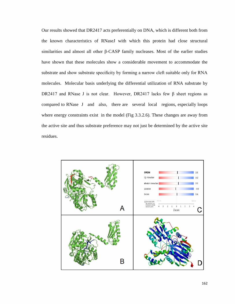
162
Our results showed that DR2417 acts preferentially on DNA, which is different both from
the known characteristics of RNaseJ with which this protein had close structural
similarities and almost all other β-CASP family nucleases. Most of the earlier studies
have shown that these molecules show a considerable movement to accommodate the
substrate and show substrate specificity by forming a narrow cleft suitable only for RNA
molecules. Molecular basis underlying the differential utilization of RNA substrate by
DR2417 and RNase J is not clear. However, DR2417 lacks few β sheet regions as
compared to RNase J and also, there are several local regions, especially loops
where energy constraints exist in the model (Fig 3.3.2.6). These changes are away from
the active site and thus substrate preference may not just be determined by the active site
residues.

163
Fig 3.3.2.6. Homology model structure of DR2417. The 3-D structure of (A) DR2417
was generated by swissmodel using (B) RNase J as a template. The red colored regions
highlight the differences between the template and the target. RNase J has well defined
beta sheets, which lacks in DR2417. (C)The Q-mean plot showing that the overall quality
of the model is satisfactory and (D) the estimated per residue inaccuracy is shown in the
model, with local inaccuracies shown in red color.
3.3.2.3 DR2417 Showed 3′ → 5′ Exonuclease and ssDNA/dsDNA Junction
Endonuclease
The proposed model of ESDSA based Double Strand Break (DSB) repair has suggested
that the processing of shattered DNA in both polarities is required for efficient DSB
repair [32,34]. However, the enzymes that process DNA during ESDSA are not precisely
known but suggested to be amongst the recombination nucleases. Recent reports
implicate RecJ as an important nuclease involved in recombination repair [32,132]. Also,
certain proteins having DNA ends processing and processive degradation of DNA
strand in 5′ → 3′ or 3′ → 5′ polarity have been reported [40,67,129]. The possible role
of DR2417 in processing of DNA substrate for ESDSA cannot be ruled out. We have
also seen that the expression of dr2417 was highest at 1 hour PIR, the time during which
extensive nucleolytic degradation is seen (Fig.3.3.2.7).

164
Fig S4
0 0.5 1 2 3 5 h
Gap
DR2417
Dr2
417
/ G
ap r
atio
Post irradiation recovery period (h)
0.0
3.0
4.0
5.0
6.0
7.0
1.0
2.0D
r24
17
/ G
ap r
atio
Post irradiation recovery period (h)
Dr2
417
/ G
ap r
atio
Post irradiation recovery period (h)
0.0
3.0
4.0
5.0
6.0
7.0
1.0
2.0
0.0
3.0
4.0
5.0
6.0
7.0
1.0
2.0D
r24
17
/ G
ap r
atio
Post irradiation recovery period (h)
Dr2
417
/ G
ap r
atio
Post irradiation recovery period (h)
0 1.0 2.0 3.0 4.0 5.00 1.0 2.0 3.0 4.0 5.0
0.0
3.0
4.0
5.0
6.0
7.0
1.0
2.0
0.0
3.0
4.0
5.0
6.0
7.0
1.0
2.0
0.0
3.0
4.0
5.0
6.0
7.0
1.0
2.0
0 1.0 2.0 3.0 4.0 5.00 1.0 2.0 3.0 4.0 5.0
0.0
3.0
4.0
5.0
6.0
7.0
1.0
2.0
0.0
3.0
4.0
5.0
6.0
7.0
1.0
2.0
0.0
3.0
4.0
5.0
6.0
7.0
1.0
2.0
0.0
3.0
4.0
5.0
6.0
7.0
1.0
2.0
A
B
Fig 3.3.2.7. Expression kinetics of dr2417 during post irradiation recovery of D.
radiodurans. Total RNA was prepared from bacterial cells collected at different time
interval (0, 0.5, 1, 2, 3 and 5 hour) of 4 kGy post irradiation and cDNA was synthesized
using standard protocol.(A) Reverse transcriptase - PCR was carried out using dr2417
(DR2417) and dr1343 (GAP) specific primers. Products were analysed on 1% agarose
gene. (B) The intensity of PCR product was quantified by densitometry and data obtained
from three such independent experiments were computed for ratios of dr2417 to dr1343
genes expression.
We therefore, checked the DNA processing activity of DR2417 on DNA substrates
having various types of DNA ends like single stranded DNA, linear double stranded
DNA, stem– loop DNA, 5′ overhang and 3′ overhang DNA substrates. DR2417 showed
very poor activity with a 5′ labeled 33mer single strand oligonucleotide [99F] substrate
(Fig. 3.3.2.8A). When 5′ labeled 99F was annealed with equal size complementary

165
oligonucleotide to make a 33mer dimer, the enzyme showed relatively faster
degradation than single stranded DNA substrate, but the release of terminal radio
labeled nucleotide was seen only after 2 hour of incubation at 37 °C (Fig. 3.3.2.8B).
These results strongly argued that although DR2417 possibly has 3′ → 5′ exonuclease
activity on single strand DNA and double strand DNA substrates, it acted slowly on
synthetic substrates lacking 5′ phosphate on opposite strand. Hence, the above 33mer
dimer substrate was phosphorylated at 5′ with cold ATP and incubated with DR2417.
Now the enzyme showed faster degradation with the release for terminal nucleotide in
less than 30 min incubation (Fig. 3.3.2.8C). Although the results indicated that DR2417
had 3′ → 5′ exonuclease activity, the possibility of the products release due to
exonucleolytic degradation in opposite polarity could not be ruled out from this result.
To check this possibility, DR2417 was incubated with DNA substrate having 37-
nucleotide long stem and 4- nucleotide long loop structure (HP78) labeled on both
ends separately. DR2417 incubated with HP78 radiolabeled at 5′ end showed a series of
typical products that are normally observed during exonucleolytic degradation of
DNA (Fig. 3.3.2.8D). This enzyme also produced the islands of products from 0.5 hour
onward incubation. This suggested that DR2417 has 3′ → 5′ exonuclease activities and a
strong possibility of an endonuclease function at the single strand/double strand DNA
junction, that could produce islands of products of 37 nucleotides and 41 nucleotides on
5′ labeled HP78. DR2417 however, showed very poor activity on 3′ radiolabeled HP78,
which had a hydroxyl group at 5′end (Fig. 3.3.2.8E) and the 3′ radiolabeled [32
P]
dNMP could be detected only after 3 hour of incubation. This further indicated that
DR2417 possibly requires both 5′ phosphate and 3′ hydroxyl group in double stranded

166
DNA for optimum activity. This hypothesis was tested with 3′ radiolabeled HP78 also
phosphorylated at 5′ end with cold ATP. The enzyme showed a faster release of 3′
terminal [32P] dNMP in less than 30 min incubation under similar condition (Fig.
3.3.2.8F). This result along with the results obtained when 3′ radiolabeled 33mer
dimer substrate was kinased at 5′ position (Fig. 3.3.2.8C) suggested the requirement of 5′
phosphate for the 3′ → 5′ exonuclease activity of DR2417.
Fig 3.3.2.8. DNA processing activity characterization of DR2417 on different types of
DNA substrates. DNA substrates like (A) 5′ labeled linear 33 bases ssDNA (B) 33 bp
dsDNA (C) 3 ′ labeled 33 bp dsDNA having 5 ′ phosphorylation in opposite strand, (D)

167
78mer (HP78) stem loop DNA , (E) HP78 with and (F) without 5′ phosphorylation, were
prepared. These substrates (S) were incubated with DR2417 at different time interval at
37 °C. Reaction was stopped with DNA sequencing dye (50% formamide, 25 mM
EDTA, pH8.0) and mixtures were heated at 95 °C for 15 min. Products were analyzed on
16% Urea-PAGE and autoradiograms were developed. Arrow marked showing limit
products (LP), terminal mononucleotide (dNMP32
) and ss/dsDNA junction
endonucleolytic products. Experiments repeated two times and data of a reproducible
experiment is shown.
The 1 hour PIR fraction readily degraded exogenously added PCR DNA. However when
DR2417 was incubated with PCR DNA, the activity was sluggish (Fig 3.3.2.9). From our
studies on bioichemical characterization (Fig 3.3.2.8E & 3.3.2.8F), we realized that
DR2417 may not act on PCR amplified DNA as both ends are hydroxylated. On
incubating DR2417 with EcoRI cut plasmid DNA which has 5’-P, it readily degraded it
(Fig 3.3.2.9). We then treated PCR dsDNA with Polynucleotide Kinase to phosphorylate
the 5’ends and when DR2417 was incubated with this modified substrate, it readily
degraded this DNA (Fig 3.3.2.9). Thus there was a possibility of a DNA binding
polynucleotide kinase in the cell lysate fraction from which DR2417 was isolated.

168
Fig 3.3.2.9. 100 ng of DR2417 was incubated either with 1 µg of PCR product (lane P,
NK & K) or 200 ng of EcoRI cut pBSK+ plasmid DNA (lane C &T) at 37°C for 30 min.
lane P-control PCR DNA, lane NK- PCR DNA with DR2417, lane K- PCR DNA with
ends phosphorylated by PNK and then incubated with DR2417, lane C- EcoRI cut
pBSK+, lane T- EcoRI cut pBSK+ treated with DR2417. Representative images of
independent assays have been provided
The requirement of 5′ phosphate for exonuclease activity has been shown as a
characteristic function of β-CASP family nuclease [133]. Interestingly, these results
suggested that (i) double stranded DNA substrates that contain both 5′ phosphate and 3′
hydroxyl group at the same end, which would eventually be present in native DNA
would be used as preferred substrate for the inherent 3′ → 5′ exonuclease activity of
DR2417, and (ii) DR2417 was a ss/ds junction endonuclease.

169
3.3.2.4. Unlike in E. coli, DR2417 Showed a Dominant Phenotype in D.
radiodurans
Several deinococcal proteins expressed in E. coli and vice versa have shown
remarkable effects on DNA damage tolerance of transgenic bacteria [124,134].
On the other hand the deletion of certain DNA repair genes from D. radiodurans genome
does not affect the normal growth of this bacterium [44,135]. While creating
deletion mutation of dr2417 gene in D. radiodurans genome, we observed that the cells
approaching complete replacement of dr2417 with nptII marker gene (Fig.
3.3.2.10), did not survive under normal growth conditions and mutants having lower
copy numbers of this gene showed poor growth under normal conditions (Fig.
3.3.2.11). These cells, survived less when subjected to γ radiation stress as compared
to the wild type D. radiodurans (Fig. 3.3.2.11). This observation was very much
similar to those favoring the essentiality of a gene in bacterial system. Recently, the
indispensability of RecJ in growth and DSB repair of D. radiodurans has been
demonstrated [28]. These results might allow us to speculate that indispensability of
DR2417 as an essential gene for survival and eventual radiation resistance in D.
radiodurans.

170
Samples M Cl4 Cl9 Cl10 R1
3.0
0.5
1.0 1.5 2.0 1.97kb
1.19kb
Samples M R1 Cl4 Cl9 Cl10
0.5 1.0 3.0
10.0 kb
521bp
Samples M Cl4 Cl9 Cl10 R1
3.0
0.5
1.0 1.5 2.0 1.97kb
1.19kb
Samples M Cl4 Cl9 Cl10 R1
3.0
0.5
1.0 1.5 2.0 1.97kb
1.19kb
Samples M Cl4 Cl9 Cl10 R1
3.0
0.5
1.0 1.5 2.0
Samples M Cl4 Cl9 Cl10 R1
3.0
0.5
1.0 1.5 2.0 3.0
0.5
1.0 1.5 2.0 1.97kb
1.19kb
Samples M R1 Cl4 Cl9 Cl10
0.5 1.0 3.0
10.0 kb
521bp
Samples M R1 Cl4 Cl9 Cl10
0.5 1.0 3.0
10.0 kb
Samples M R1 Cl4 Cl9 Cl10
0.5 1.0 3.0
10.0 kb
521bp
~2.0kb
dr2417
~1.8kb
P1
P2
P3
P4521bp
~2.0kb
dr2417
~1.8kb
P1
P2
P3
P4521bp
dr2417
~1.8kb
P1
P2
P3
P4521bp
103bp
R1 Cl4 Cl9 Cl10 Cl4*
A
B
C
D
Samples M Cl4 Cl9 Cl10 R1
3.0
0.5
1.0 1.5 2.0 1.97kb
1.19kb
Samples M R1 Cl4 Cl9 Cl10
0.5 1.0 3.0
10.0 kb
521bp
Samples M Cl4 Cl9 Cl10 R1
3.0
0.5
1.0 1.5 2.0 1.97kb
1.19kb
Samples M Cl4 Cl9 Cl10 R1
3.0
0.5
1.0 1.5 2.0 1.97kb
1.19kb
Samples M Cl4 Cl9 Cl10 R1
3.0
0.5
1.0 1.5 2.0
Samples M Cl4 Cl9 Cl10 R1
3.0
0.5
1.0 1.5 2.0 3.0
0.5
1.0 1.5 2.0 1.97kb
1.19kb
Samples M R1 Cl4 Cl9 Cl10
0.5 1.0 3.0
10.0 kb
521bp
Samples M R1 Cl4 Cl9 Cl10
0.5 1.0 3.0
10.0 kb
Samples M R1 Cl4 Cl9 Cl10
0.5 1.0 3.0
10.0 kb
521bp
~2.0kb
dr2417
~1.8kb
P1
P2
P3
P4521bp
~2.0kb
dr2417
~1.8kb
P1
P2
P3
P4521bp
dr2417
~1.8kb
P1
P2
P3
P4521bp
103bp
R1 Cl4 Cl9 Cl10 Cl4*
103bp
R1 Cl4 Cl9 Cl10 Cl4*
A
B
C
D
A
B
C
D
Fig 3.3.2.10. Characterization of D. radiodurans cells for dr2417 deletion. Deinococcus
cells transformed with linear pNOK2417 were subcultured in TGY broth supplemented
with Kanamycin (8 μg/ml) and grown several generations. Certain clones were randomly
picked up at specific time of subculturing and tentatively assigned as clone 9 (Cl9) clone
10 (Cl10) and clone 4 (Cl4). The genomic DNA was made and the status of dr2417 gene
was checked by PCR ampli fication using(A) flanking primers P1 and P2 and (B) internal
primers P3 and P4 and the amplification of the expected size products as marked in (C)
was ascertained on agarose gel. (D) almost equal levels of amplification of dr1343 gene

171
taken as an internal reference control; in all the samples equal amount of template DNA
was taken in each PCR reaction .
Fig 3.3.2.11. Effect of deletion of d2417 on growth and γ radiation response of D.
radiodurans. Genomic DNA was prepared from clone 4 (Cl4), clone 9 (Cl9), clone 10
(Cl10) as described in Fig. 3.3.2.10 , and wild type (R1) cells. Copy number of dr2417
was determined by real time PCR using internal primers of dr2417 and dr1343 as a
reference control, as described in methods. Levels of dr2417 sequence amplification in

172
each sample were normalized by taking levels of dr1343 amplification in respective
sample as reference control. (A) Fold change in dr2417 copy number was calculated by
dividing its PCR yields in respective clones with the same in wild type control .(B)These
clones were checked for growth characteristics and (C) responses to different doses of γ
radiation. Different clones having different fold reduction in dr2417 copy number
showed proportional growth retardation and sensitivity to γ radiation
Effect of DR2417 on DNA damage response of E. coli to -rays, UVC rays and to
interstrand crosslinks caused by mitomycin C was studied. The wild type E. coli
expressing this protein on a multicopy plasmid did not show any significant effect
(Fig3.3.2.12). This could be due to different levels of ATP in E. coli and D. radiodurans
or some other factor like functional redundancy by other proteins or some inhibitory
activity in E. coli. A greater possibility of E. coli not having the pathway (s) where
DR2417 works and that contribute to extreme resistance in D. radiodurans may be
suggested. It is pertinent to note that although homologs of DR2417 abound in gram
positive bacteria where they are involved in RNA metabolism they are absent in E. coli
and other enterobacteria [123]. Thus it is possible that the pathways in which β-CASP
nucleases operate upon are missing in E. coli and hence we could not see any significant
effect. Also, the threshold of DSB DNA needed for DR2417 to act upon is simply not
generated in the radiation sensitive E. coli cells.

173
Fig. 3.3.2.12. Response of E. coli cells expressing DR2417 to DNA damaging agents.
recA E. coli DH5 cells were exposed to (a) radiation (b) UVC radiation and (c)
mitomycin C at various doses. Similarly wildtype E. coli strain AB11557( recA+) and

174
were exposed to (d) radiation (e) UVC radiation and (f) mitomycin C at various doses.
Results from three independent experiments are shown
Further studies at cellular and molecular levels would be required for providing the
direct evidence on indispensability of DR2417 in this bacterium. The reported mistake
in form of a frameshift at 996th position in dr2417m ORF has been corrected and
submitted to GenBank. Recombinant protein showed both exo- and endo-nuclease and
needs a 5′ phosphate for its activity akin to Artemis, and is inhibited by ATP as observed
in hSNM1, a homolog of Artemis. However, unique to DR2417 was its preference for
double stranded DNA for its 3′ → 5′ exonuclease activity and a significantly low activity
on single stranded DNA and RNA substrates. Although, the molecular basis for low
RNase activity in DR2417 is not clear, the possibilities of other amino acids and
motifs involvement in determining the substrate specificity by Motif C of β-CASP
family nucleases could be speculated. All the prokaryotic proteins of this family like
ykqC from Bacillus subtilis, TTHA0252 from T. thermophilus, Pab-RNase J from P.
abyssi, have a conserved histidine in motif C similar to DR2417 but function as
RNases [92,126,136]. However, the DNA specific nucleases of this family like
Artemis and hSNM1 in eukaryotes have a conserved Valine in motif C and express a
considerably low activity on RNA substrates [120]. Low RNase activity in DR2417
could be attributed to certain notable differences in the amino acids contributed to 3-D
modeled structure of this protein (Fig 3.3.2.6). The strong endonuclease activity on
HP78, a stem–loop substrate (Fig. 3.3.2.8D) also confirmed that DR2417 confers
ssDNA/dsDNA endonuclease function. These results suggested that DR2417 was a DNA
processing enzyme. Although, the functional significance of this protein is not clear in

175
D. radiodurans, our results indicated that DR2417 plays an essential role in growth and
eventually γ radiation resistance in this bacterium.
We noted that at higher temperatures, DNA is degraded in the presence of Mn2+
but
DR2417 prevents this degradation (Fig 3.3.2.13a). When DR2417 is incubated for more
than 30 minutes with with double stranded DNA, DR2417 is resistant to digestion by
proteinase K proteolysis (Fig 3.3.2.13b). Thus DNA is protected by the protein and DNA
protects the protein. A possible explanation of this phenomenon can be if an inaccessible
supramolecular complex is formed and this phenomenon needs further investigation. The
inability to generate deletion mutation, a strong affinity of this protein to DNA, the
protection of DNA from higher temperature together might allow to speculate the
crucial role of this protein in structure maintenance and normal DNA metabolism.
Inhibition of its nuclease activity by ATP is a feature which could be useful in
regulation of its functions under different physiological conditions. Recently, it has been
shown that the levels of ATP changes during post irradiation recovery in D.
radiodurans [82]. With the present data it is difficult to decipher the indispensable role
of this protein.

176
Fig 3.3.2.13. (a) 100 ng of DR2417 was incubated with 1 µg of ØX 174 virion DNA in a
buffer containing 10 mM Tris Cl pH 8.0, 50 mM NaCl, 1 mM DTT and 2mM MnCl2 at
the indicated temparatures.(b) Effect of proteinase K on DNA-protein complex stability.
The 500ng purified DR2417 was incubated with 200ng of 562bp PCR amplified dsDNA
for different time interval. One set was treated proteinase K for 30 min and other was
kept as control. Reaction mixture was analysed on 1% agarose gel.
A wide occurrence of similar type of proteins in Bacilli and other bacteria might add
further to the importance of this family protein for the growth and maintenance of
living organisms. Although, further work will be needed to understand the exact

177
mechanism of DR2417 function, the provided evidences suggest that DR2417 was a
genuine DNA damage inducible novel nuclease of β- CASP family as against the
reported frameshift in genome sequence, and its DNA processing activity is regulated
by levels of high energy phosphates and may be essential for survival and radiation
resistance of D. radiodurans.

178
Chapter 4
Discussion

179
As mentioned earlier, D. radiodurans is unique amongst living organisms in having an
efficient repair process which can reseal a large number of Double Strand Breaks (DSBs)
[135]. It has adapted a time consuming modification of the homologous repair pathway
rather than the quicker Non Homologous End Joining (NHEJ) pathway, presumably to
maintain the fidelity of information. This is an efficient pathway as more than 200
fragments are efficiently resealed even when evidence exists for transposon activity [43].
The initial phase of the DNA repair pathway, named as ESDSA pathway has a lot of
resemblance with Synthesis Dependent Strand Annealing (SDSA) pathway as seen in
yeasts which is essentially a non reciprocal crossover mechanism [34]. In yeasts SDSA is
mediated by the RAD52 epistasis group comprising of RAD50, RAD51 (RecA), RAD52,
RAD54, RAD55, RAD57, Xrs2 and MRE11[35]. RAD50/MRE11 form an ATP
regulated dsDNA specific exonuclease activity and it has been shown in D. radiodurans
that this activity is contributed by SbcCD [40]. It has been shown that DdrA could be a
functional homolog of RAD52 [54] and DR1259 has been identified as a RAD54
homolog. Thus D. radiodurans has almost all the functional homologs of the yeast DNA
repair apparatus. It is interesting to note that several fungi too share the property of
radioresistance as seen in D. radiodurans [38] and it may be speculated that the non
reciprocal homologous crossing over mechanism (SDSA/ESDSA) coupled with control
of cell cycle is the reason for radio-resistance. A single and seminal study by Kota et
al.,2008 to purify a DNA repair complex during the crucial early phase of repair however
does not list these above deinococcal proteins together but listed several other proteins
including DR2417 which were associated with putative DNA repair roles [131].
However, transcriptome data show that DdrA, RecA, DR1259, SbcD all are

180
overexpressed during the repair process. RT-PCR analysis in our study also showed the
inducible expression of DRA0282 and DR2417 genes during the early phase of Post
Irradiation Recovery (PIR). Thus there could be a possibility of DdrA, RecA, DR1259,
SbcD being active in ESDSA but not directly by interacting with each other but through a
more elaborate mechanism which might include DRA0282 and DR2417.
Most authors suggest that homologous recombination is the major repair pathway in D.
radiodurans but that does not preclude NHEJ or similar pathways of DSB repair, at least
in the initial few hours. A look at the protein profile of the first hour of PIR shows several
proteins which are novel [131,137]. For example, many of these proteins are membrane
or cell wall bound and have putative nucleases/kinases/protease domains and hence they
could be involved in DNA repair or cell cycle arrest.
In this thesis, we had started with an observation that a cellular lysate fraction of
Deinococcus cells in the early phase of repair showed a nuclease activity which was
inhibited by ATP. In prokaryotic systems ATP stimulated nucleases are more prevalent, a
classic case being the RecBCD system and hence the observed ATP inhibited nuclease
activity was unusual [138]. It is also well known that extensive but regulated nuclease
activity occurs in the early phase of repair in D. radiodurans [34] and hence finding the
identity of this possible nuclease and/or kinase was of significance. Analysis of mass
spectroscopy data by MASCOT identified a large dataset >300 proteins, but none of them
was annotated as a nuclease or kinase in the database. Here it is pertinent to remember
that automatic annotation of protein function is not always accurate. As outlined by
Alfonso Valencia et al.,2005 [139], the general model is that rate of evolution decreases
from catalytic site, to protein core, to substrate specificity site and to surface

181
regulation.The degree of divergence of functional annotation is therefore, dependent on
the degree of divergence of sequence similarity. For an unknown sequence which has at
least 15% sequence identity with a known protein, the first Enzyme Commissin digit (EC
digit) can be accurately assigned which meant that whether a protein is an oxidoreductase
or a ligase can be fairly and correctly predicted. However, in proteins with even at 70%
sequence identity, the fourth EC digit which specifies substrate specificity can be and has
been wrongly assigned and this fact has been confirmed by other studies too [140] . There
have been cases of mis-annotataion before [141] and thus the presence of
nucleases/kinases amongst the annotated proteins cannot be ruled out and would be worth
studying. Analyzing all these proteins for nuclease or kinase activty experimentally
would have required high throughput protein purification and assay systems, which
would be time consuming and purifying a protein from a complex collection is equally
demanding. Hence we relied on other alternative i.e. insilico analysis. Most nuclease and
kinase domains are well characterized and are cataloged in databases. Since MASCOT
could not find any nuclease/kinase in pool of proteins exhibiting ATP sensitive nuclease
functions in our earlier studies, we focussed on the structure-function prediction of the
11 hypothetical proteins also found in this fraction.
The set of criteria we used for analyzing the data was (1) protein sequence analysis (2)
genomic context (3) subcellular location (4) peptide abundance in FTICR-MS data (5)
theoretically modeled structure and (6) phylogenetic relationships. In this study, we
employed both bioinformatics as well as experimental approaches for functional
characterization of both these proteins.

182
Our experience showed that the output obtained with each individual analysis was not
seamless. For example, the PSI-BLAST results indicated DRA0282 as Ku type protein
but the structure prediction servers did not predict DRA0282 as Ku type protein but as
human fibronectin (fibronectin type III domains bind DNA) and receptor protein tyrosine
phosphatase type protein. The best analysis came when we combined results from the
sequence search with the structure data and phylogenetic tree information. Based on
initial BLAST searches, we had presumed that we are looking into components of NHEJ
pathway and accordingly our intent was to see whether indeed these initial searches were
true. For both DR2417 and DRA0282 this method proved effective and provided us with
testable predictions but for DR0390, we could only predict its role as a kinase with
possible role in binding to lipid substrates and it failed to predict any functional role for
DR1654 save it being possibly a membrane bound protein. Both DRA0282 and DR2417
were found in a cell lysate fraction at 1 hour PIR phase and both these proteins were
unannotated and hence their study assumed importance. Our bioinformatics results
showed that these proteins may have a role in DNA processing. DR2417 showed
homology to Artemis, a nuclease involved in processing noncompatible ends for NHEJ
and DRA0282 was homologous with Ku80 which too was involved in the same pathway.
Artemis is an important DNA repair enzyme which modifies its activity from a 5’ ssDNA
exonuclease to a hairpin endonuclease. We would have expected DR2417 to show a
similar kinase dependent switch from 5’ single stranded DNA exonuclease to a hairpin
endonuclease in the presence of DRA0282 and DR0390 (presuming it had DNA-PKcs
activity, which it may not have). Our assays showed that DR2417 was a double stranded
DNA exonulease as well as a hairpin endonuclease but it did both these activities without
183
being modulated by any other protein and our preliminary investigation showed that
DRA0282 hindered this nuclease activity rather than modulating it. At the same time
DR2417 was inhibited by ATP which could be easily explained by the 3-D model
obtained. However, the model also suggested that DR2417 should act on single stranded
DNA or RNA, but our biochemical assays show that it does not degrade single stranded
DNA or RNA. More importantly, earlier bioinformatics studies on β-CASP family of
nucleases concluded that valine at motif C contributed to the discrimination between
RNase and DNase. Our study has adequate proof that motif C is inconsequential to
RNase/DNase discrimination and the reason for this lies elsewhere. Indirectly we assume
that the biochemical activity could be explained if DR2417 acts exclusively as a dimer.
Also, it may be well worth noting that the closest homolog of DR2417 is RNAse J from
Thermus thermophilus which functions an RNAse in the closely related Thermus species.
DR2417 is the first reported DNase of the β-CASP family amongst prokaryotes and this
assumes importance as members of this family are present in more than 200 species of
gram positive bacteria. Our studies indicate that possibly in all other species β-CASP
nucleases act as RNases, while in Deinococcus spp. they have evolved into DNases. A
crucial part of the evolution would be a dimer interface which would not allow single
stranded DNA but would become relaxed enough for double stranded DNA in its
presence. Artemis null mutants of mice do not survive due to a compromised immune
system as NHEJ pathway is required for T and B cell maturation, but this mutation is not
lethal per se. We found that Δdr2417 is possibly a lethal mutation in D. radiodurans and
thus DR2417 could have an essential role in the lifecycle of D. radiodurans. It was
difficult to study the biological effects as cells lacking the gene would die off. But

184
nevertheless, its nuclease activities are consistent with its role during early phase repair
where extensive double stranded DNase activity converts the double stranded DNA to
extensive tracts of single stranded DNA which is then paired with homologous strands in
a recA dependent pathway. We also overlooked a crucial piece of evidence from our own
study. Hydrophobic Cluster Analysis plots (HCA plots) highlight regions of irregular
secondary structure. Our HCA plot showed that the C-terminal of DR2417 is devoid of
regular secondary structure with an excess of proline and arginine residues as seen in
pseudogenes. This fact should have alerted us , instead we found a valine at a suitable
position in the multiple sequence alignment. Since DR2417 was a DNase, we assumed
that this valine is the site in motif C. The earlier studies had provided circumstantial
evidence for valine at motif C being discriminatory for DNase/RNase without providing
any structural/biochemical evidence. We took this evidence at face value, which now we
find was erroneous. Nevertheless, resequencing the dr2417 ORF and Peptide Mass
Fingerprinting of the protein corrected this oversight.
We identified DRA0282 as a Ku type protein. The reputable automated structure
prediction server ITASSER predicted DRA0282 as a functional homolog of a sialidase
from Micromonospora viridifaciens (PDB id 1EUT) which is quite different from a DNA
binding protein. But we combined our PSI-BLAST search and multiple sequence
alignment with Ku homologs and built 3-D model with Ku80 (PDB id1JEY). A close
inspection of both models of this proteins reveal a central β barrel domain, a N-terminal β
sheet roll, thus although the structure was similar but function was different. Our
biochemical studies show that DRA0282 did indeed bind to DNA and also protected ends
of double stranded DNA from Exo III nuclease activity. But, crucially, it did not enhance

185
intermolecular or intramolecular ligation and neither did it promote endjoining of
noncompatible ends invitro. Also DRA0282 had no ATPase activity like Ku rather it was
inhibited by ATP and although 3-D modeling showed that it had a clearly defined ATP
binding domain it was not clear how ATP binding would inhibit DNA binding. Curiously
DRA0282 shows highest affinity for single stranded DNA followed by supercoiled DNA
and shows a much lower affinity for linear double stranded DNA. This is in contrast to
Ku70/80 dimer in mammals or Ku type proteins in bacteria which show preference to
linear double stranded DNA [142]. Also phylogenetic studies show that DRA0282,
matches more closely with eukaryotic Ku80 and has only 7 orthologs in bacteria. This
would mean that its Ku like structure and function is a case of convergent evolution. Our
molecular genetic studies on DRA0282 have revealed apparently contrasting results in
the response of two different bacterial species to DNA damage. While transgenic E. coli
expressing this protein show enhanced protection to UV, the D. radiodurans deletion
mutants show a marginally improved survival. Deleting this gene causes no apparent loss
in survival of D. radiodurans or its radiation resistance. We have some evidence from E.
coli that DRA0282 functionally interacts with RecA in its physiological milieu in vivo.
The mechanism of E. coli RecA and D. radiodurans RecA is different as D. radiodurans
RecA has preference for double stranded DNA while E. coli has preference for single
stranded DNA [143]. Also DRA0282 has higher affinity for single stranded DNA
compared to double stranded DNA [144]. Although it is mere speculation, possibly this
affinity for single stranded DNA allows DRA0282 to interact better with E. coli RecA
and enhance the homologous recombination pathway in E. coli, while in D. radiodurans
its role may be to prevent DNA from premature attack by exonucleases prior to being
186
coated by RecA, akin to SSB. The 3-D model suggests that like Ku, DRA0282 encloses
DNA in a ring structure which can only be released by a cleaving the protein.
Interestingly there is a serine protease DRA0283 just upstream to DRA0282. It has been
earlier shown that this protease undergoes phosphorylation in vivo and is under the
control of PprI a global switch for radioresistance [145]. Although the null mutant of
DRA0282 was not debilitated earlier in survival or in response to radiation, this protein
has been found in many proteomics studies on D. radiodurans [137,145,146]. DRA0282
may play a redundant role, or may have an important role under environmental conditions
which cannot be mimicked under laboratory conditions.
Hence, from our studies we would like to conclude that both these proteins should be
involved in DNA metabolism especially in the repair process. Recently, it has been
shown that both DR2417 and DRA0282 are associated with the D. radiodurans nucleoid,
the condensed structures which are formed during stationary phase and especially after
extensive DNA damage [112]. Toroidal structure of D. radiodurans genome may be an
added advantage to extreme phenotypes of this
bacterium has not been ruled out. Thus it corroborates our results that DRA0282 and
DR2417 are DNA metabolic genes involved in maintenance of genome integrity. It
would indeed be interesting to further unravel the real time functions of these two
proteins in the metabolism of the cell.

187
Chapter 5
Summary, Conclusion and Future
Perspectives

188
Summary
D. radiodurans was initially isolated in 1956 from canned meat treated to 4 kGy radiation
dose and classified as Micrococcus spp.[2]. This dose is 250 times more than the
tolerance level of the gut bacterium E. coli. Over the next 60 odd years, the molecular
basis of its unusual radiation resistance has been investigated and several hypotheses
have been propounded. These include the role of antioxidants , the compactness and
toroidal structure of genome, protection of double strand breaks (DSBs) from nucleolytic
degradation and an efficient DSB repair pathway which now has been shown to be
mainly facilitated by ESDSA followed by RecF pathway of recombination [34]. While all
these have independently proven in contributing to extreme radiation resistance of this
bacterium, the concerted action of these molecular events in radioresistance has not been
sufficiently detailed. In this thesis, a fraction of DNA binding proteins showing ATP
sensitive nuclease activity and autophosphorylation of several proteins was subjected to
mass spectrometric identification of its protein components. Interestingly, none of the
known nucleases and kinases were found in a pool of 300 polypeptides detected by
FTICR-MS. Out of the 11 hypothetical proteins present in this fraction, 4 were analysed
using various tools of bioinformatics for structure and function predictions. Two of these
DRA0282 and DR2417 showed well defined functional motifs relating to DNA
metabolism while another DR0390 was having putative motifs for a kinase. DRA0282
and DR2417 proteins were found structurally close to Ku80 and Artemis type nuclease,
respectively, while DR0390 was closer to DAK/DegV kinase. DRA0282 and DR2417
were further characterized in vivo and in vitro for the known functions related to their

189
closer homologues. DRA0282 was a DNA binding protein, a function that was found to
be sensitive to ATP addition. Quite unlike Ku type proteins, DRA0282 showed higher
affinity to supercoiled double stranded DNA followed by a lower affinity to single
stranded DNA and the lowest affinity to linear dsDNA amongst these topologically
different substrates. This protein also showed DNA protection from exonuclease
degradation as observed with Ku type homologues [113]. The role of this protein in
radiation resistance of D. radiodurans was redundant as its deletion mutant did not show
change in DNA damage response of wild type while absence of this protein made D.
radiodurans more tolerant to UVC as compared to wild type. This result is intriguing.
Interestingly, deletion of drA0282 from the genome of PprA (another deinococcal protein
with nearly similar function in vitro) mutant of D. radiodurans, did not add further to
pprA mutant’s radiation sensitivity. Further, over expression of this protein increased
both and UVC resistance of transgenic E. coli. This phenotype in E. coli required a
recombinationally functional RecA and was independent of SOS response. These results
indicated that DRA0282 functions in tandem with RecA in recombination repair of DNA
damage at least in E. coli while it has functional homologues in D. radiodurans. Its
function in tandem with RecA in D. radiodurans will be investigated independently.
DR2417m was wrongly annotated with a so called genuine frameshift questioning the
functional authenticity of this protein in D. radiodurans. However, this protein was
consistently detected in the functional pools of protein in independent studies. DR2417m
was sequenced and the mistakes of initial annotation were corrected. DR2417 showed
perfect match with proteins of β-CASP family of which Artemis is a member.
Recombinant protein was made and in vitro activities were checked. DR2417 showed

190
both exonculease and ss/dsDNA junction endonuclease on artificially synthesized
heteropolymeric DNA substrates. DR2417 exhibited DNA processing activity albeit 3’-5’
exonucelase, which are integral to efficient DSB repair as observed in D. radiodurans.
Quite surprisingly, inspite of having very similar structure with conserved motifs as that
of RNase J from Thermus spp. and Bacillus spp., and other β-CASP family nuclease, this
protein showed poor RNase activity. This result is further interesting but intriguing.
Molecular genetic studies confirmed that dr2417 is an essential gene in D. radiodurans as
dose dependent reduction in copy number correlated very well with loss of cell survival
and radiation resistance.
In this study we have used integrated approach of bioinformatics tools for prediction of
structure and functions of four proteins selected from the pool of 11 hypothetical proteins
and further validated the predicted functions of two of these proteins experimentally. The
evidences gathered on the various activities of these proteins have helped us in assigning
new functions to two of the hypothetical proteins from nearly 50% of uncharacterized
proteins of D. radiodurans. Apparently, these functions are found to be integral to the
DNA metabolism reported during recombination repair of DNA double strand breaks, for
which D. radiodurans is best characterized and is being studied.
Conclusion
Our study has accentuated the role of bioinformatics in finding relevant proteins in a
complex mixture of proteins undertaking a certain biochemical activity in this case e.g.
ATP sensitive nucleolytic function. Extrapolation is avoided in science and
bioinformatics is no exception to this. While learning these facts we could assign novel

191
functions to two hypothetical proteins in D. radiodurans genome and corrected one
erroneous annotation during the course of this study. Another key finding has been that
one cannot rely heavily on a single bioinformatic tool to decipher the function of novel
proteins rather an integrated approaches and authentication by wet biology is must. Thus
out of 300 proteins, we could home onto a single nuclease DR2417 and another DNA
binding protein DRA0282 for its similarity to Ku80. The findings of this study led to
conclude the following:
• On the basis of multiple sequence alignments, phylogenetic trees and 3-D
homology model, we predicted that DR2417 and DRA0282 are functional
counterparts of Artemis and Ku 70/80, respectively
• DR2417 is a Mn2+
dependent DNase and DRA0282 is a DNA end protecting
protein.
• DRA0282 is a Mn2+
dependent DNA end protecting protein having higher affinity
to supercoiled DNA and single stranded DNA as compared to linear double
stranded DNA.
• Both these proteins display unique functional properties like 3’-5’ double
stranded exonuclease activity and endonuclease with propensity to single
stranded-double stranded DNA junctions for DR2417 and protection of linear
double stranded DNA from exonucelase III digestion and higher affinity for
supercoiled DNA by DRA0282.
• Molecular genetic studies show that DR2417 appears to be an essential gene in D.
radiodurans while much effect was not seen in E. coli.DRA0282 helps

192
significantly in UV and γ radiation tolerance of E. coli but has nearly no effect on
radiation response while it seems to have a role in UVC resistance in D.
radiodurans
Future Perspectives
As we conclude this thesis, it would not be inappropriate to raise the questions that we
have shied away from- was it possible for the already annotated protein to have an
additional nuclease/kinase function and not the hypothetical proteins? Yes. That
possibility existed and a way to analyze this would be a study to take all the sequences
and analyze for known nuclease/kinase motifs by writing user defined programs. What
we have undertaken here is the approach of using available bioinformatics tools with a
biologists ‘common sense’ approach. This had its limitations in that no clear prediction
was possible for DR0390 and DR1654. But all the same, our results allow us to peer a
little deeper into the enigmatic life of D. radiodurans. While this study has answered
some of the questions and hypotheses raised both from the experimental data of earlier
work and bioinformatic analyses of certain proteins, it has left several unanswered
questions.
1. DR2417 almost is unique amongst all β-CASP family of nucleases in its weak
exonuclease activity on single stranded DNA or RNA although at the same time it is able
to act upon gross single stranded Ø X174 DNA to form island products. From the
structure- function point of view this aspect is certainly an exciting area of research.

193
During the course of this study we gained evidence that dr2417 might be an essential
gene. Earlier RecJ, a 5’-3’ exonuclease was shown to be essential for D. radiodurans,
showing that crossover mechanisms involving single stranded DNA are a part of the
lifecycle in D. radiodurans. Rec J has been postulated to work in the RecFOR pathway
but cells expressing SbcB which degrades 3’ ssDNA and eventually arrests RecFOR,
grow normally like the wild type [100] and hence D. radiodurans may have another
single stranded DNA dependent mechanism involving Rec J and possibly DR2417
working in tandem. Thus the exact physiological role played by DR2417 is well worth
investigating. Recombinant DR2417 protects DNA from thermal and pH denaturation.
The mechanism underlying this characteristic would also be worth elucidating.
2. DRA0282 is omnipresent and has been shown to be a part of the nucleoid [110]. As
described earlier, absence of this gene product does not hamper the survival of D.
radiodurans to radiation stress in rich medium. The survival and DNA repair of the
ΔdrA0282 deletion mutant under physiological stresses like dessication would be
interesting as the DdrA deletion mutant shows a debilitated phenotype only under
dessication [62]. DRA0282 shows preference to supercoiled DNA, followed by single
stranded DNA and protection of linear double stranded DNA from exonuclease
degradation and this protein also function in tandem with recombination function of
RecA in E. coli. It is possible that this protein in brings the interaction of supercoiled
DNA with single stranded DNA. The functional significance of this interaction by
DRA0282, in the context of efficient DSB repair would be interesting to ascertain.
3.DR0390 is predicted to be a kinase. The activity characterization of this protein and
significance if any, of this protein being together with DRA0282 and DR2417 would be

194
interesting to study. Last but not the least, further studies on functional characterization
of DR0390 keeping in mind its role as kinase and its involvement in radiation resistance
may be investigated.

195
Reference List
1. Mora C, Tittensor DP, Adl S, Simpson AG, Worm B (2011) How many species are there on Earth
and in the ocean? PLoS Biol 9: e1001127.
2. Duggan DE, Anderson AW, Elliker PR (1963) Inactivation of the Radiation-Resistant Spoilage
Bacterium Micrococcus radiodurans. Ii. Radiation Inactivation Rates as Influenced by Menstruum
Temperature, Preirradiation Heat Treatment, and Certain Reducing Agents. Appl Microbiol 11:
413-417.
3. Yuan M, Zhang W, Dai S, Wu J, Wang Y, et al. (2009) Deinococcus gobiensis sp. nov., an
extremely radiation-resistant bacterium. Int J Syst Evol Microbiol 59: 1513-1517.
4. Yang Y, Itoh T, Yokobori S, Shimada H, Itahashi S, et al. (2010) Deinococcus aetherius sp. nov.,
isolated from the stratosphere. Int J Syst Evol Microbiol 60: 776-779.
5. Slade D, Radman M (2011) Oxidative stress resistance in Deinococcus radiodurans. Microbiol Mol
Biol Rev 75: 133-191.
6. Sedelnikova OA, Redon CE, Dickey JS, Nakamura AJ, Georgakilas AG, et al. (2010) Role of
oxidatively induced DNA lesions in human pathogenesis. Mutat Res 704: 152-159.
7. Battista JR (2000) Radiation resistance: the fragments that remain. Curr Biol 10: R204-205.
8. Griffiths E, Gupta RS (2004) Distinctive protein signatures provide molecular markers and
evidence for the monophyletic nature of the deinococcus-thermus phylum. J Bacteriol 186: 3097-
3107.
9. Omelchenko MV, Wolf YI, Gaidamakova EK, Matrosova VY, Vasilenko A, et al. (2005)
Comparative genomics of Thermus thermophilus and Deinococcus radiodurans: divergent routes of
adaptation to thermophily and radiation resistance. BMC Evol Biol 5: 57.
10. Daly MJ (2009) A new perspective on radiation resistance based on Deinococcus radiodurans. Nat
Rev Microbiol 7: 237-245.
11. Cantu D, Schaack J, Patel M (2009) Oxidative inactivation of mitochondrial aconitase results in
iron and H2O2-mediated neurotoxicity in rat primary mesencephalic cultures. PLoS One 4: e7095.
12. Misra HS, Khairnar NP, Barik A, Indira Priyadarsini K, Mohan H, et al. (2004)
Pyrroloquinoline-quinone: a reactive oxygen species scavenger in bacteria. FEBS Lett 578: 26-30.
13. Khairnar NP, Misra HS, Apte SK (2003) Pyrroloquinoline-quinone synthesized in Escherichia
coli by pyrroloquinoline-quinone synthase of Deinococcus radiodurans plays a role beyond mineral
phosphate solubilization. Biochem Biophys Res Commun 312: 303-308.

196
14. Lavy A, Neeman Y, Fuhrman B (2005) The antioxidative effect of the bacteria Dienococcus
radiophilus against LDL lipid peroxidation. Eur J Nutr 44: 281-284.
15. Daly MJ, Gaidamakova EK, Matrosova VY, Vasilenko A, Zhai M, et al. (2004) Accumulation of
Mn(II) in Deinococcus radiodurans facilitates gamma-radiation resistance. Science 306: 1025-1028.
16. Krisko A, Radman M (2010) Protein damage and death by radiation in Escherichia coli and
Deinococcus radiodurans. Proc Natl Acad Sci U S A 107: 14373-14377.
17. Joshi B, Schmid R, Altendorf K, Apte SK (2004) Protein recycling is a major component of post-
irradiation recovery in Deinococcus radiodurans strain R1. Biochem Biophys Res Commun 320:
1112-1117.
18. Enari M, Sakahira H, Yokoyama H, Okawa K, Iwamatsu A, et al. (1998) A caspase-activated
DNase that degrades DNA during apoptosis, and its inhibitor ICAD. Nature 391: 43-50.
19. White O, Eisen JA, Heidelberg JF, Hickey EK, Peterson JD, et al. (1999) Genome sequence of the
radioresistant bacterium Deinococcus radiodurans R1. Science 286: 1571-1577.
20. Martin SA, Lord CJ, Ashworth A (2008) DNA repair deficiency as a therapeutic target in cancer.
Curr Opin Genet Dev 18: 80-86.
21. Shuck SC, Short EA, Turchi JJ (2008) Eukaryotic nucleotide excision repair: from
understanding mechanisms to influencing biology. Cell Res 18: 64-72.
22. Pham P, Rangarajan S, Woodgate R, Goodman MF (2001) Roles of DNA polymerases V and II in
SOS-induced error-prone and error-free repair in Escherichia coli. Proc Natl Acad Sci U S A 98:
8350-8354.
23. Aguilera A, Boulton SJ (2007) How to exchange your partner. Workshop on recombination
mechanisms and the maintenance of genomic stability. EMBO Rep 8: 28-33.
24. Dillingham MS, Kowalczykowski SC (2008) RecBCD enzyme and the repair of double-stranded
DNA breaks. Microbiol Mol Biol Rev 72: 642-671, Table of Contents.
25. Weller GR, Kysela B, Roy R, Tonkin LM, Scanlan E, et al. (2002) Identification of a DNA
nonhomologous end-joining complex in bacteria. Science 297: 1686-1689.
26. Englander J, Klein E, Brumfeld V, Sharma AK, Doherty AJ, et al. (2004) DNA toroids:
framework for DNA repair in Deinococcus radiodurans and in germinating bacterial spores. J
Bacteriol 186: 5973-5977.
27. Zimmerman JM, Battista JR (2005) A ring-like nucleoid is not necessary for radioresistance in
the Deinococcaceae. BMC Microbiol 5: 17.
28. Daly MJ, Gaidamakova EK, Matrosova VY, Kiang JG, Fukumoto R, et al. (2010) Small-molecule
antioxidant proteome-shields in Deinococcus radiodurans. PLoS One 5: e12570.

197
29. Daly MJ, Gaidamakova EK, Matrosova VY, Vasilenko A, Zhai M, et al. (2007) Protein oxidation
implicated as the primary determinant of bacterial radioresistance. PLoS Biol 5: e92.
30. Gutman PD, Fuchs P, Minton KW (1994) Restoration of the DNA damage resistance of
Deinococcus radiodurans DNA polymerase mutants by Escherichia coli DNA polymerase I and
Klenow fragment. Mutat Res 314: 87-97.
31. Varghese AJ, Day RS, 3rd (1970) Excision of cytosine-thymine adduct from the DNA of
ultraviolet-irradiated Micrococcus radiodurans. Photochem Photobiol 11: 511-517.
32. Bentchikou E, Servant P, Coste G, Sommer S (2010) A major role of the RecFOR pathway in
DNA double-strand-break repair through ESDSA in Deinococcus radiodurans. PLoS Genet 6:
e1000774.
33. Zahradka K, Slade D, Bailone A, Sommer S, Averbeck D, et al. (2006) Reassembly of shattered
chromosomes in Deinococcus radiodurans. Nature 443: 569-573.
34. Slade D, Lindner AB, Paul G, Radman M (2009) Recombination and replication in DNA repair of
heavily irradiated Deinococcus radiodurans. Cell 136: 1044-1055.
35. McVey M, Adams M, Staeva-Vieira E, Sekelsky JJ (2004) Evidence for multiple cycles of strand
invasion during repair of double-strand gaps in Drosophila. Genetics 167: 699-705.
36. Paques F, Leung WY, Haber JE (1998) Expansions and contractions in a tandem repeat induced
by double-strand break repair. Mol Cell Biol 18: 2045-2054.
37. Mueller JE, Clyman J, Huang YJ, Parker MM, Belfort M (1996) Intron mobility in phage T4
occurs in the context of recombination-dependent DNA replication by way of multiple pathways.
Genes Dev 10: 351-364.
38. Holloman WK, Schirawski J, Holliday R (2007) Towards understanding the extreme radiation
resistance of Ustilago maydis. Trends Microbiol 15: 525-529.
39. Bennett CB, Lewis LK, Karthikeyan G, Lobachev KS, Jin YH, et al. (2001) Genes required for
ionizing radiation resistance in yeast. Nat Genet 29: 426-434.
40. Kamble VA, Misra HS (2010) The SbcCD complex of Deinococcus radiodurans contributes to
radioresistance and DNA strand break repair in vivo and exhibits Mre11-Rad50 type activity in
vitro. DNA Repair (Amst) 9: 488-494.
41. Aylon Y, Kupiec M (2004) DSB repair: the yeast paradigm. DNA Repair (Amst) 3: 797-815.
42. Zou L, Elledge SJ (2003) Sensing DNA damage through ATRIP recognition of RPA-ssDNA
complexes. Science 300: 1542-1548.

198
43. Pasternak C, Ton-Hoang B, Coste G, Bailone A, Chandler M, et al. (2010) Irradiation-induced
Deinococcus radiodurans genome fragmentation triggers transposition of a single resident insertion
sequence. PLoS Genet 6: e1000799.
44. Cox MM, Battista JR (2005) Deinococcus radiodurans - the consummate survivor. Nat Rev
Microbiol 3: 882-892.
45. Liu Y, Zhou J, Omelchenko MV, Beliaev AS, Venkateswaran A, et al. (2003) Transcriptome
dynamics of Deinococcus radiodurans recovering from ionizing radiation. Proc Natl Acad Sci U S A
100: 4191-4196.
46. de Groot A, Dulermo R, Ortet P, Blanchard L, Guerin P, et al. (2009) Alliance of proteomics and
genomics to unravel the specificities of Sahara bacterium Deinococcus deserti. PLoS Genet 5:
e1000434.
47. Davids W, Fuxelius HH, Andersson SG (2003) The Journey to smORFland. Comp Funct
Genomics 4: 537-541.
48. Siew N, Fischer D (2003) Unravelling the ORFan Puzzle. Comp Funct Genomics 4: 432-441.
49. Nishikimi M, Fukuyama R, Minoshima S, Shimizu N, Yagi K (1994) Cloning and chromosomal
mapping of the human nonfunctional gene for L-gulono-gamma-lactone oxidase, the enzyme for L-
ascorbic acid biosynthesis missing in man. J Biol Chem 269: 13685-13688.
50. Xue Y, Daly A, Yngvadottir B, Liu M, Coop G, et al. (2006) Spread of an inactive form of
caspase-12 in humans is due to recent positive selection. Am J Hum Genet 78: 659-670.
51. Tautz D, Domazet-Loso T (2011) The evolutionary origin of orphan genes. Nat Rev Genet 12:
692-702.
52. Pearl F, Todd A, Sillitoe I, Dibley M, Redfern O, et al. (2005) The CATH Domain Structure
Database and related resources Gene3D and DHS provide comprehensive domain family information
for genome analysis. Nucleic Acids Res 33: D247-251.
53. Tanaka A, Hirano H, Kikuchi M, Kitayama S, Watanabe H (1996) Changes in cellular proteins of
Deinococcus radiodurans following gamma-irradiation. Radiat Environ Biophys 35: 95-99.
54. Harris DR, Tanaka M, Saveliev SV, Jolivet E, Earl AM, et al. (2004) Preserving genome integrity:
the DdrA protein of Deinococcus radiodurans R1. PLoS Biol 2: e304.
55. Narumi I, Satoh K, Cui S, Funayama T, Kitayama S, et al. (2004) PprA: a novel protein from
Deinococcus radiodurans that stimulates DNA ligation. Mol Microbiol 54: 278-285.
56. Earl AM, Mohundro MM, Mian IS, Battista JR (2002) The IrrE protein of Deinococcus
radiodurans R1 is a novel regulator of recA expression. J Bacteriol 184: 6216-6224.

199
57. Sun H, Xu G, Zhan H, Chen H, Sun Z, et al. (2010) Identification and evaluation of the role of the
manganese efflux protein in Deinococcus radiodurans. BMC Microbiol 10: 319.
58. Schmid AK, Howell HA, Battista JR, Peterson SN, Lidstrom ME (2005) Global transcriptional
and proteomic analysis of the Sig1 heat shock regulon of Deinococcus radiodurans. J Bacteriol 187:
3339-3351.
59. Schmid AK, Lidstrom ME (2002) Involvement of two putative alternative sigma factors in stress
response of the radioresistant bacterium Deinococcus radiodurans. J Bacteriol 184: 6182-6189.
60. Satoh K, Ohba H, Sghaier H, Narumi I (2006) Down-regulation of radioresistance by LexA2 in
Deinococcus radiodurans. Microbiology 152: 3217-3226.
61. Martins A, Shuman S (2004) An RNA ligase from Deinococcus radiodurans. J Biol Chem 279:
50654-50661.
62. Xu G, Lu H, Wang L, Chen H, Xu Z, et al. (2010) DdrB stimulates single-stranded DNA
annealing and facilitates RecA-independent DNA repair in Deinococcus radiodurans. DNA Repair
(Amst) 9: 805-812.
63. Leiros HK, Kozielski-Stuhrmann S, Kapp U, Terradot L, Leonard GA, et al. (2004) Structural
basis of 5-nitroimidazole antibiotic resistance: the crystal structure of NimA from Deinococcus
radiodurans. J Biol Chem 279: 55840-55849.
64. Wang L, Xu G, Chen H, Zhao Y, Xu N, et al. (2008) DrRRA: a novel response regulator essential
for the extreme radioresistance of Deinococcus radiodurans. Mol Microbiol 67: 1211-1222.
65. Blasius M, Buob R, Shevelev IV, Hubscher U (2007) Enzymes involved in DNA ligation and end-
healing in the radioresistant bacterium Deinococcus radiodurans. BMC Mol Biol 8: 69.
66. Chen H, Xu G, Zhao Y, Tian B, Lu H, et al. (2008) A novel OxyR sensor and regulator of
hydrogen peroxide stress with one cysteine residue in Deinococcus radiodurans. PLoS One 3: e1602.
67. Kota S, Kumar CV, Misra HS (2010) Characterization of an ATP-regulated DNA-processing
enzyme and thermotolerant phosphoesterase in the radioresistant bacterium Deinococcus
radiodurans. Biochem J 431: 149-157.
68. Sun Z, Shen S, Wang C, Wang H, Hu Y, et al. (2009) A novel carotenoid 1,2-hydratase (CruF)
from two species of the non-photosynthetic bacterium Deinococcus. Microbiology 155: 2775-2783.
69. Altschul SF, Madden TL, Schaffer AA, Zhang J, Zhang Z, et al. (1997) Gapped BLAST and PSI-
BLAST: a new generation of protein database search programs. Nucleic Acids Res 25: 3389-3402.
70. Washietl S, Hofacker I (2010) Data Miniing Techniques for the Life Sciences; Carugo O,
Eisenhaber F, editors. New York: Humana Press.

200
71. Rost B, Sander C (1993) Prediction of protein secondary structure at better than 70% accuracy. J
Mol Biol 232: 584-599.
72. Marti-Renom MA, Stuart AC, Fiser A, Sanchez R, Melo F, et al. (2000) Comparative protein
structure modeling of genes and genomes. Annu Rev Biophys Biomol Struct 29: 291-325.
73. Grant A, Lee D, Orengo C (2004) Progress towards mapping the universe of protein folds.
Genome Biol 5: 107.
74. Lopez MD, Samuelsson T (2011) eGOB: eukaryotic Gene Order Browser. Bioinformatics 27:
1150-1151.
75. Eisen JA, Kaiser D, Myers RM (1997) Gastrogenomic delights: a movable feast. Nat Med 3: 1076-
1078.
76. Delsuc F, Brinkmann H, Philippe H (2005) Phylogenomics and the reconstruction of the tree of
life. Nat Rev Genet 6: 361-375.
77. Sambrook J, Russell D (2001) Molecular Cloning : A Laboratory manual. New York: Cold
Spring Harbor Laboratory Press.
78. Schafer M, Schmitz C, Horneck G (1998) High sensitivity of Deinococcus radiodurans to
photodynamically-produced singlet oxygen. Int J Radiat Biol 74: 249-253.
79. Mount DW, Low KB, Edmiston SJ (1972) Dominant mutations (lex) in Escherichia coli K-12
which affect radiation sensitivity and frequency of ultraviolet lght-induced mutations. J Bacteriol
112: 886-893.
80. Buxbaum E, Schoner W (1990) Blocking of Na+/K+ transport by the MgPO4 complex analogue
Co(NH3)4PO4 leaves the Na+/Na(+)-exchange reaction of the sodium pump unaltered and shifts its
high-affinity ATP-binding site to a Na(+)-like form. Eur J Biochem 193: 355-360.
81. Bradford MM (1976) A rapid and sensitive method for the quantitation of microgram quantities
of protein utilizing the principle of protein-dye binding. Anal Biochem 72: 248-254.
82. Kamble VA, Rajpurohit YS, Srivastava AK, Misra HS (2010) Increased synthesis of signaling
molecules coincides with reversible inhibition of nucleolytic activity during postirradiation recovery
of Deinococcus radiodurans. FEMS Microbiol Lett 303: 18-25.
83. Das AD, Misra HS (2011) Characterization of DRA0282 from Deinococcus radiodurans for its
role in bacterial resistance to DNA damage. Microbiology 157: 2196-2205.
84. Nishino T, Morikawa K (2002) Structure and function of nucleases in DNA repair: shape, grip
and blade of the DNA scissors. Oncogene 21: 9022-9032.

201
85. Yu NY, Wagner JR, Laird MR, Melli G, Rey S, et al. (2010) PSORTb 3.0: improved protein
subcellular localization prediction with refined localization subcategories and predictive capabilities
for all prokaryotes. Bioinformatics 26: 1608-1615.
86. Gaboriaud C, Uze G, Lutfalla G, Mogensen K (1990) Hydrophobic cluster analysis reveals
duplication in the external structure of human alpha-interferon receptor and homology with gamma-
interferon receptor external domain. FEBS Lett 269: 1-3.
87. Higgins DG, Sharp PM (1988) CLUSTAL: a package for performing multiple sequence
alignment on a microcomputer. Gene 73: 237-244.
88. Sonnhammer EL, Eddy SR, Durbin R (1997) Pfam: a comprehensive database of protein domain
families based on seed alignments. Proteins 28: 405-420.
89. Roy A, Kucukural A, Zhang Y (2010) I-TASSER: a unified platform for automated protein
structure and function prediction. Nat Protoc 5: 725-738.
90. Arnold K, Bordoli L, Kopp J, Schwede T (2006) The SWISS-MODEL workspace: a web-based
environment for protein structure homology modelling. Bioinformatics 22: 195-201.
91. Rooney S, Sekiguchi J, Zhu C, Cheng HL, Manis J, et al. (2002) Leaky Scid phenotype associated
with defective V(D)J coding end processing in Artemis-deficient mice. Mol Cell 10: 1379-1390.
92. de Villartay JP, Shimazaki N, Charbonnier JB, Fischer A, Mornon JP, et al. (2009) A histidine in
the beta-CASP domain of Artemis is critical for its full in vitro and in vivo functions. DNA Repair
(Amst) 8: 202-208.
93. Lieber MR, Ma Y, Pannicke U, Schwarz K (2003) Mechanism and regulation of human non-
homologous DNA end-joining. Nat Rev Mol Cell Biol 4: 712-720.
94. Featherstone C, Jackson SP (1999) Ku, a DNA repair protein with multiple cellular functions?
Mutat Res 434: 3-15.
95. Pitcher RS, Brissett NC, Doherty AJ (2007) Nonhomologous end-joining in bacteria: a microbial
perspective. Annu Rev Microbiol 61: 259-282.
96. Goodarzi AA, Yu Y, Riballo E, Douglas P, Walker SA, et al. (2006) DNA-PK
autophosphorylation facilitates Artemis endonuclease activity. Embo J 25: 3880-3889.
97. Siebold C, Arnold I, Garcia-Alles LF, Baumann U, Erni B (2003) Crystal structure of the
Citrobacter freundii dihydroxyacetone kinase reveals an eight-stranded alpha-helical barrel ATP-
binding domain. J Biol Chem 278: 48236-48244.
98. Schulze-Gahmen U, Pelaschier J, Yokota H, Kim R, Kim SH (2003) Crystal structure of a
hypothetical protein, TM841 of Thermotoga maritima, reveals its function as a fatty acid-binding
protein. Proteins 50: 526-530.
202
99. Klein P, Somorjai RL, Lau PC (1988) Distinctive properties of signal sequences from bacterial
lipoproteins. Protein Eng 2: 15-20.
100. Terradot L, Bayliss R, Oomen C, Leonard GA, Baron C, et al. (2005) Structures of two core
subunits of the bacterial type IV secretion system, VirB8 from Brucella suis and ComB10 from
Helicobacter pylori. Proc Natl Acad Sci U S A 102: 4596-4601.
101. Gong C, Bongiorno P, Martins A, Stephanou NC, Zhu H, et al. (2005) Mechanism of
nonhomologous end-joining in mycobacteria: a low-fidelity repair system driven by Ku, ligase D and
ligase C. Nat Struct Mol Biol 12: 304-312.
102. Gullo C, Au M, Feng G, Teoh G (2006) The biology of Ku and its potential oncogenic role in
cancer. Biochim Biophys Acta 1765: 223-234.
103. Monferran S, Paupert J, Dauvillier S, Salles B, Muller C (2004) The membrane form of the
DNA repair protein Ku interacts at the cell surface with metalloproteinase 9. Embo J 23: 3758-3768.
104. Misra HS, Khairnar NP, Kota S, Shrivastava S, Joshi VP, et al. (2006) An exonuclease I-
sensitive DNA repair pathway in Deinococcus radiodurans: a major determinant of radiation
resistance. Mol Microbiol 59: 1308-1316.
105. Khairnar NP, Kamble VA, Misra HS (2008) RecBC enzyme overproduction affects UV and
gamma radiation survival of Deinococcus radiodurans. DNA Repair (Amst) 7: 40-47.
106. Khairnar NP, Kamble VA, Mangoli SH, Apte SK, Misra HS (2007) Involvement of a
periplasmic protein kinase in DNA strand break repair and homologous recombination in
Escherichia coli. Mol Microbiol 65: 294-304.
107. Rajpurohit YS, Gopalakrishnan R, Misra HS (2008) Involvement of a protein kinase activity
inducer in DNA double strand break repair and radioresistance of Deinococcus radiodurans. J
Bacteriol 190: 3948-3954.
108. Wang J, Julin DA (2004) DNA helicase activity of the RecD protein from Deinococcus
radiodurans. J Biol Chem 279: 52024-52032.
109. Abramoff MD, Viergever MA (2002) Computation and visualization of three-dimensional soft
tissue motion in the orbit. IEEE Trans Med Imaging 21: 296-304.
110. O'Donovan A, Scherly D, Clarkson SG, Wood RD (1994) Isolation of active recombinant XPG
protein, a human DNA repair endonuclease. J Biol Chem 269: 15965-15968.
111. Hiom K (1999) Dna repair: Rad52 - the means to an end. Curr Biol 9: R446-448.
112. Toueille M, Mirabella B, Guerin P, Bouthier de la Tour C, Boisnard S, et al. (2012) A
comparative proteomic approach to better define Deinococcus nucleoid specificities. J Proteomics 75:
2588-2600.

203
113. Downs JA, Jackson SP (2004) A means to a DNA end: the many roles of Ku. Nat Rev Mol Cell
Biol 5: 367-378.
114. Lee SH, Oshige M, Durant ST, Rasila KK, Williamson EA, et al. (2005) The SET domain
protein Metnase mediates foreign DNA integration and links integration to nonhomologous end-
joining repair. Proc Natl Acad Sci U S A 102: 18075-18080.
115. Zhang N, Kaur R, Lu X, Shen X, Li L, et al. (2005) The Pso4 mRNA splicing and DNA repair
complex interacts with WRN for processing of DNA interstrand cross-links. J Biol Chem 280: 40559-
40567.
116. Lu X, Legerski RJ (2007) The Prp19/Pso4 core complex undergoes ubiquitylation and structural
alterations in response to DNA damage. Biochem Biophys Res Commun 354: 968-974.
117. Beck BD, Park SJ, Lee YJ, Roman Y, Hromas RA, et al. (2008) Human Pso4 is a metnase
(SETMAR)-binding partner that regulates metnase function in DNA repair. J Biol Chem 283: 9023-
9030.
118. Mahajan KN, Mitchell BS (2003) Role of human Pso4 in mammalian DNA repair and
association with terminal deoxynucleotidyl transferase. Proc Natl Acad Sci U S A 100: 10746-10751.
119. Chayot R, Montagne B, Mazel D, Ricchetti M (2010) An end-joining repair mechanism in
Escherichia coli. Proc Natl Acad Sci U S A 107: 2141-2146.
120. Callebaut I, Moshous D, Mornon JP, de Villartay JP (2002) Metallo-beta-lactamase fold within
nucleic acids processing enzymes: the beta-CASP family. Nucleic Acids Res 30: 3592-3601.
121. Pfaffl MW, Horgan GW, Dempfle L (2002) Relative expression software tool (REST) for group-
wise comparison and statistical analysis of relative expression results in real-time PCR. Nucleic Acids
Res 30: e36.
122. Kota S, Misra HS (2006) PprA: A protein implicated in radioresistance of Deinococcus
radiodurans stimulates catalase activity in Escherichia coli. Appl Microbiol Biotechnol 72: 790-796.
123. Mathy N, Hebert A, Mervelet P, Benard L, Dorleans A, et al. (2010) Bacillus subtilis
ribonucleases J1 and J2 form a complex with altered enzyme behaviour. Mol Microbiol 75: 489-498.
124. Connelly JC, de Leau ES, Leach DR (2003) Nucleolytic processing of a protein-bound DNA end
by the E. coli SbcCD (MR) complex. DNA Repair (Amst) 2: 795-807.
125. D'Amours D, Jackson SP (2002) The Mre11 complex: at the crossroads of DNA repair and
checkpoint signalling. Nat Rev Mol Cell Biol 3: 317-327.
126. Clouet-d'Orval B, Rinaldi D, Quentin Y, Carpousis AJ (2010) Euryarchaeal beta-CASP proteins
with homology to bacterial RNase J Have 5'- to 3'-exoribonuclease activity. J Biol Chem 285: 17574-
17583.

204
127. Ghosal D, Omelchenko MV, Gaidamakova EK, Matrosova VY, Vasilenko A, et al. (2005) How
radiation kills cells: survival of Deinococcus radiodurans and Shewanella oneidensis under oxidative
stress. FEMS Microbiol Rev 29: 361-375.
128. Shashidhar R, Kumar SA, Misra HS, Bandekar JR (2010) Evaluation of the role of enzymatic
and nonenzymatic antioxidant systems in the radiation resistance of Deinococcus. Can J Microbiol
56: 195-201.
129. Bentchikou E, Servant P, Coste G, Sommer S (2007) Additive effects of SbcCD and PolX
deficiencies in the in vivo repair of DNA double-strand breaks in Deinococcus radiodurans. J
Bacteriol 189: 4784-4790.
130. Cao Z, Julin DA (2009) Characterization in vitro and in vivo of the DNA helicase encoded by
Deinococcus radiodurans locus DR1572. DNA Repair (Amst) 8: 612-619.
131. Kota S, Misra HS (2008) Identification of a DNA processing complex from Deinococcus
radiodurans. Biochem Cell Biol 86: 448-458.
132. Cao Z, Mueller CW, Julin DA (2010) Analysis of the recJ gene and protein from Deinococcus
radiodurans. DNA Repair (Amst) 9: 66-75.
133. Dorleans A, Li de la Sierra-Gallay I, Piton J, Zig L, Gilet L, et al. (2011) Molecular basis for the
recognition and cleavage of RNA by the bifunctional 5'-3' exo/endoribonuclease RNase J. Structure
19: 1252-1261.
134. Schafer M, Schmitz C, Facius R, Horneck G, Milow B, et al. (2000) Systematic study of
parameters influencing the action of Rose Bengal with visible light on bacterial cells: comparison
between the biological effect and singlet-oxygen production. Photochem Photobiol 71: 514-523.
135. Battista JR, Earl AM, Park MJ (1999) Why is Deinococcus radiodurans so resistant to ionizing
radiation? Trends Microbiol 7: 362-365.
136. Li de la Sierra-Gallay I, Zig L, Jamalli A, Putzer H (2008) Structural insights into the dual
activity of RNase J. Nat Struct Mol Biol 15: 206-212.
137. Basu B, Apte SK (2012) Gamma radiation-induced proteome of Deinococcus radiodurans
primarily targets DNA repair and oxidative stress alleviation. Mol Cell Proteomics 11: M111 011734.
138. Chen HW, Randle DE, Gabbidon M, Julin DA (1998) Functions of the ATP hydrolysis subunits
(RecB and RecD) in the nuclease reactions catalyzed by the RecBCD enzyme from Escherichia coli. J
Mol Biol 278: 89-104.
139. Valencia A (2005) Automatic annotation of protein function. Curr Opin Struct Biol 15: 267-274.
205
140. Lukk T, Sakai A, Kalyanaraman C, Brown SD, Imker HJ, et al. (2012) Homology models guide
discovery of diverse enzyme specificities among dipeptide epimerases in the enolase superfamily.
Proc Natl Acad Sci U S A 109: 4122-4127.
141. Iyer LM, Aravind L, Bork P, Hofmann K, Mushegian AR, et al. (2001) Quod erat
demonstrandum? The mystery of experimental validation of apparently erroneous computational
analyses of protein sequences. Genome Biol 2: RESEARCH0051.
142. Doherty AJ, Jackson SP (2001) DNA repair: how Ku makes ends meet. Curr Biol 11: R920-924.
143. Kim JI, Sharma AK, Abbott SN, Wood EA, Dwyer DW, et al. (2002) RecA Protein from the
extremely radioresistant bacterium Deinococcus radiodurans: expression, purification, and
characterization. J Bacteriol 184: 1649-1660.
144. Kim JI, Cox MM (2002) The RecA proteins of Deinococcus radiodurans and Escherichia coli
promote DNA strand exchange via inverse pathways. Proc Natl Acad Sci U S A 99: 7917-7921.
145. Lu H, Gao G, Xu G, Fan L, Yin L, et al. (2009) Deinococcus radiodurans PprI switches on DNA
damage response and cellular survival networks after radiation damage. Mol Cell Proteomics 8: 481-
494.
146. Lipton MS, Pasa-Tolic L, Anderson GA, Anderson DJ, Auberry DL, et al. (2002) Global analysis
of the Deinococcus radiodurans proteome by using accurate mass tags. Proc Natl Acad Sci U S A 99:
11049-11054.

Characterization of DRA0282 from Deinococcusradiodurans for its role in bacterial resistance toDNA damage
Anubrata D. Das and Hari S. Misra
Correspondence
Hari S. Misra
Received 12 April 2010
Revised 20 January 2011
Accepted 18 April 2011
Molecular Biology Division, Bhabha Atomic Research Centre, Mumbai 400 085, India
DRA0282, a hypothetical protein, was found in a pool of nucleotide-binding proteins in
Deinococcus radiodurans cells recovering from gamma radiation stress. This pool exhibited an
unusual inhibition of nuclease activity by ATP. The N terminus of DRA0282 showed similarity to
human Ku80 homologues, while the C terminus showed no similarities to known proteins. The
recombinant protein required Mn2+ for its interaction with DNA and protected dsDNA from
exonuclease III degradation. The binding of the protein to supercoiled DNA with a Kd of ~2.93 nM
was nearly 20-fold stronger than its binding to ssDNA and nearly 67-fold stronger than its binding
to linear dsDNA. Escherichia coli cells expressing DRA0282 showed a RecA-dependent
enhancement of UV and gamma radiation tolerance. The DdrA0282 mutant of D. radiodurans
showed a dose-dependent response to gamma radiation. At 14 kGy, the DdrA0282 mutant
showed nearly 10-fold less survival, while at this dose both pprA : : catDdrA0282 and pprA : : cat
mutants were nearly 100-fold more sensitive than the wild-type. These results suggested that
DRA0282 is a DNA-binding protein with a preference for superhelical DNA, and that it plays a role
in bacterial resistance to DNA damage through a pathway in which PprA perhaps plays a
dominant role in D. radiodurans.
INTRODUCTION
Deinococcus radiodurans R1 shows extraordinary toleranceto several abiotic stresses, including ionizing and non-ionizing radiation (Makarova et al., 2001; Cox & Battista2005; Blasius et al., 2008). This bacterium possesses anefficient DNA double-strand break (DSB) repair mech-anism (Minton, 1994; Daly & Minton, 1996) and strongoxidative stress tolerance mechanisms (Markillie et al.,1999; Daly et al., 2004, 2007). DSB repair in this organismdisplays RecA-dependent biphasic kinetics (Daly et al.,1994; Slade et al., 2009). Phase I involves extendedsynthesis-dependent strand annealing (ESDSA) processes(Zahradka et al., 2006), followed by the second phase of thematuration of individual chromosomes, which presumablyinvolves homologous recombination (Daly & Minton,1996). The D. radiodurans R1 genome (White et al.,1999) shows the absence of recBC and sbcB/sbcA genes,while the recD2 protein (Montague et al., 2009) andcomponents of the complete RecF pathway are present(Blasius et al., 2008). Expression of sbcB (Misra et al., 2006)and recBC (Khairnar et al., 2008) genes of Escherichia coli in
D. radiodurans makes this bacterium sensitive to gammaradiation by delaying its DSB repair, further supporting thecontention that the RecF recombination pathway con-tributes to DSB repair and radiation resistance in thisbacterium. The role of the RecFOR pathway in DSB repairthrough an ESDSA mechanism has been suggested in D.radiodurans (Bentchikou et al., 2010). However, thesignificance of DNA protection in radiation resistanceand DSB repair during the early phase of post-irradiationrecovery (PIR) has also been shown. Two proteins, namelyDdrA (Harris et al., 2004) and PprA (Narumi et al., 2004),have been characterized for their roles in DNA protectionand radiation resistance in D. radiodurans. Also, recently,the involvement of reversible protein phosphorylation inthe regulation of nucleolytic degradation during PIR hasbeen suggested (Kamble et al., 2010).
Here, we report the characterization of DRA0282 from D.radiodurans for its role in DNA protection from exonu-clease degradation in vitro and in radiation resistance invivo. The recombinant protein showed stronger binding tosuperhelical DNA and protected dsDNA from exonucleaseIII (ExoIII) degradation in vitro. The DR_A0282 deletionmutant (DdrA0282) of D. radiodurans showed a dose-dependent effect, and these cells displayed nearly 10-foldless resistance as compared with the wild-type at a 14 kGydose of gamma radiation. Deletion of drA0282 in a pprA-minus background (pprA : : cat DdrA0282), however,
Abbreviations: DSB, double-strand break; ExoIII, exonuclease III; FTICR-MS, Fourier transform ion cyclotron resonance-MS; PIR, post-irradiationrecovery.
Five supplementary figures and two supplementary tables are availablewith the online version of this paper.
Microbiology (2011), 157, 2196–2205 DOI 10.1099/mic.0.040436-0
2196 040436 G 2011 SGM Printed in Great Britain

showed no additive effect to the pprA mutant phenotype,although a double mutant showed nearly a 10-fold highersensitivity than the DdrA0282 mutant. E. coli cellsexpressing DRA0282 showed a RecA-dependent enhance-ment of UV (254 nm) and gamma radiation resistance.Although SOS-deficient but recombination-proficient E.coli cells expressing DRA0282 showed an increase in DNAdamage tolerance, the increase was significantly less thanthat of wild-type cells expressing the same protein. Thisindicated that both recombination and SOS mechanismsare important for DRA0282 function in vivo. Thus, wecharacterized DRA0282 as a DNA-binding protein with apreference for superhelical DNA, and demonstrated that itcontributes to bacterial resistance to DNA damage bymechanism(s) that require RecA, at least in E. coli.
METHODS
Bacterial strains and materials. D. radiodurans ATCC13929 was a
generous gift from Dr M. Schafer, Germany (Schafer et al., 2000). The
wild-type and its derivatives were grown aerobically in TGY (0.5 %
Bacto tryptone, 0.3 % Bacto yeast extract, 0.1 % glucose) with or
without kanamycin (8 mg ml21). TGY agar plates were incubated at
32 uC for 48 h and colonies were counted. E. coli and its derivatives
were grown in Luria–Bertani (LB) medium supplemented with
appropriate antibiotics. Other recombinant techniques were as
described in Sambrook & Russell (2001). Molecular biology grade
chemicals were purchased from Sigma, Roche Biochemicals, New
England Biolabs and Bangalore Genei.
DNA-binding protein fractionation. Exponentially growing cells
were irradiated with 6 kGy of gamma radiation. Aliquots were
collected at 1 h intervals and cell-free extract was prepared in lysis
buffer (20 mM Tris/HCl, pH 7.6, 1 mM MgCl2, 1 mM DTT, 1 mM
PMSF, 200 mg lysozyme ml21), as described previously (Kota &
Misra, 2008). Soluble proteins were diluted with 10 volumes of buffer
A (10 mM sodium phosphate, pH 7.0, 50 mM NaCl, 1 mM EDTA,
pH 8.0, 1 mM DTT, 1 mM PMSF) and passed through a heparin–
Sepharose affinity column as described previously (Kamble et al.,
2010). Proteins were eluted with 500 mM NaCl in buffer A. The
protein concentration was determined, and the heparin-purified
fraction showing ATP inhibition of nucleolytic function was treated
with alkaline phosphatase and refractionated on a MonoQ column by
FPLC. Proteins were eluted with a salt gradient (50–500 mM NaCl) in
50 mM Tris/HCl buffer, pH 7.5, containing 10 % (v/v) glycerol and
1 mM DTT. Fractions were assayed for nuclease activity in the
presence and absence of 2 mM ATP. Fraction 12, which showed ATP
inhibition of nucleolytic function, was analysed by Fourier transform
ion cyclotron resonance-MS (FTICR-MS).
Functional domain analysis. Functional domain analysis of
DRA0282 was carried out using standard online bioinformatics tools.
In brief, a PSI-BLAST search was done with the Swiss-Prot database,
with default parameters and the keyword ‘DNA repair’. Initial
iterations showed Ku80 and PRP19/PSO4 from yeast, and these were
selected for further iterations. After five iterations, several Ku-like
proteins were found. We selected these sequences, and multiple
sequence alignment was carried out, including prokaryotic Ku
proteins, in CLUSTAL_X. This procedure was repeated further to align
DRA0282 with Ku proteins alone. DRA0282 again clustered with
eukaryotic Ku. Next we built a neighbour-joining tree and boot-
strapped it 1000 times. Secondary structure was inferred from PSIPRED,
JNET and Professor at the Quick 2D server at the Max Planck Institute
for Developmental Biology. For 3D structure determination, we
initially submitted the protein sequence to online servers such as
I-TASSER and PHYRE, and the templates with closest matches were
obtained from these servers. We subsequently built a 3D model in
SWISS-MODEL, in the alignment mode, using 1JEY, the human Ku
heterodimer structure, as the template (Arnold et al., 2006), and
residues 1–486 were modelled at minimum free energy stabilization.
Construction of a DR_A0282 expression plasmid and
recombinant protein purification. Genomic DNA of D. radio-
durans was prepared as described in Battista et al. (2001) and used for
PCR amplification of drA0282 coding sequences using a gene-specific
forward primer 282F (59-GAATTCATATGTTCATGAAGAGCAAGG-
39) and reverse primer 282R (59-ACGGGATCCTAACTGACGGG-
AGGTGA-39), which incorporated restriction enzyme sites at the 59
ends of the primers. The 1.515 kb PCR product was ligated at NdeI
and BamHI in pET28a+ to yield pET282, and at NcoI and BamHI
sites in pTrc99 to yield pTrc282. The pTrc282 plasmid was used for
studying the effect of DRA0282 expression on the radiation response
of the wild-type (E. coli AB1157) and recA mutant (E. coli JC1553) of
E. coli K-12 (Supplementary Table S1). The pET282 plasmid wastransferred to E. coli BL21(DE3) pLysS, and cells were induced with
200 mM IPTG for purification of recombinant DRA0282 using metal
affinity chromatography, as described previously (Misra et al., 1998).
DRA0282 purified under denaturing conditions was refolded by serial
dilution of urea in buffer with an increasing concentration of DTT, as
described previously (Khairnar & Misra, 2009). The refolded protein
was dialysed in 10 mM Tris/HCl, pH 7.5, 15 mM KCl, 1 mM DTT
and 50 % (v/v) glycerol, and stored at 220 uC for further use.
Isolation of a deletion mutant of D. radiodurans. The drA0282
deletion mutant was generated using protocols described previously
(Khairnar et al., 2008). In brief, the 1 kb upstream and downstream
fragments of the coding sequences of drA0282 were PCR-amplified
using sequence-specific primers. The upstream fragment was
amplified using primers 282upF (59-ATGGGCCCGCTTTGTACTCCA-
GAGCA-39) and 282upR (59-CGGAATTCTTCATGAACACCAGGT-39), and the downstream fragment was amplified using 282dnF
(59-ATGGATCCACCACCCGTCAGTAAGCT-39) and 282dnR (59-CGT-
CTAGAAGCGCCCTGGTTGACCGT-39). The products were cloned
independently and sequentially in pNOKOUT (Khairnar et al., 2008) to
yield pNOK282. This plasmid was linearized with ScaI and transformed
into D. radiodurans to generate the DdrA0282 mutant. PCR
amplification was carried out using sequence-specific primers, and
complete replacement of drA0282 with the nptII gene was confirmed.
These clones were considered as DdrA0282 mutants, and a single such
clone was used for further studies.
To generate the double mutant of pprA and drA0282, linearized
pNOK282 was transformed into the pprA : : cat mutant of D.
radiodurans (Narumi et al., 2004). Recombinant cells were sub-
cultured for many generations in TGY broth containing both
chloramphenicol (5 mg ml21) and kanamycin (10 mg ml21).
Genomic DNA was prepared, and the absence of drA0282 in the
pprA mutant background was ascertained by PCR amplification using
gene-specific primers.
Cell survival studies. Cell survival studies were carried out as
described previously (Rajpurohit et al., 2008). In brief, mutant and
wild-type D. radiodurans cells were grown in TGY broth to the late-
exponential phase at 32 uC. Cells were suspended in sterile PBS and
exposed to various doses of gamma radiation at a dose rate of
5.89 kGy h21 (Gamma 5000, 60Co, Board of Radiation and IsotopesTechnology, Department of Atomic Energy, India). Appropriate
dilutions were plated on TGY agar plates and incubated at 32 uC. E.
coli cells expressing DRA0282 on pET282 and pTrc282 were treated
with different doses of UV (254 nm) and gamma radiation as
DNA-protecting protein required for radioresistance
http://mic.sgmjournals.org 2197

described previously (Kota & Misra, 2006). In brief, exponentially
growing E. coli cells induced with 200 mM IPTG were washed with
and resuspended in PBS, and treated with various doses of gamma
radiation at a dose rate of 356 Gy h21 (Gammacell 220 irradiator,
MDS Nordion) and UV at 254 nm. UV-treated cells were plated on
LB agar plates containing kanamycin (25 mg ml21) and incubated in
the dark. The plates were incubated at 37 uC overnight for E. coli and
for 36 h at 32 uC for D. radiodurans before c.f.u. were counted.
DNA binding and ExoIII protection studies. The DNA-binding
assay was carried out as described previously (Kamble et al., 2010). In
brief, 100 ng purified DRA0282 and 500 ng heparin-purified proteins
were incubated with 200 ng PCR-amplified deinococcal DNA/super-
helical form of pBluescript SK+/single-stranded linear DNA in buffer
containing 10 mM Tris/HCl, pH 7.5, 5 mM MnCl2, 15 mM KCl and
2 % (v/v) glycerol for 20 min at 37 uC. To determine the effect of
ATP, the proteins were pre-incubated with 2 mM ATP for 20 min at
37 uC, followed by incubation with 200 ng DNA substrate for 30 min.
The ExoIII protection assay was performed as described elsewhere
(Wang et al., 2005). In brief, 200 ng DNA substrate was pre-incubated
with different concentrations of purified DRA0282 for 10 min in
Exonuclease III Buffer (New England Biolabs). The reaction mixture
was further incubated for 20 min in the presence of an increasing
concentration of ExoIII. When required, the nucleoprotein complex
was dissociated by heating at 65 uC in the presence of 95 %
formamide and 25 mM EDTA. Products were analysed on 1 %
agarose gels and DNA bands were visualized with ethidium bromide.
For determination of the affinity constant of DRA0282 for different
DNA substrates, 200 ng DNA was incubated with an increasing
concentration of recombinant DRA0282 (4 nM to 1.42 mM) for
10 min, and products were analysed on 0.8 % agarose gels. Ethidium
bromide-stained DNA fluorescence intensity was measured using
ImageJ software (Abramoff et al., 2004). The integrated intensity for
bound and unbound fractions per unit area was measured separately,
and the fraction bound to the protein was plotted as a function of the
protein concentration using GraphPad Prism 5. The Kd for curve
fitting of individual plots was determined by the software, working on
the principle of the least-squares method and applying the formula
Y5Bmax6[X]/Kd+[X], where Bmax is the maximum binding capacity,
Y is the bound fraction and [X] is the protein concentration.
RESULTS AND DISCUSSION
Identification of DRA0282 from a protein mixtureexhibiting ATP-stimulated DNA protection
Heparin-binding proteins purified from cells undergoingPIR show kinetically the appearance of nucleolytic activity(Kamble et al., 2010). The 1 h PIR proteins did not showany inherent nucleolytic activity, which, however, wasexpressed upon dephosphorylation with alkaline phospha-tase. Alkaline phosphatase-treated 1 h PIR proteins wererefractionated on a Mono-Q column (Fig. 1a). Thefractions were checked for nuclease activity towards PCR-amplified DNA with and without ATP. It was found thatthe nucleolytic activity was fractionated (Fig. 1b), and onlyfraction 12 showed ATP inhibition of nucleolytic function(Fig. 1c). Fractions 6, 7 and 8 showed strong DNA-bindingactivity. Other fractions, such as 9, 10, 13–18 and 20,showed neither nucleolytic activity nor an effect of ATP onthe quality and quantity of the DNA substrate as comparedwith the control. This suggested that the nucleolytic
function of fraction 12 was sensitive to ATP. This fractionwas analysed by FTICR-MS, and matched a number ofproteins represented by at least five or more peptides(Supplementary Table S2). These included some knownproteins as well as a large number of uncharacterized ORFs,including DR_0129, DR_A0282, DR_1124, DR_1768,DR_2417 m and DR_2563. However, no proteins wereannotated as nucleases or kinases in this fraction. Amongthese, the ORFs DR_0129, DR_1124, DR_1768, DR_2417 mand DR_2563 have also been found in a multiprotein DNA-processing complex identified independently in the samebacterium that also exhibits an uncharacterized ATP-sensitive exonucleolytic function (Kota & Misra, 2008).Fraction 12 also contained DRA0282, a hypothetical protein,with the largest number of FTICR-MS peptides (84), whichwas characterized here for its in vitro activity and in vivo role,if any, in radiation resistance.
DRA0282 shows structural similarities to Ku-typeproteins
Multiple sequence alignments of DRA0282 with humanKu80 showed a close similarity to the b-barrel of the Ku
Fig. 1. Fractionation and nucleolytic activity assay of heparin-binding proteins from D. radiodurans. Heparin-binding proteinswere isolated from cells allowed to recover for 1 h post-irradiation,and were fractionated on MonoQ columns by FPLC (a). Nearly500 ng of proteins of each fraction was tested for nucleolyticactivity with 100 ng dsDNA substrate in buffer containing 5 mMMnCl2, both in the absence (b) and the presence (c) of 2 mM ATPpre-incubated with the proteins for 20 min. (b, c) Asterisks markfraction 12, which contained the hypothetical protein DRA0282.
A. D. Das and H. S. Misra
2198 Microbiology 157

core domain (Supplementary Fig. S1). Phylogeneticanalysis showed DRA0282 clustering with eukaryotic Kuproteins (Supplementary Fig. S2). This was further verifiedby the alignment of DRA0282 with other Ku-like proteinsfrom both prokaryotes and eukaryotes. Results showed that~150 amino acid residues from the N terminus ofDRA0282 were homologous to the N terminus of humanand yeast Ku80. This forms the a/b domain, also known asthe Rossman fold, which signifies the binding of nucleo-tides by this protein. Further, the 150 amino acid residuesfrom 250–400 of DRA0282 showed the highest homologyto the central domain known as the Ku core domain in Kuproteins, which is responsible for their binding to freeDNA ends (Aravind & Koonin, 2001). The secondarystructure predicted by PyMOL showed extensive b-sheetsat regular intervals, interspersed by coils. Although theDRA0282 sequence showed the overall a/b domainstructure of Ku80m, it lacked certain segments, such asa2 and b17 (Supplementary Fig. S1 and Fig. 2). The overallsimilarities of this protein to Ku-like proteins prompted usto build a 3D model using the human Ku heterodimer astemplate (Arnold et al., 2006). Regions with gaps had highroot mean square deviation (RMSD), while regions withsequence overlap had a maximum RSMD of 4u, and themodal RMSD value was 0.8. Unlike human Ku, which isactivated by ATP, DRA0282 was inhibited by it. Thisdifference could be attributed to the absence of the a2 loopin DRA0282 and the resulting changes in its structure. Thecore domain of this protein resembled the Ku80 coredomain. The a7–b18 loop, which is responsible for dsDNAbinding in Ku80, showed a significant match withDRA0282, while b10–b13 showed a poor structuralalignment. The homology of DRA0282 to human Ku80and its homologues was therefore through its N terminus,
while its C-terminal domain was different. It could bespeculated that this protein has evolved through genomicrearrangement under the pressure of natural selection. Theresults from online servers were intriguing. PHYRE returneda receptor-type tyrosine protein phosphatase as the bestmatch [Protein Data Bank (PDB) ID: 2V5Y], while I-TASSER
returned a sialidase from Micromonospora spp. (PDB ID:1EUT), and FUGUE returned measles virus haemagglutinin(PDB ID: 2ZB6). These proteins have in common extensiveb-sheet structures. These results indicated the structuralresemblance of DRA0282 to Ku80 protein in having DNA-binding domains.
DRA0282 is a DNA-binding protein with an abilityto protect dsDNA in solution
The purified recombinant DRA0282 protein (Supple-mentary Fig. S3) was incubated with ssDNA and lineardsDNA in the presence of different metal ions. The proteinshowed strong binding to both dsDNA and ssDNA in thepresence of Mn2+, while this activity was not observed inthe presence of Ca2+ and Mg2+ (Fig. 3a). DRA0282showed stronger binding to ssDNA than to dsDNA in thepresence of Mn2+ (Fig. 3b). It did not bind to dsDNA inthe presence of Zn2+, while its binding to ssDNA in thepresence of Zn2+ was minor compared with its binding inthe presence of Mn2+. Recombinant DRA0282 showedstrong binding to superhelical plasmid DNA, withoutaffecting the topology or size of the plasmid DNA. Thiswas confirmed by the complete recovery of the substrate inits original form upon heating with 95 % formamide and25 mM EDTA at 65 uC (Fig. 3b), which breaks thenucleoprotein interaction in vitro (O’Donovan et al.,1994). This indicated that DRA0282 was a Mn2+-dependent
Fig. 2. Structural similarities of DRA0282 and human Ku80. The 3D models of DRA0282 and human Ku80 were compared tofind similarities in the dsDNA-binding motifs. The N-terminal Von Willebrand domain is coloured blue in DRA0282 and red inhuman Ku80, while the Ku core domain is coloured green in both molecules.
DNA-protecting protein required for radioresistance
http://mic.sgmjournals.org 2199

DNA-binding protein. The effects of the types of DNA endsand ssDNA on dsDNA binding were monitored by acompetition assay. A 200 bp 32P-labelled dsDNA probe wasused to study the binding preferences of DRA0282. dsDNAwith different types of ends generated by BamHI, EcoRV orPstI did not disrupt the binding of DRA0282 to the probe,which, however, was readily disrupted by ssDNA (Fig. 3c).These activities were Mn2+-dependent and sensitive to ATP(Fig. 3c). Further, the purified protein showed the highestbinding constant (Kd) of 2.93 nM for superhelical plasmid,followed by 60.22 nM for linear ssDNA and 196.9 nM forlinear dsDNA (Fig. 4).
Since this protein contains a Ku80-type DNA-bindingpocket and nucleotide interaction domain, the effect ofnucleoside phosphates on DNA binding and the protectionof DNA ends from ExoIII degradation were examined. TheDNA–DRA0282 interaction was inhibited by ATP andGTP, while AMP and GMP did not show any measurableeffect on this activity (Fig. 3d). These results suggested thatDRA0282 was a DNA-binding protein with a relativelyhigher affinity for superhelical DNA and linear ssDNA as
compared with linear dsDNA. The inhibition of the DNA–protein interaction by nucleoside triphosphates and not bymonophosphates suggested the involvement of terminalphosphates rather than the adenyl group in the inhibitionof the DNA-binding activity of this protein. Similarly,recombinant DRA0282 protected linear dsDNA fromExoIII degradation, whereby as much as 50 U nucleasecould not degrade the dsDNA substrate completely afterpre-incubation with DRA0282 (Fig. 5a). An increasingconcentration of DRA0282 afforded an increase inprotection of DNA when challenged with 10 U ExoIII,which was able to degrade the dsDNA substrate completelyin the absence of DRA0282 (Fig. 5b). Since DNA bindingby DRA0282 was inhibited by ATP, the effect of ATP onthe dsDNA protection of DRA0282 was examined. In thepresence of ATP, DRA0282 failed to protect DNA fromExoIII degradation, and eventually more than 90 % of theDNA was degraded, in spite of the presence of 72 nMDRA0282 (Fig. 5c), which had shown complete DNAprotection in an earlier assay (Fig. 5b). Since ATP aloneshowed no effect on ExoIII activity (Fig. 5c, lane 6), and the
Fig. 3. Characterization of the DNA-binding activity of DRA0282. (a) One hundred nanograms of purified recombinantDRA0282 was incubated with 200 ng M13mp18 ssDNA and 200 ng dsDNA in buffer supplemented with 5 mM ZnCl2, MnCl2,MgCl2 or CaCl2, and products were analysed on 1 % agarose. (b) DRA0282 (P) was incubated with both linear (PCR) andsuperhelical (plasmid) dsDNA (S), with BSA (BS) as a control. One set of reaction mixtures was treated with 95 % formamide,25 mM EDTA (F), while another set was kept as a control (N), and both sets were analysed on 1 % agarose gels.(c) Autoradiogram showing the 200 bp dsDNA labelled with 32P at the 59 end (lane S) incubated with either 100 ng BSA (lane 1) or100 ng DRA0282 (lanes 2–9). For the competition assay, the protein was pre-incubated separately with 50 ng BamHI- (lane 3),EcoRV- (lane 4) or PstI-digested dsDNA (lane 5), M13mp18 ssDNA (lane 6) or supercoiled plasmid DNA (lane 7), before theaddition of radiolabelled probe. DNA-binding activity (lane 2) was also checked in the absence (lane 8) and presence (lane 9) of5 mM Mn2++2 mM ATP. Products were separated by 6 % native PAGE and dried, and the autoradiogram was developed. (d) Theeffect of 2 mM ATP, AMP, GTP or GMP on the ssDNA-binding activity of DRA0282 was monitored on an agarose gel.
A. D. Das and H. S. Misra
2200 Microbiology 157

protection of dsDNA from the action of ExoIII was notseen in the presence of BSA as a control (Fig. 5c, lane 7), itis possible that the binding of ATP to DRA0282 prevents itsbinding to DNA, eventually leading to DNA degradationby ExoIII. These results suggest that DRA0282 is able toprotect dsDNA from nucleolytic degradation by directbinding to the DNA substrate. Its higher affinity forsuperhelical DNA indicated a strong possibility of thisprotein interacting directly with the bacterial genome. Thepossibility that DRA0282 has a role in the DNA damageresponse of bacteria was further investigated by expressingthis protein in E. coli and by generating a knockout of thisgene in D. radiodurans.
DRA0282 enhances the UVC and gamma radiationtolerance of E. coli
The effect of DRA0282 on DNA damage tolerance wasmonitored in wild-type and mutant strains of E. coli(Supplementary Table S1). Wild-type cells expressingDRA0282 showed a nearly 10-fold higher tolerance to gammaradiation at a dose of 200 Gy (Fig. 6a), and a nearly three-logincrease in UVC tolerance at a dose of 1.2 kJ m22, ascompared with untransformed cells (Fig. 6c). Interestingly,the recA mutant (JC1553) expressing DRA0282 did not showany increase in gamma radiation (Fig. 6b) and UVC tolerance(Fig. 6d). This indicated that DRA0282 functions in E. colithrough RecA. Since RecA is involved in both homologousrecombination and SOS response mechanisms in E. coli, thecontribution of RecA in supporting DRA0282 function in
E. coli via these pathways was examined. DRA0282 wasexpressed in E. coli strain DM49 (E. coli K-12, recA+ lexA3)(Mount et al., 1972), and the effect of both UVC and gammaradiation was monitored. These cells showed an enhancedtolerance to both UV and gamma radiation (Fig. 7), albeitslightly lower than that observed in the wild-type background.The DM49 strain has a wild-type copy of RecA but a pointmutation in the RecA co-protease cleavage site of LexA(lexA3), making it SOS-deficient but still recombination-proficient. These results suggested that DRA0282 plays animportant role in the DNA damage tolerance of E. coli andworks through mechanisms that involve RecA.
DRA0282 deletion does not alter the pprA mutantphenotype in D. radiodurans
The DdrA0282 mutant showed a dose-dependent responseto gamma radiation-induced DNA damage. It showednearly a 10-fold reduction with respect to wild-typetolerance at 14 kGy gamma radiation (Fig. 8), while nodifference in the gamma radiation response was noted ineither a pprA single or a pprA : : cat DdrA0282 doublemutant. Both these mutants showed a nearly two-log
Fig. 4. Determination of the DNA-binding constant of DRA0282.Two hundred nanograms of linear dsDNA ($), superhelical DNA(m) and linear ssDNA (&) substrates were incubated withincreasing concentrations of recombinant DRA0282 for 10 minat 37 6C. Products were analysed on a 1 % agarose gel. TheDNA–protein bound fraction was plotted against protein concen-tration, and affinity constants of the protein for different DNAsubstrates were computed using GraphPad Prism 5 graphics, asdescribed in Methods.
Fig. 5. DRA0282 protection of dsDNA from ExoIII degradation.The ~150 nM 1 kb PCR-amplified dsDNA was pre-incubated with~18 nM DRA0282 for 5 min before increasing concentrations ofExoIII were added (a). Similarly, the DNA substrate was pre-incubated with increasing concentrations (18–72 nM) DRA0282in the presence of 2 mM MnCl2 (b) and 2 mM ATP along with5 mM Mn2+ (c) before 10 U ExoIII was added. Reaction mixturescontaining both BSA (BS) and ExoIII, and ExoIII alone, were usedas controls. All reactions were carried out at 37 6C for 20 min, andproducts were analysed on a 1 % agarose gel.
DNA-protecting protein required for radioresistance
http://mic.sgmjournals.org 2201

decrease with respect to the wild-type tolerance of gammaradiation (Fig. 8). This suggests that although both theseproteins contribute to the radiation resistance of thisbacterium, PprA plays the dominant role. The levels ofexpression of the drA0282 gene were increased nearly 20-fold within 1 h of post-irradiation growth (SupplementaryFig. S4), further supporting the possible involvement ofDRA0282 in the radiation response of D. radiodurans.
Previously it has been reported that gamma-irradiated D.radiodurans R1 cells show rapid DNA degradation, which isarrested within 90 min of PIR (Khairnar et al., 2008; Sladeet al., 2009). During this period the cells show a higher rateof DNA synthesis, leading to a net increase in DNA contentand DSB repair. This period coincides with the expressionof DNA-protecting proteins such as PprA (Narumi et al.,2004) and DdrA (Harris et al., 2004), and manyuncharacterized ORFs, including DR_A0282 (Liu et al.,2003; Tanaka et al., 2004; Supplementary Fig. S4). Theprotection of linear dsDNA from ExoIII activity by PprA,DdrA and now the DRA0282 protein (Fig. 5), which is alsofound to have a role in radiation resistance of D.radiodurans (Fig. 8), suggested that DNA protection fromnucleolytic degradation plays an important role in the
radiation resistance of this bacterium. The fact thatDRA0282 enhanced the UVC and gamma radiationresistance of wild-type E. coli and a lexA3 mutant, althoughto a lesser extent than the wild-type, while not doing so in arecA mutant, suggests a role for this protein in recom-bination-dependent mechanism(s) that contribute to DNArepair in E. coli. On the other hand, the molecularmechanisms that support the role of this protein in theradiation resistance of D. radiodurans are not yet clear.However, DRA0282 showed structural homology to Kuhomologues, and the different roles of Ku-type proteins,including non-homologous end-joining (NHEJ) and DSBrepair, have been shown in higher organisms (Downs &Jackson, 2004). For example, human hPso4, a proteinhaving similarity to DRA0282 (Supplementary Fig. S5), hasbeen shown to bring Metnase, a human DSB repair proteinwith SET and transposase/nuclease domains (Lee et al.,2005), to the sites of DSB repair (Zhang et al., 2005; Lu &Legerski, 2007; Beck et al., 2008). hPso4 is known to binddsDNA non-specifically, and has pleiotropic functions inDNA recombination and error-prone repair. The hPso4protein is induced 15- to 30-fold in cells exposed to gammaradiation and chemical mutagens, but not UV radiation(Mahajan & Mitchell, 2003). These findings indicate the
Fig. 6. Effect of DRA0282 overexpression on the DNA damage response of E. coli. E. coli AB1157 (wild-type; WT) andJC1553 (recA) cells expressing DRA0282 (&) and corresponding controls ($) were treated with different doses of gammaand UV (254 nm) radiation. c.f.u. were monitored after overnight incubation at 37 6C. Cell survival of 100 % corresponds to5.423�107 and 3.124�107 cells ml”1 of the wild-type and recA mutant, respectively.
A. D. Das and H. S. Misra
2202 Microbiology 157

possible role of Ku homologues in DSB repair by bothNHEJ and recombination functions. Although the exist-ence of NHEJ-type activity has not been demonstrated inD. radiodurans, its genome encodes several other hypo-thetical proteins which could constitute an NHEJ pathway,and therefore the occurrence of such a mechanism cannotbe ruled out in this bacterium. The existence of an NHEJ-type mechanism has been reported in other bacteria,including Bacillus subtilis (Weller et al., 2002) andMycobacterium (Gong et al., 2005; Pitcher et al., 2007).Recently, an alternative mechanism of DNA ends joininghas been demonstrated in E. coli (Chayot et al., 2010).Although further studies will be required to understand themolecular mechanism underlying the roles of DRA0282 inthe radioresistance of D. radiodurans, this study reports thecharacterization of the hypothetical protein DRA0282 as aDNA-binding protein with a higher affinity for superhelicalDNA and with a role in dsDNA protection, as well as its
contribution to bacterial resistance to DNA damage bya mechanism that requires a functional RecA, at least inE. coli.
ACKNOWLEDGEMENTS
The authors thank Dr S. K. Apte for his criticism and constructive
comments during the preparation of the manuscript, Professor John
R. Battista, Louisiana State University Baton Rouge, for providing
laboratory space to H. S. M., and Professor Mary Lipton for proteome
analysis. We gratefully acknowledge Dr Knut Ivan Kristiansen, Oslo
University Hospital, Oslo, Norway, and Professor I. Narumi, Japan
Atomic Energy Research Institute, Takasaki, Japan, for providing E.
coli DM49 and the pprA mutant, respectively. H. S. M. thanks the
Department of Biotechnology, Government of India, for an overseas
senior research associateship.
REFERENCES
Abramoff, M. D., Magelhaes, P. J. & Ram, S. J. (2004). Image
processing with ImageJ. Biophoton Int 11, 36–42.
Aravind, L. & Koonin, E. V. (2001). Prokaryotic homologs of the
eukaryotic DNA-end-binding protein Ku, novel domains in the Ku
protein and prediction of a prokaryotic double-strand break repair
system. Genome Res 11, 1365–1374.
Arnold, K., Bordoli, L., Kopp, J. & Schwede, T. (2006). The SWISS-MODEL
workspace: a web-based environment for protein structure homology
modelling. Bioinformatics 22, 195–201.
Battista, J. R., Park, M. J. & McLemore, A. E. (2001). Inactivation of
two homologues of proteins presumed to be involved in the
Fig. 7. Effect of DRA0282 on DNA damage tolerance of SOS-deficient and recombination-proficient E. coli. E. coli DM49 (lexA3
mutant) cells expressing DRA0282 (&) or harbouring theexpression vector ($) were treated with different doses of gamma(a) and UV (254 nm) (b) radiation, and cell survival was monitored at37 6C. Cell survival of 100 % corresponds to 7.21�107 cells ml”1.
Fig. 8. Effect of drA0282 deletion on the gamma radiation toleranceof D. radiodurans. Cells of D. radiodurans R1 (wild-type; $) and itsDdrA0282 single mutant (&), pprA : : catDdrA0282 double mutant(.) and pprA : : cat single mutant (m) were treated with differentdoses of gamma radiation, and cell survival was monitored. Cellsurvival of 100 % corresponds to 1.45�107, 4.12�106, 2.54�107
and 1.72�106 cells ml”1 for the wild-type, DdrA0282, pprA : : cat
and pprA : : catndrA0282 strains, respectively.
DNA-protecting protein required for radioresistance
http://mic.sgmjournals.org 2203

desiccation tolerance of plants sensitizes Deinococcus radiodurans R1
to desiccation. Cryobiology 43, 133–139.
Beck, B. D., Park, S. J., Lee, Y. J., Roman, Y., Hromas, R. A. & Lee,S. H. (2008). Human Pso4 is a metnase (SETMAR)-binding partnerthat regulates metnase function in DNA repair. J Biol Chem 283,
9023–9030.
Bentchikou, E., Servant, P., Coste, G. & Sommer, S. (2010). A majorrole of the RecFOR pathway in DNA double-strand-break repair
through ESDSA in Deinococcus radiodurans. PLoS Genet 6, e1000774.
Blasius, M., Hubscher, U. & Sommer, S. (2008). Deinococcus
radiodurans: what belongs to the survival kit? Crit Rev Biochem Mol
Biol 43, 221–238.
Chayot, R., Montagne, B., Mazel, D. & Ricchetti, M. (2010). An end-
joining repair mechanism in Escherichia coli. Proc Natl Acad Sci U S A
107, 2141–2146.
Clark, A. J. & Margulies, A. D. (1965). Isolation and characterization
of recombination deficient mutants of E. coli K12. Proc Natl Acad SciU S A 53, 451–459.
Cox, M. M. & Battista, J. R. (2005). Deinococcus radiodurans – the
consummate survivor. Nat Rev Microbiol 3, 882–892.
Daly, M. J. & Minton, K. W. (1996). An alternative pathway of
recombination of chromosomal fragments precedes recA-dependentrecombination in the radioresistant bacterium Deinococcus radio-
durans. J Bacteriol 178, 4461–4471.
Daly, M. J., Ouyang, L., Fuchs, P. & Minton, K. W. (1994). In vivodamage and recA-dependent repair of plasmid and chromosomal
DNA in the radiation-resistant bacterium Deinococcus radiodurans.
J Bacteriol 176, 3508–3517.
Daly, M. J., Gaidamakova, E. K., Matrosova, V. Y., Vasilenko, A., Zhai,M., Venkateswaran, A., Hess, M., Omelchenko, M. V.,Kostandarithes, H. M. & other authors (2004). Accumulation ofMn(II) in Deinococcus radiodurans facilitates gamma-radiation
resistance. Science 306, 1025–1028.
Daly, M. J., Gaidamakova, E. K., Matrosova, V. Y., Vasilenko, A., Zhai,M., Leapman, R. D., Lai, B., Ravel, B., Li, S. M. & other authors (2007).Protein oxidation implicated as the primary determinant of bacterialradioresistance. PLoS Biol 5, e92.
Downs, J. A. & Jackson, S. P. (2004). A means to a DNA end: the
many roles of Ku. Nat Rev Mol Cell Biol 5, 367–378.
Gong, C., Bongiorno, P., Martins, A., Stephanou, N. C., Zhu, H.,Shuman, S. & Glickman, M. S. (2005). Mechanism of nonhomolo-gous end-joining in mycobacteria: a low-fidelity repair system driven
by Ku, ligase D and ligase C. Nat Struct Mol Biol 12, 304–312.
Harris, D. R., Tanaka, M., Saveliev, S. V., Jolivet, E., Earl, A. M., Cox,M. M. & Battista, J. R. (2004). Preserving genome integrity: the DdrA
protein of Deinococcus radiodurans R1. PLoS Biol 2, e304.
Kamble, V. A., Rajpurohit, Y. S., Srivastava, A. K. & Misra, H. S.(2010). Increased synthesis of signaling molecules coincides with
reversible inhibition of nucleolytic activity during postirradiationrecovery of Deinococcus radiodurans. FEMS Microbiol Lett 303,
18–25.
Khairnar, N. P. & Misra, H. S. (2009). DNA polymerase X fromDeinococcus radiodurans implicated in bacterial tolerance to DNA
damage is characterized as a short patch base excision repair
polymerase. Microbiology 155, 3005–3014.
Khairnar, N. P., Kamble, V. A. & Misra, H. S. (2008). RecBC enzyme
overproduction affects UV and gamma radiation survival ofDeinococcus radiodurans. DNA Repair (Amst) 7, 40–47.
Kota, S. & Misra, H. S. (2006). PprA: a protein implicated in
radioresistance of Deinococcus radiodurans stimulates catalase activityin Escherichia coli. Appl Microbiol Biotechnol 72, 790–796.
Kota, S. & Misra, H. S. (2008). Identification of a DNA processing
complex from Deinococcus radiodurans. Biochem Cell Biol 86,
448–458.
Lee, S.-H., Oshige, M., Durant, S. T., Rasila, K. K., Williamson, E. A.,Ramsey, H., Kwan, L., Nickoloff, J. A. & Hromas, R. (2005). The SET
domain protein Metnase mediates foreign DNA integration and links
integration to nonhomologous end-joining repair. Proc Natl Acad Sci
U S A 102, 18075–18080.
Liu, Y., Zhou, J., Omelchenko, M. V., Beliaev, A. S., Venkateswaran,A., Stair, J., Wu, L., Thompson, D. K., Xu, D. & other authors (2003).Transcriptome dynamics of Deinococcus radiodurans recovering from
ionizing radiation. Proc Natl Acad Sci U S A 100, 4191–4196.
Lu, X. & Legerski, R. J. (2007). The Prp19/Pso4 core complex
undergoes ubiquitylation and structural alterations in response to
DNA damage. Biochem Biophys Res Commun 354, 968–974.
Mahajan, K. N. & Mitchell, B. S. (2003). Role of human Pso4 in
mammalian DNA repair and association with terminal deoxynucleo-
tidyl transferase. Proc Natl Acad Sci U S A 100, 10746–10751.
Makarova, K. S., Aravind, L., Wolf, Y. I., Tatusov, R. L., Minton, K. W.,Koonin, E. V. & Daly, M. J. (2001). Genome of the extremely radiation-
resistant bacterium Deinococcus radiodurans viewed from the
perspective of comparative genomics. Microbiol Mol Biol Rev 65,
44–79.
Markillie, L. M., Varnum, S. M., Hradecky, P. & Wong, K. K. (1999).Targeted mutagenesis by duplication insertion in the radioresistant
bacterium Deinococcus radiodurans: radiation sensitivities of catalase
(katA) and superoxide dismutase (sodA) mutants. J Bacteriol 181,
666–669.
Minton, K. W. (1994). DNA repair in the extremely radioresistant
bacterium Deinococcus radiodurans. Mol Microbiol 13, 9–15.
Misra, H. S., Pandey, P. K., Modak, M. J., Vinayak, R. & Pandey, V. N.(1998). Polyamide nucleic acid–DNA chimera lacking the phosphate
backbone are novel primers for polymerase reaction catalyzed by DNA
polymerases. Biochemistry 37, 1917–1925.
Misra, H. S., Khairnar, N. P., Kota, S., Shrivastava, S., Joshi, V. P. &Apte, S. K. (2006). An exonuclease I-sensitive DNA repair pathway in
Deinococcus radiodurans: a major determinant of radiation resistance.
Mol Microbiol 59, 1308–1316.
Montague, M., Barnes, C., Smith, H. O., Chuang, R. Y. & Vashee, S.(2009). The evolution of RecD outside of the RecBCD complex. J Mol
Evol 69, 360–371.
Mount, D. W., Low, K. B. & Edmiston, S. J. (1972). Dominant
mutations (lex) in Escherichia coli K-12 which affect radiation
sensitivity and frequency of ultraviolet light-induced mutations.
J Bacteriol 112, 886–893.
Narumi, I., Satoh, K., Cui, S., Funayama, T., Kitayama, S. &Watanabe, H. (2004). PprA: a novel protein from Deinococcus
radiodurans that stimulates DNA ligation. Mol Microbiol 54, 278–285.
O’Donovan, A., Scherly, D., Clarkson, S. G. & Wood, R. D. (1994).Isolation of active recombinant XPG protein, a human DNA repair
endonuclease. J Biol Chem 269, 15965–15968.
Pitcher, R. S., Brissett, N. C. & Doherty, A. J. (2007). Nonhomologous
end-joining in bacteria: a microbial perspective. Annu Rev Microbiol
61, 259–282.
Rajpurohit, Y. S., Gopalakrishnan, R. & Misra, H. S. (2008).Involvement of a protein kinase activity inducer in DNA double
strand break repair and radioresistance of Deinococcus radiodurans.
J Bacteriol 190, 3948–3954.
Sambrook, J. & Russell, D. W. (2001). Molecular Cloning: a
Laboratory Manual, 3rd edn. Cold Spring Harbor, NY: Cold Spring
Harbor Laboratory.
A. D. Das and H. S. Misra
2204 Microbiology 157

Schafer, M., Schmitz, C., Facius, R., Horneck, G., Milow, B., Funken,K. H. & Ortner, J. (2000). Systematic study of parameters influencingthe action of Rose Bengal with visible light on bacterial cells:comparison between the biological effect and singlet-oxygen produc-tion. Photochem Photobiol 71, 514–523.
Slade, D., Lindner, A. B., Paul, G. & Radman, M. (2009).Recombination and replication in DNA repair of heavily irradiatedDeinococcus radiodurans. Cell 136, 1044–1055.
Tanaka, M., Earl, A. M., Howell, H. A., Park, M. J., Eisen, J. A., Peterson,S. N. & Battista, J. R. (2004). Analysis of Deinococcus radiodurans’stranscriptional response to ionizing radiation and desiccation revealsnovel proteins that contribute to extreme radioresistance. Genetics 168,21–33.
Wang, J., Li, T., Guo, X. & Lu, Z. (2005). Exonuclease III protectionassay with FRET probe for detecting DNA-binding proteins. NucleicAcids Res 33, e23.
Weller, G. R., Kysela, B., Roy, R., Tonkin, L. M., Scanlan, E., Della, M.,Devine, S. K., Day, J. P., Wilkinson, A. & other authors (2002).Identification of a DNA nonhomologous end-joining complex inbacteria. Science 297, 1686–1689.
White, O., Eisen, J. A., Heidelberg, J. F., Hickey, E. K., Peterson, J. D.,Dodson, R. J., Haft, D. H., Gwinn, M. L., Nelson, W. C. & other authors(1999). Genome sequence of the radioresistant bacterium Deinococcusradiodurans R1. Science 286, 1571–1577.
Zahradka, K., Slade, D., Bailone, A., Sommer, S., Averbeck, D.,Petranovic, M., Lindner, A. B. & Radman, M. (2006). Reassembly ofshattered chromosomes in Deinococcus radiodurans. Nature 443, 569–573.
Zhang, N., Kaur, R., Lu, X., Shen, X., Li, L. & Legerski, R. J. (2005). ThePso4 mRNA splicing and DNA repair complex interacts with WRN forprocessing of DNA interstrand cross-links. J Biol Chem 280, 40559–40567.
Edited by: T. Msadek
DNA-protecting protein required for radioresistance
http://mic.sgmjournals.org 2205

Biochimica et Biophysica Acta 1820 (2012) 1052–1061
Contents lists available at SciVerse ScienceDirect
Biochimica et Biophysica Acta
j ourna l homepage: www.e lsev ie r .com/ locate /bbagen
DR2417, a hypothetical protein characterized as a novel β-CASP family nuclease inradiation resistant bacterium, Deinococcus radiodurans
Anubrata D. Das, Hari S. Misra ⁎Molecular Biology Division, Bhabha Atomic Research Centre, Mumbai-400 085, India
⁎ Corresponding author. Tel.: +91 22 25595417; fax:E-mail address: [email protected] (H.S. Misra).
0304-4165/$ – see front matter © 2012 Elsevier B.V. Aldoi:10.1016/j.bbagen.2012.03.014
a b s t r a c t
a r t i c l e i n f oArticle history:
Received 9 July 2011Received in revised form 1 March 2012Accepted 20 March 2012Available online 30 March 2012Keywords:β-CASP family nucleaseDeinococcusDNA processingDSB repairJunction endonucleaseRadioresistance
Background: Deinococcus radiodurans survives extreme doses of radiations contributed by efficient DNArepair pathways. DR2417 (DncA) was detected separately both in a pool of nucleotide binding proteins andmultiprotein complex isolated from cells undergoing DNA repair.Scope of review: DR_2417m ORF was sequenced and amino acid sequence of DncA was search for structuralsimilarities with other proteins and functional motifs. Recombinant DncA was characterized for its DNAmetabolic functions in vitro and its role in radiation resistance.Major conclusions: Sequencing of DR_2417m did not show the reported frame shift at 996th nucleotideposition of this gene. DncA showed similarities with β-CASP family nucleases. Recombinant protein actedefficiently on dsDNA and showed an Mn2+ dependent 3′→5′ exonuclease and ssDNA/dsDNA junctionendonuclease activities while a very low level activity on RNA. The DNase activity of this protein wasinhibited in presence of ATP. Its transcription was induced upon γ radiation exposure and a reduction in itscopy number resulted in reduced growth rate and loss of γ radiation resistance in Deinococcus. Conclusion —
our results suggest that DncA was a novel nuclease of β CASP family having a strong dsDNA end processingactivity and it seems to be an essential gene required for both growth and γ radiation resistance of thisbacterium.General significance: Traditionally DncA should have shown both DNase and RNase functions as othermembers of β CASP family nucleases. A strong DNase and poor RNase activity possibly made it functionallysignificant in the radioresistance of D. radiodurans, which would be worth investigating independently.
© 2012 Elsevier B.V. All rights reserved.
1. Introduction
Deinococcus radiodurans R1 has a remarkable ability of survivingintense doses of γ radiation. It tolerates up to 6 kGy γ radiation withremarkable endurance, a dose, which would be lethal for almost allforms of life. During this exposure, the genome breaks down to nearly200 fragments but within 3–4 h, all this is accurately reassembledwithout a measurable loss of cell viability [1,2]. These phenotypes areattributed to an efficient DNA strand break repair mechanism [3,4] anda strong oxidative stress tolerance [5] due to antioxidant enzymes [6]and by the non-enzymatic components like pyrroloquinoline quinine(PQQ) [7], carotenoids [8] and accumulation of Mn (II) [9,10]. DNAstrand break repair in this bacterium is biphasic [11,12] with an initialESDSA phase [13] and a later homologous recombination phase.Interestingly, the involvement of RecA has been demonstrated in boththese phases [14]. The genome sequence ofD. radiodurans revealed thatit had the usual complement of DNA repair machinery such asnucleotide excision repair, base excision repair, recombination repair
+91 22 25505151.
l rights reserved.
[3], as known in other radiation sensitive bacteria [15]. However, thereare some unique features like — (i) presence of proteins involved inextended synthesis dependent strand annealing (ESDSA) [13] thatsupports phase I of DSB repair, (ii) PprA [16] and DdrA [17] involved inprotection of DNA and (iii) modulation of nucleolytic functions duringpost irradiation recovery (PIR), through protein phosphorylation/dephosphorylation [18]. The involvement of a novel DNA damageresponsive membrane protein kinase in extreme radiation resistanceand DSB repair [19] has been recently demonstrated. These processeshelp in error free reassembling of shattered genome without loss ofgenetic information. Some of these proteins characterized recently,suggest the importance of DNA processing and its functional signifi-cance in radiation resistance of this bacterium [20–22]. Also, anotherunique source for contributing to the extraordinary resistance ofD. radiodurans may lie with the remaining ~30% uncharacterizedproteins. Recently, we observed that DNA binding proteins isolatedfrom cells collected at different PIR period show a kinetic change innucleolytic activity on double stranded DNA substrate [18]. Massspectrometric identification of the proteins from this pool showed thepresence of 11 “hypothetical proteins”. Among them, DR2417m wasfound to be relatively abundant with 36 peptides [23]. Further, DR2417was also detected in a multiprotein DNA processing complex isolated

1053A.D. Das, H.S. Misra / Biochimica et Biophysica Acta 1820 (2012) 1052–1061
from this bacterium [24]. The DR_2417mORF encoding this proteinwasannotated as having a genuine frameshift at its 996th position inD. radiodurans [25]. These intriguing features and detection of thisprotein from a functionally important pool of proteins prodded us tocharacterize this protein's functions both in vitro and in vivo.
Here, we report the characterization of DR2417 as a DNAprocessing nuclease found in a multiprotein DNA processing complex(hereafter referred as DncA) and for its role in growth and radiationresistance in D. radiodurans. Interestingly, nucleotide sequencing ofPCR amplified DR_2417m ORF did not show the reported frameshiftin coding region of this protein. Recombinant DncA showed anefficient nuclease activity on double stranded DNA substrates but asignificantly low activity on both single stranded DNA and RNAsubstrates. It was found to be an endonuclease also having 3′→5′exonuclease activity with a propensity for single/double strandjunctions in vitro. The activity of this enzyme was inhibited by ATPbut not by AMP. Molecular genetic studies showed that DncA had anindispensable role in growth and eventually in γ radiation tolerancein this bacterium, as the cells approaching the complete replacementof dncA with nptII marker gene did not survive.
2. Materials and methods
2.1. Bacterial strains and materials
D. radiodurans strain R1, ATCC13939,was a generous gift fromDr.M.Schaefer [26] andwas grown in TGYmedium (0.5% Bacto tryptone, 0.3%Yeast Extract, 0.1 % Glucose). The Escherichia coli strain DH5αwas usedformaintaining the cloned genewhile E. coli BL21 (DE3) pLysSwas usedfor expression of the recombinant protein. E. coli cells harboringpET28a+, an expression vector, and its derivative were grown at 37 °Cin LB broth or LB agar plates supplemented with kanamycin (25 μg/ml).All the molecular biology grade chemicals were purchased from SigmaChemical Company, USA, Bethesda Research Laboratory, USA, and SiscoResearch Laboratory, India. The restriction enzymes andDNAmodifyingenzymes were obtained from Roche Biochemicals, Germany, NewEngland Biolabs, USA, and Bangalore Genei, India.
2.2. Gamma irradiation and cell survival studies
Gamma radiation treatment to bacterial cells was carried out asdescribed earlier [18]. In brief, the exponentially growing cells of wildtype and partial knockout mutants of D. radiodurans were washedand resuspended in 1/10th volume of normal saline. These cells wereirradiated with different doses of γ radiation on ice, at a dose rate6.47 kGy/h at 4 °C in γ chamber (GC 5000, 60Co, Board of Radiationand Isotopes Technology, DAE, India) as described earlier [27] and thecolony forming units were determined as described earlier [28]. Forthe effect of DR2417 (DncA) in E. coli, the dncA was cloned in pTRC99(Pharmacia) and recombinant protein was expressed in E. coliAB1157. The expression of protein was confirmed by SDS-PAGE.Transgenic E. coli AB1157 expressing DncA were checked for theirsurvival under various doses of γ radiation and UV radiation asdescribed in [29]. Treated cells were plated on LB agar withappropriate antibiotics and 50 μM IPTG and the cell forming units at37 °C, were determined.
2.3. Gene expression studies
Total RNA was extracted from γ radiation treated cells at differenttime interval of PIR and cDNA was prepared using modified protocolused in [30] and described in [19]. PCR amplification of internalfragment of dncA was carried out using gene specific primers. Theequal amount of template used in different samples was ascertainedwith amplification of internal fragment of dr1343 encoding glyceral-dehyde 3-phosphate dehydrogenase (Gap) as an internal control
using dr1343F (5′ CTTCACCAGCCGCGAAGGGGCCTCCAAGC 3′) anddr1343R (5′GCCCAGCACGATGGAGAAGTCCTCGCC 3′) primers. Prod-ucts were analyzed on 1% agarose gels. Amount of PCR amplifiedproduct would reflect the levels of corresponding gene transcripts atdifferent PIR period. DNA band intensity was quantified by densito-metric scanning and plotted as the ratio of dncA/dr1343 against PIRtime.
2.4. Nucleotide sequencing and bioinformatic studies
DR_2417m was PCR amplified using sequence specific primers(Forward primer, (5′GAAT TCCATATGACCAGACCAGAACAA3′ and re-verse primer, 5′CGGGATCCTCAATCAA AGAAAGGGAA3′) from genomicDNAofD. radioduransR1. PCRproductwas purified and full-length genewas sequenced using both terminal primers used for PCR amplificationand internal primers (dr2417F3 — 5′GCACGTCGTCGTTCACGT 3′ anddr2417R3 — 5′GTCGGCGAACTGCAAGACCA 3′) on automated DNAsequencer (ABI PRISM 3100 Avant Genetic Analyzer) using dideoxychain termination chemistry. Nucleotide sequence was translated intoprotein sequence. Functional motif search and structure predictionstudies were carried out as described in earlier analysis in [23]. In brief,the amino acid sequence of DncA, annotated as a hypothetical protein,was subjected to a BLAST search at the NCBI portal. The close homologswere aligned with protein sequence in ClustalX, and conserved motifswere checked and further a bootstrapped phylogenetic tree was made(Fig S1). For making structure model of complete DR2417 molecule, itsamino acid sequence was submitted to the Swiss model server inautomatedmode and themodel was visualized in PyMOL as outlined in[31].
2.5. Construction of DncA expression plasmid
Genomic DNAwas prepared from exponentially growing cultures ofD. radiodurans as described earlier [32]. The standard recombinant DNAtechniques including plasmid DNA isolation from E. coli were used asdescribed in [33]. The dncA [25] coding region was PCR amplified fromtotal genomic DNA of D. radiodurans using gene specific primers(Forward primer, 5′GAATTCCATATGACCAGACCAGAACAA3′ and re-verse primer, 5′CGGGATCCTCAATCAAAGAAAGGGAA3′) and havingrequired restriction endonuclease site incorporated at the 5′ ends inrespective primers. The PCR product was sequenced to confirm theidentity of dncA and the absence of mutation in the coding region of thegene. It was cloned at NdeI–BamHI sites in pET28a+. The recombinantplasmid containing dncA gene under IPTG inducible T7 promoter, wasnamed as pET2417.
2.6. Purification of recombinant protein
Transgenic E. coli BL21 cells harboring pET2417 were induced with100 μM IPTG and cells expressing recombinant protein were dis-rupted by mild sonication at 4 °C. Protein was purified underdenaturing conditions using nickel-affinity chromatography as de-scribed in manufacturer's protocols (QIAGEN, Germany). In brief, thecell pellet was suspended in lysis buffer A (10 mM Tris–HCl, pH 8.0,50 mM NaCl, 1 mM PMSF, 10 mM MgCl2, lysozyme (1 mg/ml) andincubated at 4 °C for 30 min. Cells were sonicated on ice with 50%duty cycle at 4 μ tip with the sonication pulse of 30 s. Cell suspensionwas centrifuged at 5000 ×g for 5 min followed by 20,000 ×g for30 min at 4 °C. The pellet presumably containing recombinant proteinin inclusion was dissolved in buffer B (20 mM Tris–HCl, pH 8.0, 8 Murea and 5 mM MgCl2), incubated at 37 °C for 30 min followed bycentrifugation at 20,000 ×g for 30 min. The clear supernatant wasmixed with required quantity of 50% Ni-NTA agarose slurry andpassed through column in a buffer system containing 10% glyceroland 2% ethanol. The column was washed with buffer C (buffer B at pH6.3). The (his)6-tagged recombinant protein was eluted from matrix

1054 A.D. Das, H.S. Misra / Biochimica et Biophysica Acta 1820 (2012) 1052–1061
with elution buffer D (buffer B, pH 5.3) followed by buffer E (buffer B,pH 4.6). The fractions containing more than 95% pure protein werepooled and refolded by employing the serial dilution of urea in arefolding buffer (100 mM Tris–HCl, 400 mM arginine, 0.5 mMoxidized glutathione, 5 mM reduced glutathione and 10% glycerol),with increasing concentration of dithiothreitol (0–5 mM). Thispreparation of protein was further purified by both Q-sepharose andSP-sepharose ion-exchange and Sephacryl S-100 HR gel filtrationchromatography. Purity of recombinant DncA was checked on SDS-PAGE. The fractions showing more than 99% purity were pooled anddialyzed overnight in buffer H (50 mM Tris–HCl, pH7.6, 50 mM NaCl,1 mM PMSF, 1 mM DTT, 5 mM MgCl2 and 50% glycerol, pH 8.0).Protein concentration was determined using Bradford's dye bindingmethod.
2.7. Mass spectrometric analysis
Matrix assisted laser desorption ionization (MALDI) mass spectro-metric analysis of this proteinwas carried out using standard procedurefor in-gel trypsin digestion [34]. In brief, the gel pieceswere dehydratedwith 100 μl of acetonitrile and dried in a vacuum centrifuge. The driedgel pieces were rehydrated with 20 μl of 50 mM ammonium bicarbon-ate containing 0.2 μg modified trypsin (Promega) for 45 min on ice.After removal of solution, 70 μl of 50 mM ammonium bicarbonate wasadded. The digestion was performed overnight at 37 °C. The peptidesolution was desalted using C18 nano column. Custom-made chro-matographic columns were used for desalting and concentration of thepeptide mixture prior to mass spectrometric analysis. A columnconsisting of 100–300 nL of poros reverse phase R2 material(20–30 μm bead size, PerSeptive Biosystems) was packed in aconstricted GE Loader tip (Eppendorf, Hamburg, Germany). A 10 mlsyringe was used to force liquid through the column by applying agentle air pressure. Thirty microliters of the peptide mixture from thedigestion supernatant was diluted 30 μl in 0.1% TFA, loaded onto thecolumn, and washed with 30 μl of 0.1% TFA. For analyses by MS,peptides were eluted with matrix solution (15–20 g/L 4HCCA in 60%acetonitrile/0.1% TFA). Aliquots (30 μl) of peptide sample solution wereloaded onto theMALDI-TOFMS sample plate together with 0.5 μl of thematrix solution (15–20 g/L 4HCCA in 60% acetonitrile/0.1% TFA). TheMALDI analysis was carried out using a delayed-extraction reflectrontime-of-flight mass spectrometer (Model Voyager DE-STR MALDI-TOF;ABI, USA) by outsourcing it at AB Sciex Instruments, USA. The mass ofpeptides was compared with the masses of in silico generated trypticfragment of DncA protein sequence.
2.8. DNA binding and nuclease activity assay
The DNA binding and nuclease activities of this protein weredetermined as described earlier [22,24]. In brief, the 1 μg of purifiedrecombinant protein was incubated with 1 μg dsDNA (PCR product)and 1 μg of ØX174/M13 mp18 virion DNA (ssDNA) separately at 37 °Cfor 10 min, in buffer containing 10 mM Tris–HCl pH8.0, 50 mM NaCland 1 mM DTT supplemented with 5 mMMnCl2 as required. Reactionwas stopped with 5 mM EDTA in 95% formamide and heated at 65 °Cfor 15 min when required to disrupt nucleoprotein complex formonitoring nucleolytic products. The reaction products were ana-lyzed on 1% agarose gel. For studying the effect of nucleotides, the2 mM each of ATP, AMP, GTP and GMP was pre-incubated withprotein before reaction was initiated with the addition of DNAsubstrate.
2.9. DNA processing activity assay
For checking the DNA processing activity, the double strandedDNA substrates with varying structure like 5′ overhang, 3′ overhang,recessed end, stem–loop, forked end and ssDNA substrates were
prepared. One of the strands was labeled at either 5′ or 3′ end asrequired, and purified. Nearly equimolar ratio of both labeled strandand its complementary unlabeled strand was mixed in annealing Buffer(50 mM Tris–HCl pH 7.6, 75 mM NaCl, 1 mM DTT), and incubated at95 °C for 3 min followed a gradual cooling to 25 °C on heating block. Thedouble stranded DNA substrate was made by annealing FDs2417(5′ AGCACATCGGCTTCGCCGGGTGACACAACCGCT3′) and RDs2417 (5′AGCGGTTGT GTCACCCGGCGAAGCCGATGTGCT3′) oligos, and 3′ and 5′overhang DNA were generated by annealing 99F (5′GGAATTCCAAT-GAACCGCAAAACCGCAAAAACC GTACCGA3′) with 5OV99F (5′TC GGTACGGTTTTTGCGGTTCATA 3′) and 3OV99F (5′ TTTTTGCGGTTCATATG-GAATTCC3′) oligos, respectively. The HP78 (5′GTTTCTA TTCAGCCCT-TTGACGTAATCCAGCCCCGGGTTTTCCCGGGGCTGGATTACGTCAAAGGGC-TGAATAGAAAC3′) as a stem–loop DNA [35], and 99F and FDs2417oligonucleotides separately as ssDNA substrates were used. DNAprocessing activity was checked as described earlier [22]. In brief,5 pmol of labeled DNA substrate was incubated with 100 ng of DncA inthe assay buffer and aliquots were taken at different time of incubation.The reaction mixture was separated on 14% denaturing Urea-PAGE gel,and products were visualized by autoradiography.
2.10. RNase activity assay
Effect of DncA on RNA was evaluated using total RNAs purified fromD. radiodurans using standard protocol as described in [33], and acommercially synthesized RNA substrate as described in [36]. Nearly1 μg total RNA of D. radiodurans was incubated with 100 ng DncA at37 °C for 10 min, in a buffer containing 10 mM Tris–HCl pH 8.0, 50 mMNaCl and 1 mMDTT supplementedwith 5 mMMnCl2/ZnCl2, as required.Reactionwas stoppedwith 5 mMEDTA in 95% formamide and heated at65 °C for 15 min when required to disrupt nucleoprotein complex formonitoring nucleolytic products. The reaction products were analyzedon 1% agarose gel. Total RNA purified from exponentially growing E. coliwas treated with DNase free of RNase (GE, Healthcare). Purified RNAwas labeled with polynucleotide kinase in standard reaction buffersupplementedwith [32P] γ-ATP. Radio labeled substrate was purified byethanol precipitation at −20 °C, and free nucleotide was removed.Approximately 500 K cpm equivalent labeled RNA substrate wasincubated with 100 ng purified DncA/1 unit exonuclease I/200 ngRNaseA at 37 °C for 1 h in a buffer containing 10 mM Tris–HCl pH 8.0,50 mM NaCl and 1 mMDTT supplemented with 5 mM ZnCl2. Reactionswere stopped with 5 mM EDTA in 95% formamide and heated at 65 °Cfor 15 min. TCA soluble counts weremonitored as described earlier [27].Further, the 20mer synthetic RNA (5′ UGGUGGUGGAUCCCGGGAUC3′)as used in earlier studies [36], was commercially synthesized and it waslabeled at 5′end with polynucleotide kinase. The 5 pmol of labeled RNAsubstratewas incubatedwith 100 ngof DncA in the assaybuffer (10 mMTris–HCl pH 8.0, 50 mMNaCl and 1 mM DTT supplemented with 2 mMZnCl2) and aliquots were taken at different time of incubation. Thereaction mixture was separated on 14% denaturing Urea-PAGE gel, andproducts were visualized by autoradiography.
2.11. Generation of dr2417 knockout and copy number studies
For generating dncA gene deletion in D. radiodurans R1 genome,the required construct was made using strategy as described in [27].In brief, 1 kb upstream to start codon and 1 kb downstream to stopcodon of DR2417 were PCR amplified using gene specific primers.The 2417upF (5′ATGGTACCGCCGCAGCCTCGCCA 3′) and 2417upR(5′ ATGGGCCCTATGCCTCCGCTTCCCT 3′) primers for upstream frag-ment and 2417dnF (5′ATGGATCCGTGGGAGCGGGCAGG 3′) and2417dnR (5′ CGTCTAGAACGTTG CTGCCTTCG 3′) primers were usedfor downstream fragment. These fragments were cloned at KpnI-ApaIand BamHI-XbaI, respectively in pNOKOUT [27] to yield pNOK2417.The pNOK2417 is a suicidal vector in D. radiodurans, containing anexpressing cassette of nptII flanked by DNA sequences homologous to

1055A.D. Das, H.S. Misra / Biochimica et Biophysica Acta 1820 (2012) 1052–1061
the regions flanking the dncA locus in the genome of this bacterium.Integration of nptII cassette in genome of D. radiodurans providesstable expression of kanamycin resistance to the cells and eventuallymarked the replacement of dncA with nptII through homologousrecombination. The recombinant plasmid was linearized and transferto D. radiodurans. Transformants were grown in TGY containingkanamycin (8 μg/ml) for several generations. The presence of dr2417and nptII genes was monitored in surviving fraction at everysubculture using both internal primers dr2417F3 (P3) and dr2417R3(P4) as well as flanking primers P1 (5′ATGACCAGACCAGAACAA3′)and P2 (5′TCAATCAAAGAAAGGGAA3′) for dncA and NptF (5′AGGCCACGTTGTGTCTCA3′) and NptR (5′TGCTCTGCCAGTGTTACA3′).The yield of PCR product using internal primers of dncA wascompared with the yield of PCR product of dr1343 gene amplifiedusing dr1343F and dr1343R primers as described above, as aninternal reference control. For copy number determination, thegenomic DNA was prepared from cells collected at different stagesof sub-culturing and treated with RNase free from DNase beforenumber of copies of dncA was determined by real time PCR (RT-PCR)using dr2417F3 and dr2417R3 internal primers as described above.Data was normalized with dr1343 as an internal reference. The cloneshaving different status of dncA in genome were checked for growthand γ radiation resistance.
3. Results and discussion
3.1. DncA is structurally similar with β-CASP family of nucleases
DR2417 (DncA) was identified from a pool of nucleotide bindingproteins of D. radiodurans, exhibiting the ATP sensitive nucleolyticdegradation of dsDNA [18] and independently in a multiprotein DNAprocessing complex also showing the ATP inhibition to its nuclease
Fig. 1. Bioinformatic analysis of DR2417 (DncA). Amino acid sequence of DncA was searchedmembers of the beta CASP family proteins in ClustalX. Multiple sequence alignment of DncA3 (M3) and 4 (M4) and Motif A, Motif B and Motif C in β CASP domain of the proteins belowere removed during analysis.
activity [24]. DncA was annotated a hypothetical protein encodedfrom DR_2417m ORF reported to have a genuine frameshift at 996thin this ORF [25]. In order to understand the functional significance ofthis protein, as it exists in the multiprotein DNA processing complex,and to ascertain the frameshift in annotated ORF, the PCR product ofDR_2417m was completely sequenced. Data showed the absence ofreported frameshift at 996th position (Fig S2), which may be referredas DncA. The correct nucleotide sequence of erstwhile DR_2417mwassubmitted to GenBank (Accession No. JQ432552). Amino acidsequences derived from dncA gene showed homology with variousmembers of β CASP family proteins including a well-characterizedNHEJ repair protein “Artemis”, RNase J and hSNM1. Earlier, Callebautand colleagues [37] had also identified DR2417m as a β-CASP familyprotein on the basis of its N-terminal similarities with other proteins.However, they evaluated it to be an inactive paralog to an active geneDRA0069 [37] perhaps due to its unusual C-terminal structure. Due towrongly annotated frameshift at 996th position, the amino acidsequences beyond 332nd position were not correct and thus motif Band motif C could not be assigned in the earlier analysis. Now, theC-terminal part of DncA has a regular structure, 19 amino acidsshorter and both motif B and motif C are very apparent, as comparedto erstwhile DR2417m. Nevertheless, it showed all the metalloβ-lactamase motifs M1-M4, motif A, motif B and motif C of theβ-CASP fold including a key motif H-X-H-X-DH, present in nucleases(Fig. 1). Interestingly, DncA shows a histidine at motif C, which issimilar to motif C of other β CASP nucleases also exhibiting RNaseactivity. Phylogenetic analysis revealed that DncA was closest toDgeo_2150 (http://www.jgi.doe.gov) and not its paralog DR_A0069and high bootstrap values indicate that this relationship is true. SinceDncA was found in a fraction processing dsDNA, we surmised, it couldhave an Artemis type function. However, the critical motif C, whichdistinguishes DNases from RNases within this family of proteins,
for similarities with existing proteins in database. DncA was subsequently aligned withwith its homologs showed the presence of conserved β lactamase motif 1 (M1), 2 (M2),nging to this family. For clarity of representation, the regions of alignment having gaps

Fig. 3. Nucleolytic activity assay of DncA on DNA substrates. The 100 ng purerecombinant DncA was incubated with M13mp18 virion DNA (ssDNA) and PCRamplified DNA (dsDNA) in buffer containing 10 mM MgCl2, in presence and absence of1 mM ATP (A). The plasmid DNA digested with HindIII was incubated for 30 min with100 ng purified DncA in buffer (10 mM Tris–HCl pH8.0, 50 mM NaCl and 1 mM DTT)supplemented with 5 mM MnCl2 (Mn), 10 mM MgCl2 (Mg) and 5 mM ZnCl2 (Zn) asrequired, and nuclease activity was checked in presence and absence of 1 mM ATP (B).Reaction mixture was heated at 85 °C for 15 min in presence of 65% formamide tobreak nucleoprotein complex. The effect of ATP, GTP, AMP and GMP was also checkedon its ssDNA binding activity (C). Products were analyzed on 1% agarose gel and changeif any, was compared with respective substrates (S) control. Experiments wererepeated at least three times and results of a typical experiment shown.
1056 A.D. Das, H.S. Misra / Biochimica et Biophysica Acta 1820 (2012) 1052–1061
showed the presence of histidine (H) in DncA while this amino acid isvaline (V) in Artemis. These analyses indicated that DncA is a memberof β-CASP family nuclease and is much closer to RNaseJ.
3.2. DncA was characterized as a manganese dependent DNase
Recombinant DncA (~63 kDa) was purified to near homogeneityfrom transgenic E. coli as described in methods (Fig S3). Massspectrometric analysis confirmed the identity of recombinant proteinas DncA (Fig S4). The mass of peptides obtained experimentally andtryptic fragments generated in silico for DncA, showed completematch (Fig. 2A) and these peptides could be mapped on DncA protein(Fig. 2B). The peptides detected from purified recombinant proteinwere similar to in silico generated tryptic fragments of C-terminal ofDncA. Since, the C-terminal of DncA is likely to different from originalpeptide due to a wrong annotation of frame shift in reading frame,MALDI analysis further confirmed that PCR cloned erstwhileDR_2417m ORF, did not have frame shift mutation as reported earlier[25]. Therefore, the recombinant protein DncA is correct proteinencoded from dncA gene on the genome of this bacterium.
Nucleases have metal ion preference for their activity e.g. mostRNases prefer Zn2+, DNases prefer Mg2+ or Mn2+ [38] which wasalso evident in functional homologs of DcnA from both bacteria andhigher organisms [21,39]. Therefore the activity of DncA was checkedin the presence of Zn2+, Mn2+ and Mg2+ on both ssDNA and dsDNA.It showed a strong binding with both ssDNA and dsDNA in presenceof Mg2+ ion (Fig. 3A). DncA incubated with DNA in presence of Mn2+
showed the better nuclease activity followed by Zn2+ and nearly nonuclease activity was observed with Mg2+ (Fig. 3B). Interestingly, theDncA interaction with DNA was inhibited by ATP and GTP but notwith either AMP or GMP (Fig. 3C). A reliable model of DncA wasobtained from the SWISSMODEL server, with RNase J from Thermusthermophilus HB27 (PDB id 3T30 and gene id TTC0775) (Q-Meanscore of 0.3 and R.M.S deviation of 0.063). RNase J can hydrolyze 5′PRNA substrate but not 5′PPP RNA. This could be accounted to thepresence of its nucleotide binding pocket just upstream to the activesite, which might prevents entry of triphosphate substrates. DncAmodel also showed placement of nucleotide-binding pocket withrespect to active site (data not shown) and that could be attributed toon the inhibition of DncA nuclease activity by triphosphates and notmonophosphates. Unlike other β-CASP family nucleases, which areambivalent [40], DncA was a Mn2+ dependent nuclease with a highspecificity for DNA. This is interesting because Mn2+ inhibits theactivity of the homologous RNase TTHA0252 from T. thermophilus [41]and there are no known cases of other β CASP family nucleases thatprefer Mn2+. Recently however, there are reports suggesting that the
Fig. 2. Mass spectrometric analysis for confirmation of protein structure derived from the corrected nucleotide sequence of dncA. Recombinant DncA was purified to nearhomogeneity and subjected to peptide mass fingerprinting. The masses of the peptides were searched in Mascot database and identity of protein was confirmed as a polypeptideencoded from DncA (Fig S2). Nine of these peptides showed absolute match with in silico generated tryptic fragment of DncA as per revised protein sequence (A). All these peptides(P1–P9) were mapping on primary structure of DncA, which showed the detection of peptides (P5–P9) corresponding to C-terminal of DncA (B).

Fig. 4. Determination of RNase activity in recombinant DncA. Total RNA was isolated fromD. radiodurans and incubatedwith DncA for different time interval in presence and absenceof 5 mM MnCl2 (Mn) and ZnCl2 (Zn), separately and products were analyzed on agarose(A). Total RNA purified from E. coliwas labeled and RNA degrading ability of recombinantDncA, and commercial preparation of exonuclease I (ExoI) and RNaseA (RNaseA) wasmeasured as TCA soluble counts (B). RNase potential of DncA was further checked onlabeled synthetic RNA substrate (C), incubated with DncA at 37 °C under standard assayconditions. Aliquots were taken at 30 min, 60 min and 120 min post incubation andproducts were analyzed on Urea-PAGE and detected by autoradiography (C).
1057A.D. Das, H.S. Misra / Biochimica et Biophysica Acta 1820 (2012) 1052–1061
different species of Deinococcus including D. radiodurans accumulateshigher levels of Mn2+, which helps these bacteria in higher oxidativestress tolerance and protection of biomolecules from oxidative damage[10,42,43]. Several DNA repair enzymes like X family DNA repairpolymerase [20,44], SbcCD enzyme [21] and DNA processing nucleaseDR0505 [22] ofD. radioduranshave been shown topreferMn2+ for theircatalytic functions. A multiprotein DNA processing complex isolatedfrom this bacterium, and contains DR2417, also prefers Mn2+ for theDNA processing activity [24]. These findings independently suggestedthat since, this bacterium accumulates higher concentration ofmanganese, its DNA metabolic proteins seem to have evolved for thebetter use of Mn2+ for their functions. Since DncA also shows a limitedactivity with Zn2+, we presume that Mn2+ dependent activity is anevolutionary modification of this protein in this bacterium.
Nucleolytic activity of DncA on RNA substrate was evaluated usingtotal RNA extracted from D. radiodurans and from E. coli. Therecombinant DncA showed undetectable levels of nuclease activityon RNA substrate assayed in presence of either Mn2+ or Zn2+,separately (Fig. 4A) and analyzed on agarose. Radio labeled RNAsubstrate when incubated with DncA showed a significant release ofTCA soluble radiolabeled products from RNA substrate (Fig. 4B), but itwas nearly 6 and 1.5 folds lower than RNaseA and exonuclease Icontrols, respectively. Incubation of DncA with 5′ kinased syntheticRNA for different time intervals showed the release of [32P]-NMPfrom 30 min onward. However, complete degradation of total RNAsubstrate was not achieved in 2 h of incubation (Fig. 4C), indicatingthat DncA had a lower preference for the RNA substrate. Thesefindings suggested that DncA was an efficient Mn2+ dependentDNase with a poor RNase activity, at least in vitro.
Our results showed that DncA acts preferentially on dsDNA, whichis different from the known characteristics of RNaseJ, with which thisprotein had close structural similarities. Most of the earlier studieshave shown that these molecules show a considerable movement toaccommodate the substrate and show substrate specificity by forminga narrow cleft suitable only for RNA molecules. Molecular basisunderlying the differential utilization of RNA substrate by DncA andRNase J is not clear. However, DncA lacks several β sheet regions ascompared to RNase J and also, there are several local regions,especially loops where energy constraints exist in the model (Fig S5).
3.3. DncA showed 3′→5′ exonuclease and ssDNA/dsDNA junctionendonuclease
The proposedmodel of ESDSA based DSB repair has suggested thatthe processing of shattered DNA in both polarities is required forefficient DSB repair [13,14]. However, the enzymes that could do DNAprocessing during ESDSA have not been precisely known butsuggested to be among the recombination nucleases. A recent reportimplicates RecJ as an important nuclease involved in recombinationrepair [45]. Also, certain proteins having DNA ends processing andprocessive degradation of DNA strand in 5′→3′ or 3′→5′ polarityhave been reported [20–22]. The possible role of DncA in processingof DNA substrate for ESDSA cannot be ruled out. We have also seenthat the expression of dncA was highest at 1 h PIR (Fig S6), the timeduring which extensive nucleolytic degradation is seen. We therefore,checked the DNA processing activity of DncA on DNA substrateshaving various types of DNA ends like ssDNA, linear dsDNA, stem–
loop DNA, 5′ overhang and 3′ overhang DNA substrates.DncA showed very poor activity with a 5′ labeled 33mer single
strand oligonucleotide [99F] substrate (Fig. 5A). When 5′ labeled 99Fwas annealed with equal size complementary oligonucleotide tomake recessed ds33mer, the enzyme showed relatively fasterdegradation than ssDNA substrate, but the release of terminal radiolabeled nucleotide was still seen after 2 h of incubation at 37 °C(Fig. 5B). These results strongly argued that although DncA possiblyhas 3′→5′ exonuclease activity on ssDNA and dsDNA substrates, it
acted slowly on synthetic substrate lacking 5′ phosphate on oppositestrand. Hence, the above ds33mer substrate was phosphorylated at 5′with cold ATP and incubated with DncA. Now the enzyme showedfaster degradation with the release for terminal nucleotide in lessthan 30 min incubation (Fig. 5C). Although the results indicated thatDncA had 3′→5′ exonuclease activity, the possibility of the productsrelease due to exonucleolytic degradation in opposite polarity couldnot be ruled from this result. To check this possibility, DncA wasincubated with DNA substrate having 37-nucleotide long stem and 4-nucleotide long loop structure (HP78) labeled on both endsseparately. DncA incubated with HP78 radiolabeled at 5′ end showeda series of typical products that are normally observed during

Fig. 5. DNA processing activity characterization of DncA on different types of DNA substrates. DNA substrates like 5′ labeled linear 33 bases ssDNA (A), recessed 33 bp dsDNA (B) and78mer (HP78) stem loop DNA (D), and 3′ labeled recessed 33 bp dsDNA having 5′ phosphorylation in opposite strand (C), HP78 with (F) and without (E) 5′ phosphorylation, wereprepared. These substrates (S) were incubated with DncA at different time interval at 37 °C. Reaction was stopped with DNA sequencing dye (50% formamide, 25 mM EDTA, pH8.0) andmixtures were heated at 95 °C for 15 min. Products were analyzed on 16% Urea-PAGE and autoradiograms were developed. Arrow marked showing limit products (LP), terminalmononucleotide (dNMP32) and ss/dsDNA junction endonucleolytic products. Experiments repeated two times and data of a reproducible experiment is shown.
1058 A.D. Das, H.S. Misra / Biochimica et Biophysica Acta 1820 (2012) 1052–1061
exonucleolytic degradation of DNA (Fig. 5D). This enzyme alsoproduced the islands of products from 0.5 h onward incubation. Thissuggested that DncA has 3′→5′ exonuclease activities and a strongpossibility of an endonuclease function at the ss/ds DNA junction, thatcould produce islands of products of 37nt and 41nt on 5′ labeledHP78. DncA however, showed very poor activity on 3′ radiolabeledHP78, which had a hydroxyl group at 5′end (Fig. 5E) and the 3′radiolabeled [32P] dNMP could be detected only after 3 h incubation.This further indicated that DncA possibly requires both 5′ phosphateand 3′ hydroxyl group in dsDNA for optimum activity. This hypothesiswas tested with 3′ radio-labeled HP78 also phosphorylated at 5′ endwith cold ATP. Enzyme showed a faster release of 3′ terminal [32P]dNMP in less than 30 min incubation under similar condition (Fig. 5F).This result along with the results obtained when 3′ radiolabeledds33mer substrate was kinased at 5′ position (Fig. 5C) suggested therequirement of 5′ phosphate for the 3′→5′ exonuclease activity ofDncA. The requirement of 5′ phosphate and 3′ hydroxyl group forefficient function of DncA was further supported with the higheractivity of DncA on kinased PCR amplified dsDNA as compare to non-phosphorylated PCR product, and the plasmid DNA linearized withrestriction enzyme (data not shown). The requirement of 5′ phosphatefor exonuclease activity has been shown as a characteristic function ofβ CASP family nuclease [46]. Interestingly, these results suggested that(i) dsDNA substrates that contain both 5′ phosphate and 3′ hydroxylgroup, which would eventually be present in natural substrates werepreferred substrate for the inherent 3′→5′ exonuclease activity ofDncA and (ii) DncA was a ss/ds junction endonuclease.
3.4. Unlike in E. coli, DncA showed a dominant phenotype inD. radiodurans
Several deinococcal proteins expressed in E. coli and vice versahave shown remarkable effects on DNA damage tolerance of
transgenic bacteria [26,35,36,47]. On the other hand the deletion ofcertain DNA repair genes from D. radiodurans genome does not affectnormal growth of this bacterium [2,4]. While creating deletionmutation of dncA gene in D. radiodurans genome, we observed thatthe cells approaching complete replacement of dncA with nptIImarker gene (Fig. 6), did not survive under normal growthconditions. Mutants having different copy numbers of this geneshowed differential growth rate under normal conditions (Fig. 7).These cells, survived less when subjected to γ radiation stress ascompared to the wild type D. radiodurans (Fig. 7). On the contrarywhen these cells were grown in absence of selection pressure, thedncA copy numbers increased several folds, which was reflected ingrowth recovery of these cells (data not given). This observation wasvery much similar to those favoring the essentiality of a gene inbacterial system. Recently, the indispensability of RecJ in growth andDSB repair of D. radiodurans has been demonstrated [45]. Theseresults might allow us to speculate that indispensability of dncA wasan essential gene for survival and eventual gamma radiationresistance in D. radiodurans. On the other hand the overexpressionof this protein in wild type E. coli showed no significant effect (Fig S7).Further studies at cellular and molecular levels would be required forproviding the direct evidence on indispensability of DncA in thisbacterium.
This study has brought forth some interesting findings to suggestthat dncA gene encodes a functional and stable protein inD. radiodurans that is a β CASP family nuclease. The reported mistakein form of a frameshift at 996th position in DR_2417m ORF has beencorrected and submitted to database. Amino acid sequences deducedfrom the correct gene dncA showed best structural similarities withother members of β CASP family nucleases. Recombinant proteinshowed both exo- and endo-nuclease and needs a 5′ phosphate for itsactivity akin to Artemis, and inhibited by ATP as observed in hSNM1, ahomolog of Artemis. However, uniqueness in DncA was its preference

Fig. 6. Characterization of D. radiodurans cells for dr2417 (dncA) deletion. Deinococcuscells transformed with linear pNOK2417 were sub-cultured in TGY broth supplemen-ted with Kan (8 μg/ml) and grown several generations. Certain clones were randomlypicked up at specific time of sub-culturing and tentatively assigned as clone 9 (Cl9)clone 10 (Cl10) and clone 4 (Cl4). The genomic DNA was made and the status of dncAgene was checked by PCR amplification using flaking primers P1 and P2 (A) andinternal primers P3 and P4 (B) and the amplification of the expected size products asmarked in (C) was ascertained on agarose gel. Almost equal levels of amplification ofdr1343 gene taken as an internal reference control, in all the samples ascertained theequal amount of template DNA in each PCR reaction (D).
Fig. 7. Effect of dncA deletion on growth and γ radiation response of D. radiodurans.Genomic DNA was prepared from clone 4 (Cl4), clone 9 (Cl9), clone 10 (Cl10) asdescribed in Fig. 6, and wild type (R1) cells. Copy number of dncA (dr2417) wasdetermined by real time PCR using internal primers of dncA and dr1343 as a referencecontrol, as described in methods. Levels of dncA sequence amplification in each samplewere normalized by taking levels of dr1343 amplification in respective sample asreference control. Fold change in dncA copy number was calculated by dividing its PCRyields in respective clones with the same in wild type control (A). These clones werechecked for growth characteristics (B) and responses to different doses of γ radiation(C). Different clones having different fold reduction in dncA copy number showedproportional growth retardation and sensitivity to γ radiation.
1059A.D. Das, H.S. Misra / Biochimica et Biophysica Acta 1820 (2012) 1052–1061
for dsDNA for its 3′→5′ exonuclease activity and a significantly lowactivity on ssDNA and RNA substrates. Although, the molecular basisfor low RNase activity in DncA DNase is not clear, the possibilities ofother amino acids and motifs involvement in determining thesubstrate specificity by Motif C of β CASP family nucleases could bespeculated. Because, all the prokaryotic proteins of this family likeykqC from Bacillus subtilis, TTHA0252 from T. thermophilus, Pab-RNaseJ from P. abyssi, have a conserved histidine in motif C similar to DncA,
but function as RNases [40,48,49]. However, the DNA specificnucleases of this family like Artemis and hSNM1 in eukaryotes havea conserved Valine in motif C and express a considerably low activityon RNA substrates [37]. Low RNase activity in DncA could beattributed to certain notable differences in the amino acids contrib-uted to 3-D modeled structure of this protein (Fig S5). The strongendonuclease activity on HP78, a stem–loop substrate (Fig. 5D) alsoconfirmed that DncA confers ssDNA/dsDNA endonuclease function.These results suggested that DncA was DNA processing enzyme.
Although, the functional significance of this protein is not clear inD. radiodurans, our results indicated that DncA plays an essential role

1060 A.D. Das, H.S. Misra / Biochimica et Biophysica Acta 1820 (2012) 1052–1061
in growth and eventually γ radiation resistance in this bacterium.Inability to generate deletion mutation, a strong affinity of thisprotein to DNA, the protection of DNA from higher temperature andincreasing the mobility of circular DNA (data not shown) togethermight allow to speculate the crucial role of this protein in structuremaintenance and normal DNA metabolism. Inhibition of its nucleaseactivity by ATP is another novel feature of this protein, which could beuseful in regulation of its functions under different physiologicalconditions. Recently, it has been shown that the levels of ATP changesduring post irradiation recovery in D. radiodurans [18]. With thepresent data it is difficult to decipher the indispensable role of thisprotein. Since, the longer incubation of DncA with dsDNA protected itfrom proteinase K proteolysis (Fig S8) and it increased the mobility ofcircular DNA on agarose gel without cleavage (data not shown), therole of this protein in DNA scaffolding and thereby maintainingintegrity of genome structure could be argued as one of the possiblemechanisms of its action. The absence of such protein could makethese processes defective and that might account to its indispens-ability in this organism. A wide occurrence of similar type of proteinsin Bacilli and other bacteria (data not shown)might add further to theimportance of this family protein for the growth and maintenance ofliving organisms. Although, further work will be needed to under-stand the exact mechanism of DncA function, the provided evidencessuggest that DncA was a genuine DNA damage inducible novelnuclease of β CASP family as against the reported frameshift ingenome sequence, and its DNA processing activity is regulated bylevels of high energy phosphate by yet unknown mechanism, whichmight provide a plausible basis for its requirement in growth andeventually DNA damage tolerance in D. radiodurans.
Acknowledgements
Authors thank Dr S. K. Apte for his constant support andencouragement while pursuing this work and Mrs Swathi Kota, andMs Kruti P. Mehta for critical readings and comments while preparationof this manuscript. Assistance of Dr (Mrs) Rita Mukhopadhyaya innucleotide sequencing of this gene and Mr S. Jaykumar for real timePCR, is gracefully acknowledged. We gratefully acknowledge Dr (Mrs)Vidya A Kamble, for her initialwork that led to detailed characterizationof this protein.
Appendix A. Supplementary data
Supplementary data to this article can be found online at doi:10.1016/j.bbagen.2012.03.014.
References
[1] J.R. Battista, Radiation resistance: the fragments that remain, Curr. Biol. 10 (2000)R204–R205.
[2] M.M. Cox, J.R. Battista, Deinococcus radiodurans - the consummate survivor, Nat.Rev. Microbiol. 3 (2005) 882–892.
[3] K.S. Makarova, L. Aravind, Y.I. Wolf, R.L. Tatusov, K.W. Minton, E.V. Koonin, M.J.Daly, Genome of the extremely radiation-resistant bacterium Deinococcus radio-durans viewed from the perspective of comparative genomics, Microbiol. Mol.Biol. Rev. 65 (2001) 44–79.
[4] M. Blasius, S. Sommer, U. Hubscher, Deinococcus radiodurans: what belongs to thesurvival kit? Crit. Rev. Biochem. Mol. Biol. 43 (2008) 221–238.
[5] D. Slade, M. Radman, Oxidative stress resistance in Deinococcus radiodurans,Microbiol. Mol. Biol. Rev. 75 (2011) 133–191.
[6] L.M. Markillie, S.M. Varnum, P. Hradecky, K.K. Wong, Targeted mutagenesis byduplication insertion in the radioresistant bacterium Deinococcus radiodurans:radiation sensitivities of catalase (katA) and superoxide dismutase (sodA)mutants, J. Bacteriol. 181 (1999) 666–669.
[7] N.P. Khairnar, H.S. Misra, S.K. Apte, Pyrroloquinoline-quinone synthesized inEscherichia coli by pyrroloquinoline-quinone synthase of Deinococcus radioduransplays a role beyond mineral phosphate solubilization, Biochem. Biophys. Res.Commun. 312 (2003) 303–308.
[8] B. Tian, Z. Sun, S. Shen, H. Wang, J. Jiao, L. Wang, Y. Hu, Y. Hua, Effects ofcarotenoids from Deinococcus radiodurans on protein oxidation, Lett. Appl.Microbiol. 49 (2009) 689–694.
[9] M.J. Daly, E.K. Gaidamakova, V.Y. Matrosova, A. Vasilenko, M. Zhai, A.Venkateswaran, et al., Accumulation of Mn(II) in Deinococcus radioduransfacilitates gamma-radiation resistance, Science 306 (2004) 1025–1028.
[10] M.J. Daly, E.K. Gaidamakova, V.Y. Matrosova, A. Vasilenko, M. Zhai, R.D. Leapman,et al., Protein oxidation implicated as the primary determinant of bacterialradioresistance, PLoS Biol. 5 (2007) e92.
[11] M.J. Daly, L. Ouyang, P. Fuchs, K.W. Minton, In vivo damage and recA-dependentrepair of plasmid and chromosomal DNA in the radiation-resistant bacteriumDeinococcus radiodurans, J. Bacteriol. 176 (1994) 3508–3517.
[12] M.J. Daly, K.W. Minton, An alternative pathway of recombination of chromosomalfragments precedes recA-dependent recombination in the radioresistant bacte-rium Deinococcus radiodurans, J. Bacteriol. 178 (1996) 4461–4471.
[13] K. Zahradka, D. Slade, A. Bailone, S. Sommer, D. Averbeck, M. Petranovic, A.B.Lindner, M. Radman, Reassembly of shattered chromosomes in Deinococcusradiodurans, Nature 443 (2006) 569–573.
[14] D. Slade, A.B. Lindner, G. Paul, M. Radman, Recombination and replication in DNArepair of heavily irradiated Deinococcus radiodurans, Cell 136 (2009) 1044–1055.
[15] A. Kuzminov, Recombinational repair of DNA damage in Escherichia coli andbacteriophage lambda, Microbiol. Mol. Biol. Rev. 63 (1999) 751–813.
[16] I. Narumi, K. Satoh, S. Cui, T. Funayama, S. Kitayama, H. Watanabe, PprA: a novelprotein from Deinococcus radiodurans that stimulates DNA ligation, Mol.Microbiol. 54 (2004) 278–285.
[17] D.R. Harris, M. Tanaka, S.V. Saveliev, E. Jolivet, A.M. Earl, M.M. Cox, J.R. Battista,Preserving genome integrity: the DdrA protein of Deinococcus radiodurans R1,PLoS Biol. 2 (2004) e304.
[18] V.A. Kamble, Y.S. Rajpurohit, A.K. Srivastava, H.S. Misra, Increased synthesis ofsignaling molecules coincides with reversible inhibition of nucleolytic activityduring postirradiation recovery of Deinococcus radiodurans, FEMS Microbiol. Lett.303 (2010) 18–25.
[19] Y.S. Rajpurohit, H.S. Misra, Characterization of a DNA damage-inducible membraneprotein kinase from Deinococcus radiodurans and its role in bacterial radioresistanceand DNA strand break repair, Mol. Microbiol. 77 (2010) 1470–1482.
[20] E. Bentchikou, P. Servant, G. Coste, S. Sommer, Additive effects of SbcCD and PolXdeficiencies in the in vivo repair of DNA double-strand breaks in Deinococcusradiodurans, J. Bacteriol. 189 (2007) 4784–4790.
[21] V.A. Kamble, H.S. Misra, The SbcCD complex of Deinococcus radioduranscontributes to radioresistance and DNA strand break repair in vivo and exhibitsMre11-Rad50 type activity in vitro, DNA Repair (Amst) 9 (2010) 488–494.
[22] S. Kota, C.V. Kumar, H.S. Misra, Characterization of an ATP-regulated DNA-processing enzyme and thermotolerant phosphoesterase in the radioresistantbacterium Deinococcus radiodurans, Biochem. J. 431 (2010) 149–157.
[23] A.D. Das, H.S. Misra, Characterization of DRA0282 from Deinococcus radioduransfor its role in bacterial resistance to DNA damage, Microbiology (SGM) 157(2011) 2196–2205.
[24] S. Kota, H.S. Misra, Identification of a DNA processing complex from Deinococcusradiodurans, Biochem. Cell Biol. 86 (2008) 448–458.
[25] O. White, J.A. Eisen, J.F. Heidelberg, E.K. Hickey, J.D. Peterson, R.J. Dodson, D.H.Haft, M.L. Gwinn, W.C. Nelson, D.L. Richardson, et al., Genome sequence of theradioresistant bacterium Deinococcus radiodurans R1, Science 286 (1999)1571–1577.
[26] M. Schaefer, C. Schmitz, R. Facius, G. Horneck, B. Milow, K.H. Funken, J. Ortner,Systematic study of parameters influencing the action of Rose Bengal with visiblelight on bacterial cells: comparison between the biological effect and singlet-oxygen production, Photochem. Photobiol. 71 (2000) 514–523.
[27] N.P. Khairnar, V.A. Kamble, H.S. Misra, RecBC enzyme overproduction affects UVand gamma radiation survival of Deinococcus radiodurans, DNA Repair (Amst) 7(2008) 40–47.
[28] H.S. Misra, N.P. Khairnar, S. Kota, S. Shrivastava, V.P. Joshi, S.K. Apte, Anexonuclease I-sensitive DNA repair pathway in Deinococcus radiodurans: amajor determinant of radiation resistance, Mol. Microbiol. 59 (2006) 1308–1316.
[29] S. Kota, H.S. Misra, PprA: A protein implicated in radioresistance of Deinococcusradiodurans stimulates catalase activity in Escherichia coli, Appl. Microbiol.Biotechnol. 72 (2006) 790–796.
[30] H. Chen, G. Xu, Y. Zhao, B. Tian, H. Lu, X. Yu, et al., A novel OxyR sensor andregulator of hydrogen peroxide stress with one cysteine residue in Deinococcusradiodurans, PLoS One 3 (2008) e1602.
[31] A. Biegert, C. Mayer, M. Remmert, J. Soding, A.N. Lupas, The MPI BioinformaticsToolkit for protein sequence analysis, Nucleic Acids Res. 34 (2006) W335–W339.
[32] J.R. Battista, M.J. Park, A.E. McLemore, Inactivation of two homologues of proteinspresumed to be involved in the desiccation tolerance of plants sensitizesDeinococcus radiodurans R1 to desiccation, Cryobiology 43 (2001) 133–139.
[33] J. Sambrook, D.W. Russell, Molecular Cloning: A Laboratory Manual, third ed. ColdSpring Harbor Laboratory Press, Cold Spring Harbor, NY, 2001.
[34] A. Shevchenko, M. Wilm, O. Vorm, M. Mann, Mass spectrometric sequencing ofproteins from silver-stained polyacrylamide gels, Anal. Chem. 68 (1996) 850–858.
[35] J.C. Connelly, E.S. de Leau, D.R. Leach, DNA cleavage and degradation by the SbcCDprotein complex from Escherichia coli, Nucleic Acids Res. 27 (1999) 1039–1046.
[36] N. Mathy, A. Hebert, P. Mervelet, L. Benard, A. Dorleans, Ines Li de la Sierra-Gallay,P. Noirot, H. Putzer, C. Condon, Bacillus subtilis ribonucleases J1 and J2 form acomplex with altered behaviour, Mol. Microbiol. 75 (2010) 489–498.
[37] I. Callebaut, D. Moshous, J.P. Mornon, J.P. de Villartay, Metallo-beta-lactamase foldwithin nucleic acids processing enzymes: the beta-CASP family, Nucleic AcidsRes. 30 (2002) 3592–3601.
[38] J.C. Connelly, E.S. de Leau, D.R. Leach, Nucleolytic processing of a protein-boundDNA end by the E. coli SbcCD (MR) complex, DNA Repair (Amst) 2 (2003)795–807.

1061A.D. Das, H.S. Misra / Biochimica et Biophysica Acta 1820 (2012) 1052–1061
[39] D. D'Amours, S.P. Jackson, The Mre11 complex: at the crossroads of DNA repairand checkpoint signaling, Nat. Rev. Mol. Cell Biol. 3 (2002) 317–327.
[40] B. Clouet-d'Orval, D. Rinaldi, Y. Quentin, A.J. Carpousis, Euryarchaeal beta-CASPproteins with homology to bacterial RNase J Have 5'- to 3'-exoribonucleaseactivity, J. Biol. Chem. 285 (2010) 17574–17583.
[41] H. Ishikawa, N. Nakagawa, S. Kuramitsu, R. Masui, Crystal structure of TTHA0252from Thermus thermophilus HB8, a RNA degradation protein of the metallo-beta-lactamase superfamily, J. Biochem. 140 (2006) 535–542.
[42] D. Ghosal, M.V. Omelchenko, E.K. Gaidamakova, V.Y. Matrosova, A. Vasilenko, A.Venkateswaran, et al., How radiation kills cells: survival of Deinococcus radio-durans and Shewanella oneidensis under oxidative stress, FEMS Microbiol. Rev.29 (2005) 361–375.
[43] R. Shashidhar, S.A. Kumar, H.S. Misra, J.R. Bandekar, Evaluation of the role ofenzymatic and nonenzymatic antioxidant systems in the radiation resistance ofDeinococcus, Can. J. Microbiol. 56 (2010) 195–201.
[44] N.P. Khairnar, H.S. Misra, DNA polymerase X from Deinococcus radioduransimplicated in bacterial tolerance to DNA damage is characterized as a short patchbase excision repair polymerase, Microbiology (SGM) 155 (2009) 3005–3014.
[45] Z. Cao, C.W. Mueller, D.A. Julin, Analysis of the recJ gene and protein fromDeinococcus radiodurans, DNA Repair (Amst) 9 (2010) 66–75.
[46] Y. Ma, H. Lu, B. Tippin, M.F. Goodman, N. Shimazaki, O. Koiwai, C.L. Hsieh, K.Schwarz, M.R. Lieber, A biochemically defined system for mammalian nonhomo-logous DNA end joining, Mol. Cell 16 (2004) 701–713.
[47] G.Gao, B. Tian, L. Liu,D. Sheng, B. Shen, Y.Hua, ExpressionofDeinococcus radioduransPprIenhances the radioresistance of Escherichia coli, DNARepair (Amst) 2 (2003)1419–1427.
[48] J.-P. de Villartay, N. Shimazaki, J.-B. Carbonnier, A. Fischer, J.-P. Mormon, M.R. Leiber,I. Callebaut, A histidine in the β-CASP domain of artemis is critical for its full in vitroand in vivo functions, DNA Repair (Amst) 8 (2009) 202–208.
[49] I.L. de la Sierra-Gallay, L. Zig, A. Jamalli, H. Putzer, Structural insights into the dualactivity of RNase J, Nat. Struct. Mol. Biol. 15 (2008) 206–212.

1 23
Journal of Molecular Evolution ISSN 0022-2844 J Mol EvolDOI 10.1007/s00239-013-9577-9
Hypothetical Proteins Present DuringRecovery Phase of Radiation ResistantBacterium Deinococcus radiodurans areUnder Purifying Selection
Anubrata D. Das & Hari S. Misra

1 23
Your article is protected by copyright and all
rights are held exclusively by Springer Science
+Business Media New York. This e-offprint is
for personal use only and shall not be self-
archived in electronic repositories. If you wish
to self-archive your article, please use the
accepted manuscript version for posting on
your own website. You may further deposit
the accepted manuscript version in any
repository, provided it is only made publicly
available 12 months after official publication
or later and provided acknowledgement is
given to the original source of publication
and a link is inserted to the published article
on Springer's website. The link must be
accompanied by the following text: "The final
publication is available at link.springer.com”.

ORIGINAL ARTICLE
Hypothetical Proteins Present During Recovery Phaseof Radiation Resistant Bacterium Deinococcus radioduransare Under Purifying Selection
Anubrata D. Das • Hari S. Misra
Received: 18 April 2013 / Accepted: 26 July 2013
� Springer Science+Business Media New York 2013
Abstract Deinococcus radiodurans has an unusual
capacity to recover from intense doses of ionizing radia-
tion. The DNA repair proteins of this organism play an
important role in repairing the heavily damaged DNA by
employing a novel mechanism of DNA double-strand
break repair. An earlier report stated that genes of many of
these repair proteins are under positive selection implying
that these genes have a tendency to mutate, which in turn
provides selective advantage to this bacterium. Several
‘‘hypothetical proteins’’ are also present during the recov-
ery phase and some of them have also been shown for their
roles in radiation resistance. Therefore, we tested the
selection pressure on the genes encoding these poorly
characterized proteins. Our results show that a number of
‘‘hypothetical proteins’’ present during the repair phase
have structural adaptations compared to their orthologs and
the genes encoding them as well as those for the DNA
repair proteins present during this phase are under purify-
ing selection. Evidence of purifying selection in these
hypothetical proteins suggests that certain novel charac-
teristics among these proteins are conserved and seem to be
under functional constraints to perform important functions
during recovery process after gamma radiation damage.
Keywords Deinococcus � Hypothetical proteins �Selection pressure
Introduction
Bacteria belonging to the Deinococcaceae family have
been isolated from the diverse environments. Deinococcus
radiodurans a member of this family is characterized for its
unusually higher resistance to several DNA damaging
agents including radiations and desiccation. While the cradiation D10 for human is 0.005 kGy, it is 0.25 kGy for
Escherichia coli and as high as 10 kGy for D. radiodurans.
Ionizing radiation kills by primarily causing DNA double-
strand breaks (DSBs) in the DNA and the resistance to
ionizing radiation is seen in but a handful of organisms
notably among the Deinococci, the cyanobacteria like
Chroococcidiopsis spp. and various fungi like Filobasidi-
um (Slade and Radman 2011). Genome sequencing showed
that D. radiodurans has a normal complement of repair
proteins (White et al. 1999) and hence the question natu-
rally arose as to how a repair complement common with
other sensitive bacteria could repair the large number of
DSBs created by exposing the cells to radiation. Molecular
genetic experiments later showed that DSBs were found to
be repaired initially by a novel pathway, the extended
synthesis-dependent strand annealing (ESDSA) which
involves non-reciprocal crossovers mediated primarily by
RecJ, Rec A, DNA pol I, and DNA pol III followed by Rec
FOR pathway of homologous recombination (Misra et al.
2006; Slade et al. 2009). ESDSA is similar to the synthesis-
dependent strand annealing (SDSA) pathway seen in yeast,
which itself can tolerate up to 0.8 kGy of c radiation
(Bennett et al. 2001). Again, D. radiodurans RecA shares
61 % identity with E. coli RecA but in contrast to the
Electronic supplementary material The online version of thisarticle (doi:10.1007/s00239-013-9577-9) contains supplementarymaterial, which is available to authorized users.
A. D. Das � H. S. Misra (&)
Molecular Biology Division, Bhabha Atomic Research Centre,
Mumbai 400085, India
e-mail: [email protected]
A. D. Das
e-mail: [email protected]
123
J Mol Evol
DOI 10.1007/s00239-013-9577-9
Author's personal copy

E. coli RecA, which prefers to bind to single-strand DNA,
Deinococcal RecA preferentially binds to double-strand
DNA (Kim and Cox 2002). Thus, possibly D. radiodurans
has evolved to overcome damage due to radiation by
improving on the SDSA pathway and by evolving minor
but functional changes in known repair proteins. In this
context, Sghaier et al. (2008) reported that the basal DNA
repair machinery including DNA polymerases and DNA
glycosylases are under positive selection. The presence of
positive selection implies that there is a tendency to gain
mutations in a protein which confers newer functions and
subsequently allows the organism to adapt better to a
environment, whereas the presence of negative selection or
purifying selection means that the proteins are under
functional constraints and there is a tendency to conserve
the amino acids which are involved in that particular
function or involved in the maintenance of structure
(Nielsen et al. 2005).
Recovery from radiation also involves many other
aspects like membrane regeneration, protein recycling, a
regulated nucleolytic activity, signaling mechanisms etc.,
which are poorly understood. Transcriptomics (Liu et al.
2003), proteomics (Tanaka et al. 1996), and biochemical
studies have shown that many hypothetical proteins are
present during the recovery from radiation damage. Almost
40 % of the annotated ORFs in Deinococcus codes for
‘‘hypothetical proteins’’ or ‘‘putative proteins,’’ which by
definition do not have any functionally, characterized
homologs (Siew and Fischer 2003). Since studies done
earlier in other species have shown that the genes which
code for hypothetical proteins (hereafter referred as OR-
Fans) are the major component of positively selected genes
in those species, implying that these are important for the
fitness of these species under the respective living condi-
tions (Soyer et al. 2009; Tai et al. 2011). We thus explored
the subset of ORFans existing in the recovery phase in D.
radiodurans for the evidence of positive selection as a
simple test for their importance to the fitness and survival
of D. radiodurans. Our results show that ORFans encoding
for hypothetical proteins present during the radiation
recovery phase are rather under purifying selection and this
tendency of conservation indicates their essentiality in the
recovery process.
Materials and Methods
Identification of Hypothetical Proteins and Orthologs
We shortlisted all hypothetical proteins obtained from the
proteomics papers on Deinococcus, which have reported
for recovery from radiation except for (Lipton et al. 2002),
which did not. Transcriptome data were obtained from (Liu
et al. 2003; Tanaka et al. 1996) and all genes with induc-
tion greater than twofold was considered significant. We
analyzed the hypothetical proteins in the pSORTb server to
find the sub cellular location of these proteins. Usually
orthologs are obtained from closely related genomes in toto
but due to lack of clear phylogenetic relationship between
radiation resistant species, we decided to obtain orthologs
from the curated eggNOG database (Powell et al. 2012).
Since the accuracy of prediction improves by increasing
the number of species involved in the study, in most cases
10 orthologs were taken for each ‘‘ORFan’’ under study.
We carried out the similar exercise for the DNA repair
proteins in this study.
Determination of Positive Selection and Homology
Model of Structures
After obtaining the pre-aligned orthologs for each query,
we obtained the corresponding set of coding sequences
(CDS) of this set of proteins from EMBLCDS database and
codon aligned them in PAL2NAL (Suyama et al. 2006). In
a few cases especially for those proteins where adequate
homologs were not available in the database, a reciprocal
BLAST search with an E-value cutoff of 0.0001 was used
to find additional homologs. Then CLUSTALX was used
to align these additional homologs to the pre-aligned
dataset obtained from the eggNOG database. Subsequently,
for each query protein and its corresponding orthologs we
built a maximum likelihood (ML) tree by DNAML in the
PHYLIP package, with gamma distributed rates and a
randomized input order. We obtained the 3-D homology
models of the proteins from Phyre2 fold prediction server
(Soding 2005) and I-Tasser server (Roy et al. 2010). These
models were visualized in PyMOL. For positive selection
test, we used the codeml program in PAML4.6 package
(Yang 2007). The F3x4 codon substitution model was used
to calculate likelihoods. The likelihood ratio test (LRT)
was calculated from the likelihoods obtained from M1a and
M2a models. Subsequently, the positively selected sites
were seen by Bayesian empirical Bayesian (BEB) analysis
using PAML.
Results and Discussion
Hypothetical Proteins were Upregulated During
Recovery Phase of c Irradiation
Deinococcus radiodurans cells exposed to c radiation
showed upregulation of *832 genes at various time
intervals of the recovery phase (Liu et al. 2003; Tanaka
et al. 2004). Out of these, around 375–500 are ORFans
(Fig. 1a). Through independent studies, the functional
J Mol Evol
123
Author's personal copy

significance of a number of hypothetical proteins in radi-
ation resistance has been demonstrated. For example, Ta-
naka et al. (2004) generated deletion mutants of some of
the hypothetical proteins, which were upregulated, and
showed that several of these mutants were unviable. Thus,
the roles of hypothetical proteins like ddrA (DR0423),
ddrB (DR0070), pprA (DRA0346), DRB0100 etc. in the
radiation resistance were discovered (Narumi et al. 2004;
Harris et al. 2004; Kota et al. 2010). On comparing the
gene expression levels at the two different doses i.e., 15
and 3 kGy, it was noticed that at 15 kGy dose, there are
473 ORFans showing greater than two-fold-induced
expression and this number is well above the 26 ORFans
induced at 3 kGy c dose (Fig. 1b). This showed that the
extent of cellular damage has a role in expression of OR-
Fans. Interestingly, some of the highly induced ORFans at
3 kGy like DR0326, DR0491, DR0533, DR1439, DR1440
were not reported at 15 kGy, while reverse is true for some
of the other ORFans like DR1358, DR1141, DR0697,
DR1359. This qualitative shift hints to a possible dose-
dependent or DNA damage-dependent gene regulation. We
surveyed the data from independent proteomics studies on
D. radiodurans and it showed that around 60 different
hypothetical proteins were present during the post irradia-
tion recovery (PIR) phase (Table S1) (Das and Misra 2011;
Lu et al. 2009; Zhang et al. 2005; Kota and Misra 2008;
Basu and Apte 2012). This number included both proteins
induced in response to c radiation as well as constitutively
expressed proteins and what is interesting for this small
subset is that most of these proteins were reported from
first 2 h of recovery period when the novel ESDSA phase is
highly active. An upregulation in the transcripts did not
necessarily lead to the detectable levels of corresponding
proteins. For example, the upregulation of DR0422 (20
fold), DR1141 (tenfold), DR2574 (sixfold), and DR1440
(fourfold) ORFans of D. radiodurans were reported in
different transcriptomic studies, but the proteins corre-
sponding of these ORFs are not yet reported. Another very
interesting feature observed was that of the 23 paralogous
genes belonging to 9 gene families, which have expanded
in D. radiodurans, only DR2179 was found to be present
during this phase (Omelchenko et al. 2005). The predicted
functions of these proteins indicated that these constitute
the group of proteins involved in lipid transport, exhibiting
protease activity and DNA binding proteins including
transcriptional activators. Almost one-third of all the
hypothetical proteins detected were membrane bound or
extracellular proteins (Fig. S1). These proteomic studies
have also reported several small molecular weight proteins
with an average molecular weight of 18 kDa having very
few orthologs, including some like DRA0281 and DR1977,
which were present only in D. radiodurans. Nevertheless,
the upregulation of several ORFans and the presence of
hypothetical proteins during the recovery phase indicated
that these proteins also contribute to the recovery of D.
radiodurans from c radiation effects.
Many Hypothetical Proteins Have Evolved Altered
Features on Common Structural Templates
Many of the proteins listed in Table S1 were previously
annotated as ‘‘hypothetical,’’ but subsequently have been
assigned functions based on their sequence and structural
features. However, in several cases we saw structural
alterations different from what has been annotated, which
could lead to the altered capabilities for substrate and/or
protein interaction. Since these proteins were found in the
recovery phase, we tried to understand their functions in
the context of recovery from radiation damage and we have
highlighted here a few such examples with greater details.
Fig. 1 Gene expression profile of ORFans encoding for hypothetical
proteins in response to c-radiation. a Box plot showing the pattern of
expression of ORFans at various time intervals during the PIR phase
as reported in (Tanaka et al. 1996). b Altered expression profile of
ORFans involved in radiation recovery at different doses at 3 and
15 kGy shows that expression of select genes sharply increases at
higher doses as reported in Liu et al. (2003)
J Mol Evol
123
Author's personal copy

DR0672 is a typical case where commonly available
structural folds have been modified. It has remote sequence
homology even within Deinococci but showed structural
homology with Neisserial surface protein A (NspA)
(Fig. 2a). A beta sheet b-5, was absent in DR0672 and this
implies that DR0672 possibly forms a much more compact
barrel. Unlike the hydrophobic residues present in NspA,
the domain of DR0672 oriented toward the outer side is
lined with charged residues in the large loops, as seen in
extracellular lipid binding domains of OmpA proteins
(LaLonde et al. 1994). This protein is highly divergent
from the canonical OmpA proteins and may be involved in
binding lipids or in maintaining membrane integrity as seen
in Porphyromonas gingivalis (Iwami et al. 2007).
DR2377 is a homolog of TTHA0849 from Thermus
spp., a member of the steroidogenic acute regulatory rela-
ted lipid-transfer domain (START) superfamily (Iyer et al.
2001). Members of this family are involved in transport of
various lipids. It has been reported that the cavity of active
site of TTHA0849 is small so that it can only accommodate
lipids smaller than cholesterol. A 3D model of DR2377
based on the template of TTHA0849 shows that its cavity
is much larger and thus the nature of lipids transported
should be different in Deinococcus and Thermus spp.
(Fig. 2b). This protein is induced twofold higher during
recovery from radiation and may be involved in the
metabolism of lipids associated with radiation resistance.
DR0459 is a membrane bound protein with an N-ter-
minal signal peptide. In DR0459, the structure could not be
completely modeled but N-terminal of the protein shows
homology to foot and mouth disease virus (FMDV) leader
protease. The multiple sequence alignment (Fig. 2c) shows
an interesting aspect. Homologs of DR0459 are present in
both Deinococci and Thermus and in both of them have the
conserved cysteine (Cys51) and aspartate (Asp163) residue
of FMDV leader protease. However, the conserved histi-
dine (His148), which completes the catalytic network, is
replaced by alanine in Deinococcus spp. A similar mutation
was observed in case of plant storage protein narbonin,
which is an inactive form of chitinase (Hennig et al. 1992).
Fig. 2 The 3D model of DR0672 and DR2377 and the multiple
sequence alignment of DR0459 with its homologs. a A cartoon
representation of the 3D homology model of DR0672 (DR0672),
which was obtained from the structure of NspA (1P4T) from N.
meningitides with an RMSD of 5.0. The beta sheet in NspA, which is
absent in DR0672 is highlighted in red color. b A space filling model
of DR2377 shows the deeper substrate binding pocket of DR2377
compared to its homolog TTHA0849 from the closely related T.
thermophilus spp. c Part of a multiple sequence alignment of DR0459
and its homologs from foot and mouth disease virus (FMDV)
sequences. The conserved sites are marked with an asterisk. DR0459,
Dgeo1751, Deide03390, DGoCA2467, Deima2270 are Deinococcus
orthologs, while TTHA0681 and TTC0322 are Thermus orthologs
(Color figure online)
J Mol Evol
123
Author's personal copy

The C-terminal has an adhesin domain, thus earmarking
DR0459 to the cell wall. DR0459 is induced threefold
higher after a 1 kGy dose of c radiation thus highlighting
its role in the recovery of radiation damaged cells (Lu et al.
2009). It would be worth examining if the presence of
Ala148 in place of mostly conserved His148 makes this
protein inactive in D. radiodurans.
DR2623 was found induced when D. radiodurans cells
were irradiated at 1 kGy dose and then allowed to undergo
repair for 1 h (Zhang et al. 2005). DR2623 is structurally
homologous to thioredoxin reductase (Fig. 3a, b). The
thioredoxin system is critical in Deinococcus as this system
works with thioredoxin-dependent thiol peroxidases, which
scavenge the harmful reactive oxygen species (ROS) gen-
erated during c radiation. The active sites of this class of
proteins have conserved thiol rich active site motif
‘‘CXXC’’ and during catalysis the electrons flow from
nicotinamide adenine dinucleotide (NADH) to the active
site disulfide via flavin adenine dinucleotide (FAD) and
then to thioredoxin (Fig. 3c) (Yamamura et al. 2009). In
DR2623 the conserved cysteine residues are replaced by
isoleucine and threonine and no contacts with FAD could
be detected at least in the generated 3-D model, ruling it
out as a reductase (Fig. 3d). Also missing is the C-terminal
dimerization domain and thus probably DR2623 functions
as a monomer. But homologs of DR2623 are widely dis-
tributed among the bacteria as seen in the 16S rDNA
phylogenetic tree (Fig. 3e). These homologs do not have
conserved residues seen in their characterized counterparts
like glutathione reductase and lipoamide dehydrogenase
and hence it would be interesting to know the exact func-
tion of this protein other than binding with FAD and
NADH.
Among the other interesting proteins, the 3D structure of
16 kDa protein DR2179, which is induced during this
phase, showed domain structure similar to 4VR domain
initially identified as a novel small molecule binding
domain (SMBD) in proteins (Fig. 4a, b) (Anantharaman
Fig. 3 Bioinformatic analysis of DR2623 with closely related
proteins. a A cartoon representation of Glutathione reductase from
B. henselae (PDB id 3T30). b A cartoon representation of the 3-D
model of DR2623 modeled on GSR of B. henselae. c A close up view
of DR2623 superimposed on 3T3O showing active site superimpo-
sition. The flavin adenine dinucleotide (FAD) molecule, which
interacts with DR2623 is shown as a collection of gray spheres and
the distant cysteine residues of DR2623 are marked in are positively
selected are marked in red , while the catalytic cysteine residues of
3T3O are marked in green. d A multiple sequence alignment of
homologs of DR2623. The site where cysteine is replaced is marked
by asterisk. e A 16S rDNA phylogenetic tree of the homologs of
DR2623. The bootstrap values are indicated at the nodes (Color figure
online)
J Mol Evol
123
Author's personal copy

et al. 2001). A distinguishing feature of this domain is the
presence of two conserved cysteine residues (Fig. 4c). A
multiple sequence alignment (Fig. 4d) shows that DR2179
and its homologs have three conserved cysteine residues.
However, the relative position of these residues is different
with respect to the canonical standalone 4VR domain as
identified in 23 kDa 4-vinyl reductase. DR2179 has been
annotated as a heme NO binding protein and is structurally
homologous to the H-NOX protein SO2144 from Shewa-
nella oneidensis. SO2144 has been shown to sense the
intracellular NO and regulate a cognate histidine kinase
SO2145 by inhibiting its autophosphorylation (Price et al.
2007). DR2179 has the conserved S109 and R111 which
interact with the heme moiety. But it lacks the conserved
Y-X-S-X-R motif reported in H-NOX proteins. Thus, it is
difficult to envisage its role as a typical H-NOX protein.
The comparison of 3D model of DR2179 with the structure
of a known pyrroloquinoline quinine (PQQ) binding pro-
tein like PqqB from Klebsiella pneumoniae did rule out the
possibility of PQQ an antioxidant and essential cofactor for
quinoprotein, interaction with this protein (Puehringer et al.
2008).
DR0390 is a 2-domain protein with its N-terminal
matching the C-terminal nucleotide binding domain of
dihydroxyacetone (Dha) kinase of Citrobacter freundii and
the C-terminal matching with DegV family lipid binding
proteins (Fig. 5a). The homolog of this protein is found
mainly in Gram-positive bacteria like Bacillus spp. and
Clostridium spp., while it is absent in Gram-negative
(Fig. 5b). In Bacilli and Clostridia, it is adjacent to RecG
but we could not ascertain any functional significance for
this linkage. DR0390 in D. radiodurans and a homolog in
Bacillus halodurans is constitutively expressed (Wallace
et al. 2012). The Dha kinases utilize either ATP or PEP to
phosphorylate dihydroxyacetone and other small aldoses or
ketoses. The N-terminal of these proteins forms barrel
shape structure comprised of 8 a-helices. In DR0390 the
catalytic site is formed of the amino acids like D57, D59,
T60, T150, S102, which are characterized as essential for
catalytic function in Dha kinase of C. freundii (Fig. 5a)
(Siebold et al. 2003). Another conserved residue D380 of
Dha kinase is replaced by a functional analog N51 in this
protein. DR0390 has a conserved threonine in the first loop
instead of a histidine and this is usually a hallmark of two
Fig. 4 Bioinformatic analysis of DR2179. The homology model of
DR2179 (a) based on the crystal-structure cartoon of SO1244 (PDB id
2KIL) (b). The porphyrin ring of the heme molecule is shown as a
collection of gray spheres for clarity. c A close up view of the
putative active site of DR2179 showing the conserved cysteine
residues (C101 and C113) forming bonds with the porphyrin ring. d A
multiple sequence alignment of DR2179, its orthologs and the
orthologs with SO2144, which are clustered within a purple lined box.
The conserved cysteine residues of DR2179 are marked by black
outlines while the conserved cysteine residues of the H-NOX
homologs are marked by red outlines (Color figure online)
J Mol Evol
123
Author's personal copy

domain Dha kinases. Unusual in DR0390 is that, unlike a
well formed capping loop as seen in PEP-dependent
kinases (Siebold et al. 2003) or an unstructured region as in
ATP-dependent kinases, it extends the helix H1 to encircle
the active site. Structural studies show that DegV domain
of both independent proteins and proteins with multi
domain are composed of the conserved serine, threonine,
arginine, and histidine residue, which interact with phos-
pholipids/lipids substrates. In the DegV domain of
DR0390, the conserved threonine is replaced by a lysine
(K40) and the histidine is absent. The pattern of conser-
vation on the 3D homology model shows that the C-ter-
minal domain of DR0390 has different distribution of
charged residues (Fig. 5a) compared to DegV proteins and
that the substrate binding pocket is fairly large compared to
the more compact Deg V family proteins. Thus, the nature
of substrates could bind to DR0390 appears to be quite
different as compared to DegV family proteins. D. radio-
durans has at least three orthologs of DegV family proteins
and none of them match to each other at their C-terminal
regions.
DR2577 (SlpA) and its homolog DR1124 are annotated
as S-layer proteins in D. radiodurans and have been shown
to maintain the cell envelope structure (Rothfuss et al.
2006). But both these proteins have a C-terminal porin
domain and N-terminal phenylalanine characteristic of
outer-membrane proteins like OmpM1 from Mitsuokella
multacida (Kalmokoff et al. 2009). Similarly, DRA0009 is
an inactive homolog of sensor histidine kinase from
Thermotoga maritima (Marina et al. 2005). All the cata-
lytic residues of this protein are conserved with histidine
kinase except asparagine the site for phosphorylation that is
replaced by alanine DRA0009. The subtle and pervasive
alterations seen in these proteins raised a pertinent question
whether these observed changes were random or were
selected during evolution.
Hypothetical Proteins Present During the Recovery
Phase are Under Purifying Selection
Since many of these hypothetical proteins showed novel
modifications of their structure, we wanted to see whether
the corresponding genes are under positive selection and
contribute to the ability of D. radiodurans to adapt to harsh
environments like doses of high c radiation. Selection
pressure is determined by the ratio of non-synonymous
substitutions to synonymous substitutions (dN/dS) for a
given site in a gene (for convenience of readers dN/dS is
Fig. 5 Structure and functional domain distribution in DR0390.
a DR0390 a 2-domain protein is comprised of N-terminal kinase
domain as modeled on C. freundii Dha kinase (PDB id 1UN8) while
C-terminal domain is modeled on DegV of S. pyogenes. The
electrostatic surface potential of conserved residues of both the
templates and DR0390 are mapped onto the respective structures to
compare the similarities in the N-terminal and differences in the
C-terminal. b The phylogenetic distribution of the homologs of
DR0390 in bacteria. The numbers in the branches are the number of
hits to DR0390 in BLAST search
J Mol Evol
123
Author's personal copy

represented as x). If x is \1, it means that mutations are
deleterious and are preferentially removed and it was the
case of purifying selection. On the other hand if x is [1
then mutations are advantageous and these mutations are
retained and this is a case of positive selection (Bielawski
and Yang 2004). We calculated the dN/dS ratio for the
hypothetical proteins and their homologs based on (a) a
single ratio model (M0) for overall selection pressure and
(b) a nested set of models (M1a vs M2a) for site-specific
selection. A LRT based on the nested pair of models
compares the probability of sequences having positively
selected sites. We used BEB analysis is a complementary
statistical tool to the above LRT and probability of x in a
given data set was estimates. Results showed that the
actual sites are under positive selection. When LRT and
BEB are in agreement, it means that the selection pressure
is robust. Several multilocus-sequence typing (MLST)
studies as well as whole genome studies have shown that
housekeeping genes in bacteria are under purifying
selection because the corresponding proteins have to
conserve their structures/active sites for participating in
similar biochemical functions across the living systems
(Lan and Reeves 2001; Dingle et al. 2001). Likewise it
has been seen that genes encoding most hypothetical
proteins have recently evolved and are species-specific
and usually they are under positive selection (Ge et al.
2008).
Orthologs selection-based studies for the hypothetical
proteins were difficult in this study because though Dei-
nococcus and Thermus are in the same phylum, there have
been a lineage-specific gene gain and loss in both these
species (Omelchenko et al. 2005). For example, Deino-
coccus has homologs of proteins including DR1252 a
saccharopine dehydrogenase with N-terminal Rossman
fold, which are well conserved in other species but are
absent in Thermus spp. Also in several cases, the orthologs
in Thermus and Deinococcus differ in length, e.g.,
DRA0009 is half the size of a homologous sensor histidine
kinase from Thermus, indicating that these proteins are
under different evolutionary constraints. For these reasons
we obtained the set of orthologs from a curated database
like eggNOG. There is an exponential decrease in the
number of hypothetical proteins having more than 100
orthologs as compared to the DNA repair genes, which had
*900 orthologs each, indicating that these have evolved
recently (Fig. 6a).
Fig. 6 Selection pressure and synteny of ORFans in D. radiodurans.
a The relationship of hypothetical proteins and the number of
orthologs. b The distribution of hypothetical proteins as a function of
global x values. c Relationship of site-specific ‘w’ values and the
number of orthologs. The proteins, which are positively selected are
marked in dark circles and labeled with LRT values in brackets.
d The distribution of %GC content in hypothetical proteins. e Lack of
synteny in Deinococcus species
J Mol Evol
123
Author's personal copy

In some cases like DR0672 where suitable orthologs
were absent we could not obtain data for selection pressure.
When we applied the single ratio model (M0) for all the
other cases, we found all the ORFans encoding for the
hypothetical proteins were under purifying selection with
x\ 1 (Fig. 6b). Thus, the whole gene per se was under
purifying selection but it was still possible that a few select
sites would be under positive selection (Yang and dos Reis
2011). Next, even the LRT for site-specific selection
showed no statistically significant evidence of site-specific
positive selection in these genes (Table S1). In the case of
DR1654, DR1314, DR0423 (DdrA), DR1940, and
DRA0282, the LRT values were positive but below the
levels of significance. In the given set of studied ORFans,
the fractions of ORFans with positive LRT values were few
(Fig. 6c), suggesting that these are newly evolved genes. In
the case of extracellular proteins DR1654 and DR1940,
analysis also showed that these proteins have positively
selected sites with a confidence[90 %. DR1654 was found
in heparin-binding fraction in cell lysate of D. radiodurans
recovering from radiation injury (Das and Misra 2011). A
low-resolution model obtained from I-Tasser (RMSD value
of 10) showed it to be a homolog of the NC4 domain of
collagen (Fig. 7a, b), which is known to bind with various
macromolecules such as proteoglycans and heparin (Lep-
panen et al. 2007). Thus, homology prediction corroborated
the observation that DR1654 is a heparin-binding protein
(Das and Misra 2011). It is a poorly distributed protein and
the homologs of these proteins are seen almost exclusively
in radiation resistant bacteria (Fig. 7c). Like wise, the
N-terminal of DR1940 is homologous to HslJ, a chaperone
in E. coli and the C-terminal is homologous to Ecotin, a
trypsin inhibitor (Fig. 7d, e). Also the homology model
shows that positively selected residues Ser13, Leu15, Ala
185 are exposed on the same face of the chaperone domain
suggesting that they may be involved in substrate binding.
This protein is fairly well distributed among bacteria
(Fig. 7e) and knowing its function can lead to insights on
survival under stress. Pertinent to note is that BEB analysis
showed positively selected sites only for the extracellular
proteins, a class which is well-documented for positive
selection, as they primarily interact with a changing envi-
ronment (Nielsen et al. 2005; Petersen et al. 2007). Both
DdrA and DRA0282 are intracellular and interact with
Fig. 7 Hypothetical proteins with positively selected sites in BEB
analysis. a The 3D homology model of DR1654 showing the
positively selected residues Gln72 and Leu104 in red color. b The
structural homolog of DR1654, the NC4 domain of collagen. c The
16S rRNA phylogeny tree showing the sparse distribution of bacteria
in which this protein is found. d The 3D homology model of DR1940
showing the positively selected residues Lys 274, Ser13, Leu15, and
Ala185 in red color. e The structural homolog of DR1940, YP557733
is a hypothetical protein from Burkholderia xenovorans, which
matches with the N-terminal of DR1940 while Ecotin, a trypsin
inhibitor matches with the C-terminal of DR1940. f The 16S rRNA
phylogeny tree showing the distribution of this protein in several
bacteria (Color figure online)
J Mol Evol
123
Author's personal copy

DNA in vitro. These are upregulated during radiation
recovery (Liu et al. 2003) and their deletion mutants show
no effect when grown in rich medium (Harris et al. 2004;
Das and Misra 2011). DR1314 is homologous to the PRC-
H barrel domain of the photosynthetic reaction center of
Rhodopseudomonas viridis. This domain is reported to be a
key regulator in electron transfer between quinones in
photosynthetic reaction center (Anantharaman and Aravind
2002). In D. radiodurans, the quinone like PQQ plays an
important role to counter oxidative stress indicating some
roles of DR1314 in electron transport processes during
radiation recovery.
Since horizontal gene transfer has been a prevalent
phenomenon in this genus, we tried to find out whether any
of the ORFans studied here were recently transferred.
Deinococcus genome is GC rich and a variation in the GC
content of a gene could be an evidence for horizontal gene
transfer. Our studies show that the hypothetical proteins
under study have the same mean GC content as that of
Deinococci and prima facie are not the case of any recent
horizontal gene transfer (Fig. 6d). Another highlight is that,
usually genes which have recently evolved like many of the
ORFans are poorly expressed (Tautz and Domazet-Loso
2011) but in our case most of these ORFans are either
constitutively expressed or are highly induced during the
recovery process. Thus, we have realized that most of the
hypothetical genes present in this phase have evolved novel
features through purifying selection, which shows the
tendency to conserve these features. Since purifying
selection occurs due to functional constraints on a protein,
it means that these proteins perform a key role in the
recovery process.
Recombination/Repair Genes are also Under Purifying
Selection
Since we found that most of the ORFans in the recovery
phase are under purifying selection we decided to check the
selection pressure on some of the recombination repair
genes like recFOR pathway genes and a few other ubiq-
uitous DNA repair proteins present in this phase (Table 1).
A couple of earlier studies had shown that recFOR is
important in the DSB repair of this bacterium (Misra et al.
2006; Bentchikou et al. 2010). Among the dozen repair
genes studied by us with the rigorous model M1a versus
M2a we found that they too were under purifying selection.
This result remains true even when very divergent ortho-
logs were selected and also has experimental support
because when the DNA polymerase of E. coli is expressed
in Deinococcus, it is able to participate in the DNA repair
process and restore DSBs, which is possible only with a
certain degree of conservation. Although, the sample size
of housekeeping genes and recombination/repair genes
analyzed under this study is not very large, this finding is in
agreement with numerous reports, which show that
housekeeping genes are under purifying selection (Petersen
et al. 2007). Global genome studies have shown that the
lack of gene order (synteny) is usually observed with
organisms when they belong to distant phyla and that
correlates with increasing dN/dS values in the corre-
sponding orthologs (Novichkov et al. 2009). Thus the
mechanism, which tend to conserve the gene order also
conserve the sequence. But Deinococci lack synteny within
themselves (Fig. 6e) and although protein sequences are
conserved, the gene order is not and this implies that
Table 1 List of DNA repair genes checked for selection pressure in this study
Repair genes (locus name) Gene name LRT Global x Site-specific
X in M2a
DR0856 DNA pol III e subunit 0.10 0.018 26.0
DR0906 DNA gyrase subunit B 0 0.031 1.0
DR1532 Transcription repair coupling factor 0 0.022 1.0
DR1707 DNA pol I 0 0.066 1.0
DR1913 DNA gyrase subunit A 0 0.006 1.0
DR1916 Rec G 0 0.026 1.0
DR2074 Methyladenine DNA glycosylase 0 0.004 1.0
DR0198 Rec R 0 0.030 1.0
DR1089 Rec F 0 0.117 1.0
DR1126 Rec J 0 0.035 1.0
DR1289 Rec Q 0 0.002 1.0
DR2340 Rec A 0 0.033 49.0
Gene sequences of the DNA recombination and repair genes were analyzed using LRT and the ratio of non-synonymous substitutions to
synonymous substitutions (x) were looked using nested set of model type M2a for site-specific selection (X)
J Mol Evol
123
Author's personal copy

operons would also not have been conserved. The DNA
transposition activity reported in D. radiodurans may
possibly be responsible for such a phenomenon (Mennecier
et al. 2006). Thus in this study we have found that these
hypothetical proteins in Deinococcus spp. are subjected to
unique evolutionary process where the genes have been
shuffled around during speciation but their phenotypes are
conserved and they are robustly expressed.
Since positive selection is an indication of adaptive
changes to the environmental conditions, we set out to find
examples of positively selected hypothetical proteins present
during the recovery phase of D. radiodurans from c radia-
tion. We have found a wide diversity in the phylogenetic
distribution of these hypothetical proteins. For example there
were conserved proteins like DR2623 with a wide phylo-
genetic distribution to proteins like DRA0281, which was
present only in D. radiodurans. Homology models for a
number of these hypothetical proteins showed distinct
adaptations of the active site or binding surfaces or rear-
rangement of domains, which should lead to newer capa-
bilities. All the ORFans encoding for these hypothetical
proteins and the key DNA repair genes present in this phase
were found to be under purifying selection. We suspect that
these proteins have evolved adaptations for performing
novel functions, which are necessary to overcome the effects
of ionizing radiation, and hence these adaptations are con-
served. A functional study of these proteins and their novel
biochemical properties could throw a new light on the phe-
nomenal radiation recovery of this organism. Moreover
earlier studies have shown that a significantly low dN/dS
value is an indication of a functional exon in eukaryotes
(Nekrutenko et al. 2002) and we have seen evidence of
purifying selection in many of these functional hypothetical
proteins. Thus a possibility exists in future for finding of
functional hypothetical proteins in Deinococcus-Thermus
phylum by detecting for purifying selection.
Acknowledgments The authors thank Dr. S. K. Apte, for his con-
stant support and encouragement while pursuing this work.
References
Anantharaman V, Aravind L (2002) The PRC-barrel: a widespread,
conserved domain shared by photosynthetic reaction center
subunits and proteins of RNA metabolism. Genome Biol
3(11):RESEARCH 0061
Anantharaman V, Koonin EV, Aravind L (2001) Regulatory potential,
phyletic distribution and evolution of ancient, intracellular small-
molecule-binding domains. J Mol Biol 307(5):1271–1292
Basu B, Apte SK (2012) Gamma radiation-induced proteome of
Deinococcus radiodurans primarily targets DNA repair and
oxidative stress alleviation. Mol Cell Proteomics 11(1):M111
011734
Bennett CB, Lewis LK, Karthikeyan G, Lobachev KS, Jin YH,
Sterling JF, Snipe JR, Resnick MA (2001) Genes required for
ionizing radiation resistance in yeast. Nat Genet 29(4):426–434
Bentchikou E, Servant P, Coste G, Sommer S (2010) A major role of
the RecFOR pathway in DNA double-strand-break repair
through ESDSA in Deinococcus radiodurans. PLoS Genet
6(1):e1000774
Bielawski JP, Yang Z (2004) A maximum likelihood method for
detecting functional divergence at individual codon sites, with
application to gene family evolution. J Mol Evol 59(1):121–132
Das AD, Misra HS (2011) Characterization of DRA0282 from
Deinococcus radiodurans for its role in bacterial resistance to
DNA damage. Microbiology 157(Pt 8):2196–2205
Dingle KE, Colles FM, Wareing DR, Ure R, Fox AJ, Bolton FE,
Bootsma HJ, Willems RJ, Urwin R, Maiden MC (2001)
Multilocus sequence typing system for Campylobacter jejuni.
J Clin Microbiol 39(1):14–23
Ge G, Cowen L, Feng X, Widmer G (2008) Protein coding gene
nucleotide substitution pattern in the apicomplexan protozoa
Cryptosporidium parvum and Cryptosporidium hominis. Comp
Funct Genomics. doi:10.1155/2008/879023
Harris DR, Tanaka M, Saveliev SV, Jolivet E, Earl AM, Cox MM,
Battista JR (2004) Preserving genome integrity: the DdrA
protein of Deinococcus radiodurans R1. PLoS Biol 2(10):e304
Hennig M, Schlesier B, Dauter Z, Pfeffer S, Betzel C, Hohne WE,
Wilson KS (1992) A TIM barrel protein without enzymatic
activity? Crystal-structure of narbonin at 1.8 A resolution. FEBS
Lett 306(1):80–84
Iwami J, Murakami Y, Nagano K, Nakamura H, Yoshimura F (2007)
Further evidence that major outer membrane proteins homolo-
gous to OmpA in Porphyromonas gingivalis stabilize bacterial
cells. Oral Microbiol Immunol 22(5):356–360
Iyer LM, Koonin EV, Aravind L (2001) Adaptations of the helix-grip
fold for ligand binding and catalysis in the START domain
superfamily. Proteins 43(2):134–144
Kalmokoff ML, Austin JW, Cyr TD, Hefford MA, Teather RM,
Selinger LB (2009) Physical and genetic characterization of an
outer-membrane protein (OmpM1) containing an N-terminal
S-layer-like homology domain from the phylogenetically Gram-
positive gut anaerobe Mitsuokella multacida. Anaerobe 15(3):
74–81
Kim JI, Cox MM (2002) The RecA proteins of Deinococcus
radiodurans and Escherichia coli promote DNA strand exchange
via inverse pathways. Proc Natl Acad Sci USA 99(12):
7917–7921
Kota S, Misra HS (2008) Identification of a DNA processing complex
from Deinococcus radiodurans. Biochem Cell Biol 86(5):
448–458
Kota S, Kamble VA, Rajpurohit YS, Misra HS (2010) ATP-type
DNA ligase requires other proteins for its activity in vitro and its
operon components for radiation resistance in Deinococcus
radiodurans in vivo. Biochem Cell Biol 88(5):783–790
LaLonde JM, Bernlohr DA, Banaszak LJ (1994) The up-and-down
beta-barrel proteins. FASEB J 8(15):1240–1247
Lan R, Reeves PR (2001) When does a clone deserve a name? A
perspective on bacterial species based on population genetics.
Trends Microbiol 9(9):419–424
Leppanen VM, Tossavainen H, Permi P, Lehtio L, Ronnholm G,
Goldman A, Kilpelainen I, Pihlajamaa T (2007) Crystal structure
of the N-terminal NC4 domain of collagen IX, a zinc binding
member of the laminin-neurexin-sex hormone binding globulin
(LNS) domain family. J Biol Chem 282(32):23219–23230
Lipton MS, Pasa-Tolic L, Anderson GA, Anderson DJ, Auberry DL,
Battista JR, Daly MJ, Fredrickson J, Hixson KK, Kostandarithes
H, Masselon C, Markillie LM, Moore RJ, Romine MF, Shen Y,
Stritmatter E, Tolic N, Udseth HR, Venkateswaran A, Wong
J Mol Evol
123
Author's personal copy

KK, Zhao R, Smith RD (2002) Global analysis of the Deino-
coccus radiodurans proteome by using accurate mass tags. Proc
Natl Acad Sci USA 99(17):11049–11054
Liu Y, Zhou J, Omelchenko MV, Beliaev AS, Venkateswaran A, Stair
J, Wu L, Thompson DK, Xu D, Rogozin IB, Gaidamakova EK,
Zhai M, Makarova KS, Koonin EV, Daly MJ (2003) Transcrip-
tome dynamics of Deinococcus radiodurans recovering from
ionizing radiation. Proc Natl Acad Sci USA 100(7):4191–4196
Lu H, Gao G, Xu G, Fan L, Yin L, Shen B, Hua Y (2009)
Deinococcus radiodurans PprI switches on DNA damage
response and cellular survival networks after radiation damage.
Mol Cell Proteomics 8(3):481–494
Marina A, Waldburger CD, Hendrickson WA (2005) Structure of the
entire cytoplasmic portion of a sensor histidine-kinase protein.
EMBO J 24(24):4247–4259
Mennecier S, Servant P, Coste G, Bailone A, Sommer S (2006)
Mutagenesis via IS transposition in Deinococcus radiodurans.
Mol Microbiol 59(1):317–325
Misra HS, Khairnar NP, Kota S, Shrivastava S, Joshi VP, Apte SK
(2006) An exonuclease I-sensitive DNA repair pathway in
Deinococcus radiodurans: a major determinant of radiation
resistance. Mol Microbiol 59(4):1308–1316
Narumi I, Satoh K, Cui S, Funayama T, Kitayama S, Watanabe H
(2004) PprA: a novel protein from Deinococcus radiodurans that
stimulates DNA ligation. Mol Microbiol 54(1):278–285
Nekrutenko A, Makova KD, Li WH (2002) The K(A)/K(S) ratio test
for assessing the protein-coding potential of genomic regions: an
empirical and simulation study. Genome Res 12(1):198–202
Nielsen R, Bustamante C, Clark AG, Glanowski S, Sackton TB,
Hubisz MJ, Fledel-Alon A, Tanenbaum DM, Civello D, White
TJ, Adams MD JJS, Cargill M (2005) A scan for positively
selected genes in the genomes of humans and chimpanzees.
PLoS Biol 3(6):e170
Novichkov PS, Wolf YI, Dubchak I, Koonin EV (2009) Trends in
prokaryotic evolution revealed by comparison of closely related
bacterial and archaeal genomes. J Bacteriol 191(1):65–73
Omelchenko MV, Wolf YI, Gaidamakova EK, Matrosova VY,
Vasilenko A, Zhai M, Daly MJ, Koonin EV, Makarova KS
(2005) Comparative genomics of Thermus thermophilus and
Deinococcus radiodurans: divergent routes of adaptation to
thermophily and radiation resistance. BMC Evol Biol 5:57
Petersen L, Bollback JP, Dimmic M, Hubisz M, Nielsen R (2007)
Genes under positive selection in Escherichia coli. Genome Res
17(9):1336–1343
Powell S, Szklarczyk D, Trachana K, Roth A, Kuhn M, Muller J,
Arnold R, Rattei T, Letunic I, Doerks T, Jensen LJ, von Mering
C, Bork P (2012) eggNOG v3.0: orthologous groups covering
1133 organisms at 41 different taxonomic ranges. Nucleic Acids
Res 40(Database issue):D284-289
Price MS, Chao LY, Marletta MA (2007) Shewanella oneidensis MR-
1 H-NOX regulation of a histidine kinase by nitric oxide.
Biochemistry 46(48):13677–13683
Puehringer S, RoseFigura J, Metlitzky M, Toyama H, Klinman JP,
Schwarzenbacher R (2008) Structural studies of mutant forms of
the PQQ-forming enzyme PqqC in the presence of product and
substrate. Proteins 78(11):2554–2562
Rothfuss H, Lara JC, Schmid AK, Lidstrom ME (2006) Involvement
of the S-layer proteins Hpi and SlpA in the maintenance of cell
envelope integrity in Deinococcus radiodurans R1. Microbiol-
ogy 152(Pt 9):2779–2787
Roy A, Kucukural A, Zhang Y (2010) I-TASSER: a unified platform
for automated protein structure and function prediction. Nat
Protoc 5(4):725–738
Sghaier H, Ghedira K, Benkahla A, Barkallah I (2008) Basal DNA
repair machinery is subject to positive selection in ionizing-
radiation-resistant bacteria. BMC Genomics 9:297
Siebold C, Arnold I, Garcia-Alles LF, Baumann U, Erni B (2003)
Crystal structure of the Citrobacter freundii dihydroxyacetone
kinase reveals an eight-stranded alpha-helical barrel ATP-
binding domain. J Biol Chem 278(48):48236–48244
Siew N, Fischer D (2003) Unravelling the ORFan puzzle. Comp
Funct Genomics 4(4):432–441
Slade D, Radman M (2011) Oxidative stress resistance in Deinococ-
cus radiodurans. Microbiol Mol Biol Rev 75(1):133–191
Slade D, Lindner AB, Paul G, Radman M (2009) Recombination and
replication in DNA repair of heavily irradiated Deinococcus
radiodurans. Cell 136(6):1044–1055
Soding J (2005) Protein homology detection by HMM–HMM
comparison. Bioinformatics 21(7):951–960
Soyer Y, Orsi RH, Rodriguez-Rivera LD, Sun Q, Wiedmann M
(2009) Genome wide evolutionary analyses reveal serotype
specific patterns of positive selection in selected Salmonella
serotypes. BMC Evol Biol 9:264
Suyama M, Torrents D, Bork P (2006) PAL2NAL: robust conversion
of protein sequence alignments into the corresponding codon
alignments. Nucleic Acids Res 34(Web Server issue):W609-612
Tai V, Poon AF, Paulsen IT, Palenik B (2011) Selection in coastal
Synechococcus (cyanobacteria) populations evaluated from
environmental metagenomes. PLoS ONE 6(9):e24249
Tanaka A, Hirano H, Kikuchi M, Kitayama S, Watanabe H (1996)
Changes in cellular proteins of Deinococcus radiodurans
following gamma-irradiation. Radiat Environ Biophys 35(2):
95–99
Tanaka M, Earl AM, Howell HA, Park M-J, Eisen JA, Peterson SN,
Battista JR (2004) Analysis of Deinococcus radiodurans’
transcriptional response to ionizing radiation and desiccation
reveals novel proteins that contribute to extreme radioresistance.
Genetics 168:21–33
Tautz D, Domazet-Loso T (2011) The evolutionary origin of orphan
genes. Nat Rev Genet 12(10):692–702
Wallace JG, Zhou Z, Breaker RR (2012) OLE RNA protects
extremophilic bacteria from alcohol toxicity. Nucleic Acids Res
40(14):6898–6907
White O, Eisen JA, Heidelberg JF, Hickey EK, Peterson JD, Dodson
RJ, Haft DH, Gwinn ML, Nelson WC, Richardson DL, Moffat
KS, Qin H, Jiang L, Pamphile W, Crosby M, Shen M,
Vamathevan JJ, Lam P, McDonald L, Utterback T, Zalewski
C, Makarova KS, Aravind L, Daly MJ, Minton KW, Fleisch-
mann RD, Ketchum KA, Nelson KE, Salzberg S, Smith HO,
Venter JC, Fraser CM (1999) Genome sequence of the radio-
resistant bacterium Deinococcus radiodurans R1. Science
286(5444):1571–1577
Yamamura A, Okada A, Kameda Y, Ohtsuka J, Nakagawa N, Ebihara
A, Nagata K, Tanokura M (2009) Structure of TTHA1623, a
novel metallo-beta-lactamase superfamily protein from Thermus
thermophilus HB8. Acta Crystallogr Sect F Struct Biol Cryst
Commun 65(Pt 5):455–459
Yang Z (2007) PAML 4: phylogenetic analysis by maximum
likelihood. Mol Biol Evol 24(8):1586–1591
Yang Z, dos Reis M (2011) Statistical properties of the branch-site
test of positive selection. Mol Biol Evol 28(3):1217–1228
Zhang C, Wei J, Zheng Z, Ying N, Sheng D, Hua Y (2005) Proteomic
analysis of Deinococcus radiodurans recovering from gamma-
irradiation. Proteomics 5(1):138–143
J Mol Evol
123
Author's personal copy