characterization of two chloroplast envelope membrane proteins · characterization of two...
TRANSCRIPT

Characterization of two chloroplast
envelope membrane proteins
Dissertation der Fakultät für Biologie
der
Ludwig-Maximilians-Universität München
vorgelegt von
Nannan Li
München
2012

Erstgutachter: Prof. Dr. J. Soll
Zweitgutachter: Prof. Dr. J. Nickelsen
Tag der mündlichen Prüfung: 27. 08. 2012

Summary
i
Summary
Plastid localized metabolite synthesis (e.g. amino acids, fatty acids or secondary compounds)
requires extensive solute exchange across the outer and inner envelope membranes. In this
thesis, the topology and function of an outer envelope protein, named OEP7, and of a novel
inner envelope protein, named FAX1, were in focus.
OEP7 is a functionally unknown chloroplast outer envelope protein with four homologs in
Arabidopsis. In the first part of my thesis, the so far unknown topology of At-OEP7.2 was
analyzed by proteolysis of chloroplasts and green fluorescence protein (GFP)-fusion labeling
in protoplasts, which indicated that OEP7.2 is an outer envelope protein with the N-terminus
in the cytosol and the C-terminus in the inter membrane space. Furthermore, in vivo targeting
of OEP7 deletion constructs fused with GFP in protoplasts suggested that the N-terminal
residues 11 to 22 of At-OEP7.2 are necessary and residues 1 to 48 of At-OEP7.2 are sufficient
for targeting to the chloroplast outer envelope membrane. I further characterized
oep7.1/oep7.2 double mutants, but although OEP7.2 transcription is strictly regulated in a
diurnal rhythm, the OEP7.2 protein amount is stable throughout the diurnal cycle and no
growth phenotype of the double mutants was detectable.
FAX1: Because fatty acid synthesis in plants exclusively takes place in plastids, export for
further lipid metabolites is required. However, until now few data indicate the mechanism of
fatty acid export from plastids. In the second part of my thesis, I selected FAX1 (fatty acid
export 1), a novel membrane-spanning protein in Arabidopsis thaliana. FAX1 is a plant
specific protein and member of the transmembrane 14C (Tmemb_14) family, which is an
uncharacterized protein family in eukaryotes and prokaryotes. According to GFP-targeting in
protoplasts and immunoblot analysis, FAX1 localizes to the plastid inner envelope membrane.
Protease treatment of pea inner envelope vesicels revealed that both N-terminus and C-
terminus are in the inter membrane space. fax1 knockout mutants show a phenotype
characterized by reduced biomass, thin inflorescence stems, and strongly impaired male
fertility, which was complemented by 35S:: FAX1 expression in the knockout background
plants. However, the overexpression of FAX1 in Col-0 cause increased biomass.
Transcriptomic, metabolic and ultrastructural analysis of FAX1 mutants implies that FAX1 is
important for synthesis of secondary metabolites, like cutin and wax (especially ketones),
secondary cell walls and pollen exine which all require previous fatty acid export from
plastids. Fatty acid analysis indicated that phosphatidylcholine in leaves of fax1 knockout
plants was reduced to 60% of Col-0 wild-type levels.

Zusammenfassung
ii
Zusammenfassung
Die in den Plastiden höherer Pflanzen lokalisierte Biosynthese von Metaboliten (z.B.
Aminosäuren, Fettsäuren oder sekundäre Inhaltsstoffe) erfordert einen ausgiebigen Austausch
und den Transport gelöster Stoffe über die äußere und innere Hüllmembran. Im Fokus dieser
Arbeit standen daher Untersuchungen zu Topologie und Funktion von OEP7 in der äußeren
Hüllmembran sowie die Charakterisierung von F AX1, einem bisher nicht bekannten
Membranprotein in der inneren Hüllmembran von Chloroplasten.
OEP7 ist ein OEP (outer envelope protein) unbekannter Funktion mit vier homologen
Proteinen in Arabidopsis. In dieser Arbeit wurde die bisher unbekannte Topologie der Isoform
At-OEP7.2 über Proteolyse-Experimente an isolierten Chloroplasten und die Signale von
GFP-Fusionskonstrukten in Protoplasten aufgeklärt. Die Experimente zeigen, dass OEP7.2
eine α-helicale Membrandomäne in der äußeren Hüllmembran besitzt und der Aminoterminus
des Proteins im Cytosol, der Carboxyterminus im Intermembranraum lokalisiert sind. Weitere
in vivo GFP-Studien mit verkürzten OEP7.2-Peptiden zeigten, dass die N-terminalen
Aminosäurereste 11-22 notwendig und die Peptidkette 1-48 hinreichend für die Insertion in
die äußere Hüllmembran sind. Weiterhin habe ich im Rahmen meiner Dissertation eine
phänotypische Charakterisierung von Doppel-Verlustmutanten der Isoformen OEP7.1/
OEP7.2 vorgenommen. Obwohl hier der Transkriptgehalt von A t-OEP7.2 sehr strikt im
Tag/Nacht-Rhythmus reguliert wurde, blieb die Proteinmenge stabil und es konnte kein
Wachstumsphänotyp der Doppelmutanten detektiert werden.
FAX1: Da die Synthese von Fettsäuren in Pflanzen ausschließlich in Plastiden stattfindet, ist
der Export von F ettsäuren und D erivaten aus den Plastiden essentiell für alle weiteren
Prozesse des Lipidstoffwechsels. Über den zugrundeliegenden Transportmechanismus ist
allerdings noch sehr wenig bekannt. Im zweiten Teil meiner Arbeit habe ich mich daher mit
der Charakterisierung des Proteins FAX1 (fatty acid export 1) beschäftigt. FAX1 ist Pflanzen-
spezifisch und gehört zu den "transmembrane 14C" (Tmemb_14) Proteinen, einer noch nicht
näher beschriebenen Familie in Eu- und Prokaryoten. Nach meinen Ergebnissen der in vivo
Lokalisierung von FAX1-GFP Signalen und einer Immunoblot-Analyse, ist FAX1 ein
integrales Membranprotein der inneren Hüllmembran von C hloroplasten. Proteolyse-
Experimente an isolierten Vesikeln der inneren Hüllmembran von C hloroplasten aus Erbse
zeigten, dass sich Amino- und Carboxy-Terminus des Proteins im Intermembranraum
befinden. Verlustmutanten von F AX1 haben im Vergleich zum Wildtyp eine geringere
Biomasse, dünnere Stängel und eine stark reduzierte männliche Fruchtbarkeit. Dieser

Zusammenfassung
iii
Phänotyp konnte durch die Expression von F AX1 unter Kontrolle des 35S Promoters im
Hintergrund der knockout Mutante komplementiert werden, während eine Überexpression von
FAX1 zu erhöhter Produktion von Biomasse führte. Analysen auf Transkript- und Metabolit-
Ebene sowie der zellulären Ultrastruktur von FAX1 Mutanten zeigten, dass die Funktion von
FAX1 wichtig ist für die Synthese von s ekundären Inhaltsstoffen wie Kutin und W achs
(insbesondere von Ketonen), dem Aufbau der sekundären Zellwand sowie der Pollen Exine.
All diese Prozesse benötigen einen vorangehenden Export von Fettsäuren aus Plastiden. Die
Analyse des Fettsäuregehalts zeigte weiterhin, dass Phosphatidylcholin in fax1 knockout
Linien auf 60% des Wildtypniveaus reduziert ist.

Table of contents
iv
Table of contents
Summary………………………………………………………………………………………i
Zusammenfassung…………………………………………………………………………….ii
Table of contents……………………………………………………………………………...iv
Abbreviations…………………………………………………………………………………vii
I. Introduction......................................................................................................1
1 Metabolite and ion transport across the plastid envelope membranes....................1
1.1 The plastid outer envelope…………………………………………………………1
1.2 The plastid inner envelope…………………………………………………………3
2 Fatty acid export from plastids………………………………………………………….3
3 Aim of the thesis…………………………………………………………………………7
II. Materials…………………………………………………………………………………….8
1 Chemicals………………………………………………………………………………..8
2 Detergents………………………………………………………………………………..8
3 Enzymes…………………………………………………………………………………..8
4 Kits………………………………………………………………………………………8
5 Molecular weight markers and DNA markers……………………………………………9
6 Antisera………………………………………………………………………………..9
7 Strain, vectors, clones and oligonucleotides…………………………………………….9
III. Methods…………………………………………………………………………………12
1 Plant methods…………………………………………………………………………..12
1.1 Plant material and growth conditions……………………………………………..12
1.2 Sub-cellular localization of GFP Fusion proteins in Arabidopsis protoplasts……12
1.3 Cross fertilization of Arabidopsis thaliana………………………………………12
2 Molecular biological methods…………………………………………………………13
2.1 General molecular biological methods……………………………………………13
2.2 Isolation of genomic DNA from Arabidopsis thaliana……………………………13
2.3 RNA extraction from Arabidopsis thaliana and RT-PCR…………………………13
2.4 Characterization of plant T-DNA insertion lines and TILLING lines…………….13
2.5 Microarray analysis……………………………………………………………….14
3 Biochemical methods……………………………………………………………………15

Table of contents
v
3.1 General biochemical methods……………………………………………………..15
3.2 Total protein extraction from Arabidopsis thaliana………………………………15
3.3 Immunoblotting…………………………………………………………………..15
3.3.1 Electrotransfer and blocking of proteins……………………………………..15
3.3.2 Alkaline phosphatase (AP) detection………………………………………..16
3.3.3 Enhanced chemiluminescence (ECL) ………………………………………16
3.4 Proteolysis of chloroplasts from Arabidopsis or inner envelope vesicles from Pisum sativum…………………………………………………………………………………16
4 Cell biology methods………………………………………………………………..16
4.1 Isolation of Arabidopsis thaliana chloroplasts……………………………………..16
4.2 Preparation of inner and outer envelope from Pisum sativum……………………..17
4.3 Preparation and transient transformation of protoplasts from Arabidopsis………17
5 Light and transmission electron microscopy……………………………………………17
6 Wax analysis……………………………………………………………………………18
7 Fatty acid analysis……………………………………………………………………..18
8 Bioinformatical methods………………………………………………………………18
IV. Results…………………………………………………………………………………….20
1 At-OEP7.2 structure and function analysis……………………………………………20
1.1 OEP7 proteins in Arabidopsis……………………………………………………20
1.2 Membrane orientation of OEP7.2 in the outer envelope of chloroplasts………….20
1.3 Targeting of At-OEP7.2 to the outer envelope membrane of chloroplasts………22
1.4 OEP7 mutant analysis in Arabidopsis……………………………………………24
2 FAX1, a putative permease for fatty acid export from chloroplasts……………………27
2.1 Identification of a pre-senescence related chloroplast envelope protein…………..27
2.2 FAX1 is localized in the inner envelope of chloroplasts…………………………..29
2.3 Membrane orientation of FAX1 in the inner envelope of chloroplasts……………31
2.4 FAX1 is a plant specific protein and a member of the transmembrane 14C family 32
2.5 FAX1 is expressed during all stages of Arabidopsis development……………….34
2.6 FAX1 mutant analysis in Arabidopsis……………………………………………36
2.7 Phenotype of FAX1 mutant lines…………………………………………………38
2.8 Wax content in stems of FAX1 mutant lines……………………………………..49
2.9 Fatty acid contents in fax1 knockout mutants and wild type……………………….51
2.10 Transcriptomic analysis of fax1 knockout mutants and wild type………………..54

Table of contents
vi
V. Discussion……………………………………………………………………………..58
1 OEP7……………………………………………………………………………………58
2 FAX1…………………………………………………………………………………..59
2.1 Plastid fatty acid export and the pollen exine formation in Arabidopsis………….59
2.2 Plastid fatty acid export affects wax and cutin biosynthesis in stems…………..61
2.3 Plastid fatty acid export and plant cell wall formation in stems……………………63
2.4 Acyl-activating enzymes…………………………………………………………..66
2.5 The role of phosphatidylcholine…………………………………………………..67
2.6 Predicted role of FAX1…………………………………………………………68
VI. Outlook…………………………………………………………………………………..69
VII. Reference list……………………………………………………………………………70
Acknowledge…………………………………………………………………………………83
Curriculum vitae
Eidesstattliche Erklärung
Erklärung

Abbreviations
vii
Abbreviations
aa amino acids AGI Arabidopsis Genome Initiative AP alkaline phosphatase At Arabidopsis thaliana ATP adenosine triphosphate BCA bicinchoninic acid bp base pair BSA bovine serum albumin cDNA complementary DNA Col-0 Columbia 0 ecotype DNA deoxyribonucleic acid dNTP deoxynucleotide triphosphates DTT dithiothreitol E. coli Escherichia coli ECL enhanced chemiluminescence EDTA ethylenediaminetetraacetic acid EST expressed sequence tag GFP green fluorescent protein he heterozygous Hepes (4-(2-hydroxyethyl)-1-piperazineethanesulfonic acid) ho homozygous IP isoelectric point kDa kilo Dalton MOPS 3-(N-morpholino) propanesulfonic acid mRNA messenger RNA MS Murashige and Skoog MW molecular weight OEPs outer envelope proteins PAGE polyacrylamide gel electrophoresis PCR polymerase chain reaction PMSF phenylmethylsulfonyl fluoride Ps Pisum sativum PVDF polyvinylidene fluoride RNA ribonucleic acid rpm revolutions per minute RT PCR reverse-transcription polymerase chain reaction RT room temp SD standard deviation SDS sodium dodecyl sulphate T-DNA Transfer-DNA Tic translocon at the inner envelope of chloroplasts TILLING targeting induced local lesions in genomes
Abbreviations
viii
Tris tris(hydroxymethyl) aminomethane UTR untranslated region VDAC voltage dependent anion channel w/v weight per volume wt wild-type x g times the force of gravity

Introduction
1
I. Introduction
Plastids and especially chloroplasts in green leaves are ubiquitous organelles found in plant
and algal cells, which are responsible for photosynthesis, as well as synthesis and storage of
many molecules and products necessary for plant cell metabolisms (Figure 1). A pair of
membranes, named the outer (OE) and inner envelope (IE), surrounds the plastid and thus
mediates metabolic communication with the cytosol and other organelles. Therefore, the
envelope of plastids contains numerous proteins for transport of metabolites and ions.
Figure 1: Schematic representation of plastid functions (copied from Weber et al., 2005) All functions shown in purple, red, and orange also take place in the stroma. Glc6P: glucose 6-phosphate, OPDA: oxophytodienoic acid, OPPP: oxidative pentose phosphate pathway, PEP: phosphoenolpyruvate.
Considering the complexity of plastid metabolite and ion transporters, only known solute
transport proteins of the outer envelope (OEP16, OEP21, OEP24 and OEP37), OEP7 (a
unknown outer envelope protein), some transporters with well-defined functions in the inner
envelope and the mechanism of fatty acid export from plastids are introduced in the
following.
1 Metabolite and ion transport across the plastid envelope membranes
1.1 The plastid outer envelope
OEP16 is the best described outer envelope solute channel with a purely alpha-helical
structure (Linke et al., 2004) and with a selectively for amino acid transport (Pohlmeyer et al.,
1997). By sequence similarity analysis, three isoforms of OEP16 in Arabidopsis thaliana: At-

Introduction
2
OEP16.1, At-OEP16.2 and At-OEP16.4 were classified as members of the preprotein and
amino acid transporter (PRAT) family (Murcha et al., 2007; Pudelski et al., 2010). Recent in
planta studies revealed that the loss of OEP16, mainly that of OEP16.2, caused metabolic
imbalance, in particular that of amino acids during ABA-controlled seed development and
early germination (Pudelski et al., 2011).
OEP21 is a β-barrel forming protein with eight β-strands and is a rectifying, anion selective
channel for phosphorylated carbohydrates and triosephosphate (Bolter et al., 1999). There are
two ATP binding sites, one internal to the pore and one in the inter membrane space which
can bind ATP and triosephosphate, thereby regulating anion selectivity (Hemmler et al.,
2006).
OEP24 was characterised as a member of β-barrel forming proteins as well (12 β-strands;
Pohlmeyer et al., 1998; Schleiff et al., 2003). In vitro reconstitution of OEP24 in liposomes
revealed that it is slightly selective for cations and highly conductive. Furthermore, it is
demonstrated that OEP24 carries the fluxes of triose phosphates , sugars, hexose-phosphates,
ATP, phosphates, and charged amino acids (Pohlmeyer et al., 1998). In yeast, OEP24, which
is targeted to the mitochondrial outer membrane, can functionally complement the
mitochondrial voltage-dependent anion channel (VDAC; Röhl et al., 1999).
In addition, OEP37 represents another β-barrel transmembrane channel with 12 β-strands
(Schleiff et al., 2003). Until now, the transport function of OEP37 in plants is still unknown
since the knockout mutant of OEP37 showed no obvious phenotype (Götze et al., 2006)
OEP7 (outer envelope protein of 7 kDa) is an outer envelope protein of chloroplasts, which
has only one α-helical transmembrane domain. It was first siolated from spinach chloroplasts
and named E6.7 at the chloroplast outer envelope membrane (Salomon et al., 1990). Import of
[35S]methionine-labeled OEP7 into intact spinach chloroplasts followed by proteolysis has
revealed that the positively charged amino acids, flanking the transmembrane domain at the
C-terminus are essential to retain the native Nin-Cout orientation during insertion into
chloroplast outer envelope (Schleiff et al., 2001). To investigate in vivo the targeting
mechanism of OEP7 in Arabidopsis, OEP7 was expressed as a fusion protein with the green
fluorescent protein (GFP) either transiently in protoplasts or stably in transgenic plants (Lee et
al., 2001). It was shown that the transmembrane domain and its C-terminal neighboring
seven-amino acid region of OEP7 were necessary and sufficient for targeting to the
chloroplast outer envelope membrane. By an yeast two-hybrid screen to screen for proteins
that interact with OEP7, AKR2, an Arabidopsis ankyrin repeat protein, was identified to play

Introduction
3
an essential role in the biogenesis and membrane insertion of the chloroplast outer envelope
proteins (Bae et al., 2008). However, few data on t he function of OEP7 exist only very
recently a homolog of OEP7 in Suaeda salsa, named SsOEP8, was indicated to confer
oxidative stress tolerance and to induce chloroplast aggregation in transgenic Arabidopsis
plants (Wang et al., 2012). Until now four isoforms of OEP7 in Arabidopsis were found.
1.2 The plastid inner envelope
Plastidic phosphate translocators (pPTs) were the first and best characterized transporters in
the plastid inner envelope, which functionally act as homodimers (Weber et al., 2005; Weber
and Linka, 2011). According to the substrate specificity, pPT proteins are classified into three
groups: i) the triose phosphate translocators (TPT), ii) glucose 6-phosphate (GPT)/xylulose 5-
phosphate (XPT) translocators, and iii) phosphoenolpyruvate translocators (PPT). In green
plants, triose phosphates generated by the Calvin-Benson cycle in chloroplasts during the day
are exported via TPT in exchange with phosphate. In vascular plants, the main function of
TPT is to provide fixed carbon for sucrose synthesis and primary metabolism (for overview
see Weber and Linka, 2011).
PIC1 (permease in chloroplasts 1) in Arabidopsis was identified as an inner envelope protein
with four α-helices and is homologous to cyanobacterial permease-like proteins (Duy et al.,
2007). Transriptomic, metabolic and ultrastructural analysis of both knockout mutants and
overexpressors of PIC1 revealed that PIC1 functions in iron transport across the inner
envelope of chloroplasts and is crucial for balancing plant iron metabolism in general (Duy et
al., 2007 a nd 2011). In a yeast-two-hybrid screen for PIC1 interacting proteins, NiCo, a
putative protein, which may function in the transport of nickel and/or cobalt, was identified.
Thus, it is implied that PIC1 and NiCo might function together in plastid iron transport (Duy
et al., 2011).
2 Fatty acid export from plastids
It is known that fatty acid biosynthesis in plants takes place in plastids (Ohlrogge et al., 1979;
Buchanan et al., 2002). α-Linolenic acid (18:3 (n−3)) ca n contribute as much as 90% of the
fatty acids in glycolipids in the photosynthetic tissue of some plants. Therefore, 18:3 plants,
such as pea and rice, are the one in which glycolipids contain exclusively C18
polyunsaturated fatty acids. And the 16:3 plants, such as spinach, Arabidopsis and Brassica.
napus, contain glycolipids having appreciable amounts of a C16 polyunsaturated fatty acid,
localized exclusively in the sn-2 position (Buchanan et al., 2002). The beginning of the fatty
acid export from plastids is the de novo fatty acid synthesis product, the acyl-ACP (acyl

Introduction
4
carrier protein; Figure 2). 90% of the acyl-ACP is firstly hydrolyzed to free fatty acids and
ACP by acyl-ACP thioesterases, which possibly act at the inner side of the IE or in the stroma
(Koo et al., 2004). Subsequently, the free fatty acids (FFA) are transported through the IE to
the OE. Until now, two hypothesis were suggested for the free fatty acid export through the
IE, since no FFA-export related protein was found so far: (i) Some believe that a transporter
protein is not necessary for the fatty acid export through the IE (Kamp and Hamilton, 2006;
Hamilton, 2007); (ii) Considering the fatty acid transport related proteins in E. coli, yeast and
animals, and also the ABC transporter PXA1 at the membrane of peroxisomes in Arabidopsis
(Kunz et al., 2009), it is otherwise believed that there should be transport proteins, possibly an
ABC transporter, involved in FFA export (Benning, 2009; Li-Beisson et al., 2010).
Figure 2 Fatty acid export from plastids and galactolipid biosynthesis in leaves of 16:3 plants (Modified according to Williams et al., 2000; Koo et al., 2004; Bate et al., 2007; Benning, 2009; Tjellström et al., 2012). Left, fatty acid export from plastids: Acyl-ACP is hydrolysed to FFA by thioesterases at the IE. Then FFA is transported to the OE by an unknown fatty acid exporter (?) or by a flip-flop mechanism. Acyl-CoA synthetase at the OE accepts FFA and synthesizes Acyl-CoA, creating an Acyl-CoA pool in the cytosol. Right, galactolipid biosynthesis consists of two pathways: a) the prokaryotic pathway, in the plastid stroma; b) the eukaryotic pathway in the cytosol. Acyl-ACP: Acyl-acyl carrier protein, Acyl-CoA: Acyl-Coenzyme A, FFA: free fatty acids, Thioesterase: Acyl-ACP thioesterase, Synthetase: Acyl-CoA synthetase, ACBP: Acyl-CoA binding protein, PA: Phosphatidic acid, DAG: Diacylglycerol, PC: Phosphatidylcholine, PE: Phosphatidylethanolamine, PS: Phosphatidylserine, PI: Phosphatidylinositol, PLD: Phospholipase D; TGD: TGD complex (PA importer), ?: unknown fatty acid exporter, PAP: PA-phosphatase, DAG: Diacylglycerol, MGDG: Monogalactosyldiacylglycerol, DGDG: Digalactosyldiacylglycerol, MGD1: MGDG synthase, DGD1: DGDG synthase, OE: outer envelope, IE: inner envelope, PLAM: the plastid envelope and ER membrane contact site.
IE
OE
Prokaryotic pathway
DGDG
DAGPAP
PA
Acyl-ACP
Acyl-CoASynthetase
ThioesteraseTGD
?
PLAM
stroma
12
3
Lyso-PA/PA
MGD1
MGDG
DGD1
cytosol
thylakoids
Eukaryotic pathway
PC
PE/PS/PI
PA
PC
DAG
acyl editing
PLD
Acyl-CoA
ACBP
FFA

Introduction
5
The exogenous long chain fatty acid transport and trafficking system in E. coli consists of
three central components, FadL, an outer membrane fatty acid transport protein (Nunn and
Simons, 1978; Black et al., 1987), FadD, an inner membrane long chain acyl-CoA synthetase
(Overath and Raufuss, 1967; Klein et al., 1971) and FadR, a long chain acyl-CoA regulator
which localizes in the cytosol (DiRusso et al., 1999; DiRusso and Black, 2004). In yeast
(Saccharomyces cerevisiae), the fatty acid transport system contains two membrane-bound
components: i) Fat1p, fatty acid transport protein, which has significant homologies to the
mammalian fatty acid transport proteins (FATPs); ii) the very long chain acyl-CoA
synthetases, Faa1p or Faa4p, the fatty acyl-CoA synthetases to activate exogenous fatty acids
(Faergeman et al., 2001; Zou et al., 2003). Unfortunately, no or thologs of FadL, Fat1p or
FATP were identified in plant genomes until now. However, two ABC transporter system
related to fatty acid transport provided hints for the fatty acid export function in plastids. The
fatty acid transport from ER to plastids in Arabidopsis needs an ABC transporter complex
which consists of the TGD1, 2 and 3 subunits: a FA-binding protein in the inter membrane
space (TGD2), the inner envelope intrinsic permease (TGD1) and the stromal ATP-binding
subunit (TGD3). The TGD1,2,3 transporter complex is proposed to accept and transfer the
phosphatidic acid from the outer envelope to phosphatidic acid phosphatase (PAP) at the
inside of the inner envelope membrane (see Figure 2; Awai et al., 2006; Lu et al., 2007; Xu et
al., 2005; Benning, 2009). Another ABC transporter in Arabidopsis involved in fatty acid
traffic is the peroxisomal ABC-transporter1 (PXA1), which is required for fatty acid
respiration via peroxisomal β-oxidation (Zolman et al., 2001; Footitt et al., 2002; Hayashi et
al., 2002; Kunz et al., 2009).
After the free fatty acid export through the inner envelope, it is widely agreed that acyl-CoA
synthetases are involved in the acyl-CoA synthesis at the outer envelope membrane (see
Figure 2; Koo et al., 2004). It is known that Arabidopsis possesses an acyl-activating enzyme
superfamily with 63 different genes, including 9 long chain acyl-CoA synthetases (LACS;
Shockey et al., 2002, 2003). However, no acyl-CoA synthetase was identified to have a role
during fatty acid export from plastids. One of the nine LACS, LACS9, was experimentally
verified to localize at the envelope of chloroplasts but has no e ssential synthetase function
during fatty acid export since the loss function of LACS9 has no phenotype (Schnurr et al.,
2002). Therefore, other members of the acyl-activating enzyme superfamily should have an
important role in fatty acid export in vivo.
Recently, phosphatidylcholine (PC) is predicted to play an important role during newly
synthesized acyl chains trafficking from plastids to the endoplasmic reticulum (Tjellström et

Introduction
6
al., 2012). Here, the eukaryotic pathway of galactolipid biosynthesis connects cytosolic, ER
and OE localized PC with the plastid export of free fatty acids (Figure 2). Free fatty acids are
transferred to be acyl-CoA by acyl-CoA synthetase in the cytosol. Kinetic labeling
experiments with [14C] acetate, [14C] gylcerol, and [14C] carbon dioxide in pea demonstrated
that acyl editing is an integral component of eukaryotic glycerolipid synthesis, which means
most newly synthesized acyl groups are incorporated directly into PC through an acyl editing
mechanism (Bates et al., 2007). Furthermore, considering that in vivo evidence for acyl-CoA
as a carrier of acyl chains from plastid to ER is lacking and according to rapid kinetic labeling
experiments Tjellström et al. (2012) propose that PC may be central to acyl fluxes that occur
between plastids and the ER. It is also suggested that determining the molecular identity and
biochemical activity of the proteins catalyzing the initial incorporation of nascent fatty acid
into PC will provide an important breakthrough for our current understanding of fatty acid
export from plastids (Benning, 2009).

Aim of the work
7
3 Aim of the thesis
The metabolite and ion transport across the outer and inner envelope of plastids is mediated
by plastid envelope proteins. OEP7, as the smallest, functionally unknown outer envelope
protein, was focused in this work. Considering that the characterization of the OEP7.2
isoform in Arabidopsis has never been described and knockout mutants of OEP7.1 in
Arabidopsis showed no phenotype, one aim of my work was to describe the topology of
OEP7.2 and the characterization of oep7.1/oep7.2 double knockout mutant in Arabidopsis.
As described above until now transport for fatty acid export proteins from chloroplasts are
unknown. Therefore, the second aim of my thesis was to identify and characterise new
envelope membrane proteins of plastids. Here the senescence related protein FAX1 at the
inner envelope was a potential candidate. The goal of my work thus included the molecular
and in planta characterisation of FAX1 involving studies on mutant plants (transcriptomics,
metabolomics and ultrastructural analyses). In the long term, my work should further reveal
the mechanism of fatty acid trafficking between plastids and ER, and the impact on secondary
metabolism in plant development.

Materials
8
II. Materials
1 Chemicals
All chemicals used in this work were purchased in high purity from AppliChem (Darmstadt,
Germany), Biomol (Hamburg, Germany), Difco (Detroit, USA), Fluka (Buchs, CH),
GibcoBRL (Paisley UK), Merck (Darmstadt, Germany), Roth (Karlsruhe, Germany), Roche
(Penzberg, Germany), Sigma-Aldrich (Steinheim, Germany), or Serva (Heidelberg,
Germany).
2 Detergents
n-dodecyl-β-D-maltoside (DoMa), sodium dodecyl sulphate (SDS), and Triton X-100 (TX-
100) were obtained from Roth (Karlsruhe, Germany).
3 Enzymes
Restriction enzymes and DNA-polymerases were purchased from Roche (Penzberg,
Germany), MBI Fermentas (St. Leon-Rot, Germany) and New England Biolabs GmbH
(Frankfurt am Main, Germany). T4-DNA ligases were obtained from MBI Fermentas (St.
Leon-Rot, Germany) and Invitrogen (Karlsruhe, Germany). Reverse Transcriptase was
purchased from Promega (Madison, USA). RNase-free DNase I was obtained from Roche
(Mannheim, Germany) and RNase from Amersham Biosciences (Uppsala, Sweden). Cellulase
R10 and Macerozyme R10 for digestion of the plant cell wall were from Yakult (Tokyo,
Japan) and Serva (Heidelberg, Germany). Taq Polymerase was purchased from Diagonal
(Münster, Germany), Eppendorf, MBI Fermentas, Clontech (Saint-Germainen-Laye, France),
Finnzymes (Espoo, Finland) and Bioron (Ludwigshafen am Rhein, Germany).
4 Kits
RNA extraction from plants was isolated using the “Plant RNAeasy Kit” from Qiagen
(Hilden, Germany). The “Plasmid Midi Kit” for high yield DNA purification and the
“Nucleospin Extract II Kit” for purification of DNA fragments from agarose gels were
purchased from Macherey and Nagel (Düren, Germany). For in vitro translation, the “Flexi
Rabbit Reticulocyte Lysate System” from Promega (Madison, USA) was used.

Materials
9
5 Molecular weight markers and DNA markers
For agarose-gel electrophoresis, PstI digested Phage DNA (MBI Fermentas) was used as a
molecular size marker. For SDS-PAGE and Tricine-SDS-PAGE, the Low Molecular Weight
Marker from Sigma-Aldrich (Steinheim, Germany) was used.
6 Antisera
The following antibodies were generated in this work: α-At-OEP7.2 NT (N-terminal region of
At-OEP7.2), α-At-OEP7.2 CT (C-terminal region of At-OEP7.2), α-Ps-FAX1-NT (N-terminal
region of Ps-FAX1), α-Ps-FAX1-CT (C-terminal region of Ps-FAX1) and α-At-FAX1 (N-
terminal region of At-FAX1) (Table 1). All peptides were analyzed, synthesized and used to
generate antiserum in rabbit by Pineda (Berlin, Germany). Primary antibodies directed against
At-OEP7.1, Ps-LSU, Ps-LHCP, Ps-OEP16.1, Ps-Tic62 and At-PIC1 used in this work were
available in the lab.
Table 1: Peptide antibodies used in this work
7 Strain, vectors, clones and oligonucleotides
Cloning in E. coli was performed using the strains DH5-α (Invitrogen, Karlsruhe, Germany)
or TOP10 (Invitrogen). The Agrobacterium tumefaciens GV3101::pMK90RK (Koncz and
Schell, 1986) strain used for stable transformation of Arabidopsis thaliana was a kind gift of
Dr. J. Meurer (Department Biologie I, Botany, LMU München). All plasmid vectors used in
this work are shown in Table 2. Oligonucleotide primers in standard desalted quality used in
this work were ordered from Qiagen /Operon (Köln, Germany) or Metabion (Martinsried,
Germany) (Table 3). All plasmid constructs created in this work are listed in Table 4.
Antibody Peptides synthesized For Protein
At-OEP7.2 NT MVEKSGGEVNFPKLEKPTGKKQ At-OEP7.2
At-OEP7.2 CT LFKKLSSSKDKSDSDDATVPPPSGA At-OEP7.2
Ps-FAX1-2 ERHEAETADTETKNTLSYAADESKLNVEEK Ps-FAX1
Ps-FAX1-CT GNPPPKKLKPSASVA Ps-FAX1 /At-FAX1
At-FAX1-2b TAEVSKPVVEKTSKPYSTVDETATNK At-FAX1
Materials
10
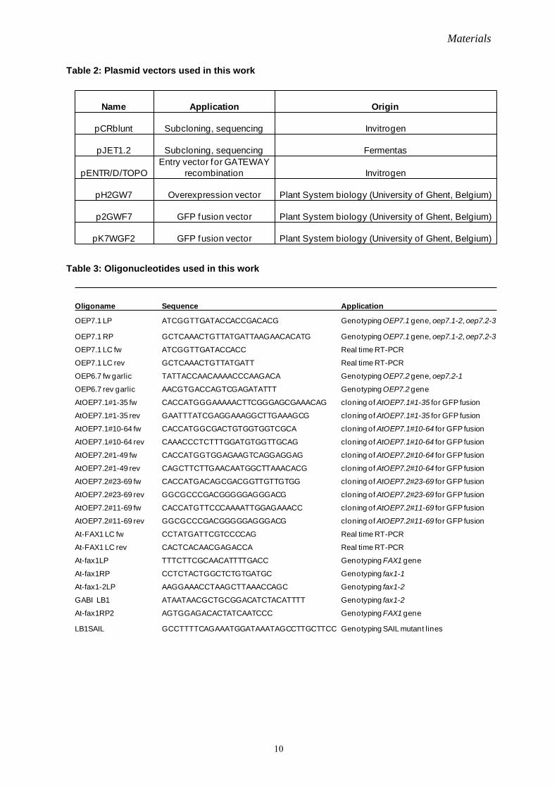
Table 2: Plasmid vectors used in this work
Table 3: Oligonucleotides used in this work
Name Application Origin
pCRblunt Subcloning, sequencing Invitrogen
pJET1.2 Subcloning, sequencing Fermentas
pENTR/D/TOPOEntry vector for GATEWAY
recombination Invitrogen
pH2GW7 Overexpression vector Plant System biology (University of Ghent, Belgium)
p2GWF7 GFP fusion vector Plant System biology (University of Ghent, Belgium)
pK7WGF2 GFP fusion vector Plant System biology (University of Ghent, Belgium)
Oligoname Sequence Application
OEP7.1 LP ATCGGTTGATACCACCGACACG Genotyping OEP7.1 gene, oep7.1-2, oep7.2-3
OEP7.1 RP GCTCAAACTGTTATGATTAAGAACACATG Genotyping OEP7.1 gene, oep7.1-2, oep7.2-3
OEP7.1 LC fw ATCGGTTGATACCACC Real time RT-PCR
OEP7.1 LC rev GCTCAAACTGTTATGATT Real time RT-PCR
OEP6.7 fw garlic TATTACCAACAAAACCCAAGACA Genotyping OEP7.2 gene, oep7.2-1
OEP6.7 rev garlic AACGTGACCAGTCGAGATATTT Genotyping OEP7.2 gene
AtOEP7.1#1-35 fw CACCATGGGAAAAACTTCGGGAGCGAAACAG cloning of AtOEP7.1#1-35 for GFP fusion
AtOEP7.1#1-35 rev GAATTTATCGAGGAAAGGCTTGAAAGCG cloning of AtOEP7.1#1-35 for GFP fusion
AtOEP7.1#10-64 fw CACCATGGCGACTGTGGTGGTCGCA cloning of AtOEP7.1#10-64 for GFP fusion
AtOEP7.1#10-64 rev CAAACCCTCTTTGGATGTGGTTGCAG cloning of AtOEP7.1#10-64 for GFP fusion
AtOEP7.2#1-49 fw CACCATGGTGGAGAAGTCAGGAGGAG cloning of AtOEP7.2#10-64 for GFP fusion
AtOEP7.2#1-49 rev CAGCTTCTTGAACAATGGCTTAAACACG cloning of AtOEP7.2#10-64 for GFP fusion
AtOEP7.2#23-69 fw CACCATGACAGCGACGGTTGTTGTGG cloning of AtOEP7.2#23-69 for GFP fusion
AtOEP7.2#23-69 rev GGCGCCCGACGGGGGAGGGACG cloning of AtOEP7.2#23-69 for GFP fusion
AtOEP7.2#11-69 fw CACCATGTTCCCAAAATTGGAGAAACC cloning of AtOEP7.2#11-69 for GFP fusion
AtOEP7.2#11-69 rev GGCGCCCGACGGGGGAGGGACG cloning of AtOEP7.2#11-69 for GFP fusion
At-FAX1 LC fw CCTATGATTCGTCCCCAG Real time RT-PCR
At-FAX1 LC rev CACTCACAACGAGACCA Real time RT-PCR
At-fax1LP TTTCTTCGCAACATTTTGACC Genotyping FAX1 gene
At-fax1RP CCTCTACTGGCTCTGTGATGC Genotyping fax1-1
At-fax1-2LP AAGGAAACCTAAGCTTAAACCAGC Genotyping fax1-2
GABI LB1 ATAATAACGCTGCGGACATCTACATTTT Genotyping fax1-2
At-fax1RP2 AGTGGAGACACTATCAATCCC Genotyping FAX1 gene
LB1SAIL GCCTTTTCAGAAATGGATAAATAGCCTTGCTTCC Genotyping SAIL mutant lines

Materials
11
Table 4: Constructs created in this work
At-FAX1 fw CACCATGGCTTCACAAATCTCTCAGCcloning of At-FAX1cDNA for overexpressinglines and complementation lines
At-FAX1 rev GTATGAAGGACTAGTCGCAGATGGcloning of At-FAX1cDNA for overexpressinglines and complementation lines
At-fax1 (+stop codon) revTCAGTATGAAGGACTAGTCGCAGATGG cloning of At-FAX1cDNA
Ps-fax1 (cacc) fw CACCATGGCGGCGACATCTCAGGCTCA cloning of Ps-FAX1cDNA to pENTR/D/TOPO
Ps-fax1 (+stop codon) revTCAGGCTACACTGGCAGATGGCTTC cloning of Ps-FAX1cDNA to pENTR/D/TOPO
attB1 ACAAGTTTGTACAAAAAAGCAGGCT Gateway vectors related
attB2 ACCACTTTGTACAAGAAAGCTGGG Gateway vectors related
Protein Plasmid-vector Application
FAX1(at3g57280) pUNI151 Sequencing, subcloning
Ps-FAX1 contig pCRblunt Sequencing, subcloning
At-FAX1 (-stop codon) pENTR/D/TOPO Gateway recombination (GFP)
At-FAX1 (-stop codon) p2GWF7 GFP fusion
At-OEP7.1(-stop codon) pENTR/D/TOPO Gateway recombination (GFP)
At-OEP7.1(-stop codon) p2GWF7 GFP fusion (At-OEP7.1:GFP)
At-OEP7.2(-stop codon) pENTR/D/TOPO Gateway recombination (GFP)
At-OEP7.2(-stop codon) p2GWF7 GFP fusion (At-OEP7.2:GFP)
At-OEP7.1 pENTR/D/TOPO Gateway recombination (GFP)
At-OEP7.1 pK7WGF2 GFP fusion (GFP:At-OEP7.1)
At-OEP7.2 pENTR/D/TOPO Gateway recombination (GFP)
At-OEP7.2 pK7WGF2 GFP fusion (GFP:At-OEP7.2)
At-OEP7.1#1-35 pENTR/D/TOPO Gateway recombination (GFP)
At-OEP7.1#10-64 pENTR/D/TOPO Gateway recombination (GFP)
At-OEP7.2#1-48 pENTR/D/TOPO Gateway recombination (GFP)
At-OEP7.2#23-69 pENTR/D/TOPO Gateway recombination (GFP)
At-OEP7.2#11-69 pENTR/D/TOPO Gateway recombination (GFP)
At-OEP7.1#1-35 p2GWF7 GFP fusion (At-OEP7.1#1-35:GFP)
At-OEP7.1#10-64 p2GWF7 GFP fusion (At-OEP7.1#10-64:GFP)
At-OEP7.2#1-48 p2GWF7 GFP fusion (At-OEP7.2#1-48:GFP)
At-OEP7.2#23-69 p2GWF7 GFP fusion (At-OEP7.2#23-69:GFP)
At-OEP7.2#11-69 p2GWF7 GFP fusion (At-OEP7.2#11-69:GFP)

Methods
12
III. Methods
1 Plant methods
1.1 Plant material and growth conditions
All experiments were performed on Arabidopsis thaliana plants, ecotype columbia (Col-0,
Lehle Seeds; Round Rock, USA). The TILLING lines oep7.1-2 and oep7.1-3 were ordered
from the Seattle Arabidopsis TILLING Service (http://tilling.fhcrc.org; Till et al., 2003) and
purchased from NASC (University of Nottingham, GB, Scholl et al., 2000). The T-DNA
insertion lines (Alonso et al., 2003; Rosso et al., 2003) SAIL_813_F06 (oep7.2-1),
SAIL_66_B09 (fax1-1) and GABI_599E01 (fax1-2) were purchased from NASC or GABI-
Kat (MPI for Plant Breeding Research, Köln, Germany), respectively. Peas (Pisum sativum)
var. “Arvica” were ordered from Bayerische Futtersaatbau (Ismaning, Germany).
Seeds were either sown directly on soil or on MS-plates (Murashige and Skoog, 1962)
supplemented with 1% (w/v) sucrose. In some cases, seedlings were transferred to soil after 2-
3 weeks. Before sowing on sterile plates, seeds of Arabidopsis thaliana were surface sterilized
in 70% (v/v) ethanol for 1-2 min, 6% (v/v) NaClO with 0.05% (v/v) Tween-20 for 3-5 min,
followed by washing in sterilized water for 3 x 1 min, and allowed to air-dry in a laminar flow
hood. To synchronize germination, seeds were vernalized at 4°C in the dark for 1-3 days. For
selection of transformed plants, seeds were grown on MS media (+1% sucrose) containing the
adequate antibiotics (25 µg/ml hygromycin or 100 µg/ml kanamycin). Unless stated
otherwise, plants were grown in a 16 h l ight (+21°C; 100 μmol photons m–2 s–1) and 8 h da rk
(+16°C) cycle (long-day) and plant tissues were generally harvested during early light phase
(2-5hrs of light).
1.2 Sub-cellular localization of GFP Fusion proteins in Arabidopsis protoplasts
Plant transformation and sub-cellular localization of GFP fusion protein in Arabidopsis
protoplasts was performed as described in Duy et al., (2007).
1.3 Cross fertilization of Arabidopsis thaliana
Crossing lists of different fax1 knockout mutants and wild type are depicted in Chapter
IV.2.7. Cross fertilization was performed as described (Detlef and Glazebrook, 2002).
Methods
13
2 Molecular biological methods
2.1 General molecular biological methods
General molecular biological methods like growing of bacteria, DNA precipitation and
determination of DNA concentration were performed as described (Sambrook et al., 1989).
The preparation of transformation-competent cells was performed according to the protocol of
Hanahan and co-worker (Hanahan, 1983). Preparation of plasmid DNA, restriction digests,
ligations and agarose gel electrophoresis were performed as described (Sambrook et al.,
1989). The reaction conditions were adjusted to the manufacturer’s recommendations.
2.2 Isolation of genomic DNA from Arabidopsis thaliana
2-3 Arabidopsis leaves were cut and transferred to a 1.5 m l microtube, 450 μl extraction
buffer (200 mM Tris-HCl with pH 7.5, 250 mM NaCl, 25 mM EDTA, 0.5% SDS, 100 μg/ml
RNase) and one small iron bead were added and the sample was lysed in a TissueLyser
(Qiagen, Hilden, Germany) for 3 minutes at maximum speed. Then the sample was incubated
at 37°C for 5-10mins. After centrifugation for 10 min at 16,000 x g and 4°C, the supernatant
was transferred to a fresh tube. To precipitate the genomic DNA, 300 μl of isopropanol was
added to the sample, carefully mixed and incubated for 5 min at room temperature. After
centrifugation for another 5 min at 16,000 x g and +4°C, the pellet was washed once with
70% ethanol, subsequently air-dried and finally resuspended in 50 μl of sterilized H2O or 10
mM Tris-HCl (pH 8.0) buffer.
2.3 RNA extraction from Arabidopsis thaliana and RT-PCR
Total RNA from leaves, stems or flowers of Arabidopsis plants was isolated using the Plant
RNeasy Extraction kit (Qiagen, Hilden, Germany). The DNA was digested with RNase-free
DNase I (Qiagen) and transcribed into cDNA using MMLV Reverse transcriptase (Promega,
Mannheim). Detection and quantification of transcripts were performed as described
previously (Philippar et al., 2004) using the LightCycler system (Roche, Penzberg).
2.4 Characterization of plant T-DNA insertion lines and TILLING lines
Genomic DNA of the T-DNA insertion lines was screened by PCR genotyping. To identify
plants with the T-DNA insertion in both alleles (homozygous), a combination of gene-specific
primers flanking the predicted T-DNA insertion sites and T-DNA-specific left border (LB)
primers (Table 5) were used. Usage of a LB primer (in combination with a corresponding
gene-specific primer) will only generate an amplification product in plants carrying at least
one T-DNA allele (heterozygous or homozygous for the T-DNA insertion). On the other

Methods
14
hand, the combination of two gene-specific primers will generate a PCR product only in DNA
of plants carrying alleles without a T-DNA (WT and heterozygous for the T-DNA insertion).
In the TILLING lines, the site of the point mutation corresponds to the site provided by
TILLING. The positions and orientations of the T-DNA inserts and the position of point
mutation in TILLING line and oligonucleotide primers in oep7.1-2, oep7.1-3, oep7.2-1, fax1-
1 and fax1-2 are shown in Figure 7 and Figure 18, respectively. To verify the T-DNA
insertion sites, PCR genotyping products were cloned and subsequently sequenced.
Table 5: PCR primer combination for genotyping mutant lines of OEP7 and FAX1 For primer sequence please refer to Table 1, and for position and orientation of primers please refer to Figure 7 and Figure 18.
2.5 Microarray analysis
150 mg tissue powder from flowers and from second to forth internode of inflorescent stems
from more than 10 individual seven-week-old plants was used for preparation of RNA
(identical sample pool that was used for fatty acid analysis). RNA (200 ng) of three samples
(n = 3) from both wild type (Col-0) and fax1-2 lines was processed and hybridized to
Affymetrix “GeneChip Arabidopsis ATH1 Genome Arrays” using the Affymetrix “3’ VIT
express” and “Hybridisation wash and stain” kits (Affymetrix UK, High Wycombe, UK)
according to the manufacturer`s instructions. The statistical significance of signal change was
calculated as described in Duy et al., (2011). The hybridization of the microarrays and
statistical analysis of data were performed by Karl Mayer and by Dr. Katrin Philippar,
respectively (Department Biologie I, Plant Biochemistry and Physiology, LMU München).
Allele Line primers for wild-typeprimers for T-DNA insertionor TILLING
oep7.1-2 TILL2 OEP7.1 fw, OEP7.1 rev OEP7.1 fw, OEP7.1 rev
oep7.1-3 TILL3 OEP7.1 fw, OEP7.1 rev OEP7.1 fw, OEP7.1 rev
oep7.2-1 SAIL_813_F06 OEP6.7 rev garlic, OEP6.7 fw garlic LB1SAIL, OEP6.7 fw garlic
fax1-1 SAIL_66_B09 fax1LP, fax1RP2 LB1SAIL, fax1RP
fax1-2 GABI_599E01 fax1LP, fax1RP2 GABI LB1, fax1-2LP
fax1-2 Complementation attB1, fax1rev attB1, fax1rev
fax1-2 Overexpressing attB1, fax1rev attB1, fax1rev

Methods
15
3 Biochemical methods
3.1 General biochemical methods
SDS-PAGE was performed as described (Laemmli, 1970). For OEP7, Tricine-SDS-PAGE
was used as described (Schägger, 2006). Gels were stained either by Coomassie Brilliant Blue
R250 (Sambrook et al., 1989) or silver-staining (Blum et al., 1987, Ansorge et al., 1985).
Determination of chlorophyll concentration was carried out as described by Arnon, (1949).
Determination of protein concentration was performed by the Bio-Rad Protein Essay Kit
(Bio-Rad, München, Germany) or the Pierce BCA Protein Assay Kit (Thermo Scientific,
Rockford, USA)
3.2 Total protein extraction from Arabidopsis thaliana
Rosette leaves or flowers (0.1 g) of four-week-old plants were harvested and flash-frozen in
liquid N2. The frozen material was thoroughly ground with mortar and pestle and extracted by
one volume extraction buffer (50 mM Tris-HCl, (pH 8), 2% LDS (Lithium dodecyl sulphate),
0.1 mM PMSF). After incubation on ice for 30 min, the sample was centrifuged at 16,000 x g
at +4°C for 15 m in to get rid of insolube components. The protein concentration was
determined with the Pierce BCA protein Assay Kit. 50 mM EDTA and 0.15% DTT were
added to the protein solution for storage and further experiment.
3.3 Immunoblotting
3.3.1 Electrotransfer and blocking of proteins
Proteins separated by SDS-PAGE were transferred onto either Nitrocellulose (PROTRAN
BA83, 0.2 μm, Whatman/Schleicher & Schüll) or PVDF membranes (Zefa Transfermembran
Immobilon-P, 0.45 μm, Zefa-Laborservice GmbH, Harthausen, Germany) by semi-dry-
blotting (Amersham Biosciences) (Kyhse-Andersen, 1984) in blotting buffers (I: 300 mM
Tris-HCl, 20% methanol, II: 25 mM Tris-HCl, 20% methanol, III: 25 mM Tris-HCl, 40 mM
aminocapron acid, 20% methanol ) for 1h at 0.8 mA per cm2 membrane surface as described
(Towbin et al., 1979). To be mentioned, the PVDF membranes have to be activated in 100%
methanol before use. Proteins of the size markers were either stained with Ponceau S solution
or amidoblack solution. Membranes with bound proteins were first incubated for 30 min in
blocking buffer containing skimmed milk powder (0-5% milk powder, 0-0.1M Tris, 0-0.15 M
NaCl, 0-0.75% Tween 20). And then the membrane was incubated with the protein-specific
primary antibody (diluted in blocking buffer 1:250-1:4000, depending on the antibody) for 2-
3h at RT or overnight at 4°C. Non-bound antibody was removed from the membrane by

Methods
16
washing for 3x10 min in TTBS (0.1M Tris, 0.15 M NaCl with 0.1% Tween 20). The
secondary antibody was selected according to the desired method of visualization.
3.3.2 Alkaline phosphatase (AP) detection
If the secondary antibody was alkaline phosphatase (AP)-conjugated secondary antibody
(goat anti-rabbit IgG (whole molecule)-AP conjugated, Sigma-Aldrich Chemie GmbH,
Taufkirchen), detection of AP signals was performed in developing buffer (100 mM Tris-HCl
(pH 9.5), 100 mM NaCl, 5 mM MgCl2) with 6.6 μl/ml NBT (nitro blue tetrazolium chloride,
50 mg/ml in 70% N,N-dimethylformamide) and 13.2 μl/ml BCIP (5- bromo-4-chloro-3-
indolyl phosphate, 12.5 mg/ml in 100% N,N-dimethylformamide). The membrane was
incubated in 50 mM EDTA to stop the reaction.
3.3.3 Enhanced Chemiluminescence (ECL)
If the secondary antibody was a horseradish peroxidase-conjugated secondary antibody (goat
anti-rabbit (whole molecule)-peroxidase conjugated, Sigma), detection of signal was
performed in 50% (v/v) solution 1 ( 100 mM Tis-HCl (pH 8.5), 1% (w/v) luminol, 0.44%
(w/v) coomaric acid) and 50% (v/v) solution 2 ( 100 mM Tris-HCl (pH 8.5), 0.018% (v/v)
H2O2). After incubation for 1 m in at RT in the dark, the solution was removed and the
luminescence detected with a film (Kodak Biomax MR; PerkinElmer, Rodgau, Germany).
3.4 Proteolysis experiments
To identify the orientation of chloroplast envelope proteins, proteolysis of Arabidopsis
chloroplasts or of inner envelope vesicles from pea was performed using the protease
thermolysin. The chloroplasts or envelope vesicles were incubated in wash buffer (330 mM
sorbitol, 50 m M Hepes (pH 7.6) and 0.5 mM CaCl2) with 10% (m/m) thermolysin and
incubated for 0-30 min on i ce. For envelope solubilisation, 1% triton was added. The
proteolysis was stopped with 5 mM EDTA per 1 µg protease. The proteins were separated on
SDS-PAGE or Tricine-SDS- PAGE and analysed by immunoblot.
4 Cell biology methods
4.1 Isolation of Arabidopsis thaliana chloroplasts
Chloroplast isolation was performed as described (Aronsson and Jarvis, 2002) with
modification. Leaf material from 21-day-old plants grown on s oil or on MS-plates
supplemented with 1% (w/v) sucrose was homogenized in 25 m l isolation buffer (0.3 M
sorbitol, 5 mM MgCl2, 5 mM EGTA, 5 mM EDTA, 20 mM HEPES/KOH (pH 8.0), 10 mM
NaHCO3, 50 m M ascorbic acid). After homogenisation and filtration steps, the soluble

Methods
17
homogenate was pelleted at 1000 g for 4 min and resuspended in isolation buffer. The sample
was separated on a t wo-step Percoll gradient (30/82% (w/v) Percoll) with centrifugation at
1500 g for 8 min. The lower band (intact chloroplasts) was transferred to a 10 ml tube and
washed in wash buffer (50 mM HEPES/KOH (pH 8.0), 0.3 M Sorbitol, 2 mM DTT). The
chloroplasts were pelleted at 1000 g for 4 min and resuspended in 0.5 ml wash buffer.
4.2 Preparation of inner and outer envelope from Pisum sativum
For isolation of IE and OE vesicles from chloroplasts, pea seedlings grown for 9-11 days on
sand under a 12/12 hours dark/light regime were used. All procedures were carried out at 4°C.
Pea leaves cut from ~ 20 trays were ground in a kitchen blender in 10-15 l isolation medium
(330 mM sorbitol, 20 mM MOPS, 13 m M Tris, 0.1 m M MgCl2, 0.02% (w/v) BSA) and
filtered through four layers of mull and one layer of gauze (30 μm pore size). The filtrate was
centrifuged for 5 m in at 1,500 x g, the pellet gently resuspended with a brush and intact
chloroplasts reisolated via a discontinuous Percoll gradient (40% and 80%). Intact
chloroplasts were washed twice with wash medium (330 mM sorbitol, Tris-base (~ pH 7.6)),
homogenized and further treated according to the modification (Waegemann et al., 1992) of
the previously described method (Keegstra and Youssif, 1986).
4.3 Preparation and transient transformation of protoplasts from Arabidopsis
Arabidopsis mesophyll protoplasts were isolated from 1 g leaves of 3 t o 4-week-old
Arabidopsis plants grown on soil under normal growth condition and transiently transformed
according to the protocol from Jen Sheen (http://molbio.mgh.harvard.edu/sheenweb/
main_page.html). GFP fluorescence was observed with a T CS-SP5 confocal laser scanning
microscope (Leica, Wetzlar, Germany).
5 Light and transmission electron microscopy
Inflorescent stems, 1 cm above the first node and anthers of flower stage 11-12 (Smyth et al.,
1990) from 2-3 individual Arabidopsis thaliana plants at the age of 34 to 40 da ys were
harvested in the morning prior to illumination. Samples were prefixed in glutaraldehyde 2.5%
(w/v) in 75 m M cacodylate buffer (pH 7.0), subsequently rinsed in cacodylate buffer and
fixed in 1% (w/v) osmium tetroxide in the same buffer for at least 2.5 h at room temperature.
The samples were stained with uranyl acetate 1% (w/v) in 20% acetone, dehydrated in a
graded acetone series and embedded in Spurr's low viscosity epoxy resin (Spurr, 1969).
Further steps and microscopic investigation were performed by Dr. Irene Gügel (Department
Biologie I, Plant Biochemistry and Physiology, LMU München).

Methods
18
6 Wax analysis
The second to forth internode region of inflorescent stems from more than 10 i ndividual,
seven-week-old plants were used for wax analysis from FAX1 mutants, wild-type and
complementation lines. Wax analysis was conducted on pools of 3-4 stems as described
(Prabhakar et al., 2010; Kurdyukov et al., 2006) and performed by Prof. Lukas Schreiber
(Department of Ecophysiology, IZMB, University of Bonn)
7 Fatty acid analysis
Tissue of cauline leaves, second to forth internode of inflorescent stems and flowers from
more than 10 individual, seven-week-old plants were grinded in liquid nitrogen and used for
fatty acid analysis. Three samples of 50 mg tissue powder from homozygous fax1 mutants and
from wild-type plants were used. Fatty acid analysis was conducted as described (Hummel et
al., 2011; Giavalisco et al., 2011) and performed by Dr. Patrick Giavalisco (Max Planck
Institute of Molecular Plant Physiology, Potsdam-Golm, Germany).
8 Bioinformatical methods
Table 6: Software, databases and algorithms used in this work
Name Reference Link
NCBI BLAST Altschul et al., 1997 http://www.ncbi.nlm.nih.gov/BLAST
Vector NTI Invitrogen
TopPred
Heijne 1992; Claros and Heijne 1994
http://mobyle.pasteur.f r/cgi-bin/portal.py?#forms::toppred
ARAMEMNON Schwacke et al., 2003 http://aramemnon.botanik.uni-koeln.de
TAIR(The Arabidopsis Information Resource Lamesch et al., 2011 http://www.arabidopsis.org
ChloroP 1..1 http://www.cbs.dtu.dk/services/ChloroP/
Arabidopsis eFP browser Schmid et al., 2005 http://bar.utoronto.ca/efp/cgi-bin/efpWeb.cgi
GenevestigatorZimmermann et al., 2004 https://www.genevestigator.com/gv/plant.jsp
AtGenExpress Consortium
http://www.weigelworld.org/resources/microarray/AtGenExpress/
GenedocNicholas and Nicholas,1997 http://www.psc.edu/biomed/genedoc
ExPASy PeptideCutter Gasteiger et al., 2005 http://web.expasy.org/peptide_cutter/EBI interpro protein sequence analysis & classif ication database http://www.ebi.ac.uk/interpro/

Methods
19
p p
MEGA 5.0 Tamura et al., 2011 http://www.megasof tware.net/
AT CHLORO database Ferro et al., 2010http://www.grenoble.prabi.f r/protehome/- grenoble-plant-proteomics/
Aida Image Analyzer v. 3.25 raytest http://www.raytest.f r/index2.html

Results
20
IV. Results
1 At-OEP7.2 structure and function analysis
1.1 OEP7 proteins in Arabidopsis
OEP7 was first identified and characterised as an outer chloroplast envelope protein in
spinach (E 6.7, Salomon et al., 1990). Currently four isoforms of OEP7 have been identified
in Arabidopsis: At-OEP7.1 (At3g52420), At-OEP7.2 (At3g63160), At-OEP7.3 (At3g19151)
and At-OEP7.4 (At2g34585) (ARAMEMNON plant membrane protein database, Schwacke
et al., 2003). The highest amino acid identity is 32% between At-OEP7.1 and At-OEP7.2. All
four OEP7 isoforms have one α-helical transmembrane domain (Figure 3) (ARAMEMNON
plant membrane protein database, Schwacke et al., 2003). It is predicted that At-OEP7.2 is
localized at the outer envelope of chloroplasts, while At-OEP7.3 is in mitochondria and At-
OEP7.4 of unknown subcellular localisation. Until the beginning of my thesis, no
experimental data on the OEP7.2 topology and function was published.
Figure 3: OEP7 proteins in Arabidopsis Identical amino acids are shaded in black, similar amino acids in grey and the transmembrane domain is depicted as red box. 1.2 Membrane orientation of OEP7.2 in the outer envelope of chloroplasts
It is known that OEP7.1, a 14 kDa protein, has the N-terminus in the intermembrane space
and the C-terminus in the cytosol orientation (Figure 4C left; Li et al., 1996). To analyse the
orientation of At-OEP7.2, Arabidopsis chloroplasts were treated with proteases in the
presence or absence of detergent and further subjected to immunoblot analysis (Figure 4).
At-OEP7.1 : At-OEP7.2 : At-OEP7.3 : At-OEP7.4 :
* 20 * 40 * 60 * 80-------------MGKTSG-AKQATVVVAAMALGWLAIEIAFKPFLDKFRSSIDKSDPTKDPDDFDTAATATTSKEGL--MVEKSGGEVNFPKLEKPTGKKQTATVVVGVLAVGWLAIELVFKPLFKKLSSSKDKSDSDDATVPPPSGA----------------------MKRTVMDNAIRSSVVVLGSLAFGYLSLELGYKPFLEKAEQYERSLQSSQQHQQQDEQEEARWDNSNVEG-----------------MAAITSAVIAIAGIILGWITIELACKPCLESGREAIDRSLNPDYDPDDQDVDSSSDVRAPLIP
: 64 : 69 : 69 : 63
At-OEP7.1 : At-OEP7.2 : At-OEP7.3 : At-OEP7.4 :
* ------------------------------------WEEKR-------------NHPPDNSDPDPTTAIKSV
: - : - : 74 : 81

Results
21
Figure 4: At-OEP7.2 protein topology analysis at the outer envelope of chloroplasts A) Thermolysin digestion sites (THL) of At-OEP7.2. Six thermolysin digestion sites, the transmembrane α- helix and regions for two peptide antisera (α-At-OEP7.2 NT, CT) are shown, respectively. B) Immunoblot analysis of At-OEP7.2 with antiserums raised against At-OEP7.2 N-terminus (NT) or At-OEP7.2 C-terminus (CT) after protease treatment with or without 1% triton x-100. 1.5, 3, 4.5 μg thermolysin were added to chloroplasts (equal 15 μg chlorophyll) and incubated for 20 min on ice, respectively. P: pellet, S: supernatant. Tic110 is marker of inner envelope proteins, which should be prevented from thermolysin digestion. C) Topology model of At-OEP7.1 and At-OEP7.2. In the presence of triton, which breaks the membrane structure, membrane proteins are fully
accessible to proteolysis. However, in the absence of triton, only the proteolysis sites of
membrane proteins at the cytosol site are accessible to proteolysis. It is firstly hypothesized
that the N-terminus is in the cytosol and C-terminus is in the inter membrane space. A 1.5
kDa region would be digested and then a 12-13 kDa band would be recognized by the
antiserum raised against At-OEP7.2 C-terminus (Figure 4A). In contrast, if the N-terminus is
in the inter membrane space and C-terminus in the cytosol, only a 0.1 kDa region of OEP7.2
would be digested and therefore a 14 kDa band, which becomes weaker with the increase of
110
14
14
α-At-Tic110
α-At-OEP7.2 NT
α-At-OEP7.2 CT
- - - - + + + + + + + + Trition X-100 THL - 1 2 3 - 1 2 3 - 1 2 3
SP
THL
A
B
α-At-OEP7.2 NT α-At-OEP7.2 CT
1.5 kDa 0.1 kDa
N
C
C
NC
OEP7.1 OEP7.2
stroma
cytosol cytosol
stroma

Results
22
thermolysin amount, would be recognized by both antisera. The three thermolysin digestion
sites near the transmembrane domain of OEP7.2 were not considered in the hypothesis
because of possible membrane protection. It is clear that the immunoblot analysis results
(Figure 4B) was matched with the first hypothesis, which revealed that At-OEP7.2 has its N-
terminus in the cytosol (Figure 4C right).
1.3 Targeting of At-OEP7.2 to the outer envelope membrane of chloroplasts
To further confirm that At-OEP7.2 is localized at the envelope membrane of chloroplasts, I
studied the At-OEP7.2: GFP and At-OEP fusion proteins in Arabidopsis protoplasts (in vivo
GFP targeting; Figure 5).
Figure 5: In vivo targeting of AtOEP7:GFP in protoplasts A) The various fusion constructs used in the experiments. At-OEP7.1:GFP or At-OEP7.2:GFP: green fluorescent protein (GFP) was fused to the C-terminus of At-OEP7.1 or At-OEP7.2, respectively. GFP:At-OEP7.1 or GFP:At-OEP7.2: GFP was fused to the N-terminus of At-OEP7.1 or At-OEP7.2, respectively. B) In vivo targeting of fusion constructs in Arabidopsis protoplasts. Only overlaps of GFP (green) and chlorophyll (red) are shown. As shown in Figure 5, At-OEP7.2, like At-OEP7.1, was targeted to the chloroplast envelope
membrane when GFP-proteins were fused to their C-terminus. However, when GFP was
attached to the N-terminus of both proteins, a punctate signals appeared, which revealed that
GFP: OEP7 was mis-targeted. The results suggested that the N-terminus of At-OEP7.2 is
critical for targeting to the outer envelope membrane. For OEP7.1, it was confirmed that the
At-OEP7.2:GFPAt-OEP7.1:GFP GFP:At-OEP7.2GFP:At-OEP7.1
At-OEP7.1:GFP
At-OEP7.2:GFP
GFPAt-OEP7.2
GFP At-OEP7.2GFP:At-OEP7.2
GFP
GFP
At-OEP7.1
At-OEP7.1
GFP:At-OEP7.1
A
B

Results
23
seven-amino acid region at the C-terminus and the transmembrane domain are determinants
for targeting to the chloroplast outer envelope membrane (Lee et al., 2001).
Figure 6: In vivo targeting of At-OEP7 deletion constructs fused with GFP in protoplasts A) Scheme of the various deletion constructs fused with GFP. B) In vivo targeting of various At-OEP7.1 and At-OEP7.2 deletion constructs, which were fused to GFP N-terminus. Protoplasts were transformed with At-OEP7.1#1-35:GFP, At-OEP7.1#10-64:GFP, At-OEP7.2#1-48:GFP, At-OEP7.2#23-69:GFP and At-OEP7.2#11-69:GFP. GFP (green), chlorophyll (red), overlap of GFP and chlorophyll and bright-field images (bright) are shown, respectively.
At-OEP7.1#1-35:GFP
At-OEP7.1#10-64:GFP
At-OEP7.2#1-48:GFP
At-OEP7.2#23-69:GFP
At-OEP7.2#11-69:GFP
GFP chlorophyll overlay bright
At-OEP7.1#1-35:GFP
At-OEP7.1#10-64:GFP
At-OEP7.2#1-48:GFP
At-OEP7.2#23-69:GFP
At-OEP7.2#11-69:GFP
GFPAt-OEP7.1#1-35
GFP
GFP
GFP
GFP
At-OEP7.1#10-64
At-OEP7.2#1-48
At-OEP7.2#23-69
At-OEP7.2#11-69
A
B

Results
24
To confirm which amino acid region is necessary and sufficient for the targeting of At-
OEP7.2, I produced various deletion constructs of At-OEP7.2 and subsequently fused them to
GFP (Figure 6A). Two deletion constructs of At-OEP7.1 with GFP were used as markers. All
these constructs were expressed transiently in protoplasts (Figure 6B).
As shown in Figure 6, the fusion proteins At-OEP7.1#1-35:GFP, At-OEP7.1#10-64:GFP, At-
OEP7.2#1-48:GFP and At-OEP7.2#11-69:GFP were targeted to the envelope membrane of
the chloroplasts. In constrast, At-OEP7.2#23-69: GFP only gave a punctate staining pattern in
the protoplasts, which revealed that it mis-targeted. Thus, these results suggest that the
residues 11 to 22 of At-OEP7.2 are necessary and residues 1 to 48 of At-OEP7.2 are sufficient
for targeting to the chloroplast envelope membrane.
1.4 OEP7 mutant analysis in Arabidopsis
Two TILLING lines oep7.1-2 and oep7.1-3 (Seattle Arabidopsis TILLING Project, Till et al.,
2003) for OEP7.1, a T-DNA insertion mutant line (SAIL_813_F06), named oep7.2-1, for
OEP7.2, and double mutant (d.m.) of oep7.1 and oep7.2-1 were available at the beginning of
my thesis. In OEP7.1 TILLING lines, a point mutation at position +146 changed the
nucleobase cytosine to thymine, which produced a stop codon at the beginning of first exon of
the OEP7.1 open reading frame (Figure 7A). In oep7.2-1, the T-DNA insertion is localized at
position +579 in the first exon of the OEP7.2 open reading frame (Figure 7B). For these
single mutants and double mutants of oep7, immunoblot analysis showed that they were
knockout mutant in Arabidopsis (Figure 7C).

Results
25
Figure 7: Characterisation of At-OEP7.1 and At-OEP7.2 mutant lines A) OEP7.1 from Arabidopsis thaliana (At3g52420). Black arrows denote exons and white line denote introns. The location of the point mutation in the TILLING line oep7.1 is detailed in the text box, which indicates a premature stop codon (C to T) at amino acid 146. Binding sites for OEP7.1 gene specific primers for real time RT-PCT are depicted. B) OEP7.2 from Arabidopsis thaliana (At3g63160). Black arrows denote exons and white lines denote introns. The T-DNA insertion sites in line SAIL_813_F06 (oep7.2-1) are depicted by triangles. Binding sites for OEP7.2 gene specific primers and T-DNA specific left (LB) primer used for genotyping and for real time RT-PCT are depicted. C) Immunoblot analysis of At-OEP7.1 and At-OEP7.2 in Arabidopsis leaf protein extract (10 µg) of mutant lines and Col-0 using antisera against At-OEP7.1 and At-OEP7.2, respectively. d.m.: double mutant. To further understand the function of OEP7, single and double mutant lines of oep7.1 and
oep7.2 were grown under several growth conditions (1: 16 h light, +21°C, 100 μmol photons
AtOEP7.2 (At3g63160)
+1
OEP6.7 rev garlicOEP7.2 LCfw
OEP7.2 LCrev
3'UTR5'UTR
oep7.2-1DAP101
LB
+579
B
AtOEP7.1 (At3g52420)
+1
OEP7.1 LC fw
OEP7.1 LC rev
3'UTR5'UTR
M G K T S G A K Q A T V V V A A
ATGGGAAAAACTTCGGGAGCGAAACAGGCGACTGTGGTGGTCGCAGCGTACCCTTTTTGAAGCCCTCGCTTTGTCCGCTGACACCACCAGCGTCGC
oep7.1-TILL2, 3: TAG = stop
+146
A
At-OEP7.1
At-OEP7.2
14
14
d.m.oep7.2Col-0.oep7.1C

Results
26
m–2 s–1 and 8 h dark ,+16°C cycle; 2: 12 h light, +21°C, 100 μmol photons m–2 s–1 and 12 h dark
,+16°C cycle; 3: 8 h light, +21°C, 100 μmol photons m–2 s–1 and 16 h dark ,+16°C cycle; 4: 16
h light, +21°C, 180 μmol photons m–2 s–1 and 8 h dark ,+16°C cycle) but no obvious phenotype
could be detected until now (data not shown). However, considering that At-OEP7.2 was
predicted as a light-sensitive gene according to Smith et al. (2004), real time RT-PCR and
immunoblot analysis were performed on oep7.2-1 and Col-0 throughout the diurnal cycle
(Figure 8).
Figure 8: Expression of At-OEP7.2 throughout the diurnal cycle Leaves were harvested from 4 weeks old Arabidopsis Col-0 plants under standard growth conditions (20 °C, in a 12-h-light/12-h-dark photoperiod with an irradiance of 180 μmol m-2 s-1). A) Diurnal changes in amounts of At-OEP7.2 protein (shown as green triangle; n= 3±SD). Protein VDAC is one control which is stable expression. B) Expression patterns of OEP7.2 gene. The mRNA amount (arbitrary units) was normalised to 10000 actin transcripts.The curve represent mean values of n=3 independent experiments. Under the conditions employed, transcripts of OEP7.2 showed peak amount at 19:00, two
hours before dark and changed relatively with light over 24 h , w hich indicated that OEP7.2
transcription is regulated in a diurnal rythm. To further identify whether OEP7.2 proteins are
regulated as well, the same samples used for real time RT-PCR were further used for
immunoblot. The result revealed that the OEP7.2 protein amount changed relatively little
throughout the diurnal cycle.
50
100
150
protein level VDACProtein level OEP7.2
5
1015
2025
3035
4045
9:00 11:00 13:00 15:00 17:00 19:00 21:00 23:00 5:00+1 7:00+1 9:00+1
light dark
RN
A m
olec
ules
(x 1
03)/
1000
0 ac
tin(a
rbitr
ary
units
)P
rote
in s
igna
l
A
B

Results
27
2 FAX1, a putative permease for fatty acid export from chloroplasts
2.1 Identification of a pre-senescence related chloroplast envelope protein
Until now, many chloroplast proteome databases were built up for plastid envelope protein
analysis. In our research, the AT CHLORO database (http://www.grenoble.prabi.fr/
protehome/-grenoble-plant-proteomics/; Ferro et al., 2010) was used for selection of new
envelope proteins. To provide a link to senescence processes, we used mutants of SAUL1
(Senescence-associated E3 ubiquitin ligase 1) that was identified as one E3 ubiquitin ligase
preventing premature senescence in Arabidopsis plants (Raab et al., 2009). To identify
premature senescence associated chloroplast envelope proteins, we performed DNA
microarray analysis of SAUL1 knockout mutants under senescence inducing conditions
(collaboration with Prof. S. Hoth, Plant Physiology, University of Hamburg; Vogelmann et
al., 2012).
Among three potential chloroplast envelope proteins from the AT CHLORO database (Ferro
et al., 2010) transcript of the protein At3g57280 was increased 5.8 and 3.2 fold after 24h and
48h of senescence induction in saul1-1 mutants compared to wild type, respectively (Figure
9).
Figure 9: At3g57280 RNA content in saul1-1 under senescence inducing conditions Total RNA was isolated from wild-type and saul1-1 mutant seedlings grown in permissive light (60 µmol m-2 s-1) for 11 days and then challenged with low light (20 µmol m-2 s-1) for 6h, 24h, or 48h. To provide biological replicates, mean values for three wild-type and three saul1-1 mutant samples were harvested for each time point (Vogelmann et al., 2012). Mean values for At3g57280 RNA (arbitray units, n= 3±SD) are shown.
At3
g572
80 s
igna
ls/ a
rbitr
ary
units
(x 1
03)

Results
28
Because of its potential function in chloroplast fatty acid transport (see Chapter IV. 2.9), the
protein At3g57280 is named FAX1 (for fatty acid export 1) in the following. At-FAX1 was
predicted to be an inner chloroplast envelope protein (Ferro et al., 2010) of 24.3 kDa, with an
isoelectric point of 9.3 (TAIR, Lamesch et al., 2011) and four predicted α-helices (Table 9,
ARAMEMNON plant membrane protein database, Schwacke et al., 2003). Further, a
chloroplast signal peptide of 33 a mino acids was predicted (ChloroP, Emanuelsson et al.,
1999). At-FAX1 cDNA was purchased as SSP pUNI151 clone U12755 (Yamada et al., 2003).
At-FAX1 was also described as a putative solute transporter of chloroplasts in Tyra et al.
(2007).
Table 9: Protein features of FAX1 from Arabidopsis and pea The name, AGI code, the isoelectric point (IP), the molecular weight (Mw) in kDa, the predicted chloroplast signal peptide and the predicted number of transmembrane domains (TMs; ARAMEMNON) are shown. At-FAX1 by proteomic analysis was identified as chloroplast envelope protein in the listed references. The pea EST contig is published in Franssen et al. (2011).
For further experiments, the cDNA of FAX1 in Pisum sativum was cloned by RT-PCR using
pea seedling cDNA as template and primers designed according to an EST pea contig
(Franssen et al., 2011). All general data of the Ps-FAX1 are shown in Table 9, the amino acid
sequences of At-FAX1 and Ps-FAX1 are depicted in Figure 10. The amino acid identity
between At-FAX1 and Ps-FAX1 is 49%, and for both proteins four α-helices transmembrane
domains are predicted (Figure 10B).
NameAGI codes/
contig IP (TAIR)Mw
(kDa)chloro signalpeptide(AraM)
TMs (AraM)
At-FAX1 At3g57280 9.3 24.3 33 4References: Fröhlich et al. (2003); Kleffmann et al. (2004); Zybailov et al. (2008); Ferro et al. (2003), (2010).
Ps-FAX1 contig012718 9.16 25 39 4

Results
29
Figure 10: Sequence analysis of At-FAX1 and Ps-FAX1 A) Graphical output of At-FAX1 sequence showing four predicted hydrophobic transmembrane domains using the TopPred program (Heijne 1992; Claros and Heijne 1994). B) Amino acid sequence of At-FAX1 and Ps-FAX1. Identical amino acids between At-FAX1 and Ps-FAX1 are shaded in black, the end of the predicted chloroplast transit peptides is marked as red triangle, the predicted transmembrane domains are lined in green, the regions used for peptide antisera are lined in red. 1: is for antiserum α-At-FAX1, 2: is for antiserum α-Ps-FAX1-NT, 3: is for antiserum α-Ps-FAX1-CT. 2.2 FAX1 is localized in the inner envelope of chloroplasts
Although At-FAX1 was predicted as an inner envelope protein according to proteomics data
(Ferro et al., 2003, 2010), no experimental data directly showed the sub-cellular localization.
Therefore, the chloroplast localization of FAX1 was verified by in vivo GFP-targeting
experiments. At-FAX1 cDNA was fused C-terminally with the green fluorescent protein
(GFP) and transiently transformed into Arabidopsis protoplasts (Figure 11).
A
At_FAX1 : Ps_FAX1 :
* 20 * 40 * 60 * 80-MASQISQLACFSSTNRQFHFQSR-SFPCPMIRP------QSFVVKSVDGNSSETPASLSYTAEVSKPVVEKTSKPYSTVMAATSQAQLFCFPTVHRSLHIQQRRSLPCLSFRHSKLSVAMSLERHEAETADTETKNTLSYAADESKLNVEEKQESYSKL
: 72 : 80
At_FAX1 : Ps_FAX1 :
* 100 * 120 * 140 * 160DETATNKESITEPVEEDVATQPIRAAKIHDFCFGIPYGGLVVSGGLLGFAFSRNLTSLSTGVLYGGGLLALSTLSLKIWREESGSEKLGLGEATQEGVVDQQKKTAIIHDFCLGIPFGGFVLTGGIIGFLFSRSPATLASGVLVGGALLFLSTLSLKVWR
: 152 : 160
At_FAX1 : Ps_FAX1 :
* 180 * 200 * 220 * EGKSSFPYILGQAVLSAVVFWKNFTAYSMTKKLFPAGVFAVISACMLCFYSYVVLSGGNPPPKKLKPSATSPSYQGKSSLPFILGQAALSGILIWKNFQSYSLAKKIFPTGISAIISSAMLCFYLYVLVSGGNPPPKKLKPSASVA--
: 226 : 232
B 1
2
3

Results
30
Figure 11: At-FAX1-GFP signals in Arabidopsis protoplast At-FAX1-GFP and At-OEP7.1-GFP constructs were transiently transformed into Arabidopsis protoplasts. GFP, chlorophyll and overlay fluorescence of both constructs are shown. At-OEP7.1-GFP was used as marker for the chloroplast outer envelope. GFP fluorescence signals only appeared surrounding the chloroplasts, which indicated that the
FAX1-GFP fusion construct localized at the envelope membrane of chloroplasts. The At-
OEP7.1-GFP fusion construct, as a marker for an envelope localized protein, also showed
fluorescence signals only around the chloroplast.
To further confirm whether FAX1 is uniquely localized at the inner envelope of plastids,
immuno-blotting analysis on fractionated pea chloroplasts was performed (Figure 12).
Antiserum raised against peptides of Ps-FAX1-NT, named α-Ps-FAX1-NT was used in
immunoblot (see Figure 10C).
Figure 12: Immunoblot of Ps-FAX1 in pea chloroplast fractions Same total protein amount (5 µg) of the pea chloroplast sub-fractions OE (outer envelope), IE (inner envelope), stroma (str) and thylakoid (thy) were separated by SDS-PAGE and immunoblots using antiserums raised against Ps-FAX1-NT were performed. LSU (large subunit of RuBisCO, 1 µg of protein) appears as a marker of the stroma, LHCP (light-harvesting chlorophyll a/b binding proteins, 0.2 µg of protein) as a marker of the thylakoids, PIC1 (permease in chloroplasts 1, 5 µg of protein) as a marker of IE, and OEP16.1 (outer envelope protein 16.1, 5 µg of protein) as a marker of OE.
GFP chlorophyll overlay
At-FAX1
At-OEP7.1
OE IE str thy
LSU66
PIC120
LHCP29
OEP16.114
FAX125

Results
31
The antiserum α-Ps-FAX1-NT showed a 24 kDa band only in the inner envelope fraction.
Antisera raised against LSU, PIC1, LHCP and OEP16.1 were used to recognize marker
proteins for stroma, inner envelopes (IE), thylakoids and outer envelopes (OE) of
chloroplasts, respectively.
2.3 Membrane orientation of FAX1 in the inner envelope of chloroplasts
Considering that FAX1 is a four transmembrane domain protein, it means that both the N- and
C-terminus should be localized either in the stroma or the inter membrane space. To verify the
orientation of FAX1 at the inner envelope membrane, pea inner envelope vesicles were
protease treated and further subjected to immunoblot analysis (Figure 13).
Figure 13: Ps-FAX1 protein topology analysis at the inner envelope of chloroplasts A) Accessible proteolytic sites of Ps-FAX1 (without transit peptide and 4 tramsmembrane domain regions). THL: thermolysin digestion sites, cTP: the chloroplast transit peptide. The molecular weight in kDa is given for each possible THL fragment of the protein. Four transmembrane domains and two regions which were designed for antisera are also shown. B) Proteolysis using thermolysin and immunoblot with antiserum α-Ps-FAX1-NT. Inner envelope membrane vesicles (IE) of pea chloroplasts were used as sample. 5 µg thermolysin was added to 50ug IE and digested the proteins for 0, 5, 10, 20, 30 min. For each lane 10 µg of protein was separated by SDS-PAGE. Tic62 is a marker of the stromal side of inner envelope vesicles, which is not accessible for thermolysin digestion. C) Proteolysis using thermolysin and immunoblot with antiserum α-Ps-FAX1-CT. Procedure is same as in B. D) Topology model for FAX1 at the inner envelope of chloroplasts. IMS: inter membrane space, IE: inner envelope, Str: stroma, NT: N-terminus, CT: C-terminus of FAX1, Cys: Cysteine residues.
CT
NT
α -Ps-FAX1-NT
Tic62
0 5 10 20 30
A
B
D
str
IMS
IE
cTP Thl α -Ps-FAX1-NT α -PsFAX1-CT
CysCys
0 5 10 20 30C
5 kDa 2.5 kDa 4.5 kDa 2 kDa 1.5 kDa2 kDa
15 18
24
62
15 18
24
62 Tic62
α -Ps-FAX1-CT
min min

Results
32
Because inner envelope membrane vesicles are isolated in a right-side out orientation, the size
of proteolysis fragments of membrane proteins can help to determine their orientation
(Keegstra and Youssif, 1986; Waegemann et al., 1992). It is firstly hypothesized that NT and
CT are in the inter membrane space, which means that only the proteolysis sites at NT and CT
are accessible to thermolysin and in immunoblot, many shifted bands between 15- 24 kDa are
able to be recognized by antisera α-Ps-FAX1-NT or α-Ps-FAX1-CT antisera (see Figure
10B). However, if both NT and CT are in the stroma, which means only the proteolysis sites
between the first and second and between third and forth transmembrane domains are
accesible to proteolysis (Figure 13A), there would be only shift bands lower than 14 kD a
recognized by antiserum α-Ps-FAX1-NT or lower than 10 kDa recognized by antiserum α-Ps-
FAX1-CT. The immunoblot results (Figure 13B and C) matched with the first hypothesis. In
addition a cysteine labelling assay (not shown) revealed that both cysteine residues of Ps-
FAX1 are embedded in the membrane (Figure 13D). Conclusively, FAX1 is an inner
envelope protein with four transmembrane domains and both NT and CT are in the inter
membrane space.
2.4 FAX1 is a plant specific protein and a member of the transmembrane 14C family
Blast analysis of the At-FAX1 protein sequence (National Center for Biotechnology
Information , NCBI, http://www.ncbi.nlm.nih.gov/BLAST) showed that FAX1 is a plant
specific protein (Figure 14). FAX1 was also annotated as a plastid targeted solute transporter
of putative "plantae-specific" origin (Tyra et al., 2007). FAX1 exists not only in di-, and
monocotyledonous plants like Arabidopsis thaliana, Populus trichocarpa, Ricinus communis,
Vitis vinifera, Plantago major, Glycine max, Pisum sativum, Picea sitchensis, Oryza sativa,
Sorghum bicolor, Zea mays but also in the green algae Chlamydomonas reinhardtii (Figure
14). Tyra et al. (2007) in addition identified more distantly related algae orthologs in
Ostreococcus tauri, Ostreococcus lucimarinus and Micromonas pusilla.

Results
33
Figure 14: FAX1 is a plant specific protein Phylogenetic tree of FAX1 was created by software MEGA 5.0 (Tamura et al., 2011). At: Arabidopsis thaliana, Pt. Populus trichocarpa, Rc: Ricinus communis, Vv: Vitis vinifera, Pm: Plantago major, Gm: Glycine max, Ps: Pisum sativum, Ss: Picea sitchensis, Os: Oryza sativa, Sb: Sorghum bicolour, Zm: Zea mays, Chlamy: Chlamydomonas reinhardtii. Further, FAX1 is described as member of the transmembrane 14C family (Tmemb_14), which
is an uncharacterised protein family UPF0136 with conserved membrane domains (EBI
InterPro protein sequence analysis & classification database; http://www.ebi.ac.uk/interpro/).
The Tmemb_14 family has members in all eukaryotes, including fungi, fruit fly, mammalians
and plants but can be found in bacteria and cyanobacteria as well. In Arabidopsis, four
Tmemb_14 proteins including FAX1, At2g38550, At3g43520 and At1g33265 are predicted to
localize in chloroplasts with four similar transmembrane domains (Figure 15). Three other
homologs At3g20510, At1g50740 and At2g26240 are predicted to localize at the plasma
membrane. The highest amino acid identity to At-FAX1 is 23.8% with At3g20510 (putative
plasma membrane protein).

Results
34
Figure 15: Graphical representation of all Tmemb_14 proteins in Arabidopsis The Tmemb_14 conserved motif is lined in red. The four transmembrane domains are depicted in black boxes. The end of chloroplast transit peptides is marked by green triangles. The other three proteins (At3g20510, At1g50740 and At2g26240) are predicted to localize at the plasma membrane. 2.5 FAX1 is expressed during all stages of Arabidopsis development
It is revealed that FAX1 is expressed in all developmental stages and tissues of Arabidopsis,
when screening the microarray data of the Arabidopsis eFP Browser (Winter et al., 2007),
which depends on data from AtGenExpress (Schmid et al., 2005). According to data from
Genevestigator (Zimmermann et al., 2004), At-FAX1 is expressed throughout all Arabidopsis
development stages as well with the highest level in young flower stages but the lowest level
during seed germination and in mature siliques (Figure 16).
To further analyse the FAX1 gene expression in flowers, microarray data of pollen
development (Honys and Twell 2004) was used (Figure 17). Here, FAX1 transcripts were high
in young pollen stage but lower in mature pollen stages (Figure 17). Conclusively, FAX1 may
be an essential gene in all Arabidopsis developmental stages, especially in young flowers and
early pollen development.
At-FAX1
At2g38550
At3g43520
At1g33265
At3g20510
At1g50740
At2g26240
Tmemb_14

Results
35
Figure 16: Gene expression of FAX1 in different developmental stages of Arabidopsis FAX1 gene code is 251624_at. Microarray data is from Genevestigator (https://www.genevestigator.com/gv/index.jsp, Zimmermann et al., 2004). Mean signal intensities (arbitrary units ± SD) were averaged from 2-3 replicates. Arabidopsis development was defined as 9 stages: germinated seed, seedling, young rosette, developed rosette, bolting, young flower, developed flower, flowers and siliques and mature siliques.
Figure 17: Transcript level of FAX1 in Arabidopsis pollen development (Honys and Twell, 2004) Mean signal intensity (arbitrary units±SD) of FAX1 mRNA was averaged from 1-2 replicates. Arabidopsis pollen development was divided into four stages: ms (microspore), bcp (bicellular pollen), tcp (tricellular pollen), mp (mature pollen).
100
200
300
400
500
600
700
800
900
ms bcp tcp mp
FAX
1 m
RN
A/ a
rbitr
ary
units

Results
36
2.6 FAX1 mutant analysis in Arabidopsis
The gene of FAX1 in Arabidopsis consists of six exons and the mRNA is named Arabidopsis
thaliana transmembrane 14C (At3g57280) in the NCBI database, with the reference sequence
NM_15588. The mRNA sequence of FAX1 is 1030bp long including 180bp 5' UTR and
169bp of 3' UTR (Figure 18).
Figure 18: The FAX1 gene in Arabidopsis Scheme of the exon and intron localization of FAX1 and T-DNA insertion sites of Arabidopsis mutants. Black arrows indicate the six exons, white lines introns. Two T-DNA insertion sites in the first intron (fax1-1, position +526) and in the first exon (fax1-2, position +388, +405, 17bp deletion of FAX1) are indicated by triangles. pCSA110 is the T-DNA sequence in the SAIL_66_B09 line (fax1-1). pAC161 is T-DNA sequence in the GABI-Kat line 599E01 (fax1-2). Binding sites for FAX1 gene specific primers and T-DNA specific left (LB) border primers used for PCR genotyping and for real time RT PCR are depicted. To understand the function of in the chloroplast inner envelope, two mutant T-DNA insertion
lines in the FAX1 gene were selected: SAIL_66_B09 (fax1-1) and GABI-Kat 599E01 (fax1-2;
Figure 18). After PCR genotyping (Figure 19A and B) and sequencing of the PCR product, T-
DNA insertion sites were identified in the first intron at position +526 (fax1-1) and in the first
exon at position +388 to +405 with a 17bp deletion of the FAX1 gene (fax1-2; Figure 18).
Subsequent RT-PCR on RNA of homozygous FAX1 mutants (Figure 19C) showed that both
lines represent knockouts for FAX1.
+1
1000bp
fax1LPfax1-2LP
LCrevfax1RP2 3'UTR5'UTR
fax1-2pAC161
LB LB
LB
fax1-1pCSA110
LCfw

Results
37
Figure 19: PCR genotyping of two FAX1 T-DNA insertion lines in Arabidopsis A) and B) PCR genotyping amplified a 648bp signal in heterozygous (he) and wild type (wt) lines by FAX1 gene specific primers fax1LP and fax1RP2 (A1 and B1). For the fax1-1 T-DNA insertion line, a 595bp product was amplified in homozygous (ho) and he mutants by a T-DNA primer from SAIL (LB1 SAIL) and the gene specific primer fax1RP (A2). For the fax1-2 T-DNA insertion line, a 682bp product in ho and he lines was detected by a T-DNA primer from GABI (GABI LB1) and a gene specific primer fax1-2LP (B2). C) RT-PCR on FAX1 RNA extracted from young leaves of fax1-1 and fax1-2 mutants. LCfw and LCrev amplified a product of 265bp. ho: homozygous, he: heterozygous, wt: wild type. For position of primers, see Figure 16. To complement the knockout of FAX1, the FAX1 cDNA under control of the 35S promoter
(plasmid vector pH2GW7; Karimi et al., 2002), was introduced into heterozygous fax1-2
plants. After selection of the following T1 and T2 generations and PCR genotyping, two lines
named Comp#7 and Comp#54 containing the complementation construct were identified as
homozygous for the original T-DNA at the FAX1 locus (Figure 20A).
To stable overexpress FAX1 in the ecotype Columbia (Col-0), the same construct
(35S::FAX1) used for the complementation lines was introduced into Col-0. T1 generation
plants containing the construct 35S::FAX1 were also selected based on hygromycin resistance
and PCR genotyping. In the T2 generation, two plants named FAX1ox2 and FAX1ox4 were
identified as heterozygous overexpressing lines of FAX1 (Figure 20B).
Figure 20: PCR genotyping of FAX1 complementation and overexpressing lines PCR genotyping amplified one 725bp signal by a FAX1 cDNA specific primer (fax1rev) and a vector specific primer (attB1) in complemented fax1-2 and overexpressing lines. he: heterozygous, wt: wild type. In the following, the T-DNA insertion lines (fax1-1, fax1-2), complementation lines (Comp#7,
Comp#54), overexpressing lines (FAX1ox2, FAX1ox4) and wild type (Col-0, wild type from
fax1-2 line) were analyzed for their FAX1 transcript and protein content by quantitative real
time (qRT)-PCR and immunoblot analysis, respectively. The qRT-PCR result (n=1, data no
shown) indicated that the FAX1 mRNA amount in five weeks old cauline leaves of Comp#7
fax1-2fax1-11
ho he wtho he wtho he wtho he wt2
648bp 595bp 648bp 682bp
1 2
A B
265bp
fax1-1 fax1-2ho he wt ho he wt
C
725bp725bp
A B
he wt he Col-0 wt he wt heCol-0 wtFAX1ox 2 FAX1ox 4Comp#7 Comp#54
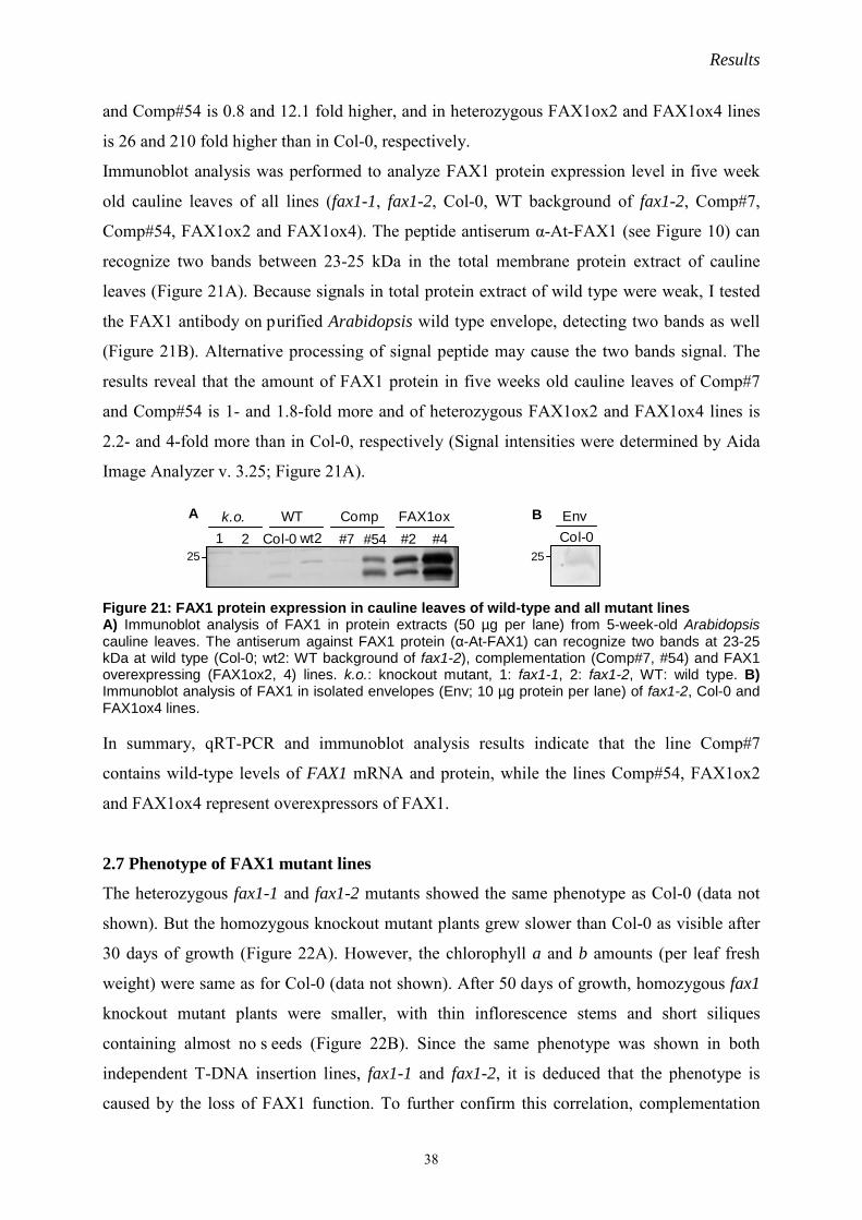
Results
38
and Comp#54 is 0.8 and 12.1 fold higher, and in heterozygous FAX1ox2 and FAX1ox4 lines
is 26 and 210 fold higher than in Col-0, respectively.
Immunoblot analysis was performed to analyze FAX1 protein expression level in five week
old cauline leaves of all lines (fax1-1, fax1-2, Col-0, WT background of fax1-2, Comp#7,
Comp#54, FAX1ox2 and FAX1ox4). The peptide antiserum α-At-FAX1 (see Figure 10) can
recognize two bands between 23-25 kDa in the total membrane protein extract of cauline
leaves (Figure 21A). Because signals in total protein extract of wild type were weak, I tested
the FAX1 antibody on purified Arabidopsis wild type envelope, detecting two bands as well
(Figure 21B). Alternative processing of signal peptide may cause the two bands signal. The
results reveal that the amount of FAX1 protein in five weeks old cauline leaves of Comp#7
and Comp#54 is 1- and 1.8-fold more and of heterozygous FAX1ox2 and FAX1ox4 lines is
2.2- and 4-fold more than in Col-0, respectively (Signal intensities were determined by Aida
Image Analyzer v. 3.25; Figure 21A).
Figure 21: FAX1 protein expression in cauline leaves of wild-type and all mutant lines A) Immunoblot analysis of FAX1 in protein extracts (50 µg per lane) from 5-week-old Arabidopsis cauline leaves. The antiserum against FAX1 protein (α-At-FAX1) can recognize two bands at 23-25 kDa at wild type (Col-0; wt2: WT background of fax1-2), complementation (Comp#7, #54) and FAX1 overexpressing (FAX1ox2, 4) lines. k.o.: knockout mutant, 1: fax1-1, 2: fax1-2, WT: wild type. B) Immunoblot analysis of FAX1 in isolated envelopes (Env; 10 µg protein per lane) of fax1-2, Col-0 and FAX1ox4 lines. In summary, qRT-PCR and immunoblot analysis results indicate that the line Comp#7
contains wild-type levels of FAX1 mRNA and protein, while the lines Comp#54, FAX1ox2
and FAX1ox4 represent overexpressors of FAX1.
2.7 Phenotype of FAX1 mutant lines
The heterozygous fax1-1 and fax1-2 mutants showed the same phenotype as Col-0 (data not
shown). But the homozygous knockout mutant plants grew slower than Col-0 as visible after
30 days of growth (Figure 22A). However, the chlorophyll a and b amounts (per leaf fresh
weight) were same as for Col-0 (data not shown). After 50 days of growth, homozygous fax1
knockout mutant plants were smaller, with thin inflorescence stems and short siliques
containing almost no s eeds (Figure 22B). Since the same phenotype was shown in both
independent T-DNA insertion lines, fax1-1 and fax1-2, it is deduced that the phenotype is
caused by the loss of FAX1 function. To further confirm this correlation, complementation
25
A B
251 2 Col-0 wt2 #4 Col-0#2#54#7
Comp FAX1oxWTk.o. Env

Results
39
and overexpressing lines were grown at the same conditions (Figure 22). Both
complementation lines Comp#7, #54 showed the same growth phenotype as wild type at 30
and 50 days, except shorter siliques (see Figure 25). Remarkably, the overexpressing lines
FAX1ox2 and 4 were slightly bigger and produced thicker inflorescence stems than wild type
(Figure 22).
Figure 22: Growth phenotypes of FAX1 mutants A) 30-day-old plants of FAX1 mutants and wild type. B) 50-day-old plants of FAX1 mutants and wild type. fax1-1, fax1-2: homozygous knockout lines; Col-0, WT fax1-2: wild-type FAX1 alleles; Comp#7, Comp#54: FAX1 complementation lines; FAX1ox2, FAX1ox4: FAX1 overexpressing lines.
A
fax1-1 Col-0 Comp#7 FAX1ox 2
fax1-2 WT fax1-2 Comp#54 FAX1ox 4
B
fax1-1 fax1-2 Col-0 WT fax1-2 Comp#7 Comp#54 FAX1ox2 FAX1ox4

Results
40
Conclusively, the phenotypes in FAX1 knockout mutants and overexpressing lines in
Arabidopsis are linked directly to the function of FAX1.
With the described growth phenotype of FAX1 mutant lines, it is clear that FAX1 affects the
biomass production, including leaves, stems, roots and flowers in Arabidopsis plants.
Therefore, rosette weight, leaf weight, fresh stem weight, dry stem weight, root weight and
stem diameter were measured in all FAX1 mutant lines (Figure 23).
Figure 23: Plant biomass of FAX1 mutants and wild type Arabidopsis plants from FAX1 mutants and wild type were harvested at 50 days.and dissected into different organs. A) Picture of stems. Bottom part of second internode of inflorescence stem from fax1-
0.2
0.4
0.6
0.8
1.0
1.2
1.4
1.6
1.8 stem diameter
stem
diam
eter
(mm
)
k.o.1 2 Col-0 WT2
WT#7 #54
Comp OX#2 #4
Col-0fax1-2 FAX1ox 2
A B
5
10
15
20
25 stem fresh weight
stem
fresh
wei
ght(
mg/
cm)
C
k.o.1 2 Col-0 WT2
WT#7 #54
Comp OX#2 #4
0.5
1
1.5
2
2.5
3 stem dry weight
stem
dry
wei
ght (
mg/
cm)
k.o.1 2 Col-0 WT2
WT#7 #54
Comp OX#2 #4
D
5E
fax1-2 Col-0 FAX1ox 2
0.05
0.1
0.15
0.2
0.25 root weight
root
wei
ght(
g)
H
k.o.1 2 Col-0 WT2
WT#7 #54
Comp OX#2 #4
0.51
1.52
2.53
3.54
4.5rosette weight
rose
ttew
eigh
t(g)
k.o.1 2 Col-0 WT2
WT#7 #54
Comp OX#2 #4
F
2
4
6
8
10
12 punched leaf weight
punc
hed
leaf
wei
ght
(mg/
0.0
9cm
2)
k.o.1 2 Col-0 WT2
WT#7 #54
Comp OX#2 #4
G

Results
41
2, Col-0 and FAX1ox2 was selected. B) Stem diameter in mm. Same internode part as in A (n=4-13±SD) was selected from FAX1 mutants and wild type. C) Stem fresh weight (mg/cm, n=4-12±SD). 1cm stem at the bottom of the second internode of inflorescence stem, washed. D) Stem dry weight (same sample as in C, n=4-12±SD). E) Pictures of rosettes from fax1-2, Col-0 and FAX1ox2. F) Rosette weight (n=8-13±SD). G) Punched leaf weight (central of rosette leaf with similar size was punched as one 0.09 cm-2 circle and weighted, n=6-11±SD). H) Root weight (n=4-10±SD). fax1-2, k.o.: homozygous knockout lines; Col-0, WT2: wild-type FAX1 alleles; Comp#7, Comp#54: FAX1 complementation lines; FAX1ox2, FAX1ox4: FAX1 overexpressing lines. Generally, adult homozygous fax1 knockout plants were smaller, and adult overexpressing
lines plants were larger in size than wild type and complementation lines. When measured in
50-day-old plants, the stem diameter, stem fresh weight and stem dry weight of FAX1 mutants
were different from the corresponding wild type alleles and complementation lines: knockout
lines were smaller and less, overexpressing lines were bigger and more (Figure 23A-D).
Leaves and roots also showed a biomass reduction in fax1 knockouts but an increase in
FAX1ox lines when compared to wild type (Figure 23E-H).
Conclusively, all measurements in FAX1 mutant tissues provided evidence that FAX1 is
involved in plant biomass production.
fax1 knockout plants show reduced male fertility
To understand the loss of function phenotype of homozygous fax1 mutants during flower
development, especially during the pollination and fertilization procedure, genetic analysis of
male and female crosses between T-DNA insertion lines and wild type was performed (Table
10). Table 10: The backcrossing of fax1 lines
: pollen and female gamtophyte pollination. No.: Number of plants analysed in the filial generation. "he fax1-1 x he fax1-1" and "he fax1-2 x he fax1-2" are self-pollination of the heterozygous fax1-1, fax1-2 mutant, respectively. "wt fax1-2 x ho fax1-2" is the backcrossing of wild type fax1-2 pollen with homozygous fax1-2 mutant female gametophyte. "he fax1-2 x ho fax1-2" is the backcrossing of heterozygous fax1-2 mutant pollen with homozygous fax1-2 mutant female gametophyte. ho%: percentage of homozygous, he%: percentage of heterozygous, wt%: percentage of FAX1 corresponding wild-type alleles.
Self-fertilization of heterozygous fax1-1 and fax1-2 mutant revealed that the segregation ratio
of homozygous progeny was 7% and 4% respectively, demonstrating an impaired pollination
+x
Crosses( ) No. ho % he % wt %
he fax1-1 x he fax1-1 280 7 65 28
he fax1-2 x he fax1-2 204 4 63 33
wt fax1-2 x ho fax1-2 20* / 100 /
he fax1-2 x ho fax1-2 171 12 88 /
* mix from 10 crossing events
+x

Results
42
or fertilization procedure. When the female gametophyte from homozygous fax1-2 mutant
was fertilized with wild-type fax1-2 pollen, normal siliques with 100% heterozygous seeds
were harvested at the filial generation, which indicates that the female gametophyte of
homozygous fax1-2 was fertile. However, when the wild-type fax1-2 female gametophyte was
fertilized with homozygous fax1-2 mutant pollen, the same shorter siliques and reduced
amount of homozygous progeny as during selfing of homozygous fax1-2 mutant plants were
harvested and seed yield was < 0.1% of wild-type (data not shown), which indicates that the
male gametophyte of the knockout mutant was almost sterile. Furthermore, during this
manual crossing, it became evident that pollen grains of homozygous fax1-2 were improperly
released by anthers. To minimize anther defects from the paternal tissue, I thus pollinated
homozygous fax1-2 gametophytes with pollen from heterozygous fax1-2 anthers thereby
achieving 12% of homozygous progeny (Table 10).
In summary, the strongly impaired male fertility phenotype of homozygous fax1 mutants is
mainly caused by impaired pollen anther development.
Anther and pollen development are impaired in homozygous fax1knockout mutants
To further analyse flower development, especially the fertilization phenotype, morphological
studies of flower and silique development of all FAX1 mutant lines were performed.
In wild-type alleles, complementation lines and overexpressing lines, bolting of flowers
occurred normally and silique and seed development were also similar to each other.
However, the stigma of homozygous fax1 knockout mutants grew slower showing non
pollinized papillae, which caused very short siliques with few seeds inside (Figure 25).

Results
43
Figure 24: FAX1 function related phenotypes in flower development 6-7 flower buds and youngest siliques from one bouquet of mature FAX1 mutant lines and wild-type were selected. fax1-1, fax1-2: homozygous knockout lines; Col-0, WT fax1-2: wild-type FAX1 alleles; Comp#7, Comp#54: FAX1 complementation lines; FAX1ox2, FAX1ox4: FAX1 overexpressing lines. Arrow heads indicate the non-pollinated stigma papillae of fax1-1, fax1-2.
After 55 days of growth, siliques of wild type alleles and complementation lines started to
senescent, which firstly appeared at siliques on inflorescence stems (Figure 25). However,
fax1-1
Col-0
fax1-2
WT fax1-2
Comp#7 Comp#54
FAX1ox 2 FAX1ox 4

Results
44
homozygous fax1 knockout mutants and overexpressing lines plants still only had green
siliques, which imply that FAX1 may have function related to senescence.
Figure 25: FAX1 function related phenotypes in silique development 8 siliques inflorescence stems of all lines (55 days old) were selected and arranged from left to right depending on the developmental stages (left side is the youngest, right side is the oldest). fax1-1, fax1-2: homozygous knockout lines; Col-0, WT fax1-2: wild-type FAX1 alleles; Comp#7, Comp#54: FAX1 complementation lines; FAX1ox2, FAX1ox4: FAX1 overexpressing lines. To visualize the strongly impaired male fertility phenotype of homozygous fax1 knockout
mutants, open flowers at development stages 12-14 (Smyth et al., 1990) from all FAX1
mutant lines and wild type were compared (Figure 26).
fax1-1
Col-0
fax1-2
WT fax1-2
Comp#7
Comp#54
FAX1ox 2
FAX1ox 4

Results
45
Figure 26: FAX1 function related phenotype in pollination Flower buds at flower stage 12-14 (Smyth et al., 1990) were selected. fax1-1, fax1-2: homozygous knockout lines; Col-0, WT fax1-2: wild-type FAX1 alleles; Comp#7, Comp#54: FAX1 complementation lines; FAX1ox2, FAX1ox4: FAX1 overexpressing lines Both two FAX1 wild-type alleles and overexpressing lines produced flowers with mature
pollen grains being released by anthers and sticking to the papillae of stigmas. However,
flowers of the two homozygous fax1 knockouts had almost no free pollen grains and stigma
papilla produced long hairs, which indicates that very few pollination happened. In
fax1-1
Col-0
fax1-2
WT fax1-2
Comp#7 Comp#54
FAX1ox 2 FAX1ox 4

Results
46
comparison to the knockout lines, flowers of complementation lines had more pollen outside
of anthers but fewer than wild type, indicating that complementation of pollen release by
anthers was not 100%. Furthermore, normal wild type pollen was yellow in colour because of
the inclusion of flavonoid and carotenoid deposits (Dobritas et al., 2011). However, impaired
pollen of homozygous fax1 knockouts as well as of complementation lines was pale and
colourless (Figure 27).
Figure 27: FAX1 function related phenotype of Arabidopsis mature pollen on the surface of anthers Anthers after flower stage 13 were selected. fax1-1, fax1-2: homozygous knockout lines; Comp#7, Comp#54: FAX1 complementation lines. Arrow heads indicate the colourless and pale pollens of fax1-1, fax1-2, Comp#7 and Comp#54. Ultrastructural analysis of FAX1 mutant lines
In the following, stem, anther and pollen morphology were analyzed in the FAX1 mutants and
Col-0 by light microscopy and transmission electron microscopy (TEM; Figure 28-30).
Microscopic investigation were performed by Dr. Irene Gügle (Department Biologie I, Plant
Biochemistry and Physiology, LMU München). Firstly, stems were pictured 1 cm above the
first node of 5-week-old inflorescence stems (Figure 28). Here the amount of hypodermal cell
layers appeared reduced (~3) in fax1-2 but increased in FAX1ox (~5), when compared with
Col-0 (~4; Figure 28A-C). Compared with Col-0 wild type, vascular bundles were smaller
and xylem and sclerenchyma contained secondary cell walls reduced in homozygous fax1-2
fax1-1 fax1-2 Col-0
Col-0 fax1-2Comp#7Comp#7 Comp#54
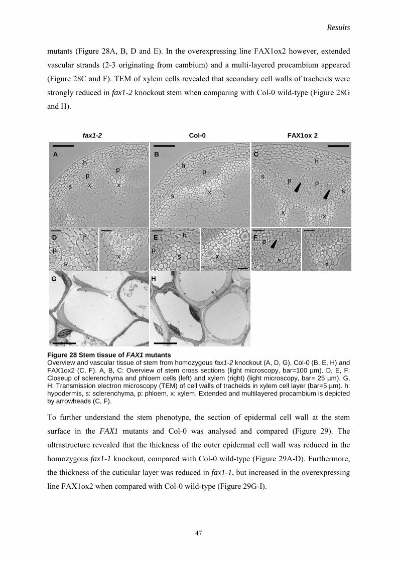
Results
47
mutants (Figure 28A, B, D and E). In the overexpressing line FAX1ox2 however, extended
vascular strands (2-3 originating from cambium) and a multi-layered procambium appeared
(Figure 28C and F). TEM of xylem cells revealed that secondary cell walls of tracheids were
strongly reduced in fax1-2 knockout stem when comparing with Col-0 wild-type (Figure 28G
and H).
Figure 28 Stem tissue of FAX1 mutants Overview and vascular tissue of stem from homozygous fax1-2 knockout (A, D, G), Col-0 (B, E, H) and FAX1ox2 (C, F). A, B, C: Overview of stem cross sections (light microscopy, bar=100 µm). D, E, F: Closeup of sclerenchyma and phloem cells (left) and xylem (right) (light microscopy, bar= 25 µm). G, H: Transmission electron microscopy (TEM) of cell walls of tracheids in xylem cell layer (bar=5 µm). h: hypodermis, s: sclerenchyma, p: phloem, x: xylem. Extended and multilayered procambium is depicted by arrowheads (C, F). To further understand the stem phenotype, the section of epidermal cell wall at the stem
surface in the FAX1 mutants and Col-0 was analysed and compared (Figure 29). The
ultrastructure revealed that the thickness of the outer epidermal cell wall was reduced in the
homozygous fax1-1 knockout, compared with Col-0 wild-type (Figure 29A-D). Furthermore,
the thickness of the cuticular layer was reduced in fax1-1, but increased in the overexpressing
line FAX1ox2 when compared with Col-0 wild-type (Figure 29G-I).
Col-0 FAX1ox 2fax1-2
A B C
D E F
G H
p
p p
ss s
s
h
x
x xx
h hp
hp
pp p
h
ss
x
xx
xs

Results
48
Figure 29 Stem epidermis of FAX1 mutants Epidermis and hypodermis of stem from homozygous fax1-1 knockout (A, B, G), Col-0 (C, D, H) and FAX1ox2 (E, F, I). A, C, E: Overview of epidermal and hypodermal cell layers (light microscopy). B, D, F: Transmission electron microscopy closeups of epidermal cell wall (bar= 500 nm), and G, H, I: Cutin layer (bar= 200 nm). cut: cutin layer, cyt: cytosol, cw: cell wall. Moreover, investigation of anther and pollen prior to dehiscence of homozygous fax1-2
knockout, Col-0 wild-type and complementation line Comp#54 by light microscopy and TEM
was performed (Figure 30). In line with the observations from stems, the vascular bundle of
anthers appeared collapsed and cutin was reduced in fax1-2 while comparing with Col-0 wild-
type (data not shown). In the homozygous fax1-2 knockout, the ultrastructure of anthers
further showed that fragmented parts of tapetum still were present and bound together with
pollen grains, while the tapetum had disappeared in Col-0 wild-type and only few parts of
tapetum were bound with some pollen grains in the complementation line Comp#54 (Figure
30A-I). Surprisingly, the pollen grains in the homozygous fax1-2 knockout were only
surrounded by the intine layer, which revealed that the exine development was strongly
impaired. In Comp#54, however bacula structures of the exine were restored, while both
tectum and bacula of exine existed in Col-0 wild-type (Figure 30G-L). In addition
accumulation of deposits unknown materials were detected in the complementation line
Comp#54 (Figure 30I, L).
Col-0 FAX1ox 2fax1-1A B C D E F
G H I
cut
cyt
cut cut
cyt cyt
cw
cwcw

Results
49
Figure 30 Anther and pollen of homozygous fax1 knockout, Col-0 and complementation line Anther and pollen of flowers from homozygous fax1-2 knockout (A; D, G, J), Col-0 (B, E, H, K) and complementation line Comp#54 (C, F, I, L). A, B, C: Overview of anther cross section prior to dehiscence (light microscopy, bar= 50 µm). D, E, F: Overview of areas between pollen grain and anther cells (TEM, bar= 5 µm). G, H, I: Closeup of areas between pollen and anther cells (TEM, bar= 1 µm). J, K, L: Closeup of exine and intine of pollen grain (TEM, bar= 500 nm). t: tapetum, a: anther cell, e: exine, i: intine, tec: tectum of exine, bac: bacula of exine, * : material deposits in exine/tapetum region. 2.8 Wax content in stems of FAX1 mutant lines
Considering the ultrastructural result on the stem cuticular layer (see Figure 29), the wax
content on inflorescence stems of FAX1 mutants and the corresponding wild-type alleles was
analysed and compared (Figure 31-32). Wax analysis was performed by Prof. Lukas
Schreiber (Department of Ecophysiology, IZMB, University of Bonn).
Col-0 Comp#54fax1-2A B C
D E F
G H I
J K L
t
ai
tec bac
tt
t
*
*a
a
ee
e
bac
i
ii
i
i

Results
50
Figure 31: Wax analysis of FAX1 mutant lines A) Wax analysis of FAX1 knockout and complementation lines. Total wax coverage in μg/cm2 stem of surface (n= 3 ± SD, n=7 for fax1-2). For each replicate 3-4 stems (internode 2-4) were pooled. B) Wax analysis of FAX1 overexpressing lines and wild type. Total wax coverage in μg/cm2 stem of surface (n= 6 ± SD). For each replicate 3-4 stems (internode 2-4) were pooled. The asterisks indicate significantly different wax levels (* P< 0.05) in FAX1ox2 and 4 when compared with Col-0 wild type. k.o. 1 and 2: homozygous knockout lines, WT fax1 1 and 2: wild-type FAX1 alleles, Comp#7, Comp#54: FAX1 complementation lines, FAX1ox2 and 4: FAX1 overexpressing lines. The homozygous fax1 knockout mutants had similar amounts of total stem wax coverage as
the corresponding wild-type alleles, and interestingly, the complementation lines slightly
increased the stem total wax amount when comparing with wild-type (Figure 31A). However,
the total wax levels of FAX1 overexpressing lines FAX1ox2 and FAX1ox4 were increased to
136% and 134% of the Col-0 loads, respectively (Figure 31B).
Furthermore, the homozygous fax1 knockout mutants differed significantly from wild type in
the levels of ketones. The fax1-1 and fax1-2 ketone levels were reduced to 33% and 47% of
the wild-type loads, respectively (Figure 32). But in FAX1 overexpressing lines, there is no
difference in composition of wax, regarding aliphatic chain length or functional groups (eg.
ketone, acid or aldehyde), when comparing with Col-0 wild type (data not shown).
5
10
15
20
25
30
35 total wax
1 2 Col-0 1 2 #54
Tota
l wax
cove
rage
(μg
cm-2
)
5
10
15
20
25
30
35
40
Col-0 FAX1ox2 FAX1ox4
total wax
Tota
l wax
cove
rage
(μg
cm-2
)
BA
k.o. WT fax1#7Comp
* *

Results
51
Figure 32: Ketone analysis of FAX1 mutant lines Total ketone coverage in μg/cm2 stem of surface (n= 3 ± SD, n=7 for fax1-2). For each replicate 3-4 stems (internode 2-4) were pooled. The asterisks indicate significantly different signal levels (** P < 0.005, * P< 0.05) in homozygous fax1 knockout mutants (fax1-1 or fax1-2) and FAX1 complementation lines (Comp#7 or Comp#54) when compared with Col-0. 2.9 Fatty acid contents in fax1 knockout mutants and wild type
Considering the phenotypes at the exine of pollen and stem outer layer of epidermis cells, the
fatty acids composition in the flowers, stems and leaves of homozygous fax1 knockout
mutants and corresponding wild-type alleles were measured by Dr. Patrick Giavalisco (Max
Planck Institute of Molecular Plant Physiology, Potsdam-Golm, Germany; Figure. 33-35).
Figure 33: Polar lipid composition in flowers of wild type and the fax1 knockout mutants Flowers were harvested from 50 days old plants. Lipid content (n=6±SD) was normalised to fresh weight (FW) and the internal standard PC34:0. The asterisks indicate significantly different signal levels (** P < 0.005, * P< 0.05) in homozygous fax1 knockout mutants (fax1-1 or fax1-2) when compared with Col-0. A) Main differences of polar lipids except free fatty acids are shown. PC: phosphatidylcholine, TAG: triacylglycerol. B) Free fatty acids (FA) different in flowers of the homozygous fax1 knockout mutants and corresponding wild-type alleles.
1
2
3
4
5
6
7 ketones
fax1-1 fax1-2 Col-0 WT fax1-1
WTfax1-2
Comp#7
Comp#54
Keto
ne c
over
age
(μg
cm-2
)
*
****
B
2
468
1012
14161820
FA 20:1 FA 24:1
Lipi
d co
nten
t/ a
rbitr
ary
units
(x 1
03)
fax1-1fax1-2WT fax1-2Col-0
*
*
A
1
2
3
4
5
6
7
PC36:6 TAG50:3 TAG52:6 TAG54:5 TAG54:6 TAG54:8 TAG54:9
lipid
con
tent
/ arb
itrar
y un
its (x
106
) fax1-1fax1-2WT fax1-2Col-0
** **
**
*

Results
52
The measurement and analysis of flower lipid composition revealed that the amount of
PC36:6, TAG50:3 and TAG54:5 were significantly reduced in the homozygous fax1 knockout
mutants comparing with Col-0 wild-type. Only amount of FA24:1 was increased in the fax1
knockout mutants (Figure 33).
Figure 34: Polar lipid composition in cauline leaves of wild type and fax1 knockout mutants Cauline leaves were harvested from 50 days old plants. Lipid content (n=6±SD) was normalised to fresh weight (FW) and the internal standard PC34:0. The asterisks indicate significantly different signal levels (** P < 0.005, * P< 0.05) in homozygous fax1 knockout mutants (fax1-1 or fax1-2) when compared with Col-0. A) Main differences of polar lipids with high expression level are shown. PC: phosphatidylcholine, DGDG: digalactosyldiacylglycerol, MGDG: monogalactosyldiacylglycerol. B) Differences of polar lipids with lower expression level than in A are shown. PE: phosphatidylethanolamine, PG: Phosphatidylglycerol. C) Free fatty acids (FA) different in leaves of the homozygous fax1 knockout mutants and corresponding wild-type alleles.
B
5
10
15
20
25
PC36:3 PE36:6PG34:1PG34:2PG34:3PG34:4
lipid
con
tent
/ arb
itrar
y un
its (x
105
)
fax1-1fax1-2WT fax1-2Col-0
*** ***
**
***
**
A
5
10
15
20
25
30
35
40
lipid
con
tent
/ arb
itrar
y un
its (x
106
)
fax1-1fax1-2WT fax1-2Col-0
DGDG36:6
MGDG34:6
MGDG36:6
PC34:3 PC36:5 PC36:6 Pheo-phytin
**
**
*******
**** ** ****
*
lipid
con
tent
/ arb
itrar
y un
its (x
105
) fax1-1fax1-2WT fax1-2Col-0
C
1
2
3
4
5
6
FA 18:0 FA 20:0 FA 26:0
**
**
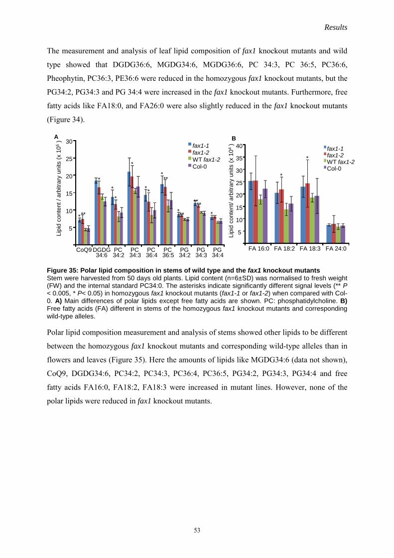
Results
53
The measurement and analysis of leaf lipid composition of fax1 knockout mutants and wild
type showed that DGDG36:6, MGDG34:6, MGDG36:6, PC 34:3, PC 36:5, PC36:6,
Pheophytin, PC36:3, PE36:6 were reduced in the homozygous fax1 knockout mutants, but the
PG34:2, PG34:3 and PG 34:4 were increased in the fax1 knockout mutants. Furthermore, free
fatty acids like FA18:0, and FA26:0 were also slightly reduced in the fax1 knockout mutants
(Figure 34).
Figure 35: Polar lipid composition in stems of wild type and the fax1 knockout mutants Stem were harvested from 50 days old plants. Lipid content (n=6±SD) was normalised to fresh weight (FW) and the internal standard PC34:0. The asterisks indicate significantly different signal levels (** P < 0.005, * P< 0.05) in homozygous fax1 knockout mutants (fax1-1 or fax1-2) when compared with Col-0. A) Main differences of polar lipids except free fatty acids are shown. PC: phosphatidylcholine. B) Free fatty acids (FA) different in stems of the homozygous fax1 knockout mutants and corresponding wild-type alleles. Polar lipid composition measurement and analysis of stems showed other lipids to be different
between the homozygous fax1 knockout mutants and corresponding wild-type alleles than in
flowers and leaves (Figure 35). Here the amounts of lipids like MGDG34:6 (data not shown),
CoQ9, DGDG34:6, PC34:2, PC34:3, PC36:4, PC36:5, PG34:2, PG34:3, PG34:4 and free
fatty acids FA16:0, FA18:2, FA18:3 were increased in mutant lines. However, none of the
polar lipids were reduced in fax1 knockout mutants.
A
5
10
15
20
25
30
CoQ9 DGDG 34:6
PC 34:2
PC 34:3
PC 36:4
PC 36:5
PG 34:2
PG 34:3
PG 34:4
Lipi
d co
nten
t/ a
rbitr
ary
units
(x 1
05)
fax1-1fax1-2WT fax1-2Col-0
*****
**
*
*
**
*
**
**
* **
*
B
5
10
15
20
25
30
35
40
FA 16:0 FA 18:2 FA 18:3 FA 24:0
Lipi
d co
nten
t/ ar
bitra
ry u
nits
(x 1
04)
fax1-1fax1-2WT fax1-2Col-0
*
*

Results
54
Figure 36: PC amount in flowers, leaves and stems of fax1 knockout mutants and wild type Flowers, leaves and stems were harvested from 50 days old plants. PC content (n=6±SD) was normalised to fresh weight (FW) and the internal standard PC34:0. As a summary, total phosphatidylcholine (PC) amount in leaves and PC36:3 in flowers of the
homozygous fax1 knockout mutants were reduced significantly when compared with the
corresponding wild-type, however in stems, total PC amount was increased (Figure 36). It is
known that PC is a key chemical involved in fatty acid transport from plastids to ER (see
Chapter I.2). Therefore, PC amount differences between the homozygous fax1 knockout
mutants and wild type provides evidence that FAX1 might be an inner envelope protein
involved in fatty acid export from chloroplasts.
2.10 Transcriptomic analysis of fax1 knockout mutant and wild type
Because of the impaired flower and stem development in homozygous fax1 knockout mutants,
gene expression in fax1-2 versus the wild type was compared by microarray analysis
(Affymetrix ATH1 GeneChip) in the same tissue pools that were used for lipid analysis and
stem wax coverage analysis. The hybridization of the microarrays and statictical analysis of
data were performed by Karl Mayer and by Dr. Katrin Philippar, respectively (Department
Biologie I, Plant Biochemistry and Physiology, LMU München). I found that 1,058 genes
could be identified as down-regulated and 515 genes were up-regulated in flowers of fax1-2,
while 410 genes could be classified as down-regulated and 1136 genes were up-regulated in
stems (Table 11). To be mentioned, the values for FAX1 in homozygous fax1-2 and wild-type
5
10
15
20
25
PC flower PC leaf PC stem
fax1-1
fax1-2
WT fax1-2
Col-0
PC c
onte
nt/ a
rbitr
ary
units
(x 1
05)

Results
55
stems are 178.6 and 774.7 (p-value 0.073), respectively. The values for FAX1 in homozygous
fax1-2 and wild-type flowers are 151.6 and 614.2 (p-value 0.022), respectively. Table 11: Differential gene expression in fax1-2 flowers and stems Functional categories of differentially expressed genes in fax1-2 versus wild-type flowers and stem are shown (Fold change >1.5, signal differeence >30). Classification of genes was performed by MAPMAN (Thimm et al., 2004).
Interestingly, fax1 knockout flowers mainly down-regulated gene expression (1058 of 1573)
while loss of FAX1 in stems resulted in mostly increased transcript content (1136 of 1546).
Further, the overlap of differentially expressed genes in flowers and stems was extraordinarily
low (only 157), indicating different physiological reactions of flowers and stem tissues.
Considering that FAX1 might function in fatty acid export across the chloroplast inner
envelope membrane, we focused on e xpression of genes related to lipid biosynthesis or
secondary cell wall development, which represented a hotspot of regulation with top fold
changes. To be mentioned, none of the top regulated genes overlapped in flowers and stems.
In stems, among 40 genes up-regulated with a fold change >10 (fax1-2 versus wild-type) were
23 genes with a function related to lipid biosynthesis or secondary cell wall (Table 12).
Interestingly, AtMAH1/AtCYP96A15 (At1g57750), which is highly up-regulated (FCH
12.7), is the midchain alkane hydroxylase responsible for formation of secondary alcohols and
ketones in stem cuticular wax (Greer et al., 2007), indicating a correlation with the changed
ketone content found in fax1 knockouts (see Figure 32). Furthermore, both AtIRX15
(At3g50220) and AtIRX15L (At5g67210), which encode DUF579-containing proteins that
are essential for normal xylan deposition in the secondary cell wall (Brown et al., 2011), are
up-regulated in homozygous fax1-2 versus wild-type Arabidopsis stem.
flowers stems
Down, 1058 genes Up, 515 genes Down, 410 genes Up, 1136 genescell wall modif ication 63 24 17 85lipid metabolism 30 7 3 32amino acid metabolism 7 9 3 18
secondary metabolism 22 32 15 32hormone metabolism 34 17 10 33Biotic stress 42 26 21 34RNA 70 70 38 121protein 108 40 42 135signalling 85 18 37 70development 46 16 24 28transport 95 27 37 37not assigned 265 133 91 347miscellaneous 120 39 47 83others 71 57 25 81

Results
56
Of the 57 genes down regulated with a fold change >3 were 14 genes with a function related
to lipid biosynthesis or secondary cell wall in fax1-2 versus wild-type Arabidopsis stems. Table 12: Top fold change genes in stems. Highly regulated genes related to lipid biosynthesis or secondary cell wall biosynthesis in homozygous fax1-2 versus wild-type Arabidopsis stems. u.k.: unknown.
In flowers, among 19 genes up regulated with a fold changes >3 and 33 genes down regulated
with a fold change >5 (top fold changes of fax1-2 versus wild-type) were 19 (7 up, 12 down)
genes with a function related to lipid biosynthesis or secondary cell wall (Table 12).
To be mentioned, in the current model of the transcriptional network of secondary cell wall
biosynthesis in Arabidopsis (Zhao and Dixon, 2010), a key myb-type transcription factor,
Name AGI Code Description(ARAMEMNON)Signal,fax1-2
Signal, wild type
Fold Change (FCH)
UPlipid biosynthesis
u.k. At1g74670 putative GASA/GAST/Snakin-type protein 310.63 18.75 16.57
AtMAH1 At1g57750 mid-chain alkane hydroxylase 2567.66 202.29 12.69secondary cell wall
AtXCP1 At4g35350 cysteine protease 2038.68 74.82 27.25AtLAC2 At2g29130 putative laccase 2898.82 161.55 17.94
AtPGSIP1 At3g18660 putative xylan glucuronosyltransferase 3880.01 243.38 15.94AtLAC10 At5g01190 putative laccase 559.23 37.16 15.05AtXCP2 At1g20850 cysteine protease 5855.62 390.10 15.01AtTED6 At1g43790 protein involved in secondary cell wall biogenesis 3852.57 269.13 14.31AtLAC17 At5g60020 putative laccase 3469.41 245.58 14.13AtMYB103 At1g63910 putative Myb-type transcription factor 1785.59 129.14 13.83
AtANAC073 At4g28500 NAC-type transcription factor 2988.57 233.74 12.79u.k. At5g01360 protein of unknown function 2045.68 170.28 12.01
AtXTH4 At2g06850 xyloglucan endotransglucosylase-hydrolase 4943.93 412.11 12.00AtPer64 At5g42180 putative class-III peroxidase 8220.49 694.18 11.84
u.k. At1g09610 protein of unknown function 1358.05 115.56 11.75u.k. At4g09990 protein of unknown function 1128.17 98.66 11.43
AtXTH6 At5g65730 xyloglucan endotransglucosylase-hydrolase 1889.98 169.11 11.18AtLAC12 At5g05390 putative laccase 2584.48 231.61 11.16AtIRX15L At5g67210 protein probably involved in xylan biosynthesis 3214.46 288.30 11.15AtCOBL4 At5g15630 putative glycosylphosphatidylinositol-anchored protein 4853.46 441.60 10.99AtIRX15 At3g50220 protein probably involved in xylan biosynthesis 2541.10 237.34 10.71AtXTH15 At4g14130 xyloglucan endotransglucosylase-hydrolase 387.87 37.11 10.45AtXyn3 At4g08160 xylanase 931.23 90.12 10.33
DOWNsecondary cell wall
AtABCB16 At3g28360 putative subfamily B ABC-type transporter 39.31 328.67 8.36AtVGD1 At2g47040 pectin methylesterase 66.25 458.57 6.92
u.k. At1g19510 putative MYB_related-type transcription factor 17.43 80.19 4.60u.k. At5g07410 putative pectinesterase 15.88 68.17 4.29u.k. At1g10770 putative pectin methylesterase inhibitor 56.05 234.11 4.18u.k. At5g07430 putative pectinesterase 26.95 107.84 4.00
AtUGT73B5 At2g15480 UDP-dependent glycosyl transferase 36.20 138.66 3.83u.k. At4g02250 putative invertase/pectin methylesterase inhibitor 22.59 82.06 3.63
AtNAC2 At3g15510 NAC-type transcription factor 276.34 968.72 3.51AtCslG1 At4g23990 putative cellulose synthase-like glycosyltransferase 103.34 321.57 3.11

Results
57
MYB103 and NAC-type transcription factors were also highly regulated in homozygous fax1
mutant versus Col-0 wild-type (Table 12 and 13).
Table 13: Top fold change genes in flowers. Highly regulated genes related to lipid biosynthesis or secondary cell wall biosynthesis in homozygous fax1-2 versus wild-type Arabidopsis flowers. u.k.: unknown.
Gene AGI Code Description(ARAMEMNON)Signal, fax1-2
Signal, wild type
Fold Change (FCH)
UPlipid biosynthesis
AtAOx1a At3g22370 alternative oxidase 2319.12 576.37 4.02AtAOC1 At3g25760 allene oxide cyclase 261.04 78.81 3.31
secondary cell wallAtPer49 At4g36430 putative class-III peroxidase 625.76 82.84 7.55AtPer58 At5g19880 putative class-III peroxidase 695.83 184.47 3.77AtPer14 At2g18140 putative class-III peroxidase 190.58 55.92 3.41AtPer39 At4g11290 putative class-III peroxidase 1163.18 366.35 3.18AtNTL1 At1g32870 putatively NAC-type transcription factor 551.81 174.17 3.17
DOWNlipid biosynthesis
u.k. At5g47350 putative palmitoyl protein thioesterase 108.18 1671.07 15.45AtANR At1g61720 dihydroflavonol 4-reductase 70.44 1023.51 14.53AtDFR At5g42800 dihydroflavonol 4-reductase 76.63 494.19 6.45PME49 At5g07420 putative pectinesterase 327.30 1778.40 5.43
AtPTEN1 At5g39400 phosphatidyl-inositol triphosphate phosphatase 203.49 1063.88 5.23PME28 At5g27870 putative pectinesterase 98.03 511.79 5.22
secondary cell wallu.k. At4g15750 putative invertase/pectin methylesterase inhibitor 94.44 3208.18 33.97
AtEXP23 At5g39280 putative expansin 73.00 561.75 7.70AtGDPD6 At5g08030 putative glycerophosphoryl diester phosphodiesterase 27.28 195.03 7.15AtXYLP4 At5g09370 putative xylogen-type arabinogalactan protein 70.32 422.77 6.01

Discussion
58
V. Discussion
1 OEP7
Whereas the OEP7.1 isoform has been described in the literature (see Chapter I.1.1), OEP7.2
from Arabidopsis is firstly characterized in this work. In 1993, t ail-anchored (TA) proteins
were classified as integral membrane proteins with a cytosolic N-terminal functional domain,
a single transmembrane domain (TMD) and a short C-terminal hydrophilic tail (Kutay et al.,
1993). In the last few years, a considerable amount of TA proteins, especially the mechanisms
of the targeting into specific membranes, was investigated in yeast and mammals. It is
demonstrated that the targeting signal of almost all TA proteins is located within the C-
terminal TMDs and flanking sequences (Borgese et al., 2007; Pedrazzini, 2009). The topology
analysis result of At-OEP7.2 (see Figure 4) revealed that OEP7.2 is a TA protein in the outer
envelope membrane of chloroplasts. With the in vivo targeting of At-OEP7.2 in protoplasts, I
could show that the N-terminus of OEP7.2, not the C-terminus, is critical for targeting to the
outer envelope. These findings are novel and complement those described for OEP7.1, where
in contrast the C-terminal part of the protein is necessary for targeting (Lee et al., 2001).
Further in vivo targeting of At-OEP7 deletion constructs fused with GFP in protoplasts
revealed that the N-terminal amino acid residues 11 to 22 (neighboring TMD) of At-OEP7.2
are necessary and residues 1 to 48 (N-terminus, TMD and C-terminal flanking sequences) are
sufficient for targeting to the chloroplast envelope membrane. Therefore, my findings on
OEP7.2 targeting provide important evidence to understand the targeting of TA proteins into
chloroplast membranes in plants. Furthermore, it could be demonstrated that the double
knockout mutant of oep7.1/oep7.2 in Arabidopsis showed no phenotype under various
conditions. Considering that there are at least four isoforms of OEP7 in Arabidopsis and that
the polypeptide of OEP7 is very short, which means that even more OEP7 isoforms might
exist, it is likely that triple or quarter mutants could be necessary to investigate a plant
phenotype of OEP7 loss of function. And as mentioned about SsOEP8, a homolog of OEP7 in
Suaeda salsa, which was described to confer oxidative stress tolerance and to induce
chloroplast aggregation in transgenic Arabidopsis plants (Wang et al., 2012), it could be also
interesting to analyse the knockout mutant of OEP7.1 and OEP7.2 under oxidative stress
conditions.

Discussion
59
2 FAX1
In the second part of this work, I identified a gene encoding a novel membrane protein in the
chloroplast inner envelope most likely involved in fatty acid or fatty acid derivative export.
2.1 Plastid fatty acid export and the pollen exine formation in Arabidopsis
The Arabidopsis mature pollen grain surface consists of three layers (from inside to outside):
i) an inner intine, made primarily of cellulose; ii) an outer exine wall, multilayered and
composed of the chemically resistant polymer sporopollenin and interrupted by openings
called apertures; iii) a pollen coat, composed of lipids, proteins, pigment, and aromatic
compounds, that fills the sculptured cavities of the pollen exine (Edlund et al., 2004). The
sporopollenin in exine layers is a complex polymer of fatty acids and phenolic compounds
(Guilford et al., 1988; Ahlers et al., 1999). The exine itself splits into outer sexine and the
inner nexine layers. The sexine comprises the tectum and the bacula. The nexine comprises
the nexine I and nexine II (Ariizumi and Toriyama, 2011; Figure 37). The ultrastructural
investigation of the pollen in FAX1 mutants showed that the pollen of fax1 knockout mutants
lacks the sexine and parts of the nexine (see Figure 30). In the recent years, several genes
involved in exine formation were identified in Arabidopsis, such as male sterility 2 (MS2;
Aarts et al., 1997; Chen et al., 2011), CYP703A2 (Morant et al., 2007), CYP704B1 (Dobritsa
et al., 2009), Acyl-CoA sythetase 5 (ACOS5; Souza et al., 2009) and an ABCG transporter
(ABCG26; Choi et al., 2011; Dou et al., 2011; Quilichini et al., 2010), which also showed a
pollen phenotype similar to fax1 knockout mutants. With the known function of proteins
mentioned above, it was summarized that the pollen exine development precedure depends on
fatty acids, which are synthesized in the plastids of tapetum cells and esterified with CoA by
Acyl-CoA synthetase (e.g. ACOS5) and are sent to the ER, where the fatty acyl-CoAs are
hydroxylated by CYP704B1 and CYP703A2, respectively. After the modification and other
unknown steps, the modified fatty acids are exported from the tapetum cell for exine
formation by the ABCG26 transporter and other unknown transporters (Choi et al., 2011).
Recently, both MS2 and the probable ortholog of MS2, defective pollen wall (DPW) of rice
(Oryza sativa) were identified as the plastid localized fatty acyl carrier protein reductase,
which participates in a conserved step in primary fatty alcohol synthesis for anther cuticle and
pollen sporopollenin biosynthesis (Chen et al., 2011; Shi et al., 2011).

Discussion
60
Figure 37 A typical angiosperm pollen grain structure (adapted from Ariizumi and Toriyama, 2011)
A) model of a pollen grain B) Transmission electron microscopy of a cross-section of exine architecture in Arabidopsis. Bar= 1 µm. Tc: tectum, Ba: bacula, Try: tryphine, In: intine, Cy: cytoplasm, Pla: plasma membrane, Ne I/II: nexine I/II.
According to the high similarity of phenotypes it is therefore proposed that FAX1 represents
the missing transporter for export of fatty acid derivatives from plastids in tapetum cells
(Figure 38). Transcriptomic analysis of flowers revealed that all the above mentioned genes
for exine formation were not highly regulated in fax1 knockout mutants while comparing with
wild type. However, an effective genetic screen in Arabidopsis on sporopollen synthesis and
exine assembly recently showed that many other genes are involved in the pollen morphology
(Dobritsa et al., 2011). Surprisingly, the genes of PTEN1 (a phosphatidyl-inositol
triphosphate phosphatase), DFR (dihydroflavonol 4-reductase), ANR (dihydroflavonol 4-
reductase) and the putative pectinesterases PME28 and PME49 were highly down regulated in
fax1 knockout mutant versus Col-0 (see Table 13). The Arabidopsis PTEN, a protein and lipid
dual phosphatase, homologous to animal PTEN (a tumor suppressor), was identified to
regulate autophagy in pollen tubes by disrupting the dynamics of phosphatidylinositol 3-
phosphate (PI3P; Zhang et al., 2011; Gupta et al., 2002). Both DFR and ANR are key
reductases during anthocyanins and flavonoid biosynthesis (Winkel-Shirley, 2001; Marinova
et al., 2007). Considering the colorless pollen phenotype in fax1 knockout mutant plants (see
Figure 27), it is suggested that the absence of exine layers prevents inclusion of flavonoids
and carotenoids thus leading to the transparent pollen and secondly affecting expression of
genes (e.g. down-regulating) involved in flavonoid biosynthesis. It was reviewed that pectin
methylesterases (PME) belong to large multigene families and are involved in vegetative and
reproductive processes, including pollen formation (Pelloux et al., 2007). Although the
function of PME 28 and PME49 are unknown, another PME, named quartet, was indicated
recently to affect the pollen tetrad separation during floral development, which may provide a
A B

Discussion
61
hint to explain the sticky pollen phenotype in fax1 knockout mutant plants (Francis et al.,
2006).
Furthermore, the complementation of fax1 by 35S:: FAX1 (Comp #7, Comp#54) is able to
rescue fertility and most of exine structures, because of 35S promoter is not active in pollen
but in tapetum cells (Wilkinson et al., 1997). As mentioned above, fatty acids, which are
necessary for pollen exine formation and fertility, are delivered by the anther. However, some
exine material, which is necessary for interrelation of flavonoid and carotenoids, should be
delivered by the pollen, such as pollen plastid fatty acids. Therefore, the complementation
lines of FAX1 still show transparent pollen grains (see Figure 27)
Other interesting genes, which were highly regulated in fax1 knockout mutant versus Col-0
and may have a function related to cutin, wax biosynthesis or structure of anther cells will be
discussed together with the stem phenotype (see Chapter V. 2.2).
Furthermore, the genes of some other proteins which may also have function during fatty acid
transport and lipid biosynthesis, were also regulated in flowers of fax1 knockout mutants
versus Col-0, such as PAP2 (purple acid phosphatase 2; down FCH= 4.28), MGD2
(Galactosyltransferase; down FCH=3.36), LACS5 (Long-chain Acyl-CoA synthetase; down
FCH=2.70), ACS8 (Acyl-CoA synthetase; up FCH= 2.17), FAR7 (Fatty acid reductase 7; up
FCH= 1.99).
Interestingly, the ultrastructure of anthers in fax1 knockout mutants clearly showed that
fragmented parts of tapetum still were present and bound together with pollen grains, while
the tapetum was totally degraded in Col-0 (see Figure 30). The timing of the tapetal
programmed cell death is critical for pollen development (Ku et al., 2003; Kawanabe et al.,
2006; Phan et al., 2011). Because, FAX1 initially was identified as premature senescence
upregulated (see Chapter IV. 2.1) and the siliques of FAX1 knockout and overexpressing lines
showed late a senescence phenotype compared to wild-type. Proper function of FAX1 seems
to be necessary for controlled cell death of tapetum in anther and also silique development.
2.2 Plastid fatty acid export affects wax and cutin biosynthesis in stems
The hydrophobic plant cuticle, which coats the epidermis to protect tissues from
environmental stresses, consists of two types of highly lipophilic materials: i) cutin, which
contains w- and mid-chain hydroxy and epoxy C16 and C18 fatty acids and glycerol (Heredia,
2003; Nawrath, 2006); ii) cuticular wax, which contains straight chain C20 to C60 aliphatics
and possible secondary metabolites. Furthermore, cuticular wax includes the intracuticular
wax, which interspersed within the cutin layer, and the epicuticular wax on the outer surface

Discussion
62
of cutin and intracuticular wax (Samuels et al., 2008). In Arabidopsis, it is estimated that
more than half of the fatty acids made by epidermal cells of the rapidly expanding stem are
transported into the cuticular cutin and wax (Suh et al., 2005). During the wax biosynthesis,
the very-long-chain fatty acids, which are elongated in the endoplasmic reticulum (ER) are
modified into wax compositions: alcohols, esters, aldehydes, alkanes and ketones (Samuels et
al., 2008). In line with a proposed function of FAX1 in plastid fatty acid or fatty acid
derivative export fax1 knockout mutants showed decreased cutin and FAX1 overexpressing
lines had increased cutin and wax contents (see Figure 31). Until now, it is known that a
cytochrome P450 enzyme (CYP96A15) is a mid-chain alkane hydroxylase (MAH1)
responsible for the formation of secondary alcohols and ketones in the stem cuticular wax of
Arabidopsis. The stem wax amount of T-DNA insertion mutants of MAH1 was measured to
be devoid or reduced of secondary alcohols and ketones (Greer et al., 2007). In this work, the
ketones amount of fax1 knockout mutants were reduced while the MAH1 RNA level was
highly up regulated in stem transcriptomic analysis comparing with Col-0 indicating a gene
regulatory in response to the lack of ketone components. Considering no da ta on t he
overexpressing of MAH1, it can be hypothesized that MAH1 not only catalyzes the
hydroxylation reaction from alkanes to secondary alcohols and ketones but also is crucial for
balancing ketones biosynthesis in stem cuticular wax of Arabidopsis.
In the recent years, many proteins involved in stem wax and cutin biosynthesis were
identified, such as ABCG11 (ABC transporter for wax and cutin precursors transport; Luo et
al., 2007; Panikashvili et al., 2007), ABCG12 (ABC transporter working together with
ABCG11 for wax precursors; Pighin et al., 2004; Panikashvili et al., 2007; McFarlane et al.,
2010), as well as enzymes like CER1 (Aarts et al., 1995), CER3 (Chen et al., 2003; Rowland
et al., 2007), CER8/LACS1 (Lü et al., 2009; Jessen et al., 2011) and LACS2 (Lü et al., 2009).
Interestingly, genes of CER1 (up FCH=5.77), CER8 (up FCH=3.87) and LACS2 (up
FCH=2.48) are up regulated in stems of fax1 knockout mutant versus Col-0. The analysis of
stem wax of cer1 mutants revealed that CER1 has the function as an aldehyde decarbonylase
during the conversion of aldehydes to alkanes (Aarts et al., 1995). Both CER8/LACS1 and
LACS2 are members of the nine Arabidopsis long-chain acyl-CoA synthetases. Analysis of
the double mutant of lacs1-1lacs2-3 revealed that LACS1 and LACS2 have overlapping
functions in both wax and cutin synthesis, furthermore, LACS1 preferentially modifies both
very long-chain fatty acids (VLCFA; > C24) for wax and long-chain (C16) fatty acids for
cutin synthesis (Lü et al., 2009). Considering the ultrastructural phenotype of stem cuticle of
FAX1 mutants and the transcriptomic analysis result mentioned above, it is suggested that

Discussion
63
FAX1, by exporting fatty acids across the inner envelope protein of plastids, affects wax and
cutin biosynthesis.
Interestingly, some genes which also relate to lipid biosynthesis are also up regulated in stem
of fax1 knockout mutant versus Col-0: At1g06120 (fatty acid desaturase; up F CH=8.22),
At1g65890 (putative thioesterase; up FCH=4.52) and LACS3 (At1g64400; up F CH=2.23).
The detailed connection of fatty acid biosynthesis, transport and the LACS family will be
discussed in V. 2.4.
Figure 38: The function of FAX1 in plastid fatty acid or fatty acid derivative export affects pollen exine formation, wax and cutin synthesis in epidermis cells as well as secondary cell wall formation Up-regulated genes related to lipid biosynthesis or secondary cell wall in fax1 knockout mutant versus wild-type are colored red, down-regulated blue. All abbreviations and names are described in text. 2.3 Plastid fatty acid export and plant cell wall formation in stems
The plant cell wall functions as regulator of cell expansion, cell adhesion, a barrier to pests
and pathogens and so on, and consists of three layers: i) the primary cell wall, which is
created in the cell plate during cell division and rapidly increases in surface area during cell
expansion; ii) the middle lamella, which forms the interface between the primary walls of
NST2, SND2, SND3,
MYB58, MYB63
C4H,CAD-C,CAD-F
PERs, LAC17
XCP1, XCP2,TED6
ACOS5
CYP704B1, CYP703A2
PTEN1,DFR, ANR,
PME28, PME49
pollen
FAX1 ?
epicuticular waxcutin/intercuticular waxcell wallplasma membrane
MAH1, CER1, CER8,
LACS2,At1g06120, At1g65890,
LACS3
plastid
ER
Tapetum cell
Epidermiscell of stem
Xylem or sclerenchyma cell of stem
COBL4, At5g01360
plasma membranesecondary cell wallcell wall
lignin cellulose xylan
TE related
GUX1, IRX15, IRX15L
MYB46, MYB83
alkanes
secondary alcohols
ketones
MAH1
MAH1wax esters
fatty acidelongationAAE(LACS)
FAX1 amino acid transporter ABCG26
unknown ABCG ABCG11 ABCG12

Discussion
64
neighboring cells, and the cell corners, which are often filled with pectin-rich
polysaccharides; iii) the secondary cell wall, which builds up complex structures exclusively
suiting to different cells' functions (Buchanan et al., 2002; Brown et al., 2005; Caffall and
Mohnen, 2009; Figure 39). In Arabidopsis, the xylem and interfascicular cells of the
inflorescence stem form a thick secondary cell wall, which predominantly consists of
cellulose, lignin and xylan (Turner and Somerville, 1997; Nieminen et al., 2004). As
mentioned in Chapter IV 2.7, in the fax1 knockout mutants, the vascular bundles were smaller
and the secondary cell walls of xylem tracheids reduced while comparing with Col-0 wild
type (see Figure 28). Therefore, I fcouse on the highly regulated genes which may function in
xylem vascular bundles or the secondary cell walls of xylem in fax1 knockouts.
Figure 39 The cell wall of Arabidopsis thaliana (copied from Caffall and Mohnen, 2009)
The primary cell wall (pw), middle lamella (ml), and secondary cell wall (sw) of WT Arabidopsis thaliana Col-0 transverse root section are shown. pm: plasma membrane, c: cytosol, v: vacuole. Bar= 2 µm.
Here, the transcripts of xylem cysteine proteases XCP1 and XCP2, which were identified to
aid micro-autolysis within the central vacuole of xylem tracheary elements during xylogenesis
and secondary cell wall thickening (Avci et al., 2008), were highly up regulated in stem of
fax1 knockout mutant versus Col-0. Tracheary elements form xylem vessels strands, which
are strengthened with secondary cell walls including cellulose, hemicellulose and lignin
(Turner et al., 2007; Endo et al., 2009). Therefore, it is suggested that the phenotype of fax1
knockout mutant stems may relate to the highly efficient micro-autolysis with increased

Discussion
65
XCP1 and XCP2 proteases activity. Furthermore, it was described that inhibition of both
TED6 (differentiation-related 6; transcripts up regulated in fax1 knockout mutant stem versus
Col-0) and TED7 enhanced the aberrant secondary cell wall formation of root vessel
elements, but co-overexpression of TED6 and TED7 increased tracheary elements
differentiation in cultured Zinnia cells (Endo et al., 2009). Another up regulated transcript in
fax1 is COBL4, which is known as a regulator that controls the culm mechanical strength and
cellulose content in the secondary cell walls of sclerenchyma (Sato et al., 2010). Although
cellulose synthase genes are not regulated in fax1, I found the gene TBL3 (Bischoff et al.,
2010), TBR-like, to be highly up regulated in fax1 knockout mutant versus Col-0. TBL3 is
described to significantly contribute to deposition of secondary cell wall cellulose.
As a potential abundant source of carbohydrate, xylan limits the ability of cell wall degrading
enzymes to access other cell wall polymers such as cellulose, and therefore contributes to the
lignocellulosic biomass (Yang and Wyman, 2004). Transcripts of three key proteins involved
in xylan deposition (GUX1, IRX15 and IRX15L) were highly up regulated in fax1 knockout
mutant versus Col-0. GUX1 is required for the addition of both glucuronic acid and 4-O-
methylglucuronic acid branches to xylan in Arabidopsis stem cell walls. The T-DNA
insertions of IRX15 (irregular xylem 15) and IRXL15 (irregular xylem like 15) contain less
xylose because of decreased xylan content and exhibit mildly distorted xylem vessels (Brown
et al., 2011).
Lignin is a product of the plant phenylpropanoid pathway. It is found mainly at the secondary
cell wall of sclerenchyma and water-conducting cells of the xylem (high levels of G lignin)
and in the structural fibers (higher level of S subunit; Ralph et al., 2004; Zhong and Ye,
2009). The lignified secondary cell wall formation occurs only after cells have attained their
final shape and size, under the control of a complex network of tissue specific transcription
factors, which mainly are NAC or MYB transcription factors (Umezawa, 2010; Zhong and
Ye, 2007; Bonawitz and Chapple, 2010). It has been shown that class III peroxidases and
laccase, two major classes of cell wall-related oxidoreduction enzymes, are involved in the
polymeriations of lignin precursors (Berthet et al., 2011; Huis et al., 2012; Marjamaa et al.,
2009). In the transcriptomic result of stems and flowers in fax1 knockout mutant, transcripts
of many class III peroxidases and laccases were regulated. Furthermore, recently, it was
revealed that disrupting of LACCASE17 (highly up regulated in fax1 knockout mutant)
principally affected the deposition of G lignin units in the interfascicular fibers (Berthet et al.,
2011). The transcripts of cinnamic acid 4-hydroxylase (C4H; up 2.72) and two members of
cinnamyl alcohol dehydrogenase (CAD-C (down 2.71) and CAD-F (up 4.35)), the enzymes

Discussion
66
involved in the monolignol biosynthesis (Schilmiller et al., 2009; Sibout et al., 2005), were
also regulated in fax1 knockout mutant versus Col-0.
As mentioned above, transcription factors, especially NAC or MYB transcription factors,
regulate the secondary cell wall formation. Interestingly, many MYB and NAC transcription
factors were regulated in stem and flower of fax1 knockout mutant versus Col-0: MYB103
(up 13.83), SND2/ANAC073 (up 12.79), MYB63 (up 8.27), MYB58 (up 7.54), SND3 (up
6.89), MYB46 (up 6.72), NST2 (up 4.80), MYB4 (up 4.54), MYB83 (up2.86), NAC2 (down
3.51), NTL1 (flower; up 3.17) . In Arabidopsis, the NAC transcription factors NST2 and
NST1 regulates secondary wall thickenings and are required for anther dehiscence, the latter
providing a link to the fax1 pollen phenotypes (see Chapter V. 2.1; Mitsuda et al., 2005).
MYB58 and MYB63 are lignin-specific transcription factors which only affect lignin
biosynthesis. MYB46 and MYB83 are direct downstream targets of NST transcription factors
(Zhao and Dixon, 2011).
Another major component of plant cell walls, pectin, is methylesterificated ubiquitously by
pectin methylesterases (PME). PMEs comprise a large multigene family, which has functions
involved in vegetative and reproductive processes. Except the function of PME mentioned
above on the exine development, PMEs play important roles in plant development, such as
cell wall extension and stiffening (Moustacas et al., 1991; Al-Qsous et al., 2004), cellular
separation (Sobry et al., 2004; Wen et al., 1999) and internode stem growth (Saher et al.,
2005; Pelloux et al., 2007). In the fax1 knockout mutant, transcripts of many putative PME
and PME inhibitors were highly down regulated while comparing with Col-0.
Transcripts of proteins, which are involved in senescence (SAG21; down 2.68) and acyl
activating enzyme (AAE12; up 4.80), are also regulated in fax1 knockout mutant stem versus
Col-0.
In summary, transcript regulation and stem secondary cell wall phenotypes of fax1 knockout
mutant do not directly relate to fatty acid or lipid transport and thus most likely represent
secondary effects due to the lack of fatty acid compounds like phosphatidylcholine (see
Chapter IV. 2.9).
2.4 Acyl-activating enzymes
The acyl-activating enzymes (AAE) superfamily includes the acyl-coenzyme A synthetases,
4-coumarate-coenzyme A ligases, luciferases, and non-ribosomal peptide synthetases. All
members contain a 12-amino acid motif that activates their acid substrates by ATP via an
enzyme-bound adenylate intermediate (Shockey et al., 2003). Until now, all nine members of

Discussion
67
the long-chain acyl-coenzyme A synthetases (LACS) were cloned and isolated from
Arabidopsis (Shockey et al., 2002). As mentioned in V. 2.2, LACS1 and LACS2 have
overlapping functions in both wax and cutin synthesis and LACS1 preferentially modifies
both very long chain fatty acids for wax synthesis and long-chain fatty acids for cutin
synthesis (Lü et al., 2009). LACS1 and LACS4 together are required for proper pollen coat
formation in Arabidopsis (Jessen et al., 2011). LACS6 and LACS7 might have overlapping
functions to initiate β-oxidation in plant peroxisomes (Fulda et al., 2002). LACS1 and LACS9
have overlapping functions in triacylglycerol biosynthesis (Zhao et al., 2010). Furthermore,
LACS9, which was localized at the outer envelope membrane of chloroplasts, is not identified
to play a key role during fatty acid export, since the knockout mutants of LACS9 in
Arabidopsis were indistinguishable from wild type in growth and appearance (Schnurr et al.,
2002). Interestingly, LACS1 (stem, up 3.87), LACS2 (stem, up 2.48), LACS3 (stem 2.23) and
LACS5 (flower, down 2.70) were regulated in fax1 knockout mutant versus Col-0.
Furthermore, LACS4 is a gene, expressed with FAX1 (ATTED-II database; Obayashi et al.,
2011; http://atted.jp/top_search.shtml#CoExSearch). Therefore, it is hypothesized that
potential fatty acid or fatty acid derivative export by FAX1 plays an important role during the
lipid biosynthesis procedure which has neighboring relation with the function of LACS.
Furthermore, long chain acyl-CoA synthetases exist in all fatty acid transport and trafficking
systems in E.coli, yeast and mammalian, which were named FadD, faa1p/faa4p and ACSL,
respectively (see Chapter I.2). Recently, the only homologous protein of LACS9 in the
cyanobacteria Synechoccystis sp. PCC 6803, named SynAas, was identified to be essential
and sufficient to mediate transfer of fatty acids across a b iological membrane (Berlepsch et
al., 2012). Therefore, the understanding of the function of the AAE superfamily, especially of
LACSs, and the connection between FAX1 and LACS provide an important evidence to
interpret the role of FAX1 during the fatty acid export procedure from plastids.
2.5 The role of phosphatidylcholine
As described in I.2, more knowledge on t he proteins catalyzing the initial incorporation of
nascent fatty acid into phosphatidylcholine (PC) will provide an important breakthrough for
our current understanding of fatty acid export from plastids. The reduced PC content of fax1
knockout mutant leaves clearly shows that FAX1 severely affects the procedure during fatty
acid export to PC formation. Although it is unknown if the measured PC are mainly from ER

Discussion
68
or outer envelope membrane, the result indicates that FAX1 is a key protein involved in
plastid fatty acid export and subsequent PC formation.
2.6 Predicted role of FAX1
With the discussion above, I can conclude that FAX1 is involved in export of fatty acids or
fatty acid derivatives from chloroplasts. Considering the other three homologs of FAX1
predicted in the envelope of plastids, it is implied that FAX1 is a key protein in lipid export
from plastids but that the other three proteins have some redundant functions (Figure 40).
Figure 40 Model of plastid fatty acid export and the hypothesized function of FAX1 and its homologs FAX1 is involved in export of free fatty acids (FFA) across the inner envelope of plastids. 2, 3, 4: three homologs of FAX1 in plastid inner envelope. For pathway description see Figure 2.
IE
OE
Prokaryotic pathway
DGDG
DAGPAP
PA
PC
Acyl-ACP
Acyl-CoASynthetase
Thioesterase
ACBP
TGD
PLAM
stroma
12
3
Lyso-PA/PA PE/PS/PI
Acyl-CoA
FFA
PA
PC
DAG
MGD1
MGDG
DGD1
cytosol
thylakoids
acyl editing
FAX12 3 4
PLD
Eukaryotic pathway

Outlook
69
VI. Outlook
To investigate a potential role of FAX1 in fatty acid or fatty acid derivative export from
plastids, it would be necessary to express the cDNA of FAX1 and its Arabidopsis homologs in
the plasma membrane (such as At3g20510) in the yeast fat1 mutant, which is defective in
fatty acid uptake through the plasma membrane of yeast cells (Zou et al., 2002). Similar
assays recently showed that the function of the acyl-acyl carrier protein synthetase (SynAas)
from the cyanobacteria Synechocystis sp. PCC 6803 is involved in mediating transfer of fatty
acids across biological membrane (Berlepsch et al., 2012).
The ultrastructural investigation of FAX1 mutants revealed that the thickness of the cuticular
layer was affected. It is suggested that this layer most likely consists of cutin, since waxes
were removed by fixation and embedding of tissue during microscopy sample preparation.
Therefore, to be sure of the difference of cuticular layer phenotype in FAX1 mutants, analysis
of cutin content will be performed soon.
Transmission electron microscopy of xylem cells showed that secondary cell walls of
tracheids were reduced in fax1-2 knockout stem when comparing with Col-0 wild-type.
However, as mentioned in V.2.3, secondary cell walls mainly consist of cellulose,
hemicellulose and lignin. Lignin amount will be analyzed and compared between FAX1
mutants and Col-0 to clarify the content difference of secondary wall of xylem cells.
It is clear that the ongoing lipid analysis and transcriptomic analysis of FAX1 overexpressing
lines will provide more evidence for the characterization and function of FAX1.
FAX1 is a member of Tmemb_14 family, which is an uncharacterized protein family.
Therefore, the characterization of the other homologs in Arabidopsis and other origin, such as
from green algae like Chlamydomonas reinhardtii, and the generation of double or triple
mutants in Arabidopsis will be necessary.

Reference List
70
VII. Reference List
Aarts MGM, Keijzer CJ, Stiekema WJ, and Pereira A (1995) Molecular characterization of the CER1 gene of Arabidopsis involved in epicuticular wax biosynthesis and pollen fertility. Plant Cell 7: 2115-2127
Aarts MGM, Hodge R, Kalantidis K, Florack D, Wilson ZA, Mulligan BJ, Stiekema WJ, Scott R, and Pereira A (1997) The Arabidopsis MALE STERILITY 2 protein shares similarity with reductases in elongation/condensation complexes. Plant J 12: 615-623
Ahlers F, Thom I, Lambert J, Kuckuk R, and Wiermann R (1999) 1H NMR analysis of sporopollenin from Typha Angustifolia. Phytochemistry 50: 1095-1098
Alonso JM, Stepanova AN, Leisse TJ, Kim CJ, Chen HM, Shinn P, Stevenson DK, Zimmerman J, Barajas P, Cheuk R, Gadrinab C, Heller C, Jeske A, Koesema E, Meyers CC, Parker H, Prednis L, Ansari Y, Choy N, Deen H, Geralt M, Hazari N, Hom E, Karnes M, Mulholland C, Ndubaku R, Schmidt I, Guzman P, Aguilar-Henonin L, Schmid M, Weigel D, Carter DE, Marchand T, Risseeuw E, Brogden D, Zeko A, Crosby WL, Berry CC, Ecker JR (2003) Genome-wide Insertional mutagenesis of Arabidopsis thaliana. Science 301: 653-657
Altschul SF, Madden TL, Schaffer AA, Zhang J, Zhang Z, Miller W and Lipman DJ (1997) Gapped BLAST and PSI-BLAST: A new generation of protein database search programs. Nucleic Acids Res 25: 3389-3402
Al-Qsons S, Carpentier E, Klein-Eude D, Burel C, Mareck A, Dauchel H, Gomord V, Balange AP (2004) Identification and isolation of a pectin methylesterase isoform that could be involved in flax cell wall stiffening. Planta 219: 369-378
Ariizumi T and Toriyama K (2011) Genetic regulation of sporopollenin synthesis and pollen exine development. Annu. Rev. Plant Biol. 62: 437-460
Aronsson H, Jarvis P (2002) A simple method for isolating import-competent Arabidopsis chloroplasts. FEBS Lett 529: 215-220
Avci U, Petzold HE, Ismail IO, Beers EP, and Haigler CH (2008) Cysteine proteases XCP1 and XCP2 aid micro-autolysis within the intact central vacuole during xylogenesis in Arabidopsis roots. Plant J 56: 303-315
Awai K, Xu C, Tamot B, and Benning C (2006) A phosphatidic acid-binding protein of the chloroplast inner envelope membrane involved in lipid trafficking. Proc. Natl Acad. Sci. USA 103: 10817-10822
Bae W, Lee YJ, Kim DH, Lee J, Kim S, Sohn EJ, and Hwang I (2008) AKR2A-mediated import of chloroplast outer membrane proteins is essential for chloroplast biogenesis. Nature Cell Biology 10: 220-227
Bates PD, Ohlrogge JB, and Pollard M (2007) Incorporation of newly synthesized fatty acids into cytosolic glycerolipids in pea leaves occurs via acyl editing. J Biol Chem 282: 31206-31216

Reference List
71
Benning C (2009) Mechanisms of lipid transport involved in organelle biogenesis in plant cells. Annu. Rev. Cell Dev. Biol 25: 71-91
Berthet S, Demont-Caulet N, Pollet B, Bidzinski P, Cezard L, Bers PL, Borrega N, Herve J, Blondet E, Balyergue S, Lapierre C, and Jouanin L (2011) Disruption of LACCASE4 and 17 results in tissue-specific alterations to lignification of Arabidopsis thaliana stems. Plant Cell 23: 1124-1137
Bessire M, Borel S, Fabre G, Carraca L, Efremova N, Yephremov A, Cao Y, Jetter R, Jacquat AC, Metraux JP, and Nawrath C (2011) A member of the PLEIOTROPIC DRUG RESISTANCE family of ATP binding cassette transporters is required for the formation of a functional cuticle in Arabidopsis. Plant Cell 23: 1958-1970
Bischoff V, Nita S, Neumetzler L, Schindelasch D, Urbain A, Eshed R, Persson A, Delmer D, and Scheible WR (2010) Trichome birefringence and its homolog at5g01360 encode plant-specific DUF231 proteins required for cellulose biosynthesis in Arabidopsis. Plant Physiol 153: 590-602
Black PN, Said B, Ghosn CR, Beach JV, and Nunn WD (1987) Purification and characterization of an outer membrane-bound protein involved in long-chain fatty acid transport in Escherichia coli. J Biol Chem 262: 1412-1419
Bölter B, Soll J, Hill K, Hemmler R, and Wagner R (1999) A rectifying ATP-regulated solute channel in the chloroplastic outer envelope from pea. EMBO J 18: 5505-5516
Borgese N, Brambillasca S, and Colombo S (2007) How tails guide tail-anchored proteins to their destinations. Current Opinion in Cell Biology 19: 368-375
Breuers FKH, Bräutigam A, and Weber APM (2011) The plastid outer envelope – a highly dynamic interface between plastid and cytoplasm. Frontiers in Plant Science doi:10.3389/fpls.2011.00050
Brown DM, Zeef LAH, Ellis J, Goodacre R, and Turner SR (2005) Identification of novel genes in Arabidopsis involved in secondary cell wall formation using expression profiling and reverse genetics. Plant Cell 17: 2281-2295
Brown D, Wightman R, Zhang Z, Gomez LD, Atanassov I, Bukowski JP, Tryfona T, McQueen-Mason SJ, Dupree P, and Turner S (2011) Arabidopsis gene Irregular Xylem (IRX15) and IRX15L encode DUF579-containing proteins that are essential for normal xylan deposition in the secondary cell wall. Plant J 66: 401-413
Buchanan B, Gruissen W, and Jones R (2002) Biochemistry & Molecular Biology of Plants ISBN: 978-0-943088-39-6
Caffall KH and Mohnen D (2009) The structure, function, and biosynthesis of plant cell wall pectic polysaccharides. Carbohydrate Research 344: 1879-1900
Chen X, Goodwin SM, Boroff VL, Liu X, and Jenks MA (2003) Cloning and characterization of the WAX2 gene of Arabidopsis involved in cuticle membrane and wax production. Plant Cell 15: 1170-1185

Reference List
72
Chen W, Yu X, Zhang K, Shi J, Oliveira SD, Schreiber L, Shanklin J, and Zhang D (2011) Male sterile2 encodes a p lastid-localized fatty acyl-carrier protein reductase required for pollen exine development in Arabidopsis. Plant Physiol 157: 842-853
Choi H, Jin J, Choi S, Hwang JU, Kim YY, Suh MC, and Lee Y (2011) An ABCG/WBC-type ABC transporter is essential for transport of sporopollenin precursors for exine formation in developing pollen. Plant J 65: 181-193
Detlef W and Glazebrook J (2001) Arabidopsis: A laboratory Manual. Cold Spring Harbor Laboratory, Cold Spring Harbor Press, New York
DiRusso CC, Heimert TL, and Metzger AK (1992) Characterization of FadR, a global transcriptional regulator of fatty acid metabolism in Escherichia coli. J Biol Chem 267: 8685-8691
DiRusso CC, Black PN, and Weimar JD (1999) Molecular inroads into the regulation and metabolism of fatty acids, lessons from bacteria. Progress in Lipid Research 38: 129-197
DiRusso CC and Black PN (2004) Bacterial long chain fatty acid transport: gateway to a fatty acid-responsive signaling system. J Biol Chem 279: 49563-49566
Dobritsa AA, Geanconteri A, Shrestha J, Carlson A, Kooyers N, Coerper D, Urbanczyk-Wochniak E, Bench BJ, Summer LW, Swanson R, and Preuss D (2011) A large-scale genetic screen in Arabidopsis to identify genes involved in pollen exine production. Plant Physiol 157: 947-970
Dobritsa AA, Lei Z, Nishikawa S, Urbanczyk-Wochniak E, Huhman DV, Preuss D, and Sumner LW (2010) LAP5 and LAP6 encode anther-specific proteins with similarity to chalcone synthase essential for pollen exine development in Arabidopsis. Plant Physiol 153: 937-955
Dobritas AA, Shrestha J, Morant M, Pinot F, Matsuno M, Swanson R, Moller BL, and Preuss D (2009) CYP704B1 is a long-chain fatty acid w-hydroxylase essential for sporopollenin synthesis in pollen of Arabidopsis. Plant Physiol 151: 574-589
Dou XY, Yang KZ, Zhang Y, Wang W, Liu X, Chen L, Zhang X, and Ye D (2011) WBC27, an adenosine Tri-phosphate-binding cassette protein, controls pollen wall formation and patterning in Arabidopsis. Journal of Integrative Plant Biology 53: 74-88
Duy D, Wanner G, Meda AR, von Wiren N, Soll J, and Philippar K (2007) PIC1, an ancient permease in Arabidopsis chloroplasts, mediates iron transport. Plant Cell 19: 986-1006
Duy D, Stübe R, Wanner G, and Phillippar K (2011) The chloroplast permease PIC1 regulates plant growth and development by directing homeostasis and transport of iron. Plant Physiol 155: 1709- 1722
Edlund AF, Swanson R, and Preuss D (2004) Pollen and stigma structure and function: the role of diversity in pollination. Plant Cell 16: S84-S97

Reference List
73
Emanuelsson O, Nielsen H, von Heijne G (1999) ChloroP, a neural network-based method for predicting chloroplast transit peptides and their cleavage sites. Protein Sci 8: 978-984
Endo S, Pesquet E, Yamaguchi M, Tashiro G, Sato M, Toyooka K, Nishikubo N, Udagawa-Motose M, Kubo M, Fukuda H, and Demura T (2009) Identifying new components participarting in the secondary cell wall formation of vessel elements in Zinnia and Arabidopsis. Plant Cell 21: 1155-1165
Faergeman NJ, Black PN, Zhao XD, Knudsen J, and DiRusso CC (2001) The acyl-CoA synthetases encoded within FAA1 and FAA4 in Saccharomyces cerevisiae function as components of the fatty acid transport system linking import, activation, and intracellular utilization. J Biol Chem 276: 37051-37059
Ferro M, Brugiere S, Salvi D, Seigneurin-Berny D, Court M, Moyet L, Ramus C, Miras S, Mellal M, Le Gall S, Kieffer-Jaquinod S, Bruley C, Garin J, Joyard J, Masselon C, and Rolland N (2010) AT_CHLORO, a comprehensive chloroplast proteome database with subplastidial localization and curated information on envelope proteins. Mol Cell Proteomics 9: 1063- 1084
Fischer K (2011) The import and export business in plastids: transport processes across the inner envelope membrane. Plant Physiol 155: 1511-1519
Footitt S, Slocombe SP, Larner V, Kurup S, Wu Y, Larson T, Graham I, Baker A, and Holdsworth M (2002) Control of germination and lipid mobilization by COMATOSE, the Arabidopsis homologue of human ALDP. EMBO J 21: 2912-2922
Francis KE, Lam SY, and Copenhaver GP (2006) Separation of Arabidopsis pollen tetrads is regulated by Quartet1, a pectin methylesterase gene. Plant Physiol 142: 1004-1013
Franssen SU, Shrestha RP, Bräutigam A, Bornberg-Bauer E, Weber AP (2011) Comprehensive transcriptome analysis of the highly complex Pisum sativum genome using next generation sequencing. BMC Genomics 12: 227
Fulda M, Shockey J, Werber M, Wolter FP, and Heinz E (2002) Two long-chain acyl-CoA synthetases from Arabidopsis thaliana involved in peroxisomal fatty acid β-oxidation. Plant J 32:93-103
Gasteiger E, Hoogland C, Gattiker A, Duvaud S, Wilkins MR, Appel RD, and Bairoch A (2005) Protein identification and analysis tools on the ExPASy server. The Proteomics Protocols Handbook 52:571-607
Goetze TA, Philippar K, Ilkavets I, Soll J, and Wagner R (2006) OEP37 is a new member of the chloroplast outer membrane ion channels. J Biol Chem 281: 17989–17998
Greer S, Wen M, Bird D, Wu X, Samuels L, Kunst L, and Jetter R (2007) The cytochrome P450 enzyme CYP96A15 is the midchain alkane hydroxylase responsible for formation of secondary alcohols and ketones in stem cuticular wax of Arabidopsis. Plant Physiol 145: 653-667

Reference List
74
Guilford WJ, Schneider DM, Labovitz J, and Opella SJ (1988) High resolution solid state 13C NMR spectroscopy of sporopollenins from different plant taxa. Plant Physiol 86: 134-136
Gupta R, Ting JTL, Sokolov LN, Johnson SA, and Luan S (2002) A tumor suppressor homolog, AtPTEN1, is essential for pollen development in Arabidopsis. Plant Cell 14: 2495-2507
Hamilton JA (2007) New insights into the roles of proteins and lipids in membrane transport of fatty acids. Prostaglandins, Leukotriennes and Essential Fatty acids 77: 355-361
Hanahan D (1983) Studies on transformation of Escherichia coli with plasmids. J Mol Biol 166: 557-580
Hayashi M, Nito K, Takei-Hoshi R, Yagi M, Kondo M, Suenaga A, Yamaya T, and Nishimura M (2002) Ped3p is a peroxisomal ATP-binding cassette transporter that might supply substrates for fatty acid β-oxidation. Plant Cell Physiol. 43: 1-11
Hemmler R, Becker T, Schleiff E, Bölter B, Stahl T, Soll J, Goetze TA, Braams S, and Wagner R (2006) Molecualr properties of Oep21, an ATP-regulated anion-selective solute channel from the outer chloroplast membrane. J Biol Chem 281: 12020-12029
Heredia A (2003) Biophysical and biochemical characteristics of cutin, a p lant barrier biopolymer. Biochimica et Biophysica Acta 1620: 1-7
Honys D und Twell D (2003) Comparative analysis of the Arabidopsis pollen transcriptome. Plant Physiol 132: 640-652
Huis R, Morreel K, Fliniaux O, Lucau-Danila A, Fenart S, Grec S, Neutelings G, Chabbert B, Mesnard F, Boerjan W, and Hawkins S (2012) Natural hypolignification is associated with extensive oligolignol accumulation in flax stems. Plant Physiol 158: 1893-1915
Jessen D, Olbrich A, Knuefer J, Krueger A, Hoppert M, Polle A, and Fulda M (2011) Combined activity of LACS1 and LACS4 is required for proper pollen coat formation in Arabidopsis. Plant J 68: 715-726
Kamp F and Hamilton JA (2006) How fatty acids of different chain length enter and leave cells by free diffusion. Prostaglandins, Leukotrienes and Essential Fatty acids 75: 149-159
Karimi M, Inzé D, Depicker A. (2002) GATEWAY vectors for Agrobacterium-mediated plant transformation. Trends Plant Sci 7:193-195
Kawanabe T, Ariizumi T, Kawai-Yamada M, Uchimiya H, and Toriyama K (2006) Abolition of the tapetum suicide program ruins microsporogenesis. Plant Cell Physiol. 47: 784-787
Keegstra K and Youssif AE (1986) Isolation and characterization of chloroplast envelope membrane. Methods in Enzymology 118: 316-325

Reference List
75
Klein K, Steinberg R, Fiethen B, and Overath P (1971) Fatty acid degradation in Escherichia coli. An inducible system for the uptake of fatty acids and further characterization of old mutants. Eur. J. Biochem. 19: 442-450
Koncz C und Schell J (1986) The promoter of TL-DNA gene 5 controls the tissue-specific expression of chimeric genes carried by a novel type of Agrobacterium binary vector. Mol Gen Genet 204: 383-396
Koo AJK, Ohlrogge JB, and Pollard M (2004) On the export of fatty acids from the chloroplast. J Biol Chem 279: 16101-16110
Kriechbaumer V, Shaw R, Mukherjee J, Bowsher CG, Harrison AM, and Abell BM (2009) Subcellular distribution of tail-anchored proteins in Arabidopsis. Traffic 10: 1753-1764
Ku S, Yoon H, Suh HS, Chung YY (2003) Male-sterility of thermosensitive genic male-sterile rice is associated with premature programmed cell death of the tapetum. Planta 217: 559-565
Kunz HH, Scharnewski M, Feussner K, Feussner I, Fluegge UI, Fulda M, and Gierth M (2009) The ABC transporter PXA1 and peroxisomal β-oxidation are vital for metabolism in mature leaves of Arabidopsis during extended darkness. Plant Cell 21: 2733-2749
Kutay U, Hartmann E, and Rapoport TA (1993) A class of membrane proteins with a C-terminal anchor. Trends in cell biology 3: 72-75
Kyhse-Anderson J (1984) Electroblotting of multiple gels-a simple apparatus without buffer tank for rapid transfer of proteins from polyacrylamid to nitrocellulose. J Biochem Biophys Methods 10: 203-209
Laemmli UK (1970) Cleavage of structural proteins during assembly of head of Bacteriophage- T4. Nature 227: 680-685
Lamesch P, Berardini TZ, Li D, Swarbreck D, Wilks C, Sasidharan R, Muller R, Dreher K, Alexander DL, Garcia-Hernandez M, Karthikeyan AS, Lee CH, Nelson WD, Ploetz L, Singh S, Wensel A, Huala E (2011) The Arabidopsis Information Resource (TAIR): improved gene annotation and new tools. Nucleic Acids Res 40: 1202-1210
Lee YJ, Kim DH, Kim YW, and Hwang I (2001) Identification of a signal that distinguishes between the chloroplast outer envelope membrane and the endomembrane system in vivo. Plane Cell 13: 2175- 2190
Li-Beisson Y, Shorrosh B, Beisson F, Andersson MX, Arondel V, Bates PD, Baud S, Bird D, Debono A, Durrett TP, Franke RB, Graham IA, Katayama K, Kelly AA, Larson T, Markham JE, Miquel M, Molina I, Nishida I, Rowland O, Samuels L, Schmid KM, Wada H, Welti R, Xu C, Zallot R, and Ohlrogge J (2010) Acyl-lipid metabolism. The Arabidopsis Book Number 8

Reference List
76
Linke D, Frank J, Pope MS, Soll J, Ilkavets I, Fromme P, Burstein EA, Reshetnyak YK, and Emelyanenko VI (2004) Folding kinetics and structure of OEP16. Biophysical J 86: 1479-1487
Lü S, Song T, Kosma DK, Parsons EP, Rowland O, and Jenks MA (2009) Arabidopsis CER8 encodes Long-chain acyl-CoA synthetase 1 ( LACS1) that has overlapping functions with LACS2 in plant wax and cutin synthesis. Plant J 59: 553-564
Lu B, Xu C, Awai K, Jones AD, and Benning C (2007) A small ATPase protein of Arabidopsis, TGD3, involved in chloroplast lipid import. J. Biol. Chem. 282: 35945-35953
Luo B, Xue XY, Hu WL, Wang LJ, and Chen XY (2007) An ABC transporter gene of Arabidopsis thaliana, AtWBC11, is involved in cuticle development and prevention of organ fusion. Plant Cell Physiol. 48: 1790-1802
Marinova K, Pourcel L, Weder B, Schwarz M, Barron D, Routaboul JM, Debeaujon I, and Klein M (2007) The Arabidopsis MATE transporter TT12 acts as a vacuolar Flavonoid/H+-antiporter active in proanthocyanidin-accumulating cells of the seed coat. Plant Cell 19: 2023-2038
Marjamaa K, Kukkola EM, and Fagerstedt KV (2009) The role of xylem class III peroxidases in lignification. Journal of Experimental Botany 60:367-376
McFarlane HE, Shin JJH, Bird DA, and Samuels AL (2010) Arabidopsis ABCG transproters, which are required for export of diverse cuticular lipids, dimerize in different combinations. Plant Cell 22: 3066-3075
Morant M, Jorgensen K, Schaller H, Pinot F, Moller BL, Werck-Reichhart D, and Bak S (2007) CYP703 is an ancient cytochrome P450 in land plants catalysing in-chain hydroxylation of lauric acid to provide building blocks for sopropollenin synthesis in pollen. Plant Cell 19: 1473-1487
Moustacas AM, Nari J, Borei M, Noat G, and Ricard J (1991) Pectin methylesterase, metal ions and plant cell-wall extension. The role of metal ions in plant cell-wall extension. Biochem. J. 279: 351-354
Murashige T, Skoog F (1962) A revised medium for rapid growth and bio assays with tobacco tissue cultures. Physiologia Plantarum 15: 473-497
Murcha MW, Elhafez D, Lister R, Tonti-Filippini J, Baumgartner M, Philippar K, Carrie C, Mokranjac D, Soll J, and Whelan J (2007) Characterization of the Preprotein and Amino acid transporter gene family in Arabidopsis. Plant Physiol. 143: 199-212
Nawrath C (2006) Unraveling the complex network of cuticular structure and function. Current Opinion in Plant Biology 9: 281-287
Nicholas KB and Nicholas HB (1997) GeneDoc: a tool for editing and annotating multiple sequence alignments. Distributed by the authors
Reference List
77
Nieminen KM, Kauppinen L, and Helariutta Y (2004) A weed for wood ? Arabidopsis as a genetic model for xylem development. Plant Physiol 135: 653-659
Nunn WD and Simons RW (1978) Transport of long-chain fatty acids by Escherichia coli: Mapping and characterization of mutants in the fadL gene. Proc. Natl. Acad. Sci. USA 75: 3377-3381
Obayashi T, Nishida K, Kasahara K, Kinoshita K (2011) ATTED-II updates condition-specific gene coexpression to extend coexpression analyses and applications to a broad range of flowering plants. Plant Cell Physiology 52: 213-219
Ohlrogge JB, Kuhn DH, and Stumpf PK (1979) Subcellular localization of acyl carrier protein in leaf protoplasts of Spinacia oleracea. Proc. Natl. Acad. Sci. USA 76: 1194-1198
Overath P and Raufuss EM (1967) The induction of the enzyme of fatty acid degradation in Escherichia coli. Biochem Biophys Res Commun 29: 28-33
Panikashvili D, Savaldi-Goldstein S, Mandel T, Yifhar T, Franke RB, Höfer R, Schreiber L, Chory J, and Aharoni A (2007) The Arabidopsis DESPERADO/AtWBC11 transporter is required for cutin and wax secretion. Plant Physiol 145: 1345-1360
Pedrazzini E (2009) Tail-anchored proteins in plants. J. Plant Biol. 52: 88-101
Pelloux J, Rusterucci C, and Mellerowicz EJ (2007) New insights into pectin methylesterase structure and function. Trends in Plant Science 12:267-277
Phan HA, Lacuone S, Li SF, and Parish RW (2011) The MYB80 transcription factor is required for pollen development and the regulation of tapetal programmed cell death in Arabidopsis thaliana. Plant Cell 23: 2209-2224
Philippar K, Ivashikina N, Ache P, Christian M, Lüthen H, Palme K, Hedrich R (2004) Auxin activates KAT1 and KAT2, two K+ channel genes expressed in seedlings of Arabidopsis thaliana. Plant J 37: 815-827
Pighin JA, Zheng H, Balakshin LJ, Goodman IP, Western TL, Jetter R, Kunst L, Samuels L (2004) Plant cuticular lipid export requires an ABC transporter. Science 206: 702-704
Pohlmeyer K, Soll J, Steinkamp T, Hinnah S, and Wagner R (1997) Isolation and characterization of an amino acid-selective channel protein present in the chloroplastic outer envelope membrane. Proc. Natl. Acad. Sci. USA 94: 9504-9509
Pohlmeyer K, Soll J, Grimm R, Hill K, and Wagner R (1998) A high-conductance solute channel in the chloroplastic outer envelope from pea. Plant Cell 10: 1207-1216
Pudelski B, Kraus S, Soll J, Philippar K (2010) The plant PRAT proteins – preprotein and amino acid transport in mitochondria and chloroplasts. Plant Biology 12: 42‐55

Reference List
78
Pudelski B, Schock A, Hoth S, Radchuk R, Weber H, Hofmann J, Sonnewald U, Soll J, and Philippar K (2011) The plastid outer envelope protein OEP16 affects metabolic fluxes during ABA-controlled seed development and germination. Journal of Experimental Botany Doi: 10.1093/jxb/err375
Quilichini TD, Friedmann MC, Samuels AL, and Douglas CJ (2010) ATP-binding cassette transporter G26 is required for male fertility and pollen exine formation in Arabidopsis. Plant Physiol 154: 678-690
Ralph J, Lundquist K, Brunow G, Lu F, Kim H, Schatz PF, Marita JM, Hatfield RD, Ralph SA, Christensen JH, and Boerjan W (2004) Lignins: natural polymers from oxidative coupling of 4-hydroxyphenyl-propanoids. Phytochemistry Review 3: 29-60
Rosso MG, Li Y, Strizhov N, Reiss B, Dekker K, Weisshaar B (2003) An Arabidopsis thaliana T-DNA mutagenized population (GABI-Kat) for flanking sequence tag-based reverse genetics. Plant Mol Biol 53: 247-259
Röhl T, Motzkus M, and Soll J (1999) The outer envelope protein OEP24 from pea chloroplasts can functionally replace the mitochondrial VDAC in yeast. FEBS letters 460: 491-494
Rowland O, Lee R, Franke R, Schreiber L, and Kunst L (2007) The CER3 wax biosynthetic gene from Arabidopsis thaliana is allelic to WAX2/YRE/FLP1. FEBS letters 581: 3538-3544
Raab S, Drechsel G, Zarepour M, Hartung W, Koshiba T, Bittner F, and Hoth S (2009) Identification of a novel E3 ubiquitin ligase that is required for suppression of premature senescence in Arabidopsis. Plant J 59: 39-51
Saher S, Piqueras A, Hellin E, and Olmos E (2005) Pectin methyl esterases and pectins in normal and hyperhydric shoots of carnation cultural in vitro. Plant Physiology and Biochemistry 43: 155-159
Salomon M, Fischer K, Flügge UI, and Soll J (1990) Sequence analysis and protein import studies of an outer chloroplast envelope polypeptide. Proc. Natl. Acad. Sci. USA 87: 5778- 5782
Sambrook J, Fritsch EF and Maniatis T (1989) Molecular Cloning. A Laboratory Manual. Cold Spring Harbor Laboratory, Cold Spring Harbor Press, New York
Samuels L, Kunst L, and Jetter R (2008) Sealing plant surfaces: cuticular wax formation by epidermal cells. Annu. Rev. Plant Biol. 59: 683-707
Sato K, Suzuki R, Nishikubo N, Takenouchi S, Ito S, Nakano Y, Nakaba S, Sano Y, Funada R, Kajita S, Kitano H, and Katayama Y (2010) Isolation of a novel cell wall architecture mutant of rice with defective Arabidopsis COBL4 ortholog BC1 required for regulated deposition of secondary cell wall components. Planta 232: 257-270
Schägger H (2006) Tricine-SDS-PAGE. Nature protocols 1: 16-23

Reference List
79
Schilmiller AL, Stout J, Weng JK, Humphreys J, Ruegger MO, and Chapple C (2009) Mutants in the cinnamate 4-hydroxylase gene impact metabolism, growth and development in Arabidopsis. Plant J 60: 771-782
Schleiff E, Tien R, Salomon M, and Soll J (2001) Lipid composition of outer leaflet of chloroplast outer envelope determines topology of OEP7. Molecular Biology of the Cell 12: 4090- 4102
Schleiff E, Eichacker LA, Eckart K, Becker T, Mirus O, Stahl T, and Soll J (2003) Prediction of the plant β- barrel proteome: A case study of the chloroplast outer envelope. Protein Science 12: 748-759
Schmid M, Davison TS, Henz SR, Pape UJ, Demar M, Vingron M, Scholkopf B, Weigel D, Lohmann JU (2005) A gene expression map of Arabidopsis thaliana development. Nature Genetics 37: 501–506
Schnurr JA, Shockey JM, de Boer G, and Browe JA (2002) Fatty acid export from the chloroplast. Molecular characterization of a m ajor plastidial acyl-coenzyme A synthetase from Arabidopsis. Plant Physiol 129: 1700-1709
Scholl RL, May ST, Ware DH (2000) Seed and molecular resources for Arabidopsis. Plant Physiol 124: 1477-1480
Schwacke R, Schneider A, van der Graaff E, Fischer K, Catoni E, Desimone M, Frommer WB, Flügge UI, Kunze R (2003) ARAMEMNON, a novel database for Arabidopsis integral membrane proteins. Plant Physiology 131: 16–26
Shockey JM, Fulda MS, and Browse JA (2002) Arabidopsis contains nine long-chain acyl-coenzyme A synthetase genes that participate in fatty acid and glycerolipid metabolism. Plant Physiol 129: 1710-1722
Shockey JM, Fulda MS, and Browse J (2003) Arabidopsis contains a large superfamily of acyl-activating enzymes. Phylogenetic and biochemical analysis reveals a new class of acyl-coenzyme A synthetases. Plant Physiol 132: 1065-1076
Sibout R, Eudes A, Mouille G, Pollet B, Lapierre C, Jouanin L, and Seguin A (2005) Cinnamyl alcohol dehydrogenase-C and –D are the primary genes involved in lignin biosynthesis in the floral stem of Arabidopsis. Plant Cell 17: 2059-2076
Smith SM, Fulton DC, Chia Tansy, Thorneycroft D, Chapple A, Dunstan H, Hylton C, Zeeman SC, and Smith AM (2004) Diurnal changes in the transcriptome encoding enzymes of starch metabolism provide evidence for both transcriptional and posttranscriptional regulation of starch metabolism in Arabidopsis leaves. Plant Physiol 136: 2687-2699
Smyth DR, Bowman JL, and Meyerowitz EM (1990) Early flower development in Arabidopsis. Plant Cell 2: 755-767
Sobry S, Havelange A, and Cutsem PV (2005) Immunocytochemistry of pectins in shoot apical meristems: consequences for intercellular adhesion. Protoplasma 225: 15-22

Reference List
80
Souza CA, Kim SS, Koch S, Kienow L, Schneider K, McKim SM, Haughn GW, Kombrink E, and Douglas CJ (2009) A novel fatty acyl-CoA synthetase is required for pollen development and sporopollenin biosynthesis in Arabidopsis. Plant Cell 21: 507-525
Suh MC, Samuels AL, Jetter R, Kunst L, Pollard M, Ohlrogge J, and Beisson F (2005) Cuticular lipid composition, surface structure, and gene expression in Arabidopsis stem epidermis. Plant Physiol 139: 1649-1665
Tamura K, Peterson D, Peterson N, Stecher G, Nei M, and Kumar S (2011) MEGA5: molecular evolutionary genetics analysis using maximum likelihood, evolutionary distance, and maximum parsimony methods. MBE May 4
Thimm O, Bläsing O, Gibon Y, Nagel A, Meyer S, Krüger P, Selbig J, Müller LA, Rhee SY, and Stitt M (2004) MAPMAN: a user-driven tool to display genomics data sets onto diagrams of metabolic pathways and other biological processes. Plant J 37: 914-939
Till BJ, Reynolds SH, Greene EA, Codomo CA, Enns LC, Johnson JE, Burtner C, Odden AR, Young K, Taylor NE, Henikoff JG, Comai L, Henikoff S (2003) Large-scale discovery of induced point mutations with high-throughput TILLING. Genome Res 13: 524-530
Tjellström H, Yang Z, Allen DK, and Ohlrogge JB (2012) Rapid kinetic labeling of Arabidopsis cell suspension cultures: implications for models of lipid export from plastids. Plant Physiol 158: 601-611
Turner SR and Somerville CR (1997) Collapsed xylem phenotype of Arabidopsis identifies mutants deficient in cellulose deposition in the secondary cell wall. Plant Cell 9: 689-701
Turner S, Gallois P, and Brown D (2007) Tracheary element differentiation. Annu. Rev. Plant Biol. 58: 407-433
Tyra HM, Linka M, Weber APM, Bhattacharya D (2007) Host origin of plastid solute transporters in the first photosynthetic eukaryotes. Genome Biol 8: R212
Umezawa T (2010) The cinnamate/monolignol pathway. Phytochem Rev 9: 1-17
von Berlepsch S, Kunz HH, Brodesser S, Fink P, Marin K, Flügge UI, Gierth M (2012) The Acyl-Acyl Carrier Protein Synthetase from Synechocystis sp. PCC6803 mediates Fatty Acid Import. Plant physiol 159: 606-617
Vogelmann K, Drechsel G, Bergler J, Subert C, Philippar K, Soll J, Engelmann JC, Engelsdorf T, Voll LM, and Hoth S (2012) Early senescence and cell death in Arabidopsis saul1 mutants involves the PAD4-dependent salicylic acid pathway. Plant Physiol DOI: 10. 1104/ pp.112. 196220
Waegemann K, Eichacker S, and Soll J (1992) Outer envelope membranes from chloroplasts are isolated as right-side-out vesicles. Planta 187: 89-94

Reference List
81
Wang F, Yang CL, Wang LL, Zhong NQ, Wu XM, Han LB, and Xia GH (2012) Heterologous expression of a chloroplast outer envelope protein from Suaeda salsa confers oxidative stress tolerance and induces chloroplast aggregation in transgenic Arabidopsis plants. Plant, Cell & Environment 35: 588-600
Weber APM and Linka N (2011) Connecting the plastid: transporters of the plastid envelope and their role in linking plastidial with cytosolic metabolism. Annu. Rev. Plant Biol. 62: 53-77
Weber APM, Schwacke R, and Fluegge UI (2005) Solute transporters of the plastid envelope membrane. Annu. Rev. Plant Biol. 56: 133-164
Wen F, Zhu Y, and Hawes MC (1999) Effect of pectin methylesterase gene expression on pea root development. Plant Cell 11: 1129-1140
Wilkinson JE, Twell D, and Lindsey K (1997) Activities of CaMV 35S and nos promoters in pollen: implications for field release of transgenic plants. Journal of Experimental Botany 48: 265-275
Williams JP, Imperial V, Khan MU, andd Hodson JN (2000) The role of phosphatidylcholine in fatty acid exchange and desaturation in Brassica napus L. leaves. Biochem. J. 349: 127-133
Winkel-Shirley B (2001) Flavonoid biosynthesis. A colourful model for genetics, biochemistry, cell biology, and biotechnology. Plant Physiol 126: 485-493
Xu C, Fan J, Froehlich JE, Awai K, and Benning C (2005) Mutation of the TGD1 chloroplast envelope protein affects phosphatidate metabolism in Arabidopsis. Plant Cell 17: 3094-3110
Yamada K, Lim J, Dale JM, Chen H, Shinn P, Palm CJ, Southwick AM, Wu HC, Kim C, Nguyen M, Pham P, Cheuk R, Karlin-Newmann G, Liu SX, Lam B, Sakano H, Wu T, Yu G, Miranda M, Quach HL, Tripp M, Chang CH, Lee JM, Toriumi M, Chan MMH, Tang CC, Onodera CS, Deng JM, Akiyama K, Ansari Y, Arakawa T, Banh J, Banno F, Bowser L, Brooks S , Carninci P, Chal Q, Choy N, Enju A, Goldsmith AD, Gurjal M, Hansen NF, Hayashizaki Y, Johnson-Hopson C, Hsuan VW, Iida K, Karnes M, Khan S, Koesema E, Ishida J, Jiang PX, Jones T, Kawai J, Kamiya A, Meyers C, Nakajima M, Narusaka M, Seki M, Sakurai T, Satou M, Tamse R, Vaysberg M, Wallender EK, Wong C, Yamamura Y, Yuan S, Shinozaki K, Davis RW, Theologis A, Ecker JR (2003) Empirical analysis of transcriptional activity in the Arabidopsis genome. Science 302: 842-847
Yang B and Wyman CE (2004) Effect of xylan and lignin removal by batch and flowthrough pretreatment on the enzymatic digestibility of corn stover cellulose. Wiley InterScience DOI: 10. 1002/bit.20043
Yuan T, Fujioka S, Takatsuto S, Matsumoto S, Gou X, He K, Russell SD, and Li J (2007) BEN1, a gene encoding a dihydroflavonol 4-reductase (DFR)- like protein, regulates the levels of brassinosteroids in Arabidopsis thaliana. Plant J 51: 220-233

Reference List
82
Zhang Y, Li S, Zhou LZ, Fox E, Pao J, Sun W, Zhou C, and McCormick S (2011) Overexpression of Arabidopsis thaliana PTEN caused accumulation of autophagic bodies in pollen tubes by disrupting phosphatidylinositol 3-phosphate dynamics. Plant J 68: 1081-1092
Zhao Q and Dixon RA (2011) Transcriptional networks for lignin biosynthesis: more complex than we thought? Trends in Plant Science 16: 227-233
Zhao L, Katavic V, Li F, Haughn GW, and Kunst L (2010) Insertional mutant analysis reveals that long-chain acyl-CoA synthetase 1 (LACS1), but not LACS8, functionally overlaps with LACS9 in Arabidopsis seed oil biosynthesis. Plant J 64: 1048-1058
Zhong R and Ye ZH (2007) Regulation of cell wall biosynthesis. Current Opinion in Plant Biology 10: 564-572
Zhong R and Ye ZH (2009) Secondary cell walls. Encyclopedia of life sciences DOI: 10.1002/978047001 5902.a0021256
Zimmermann P, Hirsch-Hoffmann M, Hennig L, and Gruissem W (2004) Genevestigator. Arabidopsis microarray database and analysis toolbox. Plant Physiol. 136: 2621-2632
Zolman BK, Silva ID, and Bartel B (2001) The Arabidopsis pxa1 mutant is defective in an ATP- binding cassette transporter-like protein required for peroxisomal fatty acid β-oxidation. Plant Physiol. 127: 1266-1278
Zou Z, Tong F, Faergeman NJ, Borsting C, Black PN, and DiRusso CC (2002) Vectorial acylation in Saccharomyces cerevisiae. Fat1p and fatty acyl-CoA synthetase are interacting components of a fatty acid import complex. J Biol Chem 278: 16414-16422
Zou Z, DiRusso CC, Ctrnacta V, and Black PN (2002) Fatty acid transport in Saccharomyces cerevisiae. Directed mutagenesis of FAT1 distinguishes the biochemical activities associated with Fat1p. J Biol Chem 34: 31062-31071
Zou Z, Tong F, Faergeman NJ, Borsting C, Black PN, and DiRusso CC (2003) Vectorial acylation in Saccharomyces cerevisiae. Fat1p and fatty acyl-CoA synthetase are interacting components of a fatty acid import complex. J Biol Chem 278: 16414-16422

Acknowledgements
83
Acknowledgements
Firstly, I would like to thank Prof. Jürgen Soll for giving me the opportunity to perform my
Ph. D work in his lab.
Secondly, I would like to thank Prof. Jörg Nickelsen for willing to be my second referee of
my doctoral thesis defense.
Thirdly, I would also like to thank my supervisor PD Dr. Katrin Philippar for her cautious
answering about my various questions and for lots of very valuable suggestion.
Thanks to Dr. Irene Gügle for her help on l ight microscopy and electron microscopy
experiments.
Thanks also to Karl Mayer for his help on lots of experiments and sample preparation. And
thanks to Prof. Lukas Schreiber for wax analysis and Dr. Patrick Giavalisco for fatty acid
analysis.
Special thanks to my colleagues Birgit, Sabrina, Ingrid, Ceci, Roland, Annette, Dani, Karl,
Julia, Angela, Kathi, Alberto, Sebastian, Lena at Philippar’s lab group. Also thanks to
everyone in the lab.
Finally, I would like to thank my parents, friends for your encouragement and share during
my last four years.

Curriculum vitae
Curriculum vitae
Personal details
Birth Date: 10. April. 1983
Birth Place: Henan, P. R. China
Citizenship: Chinese
Education and Research Experience:
September 2008- now Ludwig-Maximilians-Universität München
Department Biology I, Plant Biochemistry and Physiology
Ph. D candidate
Supervisor Prof. Jürgen Soll and PD Dr. Katrin Philippar
September 2005- July 2008 Northwest A & F University, Shaanxi, P. R. China
College of Life Science, Biochemistry and Molecular Biology
Master of Science
Supervisor Prof. Aiguang Guo
September 2002- July 2006 Northwest A & F University, Shaanxi, P. R. China
College of Life Science, Biotechnology
Bachelor of Science

Eidesstattliche Erklärung Ich versichere hiermit an Eides statt, dass die vorgelegte Dissertation von mir selbständig und ohne unerlaubte Hilfe angefertigt ist. München, den ............................................ ............................................................. (Unterschrift)
Erklärung Hiermit erkläre ich, *
dass die Dissertation nicht ganz oder in wesentlichen Teilen einer anderen Prüfungskommission vorgelegt worden ist.
dass ich mich anderweitig einer Doktorprüfung ohne Erfolg nicht unterzogen habe.
dass ich mich mit Erfolg der Doktorprüfung im Hauptfach ..................................
und in den Nebenfächern ............................................................................................
bei der Fakultät für ..................................... der .......................................................... (Hochschule/Universität)
unterzogen habe.
dass ich ohne Erfolg versucht habe, eine Dissertation einzureichen oder mich
der Doktorprüfung zu unterziehen. München, den............................... ........................................................................................ (Unterschrift) *) Nichtzutreffendes streichen