functional analysis of arabidopsis chromatin modification
TRANSCRIPT

HAL Id: tel-01124347https://tel.archives-ouvertes.fr/tel-01124347
Submitted on 6 Mar 2015
HAL is a multi-disciplinary open accessarchive for the deposit and dissemination of sci-entific research documents, whether they are pub-lished or not. The documents may come fromteaching and research institutions in France orabroad, or from public or private research centers.
L’archive ouverte pluridisciplinaire HAL, estdestinée au dépôt et à la diffusion de documentsscientifiques de niveau recherche, publiés ou non,émanant des établissements d’enseignement et derecherche français ou étrangers, des laboratoirespublics ou privés.
Functional analysis of Arabidopsis chromatinmodification and remodeling regulators (CHR5 and
JMJ15) in gene expressionYuan Shen
To cite this version:Yuan Shen. Functional analysis of Arabidopsis chromatin modification and remodeling regulators(CHR5 and JMJ15) in gene expression. Agricultural sciences. Université Paris Sud - Paris XI, 2014.English. �NNT : 2014PA112093�. �tel-01124347�

UNIVERSITE PARIS-SUD
ÉCOLE DOCTORALE : SCIENCES DU VÉGÉTAL Institut de Biologie des Plantes
DISCIPLINE : BIOLOGIE
THÈSE DE DOCTORAT
Soutenance prévue le 28/05/2014
par
Yuan SHEN
Functional analysis of Arabidopsis chromatin
modification and remodeling regulators
(CHR5 and JMJ15) in gene expression
Composition du jury :
Directeur de thèse : Dao-Xiu ZHOU PR Université Paris Sud (IBP, Orsay) Rapporteurs : Martine DEVIC DR CNRS (IRD, Montpellier) Pierre CAROL PR UPMC (Campus JUSSIEU, Paris) Examinateurs : Loïc LEPINIEC DR INRA (IJPB, Versailles) Daniel BOUYER CR CNRS (IBMP, Strasbourg) President : Graham NOCTOR PR Université Paris Sud (IBP, Orsay)


ACKNOWLEDGEMENTS This work presented here was done at the Institut de Biologie des Plantes, under the supervision of Prof. Dao-Xiu ZHOU. It is my pleasure to thank the many people who helped me during the Ph.D. study. First of all, I would like to thank my advisor, Prof. Dao-Xiu ZHOU, for giving me this opportunity to work in the “chromatin and plant development” group. Thank you for all your systematic guidance, helpful suggestions, kind support and patience throughout the course of this thesis. I would like to thank all the members of the jury, Dr. Martine DEVIC, Prof. Pierre CAROL, Dr. Loïc LEPINIEC, Prof. Graham NOCTOR and Dr. Daniel BOUYER for taking time out of teaching/research/life to give critical reading of my thesis. This thesis would not have been possible without the financial support of China Scholarship Council and French Agence Nationale de la Recherche project “CERES”. I would like to thank Natalia CONDE-E-SILVA, for teaching me many biological techniques and for the general support. I would like to thank Yves DEVEAUX, for always being prepared to help and advice. I am also thankful to the past and present colleagues in this lab. To Laure AUDONNET for the helpful discussion and continuous encouragement. To Yongfeng HU for the scientific assistance and friendship. To Caroline SERVET for the intellectual suggestion and ideas. To Tingting LEI for the warm words and support. I am quite happy to work in this friendly and cheerful group. I want to acknowledge all of the students, staff and faculty members in Institut de Biologie des Plantes for providing a productive working atmosphere and for their scientific, administrative and moral support. Last but not least, I would like to express the deepest gratitude to my parents for their love, care and constant support throughout the past years. Also thanks to my husband Lei SHI for his understanding, accompany and endless encouragement.


ABBREVIATIONS ABI3 Abscisic acid Insensitive3
ARP Actin-Related Protein
BAH Bromo-Adjacent Homology
CBP CREB-Binding Protein
CLF Curly Leaf
CHD Chromodomain Helicase DNA binding domain
ChIP Chromatin ImmunoPrecipitation
CMT Chromomethylase
CRC Cruciferin C
DCL3 Dicer-like3
DDM1 Decreased in DNA Methylation 1
DME DEMETER
DML2, 3 DEMETER like 2, 3
DNA DeoxyriboNucleic Acid
DNMT1 DNA Methyltransferase 1
DRM2 Domains Rearranged Methyltransferase2
ES Embryonic Stem
E(z) Enhancer of zeste
EMF Embryonic Flower
FAD Flavin Adenine Dinucleotide
FIE Fertilization Independent Endosperm
FIS2 Fertilization Independent Seed 2
FLC Flowering Locus C
FT Flowering Locus T
GNAT Gcn5-related N-terminal Acetyltransferase
GUS β-Glucoronidase
HAT Histone Acetyltransferase
HDAC Histone Deacetylase
HIS High-level expression of Sugar-Inducible gene
HMT Histone Methyltransferase
HR Homologous Recombination
IBM1 Increased in BONSAI Methylation 1
JMJ Jumonji domain containing protein
KYP KRYPTONITE
LEA Late Embryogenesis Abundant
LEC1, 2 Leafy Cotyledon1, 2
LHP1 Like Heterochromatin Protein 1
LSD1 Lysine Specific Demethylase1
MBD Methyl CpG-Binding Domain
MEA MEDEA
MEE27 Maternal Effecter Embryo arrest 27
MET1 Methyltransferase1
MSI1-5 Multicopy Suppressor of IRA 1-5
NFR Nucleosome Free Region
NuRD Nucleosome Remodeling and Deacetylase
PHD Plant Homeodomain
PIC Pre-initiation complex
PIE1 Photoperiod-Independent Early flowering1
PKL PICKLE
POL Polymerase
PRC1, 2 Polycomb Repressive Complex1, 2
PTGS PostTranscriptional Gene Silencing
PTM PostTranslational Modification
RdDM RNA-directed DNA Methylation
RDR2 RNA-Dependent RNA Polymerase 2
REF6 Relative Early Flowering 6
RNA Ribonucleic Acid
ROS1 Repressor of Silecing 1
RPD3 Reduced Potassium Dependency 3
SAM Shoot Apical Meristem
SET Su(var)3-9 E(z) TRX
SIR2 Silent Information Regulator 2
SSP Seed Storage Protein
SWI/SNF2 Switch/Sucrose Non-Fermenting
SWN SWINGER
Su(var)3-9 Suppressor of variegation
TE Transposable Elements
TF Transcription Factors
TRX Trithorax
TSS Transcription Start Site
UBP Ubiquitin Protease
VRN Vernalization

TABLE OF CONTENTS
Chapter 1 General introduction
Epigenetics and epigenetic regulation 1
1.1 Chromatin organization 2
1.2 Nucleosome positioning 3
1.3 Histone modification 5
1.3.1 Histone acetylation and deacetylation 7
1.3.1.1 Plant histone acetyltransferases (HATs) 7
1.3.1.2 Plant histone deacetylases (HDACs) 8
1.3.2 Histone methylation and demethylation 10
1.3.2.1 Plant histone methyltransferases (HMTs) 12
1.3.2.2 Plant histone demethylases (HDMs) 16
1.3.2.3 JmjC proteins and their functions in plant development 17
1.3.3 Histone ubiquitination and deubiquitination 23
1.4 DNA mehtylation 24
1.5 The link between DNA methylation and histone modification 26
1.6 Chromatin remodeling 27
1.6.1 SWI/SNF class 28
1.6.2 ISWI class 30
1.6.3 INO80 class 31
1.6.4 CHD class 33
1.6.4.1 The function of CHD1 protein 34
1.6.4.2 Plant CHD proteins—PKL, PKR1, PKR2 and CHR5 37
1.6.4.3 The mechanism of PKL regulating embryonic genes 39
1.7 Embryo development 39
1.7.1 LEC1/AFL transcription factors 40
1.7.1.1 Structure and targets of LEC1/AFL genes 42
1.7.1.2 Expression profiles of LEC1/AFL genes 42
1.7.2 A network of interacting LEC1/AFL factors 43
1.7.3 Epigenetic regulation of LEC1/AFL genes 44
1.8 Objective and organization of this thesis 48
Chapter 2 Functional analysis of a chromatin remodeling factor CHR5 in Arabidopsis
Chromodomain, Helicase and DNA-binding CHD1 and CHD3 proteins act antagonistically
to regulate seed maturation program in Arabidopsis
50
2.1 Abstract 51
2.2 Introduction 52
2.3 Results 55
2.3.1 CHR5 is expressed during late embryogenesis 55

2.3.2 chr5 mutants characterization 55
2.3.3 Antagonistic function between CHR5 and PKL in seed maturation gene expression 56
2.3.4 CHR5 acted on the promoter of ABI3 and FUS3 57
2.3.5 CHR5 binds directly to the promoter region of ABI3 and FUS3 58
2.3.6 Chromatin modifications of LEC1 and AFL loci in chr5 and pkl mutants 58
2.3.7 CHR5 may modulate nucleosome occupancy on FUS3 promoter 59
2.4 Discussion 61
2.4.1 CHD1 (CHR5) and CHD3 (PKL) function in embryo/seed gene expression 61
2.4.2 Mechanism of CHR5-mediated gene activation 63
2.5 Methods 65
2.6 References 68
2.7 Figures 72
2.8 Supplemental data
79
Chapter 3 Functional characterization of a histone demethylase JMJ15 in Arabidopsis
Over-expression of Histone H3K4 Demethylase Gene JMJ15 Enhances Stress Tolerance in
Arabidopsis
89
3.1 Abstract 90
3.2 Introduction 91
3.3 Materials and methods 93
3.4 Results 96
3.4.1 Expression levels of H3K4 demethylase genes 96
3.4.2 JMJ15 displayed a highly tissue-specific expression pattern 96
3.4.3 JMJ15 gain-of-function mutations showed a reduced plant height phenotype 97
3.4.4 Over-expression of JMJ15 preferentially repressed genes marked by H3K4 methylation 98
3.4.5 Over-expression of JMJ15 preferentially repressed stress regulatory genes 99
3.4.6 JMJ15 gain-of-function mutations enhanced salt stress tolerance 100
3.5 Discussion 101
3.5.1 Function of JMJ15 in stress tolerance 101
3.5.2 Developmental function of JMJ15 102
3.6 References 104
3.7 Figures 108
3.8 Supplemental materials
115
CHAPTER 4 General discussion and perspectives
4.1 Function of CHR5 in plant gene expression 124
4.2 The function of JMJ15 in plant development 126
References 127
Appendix 147

CHAPTER 1
GENERAL INTRODUCTION


CHAPTER 1—GENERAL INTRODUCTION
1
Epigenetics and epigenetic regulation
The term "epigenetics" (epi meaning over or above) was coined by C. H. Waddington
in 1942 as a portmanteau of the words epigenesis and genetics. He used it as a
conceptual model of how genes might interact with their surroundings to produce a
phenotype. Then Robin Holliday defined "epigenetics" as "the study of the
mechanisms of temporal and spatial control of gene activity during the development
of complex organisms". Thus epigenetic can be used to describe anything other than
DNA sequence that influences the development of an organism. Recently, a
consensus definition of the epigenetic trait, "stably heritable phenotype resulting from
changes in a chromosome without alterations in the DNA sequence", was made at a
Cold Spring Harbor meeting (Berger et al., 2009). In general, epigenetics is a
fascinating new field in the genetic sciences and a brief history of major discoveries in
epigenetics is shown in Figure 1.
Figure 1. Timeline of epigenetics study (http://www.epigenetic.us/disco.htm)

CHAPTER 1—GENERAL INTRODUCTION
2
Epigenetic regulation is the process by which a gene’s activity is modulated
through covalent modifications to the DNA, the histones around which it wraps, or
the physical packaging of the chromatin in which it is embedded. Mechanisms of
epigenetic regulation are likely to have originated as a defense against parasitic DNAs,
such as transposons and viruses, but they are also used to control the expression of
many genes essential for development or environmental responses. In this chapter, I
will focus on the mechanism and role of epigenetic regulation in plant gene
expression and development.
1. 1 Chromatin organization
Chromatin is the combination or complex of DNA and its associated proteins, which
makes up the contents of the nucleus in eukaryotes. The basic unit of chromatin is the
nucleosome, constituted of 147 bp of double stranded super helical DNA wrapped
around an octamer formed by two copies each of four basic proteins called histones
(H2A, H2B, H3, H4) (Figure2) (Richmond and Davey, 2003). Repeating nucleosomes
with intervening "linker" DNA are packed into a higher-order structure of 30 nm
filaments. The 30 nm fiber is arranged into loops along a central protein scaffold to
form chromosome (Figure 2). Figure 2. Nucleosome structure. Left: The crystal structure of the nucleosome core particle consisting of H2A (yellow), H2B (red), H3 (blue) and H4 (green) core histones and DNA. Right: The major structure in DNA compaction: nucleosome, 30 nm fiber and chromosome.

CHAPTER 1—GENERAL INTRODUCTION
3
Chromatin can be roughly divided into two states: (i) active or open state
called euchromatin and (ii) silent or condensed chromatin state called heterochromatin.
Euchromatin is generally gene rich, transcripitonally active and contains only few
repetitive elements. Constitutively expressed genes in plants and other organisms
typically reside within euchromatic regions and often have nucleosome free regions
within their promoters (Rando and Ahmad, 2007; Zhang et al., 2007b). On the other
hand, constitutive heterochromatin is rich in repetitive DNA, such as transposons and
other duplicated sequences, permanently condensed, generally capable of silencing
genes (Elgin and Grewal, 2003).
1. 2 Nucleosome positioning
Nucleosome positioning is a dynamic process. Owing to the rapid progress of
high-throughput array and sequencing techniques, it is possible to detect the global
nucleosome positioning map in diverse organisms and identify the positions of
individual nucleosomes at a specific time. For instance, the genome-wide nucleosome
positioning maps of some model organisms including yeast, worms, flies and humans
have been completed (Yuan et al., 2005; Lee et al., 2007; Mavrich et al., 2008;
Schones et al., 2008; Valouev et al., 2008; Li et al., 2011). These results show that
although most genomic DNA is occupied by nucleosomes, some functional regions,
such as promoters, enhancers and terminators, are depleted of nucleosomes. In
addition, nucleosomes at most genes are organized in basically the same pattern: a
nucleosome free region (NFR) near the transcriptional start site (TSS), flanked by two
well-positioned nucleosomes (the -1 and +1 nucleosomes) and followed by a
nucleosomal array that packages the gene (Figure 3). The -1 nucleosome located
upstream of TSS covers a region from -300 to -150 which regulates the accessibility
of promoter regulatory elements in that region. The -1 nucleosome will process
changes during transcription such as histone replacement, acetylation and methylation,
as well as transcription repositioning and ultimately eviction after pre-initiation
complex (PIC) formation (Li et al., 2011).The +1 nucleosome displays the strongest

CHAPTER 1—GENERAL INTRODUCTION
4
positioning which often includes histone varients (H2A.Z and H3.3) and histone
modifications such as methylation and acetylation. During transcription the +1
nucleosome might be evicted for PIC assembly. Nucleosomes at the 5’end of the gene
are generally better localized than those in the middle. It is reported that nucleosome
positioning is mainly determined by the intrinsic DNA sequence, while the NFR is
determined mainly by the binding of transcription factors (TF) (Ozonov and van
Nimwegen, 2013; Struhl and Segal, 2013). Figure 3. Nucleosomal landscape of yeast genes. The distribution of nucleosomes in a gene is shown. The peaks and valleys represent similar positioning relative to TSS. The green shading represents high H2A.Z levels and histone modification (Jiang and Pugh, 2009).
Nucleosome positioning plays both positive and negative roles in transcription
via modulating accessibility of DNA to proteins. To initiate the transcription, the
transcription start site must be made available to the transcription machinery. During
elongation, RNA polymerase II (Pol II) must overcome the transcriptional barriers
imposed by nucleosomes in chromatin. Therefore, nucleosomes positioned at
promoter region influence the initiation of transcription, and relocation of
nucleosomes has a dramatic effect on transcription rates (Parthasarthy and Gopinathan,
2006; Choi et al., 2009; Hodges et al., 2009).

CHAPTER 1—GENERAL INTRODUCTION
5
1. 3 Histone modification
Histones are small alkaline proteins (11 to 21.5 kDa), which are highly conserved
from yeast to humans. Histones are composed of a globular carboxyl (C)-terminal
domain and a protruding amino (N)-terminal “tails” that are mainly targeted by
numerous posttranslational modifications (PTM). These modifications are usually
limited to several amino acids, for example lysine acetylation, lysine and arginine
methylation, serine and threonine phosphorylation, and lysine ubiquitination and
sumoylation. Histone modifications can be abbreviated as the histone name, the
position of the mark, and the nature and number of the marks. For example,
H3K4me3 is a trimethylation of Histone H3 Lysine 4 (K4) of. At least eight types of
histone modifications have been characterized to date (Table 1) (Kouzarides, 2007).
Table 1. Overview of different classes of modification identified on histones (Kouzarides, 2007).
Histone modifications at many sites are conserved in plants, however there
also exist a few unique histone modification sites in Arabidopsis (Zhang et al., 2007a).
For example, acetylated H4K20, H2BK6, H2BK11, H2BK27, H2BK32, and
H2AK144 and monoubiquinated H2BK143 are found in Arabidopsis but not in
human or yeast cells, whereas H3K79me, highly conserved and functioning in
telomeric silencing in non-plant systems, is not modified in Arabidopsis (Zhang et al.,
2007a). Although most of the known histone modifications occur on the N-terminal
tails of histones, exceptions include monoubiquitination of the C-terminal tails of

CHAPTER 1—GENERAL INTRODUCTION
6
H2A and H2B and acetylation of H2AK144. Figure 4 shows the main known histone
acetylation (ac), methylation (me) and ubiquitination (ub) sites in Arabidopsis.
Figure 4. Major histone modification in Arabidopsis. Left: Histone acetylation (ac), methylation (me) and ubiquitination (ub) sites on core histones are shown. Green is generally associated with transcriptional activation. Red is often related to transcriptional repression. Right: Genome wide distribution pattern of histone modifications from a transcription perspective (Lauria and Rossi, 2011)
Genome wide analyses show that histone modifications particularly on H3 are
related to gene expression (Figure 4). In Arabidopsis it is found that H3K9ac and
H3K27ac are almost exclusively located within genes and invariably correlate with
transcriptional activation, with both marks being enriched towards 5’ end of genes
and peaking around transcriptional start site (TSS) (Charron et al., 2009; Zhou et al.,
2010). The distribution of H3K56ac is similar to that of H3K9ac and H3K27ac, but it
is not correlated with active transcription and seems to be a mark of transcriptional
competence (Tanurdzic et al., 2008). Histone methylation is more complicated, as it
can either activate or repress gene expression depending on the location of the lysine
and the number of methyl groups added. Here below I will discuss the role of histone
acetylation/deacetylation, histone methylation/demethylation and histone
ubiquitination/deubiquitination function in plant gene expression.

CHAPTER 1—GENERAL INTRODUCTION
7
1.3.1 Histone acetylation and deacetylation
Among these post-transcriptional modifications, histone acetylation was first reported
and most well characterized. Histone acetylation is a dynamic, reversible process that
is highly conserved among eukaryotes. It involves the transfer of an acetyl group from
acetyl-CoA to ε- amino group of lysine residues in all core histones, mainly at the
tails but also at a few residues within the globular domain (Berger, 2007). The lysine
acetylation neutralizes the positive charge of histone and therefore decreases their
affinity for negatively charged DNA. Histone acetylation can also be recognized by
histone modifications readers such as bromo-domain proteins (Filippakopoulos and
Knapp, 2012), which promote the recruitment of additional ATP-dependent chromatin
remodelers and chromatin modifier complexes, establishing a relaxed chromatin that
facilitates the recruitment of RNA polymerases and gene expression (Bannister and
Kouzarides, 2011). Histone acetylation which relaxes chromatin structure is often
associated with active gene transcription. On the other hand, histone deacetylation
which induces chromatin compaction is related to gene repression (Berger, 2007). The
level of histone acetylation is catalyzed by the activity of both histone
acetyltransferases (HATs) and histone deacetylases (HDACs).
1.3.1.1 Plant histone acetyltransferases (HATs)
Based on primary homology with their yeast and mammalian counterparts, plant
HATs are classified into four types, the GCN5-RELATED N-TERMINAL
ACETYLTRANSFERASES (GNAT), MOZE YBF2/SAS3 SAS2 TIP60 (MYST),
CREB-BINDING PROTEIN (CBP/P300), and TATA-BINDING
PROTEIN-ASSOCIATED FACTOR II 250 (TAFII250) types (Pandey et al., 2002).
The Arabidopsis genome is predicted to encode 12 histone acetyltransferases,
including three GNAT family genes (HAG1/AtGCN5, HAG3/ELP3, and HAG2,
respectively), two MYST family genes (HAG4 and HAG5), five p300/CBP family
genes (HAC1, HAC2, HAC4, HAC5 and HAC12), and two TAF1 genes (HAF1 and
HAF2). AtGCN5 is shown to have a histone H3 acetyltransferase activity in vitro
CHAPTER 1—GENERAL INTRODUCTION
8
(Earley et al., 2007) and the global H3 acetylation is reduced in Atgcn5 mutants by
Western blot (Bertrand et al., 2003). In vivo AtGCN5 is found to affect the acetylation
of H3K9, H3K14, H3K27, and H4K12 on the target promoters (Benhamed et al.,
2006). AtGCN5 is involved in many plant development pathways such as meristem
function, leaf cell differentiation, leaf and floral organogenesis, and responses to
environmental conditions such as light and cold (Bertrand et al., 2003; Benhamed et
al., 2006; Kim et al., 2009; Kornet and Scheres, 2009; Servet et al., 2010), in
according with the fact that AtGCN5 is required for both long-term developmental
gene and short-term inducible gene expression (Benhamed et al., 2008).
AtELP3/ELO3/HAG3 as a component of the Elongator complex is reported to
interact with MINIYO (IYO) to activate RNA polymerase II (RNAPII) transcriptional
elongation (Nelissen et al., 2010; Sanmartin et al., 2011). AtELP3 is found to be
involved in auxin-related process by regulating H3K14 acetylation of auxin
activatedgenes (Nelissen et al., 2010). The Arabidopsis MYST-family HAM1 and
HAM2 proteins were proved to specifically acetylate H4K5 in vitro (Earley et al.,
2007) and to function redundantly to regulate gametophyte development and
flowering transition in vivo (Latrasse et al., 2008; Xiao et al., 2013). Arabidopsis
CBP/p300-like protein AtHAC1/PCAT2 is found to possess a HAT activity on core
histones in vitro (Bordoli et al., 2001) and to be implicated in the control of flowering
time and ethylene signaling pathway (Deng et al., 2007; Han et al., 2007; Li et al.,
2014). Finally, TAF1 is found to participate in light signal by regulating H3 and/or H4
acetylation at some light-responsive loci (Bertrand et al., 2005).
1.3.1.2 Plant histone deacetylases (HDACs)
Similar to HATs, the HDACs in Arabidopsis are encoded by 18 genes and can be
classed into three types, including Reduced Potassium Dependency 3/Histone
DeAcetylase 1 (RPD3/HDA1), Silent Information Regulator 2 (SIR2) and the
plant-specific Histone Deacetylase 2 (HD2) (Pandey et al., 2002). There are 12
RPD3/HDA1 genes, many of which have been characterized. For instance,

CHAPTER 1—GENERAL INTRODUCTION
9
HDA19/HD1 down-regulation or overexpression results in a significant change of H3
and H4 acetylation (Tian et al., 2003; Zhou et al., 2005; Fong et al., 2006). Similarly,
HDA6 is also found to deacetylate multiple lysines on H3 and H4 in vitro, however
the total level of histone acetylation is only slightly affected in hda6 mutants (Probst
et al., 2004; Earley et al., 2006). HDA19 and HDA6 play redundant roles in
embryonic and flower development, responses to environmental conditions such as
JA/ethylene-mediated defense, ABA-mediated responses to drought or salinity (Zhou
et al., 2005; Tanaka et al., 2008; Chen and Wu, 2010). Recently, several lines of
evidence suggest that HDA6 and HDA19, like their counterparts in other eukaryotes,
operate histone deacetylation within a large muti-protein complex (Perrella et al.,
2013). HDA18 is found to have in vitro histone deacetylase activity and to be
implicated in root epidermal patterning (Xu et al., 2005; Alinsug et al., 2012; Liu et
al., 2013a). HDA15 negatively regulates chlorophyll biosynthetic and photosynthetic
genes in dark by decreasing histone acetylation and RNAPII-associated transcription
(Liu et al., 2013b). HDA14 is shown to deacetylate α-tubulin and partially retained on
GTP/taxol-stabilized microtubules by direct association with the PP2A-A2
phoshphatase (Tran et al., 2012). The SIR2 family contains two members (SRT1 and
SRT2) in Arabidopsis genome, which catalyze deacetylation via a reaction depending
on NAD+ (Dali-Youcef et al., 2007). SRT2 is a negative regulator of basal defense
(Wang et al., 2010) and functions in mitochondrial energy metabolism by
deacetylazing organellar proteins (Konig et al., 2014). The HD2 family proteins are
found exclusively in plants. AtHD2A, AtHD2B and AtHD2C are proposed to
function in transcriptional gene repression during seed development, plant defense,
and response to abiotic and biotic stresses (Sridha and Wu, 2006; Bourque et al., 2011;
Colville et al., 2011; Luo et al., 2012; Grandperret et al., 2013; Yano et al., 2013). The
main function and sublocalization of Arabidopsis HDACs is shown in Table 2.

CHAPTER 1—GENERAL INTRODUCTION
10
Table 2. Summary of HDAC proteins characterized in plants (modified from Grandperret et al., 2013).
1.3.2 Histone methylation and demethylation
Histone methylation mainly occurs on lysines and arginines residues of histone
N-terminal tails. Histone lysines can be mono-, di- or tri-methylated and arginines can
be mono- or di-methylated, where the two methyl groups can be added to one
(asymmetrical) or the two (symmetrical) amine groups of aginine (Bedford and
Clarke, 2009; Black et al., 2012). Histone methylation has important roles in many
biological processes, such as transcription, cell cycle, DNA repair, stress response,
and heterochromatin formation (Mosammaparast and Shi, 2010). In Arabidopsis,
histone H3 methylation at K4, K9, K27 and K36 have been well characterized in
recent years.
CHAPTER 1—GENERAL INTRODUCTION
11
In Arabidopsis, H3K4 methylation is associated exclusively with genes and
promoters (two thirds of all genes) and is absent from heterochromatic regions (Zhang
et al., 2009). H3K4me1 is abundant in the body of genes, while H3K4me2 and
H3K4me3 are enriched in promoter and 5’end regions, with H3K4me3 being further
upstream of H3K4me2 (Zhang et al., 2009). Only H3K4me3 is associated with active
gene transcription while H3K4me1 and H3K4me2 are not well correlated with gene
transcription (Zhang et al., 2009). H3K4me3 promotes transcription through
interaction with effectors including transcription factors to recruit RNAPII to target
genes (Lauberth et al., 2013).
H3K27 methylation is a repressive mark. In plant, H3K27me1 is enriched at
constitutive heterochromatin, while H3K27me3 is preferentially localized to the
transcribed regions of genes, with an increase towards the 5’ end (Zhang et al., 2007c;
Roudier et al., 2011). About 17% of coding genes show H3K27me3, indicating that
H3K27me3 is a major repressive mark for gene expression in Arabidopsis (Zhang et
al., 2007c). Several well-known Arabidopsis developmental genes, including flower
timing gene FLOWERING LOCUSC (FLC), floral organ patterning gene AGAMOUS
(AG), homeobox gene SHOOT MERISTEMLESS (STM), imprinted genes MEDEA
(MEA) and PHERES1 (PHE1) and embryo identity genes LEAFY COTYLEDON1
(LEC1), LEAFY COTYLEDON 2 (LEC2), ABSCISIC ACID-INSENSITIVE 3 (ABI3),
FUSCA3 (FUS3) have been reported to be repressed by H3K27me3 (Schubert et al.,
2006; Turck et al., 2007; Bouyer et al., 2011; Lafos et al., 2011). The expression level
of H3K27me3-marked genes is very low suggesting transcription repression by
H3K27me3 is alleviated only in the place where their expression is needed (Zhang et
al., 2007c).
Histone H3K9 methylation in Arabidopsis predominantly occurs at H3K9me1
and H3K9me2, while some H3K9me3 can be detected mainly located in genes
peaking at 5’ and 3’ ends as a mild activating transcription mark (Charron et al.,
2009). H3K9me2 is abundant in pericentromeric heterochromatin as well as in
transposons and repeated sequence region, consistent with its role in repression of

CHAPTER 1—GENERAL INTRODUCTION
12
transposon elements (Bernatavichute et al., 2008). It is found that H3K9me2 and
DNA methylation form a self-reinforcing loop in the maintenance of genome-wide
transcriptional gene silencing and genome stability in Arabidopsis (Bernatavichute et
al., 2008; Zhou et al., 2010; Du et al., 2012; Stroud et al., 2014).
H3K36me3 peaks in the first half of the coding region in Arabidopsis, in
contrast to the 3’ end localization reported in mammals (Wang et al., 2008; Roudier et
al., 2011). In fact, H3K36me3 distribution in Arabidopsis is similar to that of
H3K79me3 in other organisms (Wang et al., 2009; Zhou et al., 2011). In addition
Arabidopsis lacks H3K79me3 modification and the H3K79 methyltransferase, it is
possible that H3K36me3 in plants functions equivalently to H3K79me3 in mammals.
Furthermore, H3K36me2 in Arabidopsis peaks at the 3’ end of expressed genes,
suggesting it could play a role similar to that attributed to H3K36me3 in other
organisms (Oh et al., 2008; Roudier et al., 2011).
1.3.2.1 Plant histone methyltransferases (HMTs)
The homeostasis of the histone methylation is maintained by histone
methyltransferases (HMTs) (Liu et al., 2010). HMTs usually contain a SET domain,
named after the three Drosophila proteins: Suppressor of variegation (Su(var)3-9),
Enhancer of Zeste (E(z)) and Trithorax (TRX). In Arabidopsis, 49 genes encoding
putative SET domain-containing proteins have been identified and are divided into
five categories (www.chromDB.org) (Ng et al., 2007a; Gendler et al., 2008;
Thorstensen et al., 2011). Different classes of SET domain proteins, their histone
methyltransferase specificity, interaction partners, and interacting domains are
showed in Table 3.

CHAPTER 1—GENERAL INTRODUCTION
13
Table 3. Plant SET domain-containing proteins (modified from Thorstensen et al., 2011).

CHAPTER 1—GENERAL INTRODUCTION
14
Like animal homologs, plant E(z) proteins are part of Polycomb Repressive
Complex 2 (PRC2) that suppresses genes by mediating H3K27 trimethylation. PRC2
core complex in Drosophila is composed of four components (Figure 5): E(z),
Su(z)12, a DNA/Protein binding C2H2 Zn-finger protein; Extra Sex Combs (ESC), a
protein with a WD40 beta-propeller; Nucleosome remodeling factor 55 (N55), a
WD40 domain protein (Schwartz and Pirrotta, 2007). In Arabidopsis, the PRC2
proteins are conserved in small families, which associate in different compositions to
target different loci (Pien and Grossniklaus, 2007; Hennig and Derkacheva, 2009).
The homologs of E(z) are CURLY LEAF (CLF), SWINGER (SWN) and MEDEA
(MEA) while EMBRYONIC FLOWER 2 (EMF2) , REDUCED
VERNALIZATIONRESPONSE 2 (VRN2) and FERTILIZATION INDEPENDENT
SEED 2 (FIS2) are related to Su(z)12 (Luo et al., 1999; Gendall et al., 2001;
Chanvivattana et al., 2004). The WD-40 protein ESC is encoded by FERTILIZATION
INDEPENDENT ENDOSPERM (FIE) (Ohad et al., 1999). N55 has 5 homologs
named MULTICOPY SUPPRESSOR OF IRA 1 to 5 (MSI1-5) (Kohler et al., 2003;
Guitton and Berger, 2005).
Figure 5. PRC2 complex core components in Drosophila and Arabidopsis.
According to the Su(z)12 components, there are three distinct Arabidopsis
PRC2 complexes PRC2-FIS, PRC2-EMF and PRC2-VRN. Mutations of the
PRC2-FIS partners MEA, FIS2, FIE and MSI1 lead to autonomous endosperm
development, and the complex is thought to control embryonic development by
repressing central seed development regulators (Ohad et al., 1999; Kohler et al., 2003;
Weinhofer et al., 2010). The PRC2-EMF complex contains CLF/SWN and functions
in floral transition, floral organ development and vegetative growth (Aichinger et al.,

CHAPTER 1—GENERAL INTRODUCTION
15
2009; Hennig and Derkacheva, 2009; Bouyer et al., 2011). PRC2-VRN complex also
contains CLF/SWN which regulates flowering time mediated by vernalization (Wood
et al., 2006; De Lucia et al., 2008). clf mutant induces early flowering and pleiotropic
phenotypes, while swn mutant shows a wild type like phenotype. However, the clf
swn double mutant is severely impaired and develops to a callus-like structure, and
H3K27me3 is probably globally mitigated (Chanvivattana et al., 2004; Aichinger et
al., 2009), suggesting that CLF and SWN have partial redundant functions in plant
development.
In the Arabidopsis ASH1 class, four ASH1 HOMOLOG (ASHH) and three
ASH1 RELATED (ASHR) members are identified (Baumbusch et al., 2001).
ASHH1/SDG26 can methylate H3 and H4 in vitro and ashh1 mutant shows a delayed
flowering phenotype (Xu et al., 2008; Berr et al., 2009). ASHH2 is considered as a
major H3K36me2/me3 HMT in Arabidopsis (Zhao et al., 2005; Xu et al., 2008), and
ashh2 mutant results in a pleiotropic phenotype (Dong et al., 2008; Cazzonelli et al.,
2009; Grini et al., 2009; Berr et al., 2010a; Tang et al., 2012). The TRX class of SET
domain proteins in Arabidopsis consists of two subgroups: ARABIDOPSIS
TRITHORAX (ATX1-5) and ARABIDOPSIS TRITHORAX RELATED (ATXR1-7)
(Baumbusch et al., 2001). It is shown that ATX1 mediates H3K4 trimethylation and
ATX2 mediates H3K4 dimethylation on a few loci. atx1 mutants display an early
flowering phenotype and alter leaf morphogenesis. ATXR3/SDG2 functions in many
processes including gametophyte development, flowering time, leaf and root growth
(Berr et al., 2010b; Guo et al., 2010; Yun et al., 2012; Yao et al., 2013). In sdg2
mutants, there is a global genome-wide reduction in H3K4me3, suggesting it is a
major H3K4 trimethyltransferase in Arabidopsis (Berr et al., 2010b; Guo et al., 2010)
ATXR5 and ATXR6 have an H3K27 monomethyltransferase activity. The double
mutations atxr5 atxr6 show partial heterochromatin decondensation and
transcriptional activation of repressed heterochromatic elements, accompanied with
decreasing H3K27me1 in vivo (Jacob et al., 2009). The SU(VAR)3-9 class contain 14
proteins in Arabidopsis which are divided into two subgroups: the SU(VAR)3–9

CHAPTER 1—GENERAL INTRODUCTION
16
Homologs SUVH1-9 and the SU(VAR)3–9 Related proteins (SUVR) SUVR1-5
(Baumbusch et al., 2001). In general, members of this class have a H3K9
methyltransferase activity and are associated with inactive genes and highly
condensed constitutive heterochromatin. KRYPTONITE (KYP/SUVR4) is the earliest
and best studied member of this class. It is found in two independent genetic screens
which are relative to reactivation of loci that were transcriptionally silenced by DNA
methylation (Jackson et al., 2002; Malagnac et al., 2002). Mutations in KYP lead to a
major decrease of heterochromatic H3K9me2 but not significant effect on H3K9me1,
revealing that KYP is a major H3K9me2 methyltransferase in Arabidopsis (Jackson et
al., 2004). SUVH5 and SUVH6, two close KYP/SUVH4 homologs, were
demonstrated to methylate H3K9 in vitro and are partially redundant with KYP (Ebbs
et al., 2005; Ebbs and Bender, 2006). SUVH2 and SUVH9 function in RNA-directed
DNA methylation (RdDM) pathway, in which SUVH2 and SUVH9 bind to
methylated DNA and facilitate the recruitment of Pol V to RdDM loci (Johnson et al.,
2008; Johnson et al., 2014; Liu et al., 2014). SUVR4 requires the H3K9me1 peptide
as substrate in vitro, whereas its two close homologs SUVR1 and SUVR2 do not have
detectable HMTase activity (Thorstensen et al., 2006). SUVR4 as well as H3K27
monomethyltransferase ATXR5 and ATXR6 are found to involve in rRNA
metabolism (Pontvianne et al., 2012).
1.3.2.2 Plant histone demehylases (HDMs)
Histone methylation was considered as irreversible until the discovery of Lysine
Specific Demethylase 1 (LSD1), which was shown to remove methyl groups from
H3K4 (Shi et al., 2004). Histone demethylases can be divided into two classes
habouring distinct mechanisms: amine oxidation by LSD1 and hydroxylation by
Jumonji C (JmjC) domain–containing proteins (Liu et al., 2010). LSD1 family
proteins need flavin adenine dinucleotide (FAD) and only acts on mono- or
di-methylated but not tri-methylated lysines. The Arabidopsis genome encodes 4
Lysine-Specific Demethylase 1 (LSD1) homologs: LSD1-LIKE 1(LDL1), LDL2, LDL3
CHAPTER 1—GENERAL INTRODUCTION
17
and FLOWERING LOCUS D (FLD). FLD, LDL1 and LDL2 are involved in transition
from vegetative to reproductive phase with partial redundancy by repressing FLC
expression (Jiang et al., 2007). In ldl1ldl2 and ldl1 fld double mutants, H3K4me2 on
FLC locus is elevated suggesting an H3K4 demethylases activity of the proteins (Jiang
et al., 2007). FLD is shown to interact with the HDA6 in flowing control (Yu et al.,
2011), indicating the crosstalk between H3K4 demethylation and histone deacetylation
in transcription repression.
1.3.2.3 JmjC proteins and their functions in plant development
The jumonji (jmj) gene was first identified in mouse by gene trap approach and was
named after the morphology of the neural plates in mutant mice, which looks like a
cruciform or “jumonji” in Japanese (Takeuchi et al., 1995). Structure analysis
indicates that Jumonji proteins contain a conserved domain, named JmjC domain,
consist of conserved 2-oxoglutarate-Fe (II)-binding site found in the dioxygenase
super family (Clissold and Ponting, 2001). In recent years, a number of JmjC
domain-containing demethylases have been identified in animals, which remove
mono-, di- and trimethylated lysines in the presence of Fe (II) and α-ketoglutarate as
cofactors (Tsukada et al., 2006; Couture et al., 2007; Ng et al., 2007b). JmjC proteins
can also demethylate arginine residues, and other protein substrates or nucleotides
(Chang et al., 2007). Phylogenetic analyses of sequences from mammalians show that
JmjC proteins can be divided into several subfamilies, including JARID/KDM5,
JMJD1/JHDM2/KDM3, JMJD2/KDM4, JMJD3/UTX/KDM6, JHDM1/FBX/KDM2
and the JmjC domain-only group. Arabidopsis genome encodes 21 JmjC
domain-containing proteins which show both conservation and divergence with
animal homologs in evolution (Figure 6) (Sun and Zhou, 2008). For example,
JARID/KDM5, JMJD1/JHDM2/KDM3, JMJD2/KDM4 and JmjC domain-only
subfamilies are conserved among plant and animals, while JMJD3/UTX/KDM6 and
JHDM1/FBX/KDM2 groups have not been found in Arabidopsis and plants contain a
special group of JmjC proteins with additional protein modules (Lu et al., 2008).

CHAPTER 1—GENERAL INTRODUCTION
18
Figure 6. Phylogenetic relationship and structure of jmjC domain-containing proteins. Arabidopsis JmjC proteins are labeled in yellow (adapted from Sun and Zhou 2008).

CHAPTER 1—GENERAL INTRODUCTION
19
JMJD2/KDM4 group
The JMJD2/KDM4 subfamily is the first reported JmjC proteins in animals, which
consist of three ~130 kDa proteins (KDM4A-C) and KDM4D, a half size protein
lacking double PHD and Tudor domains (Klose and Zhang, 2007). JMJD2/KDM4
proteins are demthylases which of di- and trimethylated H3K9 and H3K36 as well as
trimethylated H1.4K26 (Berry and Janknecht, 2013). In Arabidopsis, Early Flowering
6 (ELF6/JMJ11), its close homolog Relative of Early Flowering 6 (REF6/JMJ12) and
JMJ13 belong to this group (Sun and Zhou, 2008). ELF6 and REF6 play divergent
roles in the control of flowering time, as mutations in ELF6 show an early flowering
phenotype and ref6 mutants display a late flowering phenotype (Noh et al., 2004). The
function of ELF6 as a floral repressor is related to FT repression (Jeong et al., 2009).
The activity of SDG8 (a specific H3K4/H3K36 HMT) on FLC can be balanced by
REF6, indicating REF6 may be involved in H3K36 demethylation (Ko et al., 2010).
Interestingly, ELF6 and REF6 can change H3K9 methylation status on some
brassinosteroid related genes, suggesting that ELF6 and REF6 may act as H3K9
demethylases (Yu et al., 2008). Recently REF6 is found to specifically demethylate
H3K27me3 and H3K27me2 on hundreds of genes involved in plant development (Lu
et al., 2011a), probably reflecting the effect of different potential co-factors on
substrate specificity of the enzyme.
JMJD1/JHDM2/KDM3 group
JMJD1/JHDM2/KDM3 subfamily contains JHDM2A, JHDM2B, JHDM2C and HR
in mammals, which possess JmjC and modified zinc-finger domain and have the
ability to demethylate mono- and dimethylated H3K9 (Mosammaparast and Shi,
2010). In Arabidopsis, JMJ24, JMJ25, JMJ26, JMJ27, JMJ28 and JMJ29 belong to
this group (Lu et al., 2008; Sun and Zhou, 2008). JMJ25 is also named as IBM1
(Increased in BONSAI Methylation 1), as mutation of IBM1 displays ectopic H3K9
methyation at the BONSAI locus, leading to non-CG DNA hypermethylation and
gene silencing (Saze et al., 2008b). Loss of function of IBM1 causes multiple

CHAPTER 1—GENERAL INTRODUCTION
20
developmental defects, including small and narrow leaves, pollen grain abortion,
floral organ and embryo abnormalities and decreased reproduction (Saze et al.,
2008b). Genome-wide profiling has revealed that ibm1 mutation displays ectopic
CHG DNA methylation and H3K9me2 accumulation in thousands of genes,
especially at long transcribed genes, whereas transposable elements (TEs) are
unaffected (Miura et al., 2009; Inagaki et al., 2010). These results suggest that IBM1
protects protein coding genes from repression via H3K9 and non-CG DNA
methylation (Saze et al., 2008b; Miura et al., 2009; Inagaki et al., 2010). In addition,
aberrant phenotypes in ibm1 mutants in both DNA methylation and plant
development can be suppressed by mutations in the H3K9 HMTase KYP/ SUVH4 and
the CHG methylase CMT3, showing the interplay between H3K9 methylation and
DNA methylation in regulating gene expression (Saze et al., 2008b). The relationship
between H3K9me2 and DNA methylation will be discussed in 1.5. Furthermore, it is
found that IBM1 not only protects genes from silencing via the direct association to
prevent the coupling of histone and DNA methylation, but also targets components of
RdDM pathway, RNA-DEPENDENT RNAPOLYMERASE 2 (RDR2) and
DICER-LIKE 3 (DCL3), hence indirectly participating in RdDM-directed repression
(Fan et al., 2012).
JmjC domain-only group
This group contains several JmjC domain-containing proteins that, apart from the
JmjC domain, contain no other recognizable protein domains. This group establishes
its own branch based on homology within the JmjC domain. It is proposed that the
proteins in this group might have been diverged in eukaryotes to carry out functions
that are independent of histone demethylation (Klose and Zhang, 2007). For instance,
JMJD6 was initially suggested to demethylate both asymmetrically and symmetrically
dimethylated H3 arginine 2 (H3R2me2) and H4 arginine 3 (H4R3me2) (Chang et al.,
2007), however the activity was challenged by another study showing that JMJD6 has
lysyl hydroxylation activity (Webby et al., 2009). JMJD5 (also called KDM8) has

CHAPTER 1—GENERAL INTRODUCTION
21
been reported to demethylate H3K36me2 to regulate genes that control cell cycle and
circadian rhythm (Hsia et al., 2010; Jones et al., 2010; Ishimura et al., 2012).
Similarly, the activity of JMJD5 on histone demethylation is also questioned
considering the biochemical assays in vitro and main function as a protein
hydroxylase in vivo (Del Rizzo et al., 2012; Youn et al., 2012).
In Arabidopsis, JMJ20, JMJ21, JMJ22, JMJ23, JMJ30 and JMJ31 belong to
this group. JMJ20 and JMJ22 are found to act as histone arginine demethylases that
play redundantly positive roles in seed germination (Cho et al., 2012). In vitro, JMJ20
could demethylate H3R2me2, H4R3me1, and H4R3me2s. In vivo, JMJ20/JMJ22 are
induced upon phytochrome B activation, and JMJ20 /JMJ22 promote the expression
of gibberellin anabolic genes GA3ox1/GA3ox2 by direct binding to and reducing
repressive H4R3me2s levels on these genes (Cho et al., 2012).
Similarly to the homolog of JMJD5, JMJ30 is involved in the pace of
circadian clock by regulating the center oscillators CCA1, LHY and TOC1 expression
via a potential negative feedback loop between CCA1/LHY and JMJ30 (Lu et al.,
2011b). Interestingly, human JMJD5 is able to rescue the circadian phenotype of
jmj30 mutants and vice versa, suggesting that this gene has conserved function in both
Arabidopsis and humans in circadian clock (Jones et al., 2010). However, the histone
demethylase activity of JMJ30 in Arabidopsis still remains to be determined.
JARID/KDM5 group
KDM5 proteins contain five conserved domains: JmjN, AT-rich, JmjC, PHD and
C5HC2-zinc-finger (Figure 6). This group catalyzes the demethylation of H3K4me3
and H3K4me2, and is constituted by KDM5A, KDM5B, KDM5C, KDM5D and
JARID2 in mammalian cells (Blair et al., 2011). However, Arabidopsis genome only
encodes one member JMJ17 whose molecular activity and biological function
remains to be discovered (Sun and Zhou, 2008).

CHAPTER 1—GENERAL INTRODUCTION
22
Plant specific JmjC proteins
This group contains JmjN, JmjC, a C5HC2-zinc-finger, and FYRN/FYRC domains at
their C-termini (Figure 6). Interestingly, FYRN/FYRC domains are usually found
together in the H3K4 methyltransferase Trithorax and its homologs. In Arabidopsis,
JMJ14, JMJ15, JMJ16, JMJ18 and JMJ19 belong to this group. In vitro JMJ14
effectively demethylates H3K4me3 and to a lesser extent H3K4me2 and H3K4me1
(Jeong et al., 2009; Lu et al., 2010; Yang et al., 2010). This demethylase activity is
confirmed by in vivo assay in Nicotiana benthamiana (Lu et al., 2010). In Arabidopsis,
JMJ14 is shown to demethylate H3K4me3 and H3K4me2 at the FT locus, which is
consistent with the fact that jmj14 mutants display a lower level of FT expression and
an earlier flowering time (Jeong et al., 2009; Lu et al., 2010). JMJ14 is also required
for DRM2-mediated RdDM pathway (Deleris et al., 2010). Mutation of JMJ14 causes
reduced DNA methylation in non-CG contexts at RdDM targets such as MEA-ISR,
FWA, AtSN1, but not Ta3 which is methylated by CMT3. Accompanied with reduced
DNA methylation, there is an increase in H3K4me3 at RdDM targets in jmj14
mutants. However, JMJ14 does not impact de novo DNA methylation (Deleris et al.,
2010). Furthermore, genome-wide DNA methylation analyses reveal that JMJ14 and
LSD1 genes LDL1 and LDL2 cooperate to maintain RdDM pattern by counteracting
H3K4 methylation (Greenberg et al., 2013). Another study searching for components
of RNA silencing shows that JMJ14 participates in silencing sequences targeted by
RdDM (Searle et al., 2010). Although in jmj14 mutants target sequences show
significant increases of RNA transcripts and decreases of non-CG DNA methylation,
endogenous small RNA abundance is not affected in jmj14 mutants. Further analysis
indicates that JMJ14 acts downstream from Argonaute effector complex in RdDM
(Searle et al., 2010). Similarly, JMJ14 is also identified in a screen for mutants
defective in posttranscriptional gene silencing (PTGS) (Le Masson et al., 2012). jmj14
mutants release transgene PTGS that is correlated with an increase in promoter
methylation and retardation of transcription (Le Masson et al., 2012).

CHAPTER 1—GENERAL INTRODUCTION
23
JMJ18 is found to be involved in the control of flowing time (Yang et al.,
2012b). Mutations in JMJ18 resulted in a weak late-flowering, while JMJ18
overexpressors exhibited an obvious early-flowering phenotype (Yang et al., 2012b).
In vitro, JMJ18 displays demethylase activity toward H3K4me3 and H3K4me2. In
Arabidopsis, JMJ18 directly represses FLC expression by demethylating H3K4
methylation on FLC loci, thereby promoting the expression of downsteam flowering
activator FT to stimulate flowering (Yang et al., 2012b).
JMJ15 has been first identified as MEE27 (Maternal Effecter Embryo arrest
27) in a screen for mutants defective in the female gametophyte development
(Pagnussat et al., 2005), suggesting it may play a role in reproductive development. In
tobacco cells, JMJ15 is capable of demethylating H3K4me3, H3K4me2 and
H3K4me1 (Liu et al., 2010). Similarly, JMJ15 is also proved to be a histone
demethylase of H3K4me3 by MALDI-TOF mass spectrometry in vitro (Yang et al.,
2012a).
1.3.3 Histone ubiquitination and deubiquitination
In addition to acetylation and methylation, histones can also be modified through
ubiquitination (Jason et al., 2002). Although core histones, linker histones and several
histone variants have been reported to be ubiquitinated (Hicke, 2001), most studies are
so far focused on H2A and H2B monoubiquitination (Zhang, 2003; Weake and
Workman, 2008). Similar to animals, H2A monoubiquitination (H2Aub1) in plant is
mediated by Polycomb Repressive Complex1 (PRC1) and is required for the repression
of genes when they are not necessary in a specific differentiation status (Bratzel et al.,
2010; Molitor and Shen, 2013; Feng and Shen, 2014). In contrast to H2Aub1 for gene
silencing, H2B monoubiquitination (H2Bub1) is mainly associated with gene
activation (Zhang, 2003; Weake and Workman, 2008). The E2 enzyme Rad6 and E3
enzyme Bre1 and their homologs of the ubiquitination system are responsible for
H2Bub1 in different eukaryotic organisms. In Arabidopsis, there are two Bre1
homologs, named HUB1 and HUB2, and three Rad6 homologs named ATUBC1,

CHAPTER 1—GENERAL INTRODUCTION
24
ATUBC2 and ATUBC3. Histone ubiquitination is also a reversible process.
Deubiquitination enzymes or ubiquitin proteases (UBPs) specially cleave the peptide
bond between the ubiquitin and substrate. In Arabidopsis, from over 27 putative
proteins encoding UBPs (Liu et al., 2008), only UBP26/SUP32 has so far been reported
to participate in H2B deubiquitination (Sridhar et al., 2007).
1.4 DNA methylation
Unlike the situation in mammals, where methylation in differentiated cells is found
almost exclusively in CG dinucleotides, cytosine methylation in plants often occurs in
three sequence contexts: CG, CHG and CHH (where H is either T, A or C)
(Henderson and Jacobsen, 2007; Zilberman et al., 2007). Bisulfite sequencing of the
Arabidopsis genome shows that approximately 5% of all cytosines are methylated,
with 55% of that methylation in CG, 23% in CHG and 22% in CHH contexts,
respectively (Cokus et al., 2008; Lister et al., 2008). Depending on the location in the
genome, DNA methylation can be broadly classified as either genic or non-genic. The
genic pattern is methylation in the transcribed region or gene body (excluded from
both ends and assuming a bell-like shap with a slight bias toward the 3’ end) (Feng
and Jacobsen, 2011). This genic methylation is found in ~1/3 of all protein coding
genes in Arabidopsis and takes place predominantly in the CG context (Zhang et al.,
2006; Cokus et al., 2008; Lister et al., 2008). It is found that genic methylation
somewhat positively correlates with gene transcription, with the highest methylation
level observed in genes with moderate transcription activities (Tran et al., 2005;
Zilberman et al., 2007). The non-genic pattern is methylation on transposable
elements (TEs) and other repetitive DNA, which are mostly found in pericentromeric
heterochromatic regions but also exist in small patches between genes in the
euchromatic arms (Feng and Jacobsen, 2011). Non-genic methylation happens in all
three contexts and is generally associated with transcriptional repression (Zhang et al.,
2006; Cokus et al., 2008; Lister et al., 2008). Some studies have shown that non-genic

CHAPTER 1—GENERAL INTRODUCTION
25
DNA methylation represses protein-coding gene transcription when the mark is
present in the gene’s regulatory regions or promoters (Chan et al., 2005)
In Arabidopsis, methylation at CG dinucleotides is catalyzed by DNA
METHYLTRANSFERASE 1 (MET1), the ortholog of mammalian DNA
Methyltransferase 1 (DNMT1) (Kankel et al., 2003). The CG site is symmetrical on
the opposite strand and MET1 can bind to the methylation strand and methylated the
newly synthesized strand to maintain the DNA methylation pattern. Methylation at
CHG is mediated mainly by CHROMOMETHYLASE3 (CMT3) (Lindroth et al., 2001).
This site is also symmetrical and can potentially be regenerated by a
semi-conservative mechanism. Methylation in the non-symmetric CHH context is
maintained mainly by DOMAINS REARRANGED METHYLTRANSFERASE 2 (DRM2)
and CHROMOMETHYLASE2 (CMT2) (Cao and Jacobsen, 2002a; Stroud et al., 2014).
In addition to these well-characterized context preferences, there is a degree of
redundancy for maintenance of non-CG methylation between CMT3 and DRM2 (Cao
and Jacobsen, 2002b; Stroud et al., 2013), as well as CMT2 at some loci (Zemach et
al., 2013; Stroud et al., 2014). Among all the methyltransferase, only DRM2 is
required for de novo DNA methylation in all three contexts (Cao and Jacobsen,
2002a).
There also exist mechanisms to remove cytosine methylation. DNA
demethylation can be achieved either passively or actively. Passive demethylation
takes place during DNA replication by replacing methylated cytosines with
unmethylated ones (Saze et al., 2008a), whereas active demethylation occurs in a base
excision repair pathway initiated by DNA glycosylases. Four DNA glycosylases are
known in Arabidopsis: REPRESSOR OF SILENCING 1(ROS1), DEMETER (DME),
DEMETER-LIKE2 (DML2) and DML3 (Gong et al., 2002; Penterman et al., 2007b;
Ortega-Galisteo et al., 2008; Zhu, 2009). ROS1 is a DNA repair protein shown to
repress DNA methylation at numerous endogenous loci including many transposons
(Gong et al., 2002; Penterman et al., 2007a; Zhu et al., 2007). DME is required for
genomic imprinting during female gametophyte development, in the fertilized egg cell

CHAPTER 1—GENERAL INTRODUCTION
26
and during endosperm formation (Gehring et al., 2006; Morales-Ruiz et al., 2006;
Schoft et al., 2011). The two Demeter-like genes DML2 and DML3 are also required
for appropriate distribution of DNA methylation marks within the genome
(Ortega-Galisteo et al., 2008).
1.5 The link between DNA methylation and histone modification
In Arabidopsis, DNA methylation is correlated with specific histone modifications
that vary depending on the context and genomic location of the DNA methylation. For
instance, genic DNA methylation is largely co-incidental with H3K4me1 (Zhang et al.,
2009). In contrast, non-genic DNA methylation is negatively correlated with
H3K4me2/me3 (Greenberg et al., 2013).
The relationship between H3K9me2 and CHG DNA methylation is
established by the CMT3 pathway: CMT3 binds to H3K9me2 through its eponymous
chromodomain, as well as bromo-adjacent homology (BAH) domain (Lindroth et al.,
2004; Rajakumara et al., 2011). Loss of CHG methylation in kyp suvh5 suvh6 (three
major H3K9 methyltransferases) triple mutants mimics the loss of CHG methylation
in cmt3 mutants genome-wide (Stroud et al., 2013), suggesting that KYP, SUVH5 and
SUVH6 repress transcription by directing CHG methylation via CMT3 DNA
methyltransferase. The H3K9 demethylase gene ibm1 mutations display ectopic CHG
DNA methylation and aberrant DNA methylation in ibm1 mutants can be suppressed
by mutants in KYP or CMT3 (Saze et al., 2008b). All of these illustrate the tight
correlation between H3K9me2 and DNA methylation. In addition, DRM2 maintains
CHH methylation through a small interfering RNA (siRNA)-driven signal in a
process known as RNA-directed DNA methylation (RdDM) (Law et al., 2010).
DRM2-dependent RdDM relies on two plant specific RNA polymerases: RNA
Polymerase IV and V (Pol IV and V). It is found that Pol IV occupancy requires a
factor, SAWADEE HOMEODOMAIN HOMOLOG1 (SHH1), which is a dual histone
modification sensor, preferentially binding to histones containing H3K9 methylation
CHAPTER 1—GENERAL INTRODUCTION
27
as well as lacking in H3K4me2/me3 (Law et al., 2011; Law et al., 2013). On the other
hand, mutations of JMJ14, LDL1 and LDL2 (three H3K4 demethylases) reduce
non-CG DNA methylation at RdDM targets accompanied by an increase in H3K4
methylation (Deleris et al., 2010; Greenberg et al., 2013), suggesting that H3K4
methylation antagonizes DNA methylation.
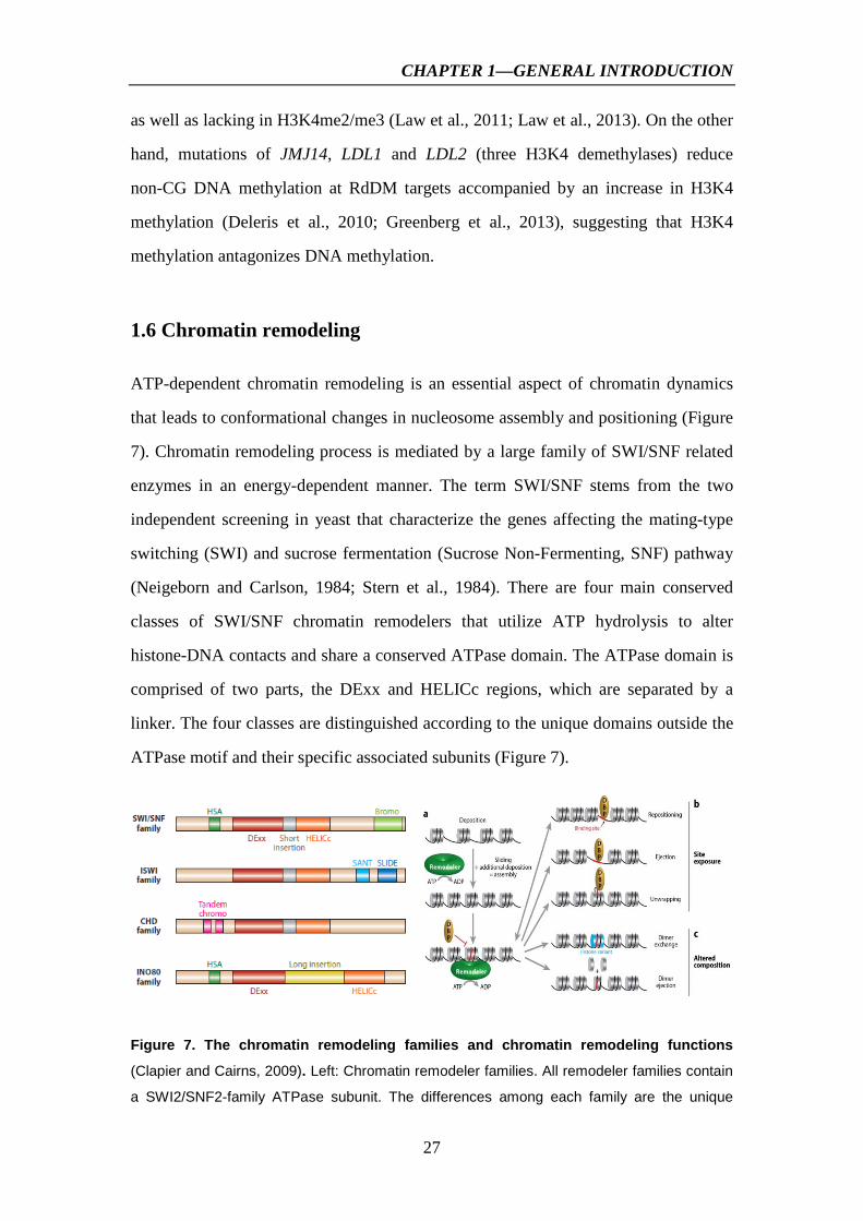
1.6 Chromatin remodeling
ATP-dependent chromatin remodeling is an essential aspect of chromatin dynamics
that leads to conformational changes in nucleosome assembly and positioning (Figure
7). Chromatin remodeling process is mediated by a large family of SWI/SNF related
enzymes in an energy-dependent manner. The term SWI/SNF stems from the two
independent screening in yeast that characterize the genes affecting the mating-type
switching (SWI) and sucrose fermentation (Sucrose Non-Fermenting, SNF) pathway
(Neigeborn and Carlson, 1984; Stern et al., 1984). There are four main conserved
classes of SWI/SNF chromatin remodelers that utilize ATP hydrolysis to alter
histone-DNA contacts and share a conserved ATPase domain. The ATPase domain is
comprised of two parts, the DExx and HELICc regions, which are separated by a
linker. The four classes are distinguished according to the unique domains outside the
ATPase motif and their specific associated subunits (Figure 7).
Figure 7. The chromatin remodeling families and chromatin remodeling functions
(Clapier and Cairns, 2009). Left: Chromatin remodeler families. All remodeler families contain
a SWI2/SNF2-family ATPase subunit. The differences among each family are the unique

CHAPTER 1—GENERAL INTRODUCTION
28
domains residing within, or adjacent to, the ATPase domain. Right: The different outcomes of
ATP-dependent chromatin remodeling. Remodelers can assist in chromatin assembly
generating room for additional deposition (a). Remodeler action on a nucleosome array results
in two categories: (b) site exposure, in which a site (red) for a DNA-binding protein (DBP)
becomes accessible by nucleosomal sliding (repositioning), or nucleosomal eviction (ejection)
or localized unwrapping, and (c) altered composition, in which the nucleosome content is
modified by dimer replacement or through dimer eviction.
SWI/SNF chromatin remodelers have been mostly conserved through
evolution across yeast, animals and plants kingdoms (Flaus et al., 2006; Narlikar et al.,
2013). Based on the presence of ATPase/helicase domain, there are 42 potential
SWI/SNF2 chromatin remodelers (http://www.chromdb.org/index.html) in
Arabidopsis. Those that have been studied are indicated in Figure 8.
Figure 8. Phylogenetic relationship and domain structure of the major SNF2 family members in Arabidopsis (Gentry and Hennig, 2014).
1.6.1 SWI/SNF class—SYD, BRM and MINU1/2 in Arabidopsis
The SWI/SNF class contains a bromodomain at the C-terminal, that binds to
acetylated N-terminal histone region, besides a helicase-SANT (SWI3, ADA2, NcoR,
TFIIIB) domain and an ATPase domain (Hassan et al., 2002; Martens and Winston,
2003). This is consistent with the fact that HAT complexes stabilize SWI/SNF
nucleosome binding at the promoter region (Hassan et al., 2001). This family has
many activities, for instance it slides and evicts nucleosome at many loci but lacks

CHAPTER 1—GENERAL INTRODUCTION
29
roles in chromatin assembly (Clapier and Cairns, 2009). Proteins in this class include
SMARCA4 (BRG1) and SMARCA2 (BRM) in human, Brahma in Drosophila and
Swi2/Snf2 and Sth1 in S. cervisiae. These proteins are mostly identified as
transcriptional activators and several studies reveal the sequential recruitment of HAT
and SWI/SNF complexes to promote transcription (Yudkovsky et al., 1999; Dilworth
et al., 2000). However, there is also evidence showing that SWI/SNF complex directly
repress transcription of some genes (Martens and Winston, 2003).
Arabidopsis SPLAYED (SYD/CHR3), BRAHMA (BRM/CHR2),
MINU1/CHR12 and MINU2/CHR23 belong to the SWI/SNF class. SYD and BRM
(3574 and 2193 amino acids, respectively) are large proteins; MINU2 and MINU3
(1132 and 1054 amino acids, respectively) are significantly smaller. Only BRM has a
C-terminal region that resembles a bromodomain domain, which is found to target
remodeling complexes to hyperacetylated chromatin in yeast (Kasten et al., 2004;
Jerzmanowski, 2007).
Loss of function alleles of SYD were first identified by genetic screening for
mutants that enhanced the lfy phenotype (Wagner and Meyerowitz, 2002). LEAFY
(LFY) is a meristem identity gene that is required for switching from vegetative to
reproductive development (Weigel et al., 1992). syd null mutants exhibit pleiotropic
developmental defects including stem cell maintenance, patterning (alteration of leaf
polarity, flower morphogenesis and ovule development), developmental transitions
(precocious onset of reproductive development) and growth (small stature, slow
growth and reduced apical dominance) (Wagner and Meyerowitz, 2002). SYD
represses LFY activity prior to the floral transition, whereas it functions as a
co-activator with LFY in the transcriptional regulation of class B and C floral
homeotic genes (Wagner and Meyerowitz, 2002). SYD is required for reproductive
shoot apical meristem (SAM) by directly regulating the expression of WUS (Kwon et
al., 2005).

CHAPTER 1—GENERAL INTRODUCTION
30
BRM was originally identified by a genetic screen for mutants that
exacerbated the defect of cotyledon separation in cuc2 (Kwon et al., 2006). syd and
brm null mutants exhibit both similar and distinct developmental defects, indicating
partially functional redundancy. For example, both single mutants share short stature
and delayed growth. The double mutant syd brm reveals embryo lethality but the
single not (Bezhani et al., 2007). Although both are required for flower patterning and
cotyledon separation, BRM and SYD may control different molecular events. During
the formation of cotyledon separation, BRM upregulates the transcription of all three
CUC genes, whereas SYD only upregulates the expression of CUC2 (Kwon et al.,
2006). During the initiation of flower patterning, the temporal recruitment of SYD to
AP3 and AG is similar to LFY, whereas the temporal binding of BRM is different
(Wu et al., 2012). In addition, BRM has also some specific role. For example, brm
mutants strongly inhibit the primary root elongation and promote more lateral roots
and secondary root branches. Some studies reveal that the phenotype of brm mutant is
similar to that of swi3c, suggesting that they may work in the same complex, which is
confirmed by yeast two hybrid assays (Farrona et al., 2004; Sarnowski et al., 2005;
Hurtado et al., 2006; Archacki et al., 2009).
MINU1 and MINU2, other two SNF2/SWI smaller members, also have
redundant roles in plant development. No significant phenotype has yet been
characterized in single mutant, however, the double mutants show embryonic lethality
which is associated with increased expression of WUSCHEL and WOX5 (Sang et al.,
2012).
1.6.2 ISWI class—CHR11 and CHR17 in Arabidopsis
The ISWI class takes its name from the Drosophila ISWI (Imitation Switch). In
Drosophila, this unique member can belong to three different complexes: NURF
(nucleosome-remodeling factor), CHRAC (chromatin-accessibility complex) and
ACF (ATP-dependent chromatin assembly and remodeling factor) (Tsukiyama and
Wu, 1995; Ito et al., 1997). All have chromatin remodeling activities and participate

CHAPTER 1—GENERAL INTRODUCTION
31
in transcription regulation (Corona and Tamkun, 2004). The ISWI members possess a
SANT adjunct to a SLIDE domain (SANT-like ISWI) at the C-terminus, both of
which together form a nucleosome recognition module that binds to an unmodified
histone tail and DNA component of nucleosomal substrates (Boyer et al., 2004).
Many ISWI complexes (ACF, CHRAC) optimize nucleosome spacing to promote
chromatin assembly and repress the gene transcription. However, certain complexes
(NURF) can randomize spacing, which can assist in RNAPII transcription (Clapier
and Cairns, 2009, 2012).
CHR 11 and CHR17 encode two ISWI proteins in Arabidopsis. Both have
conserved domains, HAND, SANT and SLIDE in the C-terminal. CHR11 is highly
expressed in sporophytic and gametophytic tissues during reproductive development
(Huanca-Mamani et al., 2005). Therefore, CHR11 is shown to be required for haploid
nuclear proliferation during megagametogenesis and cell expansion during the
sporophytic phase (Huanca-Mamani et al., 2005). One study shows that CHR11 and
CHR17 physically interact with RLT1 and RLT2, two plant-specific DDT-domain
containing proteins, together in preventing vegetative-to-reproductive transition by
regulating several key genes such as FT, SEP3 (Li et al., 2012). It is also found that
the SLIDE domain of CHR11 and the DDT domain of RLT1 together with an
adjacent sequence are responsible for the interaction (Dong et al., 2013). 1.6.3 INO80 class —INO80 and PIE1 in Arabidopsis
INO80 class of chromatin remodelers was first identified from S. cerevisiae which is
required for growth in absence of inositol (Ebbert et al., 1999). There are two INO80
members Ino80 and Swr1 in yeast, and three homologous genes INO80, SRCAP
(SNF2-related CREB-activator protein) and p400 in humans. One of the structural
characteristics of this class is a large insertion in the middle of ATPase domain, to
which the helicase-related (AAA-ATPase) Rvb1/2 proteins and one actin-related
protein (ARP) bind. INO80 complex has different functions including transcription
activation and DNA repair (Shen et al., 2000). Although SWR1 is highly related to

CHAPTER 1—GENERAL INTRODUCTION
32
INO80, it is unique in reorganizing nucleosome by recruitment and exchange of
H2A.Z variant (Krogan et al., 2003; Mizuguchi et al., 2004).
Arabidopsis encodes one homolog of Ino80 protein. Ino80 mutant reduces the
homologous recombination (HR) frequency to 15% of that in wild type. However it
does not seem to affect other DNA repair pathways (Fritsch et al., 2004). This is
unlike the situation of yeast Ino80 that is involved in both HR and non-HR DNA
repair events. Like yeast Ino80, Arabidopsis INO80 regulates a subset of genes not
functionally related to HR, suggesting its dual role in transcription and DNA repair
(Fritsch et al., 2004). Swr1 is required for replacement of histone H2A with the
H2A.Z variant in yeast. PIE1 (PHOTOPERIOD-INDEPENDENT EARLY
FLOWERING 1) encodes the single representative of SWR1 gene in Arabidopsis. pie
is originally identified as a suppressor of FRIGIDA-dependent late flowering (Noh
and Amasino, 2003). Similar to homolog in yeast, PIE1 as part of SWR complex is
responsible for deposition of H2A.Z at multiple loci (Choi et al., 2007; March-Diaz et
al., 2008). It is also found that PIE1 plays important roles in somatic DNA repair and
during meiosis (Rosa et al., 2013). Arabidopsis characterized SWI/SNF2 remodelers
are summarized in Table 4.
Table 4. Summary of the major SWI/SNF2 chromatin remodelers and their mutant phenotypes in Arabidopsis (Gentry and Hennig, 2014).

CHAPTER 1—GENERAL INTRODUCTION
33
1.6.4 CHD class
CHD chromatin remodeling proteins is named based on the three characteristic
domains: an N-terminal pair of Chromodomains, a central Helicase-like ATPase
motor, and a C-terminal DNA binding domain (Lusser and Kadonaga, 2003). The
chromodomain is a well conservedancient structural motif, since it can be found in a
vast array of organisms as diverse as protists, plants, amphibians, and mammals
(Eissenberg, 2001). Yeast has only one CHD protein (Chd1), whereas other high
organisms such as fly, human and plants have several CHD proteins, some of which
can be in large functional complex. Based on the structure and function, CHD
proteins have been broadly divided into three separate subfamilies (Hall and Georgel,
2007). Subfamily I proteins (CHD1 and CHD2 in humans) are the prototypical
examples of the family and do not contain additional identified functional domains.
Subfamily II (CHD3 and CHD4) is characterized by the presence of plant
homeodomain (PHD)-zinc-finger domain and in the absence of a clear DNA-binding
domain (Hall and Georgel, 2007). Subfamily III (CHD 5-9 in humans) is less defined
structurally and far less studied compared to the other two subgroups.
Besides CHD proteins, the chromodomain has also been identified in other
chromatin associated proteins such as HP1 (Brasher et al., 2000) and Polycomb
(Ingram et al., 1999; Brehm et al., 2004). The chromodomain of human CHD1 binds
to H3K4me2 or me3 (Flanagan et al., 2005; Sims et al., 2005). In contrast, the
chromodomain of Mi-2 (CHD3) seems to recognize DNA rather than methylated
histone tails (Brehm et al., 2004). The ATPase domain is required for the remodeling
of nucleosomes through histone displacement (Durr and Hopfner, 2006) or histone
octamer sliding (Becker and Horz, 2002). The ATP hydrolysis energy is transduced
into the conformational stress necessary to remodel chromatin (Pazin and Kadonaga,
1997). Thus the ATPase domain serves as a DNA-translocating motor to break
histone-DNA contacts and release histones from nucleosomes (Durr and Hopfner,
2006). Of the 3 defined domains for CHD proteins, the DNA-binding domain is by far
the least conserved. This domain is fairly well defined in CHD1, whereas CHD3/4, as

CHAPTER 1—GENERAL INTRODUCTION
34
part of the Nucleosome remodeling and deacetylase (NuRD) complex, may also gain
DNA binding ability through interactions with other subunits of the complex.
Moreover, the PHD domains of CHD3/4 can bind histone H3 and affect interactions
with nucleosomes (Bienz, 2006). Some CHD remodelers have the ability to slide or
evict nuleosomes to promote transcription, however, others play repressive roles such
as vertebrate Mi-2/NuRD complex, which contains histone deacelyases (HDAC1/2)
and methyl CpG-binding domain (MBD) proteins (Hall and Georgel, 2007).
1.6.4.1 The function of CHD1 protein
The subfamily I member CHD1 was initially thought to be integral to transcriptional
activity. As mentioned above, the chromodomain of human CHD1 was found to
specially bind to H3K4me3. Presently, there is in vitro evidence both for (Pray-Grant
et al., 2005) and against (Sims et al., 2005) Chd1 binding to H3K4me in budding
yeast S. cerevisiae. The fission yeast S. pombe Chd1 orthologues Hrp1 and Hrp3 may
interact and bind to H3K4me via the chromodomains of Hrp3, which have all the
consensus residues required for methyl-lysine binding (Opel et al., 2007). In
Drosophila melanogaster, the chromodomain of CHD1 is critical for chromatin
remodeling activity but is less important for localization to chromatin (Morettini et al.,
2011). The crystal structural and biochemical studies show an autoregulated domain
organization of S. cerevisiae Chd1 (Narlikar et al., 2013). The chromodomain
contacts the two lobes of ATPase domain and inhibits the ATPase motor (Hauk et al.,
2010). In contrast, the DNA binding element, containing homology to SANT and
SLIDE domains, contributes to positively regulate ATPase activity (McKnight et al.,
2011). In vitro assays reveal that Chd1 has the ability to assemble, remodel, slide and
promote regular spacing of nucleosomes (Lusser et al., 2005; Stockdale et al., 2006).
Chd1 plays important roles in transcription processes including initiation,
elongation and termination (Alen et al., 2002; Simic et al., 2003; Biswas et al., 2007;
Quan and Hartzog, 2010). It has been shown that human CHD1 associates with the
pre-initiation transcription complex through interactions with Mediator in vitro (Lin et

CHAPTER 1—GENERAL INTRODUCTION
35
al., 2011). Yeast Chd1 is found to be a factor required for remodeling the nucleosomal
PHO5 promoter and for transcriptional activation of the gene (Ehrensberger and
Kornberg, 2011) . In fly and yeast, CHD1 is localized to a transcriptionally active
gene and physically interacts with elongation factors and RNA Polymerase II (Simic
et al., 2003; Srinivasan et al., 2005). Yeast Chd1 is also an essential factor for
chromatin structure at the 3′ ends of genes at transcription, and transcriptional
termination fails to occur in its absence (Alen et al., 2002).
CHD1 is a chromatin remodeler which participates in diverse biological
processes. In Drosophila, although chd1-mutant zygotes are viable and display only a
mild notched-wing phenotype, chd1-null male and female are sterile (McDaniel et al.,
2008). In fact, maternal Chd1 in Drosophila is required for incorporation of H3.3 into
the male pronucleus during early development after fertilization. Failure to
incorporate H3.3 may render the paternal genome unable to participate in mitosis in
the zygote, resulting in non-viable haploid embryos (Konev et al., 2007). In mice,
Chd1 is essential for embryonic stem (ES) cell pluripotency and the formation of
induced pluripotent stem cells (Gaspar-Maia et al., 2009). Chd1 mice mutants show
an increase in the heterochromatic mark H3K9me3 and the ES cells display a
tendency towards neuronal differentiation (Gaspar-Maia et al., 2009). CHD1 is also
found to work as a tumor suppressor, as deletion or mutation of Chd1 is associated
with prostate cancer, with cells displaying an increase in invasiveness (Huang et al.,
2012).
Recent study points to a critical role for Chd1 in nucleosome positioning in
vivo. As mentioned above, nucleosomes typically show a stereotypic organization
over most genes: a nucleosome free region just upstream of the TSS followed by a
regular nucleosomal array over the coding region. However, in the absence of S.
cerevisiae Chd1, the nucleosome positioning is largely lost over gene bodies
(Gkikopoulos et al., 2011). Specially, NFR and +1 nucleosome over the transcribed
regions are minimally affected, but downstream nucleosomes (particularly those
starting at the +3 nucleosome) were dramatically delocalized in chd1 mutants (Figure

CHAPTER 1—GENERAL INTRODUCTION
36
9) (Gkikopoulos et al., 2011). This is consistent with in vitro data that Chd1 is able to
space nuclesome arrays on plasmid DNA in vitro (Lusser et al., 2005). A similar
situation is observed in S. pombe, as the deletion of Chd1 homologs, Hrp1 and Hrp3,
interrupts nucleosome spacing (Figure 9) (Hennig et al., 2012; Pointner et al., 2012;
Shim et al., 2012).
Distance from TSS (bp) Distance from TSS (bp)
Figure 9. Nucleosome organization is disrupted upon deletion of CHD1. Left: nucleosome organization in budding yeast S. cerevisiae (Gkikopoulos et al., 2011). Right: nucleosome positioning in fission in yeast S. pombe (Hennig et al., 2012).
In S. cerevisiae, loss of Chd1 impairs nucleosome reassembly coupled with
H2B monoubiquitination (Lee et al., 2012). Loss of Chd1 results in a substantial loss
of H2Bub levels, and nucleosomal occupancy is reduced in gene bodies in both chd1
and h2b mutants, suggesting that Chd1 may positively regulate gene expression
through promoting nucleosome reassembly coupled with H2B monoubiquitination
(Lee et al., 2012). Consistent with a role for Chd1 in maintaining chromatin structure
at active genes, Chd1 is also reported to prevent cryptic transcription within gene
bodies in S. pombe, as the deletion of hrp1 and hrp3 causes strong, genome-wide
accumulation of antisense transcripts (Hennig et al., 2012; Shim et al., 2012).
Furthermore, Chd1 is also involved in histone exchange in gene body in Drosophila
and yeast (Radman-Livaja et al., 2012; Smolle et al., 2012). It is possible that
irregular spacing of nuclesomes in chd1 mutants renders it prone to dissociation as a
result of collisions between and adjacent nucleosomes. Transient dissociation of
histones provides an opportunity for exchange with the soluble pool of nascent
histones (Radman-Livaja et al., 2012; Smolle et al., 2012; Narlikar et al., 2013).

CHAPTER 1—GENERAL INTRODUCTION
37
Genome-wide mapping of S. cerevisiae Chd1 binding on native chromatin
shows that Chd1 is highly enriched in NFRs, although it acts in activated gene body
(Gkikopoulos et al., 2011; Zentner et al., 2013). In light of these data, Chd1 plays
essential roles in regulating nucleosome dynamics during transcription at different
levels as shown in Figure 10.
Figure 10. A model for Chd1 in regulating nucleosome dynamics during transcription.
1.6.4.2 Plant CHD proteins—PKL, PKR1, PKR2 and CHR5
Phylogenetic analysis reveals that there is only one CHD1 protein (CHR5) in the
genome of Arabidopsis, no CHD2 subfamily protein exists in plants, in contrast, three
members belong to CHD3 subfamily (Figure 11) (Hu et al., 2012). In plant, CHD1
protein (CHR5) has not been studied.
Among plant CHD3 members, PICKLE (PKL) is the most studied. The
hallmark of pkl seedlings is a green, tuberous and opaque region at the primary root,
referred to as the “pickle root” (Figure 12) (Ogas et al., 1997). Pickle primary roots
can express numerous embryonic differentiation characteristics such as expression of
storage protein genes and accumulation of storage lipids and undergo spontaneous
somatic embryogenesis (Ogas et al., 1997; Henderson et al., 2004). This pickle root
phenotype is related to fail to repress embryo identity genes, such as LEC1, LEC2,

CHAPTER 1—GENERAL INTRODUCTION
38
ABI3, and FUS3 after seed germination (Figure 12) (Ogas et al., 1999; Zhang et al.,
2008; Aichinger et al., 2009; Zhang et al., 2012).
Figure 11. Relationship and structure of CHD proteins. Left: phylogenetic tree of CHD proteins (Hu et al., 2012). Right: structure and size of plant CHD proteins (Gentry and Hennig, 2014).
Figure 12. The phenotype of pkl. Left: “pickle root” phenotype (Ogas et al., 1997); Right: embryo identity genes are upregulated in pkl seedlings (Aichinger et al., 2009).
PKL contributes to other developmental process in addition to repression of
embryonic traits. PKL plays a role in repression of ectopic stipules and meristems in
leaf tissue (Hay et al., 2002). PKL is involved in repressing meristematic genes in
carpel tissue (Eshed et al., 1999). Loss of PKL results in hypersensitivity to cytokinin
with respect to callus growth and greening (Furuta et al., 2011). PKL is also required
for proper root development and has been found to work in two somewhat opposing

CHAPTER 1—GENERAL INTRODUCTION
39
ways: PKL is a negative regulator of auxin-mediated lateral root initiation by
regulating ARF7/9 transcription (Fukaki et al., 2006) and yet also promotes root
growth by increasing expression of root stem cell and meristem marker genes
(Aichinger et al., 2011). Recently, PKL is found to be a repressor of
photomorphogenesis by interacting with HY5 to regulate cell elongation-related genes
in hypocotyls (Jing et al., 2013).
1.6.4.3 The mechanism of PKL regulating embryonic genes
In contrast to animal CHD3 proteins functioning in histone deacetylation,
comparative genomic analyses reveal that PKL acts to promote trimethylation of
histone H3 lysine 27 (H3K27me3) (Zhang et al., 2008; Zhang et al., 2012). As
mentioned above, H3K27me3 is a repressive mark for gene expression in both plants
and animals (Zheng and Chen, 2011). PRC2 is the main factor responsible for
catalyzing trimethylation of H3K27 (Schwartz and Pirrotta, 2007; Schmitges et al.,
2011). In Arabidopsis, mutants of PRC2 genes substantially reduce H3K27me3 levels
and exhibit profound developmental defects including the derepression of embryonic
traits (Chanvivattana et al., 2004; Schubert et al., 2006; Bouyer et al., 2011). Presently,
it is controversial how PKL represses embryo identity genes. Some results support
that PKL directly associates and represses the embryonic genes by regulating
H3K27me3 on the loci. In particular, PKL was found to present at the promoters of
LEC1 and LEC2 during germination (Zhang et al., 2008; Zhang et al., 2012). Whereas,
other results suggest that PKL represses embryonic traits in an indirect way. In this
case, PKL, like a transcriptional activator, binds to and up-regulates PRC2 genes
rather than embryonic genes (Aichinger et al., 2009).
1.7 Embryo development
Seed development is an intriguing, specific and crucial phase of angiosperm plant life
cycle, which begins with a double fertilization process that occurs within the ovule
and ends with a dormant seed primed to become the next plant generation. Embryo

CHAPTER 1—GENERAL INTRODUCTION
40
development consists of the morphogenesis and maturation phases as shown in Figure
13. Morphogenesis refers to the differentiation of cell fates in initially equivalent cells,
which results in an embryo established with the specification of the shoot root axis
and the formation of discernible major organ and tissue systems (Capron et al., 2009).
Figure 13. Embryo development in seed plants (Braybrook and Harada, 2008).
Maturation, the second phase of zygotic embryogenesis, occurs exclusively in
seed plants. Upon entering the maturation stage, embryo ceases cell division and
continues growing by cell expansion filling up the entire seed. One of the
characteristics of this phase is the synthesis and accumulation of carbohydrates, lipids,
and seed storage proteins (SSP), which will provide nutrients to the growing seedling
before the photosynthetic capacity is fully acquired (Miquel and Browse, 1998). The
embryo also accumulates late embryogenesis abundant (LEA) proteins, dehydrins,
and osmocompatible solutes, serving to protect the embryo from desiccation (Ingram
and Bartels, 1996). Developmental arrest and the ability to withstand drying enable
the embryo to remain in a quiescent or dormant state until the seed encounters a
favorable set of conditions that promote germination (Bewley, 1997).
1.7.1 LEC1/AFL transcription factors
Genetic studies have uncovered a number of factors that act as positive regulators of
seed developmental programs. Among them, LEC1/AFL (ABI3, FUS3, LEC2), as the
main regulators, have been characterized at greater depth. The LEC and FUS3 were
originally identified as regulators of cotyledon identity, as recessive mutations in
these genes partially convert the cotyledons into vegetative leaves (Figure 14), hence,
the name LEAFY COTYLEDONS (Meinke, 1992; Meinke et al., 1994). The name

CHAPTER 1—GENERAL INTRODUCTION
41
FUSCA is derived from Greek meaning ‘to darken’, and is designated to mutant loci
with dark seeds (Figure 14) (Keith et al., 1994). The dark color of the seed is due to
the accumulation of anthocyanins in the embryo. Recessive mutations in abi3 were
originally identified in screens aiming to identify mutants insensitive to ABA during
germination (Koornneef et al., 1984). The LEC1/AFL genes, as the master regulators
of late embryogenesis and seed maturation, share overlapping but not identical mutant
phenotypes (Parcy et al., 1997; Vicient et al., 2000; Harada, 2001). Mutants of these
genes exhibit reduced dormancy and precocious germination of immature seeds.
LEC1/AFL can trigger the expression of genes of seed storage protein (SSP) and the
oil body protein, as a consequence, the seeds of the mutants are desiccation intolerant
and non dormant except lec2 embryo (Giraudat et al., 1992; Keith et al., 1994;
Meinke et al., 1994; Stone et al., 2001). Lec2 mutants are desiccation tolerant, do not
germinate precociously, and show a pattern of storage product accumulation closer to
wild type than to other mutants (Keith et al., 1994; Meinke et al., 1994). Only abi3
mutants show ABA insensitivity and defects in chlorophyll degradation (Giraudat et
al., 1992). The lec1, lec2 and fus3 mutations cause partial conversion of cotyledons
into leaf-like state by the development of trichomes and the partial leaf ultrastructure
(Keith et al., 1994; Meinke et al., 1994). The differences of lec1 and fus3 embryos are
restricted mostly on the frequency of stomata and the position of anthocyanin
accumulation (Meinke, 1992; Keith et al., 1994). In general, the analyses of
LEC1/AFL mutations indicate these genes, as the main positive regulators during seed
maturation, have redundant and distinctive roles in embryo formation, cotyledon
identity, seed storage protein expression and desiccation tolerance.
Figure 14. Seed phenotype of mutants in lec1, abi3, fus3 and lec2 (Meinke, 1992; Keith et al., 1994; To et al., 2006).

CHAPTER 1—GENERAL INTRODUCTION
42
1.7.1.1 Structure and targets of LEC1/AFL genes
LEC1/AFL genes encode two different classes of transcriptional factors. ABI3, FUS3
and LEC2 belong to the B3-domain transcription factor family, which are only found
in plants. The conserved B3 domains have similar DNA binding specificities and
recognize the RY/Sph I cis-regulatory motif (purine-pyrimidine, CATGCA as the
core sequence), which is found in the many seed storage protein genes (Kroj et al.,
2003; Monke et al., 2004; Roschzttardtz et al., 2009). It explains why these regulators
have partially redundant roles, as they are likely to share downstream targets. In
contrast, LEC1 encodes a protein homologous to HAP3 subunit of CCAAT-binding
protein complex (CBF), which is conserved among eukaryotes (Lotan et al., 1998;
Lee et al., 2003). LEC1 is able to activate the expression of cruciferin C (CRC) seed
storage protein when co-expressed with ABRE-binding bZIP transcription factors
(Yamamoto et al., 2009). In general, LEC1/AFL proteins act through direct
interactions with regulatory elements present in the promoters of maturation-related
genes to control the various features of seed development.
1.7.1.2 Expression profiles of LEC1/AFL genes
In agreement with their embryonic mutant phenotypes, LEC1/AFL genes are primarily
expressed in embryos as shown in Figure 15. LEC1 is expressed in seed (in both
embryo and endosperm). Specially, LEC1 mRNA is present at the protoderm and is
detected throughout the developing embryo by the bent-cotyledon stage (Lotan et al.,
1998). LEC2 is expressed throughout the embryo in early stages but primarily to the
hypocotyl and the embryo axis at later stages (Kroj et al., 2003; To et al., 2006). ABI3
is expressed broadly in embryo tissues throughout embryogenesis except the root
meristem (To et al., 2006). FUS3 transcripts can be detected in apical region and
suspensor of the globular early heart stage, as well as in the vasculature, root tip,
aleurone of the mature embryo (Gazzarrini et al., 2004; Tsuchiya et al., 2004). LEC2
and ABI3 are also expressed in vegetative tissues, where ABI3 is involved in lateral
meristem development (Rohde et al., 2000; Stone et al., 2001; Kroj et al., 2003; To et

CHAPTER 1—GENERAL INTRODUCTION
43
al., 2006). LEC1/AFL genes also have distinctive temporal expression profiles (Figure
15). LEC1 is expressed at early stages of embryogenesis, followed by LEC2 (Stone et
al., 2001; Kroj et al., 2003). FUS3 and ABI3 expression levels peak after the
maximum of LEC2, with FUS3 levels decreasing before maturation and ABI3 levels
remaining high throughout the late embryo development (Parcy et al., 1994; Luerssen
et al., 1998).
Figure 15. Spatial and temporal expression pattern of LEC1/AFL in developing embryos. Left: Tissue-specificity of LEC1/AFL genes in embryo which are indicated in red. (Santos-Mendoza et al., 2008). Right: temporal expression pattern from the website http://bbc.botany.utoronto.ca/efp/cgi-bin/efpWeb.cgi. (3 d, globular; 4 d, heart;5 d, torpedo; 7 d, walking stick; 9 d, curled cotyledons; 10 d, green cotyledons).
1.7.2 A network of interacting LEC1/AFL factors
Genetic analysis has revealed the existence of a complex interacting network among
the LEC1/AFL genes. According to a previously established model (To et al., 2006),
LEC1 and LEC2 act upstream of ABI3 and FUS3, and promote their expression. LEC1
and LEC2 also regulate each other. ABI3 and FUS3 also mutually regulate their own
expression (Figure 16) (To et al., 2006). However, these functional interactions may
be of direct or indirect effects and the molecular mechanism still remains to be
elicited. It was reported that LEC2 binds specially to the RY sequence on the
promoter of AGAMOUS-Like15 (AGL15) in vitro and activates AGL15 expression in
vivo (Figure 16) (Braybrook et al., 2006). AGL15 is a MADS domain transcriptional
regulator accumulating in embryos (Wang et al., 2002). By using ChIP-chip approach,
it was shown that AGL15 directly targets to LEC2, FUS3, ABI3 and activates their

CHAPTER 1—GENERAL INTRODUCTION
44
expression (Figure 16) (Zheng et al., 2009). Similarly, it was confirmed that FUS3
directly binds to LEC1, ABI3, AGL15 and itself in vivo, and upregulates these target
gene transcription levels (Figure 16) (Wang and Perry, 2013). The cross-regulation
between these embryonic regulators partially explains the complex and overlapping
phenotypes displayed by each mutant. Figure 16. A model for LEC1/AFL regulatory interaction network during embryogenesis.
1.7.3 Epigenetic regulation of LEC1/AFL genes
Genetic analysis has revealed the importance of chromatin modification in the control
of the expression of LEC1/AFL genes. As discussed above, PKL, the CHD3
chromatin remodeling factor, acts as a transcriptional repressor for LEC1/AFL genes
during germination and vegetative phases. pkl mutant displays embryonic trait in the
primary roots (Ogas et al., 1997). PKL-dependent repression of LEC1/AFL genes, to
some extent, is associated with regulation of H3K27me3 levels (Zhang et al., 2008;
Aichinger et al., 2009; Zhang et al., 2012). VP1/ABI3-LIKE (VAL, also known as
HSI) 1-3 proteins also play negative roles in seed developmental programs (Suzuki et
al., 2007). VAL genes encode B3 transcription factors, containing PHD-like and CW
domains often associated with chromatin factors (Perry and Zhao, 2003; Bienz, 2006).
Double mutants of val1val2 show strong embryonic phenotype including callus
proliferation in shoot and root regions as well as arrested shoot apical meristem
development that prevents formation of leaves and progression to vegetative
development (Suzuki et al., 2007). The embryonic phenotype of val1val2 is associated

CHAPTER 1—GENERAL INTRODUCTION
45
with de-repression of LEC1/AFL genes in seedlings. The VAL genes are
independently identified as sugar signaling factors (HIGH-LEVEL EXPRESSION OF
SUGAR-INDUCIBLE GENE2 (HSI2/VAL1), HSI2-LIKE1 (HSL1/VAL2), and
HSI2-LIKE2 (HSL2/VAL3) (Tsukagoshi et al., 2005; Tsukagoshi et al., 2007).
Interestingly, in the hsi2 hsl1 background the expression of AFL genes can be induced
by sugar, suggesting that the AFL genes may be involved in sugar signaling
(Tsukagoshi et al., 2007). Recently, it is reported that H3K27me3 levels of LEC1,
FUS3, ABI3 are reduced in val1val2, accompanied with the decreased levels of
H2Aub, indicating that VAL proteins interact with Polycomb Repressive complex1
(PRC1) and recruit PRC1-mediated H2Aub to initiate repression of embryonic genes
after germination (Yang et al., 2013).
LEC1/AFL genes are epigenetically repressed after germination by both PRC1
and PRC2 via histone modifications. As mentioned before, PRC2 mutants clf swn and
pie result in neoplastic, callus-like structures during vegetative phase because of the
elevated expression of LEC2, FUS3, ABI3 (Chanvivattana et al., 2004; Bouyer et al.,
2011). Arabidopsis PRC1, constituted of two RING1 proteins (AtRING1a and
AtRING1b), Like Heterochromatin1 (LHP1), and three BMI proteins (AtBMI1a,
AtBMI1b and AtBMI1c), functions in the modification of histone H2A
mono-ubiquitination (H2Aub) (Sanchez-Pulido et al., 2008; Bratzel et al., 2010; Chen
et al., 2010). Similar to PRC2 mutants (clf swn, fie), the double mutants Atring1a
Atring1b, Atbmi1a Atbmi1b show the embryonic traits in somatic tissues, which is
associated with increased LEC1/AFL gene expression, indicating that PRC1 represses
the embryonic program after germination (Bratzel et al., 2010; Chen et al., 2010).
Recently, it is shown that H3K27me3 at LEC1/AFL genes are strongly reduced in val1
val2 and atbmi1a atbmi1b atbmi1c, while H2Aub level at these genes is significantly
decreased in val1 val2 but not in emf1 and clf swn, indicating that VAL proteins and
AtBMI1-mediated H2Aub first initiate repression of seed maturation genes and then
the repression is maintained by PRC2-mediated H3K27me3 (Figure 17) (Yang et al.,
2013).

CHAPTER 1—GENERAL INTRODUCTION
46
Figure 17. Proposed model for the PcG-mediated repression of seed maturation genes after germination (Yang et al., 2013).
On the other hand, the histone deacetylase hda6 hda19 double mutants show
growth arrest and somatic embryo formation, and this embryonic phenotype can be
suppressed by the lec1, abi3 and fus3 mutations suggesting that LEC1, ABI3 and
FUS3 expressions are repressed by HDA6 and HDA19 (Tanaka et al., 2008).
Recently, it is found that HDA19 interacts with HSL1 (VAL2) in vitro and in vivo
(Zhou et al., 2013). HDA19 could directly bind to the chromatin of the seed
maturation genes and the ectopic expression of these genes in hda19 seedlings is
associated with increased levels of H3ac, H4ac and H3K4me3 and decreased levels of
H3K27me3, similar to that of hsl1. These results suggest that HDA19 and HSL1 may
act together to repress seed maturation gene expression during germination. The
embryonic lethal phenotype of hsl1 hda19 double mutants also indicates that HDA19
and HSL1 may play a vital role during embryogenesis (Zhou et al., 2013).
LEC2 and FUS3 expressions are also regulated by microRNA, as mutation of
DICER-LIKE1 leads to premature expression of the maturation program during early
embryogenesis accompanied with elevated levels of LEC2 and FUS3, indicating the
microRNA processing machinery negatively regulates LEC2 and FUS3 transcription
during early embryogenesis (Willmann et al., 2011).
Recently, it is found that BRM, a SNF2 chromatin remodeling factor, has a
role in repressing ABI5 expression after seed germination and during vegetative
development and ABI3 during seed germination. BRM is strongly associated with the
region of ABI5 locus occupied by the +1 nucleosome. Loss of BRM activity leads to

CHAPTER 1—GENERAL INTRODUCTION
47
destabilization of a nucleosome at ABI5 locus, indicating that BRM represses ABI5
transcription by promoting high occupancy of the +1 nucleosome and by directing
this nucleosome from a more favorable predicted position to a position more proximal
to the TSS (Han et al., 2012).
In general, the above data have shown that epigenetic modifications play
important roles in the repression of LEC1/AFL genes especially during and after seed
germination. However, little is known concerning how these transcription factors are
activated during seed development. In particular, the mechanism of initiation and
establishment the activate state of LEC1/AFL genes during embryogenesis and
maturation process is not understood and remains to be elucidated.

CHAPTER 1—GENERAL INTRODUCTION
48
1.8 Objective and Organization of this thesis
Chromatin remodeling and histone modification play important roles in the
establishment and dynamic regulation of gene expression states. However, little is
known regarding to the regulatory mechanism of chromatin modification and
remodeling that control gene expression involved in plant development and responses
to environmental cues. My thesis work concerns the analysis of a chromatin
remodeling factor CHR5 and a histone demethylase gene JMJ15 for their function in
regulating chromatin structure or in resetting chromatin modifications that control the
expression of plant developmental and stress responsive genes.
The first result chapter (Chapter 2) aims to analyze the temporal and spatial
expression pattern and function of CHR5 in late embryogenesis and seed maturation
process. The key points of this part were (1) the expression pattern of CHR5 in plant
development; (2) characterization of chr5 T-DNA mutants; (3) the effect of CHR5
mutations on the LEC1/AFL transcription during embryogenesis; (4) the antagonistic
role between CHR5 and PKL in the control of LEC1/AFL gene expression; (5) the
chromatin mechanism of LEC1/AFL gene regulation by CHR5 and PKL during seed
maturation program.
The objective of the second part of results (Chapter 3) is to investigate the
biological and molecular function of JMJ15 in plant development. To examine the
biological function, JMJ15 tissue expression pattern and jmj15 mutants
(gain-of-function and loss-of-function mutants) were characterized. The effects of the
mutations on plant growth and development were evaluated. For the molecular
function, the transcriptome of jmj15 overexpression mutants was analyzed by
microarray followed by RT-PCR validation. The de-regulated genes were analyzed
for GO function category classification, for comparison with microarray data from
atx1/sdg2 mutants, and for enrichment for histone H3K4 modification. Finally a general discussion section is provided that discusses the main results
and outlines the perspectives of the work.

CHAPTER 2
Functional analysis of a chromatin remodeling factor CHR5 in Arabidopsis


CHAPTER 2—CHR5
49
Introduction
CHR5 belongs to the CHD1 subfamily of chromatin remodeling factors, the function
of which is not yet studied in plants. LEC1/AFL genes, as the master transcriptional
factors that control seed development, have been found to be expressed during
embryo development but repressed during vegetative phase by several negative
epigenetic regulators including PKL (a CHD3 chromatin remodeler). However, the
mechanism of activation of LEC1/AFL gene expression during embryogenesis is
unknown.
In this part, I first studied the expression pattern of CHR5 by using transgenic
plants containing proCHR5-GUS construct and RT-PCR approaches. The results
showed that CHR5 was highly specifically expressed during late embryogenesis. To
study CHR5 function in gene expression and plant development, I obtained 6 lines of
chr5 T-DNA insertion mutants and the complementation of a mutant allele with the
proCHR5-CHR5-HA fusion construct was obtained. The analysis showed that CHR5
was involved in the regulation of LEC1/AFL gene expression during embryo
development and seed maturation process. In order to analyze the relationship
between CHR5 and PKL in seed maturation gene expression, the double mutant chr5
pkl was generated and analyzed. The results indicate that CHR5 and PKL function
antagonistically in the control of LEC1/AFL transcription during seed developmental
and vegetative phases. The chromatin modifications of LEC1/AFL loci in chr5 and pkl
mutants were further detected, also confirming the opposite role of CHR5 and PKL in
LEC1/AFL expression. Finally, the mechanism by which CHR5 regulates LEC1/AFL
genes was studied. The key points of this were (1) if CHR5 directly associates with
LEC1/AFL genes during embryogenesis; (2) how CHR5 regulates the transcription
level of LEC1/AFL genes—nucleosome positioning, histone modification or others.
ChIP experiment proved that CHR5 directly binds to the promoter region of FUS3
and ABI3. At the same time, CHR5 was shown to reduce nucleosome occupancy near
the transcription start site (TSS) of FUS3.
This part of the results is written in an article that is currently submitted for
publication.

CHAPTER 2—CHR5
50
Chromodomain, Helicase and DNA-binding CHD1 and CHD3 proteins act antagonistically to regulate seed maturation program in Arabidopsis
Running title: Regulation of embryo genes by CHD proteins

CHAPTER 2—CHR5
51
Chromodomain, Helicase and DNA-binding CHD1 and CHD3 proteins act antagonistically to regulate seed maturation program in Arabidopsis
2.1 Abstract
Chromatin modification and remodeling are the basis for epigenetic regulation of gene
expression. LEAFY COTYLEDON1 (LEC1), LEAFY COTYLEDON 2 (LEC2),
ABSCISIC ACID-INSENSITIVE 3 (ABI3), and FUSCA3 (FUS3) are key regulators
of seed maturation and are expressed during embryo development but epigenetically
repressed after seed germination. The CHD3 protein PICKLE (PKL) is involved in
the epigenetic silencing of the genes. However, the chromatin mechanism that
establishes the active state of the genes during embryogenesis is not clear. We show
that the Arabidopsis CHD1-related gene, CHR5, displayed a similar expression
pattern as ABI3 and FUS3 during embryo development. The mutation of the gene led
to decreased expression of LEC1, ABI3 and FUS3 in developing seeds and reduced
seed storage protein accumulation. Analysis of double mutants revealed an
antagonistic function between CHR5 and PKL in embryo gene promoter activity and
seed storage protein accumulation. CHR5 was directly associated with the promoters
of ABI3 and FUS3 and chr5 mutations led to increased nucleosome occupancy near
the transcriptional start site. The results suggest that CHR5 is involved in nucleosome
occupancy to regulate embryo identity genes expression, which is counterbalanced by
PKL during embryo development.

CHAPTER 2—CHR5
52
2.2 Introduction
Chromatin modification and remodeling are the biochemical basis for epigenetic
regulation of gene expression. The nucleosome is the basic repeating unit of
chromatin and is composed of an octamer of histones and a segment of 147 bp DNA.
Histone modification such as histone lysine acetylation and methylation play
important roles for gene activity. Histone lysine acetylation is generally involved in
gene activation, whereas histone lysine methylation may have either a positive or a
negative effect on gene expression (Berger, 2007; Mosammaparast and Shi, 2010). In
plants tri-methylation of H3 lysine 4 (H3K4me3) is enriched at the 5’ end of actively
transcribed genes, whereas tri-methylation of H3 lysine 27 (H3K27me3) marks
repressed developmental and stress responsive genes (Liu et al., 2010; Li et al., 2013).
Chromatin remodeling involves altering histone-DNA contacts, sliding
nucleosomes, and exchanging or removing histones and entire nucleosomes.
Chromatin remodelers which are ubiquitous in eukaryotic cells (Flaus et al., 2006),
are compositionally and functionally diverse, but they all share the presence of a
subunit that belongs to the SNF2-like family of ATPases, among which are the
Chromodomain, Helicase/ATPase and DNA-binding domain (CHD) proteins (Lusser
and Kadonaga, 2003). Besides Saccharomyces cerevisiae that has only one CHD
protein (Chd1), other higher eukaryotes have several CHD members that are divided
into CHD1, CHD2 and CHD3 subfamilies (Hall and Georgel, 2007). The two
chromodomains in CHD1 proteins are essential for recognition of di- or tri-methylated
lysine 4 of histone H3 (H3K4me2/3) (Flanagan et al., 2005; Sims et al., 2005;
Vermeulen et al., 2010). CHD1 proteins are implicated in gene expression at different
levels (Ho and Crabtree, 2010). Recent results suggests that CHD1 proteins have both
chromatin assembly and remodeling functions to direct the positioning of
nucleosomes required for gene transcription (Gkikopoulos et al., 2011; Torigoe et al.,
2013). In addition to the double chromodomains and the helicase/ATPase, CHD3
subfamily proteins contain one or two Plant Homeodomain (PHD) fingers at the N-
terminus. CHD3 members in Drosophila melanogaster and mammalian cells are the
central components of the Nucleosome Remodeling and histone Deacetylase (NuRD)
or Mi-2 complexes regulating transcriptional repression (Hall and Georgel, 2007;
Ramirez and Hagman, 2009). However, other studies revealed that CHD3 members

CHAPTER 2—CHR5
53
can also function as a transcriptional coactivator (Saether et al., 2007; Murawska et al.,
2008).
Several CHD3 members are identified in plants (Hu et al., 2012). The
Arabidopsis thaliana (Arabidopsis) CHD3 protein PICKLE (PKL) was initially found
to repress embryonic traits after seed germination (Ogas et al., 1997). PKL functions
as a transcriptional repressor of embryo identity genes such as LEAFY COTYLEDON1
(LEC1), LEAFY COTYLEDON 2 (LEC2), ABSCISIC ACID-INSENSITIVE 3 (ABI3),
and FUSCA3 (FUS3) in seedlings (Ogas et al., 1999; Zhang et al., 2008; Aichinger et
al., 2009; Zhang et al., 2012). In contrast to animal CHD3 proteins, PKL is found to
promote trimethylation of histone H3 lysine 27 (H3K27me3) over target genes rather
than histone deacetylation (Zhang et al., 2008). But other results suggest that the PKL
protein may be a transcriptional activator required for the expression of genes
encoding Polycomb Repressive Complex2 (PRC2) that catalyzes trimethylation of
H3K27 (Aichinger et al., 2009). Recent data revealed that the rice (Oryza sativa)
CHD3 protein, CHR729, is a bifunctional chromatin regulator that recognizes and
modulates H3K4 and H3K27 methylation over repressed tissue-specific genes (Hu et
al., 2012). By contrast, only one CHD1 gene is found in the genome of Arabidopsis or
rice (Hu et al., 2012). Its function in plant gene expression and development remains
unknown.
LEC1 and the B3-domain ABI3, FUS3, and LEC2 (referred to as AFL) are
key transcriptional regulators of zygotic embryo development (Giraudat et al., 1992;
Lotan et al., 1998; Stone et al., 2001). These factors activate the seed maturation gene
expression program in a complex network (Kagaya et al., 2005; Santos-Mendoza et al.,
2008; Suzuki and McCarty, 2008; Monke et al., 2012; Wang and Perry, 2013). These
genes are expressed specifically during embryo development and epigenetically
repressed after seed germination primarily by PRC2-mediated H3K27me3
(Makarevich et al., 2006; Aichinger et al., 2009; Berger et al., 2011). Arabidopsis
PRC2 mutants with reduced H3K27me3 display derepression of LEC1 and AFL genes
and embryonic traits in seedlings (Chanvivattana et al., 2004; Schubert et al., 2006;
Bouyer et al., 2011). Recent data indicate that the E3 H2A monoubiquitin ligase
activity of the Polycomb Repressive Complex1 (PRC1) that recognizes and binds to

CHAPTER 2—CHR5
54
H3K27me3 is also required for the post-germination repression of the genes (Bratzel
et al., 2010; Yang et al., 2013).
Besides the epigenetic repression of the embryo identity or seed maturation
genes after seed germination, little is known about the chromatin mechanism that
establishes the active state of these genes during embryogenesis and seed
development. In this work we show that the Arabidopsis CHD1 gene, known as
CHR5, is expressed during embryo development and seed maturation and is directly
involved in the activation of ABI3 and FUS3 expression. In addition, we show that
CHR5 and PKL have antagonistic function in ABI3 and FUS3 expression in
developing seeds. CHR5 binds to the promoter of ABI3 and FUS3 and regulates
nucleosome occupancy near the transcriptional start sites of the genes, whereas PKL
represses the expression of ABI3 and FUS3 genes during seed maturation. The data
suggest that interplay between CHD proteins regulates seed maturation gene
expression program.
CHAPTER 2—CHR5
55
2.3 Results
2.3.1 CHR5 is expressed during late embryogenesis
Analysis of microarray data suggested that CHR5 is highly expressed in developing
and mature seeds, shoot apex and floral organs (http://bbc.botany.utoronto.ca/efp/cgi-
bin/efpWeb.cgi?primaryGene=AT2G13370&modeInput=Absolute). To confirm the
data, we produced >10 independent transgenic plants expressing the GUS reporter
gene under the control of a 2 kb promoter region of CHR5. Most of the transgenic
lines displayed a similar GUS staining pattern that was detected in seeds, seedlings,
flower buds and floral organs such as carpel, stigma, stamen and pollen (Figure S1).
In young seedlings GUS staining was mostly detected in root tip, shoot apex, and
vasculature (Figure S1). In developing seeds GUS expression started at about late
globular-triangular stages and gradually increased till the mature stage of
embryogenesis (Figure 1A). To further study CHR5 expression in developing seeds,
we analyzed CHR5 transcript levels by RT-qPCR using mRNA isolated from siliques
from stage 3 to stage 10 as defined at https://www.genomforschung.uni-
bielefeld.de/GF-research/AtGenExpress-SeedsSiliques.html. Stages 1 to 4 correspond
to the zygote to globular stages of embryo development. The CHR5 transcript was
undetectable before stage 4 (Figure 1B). After stage 4, the CHR5 mRNA level
increased gradually till the embryo maturation stage (Figure 1B). This confirmed the
GUS staining data and indicated that CHR5 is activated during embryo development,
which roughly corresponded to that of the late AFL genes (i.e., FUS3 and ABI3)
(Wang et al., 2007). These observations suggested that CHR5 might play a role in late
embryo development and seed maturation.
2.3.2 chr5 mutants characterization
To study CHR5 function in gene expression and plant development, we characterized
Arabidopsis T-DNA insertion lines of the gene in Columbia-0 (Col-0) and
Wassilewskija (Ws) backgrounds (Figure S2). The insertions interrupted the
production of the full length transcript of the gene (Figure S2). The mutants did not
display any visible morphological defect, except a weak long hypocotyl phenotype in
different light conditions (Figure S3).

CHAPTER 2—CHR5
56
Because CHR5 expression was activated during embryo development, we
examined whether the chr5 mutations affected the expression of the embryo
regulatory genes in developing siliques (at stage 6) of by RT-qPCR. Two mutant
alleles, chr5-1 and chr5-6, were selected for the analysis. In the mutants, the mRNA
levels of LEC1, FUS3 and ABI3 were reduced compared to wild type, while that of
LEC2 was not clearly affected (Figure 2A). In addition, the expression of seed storage
protein genes (i.e., 2S2, 7S1, OLE1 and CRA1) which are downstream targets of
LEC1 and AFL (Kagaya et al., 2005), was also reduced (Figure 2A). By contrast, the
expression of PIL5, a PHYTOCHROME-INTERACTING FACTOR3-LIKE gene that
inhibits seed germination (Oh et al., 2004), was not changed in the mutants (Figure
2A). To confirm the data, we analyzed the seed storage protein accumulations by
SDS-PAGE according to previous description (Finkelstein and Somerville, 1990).
Fifty seeds per genotype were used for protein extraction; one fifth of the extraction
was loaded for the analysis. The analysis revealed a decrease of 2S albumin and 12S
globulin proteins in mature seeds of the mutants compared to wild type (Col-0)
(Figure 2B). More pronounced decrease was also detected in abi3 and fus3 loss-of-
function mutants (Figure 2B).
To confirm the effect of chr5 mutation on seed gene expression and storage
protein accumulation, we made complementation tests by transforming chr5-6 plants
with the pCHR5-CHR5-HA fusion construct under the control of the 2.0 kb promoter
region of CHR5. The production of the fusion protein in the complementation plants
was detected by western blotting methods using the anti-HA antibody (Figure S4).
The levels of seed storage proteins in chr5-6 were restored to wild type levels in the
complementation plants (Figure 2B), indicating that the decrease of storage proteins
was caused by the mutation of CHR5. These data suggested that CHR5 might be
involved in the gene expression program of embryo development and seed maturation
in Arabidopsis.
2.3.3 Antagonistic function between CHR5 and PKL in seed maturation gene
expression
The pickle (pkl) mutation derepresses embryo regulatory genes in young seedlings
with the production of embryonic traits in the primary root of seedlings, accumulation

CHAPTER 2—CHR5
57
of seed storage reserves, and formation of somatic embryos (Ogas et al., 1997).
However, this pickle root phenotype only occurs with a low penetrance (Ogas et al.,
1997). To study functional interaction between CHR5 and PKL, we generated chr5-6
pkl double mutants and examined the seedling root phenotype. The penetrance of the
embryonic root in pkl (pkl1 allele) (about 15%), revealed by Fat red dye staining of
fatty acids present in embryonic roots, was decreased to about 5% in the double
mutants (Figure 3A, 3B). Similar results were obtained in the chr5-2 pkl1 double
mutant (Figure S5). Ectopic expression of LEC1, ABI3, and FUS3 in pkl seedlings or
seedling roots was suppressed in the double mutants (Figure 3C, 3D), suggesting
antagonistic actions of the two CHD proteins on the expression of LEC1, ABI3 and
FUS3. No clear decrease of LEC2 ectopic expression in the double mutant siliques
(Figure 3E), corroborated the observation that CHR5 was not or weakly involved in
the activation of the gene (Figure 2A).
In pkl siliques, the expression levels of LEC1, ABI3 and FUS3 were higher
than in wild type (Figure 3E), indicating that PKL may also repress these genes during
embryo and seed development. This was in agreement of gradual activation of the
PKL gene during seed maturation (Fig. 1B). Consistently, a higher accumulation of
seed storage proteins was detected in pkl compared to wild type (Figure 2B). In the
siliques of the chr5-6 pkl double mutants, the expression of LEC1, ABI3 and FUS3
was lower than that in pkl (Figure 3E). In addition, the increased accumulation of seed
storage proteins in pkl was restored to wild type levels in the double mutants (Figure
2B), further supporting the idea that CHR5 and PKL have an opposite function to
regulate the seed maturation gene expression program.
2.3.4 CHR5 acted on the promoter of ABI3 and FUS3
We next studied whether chr5 and pkl mutations affected the expression pattern of the
embryo regulatory genes during embryo development. Because of the similar
expression pattern of CHR5 with ABI3 and FUS3, we chose the promoter region of
ABI3 and FUS3 (with 5’-UTR) to control GUS expression (translational fusion) in
transgenic wild type, chr5-6 and pkl single and double mutant plants. A 4 kb promoter
region of ABI3 and a 2.2 kb promoter region of FUS3 were used. Several (6-11)
independent transgenic lines for each construct in each genotype were characterized.

CHAPTER 2—CHR5
58
GUS staining revealed that ABI3 and FUS3 promoters started to be active in the
embryo during the late globular or early heart stages in the wild type (Figure 4A),
which corresponded to the expression profile of the genes (Wang et al., 2007)
indicating that the used promoter regions contained necessary elements for the
expression patterns. We also noticed that the FUS3 promoter was active in the
suspensor in the early stages of embryogenesis. The chr5 and pkl single and double
mutations did not clearly alter the expression pattern during embryo development
(Figure 4A). Quantification of the GUS transcripts in siliques (at stage 6) of several
independent transgenic plants indicated that the promoter activities of both genes were
reduced in chr5-6, but increased in pkl background compared to wild type (Figure 4B).
In the double mutants, the promoter activities were lower than that in pkl (Figure 4A,
4B). These results confirmed the above data and revealed that CHR5 and PKL act
antagonistically to regulate the promoter activity of ABI3 and FUS3 in developing
embryo.
2.3.5 CHR5 binds directly to the promoter region of ABI3 and FUS3
To examine whether CHR5 was directly associated with the embryo regulatory genes,
we isolated chromatin from chr5-6 plants complemented by pCHR5-CHR5-HA
(Figure S4). The chromatin fragments were immuno-precipitated with anti-HA and
analyzed by qPCR using primer sets corresponding to several regions of AFL and
LEC1 loci and to ACTIN2 gene and Ta3 transposon as controls (Figure 5). The
analysis revealed that only the regions upstream to or near the transcription start site
(TSS), not the gene body regions, of ABI3 and FUS3 were clearly enriched in the
precipitated fractions (Figure 5), indicating that CHR5 was associated with the
promoter and TSS of ABI3 and FUS3. No association with LEC1 or LEC2 was
detected, suggesting that CHR5 was not directly involved in their expression. The
effect of chr5 mutations on LEC1 may be due to an indirect effect.
2.3.6 Chromatin modifications of LEC1 and AFL loci in chr5 and pkl mutants
To study whether chr5 mutations affected histone modification on LEC1 and AFL
genes during seed development, chromatin fragments isolated from wild type and
chr5-6 developing seeds (from siliques at stage 6) were immunoprecipitated by

CHAPTER 2—CHR5
59
antibodies of H3K4me3 and H3K27me3 and analyzed by qPCR using primer sets
corresponding to the 5’-UTR region of FUS3, ABI3, LEC1, LEC2 and, as a control,
PIL5. The analysis revealed that LEC2 and PIL5 displayed relatively higher levels of
H3K4me3 compared to the other genes (Figure 6A). However, there was no clear
difference observed between the mutant and wild type. By contrast, relatively higher
levels of H3K27me3 were detected on FUS3, ABI3 and LEC2 and LEC1 compared to
the control genes RBCS and PIL5 (Figure 6B). The chr5 mutation led to about 2 fold
increases of H3K27me3 on LEC1 and AFL (including LEC2) genes, but not on the
control genes (Figure 6B). Similar assays with chromatin fragments directly isolated
from siliques (stage 6) of the two chr5 mutant alleles confirmed the results (Figure
6C). These data suggested that CHR5 was not involved in modulating H3K4me3 for
LEC1 and AFL activation during seed development. The increased H3K27me3 on
LEC1 and the late AFL genes may be associated with their repression in chr5 mutants.
We also analyzed histone methylation in chr5 and pkl seedlings. The results
revealed that the amount of H3K4me3 on LEC1, LEC2, FUS3 and ABI3 was not
clearly altered in chr5-6, pkl and chr5 pkl compared to wild type, except some
increase on LEC2 was observed in pkl plants (Figure S6). This corroborated the data
obtained in developing seeds/siliques (Figure 6). In pkl seedlings that ectopically
express LEC1 and AFL genes, H3K27me3 was reduced on LEC1 and LEC2, but not
on ABI3 and FUS3 (Figure S6), confirming previous observations (Aichinger et al.,
2009; Zhang et al., 2012). Unlike in developing seeds, the chr5 mutation did not lead
to any clear change of H3K27me3 on the seed maturation genes in seedlings. In
addition, the decrease of H3K27me3 on LEC1 and LEC2 in pkl was maintained in the
chr5 pkl double mutants, suggesting that the antagonistic function of CHR5 and PKL
on these genes expression in seedlings may not rely on the regulation of H3K27me3
level.
2.3.7 CHR5 may modulate nucleosome occupancy on FUS3 promoter
Recent results have shown that CHD1 protein is involved in nucleosome positioning
and turnover and thus regulates transcription rate (Zentner et al., 2013). To study
whether CHR5 was involved in chromatin structure of target genes, we next examined
nucleosome positioning and occupancy at the FUS3 TSS region in chromatin isolated

CHAPTER 2—CHR5
60
from siliques (stage 6) using high-resolution micrococal nuclease (MNase) mapping
(Chodavarapu et al., 2010; Han et al., 2012). We identified one nucleosome upstream
of the likely nucleosome-free region (NFR) between -182 and -17 relative to TSS and
two nucleosomes downstream of the NFR of FUS3. NFR that is located just upstream
of the TSS is commonly found in eukaryotic promoters. A typical nucleosome
protects about 140-150 bp of genomic DNA from MNase digestion (Yen et al., 2012),
as was the case for the three nucleosomes near the TSS of FUS3. The upstream one
was roughly located between -465 and -265, the two downstream ones protected from
-40 to +97 and from 122 to 259 relative to the TSS (Figure 7). In chr5 mutants, we
observed reproducibly a moderate increase in nucleosome occupancy at the three
positions compared to wild type (Figure 7). This was consistent with the lower
expression of FUS3 in the mutants. No alteration in positioning of the three
nucleosomes was observed. Thus, CHR5 was likely to be required to reduce
nucleosome occupancy near the NFR of FUS3, which might contribute to increased
activation of the gene in developing seeds.

CHAPTER 2—CHR5
61
2.4 Discussion
2.4.1 CHD1 (CHR5) and CHD3 (PKL) function in embryo/seed gene expression
In this work, we have shown that the Arabidopsis CHD1 gene CHR5 is involved in
embryo regulatory gene expression and seed storage protein accumulation. The
observations that chr5 mutations reduced the levels, but not patterns of LEC1 and
AFL expression in developing embryo suggest that CHR5 may be mostly involved in
modulating gene expression levels instead of defining expression pattern of the key
regulatory genes. The CHR5 expression pattern is similar to that of ABI3 and FUS3
during embryo development (Figure 1) (Wang et al., 2007). However, expression of
ABI3 and FUS3 has been detected in earlier stages (Le et al., 2010; Raissig et al.,
2013). In addition FUS3 expression was first detected in the suspensor when the
embryo was at octant stage and was not stained (Figure 4A.). Therefore, CHR5
expression in the seed partially overlaps with that of ABI3 and FUS3, suggesting that
CHR5 may be involved in establishing a favorable chromatin environment to increase
the expression levels of the seed maturation genes instead of initiating the
transcriptional activation process. This is consistent with normal embryo development
in the mutants. The high accumulation of CHR5 transcripts in mature embryo/seed
and the effects of the mutation of the gene on seed storage protein accumulation
suggest that, in addition to LEC1, AFL and the tested seed storage genes, CHR5 may
regulate many other seed maturation genes. Nevertheless, unlike lec1, abi3 or fus3
loss-of-function mutants that display a wrinkled seed phenotype with accumulation of
anthocyanin in lec1 and fus3 cotyledons or chlorophyll in lec1 and abi3, the chr5
mutant seeds germinated normally and did not display any visible abnormality.
Possibly, the reduction (about 50%) of ABI3, FUS3, LEC1 expression and storage
protein accumulation in chr5 mutants was not sufficient to lead to a seed phenotype.
In addition to developing seeds, CHR5 is also expressed in many rapidly
growing organs/tissues, suggesting that the gene may be involved in other plant
developmental processes. However, loss-of-function mutations of the gene did not
produce any severe developmental defects. This is consistent with the observations
that D. melanogaster chd1 mutant zygotes are viable and display only a mild notched-
wing phenotype (McDaniel et al., 2008). In yeast, the phenotype associated with Chd1
CHAPTER 2—CHR5
62
deletion was also minor, whereas deletion of Chd1 together with two additional
chromatin remodeling factor genes ISWI and ISW2 results in synthetic phenotypes
(Tsukiyama et al., 1999). This indicates the existence of functional redundancy
between CHD1 and other chromatin remodeling factors, which may explain partly the
mild developmental phenotype observed in chr5 mutants.
Previous studies have shown that the Arabidopsis CHD3 protein PKL
represses embryo regulatory genes after seed germination. The present data revealed
that PKL also represses these genes during embryo development and seed maturation
(Figure 2-4). Although PKL transcripts started to be detected after stage 4, its
repressive function may act mostly during the maturation stages. Since the expression
of PKL was sharply increased after stage 7 (Figure 1B), the repressive role of PKL
may be effective during late embryogenesis as well. This is consistent with previous
results showing that PKL-dependent repression of embryonic gene expression extends
to late-embryogenesis genes during germination under stress or ABA (Perruc et al.,
2007). The increase of pABI3-GUS and pFUS3-GUS expression in pkl developing
seeds suggests that PKL may act on the promoter of the genes. However, it was
shown that PKL is not directly involved in the repression of ABI3 (Aichinger et al.,
2009), while recent data indicated that PKL is present at LEC1, LEC2 and FUS3 loci
during germination to repress these master regulators of embryonic identity (Zhang et
al., 2012). Increased ABI3 and FUS3 promoter activities in pkl may be mediated by
LEC1 that was induced in pkl developing seeds (Figure 3E), and was previously
shown to activate expression of ABI3 and FUS3 (Kagaya et al., 2005). As PKL is
associated to the FUS3 locus (Zhang et al., 2012), the derepression of FUS3 promoter
activity in pkl mutants suggests that the repressive function of PKL may act on the
promoter of the gene, but it is not excluded that PKL may repress FUS3 promoter
through an indirect effect.
The opposite function of CHR5 and PKL on LEC1 and AFL expression
suggests that chromatin state and/or chromatin remodeling controlled by the CHD
proteins plays an important role for the gene expression program of embryo and seed
development. PKL is suggested to repress genes by promoting H3K27me3. The mild
increases of H3K27me3 on LEC1 and AFL genes in chr5 mutant siliques/seeds might
be related to antagonist actions of CHR5 and PKL during embryo development.
CHAPTER 2—CHR5
63
However, the suppression of ectopic expression of LEC1 and AFL in chr5 pkl
seedlings was not accompanied by an increase of H3K27me3 (Figure S6). It is
possible that increased H3K27me3 on LEC1 and AFL genes in chr5 developing seeds
might be a consequence of the repression of the genes, as H3K27me3 can be
deposited to mark down-regulated genes and to memorize the repressive state
(Bonasio et al., 2010). Although CHD1 is reported to binds to H3K4me3 (Flanagan et
al., 2005; Sims et al., 2005; Vermeulen et al., 2010), our analysis indicates that CHR5
is not involved in modulating H3K4me3 for LEC1 and AFL activation during seed
development (Figure 6). Therefore, CHR5-mediated gene activation may act at
different levels of chromatin modification or remodeling.
2.4.2 Mechanism of CHR5-mediated gene activation
CHD1 was initially thought to be integral to transcriptional activity. The relatively
higher enrichment of the Arabidopsis CHD1 protein, CHR5, near the transcriptional
start sites (TSS) of FUS3 and ABI3 is consistent with the recent data showing that S.
cerevisiae Chd1 is enriched within the nucleosome free regions in the genome
(Zentner et al., 2013). The enrichment of CHD1 proteins at TSS may be mediated
through the interaction of their tandem chromodomain with H3K4me3, a hallmark of
actively transcribed chromatin which is enriched at the TSS (Flanagan et al., 2005;
Sims et al., 2005). However, we have shown that CHR5 targets only to the promoters
of ABI3 and FUS3, but not that of LEC1, LEC2 or PIL5 (Figure 5). The underlying
mechanism of the locus-specific targeting of CHR5 is not known. It is unlikely that
the locus-specific targeting of CHR5 is only through interaction with H3K4me3 at
TSS, as the two relatively highly H3K4me3-marked genes (i.e., LEC2 and PIL5) were
not targeted by CHR5 (Figure 5; Figure 6).
It is shown that the binding of human CHD1 at TSS mediates subsequent
recruitment of post-transcriptional initiation and pre-messenger-RNA splicing factors
(Sims et al., 2005; Sims et al., 2007). More recent data indicated that CHD1 is
involved in H2B monoubiquitination (Lee et al., 2012), which may facilitate
transcriptional elongation and prevent cryptic transcriptional initiation from the gene
bodies (Hennig et al., 2012). The deletion of S. cerevisiae Chd1 results in a clear
decrease of the overall H2Bub level but not that of H3K4me3 (Lee et al., 2012).
Consistently, chr5 mutations did not affect H3K4me3 on the targeted loci (Figure 6).

CHAPTER 2—CHR5
64
However, our analysis of chr5 mutants did not reveal a clear change of the overall
level of H2Bub (Figure S7), suggesting that this function of maintaining H2Bub may
not be conserved in CHR5.
Nucleosomes are organized into uniformly spaced arrays at the 5’ end of the
genes, which starts with the +1 nucleosome at around the TSS (Jiang and Pugh, 2009).
A “-1” nucleosome located on the upstream in the promoter is positioned to
potentially control access to gene regulatory sequences. It is suggested that CHD1
enrichment within the NFR is to position nucleosomes within the promoter or around
the TSS to facilitate gene transcription and also to position nucleosomes within the
coding regions of genes, which are aligned with respect to TSS to prevent cryptic
transcription within gene bodies by suppressing histone turnover (Gkikopoulos et al.,
2011; Hennig et al., 2012; Smolle et al., 2012; Zentner et al., 2013). Our data showing
that chr5 mutations also led to a relatively higher occupancy of the nucleosomes
located near the NFR of FUS3 are in agreement with the finding that in S. cerevisiae
with the deletion of Chd1 nucleosomes near the NFR display a relatively higher
occupancy genome-wide (Gkikopoulos et al., 2011). Possibly, the reduction of
nucleosome occupancy in the promoter and near the TSS by the binding of CHD1
within NFR may facilitate the functioning of regulatory sequence such as transcription
factor binding and transcription initiation complex recruitment to stimulate gene
transcription. It remains to know whether CHR5 regulated nucleosome occupancy
near the TSS of FUS3 affects the binding of transcription factors including LEC1 and
AFL proteins to the gene.

CHAPTER 2—CHR5
65
2.5 Methods
Plant Growth
The Arabidopsis thaliana genetic resources used in this study were mostly in the
Columbia ecotype. Mutant lines chr5-1 (Sail_504_D01), chr5-2 (Salk_020296), chr5-
3 (Flag_130A16), chr5-4 (GABI_773A12), chr5-5 (Sail_1259_B05) and chr5-6
(Salk_046838) were obtained from the Nottingham Arabidopsis Stock Center and
ABRC collections. The pkl mutant used in this study was the pkl-1 allele as described
(Ogas et al., 1997). The abi3 mutant (Salk_023411) and fus3 mutant (GABI_612E06)
were used. The T-DNA insertion was confirmed by PCR using the primers described
in supplemental Table on line. Double mutants chr5-6 pkl and chr5-2 pkl and were
obtained by genetic crossed between chr5-6 and chr5-2 with pkl. Seeds were surface-
sterilized and plated on 0.5 x Murashige and Skoog medium. After stratification at
4°C for 2 d, plants were grown in a growth room under a long day photoperiod (16h
light/8h dark) at 22°C.
Constructs and Transformation
For the histochemical GUS assay, the 2 kb promoter of CHR5 was amplified from
WT DNA using proCHR5-F and proCHR5-R and then cloned into the pPR97 vector.
For the complementation experiment, the full length cDNA without a stop codon was
amplified from total cDNA of Col-0 using primers comCHR5-F and comCHR5-R.
CHR5 promoter and cDNA were inserted into the binary vector pFA1300, which was
modified based on pCAMBIA1300 (CAMBIA) and contained 2 x HA tag. The
pCHR5-GUS and pCHR5-CHR5-HA constructs were introduced into Col-0 and chr5
mutants via Agrobacterium-mediated transformation by the floral dip method. The
pABI3:GUS and pFUS3:GUS were made using a PCR-based Gateway system. The
promoter and 5’ UTR of 4 kb for ABI3 and 2.2 kb for FUS3 were inserted as
translational fusion with the uidA gene into the vector pGWB553.
Gene Expression Analysis
Definition of stages for seed and silique development were according to previous
description (Kleindt et al., 2010). Total RNA of siliques was extracted as described
(Meng and Feldman, 2010) with minor modification. The crude RNA was further

CHAPTER 2—CHR5
66
purified via the clean-up protocol of the RNeasy Plant RNA isolation kit (Qiagen)
according to the manufacturer’s protocol instead of Trizol reagent. Two micrograms
of total RNA were treated with DNase I (Promega) and transcribed into cDNA by
ImPromII reverse transcriptase (Promega). Real-time PCR was performed with the
LightCycler® 480 SYBR Green I Master (Roche) following the manufacturer’s
instructions. Primers are listed in Supplemental Table 1. Three biological replicates
were performed for each sample and the expression level was normalized with that of
ACTIN2.
ChIP Assay
The ChIP experiment was performed as previously described (Benhamed et al., 2006).
Chromatin was isolated from siliques and dissected seeds after cross-linking proteins
and DNA with 1% formaldehyde for 1 hr under vacuum and termination of the
reaction with glycine. Chromatin was fragmented to 200–1000 bp by sonication, and
ChIP was performed using the following antibodies: anti-H3H4me3 (Millipore, 07–
473), anti-H3K27me3 (Millipore, 07–449), anti-H3 (Abcam, ab1791), anti-H3ac
(Millipore, 06–599), and anti-HA antibody (Sigma, H6908).
Histochemical GUS and Lipid Staining
GUS staining was performed as previously described (Bertrand et al., 2003). Briefly,
plant siliques were fixed with 90% acetone on ice for 30 min and were washed with
staining buffer (0.2% Triton X-100, 5 mM potassium ferrocyanide, 5 mM potassium
ferricyanide, 100 mM NaH2PO4 and 100 mM Na2HPO4 pH 7.2). The dissected seeds
from the fixed siliques were immersed in GUS staining solution with 1 mM X-Gluc
and placed under vacuum for 1 h. After incubation at 37°C overnight, the staining
solution was removed and samples were cleared by sequential changes of 75 and 95%
(v/v) ethanol. Lipid staining with Fat red 7B was carried out as described previously
(Ogas et al., 1997). Whole seedlings were incubated for 1 h in filtered Fat red solution
(0.5% Fat red 7B in 60% isopropanol), washed three times with water and analyzed
under a dissecting microscope.
Seed Protein Analysis

CHAPTER 2—CHR5
67
Seed protein was extracted from 50 seeds/ genotype by grinding mature seeds in an
ice-cold motor with 20 μL/mg seed of extraction buffer (100mM Tris-HCl, PH 8.0,
0.5% SDS, 10% glycerol and 2% β-mercaptoethanol). Extracts were boiled for 3 min
and centrifuged. The gel was loaded on a seed number basis and the proteins were
resolved by 12% SDS-PAGE. Proteins were visualized by Coomassie blue staining.
MNase Assay
Siliques were harvested in liquid nitrogen after cross-linking in 1% formaldehyde.
Nuclei and chromatin were isolated as described (Chodavarapu et al., 2010) with the
following changes. The isolated chromatin was digested with 0.1 units/μL of
Micrococcal Nuclease (Takara) for 10 min in digestion buffer at 37°C.
Mononucleosomes were excised from 1.5% agarose gels and purified using a gel
purification kit (MACHEREY-NAGEL). The purified DNA was quantified using a
NanoDrop-1000 spectrophotometer. Two nanograms of purified DNA were used for
qPCR to monitor nucleosome occupancy. The fraction of input was calculated as 2-ΔCt
(2-[Ct(mono)-Ct(gDNA)]) using undigested genomic DNA followed by normalization over
that of gypsy-like retrotransposon -73 loci for each sample according to previous
description (Gevry et al., 2009). The tiled primer sets used for realtime PCR are listed
in Supplemental Table online.
Accession Numbers
Sequence data for the genes in this articles can be found in the Arabidopsis Genome
Initiative under the following accession numbers: CHR5 (AT2G13370), PKL
(AT2G25170), ABI3 (AT3G24650), FUS3 (AT3G26790), LEC1 (AT1G21970),
LEC2 (AT1G28300), 2S2 (AT4G27150), 7S1 (AT4G36700), CRA1 (AT5G44120),
OLE1(AT4G25170), PIL5 (AT2G20180), RBCS1A (AT1G67090), ACTIN2
(AT3G18780), and gypsy-like retrotransposon (AT4G07700). Mutants investigated in
this study are listed in Methods.
Acknowledgments: We thank F. Barneche for help in H2Bub detection, B. Dubreucq
and M. Miquel for helpful discussion, G. Barthole for providing fus3 and abi3
mutants. This work was supported by the Agence National de Recherche (ANR 2010-
BLAN-1238 CERES).

CHAPTER 2—CHR5
68
2.6 References
Aichinger, E., Villar, C.B.R., Farrona, S., Reyes, J.C., Hennig, L., and Kohler, C. (2009). CHD3 Proteins and Polycomb Group Proteins Antagonistically Determine Cell Identity in Arabidopsis. Plos Genet 5, e1000605.
Benhamed, M., Bertrand, C., Servet, C., and Zhou, D.X. (2006). Arabidopsis GCN5, HD1, and TAF1/HAF2 interact to regulate histone acetylation required for light-responsive gene expression. Plant Cell 18, 2893-2903.
Berger, N., Dubreucq, B., Roudier, F., Dubos, C., and Lepiniec, L. (2011). Transcriptional regulation of Arabidopsis LEAFY COTYLEDON2 involves RLE, a cis-element that regulates trimethylation of histone H3 at lysine-27. Plant Cell 23, 4065-4078.
Berger, S.L. (2007). The complex language of chromatin regulation during transcription. Nature 447, 407-412.
Bertrand, C., Bergounioux, C., Domenichini, S., Delarue, M., and Zhou, D.X. (2003). Arabidopsis histone acetyltransferase AtGCN5 regulates the floral meristem activity through the WUSCHEL/AGAMOUS pathway. J Biol Chem 278, 28246-28251.
Bonasio, R., Tu, S., and Reinberg, D. (2010). Molecular signals of epigenetic states. Science 330, 612-616.
Bouyer, D., Roudier, F., Heese, M., Andersen, E.D., Gey, D., Nowack, M.K., Goodrich, J., Renou, J.P., Grini, P.E., Colot, V., and Schnittger, A. (2011). Polycomb repressive complex 2 controls the embryo-to-seedling phase transition. Plos Genet 7, e1002014.
Bratzel, F., Lopez-Torrejon, G., Koch, M., Del Pozo, J.C., and Calonje, M. (2010). Keeping cell identity in Arabidopsis requires PRC1 RING-finger homologs that catalyze H2A monoubiquitination. Curr Biol 20, 1853-1859.
Chanvivattana, Y., Bishopp, A., Schubert, D., Stock, C., Moon, Y.H., Sung, Z.R., and Goodrich, J. (2004). Interaction of Polycomb-group proteins controlling flowering in Arabidopsis. Development 131, 5263-5276.
Chodavarapu, R.K., Feng, S., Bernatavichute, Y.V., Chen, P.Y., Stroud, H., Yu, Y., Hetzel, J.A., Kuo, F., Kim, J., Cokus, S.J., Casero, D., Bernal, M., Huijser, P., Clark, A.T., Kramer, U., Merchant, S.S., Zhang, X., Jacobsen, S.E., and Pellegrini, M. (2010). Relationship between nucleosome positioning and DNA methylation. Nature 466, 388-392.
Finkelstein, R.R., and Somerville, C.R. (1990). Three Classes of Abscisic Acid (ABA)-Insensitive Mutations of Arabidopsis Define Genes that Control Overlapping Subsets of ABA Responses. Plant Physiol 94, 1172-1179.
Flanagan, J.F., Mi, L.Z., Chruszcz, M., Cymborowski, M., Clines, K.L., Kim, Y., Minor, W., Rastinejad, F., and Khorasanizadeh, S. (2005). Double chromodomains cooperate to recognize the methylated histone H3 tail. Nature 438, 1181-1185.
Flaus, A., Martin, D.M., Barton, G.J., and Owen-Hughes, T. (2006). Identification of multiple distinct Snf2 subfamilies with conserved structural motifs. Nucleic Acids Res 34, 2887-2905.
Gevry, N., Svotelis, A., Larochelle, M., and Gaudreau, L. (2009). Nucleosome mapping. Methods Mol Biol 543, 281-291.
Giraudat, J., Hauge, B.M., Valon, C., Smalle, J., Parcy, F., and Goodman, H.M. (1992). Isolation of the Arabidopsis ABI3 gene by positional cloning. Plant Cell 4, 1251-1261.
Gkikopoulos, T., Schofield, P., Singh, V., Pinskaya, M., Mellor, J., Smolle, M., Workman, J.L., Barton, G.J., and Owen-Hughes, T. (2011). A role for Snf2-related nucleosome-spacing enzymes in genome-wide nucleosome organization. Science 333, 1758-1760.
Hall, J.A., and Georgel, P.T. (2007). CHD proteins: a diverse family with strong ties. Biochem Cell Biol 85, 463-476.
Han, S.K., Sang, Y., Rodrigues, A., Wu, M.F., Rodriguez, P.L., and Wagner, D. (2012). The SWI2/SNF2 chromatin remodeling ATPase BRAHMA represses abscisic acid

CHAPTER 2—CHR5
69
responses in the absence of the stress stimulus in Arabidopsis. Plant Cell 24, 4892-4906.
Hennig, B.P., Bendrin, K., Zhou, Y., and Fischer, T. (2012). Chd1 chromatin remodelers maintain nucleosome organization and repress cryptic transcription. EMBO Rep 13, 997-1003.
Ho, L., and Crabtree, G.R. (2010). Chromatin remodelling during development. Nature 463, 474-484.
Hu, Y., Liu, D., Zhong, X., Zhang, C., Zhang, Q., and Zhou, D.X. (2012). CHD3 protein recognizes and regulates methylated histone H3 lysines 4 and 27 over a subset of targets in the rice genome. Proc Natl Acad Sci U S A 109, 5773-5778.
Jiang, C., and Pugh, B.F. (2009). Nucleosome positioning and gene regulation: advances through genomics. Nat Rev Genet 10, 161-172.
Kagaya, Y., Toyoshima, R., Okuda, R., Usui, H., Yamamoto, A., and Hattori, T. (2005). LEAFY COTYLEDON1 controls seed storage protein genes through its regulation of FUSCA3 and ABSCISIC ACID INSENSITIVE3. Plant Cell Physiol 46, 399-406.
Kleindt, C.K., Stracke, R., Mehrtens, F., and Weisshaar, B. (2010). Expression analysis of flavonoid biosynthesis genes during Arabidopsis thaliana silique and seed development with a primary focus on the proanthocyanidin biosynthetic pathway. BMC Res Notes 3, 255.
Le, B.H., Cheng, C., Bui, A.Q., Wagmaister, J.A., Henry, K.F., Pelletier, J., Kwong, L., Belmonte, M., Kirkbride, R., Horvath, S., Drews, G.N., Fischer, R.L., Okamuro, J.K., Harada, J.J., and Goldberg, R.B. (2010). Global analysis of gene activity during Arabidopsis seed development and identification of seed-specific transcription factors. Proc Natl Acad Sci U S A 107, 8063-8070.
Lee, J.S., Garrett, A.S., Yen, K., Takahashi, Y.H., Hu, D., Jackson, J., Seidel, C., Pugh, B.F., and Shilatifard, A. (2012). Codependency of H2B monoubiquitination and nucleosome reassembly on Chd1. Genes Dev 26, 914-919.
Li, T., Chen, X., Zhong, X., Zhao, Y., Liu, X., Zhou, S., Cheng, S., and Zhou, D.X. (2013). Jumonji C Domain Protein JMJ705-Mediated Removal of Histone H3 Lysine 27 Trimethylation Is Involved in Defense-Related Gene Activation in Rice. Plant Cell 25, 4725-4736.
Liu, C., Lu, F., Cui, X., and Cao, X. (2010). Histone methylation in higher plants. Annu Rev Plant Biol 61, 395-420.
Lotan, T., Ohto, M., Yee, K.M., West, M.A., Lo, R., Kwong, R.W., Yamagishi, K., Fischer, R.L., Goldberg, R.B., and Harada, J.J. (1998). Arabidopsis LEAFY COTYLEDON1 is sufficient to induce embryo development in vegetative cells. Cell 93, 1195-1205.
Lusser, A., and Kadonaga, J.T. (2003). Chromatin remodeling by ATP-dependent molecular machines. Bioessays 25, 1192-1200.
Makarevich, G., Leroy, O., Akinci, U., Schubert, D., Clarenz, O., Goodrich, J., Grossniklaus, U., and Kohler, C. (2006). Different Polycomb group complexes regulate common target genes in Arabidopsis. EMBO Rep 7, 947-952.
McDaniel, I.E., Lee, J.M., Berger, M.S., Hanagami, C.K., and Armstrong, J.A. (2008). Investigations of CHD1 function in transcription and development of Drosophila melanogaster. Genetics 178, 583-587.
Meng, L., and Feldman, L. (2010). A rapid TRIzol-based two-step method for DNA-free RNA extraction from Arabidopsis siliques and dry seeds. Biotechnol J 5, 183-186.
Monke, G., Seifert, M., Keilwagen, J., Mohr, M., Grosse, I., Hahnel, U., Junker, A., Weisshaar, B., Conrad, U., Baumlein, H., and Altschmied, L. (2012). Toward the identification and regulation of the Arabidopsis thaliana ABI3 regulon. Nucleic Acids Res 40, 8240-8254.

CHAPTER 2—CHR5
70
Mosammaparast, N., and Shi, Y. (2010). Reversal of histone methylation: biochemical and molecular mechanisms of histone demethylases. Annu Rev Biochem 79, 155-179.
Murawska, M., Kunert, N., van Vugt, J., Langst, G., Kremmer, E., Logie, C., and Brehm, A. (2008). dCHD3, a novel ATP-dependent chromatin remodeler associated with sites of active transcription. Mol Cell Biol 28, 2745-2757.
Ogas, J., Cheng, J.C., Sung, Z.R., and Somerville, C. (1997). Cellular differentiation regulated by gibberellin in the Arabidopsis thaliana pickle mutant. Science 277, 91-94.
Ogas, J., Kaufmann, S., Henderson, J., and Somerville, C. (1999). PICKLE is a CHD3 chromatin-remodeling factor that regulates the transition from embryonic to vegetative development in Arabidopsis. Proc Natl Acad Sci U S A 96, 13839-13844.
Oh, E., Kim, J., Park, E., Kim, J.I., Kang, C., and Choi, G. (2004). PIL5, a phytochrome-interacting basic helix-loop-helix protein, is a key negative regulator of seed germination in Arabidopsis thaliana. Plant Cell 16, 3045-3058.
Perruc, E., Kinoshita, N., and Lopez-Molina, L. (2007). The role of chromatin-remodeling factor PKL in balancing osmotic stress responses during Arabidopsis seed germination. Plant J 52, 927-936.
Raissig, M.T., Bemer, M., Baroux, C., and Grossniklaus, U. (2013). Genomic Imprinting in the Arabidopsis Embryo Is Partly Regulated by PRC2. Plos Genet 9, e1003862.
Ramirez, J., and Hagman, J. (2009). The Mi-2/NuRD complex: a critical epigenetic regulator of hematopoietic development, differentiation and cancer. Epigenetics 4, 532-536.
Saether, T., Berge, T., Ledsaak, M., Matre, V., Alm-Kristiansen, A.H., Dahle, O., Aubry, F., and Gabrielsen, O.S. (2007). The chromatin remodeling factor Mi-2alpha acts as a novel co-activator for human c-Myb. J Biol Chem 282, 13994-14005.
Santos-Mendoza, M., Dubreucq, B., Baud, S., Parcy, F., Caboche, M., and Lepiniec, L. (2008). Deciphering gene regulatory networks that control seed development and maturation in Arabidopsis. Plant J 54, 608-620.
Schubert, D., Primavesi, L., Bishopp, A., Roberts, G., Doonan, J., Jenuwein, T., and Goodrich, J. (2006). Silencing by plant Polycomb-group genes requires dispersed trimethylation of histone H3 at lysine 27. EMBO J 25, 4638-4649.
Sims, R.J., 3rd, Chen, C.F., Santos-Rosa, H., Kouzarides, T., Patel, S.S., and Reinberg, D. (2005). Human but not yeast CHD1 binds directly and selectively to histone H3 methylated at lysine 4 via its tandem chromodomains. J Biol Chem 280, 41789-41792.
Sims, R.J., 3rd, Millhouse, S., Chen, C.F., Lewis, B.A., Erdjument-Bromage, H., Tempst, P., Manley, J.L., and Reinberg, D. (2007). Recognition of trimethylated histone H3 lysine 4 facilitates the recruitment of transcription postinitiation factors and pre-mRNA splicing. Mol Cell 28, 665-676.
Smolle, M., Venkatesh, S., Gogol, M.M., Li, H., Zhang, Y., Florens, L., Washburn, M.P., and Workman, J.L. (2012). Chromatin remodelers Isw1 and Chd1 maintain chromatin structure during transcription by preventing histone exchange. Nat Struct Mol Biol 19, 884-892.
Stone, S.L., Kwong, L.W., Yee, K.M., Pelletier, J., Lepiniec, L., Fischer, R.L., Goldberg, R.B., and Harada, J.J. (2001). LEAFY COTYLEDON2 encodes a B3 domain transcription factor that induces embryo development. Proc Natl Acad Sci U S A 98, 11806-11811.
Suzuki, M., and McCarty, D.R. (2008). Functional symmetry of the B3 network controlling seed development. Curr Opin Plant Biol 11, 548-553.
Torigoe, S.E., Patel, A., Khuong, M.T., Bowman, G.D., and Kadonaga, J.T. (2013). ATP-dependent chromatin assembly is functionally distinct from chromatin remodeling. Elife 2, e00863.

CHAPTER 2—CHR5
71
Tsukiyama, T., Palmer, J., Landel, C.C., Shiloach, J., and Wu, C. (1999). Characterization of the imitation switch subfamily of ATP-dependent chromatin-remodeling factors in Saccharomyces cerevisiae. Genes Dev 13, 686-697.
Vermeulen, M., Eberl, H.C., Matarese, F., Marks, H., Denissov, S., Butter, F., Lee, K.K., Olsen, J.V., Hyman, A.A., Stunnenberg, H.G., and Mann, M. (2010). Quantitative interaction proteomics and genome-wide profiling of epigenetic histone marks and their readers. Cell 142, 967-980.
Wang, F., and Perry, S.E. (2013). Identification of direct targets of FUSCA3, a key regulator of Arabidopsis seed development. Plant Physiol 161, 1251-1264.
Wang, H., Guo, J., Lambert, K.N., and Lin, Y. (2007). Developmental control of Arabidopsis seed oil biosynthesis. Planta 226, 773-783.
Yang, C., Bratzel, F., Hohmann, N., Koch, M., Turck, F., and Calonje, M. (2013). VAL- and AtBMI1-mediated H2Aub initiate the switch from embryonic to postgerminative growth in Arabidopsis. Curr Biol 23, 1324-1329.
Yen, K., Vinayachandran, V., Batta, K., Koerber, R.T., and Pugh, B.F. (2012). Genome-wide nucleosome specificity and directionality of chromatin remodelers. Cell 149, 1461-1473.
Zentner, G.E., Tsukiyama, T., and Henikoff, S. (2013). ISWI and CHD chromatin remodelers bind promoters but act in gene bodies. Plos Genet 9, e1003317.
Zhang, H., Bishop, B., Ringenberg, W., Muir, W.M., and Ogas, J. (2012). The CHD3 Remodeler PICKLE Associates with Genes Enriched for Trimethylation of Histone H3 Lysine 27. Plant Physiol 159, 418-432.
Zhang, H., Rider, S.D., Henderson, J.T., Fountain, M., Chuang, K., Kandachar, V., Simons, A., Edenberg, H.J., Romero-Severson, J., Muir, W.M., and Ogas, J. (2008). The CHD3 remodeler PICKLE promotes trimethylation of histone H3 lysine 27. J Biol Chem 283, 22637-22648.

CHAPTER 2—CHR5
72
2.7 Figures
Figure 1. Expression pattern of CHR5 in developing embryo. A. Developing seeds of
Arabidopsis plants transformed by pCHR5-GUS were stained for GUS activity
detection. Photographs of representative seeds at different stages of embryo
development (a - j) are shown. White arrow heads indicate unstained embryo at the
early stages. Scale bars= 0.1 mm. B. qRT-PCR detection of CHR5 and PKL
transcripts during the different stages of silique development as defined previously
(https://www.genomforschung.uni-bielefeld.de/GF-research/AtGenExpress-
SeedsSiliques.html). Relative expression levels (with ACTIN2 mRNA set as 1) are
shown.

CHAPTER 2—CHR5
73
Figure 2. CHR5 mutations reduced the mRNA levels of embryo and seed maturation
genes and seed storage protein accumulation. A. transcript levels of LEC1, AFL and
the indicated seed storage genes in siliques harvested at stage 6. Bars = means values
+/-SD from three biological repeats. B. SDS-PAGE analysis of mature seed proteins
from the wild type (Col-0), mutants (chr5-6, fus3, abi3, pkl and chr5-6 pkl ) and chr5-
6 complementation (pCHR5-CHR5-HA) plants. Peptides corresponding to 2S
albumin and 12S globulin are indicated.

CHAPTER 2—CHR5
74
Figure 3. CHR5 and PKL have antagonistic function in the expression of embryo
regulatory genes. A. Seedling phenotype of wild type (Col-0), chr5-6, pkl and chr5
pkl double mutants stained by Fat red dye. Bar= 5mm. B. Embryonic root penetrance
of the 4 genotypes calculated from 82 – 129 plants. Asterisks indicate the significance
of difference by Student’s t-test. C-E. Relative expression of LEC1 and AFL genes in
5 day-old seedlings (C), 5 day-old seedling roots (D) and Siliques (E), with the levels
in wild type set as 1. Bars = means values +/-SD from three biological repeats.

CHAPTER 2—CHR5
75
Figure 4. Promoter activity of ABI3 and FUS3 in wild type (Col-0), chr5-6, pkl and
chr5-6 pkl double mutants. A. GUS staining of developing seeds of plants
transformed by pABI3-GUS (left panels) and pFUS3-GUS right panels. B.
Quantification of GUS transcripts (relative to ACTIN2 mRNA) from 6-10 independent
transgenic lines per genotype for each construct. Insets: means values +/-SD are
shown. Significance of differences by Student’s t-test is indicated by asterisks.

CHAPTER 2—CHR5
76
Figure 5. CHR5 is enriched near the transcriptional start site of ABI3 and FUS3.
ChIP-qPCR analyses for the binding of CHR5 to LEC1 and AFL genes. Chromatin
isolated from siliques of chr5-6 complementation plants by pCHR5-CHR5-HA
(CHR5-HA) was immunoprecipitated with anti-HA antibody, and the amount of
precipitated DNA was measured by quantitative PCR using 3-4 primer sets (A-D) as
indicated on the 4 genes (top). Transcriptional start sites of the four genes are
indicated by arrows. Levels in non-complemented mutant (chr5-6) were set to 1 after
normalization to the levels of input DNA. ACTIN2 and Ta3 DNA were tested as
controls.

CHAPTER 2—CHR5
77
Figure 6. Histone methylation on LEC1, AFL and PIL5 genes in wild type and chr5
mutants. A. H3K4me3 on the 5 genes in seeds harvested from developing siliques (at
stage 6) of wild type and chr5-6. B. H3K27me3 on the 5 genes and RBCS (as a
control) in seeds harvested from developing siliques (at stage 6) of wild type and
chr5-6. C. H3K27me3 on the 5 genes in developing siliques (at stage 6) of wild type,
chr5-1 and chr5-6. Levels are relative to H3K4me3 or H3K27me3 levels on the
ACTIN2 locus.

CHAPTER 2—CHR5
78
Figure 7. CHR5 mutations lead to increased nucleosome occupancy near the
transcriptional start site of FUS3. Chromatin isolated from wild type (Col-0), chr5-1
and chr5-6 was digested by micrococcal nuclease, then analyzed tiled primer qPCR to
monitor nucleosome positioning and occupancy at the 5’-end of the FUS3 locus. The
fractions of input were calculated as 2-ΔCt (2-[Ct(mono)-Ct(gDNA)]) using undigested
genomic DNA followed by normalization over that of gypsy-like retrotransposon -73
loci for each sample. NFR, nucleosome free region.

CHAPTER 2—CHR5
79
2.8 Supplemental data
Figure S1. The CHR5 2 kb promoter activity in transgenic plants. GUS staining of germinating seeds (1-5 days, A-D), young seedlings (G), flowers and floral organs (H-K) of transgenic plants with the pCHR5-GUS construct. GUS activity in a 5 day-old apex (E), in a 4 day-old primary root (F) and in funiculus (L, M) are shown. Bars=0.1 mm.

CHAPTER 2—CHR5
80
Figure S2. Characterization of chr5 T-DNA insertion mutants. Upper part: six T-DNA insertions in the locus are indicated by arrows. The positions of the primer sets used for genotyping and RT-PCR analysis are indicated. Lower parts: RT-PCR analysis of CHR5 transcripts in wild type (Col-0 or Ws) and the 6 mutant alleles by using the indicated primer sets.

CHAPTER 2—CHR5
81
Figure S3. CHR5 mutations display a slight long hypocotyl phenotype. A. Hypocotyl length of chr5 mutants grown in the dark, low white light (12 µmol m-2s-1) and high white light (35 µmol m-2s-1) with 8h light/16h dark cycles for 5 days. Bar = 1 cm. B. Quantification of hypocotyl lengths of the seedlings shown in A. Data represent the mean ± SD of 50 seedlings. The asterisks indicate that the difference is significant at P < 0.01 between mutants and Col-0 according to Student’s t test.

CHAPTER 2—CHR5
82
Figure S4. Characterization of a complementation line of the chr5-6 allele. Western blot analysis of nuclear proteins extracted from chr5-6 and chr5-6 transformed with pCHR5-CHR5-HA construct using anti HA antibody.

CHAPTER 2—CHR5
83
Figure S5. Comparison of Fat red dye stained seedling phenotypes of wild type, chr5-2, pkl and chr5-2 pkl double mutants. Percentages of embryonic roots (stained red) were calculated from 85-94 seedlings.

CHAPTER 2—CHR5
84
Figure S6. Histone modifications on LEC1 and AFL loci in seedlings of wild type, chr5-6, pkl, and chr5-6 pkl double mutants. Chromatin fragments isolated from 5 day-old seedlings were immunoprecipitated with antibodies of histone H3, H3K4me3, H3K27me3 and acetylated H3 (H3ac), and quantified by qPCR using primer sets corresponding to the 4 genes and, as a control, to the Ta3 locus. Y-axis: relative levels to histone modifications on ACTIN2.

CHAPTER 2—CHR5
85
Figure S7. CHR5 mutations do not affect the overall levels of histone H2B monoubiquitination (H2Bub). Histones isaloted from wild type and chr5-1 and chr5-6 were analyzed by western blotting with anti H2B antibody.
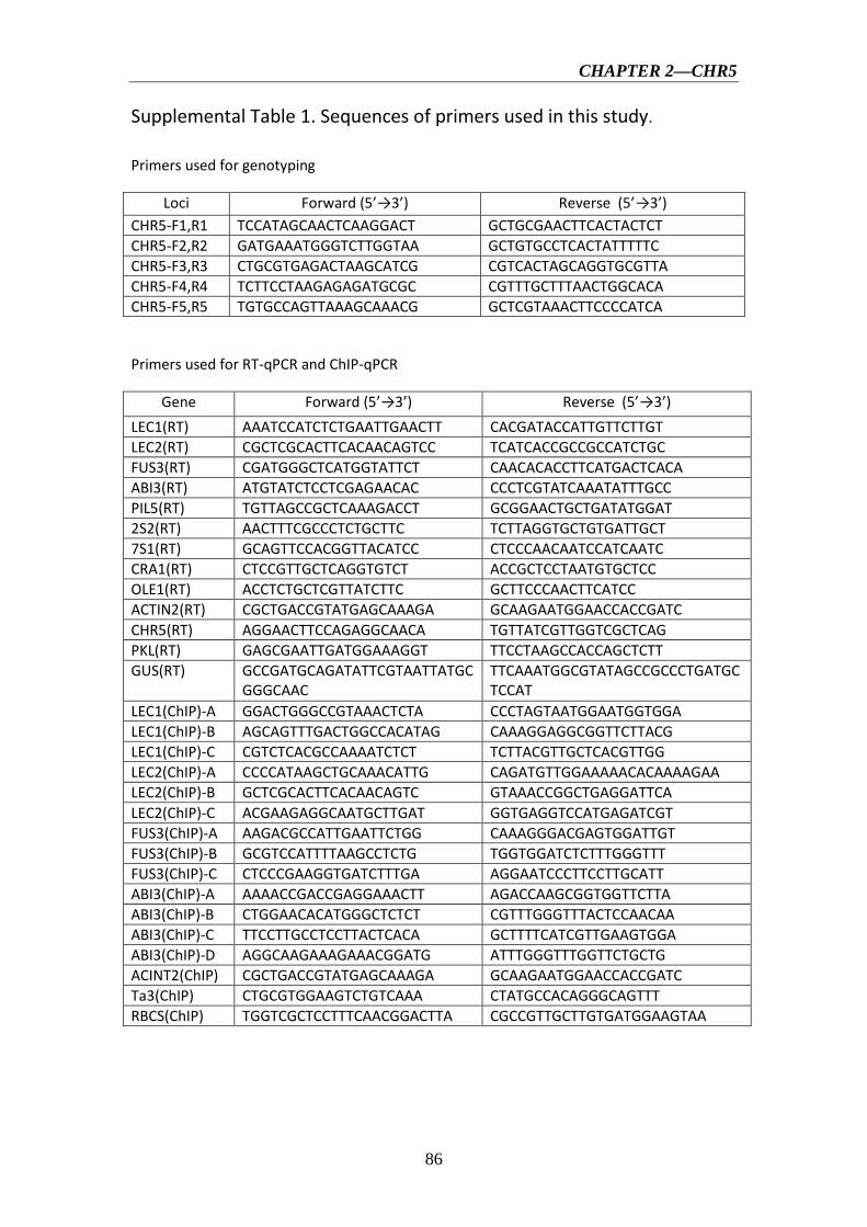
CHAPTER 2—CHR5
86
Supplemental Table 1. Sequences of primers used in this study.
Primers used for genotyping
Loci Forward (5’→3’) Reverse (5’→3’) CHR5-F1,R1 TCCATAGCAACTCAAGGACT GCTGCGAACTTCACTACTCT CHR5-F2,R2 GATGAAATGGGTCTTGGTAA GCTGTGCCTCACTATTTTTC CHR5-F3,R3 CTGCGTGAGACTAAGCATCG CGTCACTAGCAGGTGCGTTA CHR5-F4,R4 TCTTCCTAAGAGAGATGCGC CGTTTGCTTTAACTGGCACA CHR5-F5,R5 TGTGCCAGTTAAAGCAAACG GCTCGTAAACTTCCCCATCA
Primers used for RT-qPCR and ChIP-qPCR
Gene Forward (5’→3’) Reverse (5’→3’) LEC1(RT) AAATCCATCTCTGAATTGAACTT CACGATACCATTGTTCTTGT LEC2(RT) CGCTCGCACTTCACAACAGTCC TCATCACCGCCGCCATCTGC FUS3(RT) CGATGGGCTCATGGTATTCT CAACACACCTTCATGACTCACA ABI3(RT) ATGTATCTCCTCGAGAACAC CCCTCGTATCAAATATTTGCC PIL5(RT) TGTTAGCCGCTCAAAGACCT GCGGAACTGCTGATATGGAT 2S2(RT) AACTTTCGCCCTCTGCTTC TCTTAGGTGCTGTGATTGCT 7S1(RT) GCAGTTCCACGGTTACATCC CTCCCAACAATCCATCAATC CRA1(RT) CTCCGTTGCTCAGGTGTCT ACCGCTCCTAATGTGCTCC OLE1(RT) ACCTCTGCTCGTTATCTTC GCTTCCCAACTTCATCC ACTIN2(RT) CGCTGACCGTATGAGCAAAGA GCAAGAATGGAACCACCGATC CHR5(RT) AGGAACTTCCAGAGGCAACA TGTTATCGTTGGTCGCTCAG PKL(RT) GAGCGAATTGATGGAAAGGT TTCCTAAGCCACCAGCTCTT GUS(RT) GCCGATGCAGATATTCGTAATTATGC
GGGCAAC TTCAAATGGCGTATAGCCGCCCTGATGCTCCAT
LEC1(ChIP)-A GGACTGGGCCGTAAACTCTA CCCTAGTAATGGAATGGTGGA LEC1(ChIP)-B AGCAGTTTGACTGGCCACATAG CAAAGGAGGCGGTTCTTACG LEC1(ChIP)-C CGTCTCACGCCAAAATCTCT TCTTACGTTGCTCACGTTGG LEC2(ChIP)-A CCCCATAAGCTGCAAACATTG CAGATGTTGGAAAAACACAAAAGAA LEC2(ChIP)-B GCTCGCACTTCACAACAGTC GTAAACCGGCTGAGGATTCA LEC2(ChIP)-C ACGAAGAGGCAATGCTTGAT GGTGAGGTCCATGAGATCGT FUS3(ChIP)-A AAGACGCCATTGAATTCTGG CAAAGGGACGAGTGGATTGT FUS3(ChIP)-B GCGTCCATTTTAAGCCTCTG TGGTGGATCTCTTTGGGTTT FUS3(ChIP)-C CTCCCGAAGGTGATCTTTGA AGGAATCCCTTCCTTGCATT ABI3(ChIP)-A AAAACCGACCGAGGAAACTT AGACCAAGCGGTGGTTCTTA ABI3(ChIP)-B CTGGAACACATGGGCTCTCT CGTTTGGGTTTACTCCAACAA ABI3(ChIP)-C TTCCTTGCCTCCTTACTCACA GCTTTTCATCGTTGAAGTGGA ABI3(ChIP)-D AGGCAAGAAAGAAACGGATG ATTTGGGTTTGGTTCTGCTG ACINT2(ChIP) CGCTGACCGTATGAGCAAAGA GCAAGAATGGAACCACCGATC Ta3(ChIP) CTGCGTGGAAGTCTGTCAAA CTATGCCACAGGGCAGTTT RBCS(ChIP) TGGTCGCTCCTTTCAACGGACTTA CGCCGTTGCTTGTGATGGAAGTAA

CHAPTER 2—CHR5
87
Primers used for cloning
Name Forward (5’→3’) Reverse (5’→3’)
ProCHR5 AAGCTTCGCTAAATACAAATGTCTCG added HindIII site underlined
GGTACCGTAAAGAAAGCCATCTCGTCT added KpnI site underlined
CHR5(cDNA) GGTACCAGACGAGATGGCTTTCTTTAGGA added KpnI site underlined
ACTAGTTCTTGGGGGAAATCCGGT added SpeI site underlined
Primers used for FUS3 nucleosome mapping
Center of amplicon
Forward (5’→3’) Reverse (5’→3’)
FUS3(-521) TGAGACAAATGTGCTATTTCAACC TGATTTGGAAGAGAAGTGAAGGA FUS3(-465) TCCTTCACTTCTCTTCCAAATCA GAACATTTTTGCGACCAAGA FUS3(-405) TTCTTGGTCGCAAAAATGTTC CTTACTGGTCAACATGTGATTTG FUS3(-338) CAAATCACATGTTGACCAGTAAG CCAGAATTCAATGGCGTCTT FUS3(-265) AAGACGCCATTGAATTCTGG CAAAGGGACGAGTGGATTGT FUS3(-182) CTTGGTTCTAAACTTTAACCTAACC CGCAACAAGATCTAATGCCACT FUS3(-118) GGCATTAGATCTTGTTGCGTTA TTCATGAATCTAGGGTTTTGGAA FUS3(-40) AATCTTCCAAAACCCTAGATTCA ACGCTGGGCAATCAGAGAT FUS3(-17) CAAATGTCACTTCCTTACCATAAA ACGCTGGGCAATCAGAGAT FUS3(21) CCAATGGTAAACCCAAATCTC TGTGTGTTGTGCAGATCGAA FUS3(49) GCGTCCATTTTAAGCCTCTG CTGTGGTTTGTGTTGTGTTGTG FUS3(62) AAGCCTCTGTTTCGATCTGC GAGAGAAAGAGAGACTGTGGTTTG FUS3(97) TTCGATCTGCACAACACACA TGGTGGATCTCTTTGGGTTT FUS3(122) CACAACACAACACAAACCACAG GAAAACAGTTCTCTGTGTGAAGGA FUS3(176) AAACCCAAAGAGATCCACCA TGCCTAGTAAAAAGGAAGTGTGG FUS3(231) ACTGTTTTCTTCCACACTTCCT AGTAGAGGCCTTGGTTTCCA FUS3(259) GCAGTGTTAACCAATTGAGAGAGAA ACCCATGATCAACACTTGCCA gypsy(-73) TACACATTCCTCAGCCGTTG CCTATCTCCCAGCTAGCAACC


CHAPTER 3
Functional characterization of a histone
demethylase JMJ15 in Arabidopsis

CHAPTER 3—JMJ15
88
Introduction
JMJ15 belongs to the KDM5/JARID1 group of JmjC-domain-containing proteins,
which was previously shown to be a H3K4 demethylase (Liu et al., 2010; Yang et al.,
2012a). However, little is known concerning to its function in stress-responsive gene
regulation and plant growth. In this work, I analyzed the tissue expression pattern of
JMJ15 during different plant development stages using proJMJ15-GUS construct. To
study the function of JMJ15 in gene expression and plant development, I obtained 3
alleles of T-DNA insertion mutants: jmj15-1 and jmj15-2 were gain-of-function lines,
and jmj15-3 was loss-of-function line. The jmj15-3 mutant did not show obvious
phenotypes, whereas gain-of-function mutations reduced the length of hypocotyls and
inflorescence stems with higher accumulation of lignin. Furthermore, the
gain-of-function mutants showed enhanced salt tolerance whereas the loss-of-function
mutant was more sensitive to salt compared to the wild type. To study the effect of
JMJ15 over-expression on gene expression, we analyzed the transcriptome of the
jmj15-1 and jmj15-2 mutant alleles using Affymetrix ATH1 microarrays. The
transcriptomic analysis revealed that there were much more down-regulated genes
compared to up-regulated ones in the over-expression plants. Most of the
down-regulated genes are related to stress, among which most encode transcription
factors, suggesting that JMJ15 may be a higher hierarchical regulator involved in
stress-responsive gene expression. On the other hand, the H3K4 methylation level of
down-regulated genes was analyzed using publicly epigenomic datasets. The result
showed that most down-regulated genes were highly enriched for H3K4me3 and
H3K4me2. In general, our results indicate that JMJ15, as a H3K4 demethylase, plays
important roles in plant responses to stress. This part of the results is written in an
article that will be submitted for publication.

CHAPTER 3—JMJ15
89
Over-expression of Histone H3K4 Demethylase Gene JMJ15
Enhances Stress Tolerance in Arabidopsis
Yuan Shen1, Natalia Conde e Silva1,2, Laure Audonnet1, Caroline Servet2, Dao-Xiu Zhou1 ,2*
1Institut de Biologie des Plantes, Université Paris-Sud 11, Orsay, France
2UMR 8618, CNRS, Orsay, France
Running title: Function of H3K4me3 demethylation in stress tolerance
*Corresponding author: Dao-Xiu Zhou Université Paris sud 11 Institut de biologie des plantes B630 91405 Orsay, France [email protected] Keywords: Histone methylation; Jumonji demethylase; JMJ15; Abiotic stress tolerance gene; epigenetic regulation; H3K4me3; chromatin modification

CHAPTER 3—JMJ15
90
3.1 Abstract
Histone H3 lysine 4 trimethylation (H3K4me3) has been shown to be involved in
stress-responsive gene expression and gene priming in plants. However, the role of
H3K4me3 resetting in stress tolerance is not clear. In this work we studied the
expression and function of Arabidopsis H3K4 demethylase gene JMJ15. We show
that the expression of JMJ15 was restricted to a few tissues during vegetative growth
but relatively highly expressed in young floral organs. Over-expression of the gene in
gain-of-function mutations reduced the length of seedling hypocotyls and
inflorescence stems with higher accumulation of lignin in the stem, while the
loss-of-function mutants did not show any visible phenotype. The gain-of-function
mutants showed enhanced salt tolerance, whereas the loss-of-function mutants were
more sensitive to salt compared to the wild type. Transcriptomic analysis revealed a
much higher number of genes down-regulated in JMJ15 over-expression plants,
which are highly enriched for H3K4me3 and H3K4me2. Among the down-regulated
genes, many encode transcription regulators of stress responsive genes. The data
suggest that increased JMJ15 levels may regulate the gene expression program that
enhances stress tolerance.

CHAPTER 3—JMJ15
91
3.2 Introduction
Histone lysine methylation is an important epigenetic modification for gene
expression in eukaryotic cells (Martin and Zhang, 2005; Kouzarides, 2007). Histone
lysines can be mono-, di-, and tri- methylated. Genome-wide analysis has indicated
that about two-thirds of Arabidopsis genes are marked by mono-, di- or trimethylation
on histone H3 at residue lysine 4 (H3K4me1/2/3) (Zhang et al., 2009). H3K4me3 is
predominantly found at the promoter and 5’ regions of genes and is strongly
associated with transcriptional activation in plants. H3K4me3 was found to be
increased on responsive genes upon stress treatment, but the increase was found to be
lagged behind gene activation in a few experiments (Kim et al., 2008; Hu et al., 2011),
suggesting that H3K4me3 may have a function to mark the active gene state. In
addition, H3K4me3 in gene bodies has been suggested to play a role in transcriptional
memory of stress-responsive genes in Arabidopsis (Alvarez-Venegas et al., 2007;
Jaskiewicz et al., 2011). Recent results indicated that the enriched H3K4me3 in gene
bodies decreased after stress recovery but remained higher than basal state, suggesting
that a regulated resetting mechanism is involved for partial removal of H3K4me3 and
that remaining H3K4me3 may contribute for the transcriptional memory in
Arabidopsis (Ding et al., 2012; Kim et al., 2012).
Histone methylation marks are established by evolutionarily conserved
SET-domain proteins (named after 3 Drosophila genes: Su(var)3-9, Enhancer of zeste
and Trithorax). H3K4 methylation is mediated by the Trithorax group proteins (TRX).
Arabidopsis Trithorax ATX1 and ATX2 respectively trimethylates and dimethylates
H3K4 (Saleh et al., 2008). ATX1 is found to be necessary for stress-induced gene
expression (Alvarez-Venegas and Avramova, 2005; Alvarez-Venegas et al., 2007;
Ding et al., 2011). Other SET-domain genes (SDG) such as SDG4 and SDG2 also
methylate H3K4 and are involved in the control of many aspects of plant development
(Cartagena et al., 2008; Berr et al., 2010; Guo et al., 2010).
Histone methylation is reversed by histone demethylases. Lysine Specific
Demethylase 1 (LSD1) is the first identified histone demethylase to remove mono-

CHAPTER 3—JMJ15
92
and di-methyl groups from H3K4 (Shi et al., 2004). In Arabidopsis there are 4
LSD1-like genes including FLOWERING LOCUS D (FLD), LSD1-LIKE 1 (LDL1)
and LSD1-LIKE 2 (LDL2) that are shown to be involved in flowering time control
(Jiang et al., 2007). The second class of histone demethylases containing the jumonji
C (JmjC) domain catalyze histone lysine demethylation through a ferrous ion (Fe(II))
and α-ketoglutaric acid (α-KG)-dependent oxidative reaction (Tsukada et al., 2006).
Multiple JmjC domain-containing histone demethylases are identified in animal cells,
which are divided into distinct groups including JARID/KDM5,
JMJD1/JHDM2/KDM3, JMJD2/KDM4, JMJD3/UTX/KDM6, JHDM1/FBX/KDM2
and the “JmjC domain-only” group. Members of each group target to specific histone
lysine residues at different methylation states (Klose et al., 2006). About 20 JmjC
domain-containing protein genes are found in Arabidopsis (Lu et al., 2008; Sun and
Zhou, 2008; Chen et al., 2011). Most animal and plant JmjC proteins are conserved,
while some animal proteins, such as JMJD3/UTX/KDM6 group that has the H3K27
demethylase activity, is not found in plants. Recent data have shown that plant
JMJD2/KDM4 homologues can demethylate H3K27 (Lu et al., 2011; Li et al., 2013).
The JARID/KDM5 group catalyzes H3K4me2/3 demethylation in mammalian
cells. Arabidopsis genome has one JARID/KDM5-like gene (JMJ17), whose function
is presently unknown. There is a specific group in plants which includes Arabidopsis
JMJ14, JMJ15, JMJ16, JMJ18 and JMJ19. The JmjC domains of this group are more
closely related to that of the JARID, but structurally similar to that of JMJD2/KDM4
(Chen et al., 2013). JMJ14, JMJ15 and JMJ18 have been reported to have the
H3K4me2/3 demethylase activity and to regulate diverse aspects of chromatin
function and plant development (Deleris et al., 2010; Lu et al., 2010; Searle et al.,
2010; Le Masson et al., 2012; Yang et al., 2012a; Yang et al., 2012b; Cui et al., 2013).
However, the function of these H3K4 demethylases in plant stress tolerance has not
been evaluated. In this work we provide evidence that increased expression of JMJ15
preferentially down-regulates H3K4me2/3-marked stress-related genes and enhance
salt stress tolerance.

CHAPTER 3—JMJ15
93
3.3 Materials and methods
Plant growth
The Arabidopsis thaliana ecotype Columbia (Col-0) was used throughout this study.
T-DNA mutant lines jmj15-1 (GABI_257F10), jmj15-2 (GABI_876B01) and jmj15-3
(GABI_663C11) were obtained from the Nottingham Arabidopsis Stock Center
(NASC) and confirmed by PCR. Seeds were surface-sterilized and plants were grown
on 0.5 x Murashige and Skoog (MS) medium after stratification at 4°C for 2 days.
Plants were analyzed on plates under long-day (LD, 16h light/8h dark) or short-day
(SD, 8h light/16h dark) photoperiods at 20°C. Ten days after germination, plants were
transferred to soil and kept in growth rooms under LD condition.
To test JmjC gene expression in response to salt, experiments were carried out
with 8 day-old plants, treated with 0.5 x MS supplemented with or without 100 mM
NaCl for 5h. For germination tests, seeds of WT and jmj15 mutants were sown on
medium containing indicated concentration NaCl. Images of the Petri dishes were
taken 10 days after germination. Constructs and Transformation
For the histochemical GUS assay, the 2 kb promoter of JMJ15 was amplified from
wild type genomic DNA using the following primers:
5’-GGATCCAGAGCTTGGCCATTTCTTGA-3’ (forward) and
5’-GGTACCGCACTGAAAGGCTCCATTG-3’ (reverse). BamHI and KpnI
(underlined) were used for digestions. The JMJ15 promoter fragment was inserted as
translational fusion with the uidA gene into the pPR97 vector. To generate the
35S-JMJ15-HA construct, the full length cDNA without a stop codon was amplified
from total cDNA of Col-0 using primers: TCTAGACCTTTGGGTTTTGTGGAGTG
(forward) and TCTAGACCAATTCAAATCAACCCCAAA (reverse). Using XbaI
site, JMJ15 cDNA was inserted into the binary vector pFA121, which was modified
based on pBI121 and contained 2 x FLAG-HA tag. The pJMJ15-GUS and

CHAPTER 3—JMJ15
94
35S-JMJ15-HA constructs were transformed into Agrobacterium tumefaciens strain
GV3101 and then transformed the plants using floral dip method.
Microarray analysis
Total RNA was extracted from 12 day-old seedling using Trizol (Invitrogen) and
cleaned using the RNeasy isolation kit (Qiagen). Hybridization with Affymetrix
GeneChip Arabidopsis ATH1 Genome Array was performed at CapitalBio
Corporation. Wild type and both jmj15 overexpression alleles were performed in two
biological repeats. Gene expression changes between the samples were analyzed by
the AffylmGUI package from R software. GO annotation was carried out with the GO
terms of the TAIR database (http://arabidopsis.org/tools/bulk/go/index.jsp). The
percent of significantly changed genes in each TAIR annotated category was
calculated as follows: percent = the number of significantly changed genes divided by
N x 100, where N represents the total number of genes annotated in each ontology.
Significantly changed genes were subsequently analyzed for their H3K4 methylation
levels at epigenomics database (http://epigenomics.mcdb.ucla.edu/H3K4m1m2m3/).
The microarray data are deposited to the NCBI databases (accession number
pending).
Real-Time PCR
For gene expression analysis, two micrograms of total RNA were reversed transcribed
into cDNA by ImPromII reverse transcriptase (Promega). Real-time PCR was
performed with the LightCycler® 480 SYBR Green I Master (Roche) on a
LightCycler 480 (Roche). At least two biological replicates and two technical repeats
for every biological replicate were tested. The primers used in this study are listed in
supplementary Table 1.

CHAPTER 3—JMJ15
95
Histochemical GUS and Lignin Staining
GUS staining was performed as previously described (Bertrand et al., 2003). Briefly,
plant samples were fixed with 90% acetone on ice for 20 min and were washed with
staining buffer (0.2% Triton X-100, 5 mM potassium ferrocyanide, 5 mM potassium
ferricyanide, 100 mM NaH2PO4 and 100 mM Na2HPO4 pH 7.2). Then the samples
were immersed in GUS staining solution with 1 mM X-Gluc and placed under
vacuum for 20 minutes. After incubation at 37°C overnight, the staining solution was
removed and samples were cleared by sequential changes of 70% (v/v) ethanol and
stored at 4°C.
The histological comparative analysis of inflorescence stems between Col-0
and jmj15 mutants was done at the stage of newly formed green siliques, about 2
weeks after bolting, when the inflorescence stems of wild type reach 20 cm in height.
Cross-sections of the inflorescence stems at the basal end were stained for 3 min in
phloroglucinol-HCl reagent (Prolabo, VMR International, France) and then observed
in ethanol 100 % : HCl 37 % (9/1, v/v) using a light microscope (Nikon,
MULTIZOOM AZ 100).

CHAPTER 3—JMJ15
96
3.4 Results
3.4.1 Expression levels of H3K4 demethylase genes
To investigate whether H3K4 demethylase genes are involved in plant stress
responses, we analyzed the mRNA levels of JMJ14, JMJ15, JMJ16, JMJ17, JMJ18
and JMJ19 genes in 8 day-old seedlings grown in ½ MS media under continuous light,
then transferred to 100 mM NaCl or to ½ MS solution for 5 hours. In untreated (½ MS)
seedlings, the expression levels of the 6 genes varied considerably. The relative
expression levels of JMJ17, JMJ18 and JMJ19 were much higher (>102) than that of
JMJ14, JMJ15 and JMJ16 (Supplementary Figure 1). NaCl treatment did not
dramatically affect the expression of these genes, although some decrease of JMJ14
and JMJ18 and some increase of JMJ15 transcript levels were detected.
3.4.2 JMJ15 displayed a highly tissue-specific expression pattern
The relatively low expression level of JMJ15 was in agreement with previous data
showing that the 1.5 kb promoter region of JMJ15 is weak in vegetative tissues (Hong
et al., 2009). To study the temporal and spatial expression pattern of JMJ15, we used
a larger promoter region of JMJ15 (-2051 bp to +14 bp relative to ATG) to make a
GUS reporter translational fusion construct and transformed Arabidopsis plants. Three
independent GUS reporter lines were characterized. All showed the same pattern of
GUS expression. In seedlings, GUS activity was detected only at the base of rosette
leaves and root vascular tissues, but not in the root tip (Figure 1). Interestingly, a
higher accumulation of GUS activity was detected in pericycle cells that initiated to
lateral root meristem. The GUS activity remained to be detected at the base of the
growing lateral roots (Figure 1). In the inflorescence, GUS activity was strong in the
young anthers and detected in carpels, but the activities become weaker in the mature
flower (Figure 1). This temporal and tissue-specific expression pattern suggested that
JMJ15 may have a function in plant development.

CHAPTER 3—JMJ15
97
3.4.3 JMJ15 gain-of-function mutations showed a reduced plant height phenotype The JMJ15 gene contains 10 exons and encodes a polypeptide of 806 amino acids
with distinct domains, including the JmjN domain, JmjC domain, a C5HC2 zinc
finger and the FY-rich N-terminus (FYRN)/FY-rich C-terminus (FYRC) domains,
which are conserved in JMJ14, JMJ16, and JMJ18 (Supplementary Figure 2) (Lu et
al., 2008). To study the function of JMJ15 in gene expression and plant development,
we characterized 3 T-DNA insertion mutants: jmj15-1(GK-257F10), jmj15-2
(GK-876B01) and jmj15-3 (GK-663C11). In jmj15-1 and jmj15-2, the T-DNA was
inserted in the 5’ end, and in jmj15-3 the T-DNA was inserted in the seventh exon of
the gene (Figure 2A). RT-PCR analysis with 4 pairs of primers that covered the whole
coding region of the gene, revealed that the transcript level of JMJ15 was
dramatically increased in jmj15-1 and jmj15-2, but the transcript was interrupted in
jmj15-3 compared to wild-type (Figure 2B). The insertion in jmj15-1 and jmj15-2 did
not alter the 5’ end of the coding region, as the primer set F1 (that cover the 5’end of
the coding region) and R1 successfully amplify the transcripts from the mutants. The
data suggested that jmj15-1 and jmj15-2 were gain-of-function mutants that
overexpressed the gene and that jmj15-3 was a loss-of-function mutant. The jmj15-3 loss-of-function mutation did not display any visible phenotype in
normal growth conditions, confirming previous results (Yang et al., 2012a). However,
in short day (8h light/16h dark) grown seedlings, jmj15-1 and jmj15-2 mutants
produced slightly shorter hypocotyls compared to wild type (Figure 3A). At the
mature stage, the plant height of jmj15-1 and jmj15-2 were also reduced compared to
wild type (Figure 3B). The stronger phenotypes in jmj15-1 were correlated with the
higher expression of the gene in the allele compared to in jmj15-2. To study whether
the plant height phenotype of jmj15-1 and jmj15-2 was due to increased expression of
the gene, we made 35S-JMJ15-HA construct and obtained JMJ15 over-expression
transgenic plants. The transgenic plants also displayed the reduced plant height
phenotype at mature stage (Figure 3B).

CHAPTER 3—JMJ15
98
The plant height phenotype of the jmj15 gain-of-function mutants prompted us
to further investigate the stem cell structure by using histochemical method. Sections
of the basal part of the inflorescence stem of 5 week-old plants (grown in long day)
were stained with phloroglucinol and examined by light microscopy. Phloroglucinol
reacts with coniferaldehyde groups in lignin, and the color intensity reflects the total
lignin content. The analysis revealed that jmj15-1 and jmj15-2 exhibited a
significantly deeper red staining in the stem vascular system and interfascicular fibers
compared to that in wild type and jmj15-3 (Figure 4). This observation suggested that
over-expression of JMJ15 resulted in an increase of the total lignin content in the
stems concurrently with stem growth reduction. 3.4.4 Over-expression of JMJ15 preferentially repressed genes marked by H3K4 methylation
To determine the effect of JMJ15 over-expression on gene expression, the
transcriptomes of jmj15-1, jmj15-2 and wild type seedlings (12 day-old) were
analyzed by using the Affymetrix Arabidopsis ATH1 Genome Array. Two biological
replicates for each sample were analyzed. Pair-wise plots of the microarray data
revealed a good correlation of the hybridization signals between the biological
replicates of each sample and between the 2 mutant alleles (Figure 5A). The average
hybridization signals of the replicates of both mutants were normalized and compared
with the wild type signals. Up- and down-regulated genes in both jmj15-1 and jmj15-2
were filtrated with the threshold >2 fold changes compared to wild type. The analysis
revealed 23 up-regulated and 194 down-regulated genes in the jmj15 mutant lines
(Supplementary Dataset 1). The analysis revealed a much high expression level of
JMJ15 itself (>7-8 folds) in the mutants compared to wild type (Supplementary
Dataset 1), confirming the over-expression of the gene in the mutants. The higher
number of down-regulated genes compared to up-regulated ones suggested that
elevated JMJ15 expression mainly repressed genes and that JMJ15 acted as a
transcriptional repressor, consistent with its H3K4 demethylase activity (Liu et al.,
2010; Yang et al., 2012a). To validate the microarray data, we checked 4
CHAPTER 3—JMJ15
99
down-regulated genes by RT-qPCR. The relative transcript level changes in the
mutants compared to wild type detected by RT-qPCR were in agreement with that
from the microarray analysis (Figure 5B).
To evaluate whether there was any enrichment of the deregulated genes for
H3K4me2/3, we compared the deregulated genes with the genome-wide
H3K4me2/3/1 data obtained from wild type seedlings (Zhang et al., 2009). The
analysis revealed that about 85% of the down-regulated genes were marked by the
H3K4 methylation (mostly by H3K4me2, H3K4me3, or both) in the gene bodies,
compared to about 50% of up-regulated genes (Figure 6, Supplementary Dataset 1).
About 60% of the down-regulated genes were marked by H3K4me3, H3K4me2 or
H3K4me2/3 in the promoter region (in the -500 bp region relative to TSS), compared
to about 30% of up-regulated genes. Compared to up-regulated genes, the
down-regulated ones were clearly enriched for the H3K4me2/3 double methylation
marks. This analysis suggested that JMJ15-mediated gene repression might be
achieved by demethylating H3K4 and indicated that JMJ15 preferentially repressed
genes that have the H3K4me2/3 double methylation marks. 3.4.5 Over-expression of JMJ15 preferentially repressed stress regulatory genes Gene ontology (GO) analysis of the deregulated genes using the GO Slim
Classification (http://www.arabidopsis.org/help/helppages/go_slim_help.jsp) revealed
that a large proportion of the deregulated genes (23.73%) in the jmj15 mutants had a
function in the nucleus (Supplementary Figure 3A). Interestingly, 37 out of the 194
down-regulated genes were transcriptional regulators (Supplementary Dataset 1).
Remarkably, more than 32% of the down-regulated genes belonged to the
stress-responsive categories (Supplementary Figure 3B), suggesting that JMJ15 may
have a function in stress-responsive gene expression. We noticed that among the
greater than 5 fold down-regulated genes, more than 50% were transcription factors or
stress-responsive signalling protein genes (Supplementary Dataset 1). These included
the stress-responsive zinc finger protein STZ/Zat10 (At1g27730) (Sakamoto et al.,
2000), the stress-responsive WRKY proteins WRKY40 (At1g80840) and WRKY33

CHAPTER 3—JMJ15
100
(At2g38470) (Jiang and Deyholos, 2009), the cold-responsive factor CBF2
(At4g25470) (Vogel et al., 2005), the ethylene-responsive-element binding proteins
ATERF6 (At4g17490) and ATERF11 (At1g28370) (Li et al., 2011; Dubois et al., 2013),
the stress-responsive F-box protein ATFBS1 (At1g61340) and the cytochrome P450
CYP707A3 (At5g45340) genes (Umezawa et al., 2006; Maldonado-Calderon et al.,
2012) (Supplementary Dataset 1). The decreased expression of WRKY33 and
CYP707A3 genes was validated by RT-qPCR (Figure 5B). 3.4.6 JMJ15 gain-of-function mutations enhanced salt stress tolerance To study whether JMJ15 mutations affected plant tolerance to stress, we germinated
seeds of wild type, jmj15-1, jmj15-2 and jmj15-3 mutants on ½ MS media containing
130 mM or 150 mM NaCl. The seedling growth phenotype shown in Figure 6 indicated
that the gain-of-function mutations (jmj15-1 and jmj15-2) enhanced plant tolerance to
salt stress, whereas the loss-of-function mutation (jmj15-3) reduced the stress
resistance.
CHAPTER 3—JMJ15
101
3.5 Discussion
3.5.1 Function of JMJ15 in stress tolerance In this work we have shown that JMJ15 displayed a high tissue-specific expression
pattern. Constitutive or over-expression of the gene, as a result of T-DNA insertions
in the 5’ region, preferentially repressed genes marked by H3K4me2/3, suggesting
that JMJ15-mediated gene repression may be mainly mediated through H3K4me2/3
demethylation. The observation that about a third of the down-regulated genes are
related to stress implies that H3K4me2/3 levels are important for the expression of
this category of genes. This is consistent with numerous observations that H3K4me3
is associated with the induction of biotic and abiotic stress-responsive genes (van Dijk
et al., 2010; Hu et al., 2011; Jaskiewicz et al., 2011; Zong et al., 2013; To and Kim,
2014), and ATX1 that trimethylates H3K4 in the genic region is required for
stress-responsive gene expression (Ding et al., 2009; Ding et al., 2011). Microarray
analysis of atx1 mutant seedlings revealed that 424 genes were up-regulated and 328
genes were down-regulated more than 2 folds compared to the wild type
(Alvarez-Venegas et al., 2006). Similarly, 271 genes were found to be up-regulated
and 321 genes down-regulated in mutant seedlings of another H3K4
methyltransferase gene SDG2 (Guo et al., 2010). However there was no clear
correlation between transcription changes in sdg2 and atx1 (Guo et al., 2010),
suggesting that the two enzymes may regulate different targets. Comparison of the
deregulated genes in jmj15 and atx1 or sdg2 did not reveal a clear overlap, although
there were a relatively higher number of overlapped genes between jmj15 and atx1
than between jmj15 and sdg2 (Supplementary Figure 4). These proteins may targets to
different loci. In addition, the data showing that the mostly down-regulated genes
were those encoding transcription factors and signaling proteins involved in stress
responses raise the hypothesis that JMJ15 might be a higher hierarchical regulator
primarily to regulate stress-responsive gene transcription programs in Arabidopsis.
Since JMJ15 is closely related to JMJ14, JMJ16 and JMJ18 (Lu et al., 2008; Sun and

CHAPTER 3—JMJ15
102
Zhou, 2008), the ectopically expressed JMJ15 may also regulate the targets of the
other related demethylases.
The observations that the jmj15 gain-of-function mutants showed enhanced salt
tolerance at seedling stage and that the loss-of-function mutant was more sensitive to
salt stress than the wild type, suggest that JMJ15 is required for stress tolerance. The
data showing that the gain-of-function mutants displayed reduced growth and increased
stem lignification, which are suggested to be associated with stress responses (Moura et
al., 2010; Golldack et al., 2013), support the hypothesis that increased JMJ15 levels
may regulate the gene expression program that integrates plant growth to stress
tolerance. Among the mostly repressed genes in jmj15 gain-of-function mutants was
STZ/Zat10 that encodes a C2H2-zinc finger protein associated with the ERF
amphiphilic repression (EAR) domain (Supplementary Dataset 1). STZ/Zat10 has been
shown to be a transcriptional suppressor of stress-responsive genes (Sakamoto et al.,
2004). Knockout and RNAi of the gene could enhance plant tolerance to abiotic stress
(Mittler et al., 2006). As the STZ locus displays a high level of H3K4me3
(Supplementary Figure 5), it is possible that the repression of STZ/Zat10 by JMJ15
through H3K4me2/3 demethylation is associated with the enhanced salt tolerance
phenotype of the gain-of-function mutants. STZ/Zat10 may be a major player in
JMJ15-mediated regulatory network of stress tolerance.
3.5.2 Developmental function of JMJ15 Consistent with previous results (Yang et al., 2012a), the loss-of-function mutation
identified in this study (jmj15-3) did not produce any visible phenotype. JMJ15 was
first identified as Maternal Effect Embryo Arrest 27 (MEE27) in a genetic screen for
mutants defective in female gametophyte development (Pagnussat et al., 2005).
However, no embryonic defect was observed in jmj15 loss-of-function mutants (Yang
et al., 2012a). Either the mutation was compensated by highly expressed homologues
(e.g., JMJ18, Hong et al, 2009) or JMJ15-dependent H3K4 demethylation is not
sufficient to lead to any morphological change. In addition, another study has
identified JMJ15 as a maternally imprinted gene (Hsieh et al., 2011), however, our

CHAPTER 3—JMJ15
103
data showing the high promoter activity of JMJ15 in anthers do not support that
observation.
It is reported that JMJ14 demethylates H3K4me2/3 at the Flowering Locus T
(FT) locus and represses expression of the gene and that jmj14 loss-of-function
mutants display an early flowering phenotype (Jeong et al., 2009; Lu et al., 2010).
Conversely, JMJ18 directly binds to and represses the flowering repressor gene,
Flowering Locus C (FLC), through H3K4me2/3 demethylation. Consequently,
loss-of-function mutations of JMJ18 result in a weak late-flowering phenotype, while
JMJ18 overexpressors exhibit an early flowering phenotype (Yang et al., 2012b).
These observations support the notion that members of this H3K4 demethylase group
target to different loci and have distinct functions in plant development control.
However, Yang et al have shown that, like JMJ18, JMJ15 over-expression plants
showed repressed FLC expression and produced an early flower phenotype (Yang et
al., 2012a). But unlike jmj18 mutants (Yang et al., 2012a), the jmj15-3
loss-of-function mutation did not alter the flowering phenotype. Possibly, JMJ15 at
elevated levels may demethylate and repress genes that normally targeted by JMJ18 in
wild type plants. However, the jmj15-1 and jmj15-2 gain-of-mutation mutants did not
show any clear flowering phenotype. This discrepancy may be due to difference in
expression levels of JMJ15 in the overexpression plants and the mutant alleles. Acknowledgements This work was supported by the French Agence Nationale de Recherche grant
ANR-10-BLAN-1238 (CERES) and ANR-12-BSV6-0010 (NERTHPATH). Y. Shen
was supported by a PhD Fellowship from the Chinese Scholar Council.

CHAPTER 3—JMJ15
104
3.6 References Alvarez-Venegas, R., and Avramova, Z. (2005). Methylation patterns of histone H3 Lys 4, Lys
9 and Lys 27 in transcriptionally active and inactive Arabidopsis genes and in atx1 mutants. Nucleic Acids Res 33, 5199-5207.
Alvarez-Venegas, R., Al Abdallat, A., Guo, M., Alfano, J.R., and Avramova, Z. (2007). Epigenetic control of a transcription factor at the cross section of two antagonistic pathways. Epigenetics 2, 106-113.
Alvarez-Venegas, R., Sadder, M., Hlavacka, A., Baluska, F., Xia, Y.N., Lu, G.Q., Firsov, A., Sarath, G., Moriyama, H., Dubrovsky, J.G., and Avramova, Z. (2006). The Arabidopsis homolog of trithorax, ATX1, binds phosphatidylinositol 5-phosphate, and the two regulate a common set of target genes. Proc Natl Acad Sci U S A 103, 6049-6054.
Berr, A., McCallum, E.J., Menard, R., Meyer, D., Fuchs, J., Dong, A., and Shen, W.H. (2010). Arabidopsis SET DOMAIN GROUP2 is required for H3K4 trimethylation and is crucial for both sporophyte and gametophyte development. Plant Cell 22, 3232-3248.
Bertrand, C., Bergounioux, C., Domenichini, S., Delarue, M., and Zhou, D.X. (2003). Arabidopsis histone acetyltransferase AtGCN5 regulates the floral meristem activity through the WUSCHEL/AGAMOUS pathway. J Biol Chem 278, 28246-28251.
Cartagena, J.A., Matsunaga, S., Seki, M., Kurihara, D., Yokoyama, M., Shinozaki, K., Fujimoto, S., Azumi, Y., Uchiyama, S., and Fukui, K. (2008). The Arabidopsis SDG4 contributes to the regulation of pollen tube growth by methylation of histone H3 lysines 4 and 36 in mature pollen. Dev Biol 315, 355-368.
Chen, Q.F., Chen, X.S., Wang, Q., Zhang, F.B., Lou, Z.Y., Zhang, Q.F., and Zhou, D.X. (2013). Structural Basis of a Histone H3 Lysine 4 Demethylase Required for Stem Elongation in Rice. Plos Genet 9, e1003239.
Chen, X.S., Hu, Y.F., and Zhou, D.X. (2011). Epigenetic gene regulation by plant Jumonji group of histone demethylase. Biochim Biophys Acta 1809, 421-426.
Cui, X., Jin, P., Gu, L., Lu, Z., Xue, Y., Wei, L., Qi, J., Song, X., Luo, M., An, G., and Cao, X. (2013). Control of transposon activity by a histone H3K4 demethylase in rice. Proc Natl Acad Sci U S A 110, 1953-1958.
Deleris, A., Greenberg, M.V., Ausin, I., Law, R.W., Moissiard, G., Schubert, D., and Jacobsen, S.E. (2010). Involvement of a Jumonji-C domain-containing histone demethylase in DRM2-mediated maintenance of DNA methylation. EMBO Rep 11, 950-955.
Ding, Y., Avramova, Z., and Fromm, M. (2011). The Arabidopsis trithorax-like factor ATX1 functions in dehydration stress responses via ABA-dependent and ABA-independent pathways. Plant J 66, 735-744.
Ding, Y., Ndamukong, I., Xu, Z., Lapko, H., Fromm, M., and Avramova, Z. (2012). ATX1-generated H3K4me3 is required for efficient elongation of transcription, not initiation, at ATX1-regulated genes. PLoS Genet 8, e1003111.
Ding, Y., Lapko, H., Ndamukong, I., Xia, Y., Al-Abdallat, A., Lalithambika, S., Sadder, M., Saleh, A., Fromm, M., Riethoven, J.J., Lu, G., and Avramova, Z. (2009). The

CHAPTER 3—JMJ15
105
Arabidopsis chromatin modifier ATX1, the myotubularin-like AtMTM and the response to drought. Plant Signal Behav 4, 1049-1058.
Dubois, M., Skirycz, A., Claeys, H., Maleux, K., Dhondt, S., De Bodt, S., Vanden Bossche, R., De Milde, L., Yoshizumi, T., Matsui, M., and Inze, D. (2013). Ethylene Response Factor6 acts as a central regulator of leaf growth under water-limiting conditions in Arabidopsis. Plant Physiol 162, 319-332.
Golldack, D., Li, C., Mohan, H., and Probst, N. (2013). Gibberellins and abscisic acid signal crosstalk: living and developing under unfavorable conditions. Plant Cell Reports 32, 1007-1016.
Guo, L., Yu, Y.C., Law, J.A., and Zhang, X.Y. (2010). SET DOMAIN GROUP2 is the major histone H3 lysie 4 trimethyltransferase in Arabidopsis. Proc Natl Acad Sci U S A 107, 18557-18562.
Hong, E.H., Jeong, Y.M., Ryu, J.Y., Amasino, R.M., Noh, B., and Noh, Y.S. (2009). Temporal and spatial expression patterns of nine Arabidopsis genes encoding Jumonji C-domain proteins. Mol Cells 27, 481-490.
Hsieh, T.F., Shin, J., Uzawa, R., Silva, P., Cohen, S., Bauer, M.J., Hashimoto, M., Kirkbride, R.C., Harada, J.J., Zilberman, D., and Fischer, R.L. (2011). Regulation of imprinted gene expression in Arabidopsis endosperm. Proc Natl Acad Sci U S A 108, 1755-1762.
Hu, Y.F., Shen, Y., Silva, N.C.E., and Zhou, D.X. (2011). The role of histone methylation and H2A.Z occupancy during rapid activation of ethylene responsive genes. Plos One 6, e28224.
Jaskiewicz, M., Conrath, U., and Peterhansel, C. (2011). Chromatin modification acts as a memory for systemic acquired resistance in the plant stress response. EMBO Rep 12, 50-55.
Jeong, J.H., Song, H.R., Ko, J.H., Jeong, Y.M., Kwon, Y.E., Seol, J.H., Amasino, R.M., Noh, B., and Noh, Y.S. (2009). Repression of FLOWERING LOCUS T Chromatin by Functionally Redundant Histone H3 Lysine 4 Demethylases in Arabidopsis. Plos One 4, e8033.
Jiang, D., Yang, W., He, Y., and Amasino, R.M. (2007). Arabidopsis relatives of the human lysine-specific Demethylase1 repress the expression of FWA and FLOWERING LOCUS C and thus promote the floral transition. Plant Cell 19, 2975-2987.
Jiang, Y., and Deyholos, M.K. (2009). Functional characterization of Arabidopsis NaCl-inducible WRKY25 and WRKY33 transcription factors in abiotic stresses. Plant Mol Biol 69, 91-105.
Kim, J.M., To, T.K., Ishida, J., Matsui, A., Kimura, H., and Seki, M. (2012). Transition of Chromatin Status During the Process of Recovery from Drought Stress in Arabidopsis thaliana. Plant Cell Physiol 53, 847-856.
Kim, J.M., To, T.K., Ishida, J., Morosawa, T., Kawashima, M., Matsui, A., Toyoda, T., Kimura, H., Shinozaki, K., and Seki, M. (2008). Alterations of lysine modifications on the histone H3 N-tail under drought stress conditions in Arabidopsis thaliana. Plant Cell Physiol 49, 1580-1588.
Klose, R.J., Kallin, E.M., and Zhang, Y. (2006). JmjC-domain-containing proteins and histone demethylation. Nat Rev Genet 7, 715-727.
Kouzarides, T. (2007). Chromatin modifications and their function. Cell 128, 693-705.

CHAPTER 3—JMJ15
106
Le Masson, I., Jauvion, V., Bouteiller, N., Rivard, M., Elmayan, T., and Vaucheret, H. (2012). Mutations in the Arabidopsis H3K4me2/3 demethylase JMJ14 suppress posttranscriptional gene silencing by decreasing transgene transcription. Plant Cell 24, 3603-3612.
Li, T.T., Chen, X.S., Zhong, X.C., Zhao, Y., Liu, X.Y., Zhou, S.L., Cheng, S.F., and Zhou, D.X. (2013). Jumonji C domain protein JMJ705-mediated removal of histone H3 lysine 27 trimethylation is involved in defense-related gene activation in rice. Plant Cell 25, 4725-4736.
Li, Z.F., Zhang, L.X., Yu, Y.W., Quan, R.D., Zhang, Z.J., Zhang, H.W., and Huang, R.F. (2011). The ethylene response factor AtERF11 that is transcriptionally modulated by the bZIP transcription factor HY5 is a crucial repressor for ethylene biosynthesis in Arabidopsis. Plant J 68, 88-99.
Liu, C.Y., Lu, F.L., Cui, X., and Cao, X.F. (2010). Histone methylation in higher plants. Annu Rev Plant Biol 61, 395-420.
Lu, F., Cui, X., Zhang, S., Jenuwein, T., and Cao, X. (2011). Arabidopsis REF6 is a histone H3 lysine 27 demethylase. Nat Genet 43, 715-719.
Lu, F.L., Cui, X., Zhang, S.B., Liu, C.Y., and Cao, X.F. (2010). JMJ14 is an H3K4 demethylase regulating flowering time in Arabidopsis. Cell Res 20, 387-390.
Lu, F.L., Li, G.L., Cui, X., Liu, C.Y., Wang, X.J., and Cao, X.F. (2008). Comparative analysis of JmjC domain-containing proteins reveals the potential histone demethylases in Arabidopsis and rice. J Integr Plant Biol 50, 886-896.
Maldonado-Calderon, M.T., Sepulveda-Garcia, E., and Rocha-Sosa, M. (2012). Characterization of novel F-box proteins in plants induced by biotic and abiotic stress. Plant Sci 185, 208-217.
Martin, C., and Zhang, Y. (2005). The diverse functions of histone lysine methylation. Nat Rev Mol Cell Biol 6, 838-849.
Mittler, R., Kim, Y., Song, L.H., Coutu, J., Coutu, A., Ciftci-Yilmaz, S., Lee, H., Stevenson, B., and Zhu, J.K. (2006). Gain- and loss-of-function mutations in Zat10 enhance the tolerance of plants to ablotic stress. Febs Lett 580, 6537-6542.
Moura, J.C.M.S., Bonine, C.A.V., Viana, J.D.F., Dornelas, M.C., and Mazzafera, P. (2010). Abiotic and biotic stresses and changes in the lignin content and composition in plants. J Integr Plant Biol 52, 360-376.
Pagnussat, G.C., Yu, H.J., Ngo, Q.A., Rajani, S., Mayalagu, S., Johnson, C.S., Capron, A., Xie, L.F., Ye, D., and Sundaresan, V. (2005). Genetic and molecular identification of genes required for female gametophyte development and function in Arabidopsis (vol 132, pg 603, 2005). Development 132, 1161-1161.
Sakamoto, H., Araki, T., Meshi, T., and Iwabuchi, M. (2000). Expression of a subset of the Arabidopsis Cys(2)/His(2)-type zinc-finger protein gene family under water stress. Gene 248, 23-32.
Sakamoto, H., Maruyama, K., Sakuma, Y., Meshi, T., Iwabuchi, M., Shinozaki, K., and Yamaguchi-Shinozaki, K. (2004). Arabidopsis Cys2/His2-type zinc-finger proteins function as transcription repressors under drought, cold, and high-salinity stress conditions. Plant Physiol 136, 2734-2746.

CHAPTER 3—JMJ15
107
Saleh, A., Alvarez-Venegas, R., Yilmaz, M., Le, O., Hou, G., Sadder, M., Al-Abdallat, A., Xia, Y., Lu, G., Ladunga, I., and Avramova, Z. (2008). The highly similar Arabidopsis homologs of trithorax ATX1 and ATX2 encode proteins with divergent biochemical functions. Plant Cell 20, 568-579.
Searle, I.R., Pontes, O., Melnyk, C.W., Smith, L.M., and Baulcombe, D.C. (2010). JMJ14, a JmjC domain protein, is required for RNA silencing and cell-to-cell movement of an RNA silencing signal in Arabidopsis. Genes & development 24, 986-991.
Shi, Y., Lan, F., Matson, C., Mulligan, P., Whetstine, J.R., Cole, P.A., Casero, R.A., and Shi, Y. (2004). Histone demethylation mediated by the nuclear amine oxidase homolog LSD1. Cell 119, 941-953.
Sun, Q.W., and Zhou, D.X. (2008). Rice jmjC domain-containing gene JMJ706 encodes H3K9 demethylase required for floral organ development. Proc Natl Acad Sci U S A 105, 13679-13684.
To, T.K., and Kim, J.M. (2014). Epigenetic regulation of gene responsiveness in Arabidopsis. Front Plant Sci 4, 548.
Tsukada, Y., Fang, J., Erdjument-Bromage, H., Warren, M.E., Borchers, C.H., Tempst, P., and Zhang, Y. (2006). Histone demethylation by a family of JmjC domain-containing proteins. Nature 439, 811-816.
Umezawa, T., Okamoto, M., Kushiro, T., Nambara, E., Oono, Y., Seki, M., Kobayashi, M., Koshiba, T., Kamiya, Y., and Shinozaki, K. (2006). CYP707A3, a major ABA 8 '-hydroxylase involved in dehydration and rehydration response in Arabidopsis thaliana. Plant J 46, 171-182.
van Dijk, K., Ding, Y., Malkaram, S., Riethoven, J.J.M., Liu, R., Yang, J.Y., Laczko, P., Chen, H., Xia, Y.N., Ladunga, I., Avramova, Z., and Fromm, M. (2010). Dynamic changes in genome-wide histone H3 lysine 4 methylation patterns in response to dehydration stress in Arabidopsis thaliana. BMC Plant Biol 10, 238.
Vogel, J.T., Zarka, D.G., Van Buskirk, H.A., Fowler, S.G., and Thomashow, M.F. (2005). Roles of the CBF2 and ZAT12 transcription factors in configuring the low temperature transcriptome of Arabidopsis. Plant J 41, 195-211.
Yang, H., Mo, H., Fan, D., Cao, Y., Cui, S., and Ma, L. (2012a). Overexpression of a histone H3K4 demethylase, JMJ15, accelerates flowering time in Arabidopsis. Plant Cell Rep 31, 1297-1308.
Yang, H.C., Han, Z.F., Cao, Y., Fan, D., Li, H., Mo, H.X., Feng, Y., Liu, L., Wang, Z., Yue, Y.L., Cui, S.J., Chen, S., Chai, J.J., and Ma, L.G. (2012b). A companion cell-dominant and developmentally regulated H3K4 demethylase controls flowering time in Arabidopsis via the repression of FLC expression. Plos Genet 8, 496-512.
Zhang, X., Bernatavichute, Y.V., Cokus, S., Pellegrini, M., and Jacobsen, S.E. (2009). Genome-wide analysis of mono-, di- and trimethylation of histone H3 lysine 4 in Arabidopsis thaliana. Genome Biol 10, R62.
Zong, W., Zhong, X.C., You, J., and Xiong, L.Z. (2013). Genome-wide profiling of histone H3K4-tri-methylation and gene expression in rice under drought stress. Plant Mol Biol 81, 175-188.

CHAPTER 3—JMJ15
108
3.7 Figures
Figure 1. The 2.0 kb promoter activity of JMJ15 in transgenic plants. In seedlings, the GUS activity was detected in the base of rosette leaves (A), root vascular tissues (B), and bases of growing lateral roots (C-G), but not in root tip (H). In flower buds, the GUS activity was detected in anthers and in carpels (I, J), which become weak in opened flowers, but remained in stamen filaments (K, I). Bar = 0.1mm.

CHAPTER 3—JMJ15
109
Figure 2. Cauterization of JMJ15 T-DNA insertion mutants. The insertion positions of the 3 alleles are indicated by open arrows. The exons are represented by black boxes. The positions of forward (F) and reverse (R) primers are indicated by arrows. The expression levels (relative to ACTIN2 mRNA) in jmj15-1 and jmj15-2 compared to wild type were tested by qRT-PCR using the 4 indicated primer sets. The transcript in jmj15-3 compared to wild type was analyzed by RT-PCR using the indicated primers.

CHAPTER 3—JMJ15
110
Figure 3. jmj15 gain-of-function mutations reduced plant height. A. Phenotype of hypocotyls of short day-grown seedlings of jmj15-1 and jmj15-2 compared to wild type (left). Lengths of hypocotyls were measured from 30 wild type, jmj15-1 and jmj15-2 plants (right). B. Plant height at mature stage of jmj15-1 and jmj15-2 compared to wild type (left) and of 35S-JMJ15-HA overexpression plants compared to negative transgenic plants (middle). Plant height was measured from 10 wild type, jmj15-1 and jmj15-2 plants (right). Bar = means +/-SD. Asterisks indicate the significance of difference from wild type by student t-tests (P<0.01).

CHAPTER 3—JMJ15
111
Figure 4. Lignin accumulation in jmj15-1, jmj15-2, and jmj15-3 mutants compared to wild type. Inflorescence sections at the base were stained by Phloroglucinol stain. Bar = 0.1 mm

CHAPTER 3—JMJ15
112
Figure 5. Transcriptomic analysis of jmj15-1 and jmj15-2 (12 day-old) seedlings compared to wild type. A. Pair-wise plots of the microarray hybridization signals between the biological replicates of each sample and the between wild type and two 2 mutant alleles. B. Four down-regulated genes in the two mutants (microarray signals relative to wild type are indicated below the respective genes) were validated by qRT-PCR. Bar = means +/-SD from 3 replicates.

CHAPTER 3—JMJ15
113
Figure 6. Down-regulated genes are highly enriched for H3K4me2/3. Up-regulated (23) and down-regulated (194) genes in jmj15-1 or jmj15-2 mutants (changes >2 folds) were compared with genome-wide H3K4 methylation data. Percentages of H3K4me3, H3K4me2, H3K4me1 and their combinations on the promoter (A) and the gene body (B) regions are presented.

CHAPTER 3—JMJ15
114
Figure 7. Comparison of sensitivity to NaCl of jmj15 mutants with wild type during seed germination. Wild type, jmj15-1, jmj15-2, and jmj15-3 seeds were germinated for 12 days on ½ MS supplemented with indicated concentrations of NaCl.

CHAPTER 3—JMJ15
115
3.8 Supplementary Material
Figure S1. Expression levels of 6 potential H3K4 demethylase genes in Arabidopsis seedlings treated with or without 100 mM NaCl for 5 hours. The transcript levels detected by RT-qPCR are presented as relative to that of the reference gene At4g34270.

CHAPTER 3—JMJ15
116
Figure S2. Comparison of protein structures of the 6 JmjC genes specific to plants.

CHAPTER 3—JMJ15
117
Figure S3. Gene ontology analysis of down-regulated genes in jmj15 gain-of-function mutants.

CHAPTER 3—JMJ15
118
Figure S4. Overlaps of de-regulated genes between jmj15 gain-of-function mutants and atx1 or sdg2 mutants.

CHAPTER 3—JMJ15
119
Figure S5. H3K4 methylation on the STZ/Zat10 locus.
Supplementary Dataset 1. Down- and up-regulated genes in jmj15-1 and jmj15-2 mutants compared to wild type. Genes encoding transcription factors or signaling regulators are indicated. Genes that are marked H3K4me3/2/1 are also indicated.
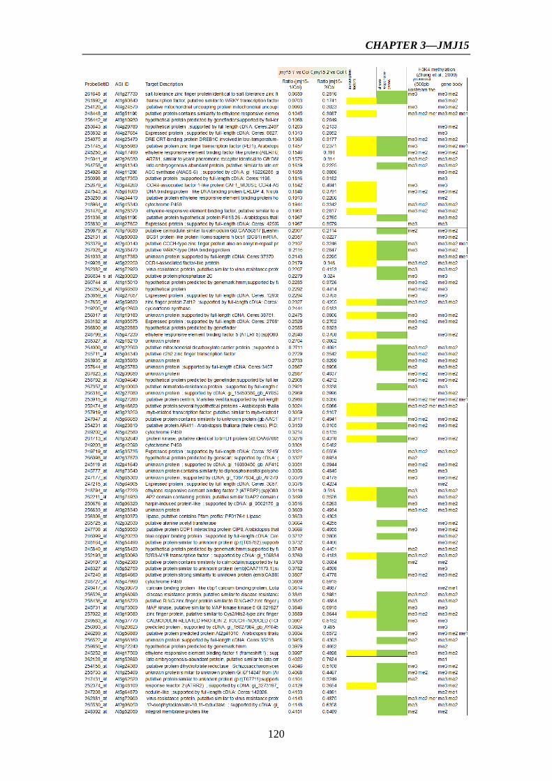
CHAPTER 3—JMJ15
120

CHAPTER 3—JMJ15
121

CHAPTER 3—JMJ15
122

CHAPTER 3—JMJ15
123
Table S1. Sequences of the primers used in this study.
Gene Forward (5’→3’) Reverse (5’→3’)
JMJ15-F1, R1 CCTTTGGGTTTTGTGGAGTG CACCAATGTCTGGCCTCTTT JMJ15-F2, R2 CCTTGGAGCAGCTTTTGAAG AACCATTTCCAAGAGCCTCA JMJ15-F3, R3 ATCTTGGGTTCTTGGTCGTG GAAGAGTGGACCCAGAAGTCC JMJ15-F4, R4 GAGATCATGAGACGCAGCAA TCATTTCCAGCGAATCCTTC JMJ14 TGGTTGTTTAGGAGGGAAGG TGGTTAGGTTTGTTGGGTCA JMJ16 GGAGATTCGCTGCGTGATAC CGTGGCTGTTAGTTGCCATT JMJ17 CATGCAGAGCAGGAACTTCA GTGGAGTGGTTTCTGCCAGT JMJ18 GAAATGGTGATGCGGAGAGT AGTCGATGGTTTGGGTCAAG JMJ19 AATCGCAAAGGATGGACAAG CCTCATGTGGTTCAGACACG WRKY33 AAGGGAAACCCAAATCCAAG AAGACGAATCCTGTGGTGCT CYP707A3 AAAGCAGGATTAACCGACGA TTGCCATTTGCTCTTCAGTG ARR15 GAAGGAGCAGAGGAGTTTTTG GTCGTCATCAAGGGAGGAA ARR5 TGTCCTGATTCTTTCGGCTTAC
A AAAAACCCATCTTTGTCACTCTTGA
ACTIN2 CGCTGACCGTATGAGCAAAGA GCAAGAATGGAACCACCGATC At4g34270 GCACCAACTGTTCTTCGTGA GGTCGCTCCAGACTGCTAAG

CHAPTER 4
GENERAL DISCUSSION & PERSPECTIVES


CHAPTER 4—GENERAL DISCUSSION & PERSPECTIVES
124
4.1 Function of CHR5 in plant gene expression
Several CHD3 proteins have been reported in plants. Arabidopsis CHD3 protein, PKL,
represses LEC1/AFL gene expression via promoting H3K27me3 on these loci
(Aichinger et al., 2009; Zhang et al., 2012). Rice CHD3 protein, CHR729, recognizes
and modulates H3K4 and H3K27 methylation over repressed tissue-specific genes
(Hu et al., 2012). Unlike the CHD3 family, only one CHD1 gene (CHR5) is found in
the genome of Arabidopsis. CHR5 is mostly expressed in growing or developmental
tissues/organs, suggesting that this chromatin remodeler may play an important role in
facilitating gene expression reprogramming during plant development. Our data on
the function of CHR5 to modulate seed maturation gene expression during
embryogenesis support this hypothesis. The role of CHR5 in other developmental
aspects could be exploited to further understand the function and mode of action of
CHR5.
The CHD1 proteins in animal and yeast cells have shown to play various roles
in regulating chromatin dynamics during transcription at different levels. For instance,
the chromodomain of human CHD1 was found to specially bind to H3K4me3, a mark
for gene activation, and to be associated with the pre-initiation transcription complex
(PIC) through interacting with Mediator (Lin et al., 2011). In fly and yeast, CHD1 is
localized to transcriptionally active genes and physically interacts with elongation
factors and RNA Polymerase II (Simic et al., 2003; Srinivasan et al., 2005). In S.
cerevisiae, Chd1 was shown to positively regulate gene expression through promoting
nucleosome reassembly coupled with H2B monoubiquitination (Lee et al., 2012). In
yeast, Chd1 was also reported to maintain chromatin structure at active genes via
modulating nucleosome spacing integrated with the prevention of cryptic transcription
and histone exchange within gene bodies (Hennig et al., 2012; Shim et al., 2012;
Smolle et al., 2012). We have shown that the mutation of Arabidopsis CHR5 led to a
decrease of the transcription level of LEC1/AFL genes accompanied with an increase
of H3K27me3 on these loci. Furthermore, CHR5 was shown to directly associate with
the promoter region of FUS3 and ABI3, and chr5 mutations resulted in increased
nucleosome occupancy near TSS on FUS3 loci. The data indicated that CHR5
regulates embryo identity gene expression via modulating nucleosome positioning.
Only recently was reported the effect of nucleosome positioning on gene transcription

CHAPTER 4—GENERAL DISCUSSION & PERSPECTIVES
125
during plant development and responsive to stress. BRM, another SNF2 chromatin
remodeling factor, was reported to repress ABI5 and ABI3 expression by directing the
nucleosome occupancy during postgermination development (Han et al., 2012).
H2A.Z-containing nucleosomes mediate the thermosensory responsive in Arabidopsis,
linking the perception of temperature through DNA-nucleosome fluctuations (Kumar
and Wigge, 2010). Furthermore, H2A.Z is enrichment across gene bodies which is
negatively correlated with gene transcription levels and positively correlated with
gene responsiveness—the degree to which a gene is differentially expressed in
responsive to environmental and developmental stimuli (Coleman-Derr and
Zilberman, 2012).
Owing to the rapid progress of high-throughput array and sequencing
techniques, it is possible to detect the global nucleosome positioning and identify the
position of individual nucleosome at a specific time. Given these reasons, it will be
very important to investigate the genome-wide target genes of CHR5 and its effect on
nucleosome positioning and global gene activities. Comparison of these data will help
us to better understanding chromatin remodeling mediated by CHR5 in gene
transcription during the plant development. In addition, the function of CHR5 in gene
expression related to stress responses could be evaluated to study the role of
CHR-dependent nucleosome positioning in inducible gene expression, which would
be of significance for plant adaptation to environmental constraints. Finally, it will of
great importance to study the interplay between CHR5 and other chromatin modifiers
(such as enzymes involved in histone modifications) and remodelers (such as, BRM,
other CHD members and proteins involved in H2A.Z deposition) in genome-wide
nucleosome dynamics related to plant growth and development.
CHAPTER 4—GENERAL DISCUSSION & PERSPECTIVES
126
4.2 The function of JMJ15 in plant development
In plant development, JMJ15 was particularly expressed in shoot meristem and during
lateral root initiation, raising the hypothesis that JMJ15 may regulate meristem
function, and lateral root formation. A more detailed examination of these aspects
during the plant development will be needed. On the other hand, as the
stress-responsive genes account for the largest proportion of regulated genes in jmj15
overxpression mutants, the function of JMJ15 in response to salt and other stress
stimuli would be chosen in a future analysis.
JMJ15 represses the expression of ARR15, ARR5, WRKY33, CYP707A3 and
other genes, which may account for some of the phenotypic changes seen in the jmj15
overexpression mutants. It would be important to study whether JMJ15 could directly
associate with these genes and regulate their expression via demethylating H3K4 on
these loci. We produced transgenic overexpression plants of FLAG-HA-tagged JMJ15
by using the 35S promoter. These transgenic lines will be used for ChIP with anti-HA
antibody to examine if JMJ15 directly binds to these genes in vivo. On the other hand,
ChIP experiment will also be carried out to test if there is any change of histone
modification on these loci in jmj15 mutants. In fact, it’s also worth identifying the
genome-wide targets and histone methylation of JMJ15 by ChIP-seq and ChIP-chip.
Then comparison of genomic targets and histone modification in combination with
gene expression profiling will unravel the role of JMJ15 in the regulation of gene
expression in Arabidopsis. Because the expression of JMJ15 is restricted in a limited
numbers of tissues, it will be usefully to perform the above experiments using specific
tissues/organs instead of the whole plant.


REFERENCES

REFERENCES
127
Aichinger, E., Villar, C.B.R., Di Mambro, R., Sabatini, S., and Kohler, C. (2011). The CHD3 chromatin remodeler PICKLE and polycomb group proteins antagonistically regulate meristem activity in the Arabidopsis root. Plant Cell 23, 1047-1060.
Aichinger, E., Villar, C.B., Farrona, S., Reyes, J.C., Hennig, L., and Kohler, C. (2009). CHD3 proteins and polycomb group proteins antagonistically determine cell identity in Arabidopsis. PLoS Genet 5, e1000605.
Alen, C., Kent, N.A., Jones, H.S., O'Sullivan, J., Aranda, A., and Proudfoot, N.J. (2002). A role for chromatin remodeling in transcriptional termination by RNA polymerase II. Mol Cell 10, 1441-1452.
Alinsug, M.V., Chen, F.F., Luo, M., Tai, R., Jiang, L.W., and Wu, K.Q. (2012). Subcellular localization of class II HDAs in Arabidopsis thaliana: nucleocytoplasmic shuttling of HDA15 is driven by light. PLoS One 7, e30846.
Archacki, R., Sarnowski, T.J., Halibart-Puzio, J., Brzeska, K., Buszewicz, D., Prymakowska-Bosak, M., Koncz, C., and Jerzmanowski, A. (2009). Genetic analysis of functional redundancy of BRM ATPase and ATSWI3C subunits of Arabidopsis SWI/SNF chromatin remodelling complexes. Planta 229, 1281-1292.
Bannister, A.J., and Kouzarides, T. (2011). Regulation of chromatin by histone modifications. Cell Res 21, 381-395.
Baumbusch, L.O., Thorstensen, T., Krauss, V., Fischer, A., Naumann, K., Assalkhou, R., Schulz, I., Reuter, G., and Aalen, R.B. (2001). The Arabidopsis thaliana genome contains at least 29 active genes encoding SET domain proteins that can be assigned to four evolutionarily conserved classes. Nucleic Acids Res 29, 4319-4333.
Becker, P.B., and Horz, W. (2002). ATP-dependent nucleosome remodeling. Annu Rev Biochem 71, 247-273.
Bedford, M.T., and Clarke, S.G. (2009). Protein arginine methylation in mammals: who, what, and why. Mol Cell 33, 1-13.
Benhamed, M., Bertrand, C., Servet, C., and Zhou, D.X. (2006). Arabidopsis GCN5, HD1, and TAF1/HAF2 interact to regulate histone acetylation required for light-responsive gene expression. Plant Cell 18, 2893-2903.
Benhamed, M., Martin-Magniette, M.L., Taconnat, L., Bitton, F., Servet, C., De Clercq, R., De Meyer, B., Buysschaert, C., Rombauts, S., Villarroel, R., Aubourg, S., Beynon, J., Bhalerao, R.P., Coupland, G., Gruissem, W., Menke, F.L., Weisshaar, B., Renou, J.P., Zhou, D.X., and Hilson, P. (2008). Genome-scale Arabidopsis promoter array identifies targets of the histone acetyltransferase GCN5. Plant J 56, 493-504.
Berger, S.L. (2007). The complex language of chromatin regulation during transcription. Nature 447, 407-412.
Berger, S.L., Kouzarides, T., Shiekhattar, R., and Shilatifard, A. (2009). An operational definition of epigenetics. Genes Dev 23, 781-783.
Bernatavichute, Y.V., Zhang, X., Cokus, S., Pellegrini, M., and Jacobsen, S.E. (2008). Genome-wide association of histone H3 lysine nine methylation with CHG DNA methylation in Arabidopsis thaliana. PLoS One 3, e3156.
Berr, A., McCallum, E.J., Alioua, A., Heintz, D., Heitz, T., and Shen, W.H. (2010a). Arabidopsis histone methyltransferase SET DOMAIN GROUP8 mediates induction of the jasmonate/ethylene pathway genes in plant defense response to necrotrophic fungi. Plant Physiol 154, 1403-1414.
Berr, A., Xu, L., Gao, J., Cognat, V., Steinmetz, A., Dong, A., and Shen, W.H. (2009). SET DOMAIN GROUP25 encodes a histone methyltransferase and is involved in FLOWERING LOCUS C activation and repression of flowering. Plant Physiol 151, 1476-1485.

REFERENCES
128
Berr, A., McCallum, E.J., Menard, R., Meyer, D., Fuchs, J., Dong, A., and Shen, W.H. (2010b). Arabidopsis SET DOMAIN GROUP2 is required for H3K4 trimethylation and is crucial for both sporophyte and gametophyte development. Plant Cell 22, 3232-3248.
Berry, W.L., and Janknecht, R. (2013). KDM4/JMJD2 histone demethylases: epigenetic regulators in cancer cells. Cancer Res 73, 2936-2942.
Bertrand, C., Bergounioux, C., Domenichini, S., Delarue, M., and Zhou, D.X. (2003). Arabidopsis histone acetyltransferase AtGCN5 regulates the floral meristem activity through the WUSCHEL/AGAMOUS pathway. J Biol Chem 278, 28246-28251.
Bertrand, C., Benhamed, M., Li, Y.F., Ayadi, M., Lemonnier, G., Renou, J.P., Delarue, M., and Zhou, D.X. (2005). Arabidopsis HAF2 gene encoding TATA-binding protein (TBP)-associated factor TAF1, is required to integrate light signals to regulate gene expression and growth. J Biol Chem 280, 1465-1473.
Bewley, J.D. (1997). Seed germination and dormancy. Plant Cell 9, 1055-1066. Bezhani, S., Winter, C., Hershman, S., Wagner, J.D., Kennedy, J.F., Kwon, C.S., Pfluger, J., Su,
Y.H., and Wagner, D. (2007). Unique, shared, and redundant roles for the Arabidopsis SWI/SNF chromatin remodeling ATPases BRAHMA and SPLAYED. Plant Cell 19, 403-416.
Bienz, M. (2006). The PHD finger, a nuclear protein-interaction domain. Trends Biochem Sci 31, 35-40.
Biswas, D., Dutta-Biswas, R., and Stillman, D.J. (2007). Chd1 and yFACT act in opposition in regulating transcription. Mol Cell Biol 27, 6279-6287.
Black, J.C., Van Rechem, C., and Whetstine, J.R. (2012). Histone lysine methylation dynamics: establishment, regulation, and biological impact. Mol Cell 48, 491-507.
Blair, L.P., Cao, J., Zou, M.R., Sayegh, J., and Yan, Q. (2011). Epigenetic regulation by lysine demethylase 5 (KDM5) enzymes in cancer. Cancers (Basel) 3, 1383-1404.
Bordoli, L., Husser, S., Luthi, U., Netsch, M., Osmani, H., and Eckner, R. (2001). Functional analysis of the p300 acetyltransferase domain: the PHD finger of p300 but not of CBP is dispensable for enzymatic activity. Nucleic Acids Res 29, 4462-4471.
Bourque, S., Dutartre, A., Hammoudi, V., Blanc, S., Dahan, J., Jeandroz, S., Pichereaux, C., Rossignol, M., and Wendehenne, D. (2011). Type-2 histone deacetylases as new regulators of elicitor-induced cell death in plants. New Phytol 192, 127-139.
Bouyer, D., Roudier, F., Heese, M., Andersen, E.D., Gey, D., Nowack, M.K., Goodrich, J., Renou, J.P., Grini, P.E., Colot, V., and Schnittger, A. (2011). Polycomb repressive complex 2 controls the embryo-to-seedling phase transition. PLoS Genet 7, e1002014.
Boyer, L.A., Latek, R.R., and Peterson, C.L. (2004). The SANT domain: a unique histone-tail-binding module? Nat Rev Mol Cell Biol 5, 158-163.
Brasher, S.V., Smith, B.O., Fogh, R.H., Nietlispach, D., Thiru, A., Nielsen, P.R., Broadhurst, R.W., Ball, L.J., Murzina, N.V., and Laue, E.D. (2000). The structure of mouse HP1 suggests a unique mode of single peptide recognition by the shadow chromo domain dimer. Embo J 19, 1587-1597.
Bratzel, F., Lopez-Torrejon, G., Koch, M., Del Pozo, J.C., and Calonje, M. (2010). Keeping cell identity in Arabidopsis requires PRC1 RING-finger homologs that catalyze H2A monoubiquitination. Curr Biol 20, 1853-1859.
Braybrook, S.A., and Harada, J.J. (2008). LECs go crazy in embryo development. Trends Plant Sci 13, 624-630.
Braybrook, S.A., Stone, S.L., Park, S., Bui, A.Q., Le, B.H., Fischer, R.L., Goldberg, R.B., and Harada, J.J. (2006). Genes directly regulated by LEAFY COTYLEDON2 provide insight into the control of embryo maturation and somatic embryogenesis. Proc Natl Acad Sci U S A 103, 3468-3473.

REFERENCES
129
Brehm, A., Tufteland, K.R., Aasland, R., and Becker, P.B. (2004). The many colours of chromodomains. Bioessays 26, 133-140.
Cao, X., and Jacobsen, S.E. (2002a). Role of the arabidopsis DRM methyltransferases in de novo DNA methylation and gene silencing. Curr Biol 12, 1138-1144.
Cao, X.F., and Jacobsen, S.E. (2002b). Locus-specific control of asymmetric and CpNpG methylation by the DRM and CMT3 methyltransferase genes. Proc Natl Acad Sci USA 99, 16491-16498.
Capron, A., Chatfield, S., Provart, N., and Berleth, T. (2009). Embryogenesis: pattern formation from a single cell. Arabidopsis Book 7, e0126.
Cazzonelli, C.I., Cuttriss, A.J., Cossetto, S.B., Pye, W., Crisp, P., Whelan, J., Finnegan, E.J., Turnbull, C., and Pogson, B.J. (2009). Regulation of carotenoid composition and shoot branching in Arabidopsis by a chromatin modifying histone methyltransferase, SDG8. Plant Cell 21, 39-53.
Chan, S.W., Henderson, I.R., and Jacobsen, S.E. (2005). Gardening the genome: DNA methylation in Arabidopsis thaliana. Nat Rev Genet 6, 351-360.
Chang, B., Chen, Y., Zhao, Y., and Bruick, R.K. (2007). JMJD6 is a histone arginine demethylase. Science 318, 444-447.
Chanvivattana, Y., Bishopp, A., Schubert, D., Stock, C., Moon, Y.H., Sung, Z.R., and Goodrich, J. (2004). Interaction of Polycomb-group proteins controlling flowering in Arabidopsis. Development 131, 5263-5276.
Charron, J.B., He, H., Elling, A.A., and Deng, X.W. (2009). Dynamic landscapes of four histone modifications during deetiolation in Arabidopsis. Plant Cell 21, 3732-3748.
Chen, D., Molitor, A., Liu, C., and Shen, W.H. (2010). The Arabidopsis PRC1-like ring-finger proteins are necessary for repression of embryonic traits during vegetative growth. Cell Res 20, 1332-1344.
Chen, L.T., and Wu, K. (2010). Role of histone deacetylases HDA6 and HDA19 in ABA and abiotic stress response. Plant Signal Behav 5, 1318-1320.
Cho, J.N., Ryu, J.Y., Jeong, Y.M., Park, J., Song, J.J., Amasino, R.M., Noh, B., and Noh, Y.S. (2012). Control of seed germination by light-Induced histone arginine demethylation activity. Dev Cell 22, 736-748.
Choi, J.K., Bae, J.B., Lyu, J., Kim, T.Y., and Kim, Y.J. (2009). Nucleosome deposition and DNA methylation at coding region boundaries. Genome Biol 10, R89.
Choi, K., Park, C., Lee, J., Oh, M., Noh, B., and Lee, I. (2007). Arabidopsis homologs of components of the SWR1 complex regulate flowering and plant development. Development 134, 1931-1941.
Clapier, C.R., and Cairns, B.R. (2009). The biology of chromatin remodeling complexes. Annu Rev Biochem 78, 273-304.
Clapier, C.R., and Cairns, B.R. (2012). Regulation of ISWI involves inhibitory modules antagonized by nucleosomal epitopes. Nature 492, 280-284.
Clissold, P.M., and Ponting, C.P. (2001). JmjC: cupin metalloenzyme-like domains in jumonji, hairless and phospholipase A2beta. Trends Biochem Sci 26, 7-9.
Cokus, S.J., Feng, S., Zhang, X., Chen, Z., Merriman, B., Haudenschild, C.D., Pradhan, S., Nelson, S.F., Pellegrini, M., and Jacobsen, S.E. (2008). Shotgun bisulphite sequencing of the Arabidopsis genome reveals DNA methylation patterning. Nature 452, 215-219.
Coleman-Derr, D., and Zilberman, D. (2012). Deposition of Histone Variant H2A.Z within Gene Bodies Regulates Responsive Genes. Plos Genet 8, e1002988.
Colville, A., Alhattab, R., Hu, M., Labbe, H., Xing, T., and Miki, B. (2011). Role of HD2 genes in seed germination and early seedling growth in Arabidopsis. Plant Cell Rep 30, 1969-1979.

REFERENCES
130
Corona, D.F.V., and Tamkun, J.W. (2004). Multiple roles for ISWI in transcription, chromosome organization and DNA replication. Biochim Biophys Acta 1677, 113-119.
Couture, J.F., Collazo, E., Ortiz-Tello, P.A., Brunzelle, J.S., and Trievel, R.C. (2007). Specificity and mechanism of JMJD2A, a trimethyllysine-specific histone demethylase. Nat Struct Mol Biol 14, 689-695.
Dali-Youcef, N., Lagouge, M., Froelich, S., Koehl, C., Schoonjans, K., and Auwerx, J. (2007). Sirtuins: the 'magnificent seven', function, metabolism and longevity. Ann Med 39, 335-345.
De Lucia, F., Crevillen, P., Jones, A.M.E., Greb, T., and Dean, C. (2008). A PHD-Polycomb Repressive Complex 2 triggers the epigenetic silencing of FLC during vernalization. Proc Natl Acad Sci U S A 105, 16831-16836.
Del Rizzo, P.A., Krishnan, S., and Trievel, R.C. (2012). Crystal structure and functional analysis of JMJD5 indicate an alternate specificity and function. Mol Cell Biol 32, 4044-4052.
Deleris, A., Greenberg, M.V., Ausin, I., Law, R.W., Moissiard, G., Schubert, D., and Jacobsen, S.E. (2010). Involvement of a Jumonji-C domain-containing histone demethylase in DRM2-mediated maintenance of DNA methylation. EMBO Rep 11, 950-955.
Deng, W., Liu, C., Pei, Y., Deng, X., Niu, L., and Cao, X. (2007). Involvement of the histone acetyltransferase AtHAC1 in the regulation of flowering time via repression of FLOWERING LOCUS C in Arabidopsis. Plant Physiol 143, 1660-1668.
Dilworth, F.J., Fromental-Ramain, C., Yamamoto, K., and Chambon, P. (2000). ATP-driven chromatin remodeling activity and histone acetyltransferases act sequentially during transactivation by RAR/RXR In vitro. Mol Cell 6, 1049-1058.
Dong, G.F., Ma, D.P., and Li, J.X. (2008). The histone methyltransferase SDG8 regulates shoot branching in Arabidopsis. Biochem Bioph Res Co 373, 659-664.
Dong, J.Q., Gao, Z., Liu, S.J., Li, G., Yang, Z.N., Huang, H., and Xu, L. (2013). SLIDE, the protein interacting domain of Imitation Switch remodelers, binds DDT-domain proteins of different subfamilies in chromatin remodeling complexes. J Integr Plant Biol 55, 928-937.
Du, J., Zhong, X., Bernatavichute, Y.V., Stroud, H., Feng, S., Caro, E., Vashisht, A.A., Terragni, J., Chin, H.G., Tu, A., Hetzel, J., Wohlschlegel, J.A., Pradhan, S., Patel, D.J., and Jacobsen, S.E. (2012). Dual binding of chromomethylase domains to H3K9me2-containing nucleosomes directs DNA methylation in plants. Cell 151, 167-180.
Durr, H., and Hopfner, K.P. (2006). Structure-function analysis of SWI2/SNF2 enzymes. Method Enzymol 409, 375-388.
Earley, K., Lawrence, R.J., Pontes, O., Reuther, R., Enciso, A.J., Silva, M., Neves, N., Gross, M., Viegas, W., and Pikaard, C.S. (2006). Erasure of histone acetylation by Arabidopsis HDA6 mediates large-scale gene silencing in nucleolar dominance. Genes Dev 20, 1283-1293.
Earley, K.W., Shook, M.S., Brower-Toland, B., Hicks, L., and Pikaard, C.S. (2007). In vitro specificities of Arabidopsis co-activator histone acetyltransferases: implications for histone hyperacetylation in gene activation. Plant J 52, 615-626.
Ebbert, R., Birkmann, A., and Schuller, H.J. (1999). The product of the SNF2/SWI2 paralogue INO80 of Saccharomyces cerevisiae required for efficient expression of various yeast structural genes is part of a high-molecular-weight protein complex. Mol Microbiol 32, 741-751.
Ebbs, M.L., and Bender, J. (2006). Locus-specific control of DNA methylation by the Arabidopsis SUVH5 histone methyltransferase. Plant Cell 18, 1166-1176.
Ebbs, M.L., Bartee, L., and Bender, J. (2005). H3 lysine 9 methylation is maintained on a transcribed inverted repeat by combined action of SUVH6 and SUVH4 methyltransferases. Mol Cell Biol 25, 10507-10515.

REFERENCES
131
Ehrensberger, A.H., and Kornberg, R.D. (2011). Isolation of an activator-dependent, promoter-specific chromatin remodeling factor. Proc Natl Acad Sci U S A 108, 10115-10120.
Eissenberg, J.C. (2001). Molecular biology of the chromo domain: an ancient chromatin module comes of age. Gene 275, 19-29.
Elgin, S.C., and Grewal, S.I. (2003). Heterochromatin: silence is golden. Curr Biol 13, R895-898.
Eshed, Y., Baum, S.F., and Bowman, J.L. (1999). Distinct mechanisms promote polarity establishment in carpels of Arabidopsis. Cell 99, 199-209.
Fan, D., Dai, Y., Wang, X.C., Wang, Z.J., He, H., Yang, H.C., Cao, Y., Deng, X.W., and Ma, L.G. (2012). IBM1, a JmjC domain-containing histone demethylase, is involved in the regulation of RNA-directed DNA methylation through the epigenetic control of RDR2 and DCL3 expression in Arabidopsis. Nucleic Acids Res 40, 8905-8916.
Farrona, S., Hurtado, L., Bowman, J.L., and Reyes, J.C. (2004). The Arabidopsis thaliana SNF2 homolog AtBRM controls shoot development and flowering. Development 131, 4965-4975.
Feng, J., and Shen, W.H. (2014). Dynamic regulation and function of histone monoubiquitination in plants. Front Plant Sci 5, 83.
Feng, S.H., and Jacobsen, S.E. (2011). Epigenetic modifications in plants: an evolutionary perspective. Curr Opin Plant Biol 14, 179-186.
Filippakopoulos, P., and Knapp, S. (2012). The bromodomain interaction module. Febs Lett 586, 2692-2704.
Flanagan, J.F., Mi, L.Z., Chruszcz, M., Cymborowski, M., Clines, K.L., Kim, Y., Minor, W., Rastinejad, F., and Khorasanizadeh, S. (2005). Double chromodomains cooperate to recognize the methylated histone H3 tail. Nature 438, 1181-1185.
Flaus, A., Martin, D.M., Barton, G.J., and Owen-Hughes, T. (2006). Identification of multiple distinct Snf2 subfamilies with conserved structural motifs. Nucleic Acids Res 34, 2887-2905.
Fong, P.M., Tian, L., and Chen, Z.J. (2006). Arabidopsis thaliana histone deacetylase 1 (AtHD1) is localized in euchromatic regions and demonstrates histone deacetylase activity in vitro. Cell Res 16, 479-488.
Fritsch, O., Benvenuto, G., Bowler, C., Molinier, J., and Hohn, B. (2004). The INO80 protein controls homologous recombination in Arabidopsis thaliana. Mol Cell 16, 479-485.
Fukaki, H., Taniguchi, N., and Tasaka, M. (2006). PICKLE is required for SOLITARY-ROOT/IAA14-mediated repression of ARF7 and ARF19 activity during Arabidopsis lateral root initiation. Plant J 48, 380-389.
Furuta, K., Kubo, M., Sano, K., Demura, T., Fukuda, H., Liu, Y.G., Shibata, D., and Kakimoto, T. (2011). The CKH2/PKL chromatin remodeling factor negatively regulates cytokinin responses in Arabidopsis calli. Plant Cell Physiol 52, 618-628.
Gaspar-Maia, A., Alajem, A., Polesso, F., Sridharan, R., Mason, M.J., Heidersbach, A., Ramalho-Santos, J., McManus, M.T., Plath, K., Meshorer, E., and Ramalho-Santos, M. (2009). Chd1 regulates open chromatin and pluripotency of embryonic stem cells. Nature 460, 863-U897.
Gazzarrini, S., Tsuchiya, Y., Lumba, S., Okamoto, M., and McCourt, P. (2004). The transcription factor FUSCA3 controls developmental timing in Arabidopsis through the hormones gibberellin and abscisic acid. Dev Cell 7, 373-385.
Gehring, M., Huh, J.H., Hsieh, T.F., Penterman, J., Choi, Y., Harada, J.J., Goldberg, R.B., and Fischer, R.L. (2006). DEMETER DNA glycosylase establishes MEDEA polycomb gene self-imprinting by allele-specific demethylation. Cell 124, 495-506.
Gendall, A.R., Levy, Y.Y., Wilson, A., and Dean, C. (2001). The VERNALIZATION 2 gene mediates the epigenetic regulation of vernalization in Arabidopsis. Cell 107, 525-535.
REFERENCES
132
Gendler, K., Paulsen, T., and Napoli, C. (2008). ChromDB: the chromatin database. Nucleic Acids Res 36, D298-302.
Gentry, M., and Hennig, L. (2014). Remodelling chromatin to shape development of plants. Exp Cell Res 321, 40-46.
Giraudat, J., Hauge, B.M., Valon, C., Smalle, J., Parcy, F., and Goodman, H.M. (1992). Isolation of the Arabidopsis ABI3 gene by positional cloning. Plant Cell 4, 1251-1261.
Gkikopoulos, T., Schofield, P., Singh, V., Pinskaya, M., Mellor, J., Smolle, M., Workman, J.L., Barton, G.J., and Owen-Hughes, T. (2011). A role for Snf2-related nucleosome-spacing enzymes in genome-wide nucleosome organization. Science 333, 1758-1760.
Gong, Z.H., Morales-Ruiz, T., Ariza, R.R., Roldan-Arjona, T., David, L., and Zhu, J.K. (2002). ROS1, a repressor of transcriptional gene silencing in Arabidopsis, encodes a DNA glycosylase/lyase. Cell 111, 803-814.
Grandperret, V., Nicolas-Frances, V., Wendehenne, D., and Bourque, S. (2013). Type-II histone deacetylases: elusive plant nuclear signal transducers. Plant Cell Environ.
Greenberg, M.V., Deleris, A., Hale, C.J., Liu, A., Feng, S., and Jacobsen, S.E. (2013). Interplay between active chromatin marks and RNA-directed DNA methylation in Arabidopsis thaliana. PLoS Genet 9, e1003946.
Grini, P.E., Thorstensen, T., Alm, V., Vizcay-Barrena, G., Windju, S.S., Jorstad, T.S., Wilson, Z.A., and Aalen, R.B. (2009). The ASH1 HOMOLOG 2 (ASHH2) histone H3 methyltransferase is required for ovule and anther development in Arabidopsis. PLoS One 4, e7817.
Guitton, A.E., and Berger, F. (2005). Control of reproduction by Polycomb Group complexes in animals and plants. Int J Dev Biol 49, 707-716.
Guo, L., Yu, Y.C., Law, J.A., and Zhang, X.Y. (2010). SET DOMAIN GROUP2 is the major histone H3 lysie 4 trimethyltransferase in Arabidopsis. Proc Natl Acad Sci U S A 107, 18557-18562.
Hall, J.A., and Georgel, P.T. (2007). CHD proteins: a diverse family with strong ties. Biochem Cell Biol 85, 463-476.
Han, S.K., Song, J.D., Noh, Y.S., and Noh, B. (2007). Role of plant CBP/p300-like genes in the regulation of flowering time. Plant J 49, 103-114.
Han, S.K., Sang, Y., Rodrigues, A., Wu, M.F., Rodriguez, P.L., and Wagner, D. (2012). The SWI2/SNF2 chromatin remodeling ATPase BRAHMA represses abscisic acid responses in the absence of the stress stimulus in Arabidopsis. Plant Cell 24, 4892-4906.
Harada, J.J. (2001). Role of Arabidopsis LEAFY COTYLEDON genes in seed development. J Plant Physiol 158, 405-409.
Hassan, A.H., Neely, K.E., and Workman, J.L. (2001). Histone acetyltransferase complexes stabilize SWI/SNF binding to promoter nucleosomes. Cell 104, 817-827.
Hassan, A.H., Prochasson, P., Neely, K.E., Galasinski, S.C., Chandy, M., Carrozza, M.J., and Workman, J.L. (2002). Function and selectivity of bromodomains in anchoring chromatin-modifying complexes to promoter nucleosomes. Cell 111, 369-379.
Hauk, G., McKnight, J.N., Nodelman, I.M., and Bowman, G.D. (2010). The chromodomains of the Chd1 chromatin remodeler regulate DNA access to the ATPase motor. Mol Cell 39, 711-723.
Hay, A., Kaur, H., Phillips, A., Hedden, P., Hake, S., and Tsiantis, M. (2002). The gibberellin pathway mediates KNOTTED1-type homeobox function in plants with different body plans. Curr Biol 12, 1557-1565.
Henderson, I.R., and Jacobsen, S.E. (2007). Epigenetic inheritance in plants. Nature 447, 418-424.
Henderson, J.T., Li, H.C., Rider, S.D., Mordhorst, A.P., Romero-Severson, J., Cheng, J.C., Robey, J., Sung, Z.R., de Vries, S.C., and Ogas, J. (2004). PICKLE acts throughout the

REFERENCES
133
plant to repress expression of embryonic traits and may play a role in gibberellin-dependent responses. Plant Physiol 134, 995-1005.
Hennig, B.P., Bendrin, K., Zhou, Y., and Fischer, T. (2012). Chd1 chromatin remodelers maintain nucleosome organization and repress cryptic transcription. EMBO Rep 13, 997-1003.
Hennig, L., and Derkacheva, M. (2009). Diversity of Polycomb group complexes in plants: same rules, different players? Trends Genet 25, 414-423.
Hicke, L. (2001). Protein regulation by monoubiquitin. Nat Rev Mol Cell Bio 2, 195-201. Hodges, C., Bintu, L., Lubkowska, L., Kashlev, M., and Bustamante, C. (2009). Nucleosomal
fluctuations govern the transcription dynamics of RNA polymerase II. Science 325, 626-628.
Hsia, D.A., Tepper, C.G., Pochampalli, M.R., Hsia, E.Y., Izumiya, C., Huerta, S.B., Wright, M.E., Chen, H.W., Kung, H.J., and Izumiya, Y. (2010). KDM8, a H3K36me2 histone demethylase that acts in the cyclin A1 coding region to regulate cancer cell proliferation. Proc Natl Acad Sci U S A 107, 9671-9676.
Hu, Y., Liu, D., Zhong, X., Zhang, C., Zhang, Q., and Zhou, D.X. (2012). CHD3 protein recognizes and regulates methylated histone H3 lysines 4 and 27 over a subset of targets in the rice genome. Proc Natl Acad Sci U S A 109, 5773-5778.
Huanca-Mamani, W., Garcia-Aguilar, M., Leon-Martinez, G., Grossniklaus, U., and Vielle-Calzada, J.P. (2005). CHR11, a chromatin-remodeling factor essential for nuclear proliferation during female gametogenesis in Arabidopsis thaliana. Proc Natl Acad Sci U S A 102, 17231-17236.
Huang, S., Gulzar, Z.G., Salari, K., Lapointe, J., Brooks, J.D., and Pollack, J.R. (2012). Recurrent deletion of CHD1 in prostate cancer with relevance to cell invasiveness. Oncogene 31, 4164-4170.
Hurtado, L., Farrona, S., and Reyes, J.C. (2006). The putative SWI/SNF complex subunit BRAHMA activates flower homeotic genes in Arabidopsis thaliana. Plant Mol Biol 62, 291-304.
Inagaki, S., Miura-Kamio, A., Nakamura, Y., Lu, F.L., Cui, X., Cao, X.F., Kimura, H., Saze, H., and Kakutani, T. (2010). Autocatalytic differentiation of epigenetic modifications within the Arabidopsis genome. Embo J 29, 3496-3506.
Ingram, J., and Bartels, D. (1996). The molecular basis of dehydration tolerance in plants. Annu Rev Plant Phys 47, 377-403.
Ingram, R., Charrier, B., Scollan, C., and Meyer, P. (1999). Transgenic tobacco plants expressing the Drosophila Polycomb (Pc) chromodomain show developmental alterations: possible role of Pc chromodomain proteins in chromatin-mediated gene regulation in plants. Plant Cell 11, 1047-1060.
Ishimura, A., Minehata, K., Terashima, M., Kondoh, G., Hara, T., and Suzuki, T. (2012). Jmjd5, an H3K36me2 histone demethylase, modulates embryonic cell proliferation through the regulation of Cdkn1a expression. Development 139, 749-759.
Ito, T., Bulger, M., Pazin, M.J., Kobayashi, R., and Kadonaga, J.T. (1997). ACF, an ISWI-containing and ATP-utilizing chromatin assembly and remodeling factor. Cell 90, 145-155.
Jackson, J.P., Lindroth, A.M., Cao, X., and Jacobsen, S.E. (2002). Control of CpNpG DNA methylation by the KRYPTONITE histone H3 methyltransferase. Nature 416, 556-560.
Jackson, J.P., Johnson, L., Jasencakova, Z., Zhang, X., PerezBurgos, L., Singh, P.B., Cheng, X., Schubert, I., Jenuwein, T., and Jacobsen, S.E. (2004). Dimethylation of histone H3 lysine 9 is a critical mark for DNA methylation and gene silencing in Arabidopsis thaliana. Chromosoma 112, 308-315.
Jacob, Y., Feng, S.H., LeBlanc, C.A., Bernatavichute, Y.V., Stroud, H., Cokus, S., Johnson, L.M., Pellegrini, M., Jacobsen, S.E., and Michaels, S.D. (2009). ATXR5 and ATXR6 are

REFERENCES
134
H3K27 monomethyltransferases required for chromatin structure and gene silencing. Nat Struct Mol Biol 16, 763-768.
Jason, L.J., Moore, S.C., Lewis, J.D., Lindsey, G., and Ausio, J. (2002). Histone ubiquitination: a tagging tail unfolds? Bioessays 24, 166-174.
Jeong, J.H., Song, H.R., Ko, J.H., Jeong, Y.M., Kwon, Y.E., Seol, J.H., Amasino, R.M., Noh, B., and Noh, Y.S. (2009). Repression of FLOWERING LOCUS T chromatin by functionally redundant histone H3 lysine 4 demethylases in Arabidopsis. PLoS One 4, e8033.
Jerzmanowski, A. (2007). SWI/SNF chromatin remodeling and linker histones in plants. Biochim Biophys Acta 1769, 330-345.
Jiang, C.Z., and Pugh, B.F. (2009). Nucleosome positioning and gene regulation: advances through genomics. Nat Rev Genet 10, 161-172.
Jiang, D., Yang, W., He, Y., and Amasino, R.M. (2007). Arabidopsis relatives of the human lysine-specific Demethylase1 repress the expression of FWA and FLOWERING LOCUS C and thus promote the floral transition. Plant Cell 19, 2975-2987.
Jing, Y.J., Zhang, D., Wang, X., Tang, W.J., Wang, W.Q., Huai, J.L., Xu, G., Chen, D.Q., Li, Y.L., and Lin, R.C. (2013). Arabidopsis chromatin remodeling factor PICKLE interacts with transcription factor HY5 to regulate hypocotyl cell elongation. Plant Cell 25, 242-256.
Johnson, L.M., Law, J.A., Khattar, A., Henderson, I.R., and Jacobsen, S.E. (2008). SRA-domain proteins required for DRM2-mediated de novo DNA methylation. PLoS Genet 4, e1000280.
Johnson, L.M., Du, J., Hale, C.J., Bischof, S., Feng, S., Chodavarapu, R.K., Zhong, X., Marson, G., Pellegrini, M., Segal, D.J., Patel, D.J., and Jacobsen, S.E. (2014). SRA- and SET-domain-containing proteins link RNA polymerase V occupancy to DNA methylation. Nature 507, 124-128.
Jones, M.A., Covington, M.F., DiTacchio, L., Vollmers, C., Panda, S., and Harmer, S.L. (2010). Jumonji domain protein JMJD5 functions in both the plant and human circadian systems. Proc Natl Acad Sci U S A 107, 21623-21628.
Kankel, M.W., Ramsey, D.E., Stokes, T.L., Flowers, S.K., Haag, J.R., Jeddeloh, J.A., Riddle, N.C., Verbsky, M.L., and Richards, E.J. (2003). Arabidopsis MET1 cytosine methyltransferase mutants. Genetics 163, 1109-1122.
Kasten, M., Szerlong, H., Erdjument-Bromage, H., Tempst, P., Werner, M., and Cairns, B.R. (2004). Tandem bromodomains in the chromatin remodeler RSC recognize acetylated histone H3 Lys14. Embo J 23, 1348-1359.
Keith, K., Kraml, M., Dengler, N.G., and McCourt, P. (1994). fusca3: a heterochronic mutation affecting late embryo development in Arabidopsis. Plant Cell 6, 589-600.
Kim, W., Benhamed, M., Servet, C., Latrasse, D., Zhang, W., Delarue, M., and Zhou, D.X. (2009). Histone acetyltransferase GCN5 interferes with the miRNA pathway in Arabidopsis. Cell Res 19, 899-909.
Klose, R.J., and Zhang, Y. (2007). Regulation of histone methylation by demethylimination and demethylation. Nat Rev Mol Cell Biol 8, 307-318.
Ko, J.H., Mitina, I., Tamada, Y., Hyun, Y., Choi, Y., Amasino, R.M., Noh, B., and Noh, Y.S. (2010). Growth habit determination by the balance of histone methylation activities in Arabidopsis. Embo J 29, 3208-3215.
Kohler, C., Hennig, L., Bouveret, R., Gheyselinck, J., Grossniklaus, U., and Gruissem, W. (2003). Arabidopsis MSI1 is a component of the MEA/FIE Polycomb group complex and required for seed development. Embo J 22, 4804-4814.
Konev, A.Y., Tribus, M., Park, S.Y., Podhraski, V., Lim, C.Y., Emelyanov, A.V., Vershilova, E., Pirrotta, V., Kadonaga, J.T., Lusser, A., and Fyodorov, D.V. (2007). CHD1 motor protein is required for deposition of histone variant h3.3 into chromatin in vivo. Science 317, 1087-1090.

REFERENCES
135
Konig, A.C., Hartl, M., Pham, P.A., Laxa, M., Boersema, P.J., Orwat, A., Kalitventseva, I., Plochinger, M., Braun, H.P., Leister, D., Mann, M., Wachter, A., Fernie, A.R., and Finkemeier, I. (2014). The Arabidopsis class II sirtuin is a lysine deacetylase and interacts with mitochondrial energy metabolism. Plant Physiol 164, 1401-1414.
Koornneef, M., Reuling, G., and Karssen, C.M. (1984). The isolation and characterization of abscisic-acid insensitive mutants of Arabidopsis-thaliana. Physiol Plantarum 61, 377-383.
Kornet, N., and Scheres, B. (2009). Members of the GCN5 histone acetyltransferase complex regulate PLETHORA-mediated root stem cell niche maintenance and transit amplifying cell proliferation in Arabidopsis. Plant Cell 21, 1070-1079.
Kouzarides, T. (2007). Chromatin modifications and their function. Cell 128, 693-705. Krogan, N.J., Keogh, M.C., Datta, N., Sawa, C., Ryan, O.W., Ding, H.M., Haw, R.A., Pootoolal,
J., Tong, A., Canadien, V., Richards, D.P., Wu, X.R., Emili, A., Hughes, T.R., Buratowski, S., and Greenblatt, J.F. (2003). A Snf2 family ATPase complex required for recruitment of the histone H2A variant Htz1. Mol Cell 12, 1565-1576.
Kroj, T., Savino, G., Valon, C., Giraudat, J., and Parcy, F. (2003). Regulation of storage protein gene expression in Arabidopsis. Development 130, 6065-6073.
Kumar, S.V., and Wigge, P.A. (2010). H2A.Z-containing nucleosomes mediate the thermosensory response in Arabidopsis. Cell 140, 136-147.
Kwon, C.S., Chen, C.B., and Wagner, D. (2005). WUSCHEL is a primary target for transcriptional regulation by SPLAYED in dynamic control of stem cell fate in Arabidopsis. Gene Dev 19, 992-1003.
Kwon, C.S., Hibara, K.I., Pfluger, J., Bezhani, S., Metha, H., Aida, M., Tasaka, M., and Wagner, D. (2006). A role for chromatin remodeling in regulation of CUC gene expression in the Arabidopsis cotyledon boundary. Development 133, 3223-3230.
Lafos, M., Kroll, P., Hohenstatt, M.L., Thorpe, F.L., Clarenz, O., and Schubert, D. (2011). Dynamic regulation of H3K27 trimethylation during Arabidopsis differentiation. PLoS Genet 7, e1002040.
Latrasse, D., Benhamed, M., Henry, Y., Domenichini, S., Kim, W., Zhou, D.X., and Delarue, M. (2008). The MYST histone acetyltransferases are essential for gametophyte development in Arabidopsis. BMC Plant Biol 8, 121.
Lauberth, S.M., Nakayama, T., Wu, X., Ferris, A.L., Tang, Z., Hughes, S.H., and Roeder, R.G. (2013). H3K4me3 interactions with TAF3 regulate preinitiation complex assembly and selective gene activation. Cell 152, 1021-1036.
Lauria, M., and Rossi, V. (2011). Epigenetic control of gene regulation in plants. Biochim Biophys Acta 1809, 369-378.
Law, J.A., Vashisht, A.A., Wohlschlegel, J.A., and Jacobsen, S.E. (2011). SHH1, a homeodomain protein required for DNA methylation, as well as RDR2, RDM4, and chromatin remodeling factors, associate with RNA polymerase IV. Plos Genet 7, e1002195.
Law, J.A., Ausin, I., Johnson, L.M., Vashisht, A.A., Zhu, J.K., Wohlschlegel, J.A., and Jacobsen, S.E. (2010). A protein complex required for polymerase V transcripts and RNA-directed DNA methylation in Arabidopsis. Curr Biol 20, 951-956.
Law, J.A., Du, J.M., Hale, C.J., Feng, S.H., Krajewski, K., Palanca, A.M.S., Strahl, B.D., Patel, D.J., and Jacobsen, S.E. (2013). Polymerase IV occupancy at RNA-directed DNA methylation sites requires SHH1. Nature 498, 385-389.
Le Masson, I., Jauvion, V., Bouteiller, N., Rivard, M., Elmayan, T., and Vaucheret, H. (2012). Mutations in the Arabidopsis H3K4me2/3 demethylase JMJ14 suppress posttranscriptional gene silencing by decreasing transgene transcription. Plant Cell 24, 3603-3612.

REFERENCES
136
Lee, H., Fischer, R.L., Goldberg, R.B., and Harada, J.J. (2003). Arabidopsis LEAFY COTYLEDON1 represents a functionally specialized subunit of the CCAAT binding transcription factor. Proc Natl Acad Sci U S A 100, 2152-2156.
Lee, J.S., Garrett, A.S., Yen, K., Takahashi, Y.H., Hu, D., Jackson, J., Seidel, C., Pugh, B.F., and Shilatifard, A. (2012). Codependency of H2B monoubiquitination and nucleosome reassembly on Chd1. Genes Dev 26, 914-919.
Lee, W., Tillo, D., Bray, N., Morse, R.H., Davis, R.W., Hughes, T.R., and Nislow, C. (2007). A high-resolution atlas of nucleosome occupancy in yeast. Nat Genet 39, 1235-1244.
Li, C., Xu, J., Li, J., Li, Q., and Yang, H. (2014). Involvement of arabidopsis histone acetyltransferase HAC family genes in the ethylene signaling pathway. Plant Cell Physiol 55, 426-435.
Li, G., Zhang, J.W., Li, J.Q., Yang, Z.N., Huang, H., and Xu, L. (2012). Imitation Switch chromatin remodeling factors and their interacting RINGLET proteins act together in controlling the plant vegetative phase in Arabidopsis. Plant J 72, 261-270.
Li, Z., Schug, J., Tuteja, G., White, P., and Kaestner, K.H. (2011). The nucleosome map of the mammalian liver. Nat Struct Mol Biol 18, 742-746.
Lin, J.J., Lehmann, L.W., Bonora, G., Sridharan, R., Vashisht, A.A., Tran, N., Plath, K., Wohlschlegel, J.A., and Carey, M. (2011). Mediator coordinates PIC assembly with recruitment of CHD1. Gene Dev 25, 2198-2209.
Lindroth, A.M., Cao, X.F., Jackson, J.P., Zilberman, D., McCallum, C.M., Henikoff, S., and Jacobsen, S.E. (2001). Requirement of CHROMOMETHYLASE3 for maintenance of CpXpG methylation. Science 292, 2077-2080.
Lindroth, A.M., Shultis, D., Jasencakova, Z., Fuchs, J., Johnson, L., Schubert, D., Patnaik, D., Pradhan, S., Goodrich, J., Schubert, I., Jenuwein, T., Khorasanizadeh, S., and Jacobsen, S.E. (2004). Dual histone H3 methylation marks at lysines 9 and 27 required for interaction with CHROMOMETHYLASE3. Embo J 23, 4146-4155.
Lister, R., O'Malley, R.C., Tonti-Filippini, J., Gregory, B.D., Berry, C.C., Millar, A.H., and Ecker, J.R. (2008). Highly integrated single-base resolution maps of the epigenome in Arabidopsis. Cell 133, 523-536.
Liu, C., Lu, F., Cui, X., and Cao, X. (2010). Histone methylation in higher plants. Annu Rev Plant Biol 61, 395-420.
Liu, C., Li, L.C., Chen, W.Q., Chen, X., Xu, Z.H., and Bai, S.N. (2013a). HDA18 affects cell fate in Arabidopsis root epidermis via histone acetylation at four kinase genes. Plant Cell 25, 257-269.
Liu, X., Chen, C.Y., Wang, K.C., Luo, M., Tai, R., Yuan, L., Zhao, M., Yang, S., Tian, G., Cui, Y., Hsieh, H.L., and Wu, K. (2013b). PHYTOCHROME INTERACTING FACTOR3 associates with the histone deacetylase HDA15 in repression of chlorophyll biosynthesis and photosynthesis in etiolated Arabidopsis seedlings. Plant Cell 25, 1258-1273.
Liu, Y.F., Wang, F., Zhang, H.Y., He, H., Ma, L.G., and Deng, X.W. (2008). Functional characterization of the Arabidopsis ubiquitin-specific protease gene family reveals specific role and redundancy of individual members in development. Plant J 55, 844-856.
Liu, Z.W., Shao, C.R., Zhang, C.J., Zhou, J.X., Zhang, S.W., Li, L., Chen, S., Huang, H.W., Cai, T., and He, X.J. (2014). The SET domain proteins SUVH2 and SUVH9 are required for Pol V occupancy at RNA-directed DNA methylation loci. PLoS Genet 10, e1003948.
Lotan, T., Ohto, M., Yee, K.M., West, M.A.L., Lo, R., Kwong, R.W., Yamagishi, K., Fischer, R.L., Goldberg, R.B., and Harada, J.J. (1998). Arabidopsis LEAFY COTYLEDON1 is sufficient to induce embryo development in vegetative cells. Cell 93, 1195-1205.
Lu, F., Cui, X., Zhang, S., Liu, C., and Cao, X. (2010). JMJ14 is an H3K4 demethylase regulating flowering time in Arabidopsis. Cell Res 20, 387-390.

REFERENCES
137
Lu, F.L., Cui, X., Zhang, S.B., Jenuwein, T., and Cao, X.F. (2011a). Arabidopsis REF6 is a histone H3 lysine 27 demethylase. Nat Genet 43, 715-719.
Lu, F.L., Li, G.L., Cui, X., Liu, C.Y., Wang, X.J., and Cao, X.F. (2008). Comparative analysis of JmjC domain-containing proteins reveals the potential histone demethylases in Arabidopsis and rice. J Integr Plant Biol 50, 886-896.
Lu, S.X., Knowles, S.M., Webb, C.J., Celaya, R.B., Cha, C., Siu, J.P., and Tobin, E.M. (2011b). The Jumonji C domain-containing protein JMJ30 regulates period length in the Arabidopsis circadian clock. Plant Physiol 155, 906-915.
Luerssen, K., Kirik, V., Herrmann, P., and Misera, S. (1998). FUSCA3 encodes a protein with a conserved VP1/ABI3-like B3 domain which is of functional importance for the regulation of seed maturation in Arabidopsis thaliana. Plant J 15, 755-764.
Luo, M., Bilodeau, P., Koltunow, A., Dennis, E.S., Peacock, W.J., and Chaudhury, A.M. (1999). Genes controlling fertilization-independent seed development in Arabidopsis thaliana. Proc Natl Acad Sci U S A 96, 296-301.
Luo, M., Wang, Y.Y., Liu, X.C., Yang, S.G., Lu, Q., Cui, Y.H., and Wu, K.Q. (2012). HD2C interacts with HDA6 and is involved in ABA and salt stress response in Arabidopsis. J Exp Bot 63, 3297-3306.
Lusser, A., and Kadonaga, J.T. (2003). Chromatin remodeling by ATP-dependent molecular machines. Bioessays 25, 1192-1200.
Lusser, A., Urwin, D.L., and Kadonaga, J.T. (2005). Distinct activities of CHD1 and ACF in ATP-dependent chromatin assembly. Nat Struct Mol Biol 12, 160-166.
Malagnac, F., Bartee, L., and Bender, J. (2002). An Arabidopsis SET domain protein required for maintenance but not establishment of DNA methylation. Embo J 21, 6842-6852.
March-Diaz, R., Garcia-Dominguez, M., Lozano-Juste, J., Leon, J., Florencio, F.J., and Reyes, J.C. (2008). Histone H2A.Z and homologues of components of the SWR1 complex are required to control immunity in Arabidopsis. Plant J 53, 475-487.
Martens, J.A., and Winston, F. (2003). Recent advances in understanding chromatin remodeling by Swi/Snf complexes. Curr Opin Genet Dev 13, 136-142.
Mavrich, T.N., Jiang, C., Ioshikhes, I.P., Li, X., Venters, B.J., Zanton, S.J., Tomsho, L.P., Qi, J., Glaser, R.L., Schuster, S.C., Gilmour, D.S., Albert, I., and Pugh, B.F. (2008). Nucleosome organization in the Drosophila genome. Nature 453, 358-362.
McDaniel, I.E., Lee, J.M., Berger, M.S., Hanagami, C.K., and Armstrong, J.A. (2008). Investigations of CHD1 function in transcription and development of Drosophila melanogaster. Genetics 178, 583-587.
McKnight, J.N., Jenkins, K.R., Nodelman, I.M., Escobar, T., and Bowman, G.D. (2011). Extranucleosomal DNA binding directs nucleosome sliding by Chd1. Mol Cell Biol 31, 4746-4759.
Meinke, D.W. (1992). A homeotic mutant of Arabidopsis thaliana with leafy cotyledons. Science 258, 1647-1650.
Meinke, D.W., Franzmann, L.H., Nickle, T.C., and Yeung, E.C. (1994). Leafy cotyledon mutants of Arabidopsis. Plant Cell 6, 1049-1064.
Miquel, M., and Browse, J. (1998). Arabidopsis lipids: a fat chance. Plant Physiol Bioch 36, 187-197.
Miura, A., Nakamura, M., Inagaki, S., Kobayashi, A., Saze, H., and Kakutani, T. (2009). An Arabidopsis jmjC domain protein protects transcribed genes from DNA methylation at CHG sites. Embo J 28, 1078-1086.
Mizuguchi, G., Shen, X.T., Landry, J., Wu, W.H., Sen, S., and Wu, C. (2004). ATP-Driven exchange of histone H2AZ variant catalyzed by SWR1 chromatin remodeling complex. Science 303, 343-348.
Molitor, A., and Shen, W.H. (2013). The polycomb complex PRC1: composition and function in plants. J Genet Genomics 40, 231-238.

REFERENCES
138
Monke, G., Altschmied, L., Tewes, A., Reidt, W., Mock, H.P., Baumlein, H., and Conrad, U. (2004). Seed-specific transcription factors ABI3 and FUS3: molecular interaction with DNA. Planta 219, 158-166.
Morales-Ruiz, T., Ortega-Galisteo, A.P., Ponferrada-Marin, M.I., Martinez-Macias, M.I., Ariza, R.R., and Roldan-Arjona, T. (2006). DEMETER and REPRESSOR OF SILENCING 1 encode 5-methylcytosine DNA glycosylases. Proc Natl Acad Sci U S A 103, 6853-6858.
Morettini, S., Tribus, M., Zeilner, A., Sebald, J., Campo-Fernandez, B., Scheran, G., Worle, H., Podhraski, V., Fyodorov, D.V., and Lusser, A. (2011). The chromodomains of CHD1 are critical for enzymatic activity but less important for chromatin localization. Nucleic Acids Res 39, 3103-3115.
Mosammaparast, N., and Shi, Y. (2010). Reversal of histone methylation: biochemical and molecular mechanisms of histone demethylases. Annu Rev Biochem 79, 155-179.
Narlikar, G.J., Sundaramoorthy, R., and Owen-Hughes, T. (2013). Mechanisms and Functions of ATP-dependent chromatin-remodeling enzymes. Cell 154, 490-503.
Neigeborn, L., and Carlson, M. (1984). Genes affecting the regulation of SUC2 gene expression by glucose repression in Saccharomyces cerevisiae. Genetics 108, 845-858.
Nelissen, H., De Groeve, S., Fleury, D., Neyt, P., Bruno, L., Bitonti, M.B., Vandenbussche, F., Van der Straeten, D., Yamaguchi, T., Tsukaya, H., Witters, E., De Jaeger, G., Houben, A., and Van Lijsebettens, M. (2010). Plant Elongator regulates auxin-related genes during RNA polymerase II transcription elongation. Proc Natl Acad Sci U S A 107, 1678-1683.
Ng, D.W., Wang, T., Chandrasekharan, M.B., Aramayo, R., Kertbundit, S., and Hall, T.C. (2007a). Plant SET domain-containing proteins: structure, function and regulation. Biochim Biophys Acta 1769, 316-329.
Ng, S.S., Kavanagh, K.L., McDonough, M.A., Butler, D., Pilka, E.S., Lienard, B.M.R., Bray, J.E., Savitsky, P., Gileadi, O., von Delft, F., Rose, N.R., Offer, J., Scheinost, J.C., Borowski, T., Sundstrom, M., Schofield, C.J., and Oppermann, U. (2007b). Crystal structures of histone demethylase JMJD2A reveal basis for substrate specificity. Nature 448, 87-91.
Noh, B., Lee, S.H., Kim, H.J., Yi, G., Shin, E.A., Lee, M., Jung, K.J., Doyle, M.R., Amasino, R.M., and Noh, Y.S. (2004). Divergent roles of a pair of homologous jumonji/zinc-finger-class transcription factor proteins in the regulation of Arabidopsis flowering time. Plant Cell 16, 2601-2613.
Noh, Y.S., and Amasino, R.M. (2003). PIE1, an ISWI family gene, is required for FLC activation and floral repression in Arabidopsis. Plant Cell 15, 1671-1682.
Ogas, J., Cheng, J.C., Sung, Z.R., and Somerville, C. (1997). Cellular differentiation regulated by gibberellin in the Arabidopsis thaliana pickle mutant. Science 277, 91-94.
Ogas, J., Kaufmann, S., Henderson, J., and Somerville, C. (1999). PICKLE is a CHD3 chromatin-remodeling factor that regulates the transition from embryonic to vegetative development in Arabidopsis. Proc Natl Acad Sci U S A 96, 13839-13844.
Oh, S.Y., Park, S., and van Nocker, S. (2008). Genic and global functions for Paf1C in chromatin modification and gene expression in Arabidopsis. Plos Genet 4, e1000077.
Ohad, N., Yadegari, R., Margossian, L., Hannon, M., Michaeli, D., Harada, J.J., Goldberg, R.B., and Fischer, R.L. (1999). Mutations in FIE, a WD polycomb group gene, allow endosperm development without fertilization. Plant Cell 11, 407-415.
Opel, M., Lando, D., Bonilla, C., Trewick, S.C., Boukaba, A., Walfridsson, J., Cauwood, J., Werler, P.J., Carr, A.M., Kouzarides, T., Murzina, N.V., Allshire, R.C., Ekwall, K., and Laue, E.D. (2007). Genome-wide studies of histone demethylation catalysed by the fission yeast homologues of mammalian LSD1. PLoS One 2, e386.

REFERENCES
139
Ortega-Galisteo, A.P., Morales-Ruiz, T., Ariza, R.R., and Roldan-Arjona, T. (2008). Arabidopsis DEMETER-LIKE proteins DML2 and DML3 are required for appropriate distribution of DNA methylation marks. Plant Mol Biol 67, 671-681.
Ozonov, E.A., and van Nimwegen, E. (2013). Nucleosome free regions in yeast promoters result from competitive binding of transcription factors that interact with chromatin modifiers. PLoS Comput Biol 9, e1003181.
Pagnussat, G.C., Yu, H.J., Ngo, Q.A., Rajani, S., Mayalagu, S., Johnson, C.S., Capron, A., Xie, L.F., Ye, D., and Sundaresan, V. (2005). Genetic and molecular identification of genes required for female gametophyte development and function in Arabidopsis. Development 132, 603-614.
Pandey, R., Muller, A., Napoli, C.A., Selinger, D.A., Pikaard, C.S., Richards, E.J., Bender, J., Mount, D.W., and Jorgensen, R.A. (2002). Analysis of histone acetyltransferase and histone deacetylase families of Arabidopsis thaliana suggests functional diversification of chromatin modification among multicellular eukaryotes. Nucleic Acids Res 30, 5036-5055.
Parcy, F., Valon, C., Kohara, A., Misera, S., and Giraudat, J. (1997). The ABSCISIC ACID-INSENSITIVE3, FUSCA3, and LEAFY COTYLEDON1 loci act in concert to control multiple aspects of Arabidopsis seed development. Plant Cell 9, 1265-1277.
Parcy, F., Valon, C., Raynal, M., Gaubiercomella, P., Delseny, M., and Giraudat, J. (1994). Regulation of gene expression programs during Arabidopsis seed development: roles of the ABI3 locus and of endogenous abscisic acid. Plant Cell 6, 1567-1582.
Parthasarthy, A., and Gopinathan, K.P. (2006). Transcriptional activation of a moderately expressed tRNA gene by a positioned nucleosome. Biochem J 396, 439-447.
Pazin, M.J., and Kadonaga, J.T. (1997). SWI2/SNF2 and related proteins: ATP-driven motors that disrupt protein-DNA interactions? Cell 88, 737-740.
Penterman, J., Uzawa, R., and Fischer, R.L. (2007a). Genetic interactions between DNA demethylation and methylation in Arabidopsis. Plant Physiol 145, 1549-1557.
Penterman, J., Zilberman, D., Huh, J.H., Ballinger, T., Henikoff, S., and Fischer, R.L. (2007b). DNA demethylation in the Arabidopsis genome. Proc Natl Acad Sci U S A 104, 6752-6757.
Perrella, G., Lopez-Vernaza, M.A., Carr, C., Sani, E., Gossele, V., Verduyn, C., Kellermeier, F., Hannah, M.A., and Amtmann, A. (2013). Histone deacetylase complex1 expression level titrates plant growth and abscisic acid sensitivity in Arabidopsis. Plant Cell 25, 3491-3505.
Perry, J., and Zhao, Y. (2003). The CW domain, a structural module shared amongst vertebrates, vertebrate-infecting parasites and higher plants. Trends Biochem Sci 28, 576-580.
Pien, S., and Grossniklaus, U. (2007). Polycomb group and trithorax group proteins in Arabidopsis. Biochim Biophys Acta 1769, 375-382.
Pointner, J., Persson, J., Prasad, P., Norman-Axelsson, U., Stralfors, A., Khorosjutina, O., Krietenstein, N., Svensson, J.P., Ekwall, K., and Korber, P. (2012). CHD1 remodelers regulate nucleosome spacing in vitro and align nucleosomal arrays over gene coding regions in S. pombe. Embo J 31, 4388-4403.
Pontvianne, F., Blevins, T., Chandrasekhara, C., Feng, W., Stroud, H., Jacobsen, S.E., Michaels, S.D., and Pikaard, C.S. (2012). Histone methyltransferases regulating rRNA gene dose and dosage control in Arabidopsis. Genes Dev 26, 945-957.
Pray-Grant, M.G., Daniel, J.A., Schieltz, D., Yates, J.R., and Grant, P.A. (2005). Chd1 chromodomain links histone H3 methylation with SAGA- and SLIK-dependent acetylation. Nature 433, 434-438.
Probst, A.V., Fagard, M., Proux, F., Mourrain, P., Boutet, S., Earley, K., Lawrence, R.J., Pikaard, C.S., Murfett, J., Furner, I., Vaucheret, H., and Scheid, O.M. (2004).

REFERENCES
140
Arabidopsis histone deacetylase HDA6 is required for maintenance of transcriptional gene silencing and determines nuclear organization of rDNA repeats. Plant Cell 16, 1021-1034.
Quan, T.K., and Hartzog, G.A. (2010). Histone H3K4 and K36 methylation, Chd1 and Rpd3S oppose the functions of Saccharomyces cerevisiae Spt4-Spt5 in transcription. Genetics 184, 321-U329.
Radman-Livaja, M., Quan, T.K., Valenzuela, L., Armstrong, J.A., van Welsem, T., Kim, T., Lee, L.J., Buratowski, S., van Leeuwen, F., Rando, O.J., and Hartzog, G.A. (2012). A key role for Chd1 in histone H3 dynamics at the 3 ' ends of long genes in yeast. Plos Genet 8, e1002811.
Rajakumara, E., Law, J.A., Simanshu, D.K., Voigt, P., Johnson, L.M., Reinberg, D., Patel, D.J., and Jacobsen, S.E. (2011). A dual flip-out mechanism for 5mC recognition by the Arabidopsis SUVH5 SRA domain and its impact on DNA methylation and H3K9 dimethylation in vivo. Gene Dev 25, 137-152.
Rando, O.J., and Ahmad, K. (2007). Rules and regulation in the primary structure of chromatin. Curr Opin Cell Biol 19, 250-256.
Richmond, T.J., and Davey, C.A. (2003). The structure of DNA in the nucleosome core. Nature 423, 145-150.
Rohde, A., Kurup, S., and Holdswoth, M. (2000). ABI3 emerges from the seed. Trends Plant Sci 5, 418-419.
Rosa, M., Von Harder, M., Cigliano, R.A., Schlogelhofer, P., and Scheid, O.M. (2013). The Arabidopsis SWR1 chromatin-remodeling complex is important for DNA repair, somatic recombination, and meiosis. Plant Cell 25, 1990-2001.
Roschzttardtz, H., Fuentes, I., Vasquez, M., Corvalan, C., Leon, G., Gomez, I., Araya, A., Holuigue, L., Vicente-Carbajosa, J., and Jordana, X. (2009). A nuclear gene fncoding the iron-sulfur subunit of mitochondrial complex II is regulated by B3 domain transcription factors during seed development in Arabidopsis. Plant Physiol 150, 84-95.
Roudier, F., Ahmed, I., Berard, C., Sarazin, A., Mary-Huard, T., Cortijo, S., Bouyer, D., Caillieux, E., Duvernois-Berthet, E., Al-Shikhley, L., Giraut, L., Despres, B., Drevensek, S., Barneche, F., Derozier, S., Brunaud, V., Aubourg, S., Schnittger, A., Bowler, C., Martin-Magniette, M.L., Robin, S., Caboche, M., and Colot, V. (2011). Integrative epigenomic mapping defines four main chromatin states in Arabidopsis. Embo J 30, 1928-1938.
Sanchez-Pulido, L., Devos, D., Sung, Z.R., and Calonje, M. (2008). RAWUL: A new ubiquitin-like domain in PRC1 ring finger proteins that unveils putative plant and worm PRC1 orthologs. BMC Genomics 9, 308.
Sang, Y., Silva-Ortega, C.O., Wu, S., Yamaguchi, N., Wu, M.F., Pfluger, J., Gillmor, C.S., Gallagher, K.L., and Wagner, D. (2012). Mutations in two non-canonical Arabidopsis SWI2/SNF2 chromatin remodeling ATPases cause embryogenesis and stem cell maintenance defects. Plant J 72, 1000-1014.
Sanmartin, M., Sauer, M., Munoz, A., Zouhar, J., Ordonez, A., van de Ven, W.T., Caro, E., de la Paz Sanchez, M., Raikhel, N.V., Gutierrez, C., Sanchez-Serrano, J.J., and Rojo, E. (2011). A molecular switch for initiating cell differentiation in Arabidopsis. Curr Biol 21, 999-1008.
Santos-Mendoza, M., Dubreucq, B., Baud, S., Parcy, F., Caboche, M., and Lepiniec, L. (2008). Deciphering gene regulatory networks that control seed development and maturation in Arabidopsis. Plant J 54, 608-620.
Sarnowski, T.J., Rios, G., Jasik, J., Swiezewski, S., Kaczanowski, S., Li, Y., Kwiatkowska, A., Pawlikowska, K., Kozbial, M., Kozbial, P., Koncz, C., and Jerzmanowski, A. (2005).
REFERENCES
141
SWI3 subunits of putative SWI/SNF chromatin-remodeling complexes play distinct roles during Arabidopsis development. Plant Cell 17, 2454-2472.
Saze, H., Sasaki, T., and Kakutani, T. (2008a). Negative regulation of DNA methylation in plants. Epigenetics 3, 122-124.
Saze, H., Shiraishi, A., Miura, A., and Kakutani, T. (2008b). Control of genic DNA methylation by a jmjC domain - Containing protein in Arabidopsis thaliana. Science 319, 462-465.
Schmitges, F.W., Prusty, A.B., Faty, M., Stutzer, A., Lingaraju, G.M., Aiwazian, J., Sack, R., Hess, D., Li, L., Zhou, S.L., Bunker, R.D., Wirth, U., Bouwmeester, T., Bauer, A., Ly-Hartig, N., Zhao, K.H., Chan, H.M., Gu, J., Gut, H., Fischle, W., Muller, J., and Thoma, N.H. (2011). Histone methylation by PRC2 Is inhibited by active chromatin marks. Mol Cell 42, 330-341.
Schoft, V.K., Chumak, N., Choi, Y., Hannon, M., Garcia-Aguilar, M., Machlicova, A., Slusarz, L., Mosiolek, M., Park, J.S., Park, G.T., Fischer, R.L., and Tamaru, H. (2011). Function of the DEMETER DNA glycosylase in the Arabidopsis thaliana male gametophyte. Proc Natl Acad Sci U S A 108, 8042-8047.
Schones, D.E., Cui, K., Cuddapah, S., Roh, T.Y., Barski, A., Wang, Z., Wei, G., and Zhao, K. (2008). Dynamic regulation of nucleosome positioning in the human genome. Cell 132, 887-898.
Schubert, D., Primavesi, L., Bishopp, A., Roberts, G., Doonan, J., Jenuwein, T., and Goodrich, J. (2006). Silencing by plant Polycomb-group genes requires dispersed trimethylation of histone H3 at lysine 27. Embo J 25, 4638-4649.
Schwartz, Y.B., and Pirrotta, V. (2007). Polycomb silencing mechanisms and the management of genomic programmes. Nat Rev Genet 8, 9-22.
Searle, I.R., Pontes, O., Melnyk, C.W., Smith, L.M., and Baulcombe, D.C. (2010). JMJ14, a JmjC domain protein, is required for RNA silencing and cell-to-cell movement of an RNA silencing signal in Arabidopsis. Genes Dev 24, 986-991.
Servet, C., Silva, N.C.E., and Zhou, D.X. (2010). Histone acetyltransferase AtGCN5/HAG1 is a versatile regulator of developmental and inducible gene expression in Arabidopsis. Mol Plant 3, 670-677.
Shen, X.T., Mizuguchi, G., Hamiche, A., and Wu, C. (2000). A chromatin remodelling complex involved in transcription and DNA processing. Nature 406, 541-544.
Shi, Y., Lan, F., Matson, C., Mulligan, P., Whetstine, J.R., Cole, P.A., Casero, R.A., and Shi, Y. (2004). Histone demethylation mediated by the nuclear amine oxidase homolog LSD1. Cell 119, 941-953.
Shim, Y.S., Choi, Y., Kang, K., Cho, K., Oh, S., Lee, J., Grewal, S.I.S., and Lee, D. (2012). Hrp3 controls nucleosome positioning to suppress non-coding transcription in eu- and heterochromatin. Embo J 31, 4375-4387.
Simic, R., Lindstrom, D.L., Tran, H.G., Roinick, K.L., Costa, P.J., Johnson, A.D., Hartzog, G.A., and Arndt, K.M. (2003). Chromatin remodeling protein Chd1 interacts with transcription elongation factors and localizes to transcribed genes. Embo J 22, 1846-1856.
Sims, R.J., 3rd, Chen, C.F., Santos-Rosa, H., Kouzarides, T., Patel, S.S., and Reinberg, D. (2005). Human but not yeast CHD1 binds directly and selectively to histone H3 methylated at lysine 4 via its tandem chromodomains. J Biol Chem 280, 41789-41792.
Smolle, M., Venkatesh, S., Gogol, M.M., Li, H., Zhang, Y., Florens, L., Washburn, M.P., and Workman, J.L. (2012). Chromatin remodelers Isw1 and Chd1 maintain chromatin structure during transcription by preventing histone exchange. Nat Struct Mol Biol 19, 884-892.
Sridha, S., and Wu, K.Q. (2006). Identification of AtHD2C as a novel regulator of abscisic acid responses in Arabidopsis. Plant J 46, 124-133.

REFERENCES
142
Sridhar, V.V., Kapoor, A., Zhang, K.L., Zhu, J.J., Zhou, T., Hasegawa, P.M., Bressan, R.A., and Zhu, J.K. (2007). Control of DNA methylation and heterochromatic silencing by histone H2B deubiquitination. Nature 447, 735-U718.
Srinivasan, S., Armstrong, J.A., Deuring, R., Dahlsveen, I.K., McNeill, H., and Tamkun, J.W. (2005). The Drosophila trithorax group protein Kismet facilitates an early step in transcriptional elongation by RNA Polymerase II. Development 132, 1623-1635.
Stern, M., Jensen, R., and Herskowitz, I. (1984). Five SWI genes are required for expression of the HO gene in yeast. J Mol Biol 178, 853-868.
Stockdale, C., Flaus, A., Ferreira, H., and Owen-Hughes, T. (2006). Analysis of nucleosome repositioning by yeast ISWI and Chd1 chromatin remodeling complexes. J Biol Chem 281, 16279-16288.
Stone, S.L., Kwong, L.W., Yee, K.M., Pelletier, J., Lepiniec, L., Fischer, R.L., Goldberg, R.B., and Harada, J.J. (2001). LEAFY COTYLEDON2 encodes a B3 domain transcription factor that induces embryo development. Proc Natl Acad Sci U S A 98, 11806-11811.
Stroud, H., Greenberg, M.V.C., Feng, S.H., Bernatavichute, Y.V., and Jacobsen, S.E. (2013). Comprehensive analysis of silencing mutants reveals complex regulation of the Arabidopsis methylome. Cell 152, 352-364.
Stroud, H., Do, T., Du, J., Zhong, X., Feng, S., Johnson, L., Patel, D.J., and Jacobsen, S.E. (2014). Non-CG methylation patterns shape the epigenetic landscape in Arabidopsis. Nat Struct Mol Biol 21, 64-72.
Struhl, K., and Segal, E. (2013). Determinants of nucleosome positioning. Nat Struct Mol Biol 20, 267-273.
Sun, Q., and Zhou, D.X. (2008). Rice jmjC domain-containing gene JMJ706 encodes H3K9 demethylase required for floral organ development. Proc Natl Acad Sci U S A 105, 13679-13684.
Suzuki, M., Wang, H.H., and McCarty, D.R. (2007). Repression of the LEAFY COTYLEDON 1/B3 regulatory network in plant embryo development by VP1/ABSCISIC ACID INSENSITIVE 3-LIKE B3 genes. Plant Physiol 143, 902-911.
Takeuchi, T., Yamazaki, Y., Katohfukui, Y., Tsuchiya, R., Kondo, S., Motoyama, J., and Higashinakagawa, T. (1995). Gene trap capture of a novel mouse gene, Jumonji, required for neural-tube formation. Gene Dev 9, 1211-1222.
Tanaka, M., Kikuchi, A., and Kamada, H. (2008). The Arabidopsis histone deacetylases HDA6 and HDA19 contribute to the repression of embryonic properties after germination. Plant Physiol 146, 149-161.
Tang, X.R., Lim, M.H., Pelletier, J., Tang, M.J., Nguyen, V., Keller, W.A., Tsang, E.W.T., Wang, A.M., Rothstein, S.J., Harada, J.J., and Cui, Y.H. (2012). Synergistic repression of the embryonic programme by SET DOMAIN GROUP 8 and EMBRYONIC FLOWER 2 in Arabidopsis seedlings. J Exp Bot 63, 1391-1404.
Tanurdzic, M., Vaughn, M.W., Jiang, H., Lee, T.J., Slotkin, R.K., Sosinski, B., Thompson, W.F., Doerge, R.W., and Martienssen, R.A. (2008). Epigenomic consequences of immortalized plant cell suspension culture. PLoS Biol 6, 2880-2895.
Thorstensen, T., Grini, P.E., and Aalen, R.B. (2011). SET domain proteins in plant development. Biochim Biophys Acta 1809, 407-420.
Thorstensen, T., Fischer, A., Sandvik, S.V., Johnsen, S.S., Grini, P.E., Reuter, G., and Aalen, R.B. (2006). The Arabidopsis SUVR4 protein is a nucleolar histone methyltransferase with preference for monomethylated H3K9. Nucleic Acids Res 34, 5461-5470.
Tian, L., Wang, J., Fong, M.P., Chen, M., Cao, H., Gelvin, S.B., and Chen, Z.J. (2003). Genetic control of developmental changes induced by disruption of Arabidopsis histone deacetylase 1 (AtHD1) expression. Genetics 165, 399-409.

REFERENCES
143
To, A., Valon, C., Savino, G., Guilleminot, J., Devic, M., Giraudat, J., and Parcy, F. (2006). A network of local and redundant gene regulation governs Arabidopsis seed maturation. Plant Cell 18, 1642-1651.
Tran, H.T., Nimick, M., Uhrig, R.G., Templeton, G., Morrice, N., Gourlay, R., DeLong, A., and Moorhead, G.B. (2012). Arabidopsis thaliana histone deacetylase 14 (HDA14) is an alpha-tubulin deacetylase that associates with PP2A and enriches in the microtubule fraction with the putative histone acetyltransferase ELP3. Plant J 71, 263-272.
Tran, R.K., Henikoff, J.G., Zilberman, D., Ditt, R.F., Jacobsen, S.E., and Henikoff, S. (2005). DNA methylation profiling identifies CG methylation clusters in Arabidopsis genes. Curr Biol 15, 154-159.
Tsuchiya, Y., Nambara, E., Naito, S., and McCourt, P. (2004). The FUS3 transcription factor functions through the epidermal regulator TTG1 during embryogenesis in Arabidopsis. Plant J 37, 73-81.
Tsukada, Y., Fang, J., Erdjument-Bromage, H., Warren, M.E., Borchers, C.H., Tempst, P., and Zhang, Y. (2006). Histone demethylation by a family of JmjC domain-containing proteins. Nature 439, 811-816.
Tsukagoshi, H., Morikami, A., and Nakamura, K. (2007). Two B3 domain transcriptional repressors prevent sugar-inducible expression of seed maturation genes in Arabidopsis seedlings. Proc Natl Acad Sci U S A 104, 2543-2547.
Tsukagoshi, H., Saijo, T., Shibata, D., Morikami, A., and Nakamura, K. (2005). Analysis of a sugar response mutant of Arabidopsis identified a novel B3 domain protein that functions as an active transcriptional repressor. Plant Physiol 138, 675-685.
Tsukiyama, T., and Wu, C. (1995). Purification and properties of an ATP-dependent nucleosome remodeling factor. Cell 83, 1011-1020.
Turck, F., Roudier, F., Farrona, S., Martin-Magniette, M.L., Guillaume, E., Buisine, N., Gagnot, S., Martienssen, R.A., Coupland, G., and Colot, V. (2007). Arabidopsis TFL2/LHP1 specifically associates with genes marked by trimethylation of histone H3 lysine 27. PLoS Genet 3, e86.
Valouev, A., Ichikawa, J., Tonthat, T., Stuart, J., Ranade, S., Peckham, H., Zeng, K., Malek, J.A., Costa, G., McKernan, K., Sidow, A., Fire, A., and Johnson, S.M. (2008). A high-resolution, nucleosome position map of C. elegans reveals a lack of universal sequence-dictated positioning. Genome Res 18, 1051-1063.
Vicient, C.M., Bies-Etheve, N., and Delseny, M. (2000). Changes in gene expression in the leafy cotyledon1 (lec1) and fusca3 (fus3) mutants of Arabidopsis thaliana L. J Exp Bot 51, 995-1003.
Wagner, D., and Meyerowitz, E.M. (2002). SPLAYED, a novel SWI/SNF ATPase homolog, controls reproductive development in Arabidopsis. Curr Biol 12, 85-94.
Wang, C., Gao, F., Wu, J., Dai, J., Wei, C., and Li, Y. (2010). Arabidopsis putative deacetylase AtSRT2 regulates basal defense by suppressing PAD4, EDS5 and SID2 expression. Plant Cell Physiol 51, 1291-1299.
Wang, F.F., and Perry, S.E. (2013). Identification of direct targets of FUSCA3, a key regulator of Arabidopsis seed development. Plant Physiol 161, 1251-1264.
Wang, H., Tang, W.N., Zhu, C., and Perry, S.E. (2002). A chromatin immunoprecipitation (ChIP) approach to isolate genes regulated by AGL15, a MADS domain protein that preferentially accumulates in embryos. Plant J 32, 831-843.
Wang, Z., Zang, C., Rosenfeld, J.A., Schones, D.E., Barski, A., Cuddapah, S., Cui, K., Roh, T.Y., Peng, W., Zhang, M.Q., and Zhao, K. (2008). Combinatorial patterns of histone acetylations and methylations in the human genome. Nat Genet 40, 897-903.
Wang, Z.B., Schones, D.E., and Zhao, K.J. (2009). Characterization of human epigenomes. Curr Opin Genet Dev 19, 127-134.

REFERENCES
144
Weake, V.M., and Workman, J.L. (2008). Histone ubiquitination: Triggering gene activity. Mol Cell 29, 653-663.
Webby, C.J., Wolf, A., Gromak, N., Dreger, M., Kramer, H., Kessler, B., Nielsen, M.L., Schmitz, C., Butler, D.S., Yates, J.R., 3rd, Delahunty, C.M., Hahn, P., Lengeling, A., Mann, M., Proudfoot, N.J., Schofield, C.J., and Bottger, A. (2009). Jmjd6 catalyses lysyl-hydroxylation of U2AF65, a protein associated with RNA splicing. Science 325, 90-93.
Weigel, D., Alvarez, J., Smyth, D.R., Yanofsky, M.F., and Meyerowitz, E.M. (1992). Leafy Controls Floral Meristem Identity in Arabidopsis. Cell 69, 843-859.
Weinhofer, I., Hehenberger, E., Roszak, P., Hennig, L., and Kohler, C. (2010). H3K27me3 profiling of the endosperm implies exclusion of polycomb group protein targeting by DNA methylation. PLoS Genet 6, e1001152.
Willmann, M.R., Mehalick, A.J., Packer, R.L., and Jenik, P.D. (2011). MicroRNAs regulate the timing of embryo maturation in Arabidopsis. Plant Physiol 155, 1871-1884.
Wood, C.C., Robertson, M., Tanner, G., Peacock, W.J., Dennis, E.S., and Helliwell, C.A. (2006). The Arabidopsis thaliana vernalization response requires a polycomb-like protein complex that also includes VERNALIZATION INSENSITIVE 3. Proc Natl Acad Sci U S A 103, 14631-14636.
Wu, M.F., Sang, Y., Bezhani, S., Yamaguchi, N., Han, S.K., Li, Z.T., Su, Y.H., Slewinski, T.L., and Wagner, D. (2012). SWI2/SNF2 chromatin remodeling ATPases overcome polycomb repression and control floral organ identity with the LEAFY and SEPALLATA3 transcription factors. Proc Natl Acad Sci U S A 109, 3576-3581.
Xiao, J., Zhang, H., Xing, L., Xu, S., Liu, H., Chong, K., and Xu, Y. (2013). Requirement of histone acetyltransferases HAM1 and HAM2 for epigenetic modification of FLC in regulating flowering in Arabidopsis. J Plant Physiol 170, 444-451.
Xu, C.R., Liu, C., Wang, Y.L., Li, L.C., Chen, W.Q., Xu, Z.H., and Bai, S.N. (2005). Histone acetylation affects expression of cellular patterning genes in the Arabidopsis root epidermis. Proc Natl Acad Sci U S A 102, 14469-14474.
Xu, L., Zhao, Z., Dong, A., Soubigou-Taconnat, L., Renou, J.P., Steinmetz, A., and Shen, W.H. (2008). Di- and tri- but not monomethylation on histone H3 lysine 36 marks active transcription of genes involved in flowering time regulation and other processes in Arabidopsis thaliana. Mol Cell Biol 28, 1348-1360.
Yamamoto, A., Kagaya, Y., Toyoshima, R., Kagaya, M., Takeda, S., and Hattori, T. (2009). Arabidopsis NF-YB subunits LEC1 and LEC1-LIKE activate transcription by interacting with seed-specific ABRE-binding factors. Plant J 58, 843-856.
Yang, C., Bratzel, F., Hohmann, N., Koch, M., Turck, F., and Calonje, M. (2013). VAL- and AtBMI1-mediated H2Aub initiate the switch from embryonic to postgerminative growth in Arabidopsis. Curr Biol 23, 1324-1329.
Yang, H., Mo, H., Fan, D., Cao, Y., Cui, S., and Ma, L. (2012a). Overexpression of a histone H3K4 demethylase, JMJ15, accelerates flowering time in Arabidopsis. Plant Cell Rep 31, 1297-1308.
Yang, H.C., Han, Z.F., Cao, Y., Fan, D., Li, H., Mo, H.X., Feng, Y., Liu, L., Wang, Z., Yue, Y.L., Cui, S.J., Chen, S., Chai, J.J., and Ma, L.G. (2012b). A companion cell-dominant and developmentally regulated H3K4 demethylase controls flowering time in Arabidopsis via the repression of FLC expression. Plos Genet 8, 496-512.
Yang, W., Jiang, D., Jiang, J., and He, Y. (2010). A plant-specific histone H3 lysine 4 demethylase represses the floral transition in Arabidopsis. Plant J 62, 663-673.
Yano, R., Takebayashi, Y., Nambara, E., Kamiya, Y., and Seo, M. (2013). Combining association mapping and transcriptomics identify HD2B histone deacetylase as a genetic factor associated with seed dormancy in Arabidopsis thaliana. Plant J 74, 815-828.

REFERENCES
145
Yao, X.Z., Feng, H.Y., Yu, Y., Dong, A.W., and Shen, W.H. (2013). SDG2-mediated H3K4 methylation is required for proper Arabidopsis root growth and development. PLoS One 8, e56537.
Youn, M.Y., Yokoyama, A., Fujiyama-Nakamura, S., Ohtake, F., Minehata, K., Yasuda, H., Suzuki, T., Kato, S., and Imai, Y. (2012). JMJD5, a Jumonji C (JmjC) domain-containing protein, negatively regulates osteoclastogenesis by facilitating NFATc1 protein degradation. J Biol Chem 287, 12994-13004.
Yu, C.W., Liu, X., Luo, M., Chen, C., Lin, X., Tian, G., Lu, Q., Cui, Y., and Wu, K. (2011). HISTONE DEACETYLASE6 interacts with FLOWERING LOCUS D and regulates flowering in Arabidopsis. Plant Physiol 156, 173-184.
Yu, X., Li, L., Li, L., Guo, M., Chory, J., and Yin, Y. (2008). Modulation of brassinosteroid-regulated gene expression by Jumonji domain-containing proteins ELF6 and REF6 in Arabidopsis. Proc Natl Acad Sci U S A 105, 7618-7623.
Yuan, G.C., Liu, Y.J., Dion, M.F., Slack, M.D., Wu, L.F., Altschuler, S.J., and Rando, O.J. (2005). Genome-scale identification of nucleosome positions in S. cerevisiae. Science 309, 626-630.
Yudkovsky, N., Logie, C., Hahn, S., and Peterson, C.L. (1999). Recruitment of the SWI/SNF chromatin remodeling complex by transcriptional activators. Gene Dev 13, 2369-2374.
Yun, J.Y., Tamada, Y., Kang, Y.E., and Amasino, R.M. (2012). ARABIDOPSIS TRITHORAX-RELATED3/SET DOMAIN GROUP2 is required for the winter-annual habit of Arabidopsis thaliana. Plant Cell Physiol 53, 834-846.
Zemach, A., Kim, M.Y., Hsieh, P.H., Coleman-Derr, D., Eshed-Williams, L., Thao, K., Harmer, S.L., and Zilberman, D. (2013). The Arabidopsis nucleosome remodeler DDM1 allows DNA methyltransferases to access H1-containing heterochromatin. Cell 153, 193-205.
Zentner, G.E., Tsukiyama, T., and Henikoff, S. (2013). ISWI and CHD chromatin remodelers bind promoters but act in gene bodies. Plos Genet 9, e1003317.
Zhang, H., Bishop, B., Ringenberg, W., Muir, W.M., and Ogas, J. (2012). The CHD3 remodeler PICKLE associates with genes enriched for trimethylation of histone H3 lysine 27. Plant Physiol 159, 418-432.
Zhang, H., Rider, S.D., Henderson, J.T., Fountain, M., Chuang, K., Kandachar, V., Simons, A., Edenberg, H.J., Romero-Severson, J., Muir, W.M., and Ogas, J. (2008). The CHD3 remodeler PICKLE promotes trimethylation of histone H3 lysine 27. J Biol Chem 283, 22637-22648.
Zhang, K., Sridhar, V.V., Zhu, J., Kapoor, A., and Zhu, J.K. (2007a). Distinctive core histone post-translational modification patterns in Arabidopsis thaliana. PLoS One 2, e1210.
Zhang, W., Mellone, B.G., and Karpen, G.H. (2007b). A specialized nucleosome has a "point" to make. Cell 129, 1047-1049.
Zhang, X., Bernatavichute, Y.V., Cokus, S., Pellegrini, M., and Jacobsen, S.E. (2009). Genome-wide analysis of mono-, di- and trimethylation of histone H3 lysine 4 in Arabidopsis thaliana. Genome Biol 10, R62.
Zhang, X., Clarenz, O., Cokus, S., Bernatavichute, Y.V., Pellegrini, M., Goodrich, J., and Jacobsen, S.E. (2007c). Whole-genome analysis of histone H3 lysine 27 trimethylation in Arabidopsis. PLoS Biol 5, e129.
Zhang, X.Y., Yazaki, J., Sundaresan, A., Cokus, S., Chan, S.W.L., Chen, H.M., Henderson, I.R., Shinn, P., Pellegrini, M., Jacobsen, S.E., and Ecker, J.R. (2006). Genome-wide high-resolution mapping and functional analysis of DNA methylation in Arabidopsis. Cell 126, 1189-1201.
Zhang, Y. (2003). Transcriptional regulation by histone ubiquitination anl deubiquitination. Gene Dev 17, 2733-2740.

REFERENCES
146
Zhao, Z., Yu, Y., Meyer, D., Wu, C., and Shen, W.H. (2005). Prevention of early flowering by expression of FLOWERING LOCUS C requires methylation of histone H3 K36. Nat Cell Biol 7, 1256-1260.
Zheng, B.L., and Chen, X.M. (2011). Dynamics of histone H3 lysine 27 trimethylation in plant development. Curr Opin Plant Biol 14, 123-129.
Zheng, Y., Ren, N., Wang, H., Stromberg, A.J., and Perry, S.E. (2009). Global identification of targets of the Arabidopsis MADS domain protein AGAMOUS-Like15. Plant Cell 21, 2563-2577.
Zhou, C.H., Zhang, L., Duan, J., Miki, B., and Wu, K.Q. (2005). HISTONE DEACETYLASE19 is involved in jasmonic acid and ethylene signaling of pathogen response in Arabidopsis. Plant Cell 17, 1196-1204.
Zhou, J.L., Wang, X.F., He, K., Charron, J.B.F., Elling, A.A., and Deng, X.W. (2010). Genome-wide profiling of histone H3 lysine 9 acetylation and dimethylation in Arabidopsis reveals correlation between multiple histone marks and gene expression. Plant Mol Biol 72, 585-595.
Zhou, V.W., Goren, A., and Bernstein, B.E. (2011). Charting histone modifications and the functional organization of mammalian genomes. Nat Rev Genet 12, 7-18.
Zhou, Y., Tan, B., Luo, M., Li, Y., Liu, C., Chen, C., Yu, C.W., Yang, S., Dong, S., Ruan, J., Yuan, L., Zhang, Z., Zhao, L., Li, C., Chen, H., Cui, Y., Wu, K., and Huang, S. (2013). HISTONE DEACETYLASE19 interacts with HSL1 and participates in the repression of seed maturation genes in Arabidopsis seedlings. Plant Cell 25, 134-148.
Zhu, J.H., Kapoor, A., Sridhar, V.V., Agius, F., and Zhu, J.K. (2007). The DNA glycosylase/lyase ROS1 functions in pruning DNA methylation patterns in Arabidopsis. Curr Biol 17, 54-59.
Zhu, J.K. (2009). Active DNA demethylation mediated by DNA glycosylases. Annu Rev Genet 43, 143-166.
Zilberman, D., Gehring, M., Tran, R.K., Ballinger, T., and Henikoff, S. (2007). Genome-wide analysis of Arabidopsis thaliana DNA methylation uncovers an interdependence between methylation and transcription. Nat Genet 39, 61-69.

APPENDIX


The Role of Histone Methylation and H2A.Z Occupancyduring Rapid Activation of Ethylene Responsive GenesYongfeng Hu, Yuan Shen, Natalia Conde e Silva, Dao-Xiu Zhou*
Institut de Biologie des Plantes, Universite Paris-Sud, Orsay, France
Abstract
Ethylene signaling pathway leads to rapid gene activation by two hierarchies of transcription factors with EIN3/EIL proteinsas primary ones and ERF proteins as secondary ones. The role of chromatin modifications during the rapid gene activation isnot known. In this work we studied trimethylated histone H3 lysine 4 (H3K4me3) and lysine 27 (H3K27me3), two oppositehistone methylation marks for gene activity, during the induction course of three ethylene-responsive genes (ERF1, AtERF14and ChiB). We found that the three genes displayed different histone modification profiles before induction. After induction,H3K4me3 was increased in the 59 region and the gene body of ERF1, while H3K27me3 was decreased in the promoter ofAtERF14. But the modification changes were later than the gene activation. Analysis of other rapidly inducible ERF genesconfirmed the observation. In addition, histone H2A.Z occupancy on the three genes and the association of the H3K27me3-binding protein LHP1 with AtERF14 and ChiB were not affected by the inductive signal. However, the mutation of genesencoding H2A.Z and LHP1 attenuated and enhanced respectively the induction of target genes and altered H3K4me3.These results indicate that the induction of ethylene-responsive genes does not require immediate modulation of H3K4me3and H3K27me3 and dissociation of LHP1 and H2A.Z from the targets, and suggest that the chromatin structure of the genesbefore induction is committed for transcriptional activation and that H3K4me3 is not required for ethylene-responsive geneactivation, but may serve as a mark for gene activity.
Citation: Hu Y, Shen Y, Conde e Silva N, Zhou D-X (2011) The Role of Histone Methylation and H2A.Z Occupancy during Rapid Activation of Ethylene ResponsiveGenes. PLoS ONE 6(11): e28224. doi:10.1371/journal.pone.0028224
Editor: Mohamed-Ali Hakimi, French National Centre for Scientific Research, France
Received August 9, 2011; Accepted November 3, 2011; Published November 28, 2011
Copyright: � 2011 Hu et al. This is an open-access article distributed under the terms of the Creative Commons Attribution License, which permits unrestricteduse, distribution, and reproduction in any medium, provided the original author and source are credited.
Funding: This work was supported by a grant from the French Agence Nationale de la Recherche. The funders had no role in study design, data collection andanalysis, decision to publish, or preparation of the manuscript.
Competing Interests: The authors have declared that no competing interests exist.
* E-mail: [email protected]
Introduction
In addition to transcription factors chromatin structure plays an
important role in the regulation of gene expression. The basic unit
of chromatin is nucleosome that is formed by histone octamer
containing two copies of H3, H4, H2A and H2B wrapped around
by 147 base pairs of DNA. Chromatin structure change includes
histone modifications and DNA methylation, histone variant
deposition and chromatin remodeling. Histone modifications,
especially H3K4 trimethylation and H3K27 trimethylation, have
been largely reported to be tightly associated with gene
transcription activity [1,2]. H3K4me3 is associated with highly
expressed and/or housekeeping genes whereas H3K27me3 marks
under-expressed and/or repressed tissue-specific genes [1,2]. Both
modification marks could be recognized by different chromatin
factors through specific protein domains. For example, the Plant
Homeodomain (PHD) of ING2 (Inhibitor of Growth 2) can bind
to H3K4me3 and the chromodomain of Polycomb proteins in
animal cells and LIKE HETEROCHROMATIN PROTEIN1
(LHP1) in Arabidopsis can bind to H3K27me3 [3,4]. The
recognitions may serve as a mechanism by which histone
modifications regulate gene expression. Histone variant H2A.Z
is another important regulator of gene expression which is
deposited into nucleosome by SWR complex. Recent analysis in
various species has revealed that activation of H2A.Z-regulated
genes was accompanied by eviction of H2A.Z or replacement of
H2A.Z with H2A by INO80 complex [5,6,7]. Other studies have
suggested that H2A.Z may act as an epigenetic mark to promote
gene reactivation [8,9].
In plants, H3K4me3 and the H3K27me3/LHP1 module have
been shown to mediate developmental genes expression such as
FLC (FLOWERING LOCUS C), AG (AGAMOUS), FUS3 (FUSCA 3)
and FT (FLOWERING LOCUS T) [10,11,12,13]. However, how
these modifications affect rapidly induced gene activation was not
clear. Ethylene is a plant hormone participating in different
processes including germination, flower and leaf senescence, fruit
ripening, leaf abscission, root nodulation, programmed cell death,
and response to stress and pathogen attack. Genetic and molecular
analyses have revealed a response pathway from perception to a
series of MAP kinase and finally transduced to two hierarchies of
transcription regulation [14]. The primary transcription regulation
is that transcription factors EIN3 (ETHYLENE-INSENSI-
TIVE3)/EIL1 (ETHYLENE-INSENSITIVE3-LIKE 1) directly
bind to EREBP (ethylene-responsive element binding protein)
genes such as ERF1 (ETHYLENE RESPONSE FACTOR 1) to
activate their expression. Subsequently EREBP proteins activate
downstream effecter genes (e.g. ChiB, basic chitinase and PDF1.2,
Plant Defensin 1.2). However, it was not known whether the rapid
activation of ethylene-responsive genes involves change of
chromatin structure. Here, we chose ERF1 and AtERF14
(Arabidopsis thaliana Ethylene-responsive element binding factor 14) as well
as 5 other ERF genes as primary and ChiB as secondary regulation
PLoS ONE | www.plosone.org 1 November 2011 | Volume 6 | Issue 11 | e28224

targets to analyze whether chromatin structures of these target
genes changed during rapid induction by ethylene. We used 1-
aminocyclopropane-1-carboxylic acid (ACC) which is converted to
ethylene by 1-aminocyclopropane-1-carboxylic acid oxidase
(ACO) in plants to treat 12 day-old seedlings. Increase of
H3K4me3 and decrease of H3K27me3 were observed during
the treatment, but the changes of both marks were much later than
the gene activation. H2A.Z occupancy and LHP1 binding did not
respond to the treatment indicating that the gene induction by
ethylene signaling did not require immediate change of the
cognate chromatin structure. However, mutation of genes
encoding H2A.Z and LHP1 affected the induction of ethylene-
responsive genes, suggesting that the committed chromatin
structure of these genes before induction is important for the
transcriptional activation.
Results
Histone methylation profile and H2A.Z deposition overethylene-responsive genes before induction
To assess the chromatin structure of ethylene-responsive genes
before induction we tested H3K4me3, H3K27me3 and H2A.Z
deposition in the promoter, the 59 region and the gene body of
ERF1, AtERF14 and ChiB (Fig. 1A). RBCS-1A (RIBULOSE
BISPHOSPHATE CARBOXYLASE SMALL CHAIN 1A), AG and
HSP70 (heat shock protein 70) were used as positive controls
Figure 1. Chromatin status of ERF1, AtERF14 and ChiB before ACC induction. (A) Diagrams of the gene structure of ERF1, AtERF14, ChiB, AG,and RBCS1A. The solid boxes indicate the coding regions, and the open boxes indicate untranslated regions (UTRs). The solid bars indicate the regionsin which primers were designed for ChIP tests. (B) H3K4me3 on the three genes before induction. The relative enrichments were calculated bycompared to input. RBCS was used as a positive control. The regions (P1, P2 and P3) shown in A represent promoter, 59 region and gene bodyrespectively. (C) H3K27me3 on the three genes before induction. AG was used as a positive control. (D) H2A.Z abundance on the three genes beforeinduction. HSP70 was used as a positive control and At4g07700 was used as a negative control. Bars represent mean values +/2SD from threerepeats.doi:10.1371/journal.pone.0028224.g001
Chromatin Dynamics during Rapid Gene Activation
PLoS ONE | www.plosone.org 2 November 2011 | Volume 6 | Issue 11 | e28224

respectively for H3K4me3, H3K27me3 and H2A.Z deposition
[1,5,13]. The At4g07700 locus was used as negative control for
H2A.Z deposition [5]. Moderate levels of H3K4me3 were
detected in the 59 region and the gene body, but not the
promoter, of ERF1 and ChiB compared to that of RBCS-1A
(Fig. 1B). In contrast, H3K4me3 was not detected over AtERF14
(Fig. 1B). H3K27me3 was enriched in the 59 region and the gene
body of both AtERF14 and ChiB but not in ERF1 (Fig. 1C).
Analysis of five additional ERF genes (ORA59, TDR1, AtERF1,
AtERF2 and AtERF11) revealed that TDR1 displayed a high level
of H3K27me3 but a low level of H3K4me3, while the other four
genes showed a high level of H3K4me3 but a low level of
H3K27me3 (Fig. S2). Similar to what found in ERF1 and ChiB,
H3K4me3 levels on the promoter of these genes were relatively
low (Fig. S2). This analysis revealed that the ethylene-inducible
genes displayed different histone modification profiles before
induction.
To test whether H2A.Z was present in the chromatin of these
genes, chromatin fragments isolated from H2A.Z-GFP transgenic
plants were precipitated with GFP antibody. We found that
H2A.Z was incorporated into chromatin over the three genes with
highest levels in the gene bodies and lowest levels in the promoters
(Fig. 1D). The presence of H2A.Z was detected also over the five
additional ERF genes (Fig. S2).
Histone methylation dynamics during induction ofethylene responsive genes
In order to study histone modification dynamics during gene
activation, we chose five time points to monitor ACC induction
time course of ethylene-responsive genes by quantitative RT-PCR.
The induction of ERF1 by ACC was early, which was 4 folds after
1 hour and elevated to 40 folds after 8 hours (Fig. 2A). For
AtERF14, the expression began to increase after 2 hours and
reached to 12 folds after 8 hours (Fig. 2A). The induction of ChiB
was moderate, only 2 to 3 folds after 8 hours (Fig. 2A). ORA59,
AtERF1, AtERF2 and AtERF11were induced as early as ERF1,
while the induction of TDR1 was delayed (Fig. S2).
H3K4me3 and H3K27me3 are two opposite histone modifica-
tion marks associated with gene transcription activity. However, it
is still not clear whether the two modifications are involved in gene
activation process. Therefore, we tested the levels of these
modifications over ethylene responsive genes during the ACC
induction process. For ERF1, H3K4me3 in the 59 region and the
gene body began to increase only after 4 hours, which was later
than the initial increase of gene expression (Fig. 2B). Similarly, the
increase of H3K4me3 over ORA59, AtERF1, AtERF2 and AtERF11
was also later than gene activation (Fig. S2). This suggested that
the induction of ERF1 did not require a concurrent increase of
H3K4me3. The late increase might indicate that H3K4me3
served as a mark of elevated transcription activity of the genes.
H3K4me3 remained undetectable over AtERF14 and TDR1and
did not change over ChiB during the induction (Fig. 2B) (Fig. S2).
These results indicated that H3K4me3 was not necessary for the
induction of AtERF14 and TDR1. However, it was not clear
whether the basal levels of H3K4me3 over ERF1 and ChiB before
induction was required for the induction of the genes.
Due to the low level of H3K27me3 over ERF1 before induction
we did not expect that there would be any change during the
induction. So we tested H3K27me3 over AtERF14 and ChiB.
H3K27me3 was not much changed in the gene body of both genes
during induction, but was decreased in the promoter of the genes,
especially AtERF14 (Fig. 2C). However the decrease of
H3K27me3 was delayed compared to the gene induction,
suggesting that rapid gene activation did not require or lead to
immediate demethylation of H3K27me3 and that the presence of
H3K27me3 did not prevent the induction process. Analysis of
H3K27me3 over ORA59, TDR1, AtERF1, AtERF2 and AtERF11
confirmed the results (Fig. S2).
Negative function of LHP1 on the induction of AtERF14and ChiB
H3K27me3 is recognized and bound by LHP1 that is suggested
to be an H3K27me3 effector. To explore the role of LHP1 in rapid
gene activation, we analyzed the induction of ethylene responsive
genes in the lhp1 mutant. For AtERF14 and ChiB that displayed high
levels of H3K27me3 the induction by ACC was clearly enhanced in
the mutant (Fig. 3A), indicating that LHP1 had a repressive function
on induction of the two genes. However, we also detected an
elevated expression of ERF1 in lhp1 (Fig. 3A). Considering that there
was a low level of H3K27me3 over ERF1 we speculated that this
might be an indirect effect of increased expression of AtERF14, as it
has been reported that overexpression of AtERF14 could lead to
increased expression of ERF1 [15]. In addition, we tested
H3K4me3 levels over the target genes in lhp1 in comparison with
the wild type. We found that in lhp1 H3K4me3 was increased in the
59 region and the gene body, but not the promoter, of ERF1 and
ChiB. The increased H3K4me3 levels may be also a consequence of
increased transcription activity of the genes as mentioned before.
However, H3K4me3 remained undetectable over AtERF14 despite
the increased expression of this gene in lhp1 (Fig. 3B). The early
induction of 4 of the 5 additional ERF genes was found to be
enhanced in the lhp1 mutant. Except TDR1 that had no H3K4me3,
the other three genes displayed increased H3K4me3 (Fig. S3).
To study whether LHP1 was bound to ChiB and AtERF14, ChIP
analysis of the lhp1 mutant complemented by LHP1::LHP1-MYC
was performed by using anti-MYC antibodies[16]. The analysis
revealed that LHP1 was associated with ChiB and AtERF14 as well
as with AG, but not with ERF1 (Fig. 4). Importantly, ACC
treatment did not lead to dissociation of LHP1 from these genes
(Fig. 4). Therefore, although the lhp1 mutation had an effect on the
induction of ethylene-induced genes, the presence of the
H3K27me3 /LHP1 module on the genes was irresponsive to the
inductive signal.
Requirement of H2A.Z for the induction of ERF1 and ChiBIn Arabidopsis, H2A.Z has been shown to be involved in
multiple responses such as temperature and phosphate starvation
[5,7]. It either activates or represses target genes expression by
eviction from nucleosomes occupying around the transcription
start site. The presence of H2A.Z was detected over the three
ethylene-inducible genes before ACC induction (Fig. 1D). During
ACC treatment, no immediate decrease of H2A.Z abundance
over ethylene-responsive genes was detected, albeit a slight
decrease was observed after induction (Fig. 5; Fig. S2), suggesting
that a clear H2A.Z eviction was not required for the initial
induction of the genes.
In order to study whether H2A.Z was required for the
induction, we tested the expression of these genes in hta9/hta11
double mutants that have a reduced level of H2A.Z [17]. In the
mutant the induction of ERF1 was reduced, while that of AtERF14
was not clearly affected (Fig. 6A). The effect of the mutations on
the induction of ChiB was detected after 8 hours, but was more
severe after 24 hours (Fig. 6A). These results indicated that H2A.Z
was involved in the induction of ERF1 and ChiB. Then we tested
H3K4me3 over ERF1 and ChiB during ACC induction in the
hta9/hta11 mutants. Increase of H3K4me3 over ERF1 observed in
wild type was delayed in the mutants, but the basal levels of
H3K4me3 over both ERF1 and ChiB were not affected (Fig. 6B).
Chromatin Dynamics during Rapid Gene Activation
PLoS ONE | www.plosone.org 3 November 2011 | Volume 6 | Issue 11 | e28224

Figure 2. Expression and histone modification changes of three ethylene responsive genes during ACC induction. (A) Induction timecourse of the three genes by ACC. Twelve day-old seedlings were treated with 50 mM ACC and harvested at the indicated time points. Relative foldchanges were determined by normalization with ACTIN2 transcript levels. (B) H3K4me3 detected on the three genes during ACC induction. Therelative enrichments were calculated by first comparing to input and then to the reference gene RBCS-1A. The three regions were analyzed for ChIP.(C) H3K27me3 detected on the three genes during ACC induction. The relative enrichments were calculated by first comparing to input and then to
Chromatin Dynamics during Rapid Gene Activation
PLoS ONE | www.plosone.org 4 November 2011 | Volume 6 | Issue 11 | e28224

We speculated that H2A.Z had no effect on H3K4me3 and the
delayed increase of H3K4me3 over ERF1 might be a result of
decreased transcription activity of the gene in the mutants.
Discussion
H3K4me3 serves as a mark of gene transcription activityGenome-wide analysis in plants indicates that H3K4me3 is
associated with actively transcribed genes. But how it affects gene
expression remains unclear [1,18]. Some researchers have
proposed that this modification may be recognized and bound
by specific proteins which act as effectors to control transcription
[3,19,20]. But other studies have suggested that H3K4me3 could
serve as a memory or a mark of active genes [21]. In this study we
tried to find out the role of H3K4me3 during activation of
ethylene-inducible genes. Our results indicated that elevated
H3K4me3 was not necessary for the ethylene-induced gene
activation but may serve as a mark of transcription activity of the
genes. First, H3K4me3 was not detected over AtERF14 and TDR1
before induction and was not increased after ACC treatment
Figure 3. The role of LHP1 on the induction of ethylene responsive genes. (A) Transcript levels of the three genes at different time pointsduring ACC induction in wild type (WT) and lhp plants. Relative fold changes were determined by normalization with ACTIN2 transcripts. (B) H3K4me3levels on ChiB, ERF1 and AtERF14 in wild type (WT) and lhp1 mutants during treatment. The relative enrichments (to RBCS-1A) on three regions of thethree genes are presented. Bars represent mean values +/2SD from three repeats. Significance of H3K4me3 differences between WT and lhp1atdifferent time points was determined by two-tailed Student’s t-test, * p,0.05, **p,0.005.doi:10.1371/journal.pone.0028224.g003
the reference gene AGAMOUS (AG). The three regions were analyzed for ChIP. Bars represent mean values +/2SD from three repeats. Significance ofH3K4me3 and H3K27me3 differences between after ACC treatment (at different time points) and before treatment (0) was tested by two-tailedStudent’s t-test, * p,0.05, **p,0.005.doi:10.1371/journal.pone.0028224.g002
Chromatin Dynamics during Rapid Gene Activation
PLoS ONE | www.plosone.org 5 November 2011 | Volume 6 | Issue 11 | e28224

(Fig. 1B, Fig. 2B; Fig. S2). Second, although some increase of
H3K4me3 was detected in the 59 region and/or the gene body of
ERF1, ORA59, AtERF1, AtERF2 and AtERF11 after induction, the
increase of H3K4me3 lagged behind that of gene activation
(Fig. 2B; Fig. S2). Finally, the level of H3K4me3 was associated
with that of gene expression in lhp1 and hta9/hta11 mutant and
wild type plants. For instance, H3K4me3 was increased when the
induction of ERF1 and ChiB was enhanced in lhp1 over these genes
(Fig. 3). Conversely, the increase of H3K4me3 was delayed when
the induction of the genes was attenuated by the mutation of
H2A.Z genes (Fig. 6). Although there was no concomitant increase
of H3K4me3 with activation of gene expression, it is not excluded
the possibility that the basal level of H3K4me3 over ERF1 and
ChiB might contribute to the chromatin commitment of these
genes for the induction.
Repressive function of the H3K27me3/LHP1 module oninducible genes
The observations that H3K27me3 did not change over
AtERF14, ChiB and TDR1 during ACC induction are consistent
with recent results showing that although H3K27me3 on the floral
time repressor FLC is inversely correlated with transcriptional
activity, the abundance of this mark is not diminished in the first
12h following activation of transcription [22]. Analysis of cold-
inducible genes has detected H3K27me3 to decline only one to
several days after application of the inductive signals [23]. These
observations suggest that transcription activation may not involve
immediate demethylation of H3K27me3, or the presence of
H3K27me3 is not sufficient to impair transcriptional activation
during induction. This is supported by a recent result showing that
vernalization-mediated induction of VERNALISATION INSENSI-
TIVE 3 (VIN3) does not lead to any decrease of H3K27me3 on the
locus 40 days after exposure to cold temperature [24,25].
The observations that the binding of LHP1 to AtERF14 and ChiB
was not affected by ACC induction and that the mutation of LHP1
led to increased induction of the genes suggest that the H3K27me3
/LHP1 module is required for the repression of the genes and the
repressive effect could be lifted by additional events during the
induction. The constitutive association of LHP1 with these targets is
reminiscent of the data showing that LHP1 remains to be associated
with VIN3 chromatin many days after induction by vernalization
[25], indicating that the LHP1 binding in that conditions does not
lead to gene silencing. These observations collectively suggest that
additional elements associated with the H3K27me3/LHP1 module,
which can be inactivated by inductive signals, might be involved in
H3K27me3/LHP1–mediated gene silencing.
Involvement of H2A.Z in the induction of geneexpression
Our data showing that deposition of histone variant H2A.Z
over the eight ethylene-responsive genes was not evicted after
ACC induction are in agreement with the findings that H2A.Z is
present in both silent and active FLC chromatin (Fig. 5; Fig. S2)
[8]. Probably, the presence of H2A.Z may mark these genes for
induction, supporting the notion that H2A.Z serves to mark active
gene and poise silent genes for reactivation [8,9]. Recent results
have shown that H2A.Z is required for both gene activation and
repression in responding to warmer temperature [5]. In contrast to
the observations on the ethylene-responsive genes, H2A.Z-
containing nucleosomes are found to be lost from both up-
Figure 4. LHP1 binding to different genes before and after ACC treatment. WT and lhp1 mutants complemented by LHP1::LHP1-MYC wereused for ChIP analysis with anti-MYC antibodies. The relative enrichments were normalized with input. Bars represent mean values +/2SD from threerepeats.doi:10.1371/journal.pone.0028224.g004
Chromatin Dynamics during Rapid Gene Activation
PLoS ONE | www.plosone.org 6 November 2011 | Volume 6 | Issue 11 | e28224

regulated and down-regulated genes after an increase of
temperature. Therefore, the role of H2A.Z in chromatin structure
and in gene activity is complex, which may be dependent on the
chromatin context of the gene. In addition, our results showed that
mutation of H2A.Z genes had an obvious negative effect on the
induction of ERF1 and ChiB but not AtERF14. Considering the
undetectable level of H3K4me3 over AtERF14 and moderate
levels over ERF1 and ChiB we speculate that H2A.Z may have a
coordinated effect with H3K4me3 on the activation of these genes
during ACC induction.
Chromatin structure is considered as an important regulator of
transcription in addition to transcription factors especially for the
developmental genes. In this work we tried to figure out whether
chromatin modifications take place during activation of rapidly
inducible genes. Our work revealed that histone modifications
including H3K4me3 and H3K27me3 and presence of chromatin
proteins such as LHP1 and H2A.Z did not display any immediate
change upon ACC treatment. However, mutation of LHP1 and
H2A.Z genes had an effect on the induction suggesting that basal
chromatin structure before induction is important for the induction.
Materials and Methods
Plant material and Exogenous ACC treatmentThe mutants used in this study are lhp1 [26], hta9/hta11 [17],
lhp1 complemented by LHP1::MYC [16] and H2A.Z-GFP
transgenic plants [5]. Arabidopsis seeds were surface-sterilized
and growth at 22uC with a 16 h light/8 h dark (long day) cycle.
Twelve days after germination 50 mM ACC solution was added.
The samples were harvested at indicated time points.
RNA extraction and reverse transcriptionTotal RNA was extracted from twelve day-old seedling using
Trizol (Invitrogen). Four mg total RNA were treated first with 1
unit of DNase I (Promega) and then reverse transcribed in a total
volume of 20 mL with 0.5 mg oligo(dT)15, 0.75 mM dNTPs,
2.5 mM MgCl2, 1 ml ImProm-II reverse transcriptase (Promega).
The resulting products were tested by Real-Time PCR with gene
specific primers (Table S1).
Chromatin ImmunoprecipitationChromatin Immunoprecipitation (ChIP) experiment was per-
formed most as described in [27]. One gram of 12 day-old seedlings
before and after ACC treatment were harvested and crosslinked in
1% formaldehyde under vacuum. Nuclei were then extracted with
extraction buffers. Chromatin was fragmented to 200–2000 bp by
sonication and ChIP was performed using antibodies: c-Myc
(Sigma, M4439), H3K4me3 (Cell Signaling, 9751S) H3K27me3
(Millipore, 07–449) and GFP antibody (Abcam, ab290). The
precipitated and input DNAs were then analyzed by real-time
PCR with gene specific primer sets (Fig. S1, Table S1). At least three
biological repeats were performed for the ChIP experiments.
Real-Time PCRReal-time PCR was performed in a total volume of 20 mL with
1.0 ml of the reverse transcription or ChIP products, 0.25 mM
Figure 5. H2A.Z deposition over ethylene responsive genes during ACC induction. H2A.Z-GFP transgenic plants were used for ChIPanalysis with GFP antibody. The enrichments on three regions are shown. Bars represent mean values +/2SD from three repeats. Significance ofH2A.Z level difference between induced (at different time points) and non induced (0) was determined by two-tailed Student’s t-test, * p,0.05,**p,0.005.doi:10.1371/journal.pone.0028224.g005
Chromatin Dynamics during Rapid Gene Activation
PLoS ONE | www.plosone.org 7 November 2011 | Volume 6 | Issue 11 | e28224

primers, and 10 ml SYBR Green Master mix (Roche) on a
LightCycler 480 real-time PCR machine (Roche) according to the
manufacturer’s instructions. All primers were annealed at 60uCand run 45 cycles. The ChIP enrichment for GFP, H3K27me3
and H3K4me3 was quantified by comparing the thresholdcycle
(Ct) of the ChIP samples with that of the input and then
normalized with the levels of control genes: 2(Ct of input-Ct of sample
ChIP) /2(Ct of input-Ct of control ChIP). The expression level of target
genes was normalized with that of ACTIN: 2(Ct of actin- Ct of target).
Supporting Information
Figure S1 Genes used as controls in this study were notaffected by ACC treatment. The expression of ACTIN2,
AGAMOUS (AG) and RBCS1A was not affected by ACC treatment.
For ACTIN2, three biological replication of ACC induction were
performed. Data represent average means and the expression
before ACC induction was set as 1. The expressions of AG and
RBCS-1A were normalized with that of ACTIN2.
(TIF)
Figure S2 Expression, histone methylation and H2A.Zdeposition of five additional ethylene responsive factor(ERF) genes during ACC induction. RNA levels (A),
H3K4me3 (B, C), H3K27me3 (D) and H2A.Z (E) of ORA59
(At1g066160), TDR1 (At3g23230), AtERF1(At4g17500), ATER-
F2(At4g47220) and ATERF11(At1g28370) were measured at the
different time points during ACC treatment as indicated. Bars
represent mean values +/2 SD from three repeats. For ChIP
experiments, primers corresponding to the promoter (B) and gene
bodies (C-E) were used. Significance of H3K4me3, H3K27me3
and H2A.Z induction compared to that before treatment (0) was
determined by two-tailed Student’s t-test, * p,0.05, **p,0.005.
(TIF)
Figure S3 Expression and H3K4me3 of additionalethylene responsive factor (ERF) genes between WTand lhp1 during ACC induction. RNA levels (upper) and
H3K4me3 (lower) were measured during ACC treatment. Bars
represent mean values +/2 SD from three repeats. For ChIP
experiments, primers corresponding to the gene bodies were used.
Figure 6. Function of H2A.Z in the ACC induction of ERF1, AtERF14 and ChiB. (A) Transcript levels of the three genes at different time pointsduring ACC induction in wild type (WT) and hta9/hta1 mutants. Relative fold changes were determined by normalization with ACTIN2 transcripts. (B)H3K4me3 levels on ERF1 and ChiB during ACC induction in WT and hta9/hta11. The relative enrichments (to RBCS-1A) on three regions are shown.Bars represent mean values +/2SD from three repeats. Significance of H3K4me3 difference between WT and hta9/hta11at different time points wasdetermined by two-tailed Student’s t-test, * p,0.05, **p,0.005.doi:10.1371/journal.pone.0028224.g006
Chromatin Dynamics during Rapid Gene Activation
PLoS ONE | www.plosone.org 8 November 2011 | Volume 6 | Issue 11 | e28224

Significance of H3K4me3 levels between WT and lhp1 before and
after ACC treatment was determined by two-tailed Student’s t-
test, *p,0.05, **p,0.005.
(TIF)
Table S1 Sequences of primers used in this study.(DOC)
Author Contributions
Conceived and designed the experiments: YH DXZ. Performed the
experiments: YH YS NCES. Analyzed the data: YH DXZ. Wrote the
paper: YH DXZ.
References
1. Zhang X, Bernatavichute YV, Cokus S, Pellegrini M, Jacobsen SE (2009)Genome-wide analysis of mono-, di- and trimethylation of histone H3 lysine 4 in
Arabidopsis thaliana. Genome Biol 10: R62.
2. Zhang X, Clarenz O, Cokus S, Bernatavichute YV, Pellegrini M, et al. (2007)Whole-genome analysis of histone H3 lysine 27 trimethylation in Arabidopsis.
PLoS Biol 5: e129.3. Shi X, Hong T, Walter KL, Ewalt M, Michishita E, et al. (2006) ING2 PHD
domain links histone H3 lysine 4 methylation to active gene repression. Nature442: 96–99.
4. Zhang X, Germann S, Blus BJ, Khorasanizadeh S, Gaudin V, et al. (2007) The
Arabidopsis LHP1 protein colocalizes with histone H3 Lys27 trimethylation. NatStruct Mol Biol 14: 869–871.
5. Kumar SV, Wigge PA (2010) H2A.Z-containing nucleosomes mediate thethermosensory response in Arabidopsis. Cell 140: 136–147.
6. Papamichos-Chronakis M, Watanabe S, Rando OJ, Peterson CL (2011) Global
regulation of H2A.Z localization by the INO80 chromatin-remodeling enzymeis essential for genome integrity. Cell 144: 200–213.
7. Smith AP, Jain A, Deal RB, Nagarajan VK, Poling MD, et al. (2010) HistoneH2A.Z regulates the expression of several classes of phosphate starvation
response genes but not as a transcriptional activator. Plant Physiol 152: 217–225.8. Deal RB, Topp CN, McKinney EC, Meagher RB (2007) Repression of
flowering in Arabidopsis requires activation of FLOWERING LOCUS C
expression by the histone variant H2A.Z. Plant Cel 19: 74–83.9. Kelly TK, Miranda TB, Liang G, Berman BP, Lin JC, et al. (2010) H2A.Z
maintenance during mitosis reveals nucleosome shifting on mitotically silencedgenes. Mol Cell 39: 901–911.
10. Adrian J, Farrona S, Reimer JJ, Albani MC, Coupland G, et al. (2010) cis-
Regulatory elements and chromatin state coordinately control temporal andspatial expression of FLOWERING LOCUS T in Arabidopsis. Plant Cell 22:
1425–1440.11. Makarevich G, Leroy O, Akinci U, Schubert D, Clarenz O, et al. (2006)
Different Polycomb group complexes regulate common target genes inArabidopsis. EMBO Rep 7: 947–952.
12. Mylne JS, Barrett L, Tessadori F, Mesnage S, Johnson L, et al. (2006) LHP1, the
Arabidopsis homologue of HETEROCHROMATIN PROTEIN1, is requiredfor epigenetic silencing of FLC. Proc Natl Acad Sci U S A 103: 5012–5017.
13. Schubert D, Primavesi L, Bishopp A, Roberts G, Doonan J, et al. (2006)Silencing by plant Polycomb-group genes requires dispersed trimethylation of
histone H3 at lysine 27. Embo J 25: 4638–4649.
14. Guo H, Ecker JR (2004) The ethylene signaling pathway: new insights. CurrOpin Plant Biol 7: 40–49.
15. Onate-Sanchez L, Anderson JP, Young J, Singh KB (2007) AtERF14, a memberof the ERF family of transcription factors, plays a nonredundant role in plant
defense. Plant Physiol 143: 400–409.
16. Latrasse D, Germann S, Houba-Herin N, Dubois E, Bui-Prodhomme D, et al.(2011) Control of flowering and cell fate by LIF2, an RNA binding partner of the
polycomb complex component LHP1. PLoS One 6: e16592.17. March-Diaz R, Garcia-Dominguez M, Lozano-Juste J, Leon J, Florencio FJ,
et al. (2008) Histone H2A.Z and homologues of components of the SWR1complex are required to control immunity in Arabidopsis. Plant J 53: 475–487.
18. Li X, Wang X, He K, Ma Y, Su N, et al. (2008) High-resolution mapping of
epigenetic modifications of the rice genome uncovers interplay between DNAmethylation, histone methylation, and gene expression. Plant Cell 20: 259–276.
19. Flanagan JF, Mi LZ, Chruszcz M, Cymborowski M, Clines KL, et al. (2005)Double chromodomains cooperate to recognize the methylated histone H3 tail.
Nature 438: 1181–1185.
20. Vermeulen M, Mulder KW, Denissov S, Pijnappel WW, van Schaik FM, et al.(2007) Selective anchoring of TFIID to nucleosomes by trimethylation of histone
H3 lysine 4. Cell 131: 58–69.21. Muramoto T, Muller I, Thomas G, Melvin A, Chubb JR (2010) Methylation of
H3K4 Is required for inheritance of active transcriptional states. Curr Biol 20:397–406.
22. Buzas DM, Robertson M, Finnegan EJ, Helliwell CA (2010) Transcription-
dependence of histone H3 lysine 27 trimethylation at the Arabidopsis polycombtarget gene FLC. Plant J.
23. Kwon CS, Lee D, Choi G, Chung WI (2009) Histone occupancy-dependent and-independent removal of H3K27 trimethylation at cold-responsive genes in
Arabidopsis. Plant J 60: 112–121.
24. Jean Finnegan E, Bond DM, Buzas DM, Goodrich J, Helliwell CA, et al. (2011)Polycomb proteins regulate the quantitative induction of VERNALIZATION
INSENSITIVE 3 in response to low temperatures. Plant J 65: 382–391.25. Kim DH, Zografos BR, Sung S (2010) Vernalization-mediated VIN3 Induction
Overcomes the LIKE-HETEROCHROMATIN PROTEIN1/POLYCOMBREPRESSION COMPLEX2-mediated epigenetic repression. Plant Physiol
154: 949–957.
26. Gaudin V, Libault M, Pouteau S, Juul T, Zhao G, et al. (2001) Mutations inLIKE HETEROCHROMATIN PROTEIN 1 affect flowering time and plant
architecture in Arabidopsis. Development 128: 4847–4858.27. Huang L, Sun Q, Qin F, Li C, Zhao Y, et al. (2007) Down-regulation of a
SILENT INFORMATION REGULATOR2-related histone deacetylase gene,
OsSRT1, induces DNA fragmentation and cell death in rice. Plant Physiol 144:1508–1519.
Chromatin Dynamics during Rapid Gene Activation
PLoS ONE | www.plosone.org 9 November 2011 | Volume 6 | Issue 11 | e28224


RESUME Le remodelage de la chromatine et la modification des histones jouent des rôles très importants dans l’établissement et la reprogrammation de l’état de l’expression génique. Il reste largement inconnu concernant les mécanismes de la régulation de ces processus chromatiniens dans le contrôle de l’expression génique impliquée dans le développement de la plante et son adaptation à l’environnement. Mon sujet de thèse se focalise sur l’analyse fonctionnelle d’un facteur de remodelage de la chromatine de type Chromodomain/Hélicase/DNA-binding 1 (CHD1) d’Arabidopsis, appelé CHR5 et une histone démethylase qui est spécifiquement impliquée dans la démethylation de l’histone H3 lysine 4 (H3K4), appelée JMJ15. Dans la première partie de cette étude, nous avons montré que le gène CHR5 est activé au cours de l’embryogénèse et que son expression se maintient élevé dans les tissues/organes en développement. L’analyse de mutants révèle que la perte de fonction de ce gène fait réprimer l’expression de gènes régulateurs de la maturation de l’embryon tels que LEC1, ABI3 et FUS3 pendant le développement des graines, et fait baisser l’accumulation des protéines de réserve. L’analyse de double mutants a permis de démontrer une fonction antagoniste entre CHR5 et PKL, une protéine du groupe « CHD3 », dans l’activité du promoteur de gènes régulateurs du développement de l’embryon et l’accumulation de réserve de graine. Nous avons montré que la protéine CHR5 s’associe directement avec les promoteurs d’ABI3 et FUS3 et que la mutation du gène CHR5 conduit à l’augmentation de présence de nucléosome dans la région du départ de transcription. Ces résultats suggèrent que CHR5 est impliquée dans le positionnement de nucléosome pour stimuler l’expression de gènes de la maturation de l’embryon, ce qui est contrebalancé par l’action de PKL au cours du développement de l’embryon. La deuxième partie de cette étude a permis de montrer que l’expression du gène de l’histone démethylase JMJ15 manifeste une forte spécificité tissulaire. L’analyse de mutants du gène a permis de l’identification de 2 allèles de gain de fonction (avec surexpression du gène), et un allèle de perte de fonction. La surexpression du gène réduit la croissance d’hypocotyle et de tige de la plante avec accumulation de lignine dans la tige, mais le perte de fonction du gène ne produise pas de phénotype apparent. Par ailleurs, la surexpression du gène renforce la tolérance de la plante au stress salin, alors la perte de fonction du gène rend la plante plus sensible. L’analyse du transcriptome a révélé beaucoup plus de gènes réprimés qu’activés par la surexpression du gène JMJ15. Ces gènes réprimés sont préférentiellement marqué la H3K4me2 ou H3K4me3, parmi lesquels beaucoup codent de facteurs de transcription. Ces données suggèrent que l’induction de JMJ15 pourrait réguler le programme de l’expression génique qui coordonne la restriction de la croissance de la plante et la tolérance au stress. Ces travaux de thèse a permis ‘identifier quelques nouveaux éléments dans la compréhension de la fonction de régulateurs chromatiniens dans l’expression génique de la plante. SUMMARY Chromatin remodeling and histone modification play important roles in the establishment and dynamic regulation of gene expression states. However, little is known regarding to the regulatory mechanism of chromatin modification and remodeling that control gene expression involved in plant development and responses to environmental cues. My thesis work concerns functional analysis of an Arabidopsis Chromodomain/Helicase/DNA-binding 1 (CHD1) type chromatin remodeling gene known as CHR5 and a histone demethylase gene that specifically removes methyl groups from methylated histone H3 lysine 4 (H3K4me), called JMJ15 in regulating chromatin structure or in resetting chromatin modifications that control the expression of plant developmental and stress responsive genes. In the first part of the study we found that CHR5 expression is activated during embryogenesis and remained to be expressed in developing organs/tissues. Analysis of mutants revealed that loss-of-function of the genes led to decreased expression of key embryo maturation genes LEC1, ABI3 and FUS3 in developing seeds and reduced seed storage protein accumulation. Analysis of double mutants revealed an antagonistic function between CHR5 and PKL, a CHD3 gene, in embryo gene promoter activity and seed storage protein accumulation. CHR5 was directly associated with the promoters of ABI3 and FUS3 and chr5 mutations led to increased nucleosome occupancy near the transcriptional start site. The results suggest that CHR5 is involved in nucleosome occupancy to regulate embryo identity genes expression, which is counterbalanced by PKL during embryo development. The second part of this study showed that expression of JMJ15 was restricted to a few tissues during vegetative growth. The jmj15 gain-of-function mutations reduced the length of seedling hypocotyls and inflorescence stems with higher accumulation of lignin in the stem, while the loss-of-function mutants did not show any visible phenotype. The gain-of-function mutants enhanced salt tolerance, whereas the loss-of-function mutants were more sensitive to salt. Transcriptomic analysis revealed a much higher number of genes down-regulated in JMJ15 over-expression plants, which are highly enriched for H3K4me3 and H3K4me2. Among the down-regulated genes, many encode transcription regulators of stress responsive genes. The data suggest that increased JMJ15 levels may regulate the gene expression program that may coordinate plant growth restrains and enhances stress tolerance. Taken together, my thesis work brought a few new elements to the current understanding of chromatin regulators function in plant gene expression.