embryogenic potential and expression of embryogenesis ... · chd3-chromatin-remodeling factor...
TRANSCRIPT

ORIGINAL ARTICLE
Embryogenic potential and expression of embryogenesis-relatedgenes in conifers are affected by treatment with a histonedeacetylase inhibitor
Daniel Uddenberg • Silvia Valladares •
Malin Abrahamsson • Jens Fredrik Sundstrom •
Annika Sundas-Larsson • Sara von Arnold
Received: 4 February 2011 / Accepted: 12 April 2011 / Published online: 4 May 2011
� The Author(s) 2011. This article is published with open access at Springerlink.com
Abstract Somatic embryogenesis is used for vegetative
propagation of conifers. Embryogenic cultures can be
established from zygotic embryos; however, the embryo-
genic potential decreases during germination. In Arabid-
opsis, LEAFY COTYLEDON (LEC) genes are expressed
during the embryonic stage, and must be repressed to allow
germination. Treatment with the histone deacetylase
inhibitor trichostatin A (TSA) causes de-repression of LEC
genes. ABSCISIC ACID3 (ABI3) and its Zea mays ortholog
VIVIPAROUS1 (VP1) act together with the LEC genes to
promote embryo maturation. In this study, we have asked
the question whether TSA treatment in a conifer affects the
embryogenic potential and the expression of embryogene-
sis-related genes. We isolated two conifer LEC1-type
HAP3 genes, HAP3A and HAP3B, from Picea abies and
Pinus sylvestris. A comparative phylogenetic analysis of
plant HAP3 genes suggests that HAP3A and HAP3B are
paralogous genes originating from a duplication event in
the conifer lineage. The expression of HAP3A is high, in
both somatic and zygotic embryos, during early embryo
development, but decreases during late embryogeny. In
contrast, the expression of VP1 is initially low but
increases during late embryogeny. After exposure to TSA,
germinating somatic embryos of P. abies maintain the
competence to differentiate embryogenic tissue, and
simultaneously the germination progression is partially
inhibited. Furthermore, when embryogenic cultures of P.
abies are exposed to TSA during embryo maturation, the
maturation process is arrested and the expression levels of
PaHAP3A and PaVP1 are maintained, suggesting a pos-
sible link between chromatin structure and expression of
embryogenesis-related genes in conifers.
Keywords Conifer embryogenesis � Embryogenic
potential � HAP3 gene family � LEAFY COTYLEDON1 �Trichostatin A � VIVIPAROUS1
Abbreviations
ABI3 ABSCISIC ACID INSENSITIVE3
HDAC Histone deacetylase
LEC1 LEAFY COTYLEDON1
TSA Trichostatin A
VP1 VIVIPAROUS1
Introduction
Somatic embryogenesis is the process of differentiation of
embryos from somatic cells. This requires a signal that
induces a somatic cell to dedifferentiate and gain
J. F. Sundstrom and A. Sundas-Larsson contributed equally to this
work.
Electronic supplementary material The online version of thisarticle (doi:10.1007/s00425-011-1418-8) contains supplementarymaterial, which is available to authorized users.
D. Uddenberg (&) � S. Valladares � M. Abrahamsson �J. F. Sundstrom � S. von Arnold
Uppsala Biocenter, Department of Plant Biology and Forest
Genetics, Swedish University of Agricultural Sciences (SLU),
7080, 750 07 Uppsala, Sweden
e-mail: [email protected]
A. Sundas-Larsson
Evolutionary Biology Center, Physiological Botany,
Uppsala University, 752 36 Uppsala, Sweden
S. Valladares
Department of Plant Physiology, Instituto de Investigaciones
Agrobiologicas de Galicia (CSIC), Apartado 122,
15780 Santiago de Compostela, Spain
123
Planta (2011) 234:527–539
DOI 10.1007/s00425-011-1418-8
embryogenic competence as well as the expression of an
appropriate cellular environment for the response of the
inductive signal (Braybrook and Harada 2008). The
molecular mechanisms involved in this transition, from a
differentiated vegetative cell to a cell with embryogenic
competence, have been best described in model angio-
sperm species, but are largely uncharacterized in gymno-
sperms. This process is, however, the foundation for
propagation of conifers through somatic embryos.
Seed development can be divided into distinct phases,
an early morphogenesis phase and a late maturation phase
followed by desiccation (West and Harada 1993; Goldberg
et al. 1994). During morphogenesis, most cell divisions and
differentiation occur and the basic body plan of the embryo
is established. During the maturation phase, embryo mor-
phogenesis is arrested and the embryo increases in size by
cell elongation. Furthermore, storage compounds are syn-
thesized and at the end of the maturation phase the embryo
becomes desiccation tolerant. Seed germination marks the
end of the embryonic development, and rapid repression of
embryonic genes has been observed with seed imbibition
(Tai et al. 2005). Several transcriptional regulators that
play critical roles in promoting expression of seed tran-
scriptional programs have been identified in Arabidopsis
thaliana (Arabidopsis) (Zhang and Ogas 2009; Le et al.
2010). The LEAFY COTYLEDON (LEC) genes LEAFY
COTYLEDON1 (LEC1), LEAFY COTYLEDON2 (LEC2)
and FUSCA3 (FUS3) are transcription factors that act as
master regulators of embryogenesis and they have been
used as tools to define the mechanisms that underlie the
initiation of somatic embryogenesis (Braybrook and Ha-
rada 2008). All three LEC genes encode transcriptional
activators that are primarily expressed in the seed (Santos-
Mendoza et al. 2008). LEC1 encodes a HAP3 subunit of
the CCAAT-box binding factor (CBF) (Lotan et al. 1998;
Lee et al. 2003). LEC2 and FUS3 encode transcription
factors of the plant-specific B3 family (Luerssen et al.
1998; Stone et al. 2001). During early embryogenesis, the
LEC genes are required to maintain the embryonic fate and
to specify cotyledon identity (reviewed by Santos-Mendoza
et al. 2008). The activity of the LEC genes must be
repressed post-embryonically to allow vegetative devel-
opment to proceed (Braybrook and Harada 2008). Ectopic
expression of all three LEC genes causes cells in vegetative
and reproductive tissues to adopt characteristics of matu-
ration phase embryos (reviewed by Braybrook and Harada
2008). ABSCISIC ACID3 (ABI3) is another master regu-
lator that together with the LEC genes promotes maturation
(Giraudat et al. 1992; Parcy et al. 1997; To et al. 2006).
ABI3 is orthologous to VP1 from maize (Zea mays)
(McCarty et al. 1991) and belongs to the same subfamily as
FUS3 and LEC2, commonly jointly referred to with the
acronym AFL genes (Suzuki and McCarty 2008 with
references). The role of B3 domain transcription factors in
the regulation of embryo maturation and ABA-regulated
gene expression in seeds has been studied extensively
(Gutierrez et al. 2007). Regulators of LEC1 and AFL genes
include the VP1/ABI3-LIKE (VAL) family of B3 domain
transcription factors, which forms a sister clade to the AFL
family (Suzuki et al. 2007). Furthermore, LEC1 seems to
act earlier and as a regulator of AFL genes, since ectopic
expression of LEC1 also activates the expression of the
AFL genes (Kagaya et al. 2005).
The intricate control of regulatory genes during devel-
opment of the seed has in several studies been shown to
involve altered histone modifications and epigenetic regu-
lation (reviewed by Zhang and Ogas 2009). Inhibition of
histone deacetylases (HDACs), by mutant analysis or
treatment with a chemical inhibitor, affects development of
the embryo as well as the expression of seed associated
genes including transcription factors (Tai et al. 2005; Ta-
naka et al. 2008). In addition, it has been postulated that the
CHD3-chromatin-remodeling factor PICKLE (PKL) is a
regulator of the LEC genes in Arabidopsis acting to repress
embryonic identity during germination (Dean Rider et al.
2003).
Mutant analysis has been used, most extensively in
Arabidopsis, to elucidate the genetic regulation of embryo
development in angiosperms (Laux et al. 2004). In gym-
nosperms, however, knowledge about the regulation of
embryo development is limited. Molecular and fossil data
suggest that extant seed plants (gymnosperms and angio-
sperms) share a common ancestor approximately 300
million years ago (Smith et al. 2010). Despite this, the
complement of genes expressed during embryogenesis in
both groups shares striking sequence similarity (Cairney
and Pullman 2007). Furthermore, it has been shown that
certain regulatory pathways controlling seed- and spore-
specific gene expression are conserved across phylogenet-
ically distant species, ranging from ferns through cycads,
and gymnosperms to angiosperms (Schallau et al. 2008).
This suggests that genes central to embryogenesis may
exhibit a high degree of conservation between angiosperms
and gymnosperms, despite the fact that patterning during
embryo development is very different between gymno-
sperms and angiosperms (von Arnold et al. 2002).
In this study, we provide insights into the molecular
regulation of the transition from the embryonic to the
vegetative stage during embryo development in conifers.
Both Norway spruce (Picea abies) and Scots pine (Pinus
sylvestris) are included, since the developmental pattern of
the zygotic embryos differs between the two species. We
have isolated two LEC1-type conifer HAP3A genes, Pa-
HAP3A and PsHAP3A, as well as the Scots pine ABI3
homolog, PsVP1 (PaVP1 has been reported earlier by
Footitt et al. 2003). A comparative phylogenetic analysis of
528 Planta (2011) 234:527–539
123
plant HAP3 genes suggests that HAP3A and HAP3B are
paralogous genes originating from a duplication event in
the conifer lineage. The expression levels of both
PsHAP3A and PsVP1 are similar during development of
zygotic and somatic embryos. In addition, changes in the
expression levels of the HAP3A and VP1 genes during
somatic embryogenesis show similar trends in the two
conifer species and resemble that of homologous genes in
angiosperms. Treatment of germinating embryos of Nor-
way spruce with an HDAC inhibitor partially inhibits the
progression of germination and maintains the embryogenic
potential. Furthermore, we show that HDAC inhibitor
treatment during embryo maturation arrests maturation and
affects the expression of PaHAP3A and PaVP1, suggesting
a possible link between chromatin structure and expression
of embryogenesis-related genes in conifers.
Materials and methods
Plant material and growth conditions
Embryogenic cell lines 06.21.00 and 06.28.05 of Norway
spruce (Picea abies L. Karst.) and 12:12 of Scots pine
(Pinus sylvestris L.) were used in this study. The
embryogenic cultures were treated as described previously
for Norway spruce (von Arnold and Clapham 2008) and
Scots pine (Burg et al. 2007), except that the cultures of
Norway spruce were proliferated on solidified medium.
Briefly, the cultures proliferated as proembryogenic masses
(PEMs) on proliferation medium, i.e., medium supple-
mented with the plant growth regulators (PGRs), auxin
(2,4-dichlorophenoxyacetic acid) and cytokinin (N6-ben-
zyladenine). To stimulate differentiation of early somatic
embryos from PEMs, the cultures were transferred to pre-
maturation medium, i.e., medium lacking PGRs for 1 week
(Norway spruce) or 2 weeks (Scots pine). Maturation of
embryos was stimulated as cultures were transferred to
maturation medium containing abscisic acid (ABA). Cot-
yledonary somatic embryos were formed after 4–8 weeks
of maturation treatment. Before germination, the embryos
were partially desiccated (von Arnold and Clapham 2008).
New embryogenic cultures of Norway spruce were ini-
tiated from mature cotyledonary somatic embryos and from
germinating somatic embryos incubated on proliferation
medium containing PGRs for 4–5 weeks.
Zygotic embryos of Scots pine representing eight
developmental stages were isolated from seeds collected
every second to the third day during late June to late July
2009, from trees growing around Uppsala, Sweden.
Somatic embryos of Scots pine at corresponding develop-
mental stages as the zygotic embryos were isolated from
embryogenic cultures during proliferation, pre-maturation
and maturation. PEMs, early-, late- and cotyledonary
embryos were collected from embryogenic cultures of
Norway spruce after 2 weeks on proliferation medium,
1 week on pre-maturation medium, 1 week on maturation
medium and 5 weeks on maturation medium. After sam-
pling, the embryos were stored at -70�C until future use.
TSA treatments
The inhibitor of histone deacetylases, trichostatin A (TSA)
(Sigma-Aldrich, St. Louis, MO, USA) was dissolved in
DMSO to obtain a stock solution of 10 mM. Initially
mature zygotic embryos of Norway spruce were screened
for their potential of initiating embryogenic cultures on
media containing 0.01, 0.1, 1 and 10 lM TSA. The highest
initiation frequency was obtained on media containing 1
and 10 lM TSA (data not shown). In this study, we have
used 10 lM TSA. A medium lacking TSA was used as
control in each experiment. Since no effect of DMSO alone
was observed, we did not include data from DMSO
controls.
To assess the effects of TSA on embryo maturation and
gene expression, the cultures were exposed to TSA during
the whole maturation phase. Tissues for gene expression
analyses were sampled weekly. The presented data are
based on three biological replicates.
To elucidate if the embryogenic potential in germinated
embryos was influenced by inhibition of histone deacety-
lases, TSA was added to the germination medium for
10 days. Thereafter, the germinated embryos were trans-
ferred to proliferation medium containing PGRs but lacking
TSA. The embryogenic potential of germinated embryos
was determined 4–5 weeks after transfer to the proliferation
medium. The data presented are based on three biological
replicates with each replicate including at least 100
embryos per treatment. To examine the possibility of
recovering the embryogenic potential after germination,
embryos were first germinated for 10 days under standard
germination conditions and then transferred to germination
medium supplemented with TSA for an additional 5 days,
before incubation on proliferation medium.
RNA isolation
Total RNA was isolated from zygotic embryos of Scots
pine using the RNAqueous Micro Kit (Ambion-Applied
Biosystems, Austin, TX, USA) and somatic embryos of
Scots pine and Norway spruce with the Spectrum Plant
Total RNA kit (Sigma-Aldrich). Total RNA was subjected
to DNase digestion, according to the manufacturer’s
instruction (DNA-free, Ambion). cDNA was prepared from
0.5 to 1 lg total RNA using the Quanta cDNA synthesis kit
(Quanta Biosciences, Gaithersburg, MD, USA).
Planta (2011) 234:527–539 529
123
Isolation and sequencing of PaHAP3A, PsHAP3A
and PsVP1
Putative LEC1 homologs from the HAP3 gene family in
Norway spruce and Scots pine as well as the VP1/ABI3
gene from the B3 gene superfamily in Scots pine were
isolated from cDNA derived from embryogenic cultures.
PaVP1 has previously been isolated in Norway spruce
(Footitt et al. 2003). Primers for isolating conifer homologs
of LEC1 and ABI3 were designed from loblolly pine (Pinus
taeda) and white spruce (Picea glauca) expressed sequence
tags (ESTs) and the PaVP1 gene (Supplementary Table 1).
PCR amplifications were performed in 50 ll reactions
using the Phusion enzyme (Finnzymes Oy, Espoo, Finland)
according to the manufacturer’s protocol. The products
were separated on a 1% TBE-gel and bands were excised
and purified using the Gel purification kit (Fermentas-
Thermo Fischer Scientific, Burlington, Canada). Purified
products were sent to GATC Biotech, Konstanz, Germany,
for sequence verification.
Sequence alignment and analysis
Additional HAP3 and VP1/ABI3 sequences were selected
based on previously published data and BLAST searches
(Altschul et al. 1997; for references and accession num-
bers, see Supplementary Table 2). In cases where the
database contained identical or nearly identical sequences
from the same taxon, only one representative was included.
Full-length amino acid alignments of 28 HAP3 genes
were initially compiled using the MUSCLE and ClustalX
algorithms in the program Geneious 5.0 (Drummond et al.
2010). Both alignments were then compared for discrep-
ancies using the AltaVist alternative alignment visualiza-
tion tool v. 1.0 (Morgenstern et al. 2003) before manual
refinement and back-translation using Geneious. The
nucleotide alignment was used for phylogenetic analyses,
while the equivalent amino acid alignment was used only
to identify shared sequence characters and sequence motifs.
Bayesian phylogenetic analyses of the HAP3 genes were
based on the conserved B-domain of the sequences, using
the program MrBayes v3.1.2 (Ronquist and Huelsenbeck
2003). Owing to the long evolutionary distances of the
HAP3 genes, a test for substitution saturation was per-
formed using the software Dambe v4.5.56 (Xia et al. 2003).
The variable third position was found to be saturated and
was omitted in subsequent analyses. The model of evolu-
tion selected was GTR ? I ? G, which assumes a general
time reversibility (GTR), a certain proportion of invariable
sites (I) and a gamma approximation of the rate variation
among sites (G). We ran four heated chains of the Markov
chain Monte Carlo in parallel, sampling one tree every 500
generations for 1,500,000 generations starting with a
random tree. The search reached stationarity after
*110,000 generations. The first 110,000 generations were
considered the ‘‘burn-in’’ period and were discarded from
generating the consensus phylogeny. Parsimony analysis
was performed using PAUP* 4.0 (Swofford, 2001). Trees
were generated using heuristic search replicates with 1,000
random stepwise taxon additions with a Tree Bisection–
Reconnection branch-swapping algorithm and MulTrees
ON. Gaps were treated as missing data and third position
nucleotides were assigned zero weight. Bootstrap support
for nodes (Felsenstein 1985) was estimated with 1,000
heuristic search replicates, using the same settings as the
original search, with 1,000 random stepwise additions for
each bootstrap replicate.
The analyses of PaVP1 and PsVP1 were made using all
B3 family genes from Arabidopsis and the VP1 gene from
maize. The conserved B3 region was used for alignment and
phylogenetic analyses. Arabidopsis B3 protein sequences
were collected using domain search (http://www.sanger.
ac.uk/). The amino acids were then back-translated to
nucleotide sequences using the Protogene server (Moretti
et al. 2006). Duplicate sequences were omitted before
sequence alignment. Alignments and Bayesian phyloge-
netic analyses were performed as described for the HAP3
genes. Starting with a random tree, one tree was sampled
every 500 generations for 11,500,000 generations. The
search reached stationarity after about 1,150,050 genera-
tions, and the ‘‘burn-in’’ period was set to the first 1,150,050
generations. The consensus tree showed a clear separation
of known subgroups in the phylogeny of B3 genes, i.e., the
AFL (ABI3/FUS3/LEC2) genes and the VAL (VP1/ABI3-
LIKE) embryonic repressor genes (VAL1, VAL2 and VAL3),
as suggested in Swaminathan et al. (2008); Romanel et al.
(2009). From the consensus tree, a subsample of 35
sequences were selected for the final analysis, which was
run for 2,000,000 generations. The search reached sta-
tionarity after 200,000 generations and the ‘‘burn-in’’ period
was set to 200,000 generations.
Quantitative real-time PCR
Quantitative real-time PCR (qRT-PCR) was performed
using the iQ5 Real-Time Detection System in iCycler iQ
96-well PCR plates with adhesive seals (Bio-Rad Labora-
tories, Hercules, CA, USA). Primers used to quantify
expression levels are presented in Supplementary Table 3.
The expression data of each gene was normalized against
the expression of three reference genes, PHOSPHOGLU-
COMUTASE, CELL DIVISION CYCLE 2 (CDC2) and
ELONGATION FACTOR1-a (EF1a), previously selected
based on their stability during embryo development (Vest-
man et al. 2011) using the geNorm software (Vandesompele
et al. 2002). Amplifications were carried out using the
530 Planta (2011) 234:527–539
123

DyNAmo Flash Sybr Green qPCR kit (Finnzymes). PCR
cycling conditions were as advised by the manufacturer,
with annealing and extension at 60�C for 30 s. The reac-
tions were run for 40 cycles and at the end of each run melt
curves were generated to ensure product uniformity. Three
independent biological replicate samples were assessed and
samples were added to the plates in triplicates. In all studies,
inter-run connector samples were included to correct for the
use of multiple plates. All calculations and normalizations
were done using the iQ5 software (Bio-Rad).
Statistical methods
Data from the qRT-PCR were analyzed using a mixed
model approach, see e.g., Littell et al. (2007), as imple-
mented in the mixed procedure of the SAS (2008) system.
The data from the zygotic and somatic embryo comparison
in Scots pine were assessed with one-way ANOVA anal-
yses, followed by post hoc Tukey’s HSD comparison tests.
For the data from TSA-treated Norway spruce embryos, the
model included time, treatment and the interaction between
time and treatment. An unstructured covariance matrix was
used to model the within-treatment covariances over time.
Different genes were analyzed separately. Differences of
P \ 0.05 were regarded as significant.
Results
The embryogenic potential decreases during
germination, but can be maintained with a histone
deacetylase inhibitor
Embryogenic cultures of Norway spruce are routinely
established from mature non-desiccated zygotic embryos
(von Arnold and Clapham 2008). The potential to initiate
embryogenic cultures in conifers decreases successively as
the embryos germinate (Klimaszewska et al. 2010b). Ini-
tially, we analyzed this decrease in embryogenic potential
during germination in Norway spruce (Fig. 1a). About 70%
of the cotyledonary embryos and embryos germinated for
1 day differentiated embryogenic tissue within 5 weeks on
proliferation medium. When embryogenic tissue was
induced on cotyledonary embryos, usually the whole explant
developed protruding embryogenic tissue (Fig. 1b). The
embryogenic potential decreased drastically during germi-
nation (Fig. 1a). Furthermore, when germinating embryos
were used as explants, embryogenic tissue differentiated
from fewer more localized regions on the explant (Fig. 1c).
To establish whether treatment with an HDAC inhibitor
affects the embryogenic potential, cotyledonary somatic
embryos were germinated for 10 days on medium supple-
mented with TSA and thereafter transferred to proliferation
medium lacking TSA to stimulate differentiation of
embryogenic tissue. The germination progression was par-
tially inhibited when the embryos were exposed to TSA
(Fig. 2a, b). Among untreated embryos, the frequency of
initiating embryogenic tissue was on average only 35%.
However, when embryos were TSA treated during germi-
nation, the average frequency of initiation of embryogenic
tissue was high, 85% (Fig. 2c). This is similar to the initi-
ation frequency from cotyledonary embryos (Fig. 1a). This
shows that the embryogenic potential of embryos (both
cotyledonary embryos and embryos germinated for
10 days) was maintained when they were transferred to
medium containing TSA. To test if treatment with TSA
could also affect the embryogenic potential of already ger-
minated embryos, we first germinated embryos for 10 days
and then exposed them to TSA for 5 days before transfer to
the proliferation medium. The initiation frequency was only
5% in untreated control embryos germinated for 15 days. In
contrast, this 5-day treatment of already germinated
embryos resulted in an initiation frequency of 22%, which
was significantly higher (P \ 0.05, Fisher’s exact test) than
for untreated control embryos. The embryogenic tissue from
TSA-treated germinating embryos differentiated from
localized regions on the explant, similar to the patterns seen
on germinated control embryos (Fig. 1c). Embryogenic
cultures derived from TSA-treated embryos proliferated and
differentiated into cotyledonary embryos as in the original
embryogenic culture (data not shown).
Isolation and phylogenetic analysis of LEC1 and VP1/
ABI3 homologs from Norway spruce and Scots pine
We isolated two HAP3 genes from both Norway spruce
and Scots pine using primers designed from publicly
available homologous conifer sequences (Supplementary
Table 1). The genes were annotated PaHAP3A (accession
number JF280794), PaHAP3B, PsHAP3A (accession
number JF280795) and PsHAP3B. Complete putative
coding sequences were obtained for both HAP3A genes
comprising 540 bp of coding sequence corresponding to
translated regions of 180 amino acids. Both peptides,
generated from the nucleotide sequences, are highly similar
to those of the two LEC1-type Arabidopsis genes LEC1
and LEAFY COTYLEDON1-LIKE (L1L). The conserved
B-domain of PaHAP3A shares 83 and 89% of the amino
acids with LEC1 and L1L, respectively, and PsHAP3A
shares 80 and 86% of translated amino acid identity. Both
PaHAP3A and PsHAP3A contain all characteristic amino
acids that define the LEC1-type HAP3 genes (Kwong et al.
2003), except that the glutamic acid (77) is replaced by
asparagine in PsHAP3A. So far, only partial transcripts of
the HAP3B genes have been sequenced, including the
conserved B-domain.
Planta (2011) 234:527–539 531
123

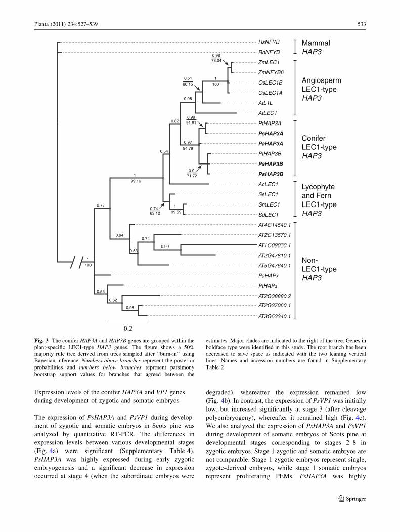
To analyze the relationship between the conifer HAP3
genes and HAP3 genes from other species, phylogenetic
analyses based on nucleotide sequences from the conserved
B-domain were made (Fig. 3). The phylogenetic analyses
of HAP3 genes using Bayesian inference or heuristic
searches of most parsimonious trees are in general agree-
ment with Kwong et al. (2003) and Xie et al. (2008),
supporting that plant HAP3 genes can be subdivided into
LEC1-type and non-LEC1-type HAP3 genes (Fig. 3). Ly-
cophyte and fern LEC1-type genes group at the base of the
seed plant LEC1-type HAP3 genes. Both seed plant lin-
eages harbor paralogous LEC1-type HAP3 genes, as
exemplified by the presence of two LEC1-type HAP3
genes in rice (Oryza sativa) and Arabidopsis, as well as in
conifers (HAP3A and HAP3B).
ABI3 belongs to the large B3 family of plant-specific
transcription factors (Giraudat et al. 1992; Suzuki and
McCarty 2008) The full-length PaVP1 cDNA (accession
number AF175576), which has been reported earlier
(Footitt et al. 2003), contains all described conserved
regions (A1, B1–B3) reported for ABI3 andVP1 (McCarty
et al. 1991; Giraudat et al. 1992). The putative Scots pine
VP1/ABI3 homolog PsVP1 was isolated using primers
designed from a loblolly pine EST and PaVP1 (Supple-
mentary Table 1). The partial PsVP1 cDNA, starting in the
A1 region and extending beyond the conserved B3 region,
included 1,939 nucleotides and shares 78% similarity with
PaVP1.
To analyze the correspondence and evolution of PaVP1
and PsVP1, a Bayesian phylogenetic analysis was per-
formed using nucleotide sequences from the B3 domains of
the two conifer genes, the maize VP1 gene and all 118 B3
genes of Arabidopsis. The consensus tree showed a clear
separation of known subgroups in the phylogeny of B3
genes, i.e., the AFL genes and the embryonic repressor
genes (VAL1, VAL2 and VAL3), as suggested previously
(Swaminathan et al. 2008; Romanel et al. 2009). From the
consensus tree, a subsample of 35 genes was selected for a
final analysis (Supplementary Fig. 1) The conifer genes
were positioned closest to the maize VP1 gene and ABI3
from Arabidopsis, confirming a close relationship between
the conifer and angiosperm ABI3/VP1 genes.
Age (Days)
Initi
atio
n fr
eque
ncy
(%) b ca
0
50
100
0 10 155
Fig. 1 The embryogenic potential decreases during germination of
Norway spruce. a Cotyledonary somatic embryos germinated for
0–14 days were stimulated to differentiate embryogenic tissue. The
proportion of embryos that had initiated embryogenic tissue within
5 weeks is presented. Frequencies are based on three biological
replicates with 30 embryos per treatment. b Embryogenic tissue
differentiated from a mature cotyledonary embryo. c Embryogenic
(arrow) and non-embryogenic tissue differentiated from a 2 week-old
germinated embryo. Bars, 2 mm
a b
Initi
atio
n fr
eque
ncy
(%)
-TSA
c100
75
50
25
0+TSA
Fig. 2 The HDAC inhibitor TSA inhibits post-germination growth
and maintains the embryogenic potential in germinating somatic
embryos of Norway spruce. a Control embryos germinated for
10 days and b embryos germinated on medium containing 10 lM
TSA for 10 days. Bars, 10 mm. c After 10 days germination on
medium lacking (-TSA) or supplemented (?TSA) with TSA, the
embryos were incubated on proliferation medium for 5 weeks to
stimulate initiation of embryogenic tissue. The frequency of initiation
is based on three biological replicates with 100 embryos per
treatment. The difference in initiation frequency between -TSA
and ?TSA was significant, P \ 0.001, Fisher’s exact test
532 Planta (2011) 234:527–539
123
Expression levels of the conifer HAP3A and VP1 genes
during development of zygotic and somatic embryos
The expression of PsHAP3A and PsVP1 during develop-
ment of zygotic and somatic embryos in Scots pine was
analyzed by quantitative RT-PCR. The differences in
expression levels between various developmental stages
(Fig. 4a) were significant (Supplementary Table 4).
PsHAP3A was highly expressed during early zygotic
embryogenesis and a significant decrease in expression
occurred at stage 4 (when the subordinate embryos were
degraded), whereafter the expression remained low
(Fig. 4b). In contrast, the expression of PsVP1 was initially
low, but increased significantly at stage 3 (after cleavage
polyembryogeny), whereafter it remained high (Fig. 4c).
We also analyzed the expression of PsHAP3A and PsVP1
during development of somatic embryos of Scots pine at
developmental stages corresponding to stages 2–8 in
zygotic embryos. Stage 1 zygotic and somatic embryos are
not comparable. Stage 1 zygotic embryos represent single,
zygote-derived embryos, while stage 1 somatic embryos
represent proliferating PEMs. PsHAP3A was highly
HsNFYB
RnNFYB
ZmLEC1
ZmNFYB6
OsLEC1B
OsLEC1A
AtL1L
AtLEC1
PtHAP3A
PsHAP3A
PaHAP3A
PtHAP3B
PaHAP3B
PsHAP3B
AcLEC1
SsLEC1
SmLEC1
SdLEC1
AT4G14540.1
PaHAPx
PtHAPx
AT2G13570.1
AT1G09030.1
AT2G47810.1
AT5G47640.1
AT2G38880.2
AT2G37060.1
AT3G53340.1
Angiosperm LEC1-type HAP3
Mammal HAP3
Conifer LEC1-type HAP3
Lycophyte and Fern LEC1-type HAP3
Non-LEC1-type HAP3
199.16
0.54
1
0.97
0.82
0.98
0.51
0.98
1
0.77
0.940.74
0.990.53
0.53
0.62
0.98
1
100
78.04
10080.15
94.79
0.7463.12 99.59
0.971.72
0.9991.61
Fig. 3 The conifer HAP3A and HAP3B genes are grouped within the
plant-specific LEC1-type HAP3 genes. The figure shows a 50%
majority rule tree derived from trees sampled after ‘‘burn-in’’ using
Bayesian inference. Numbers above branches represent the posterior
probabilities and numbers below branches represent parsimony
bootstrap support values for branches that agreed between the
estimates. Major clades are indicated to the right of the tree. Genes in
boldface type were identified in this study. The root branch has been
decreased to save space as indicated with the two leaning vertical
lines. Names and accession numbers are found in Supplementary
Table 2
Planta (2011) 234:527–539 533
123
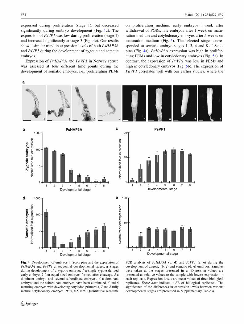
expressed during proliferation (stage 1), but decreased
significantly during embryo development (Fig. 4d). The
expression of PsVP1 was low during proliferation (stage 1)
and increased significantly at stage 3 (Fig. 4e). Our results
show a similar trend in expression levels of both PsHAP3A
and PsVP1 during the development of zygotic and somatic
embryos.
Expression of PaHAP3A and PaVP1 in Norway spruce
was assessed at four different time points during the
development of somatic embryos, i.e., proliferating PEMs
on proliferation medium, early embryos 1 week after
withdrawal of PGRs, late embryos after 1 week on matu-
ration medium and cotyledonary embryos after 5 weeks on
maturation medium (Fig. 5). The selected stages corre-
sponded to somatic embryo stages 1, 3, 4 and 8 of Scots
pine (Fig. 4a). PaHAP3A expression was high in prolifer-
ating PEMs and low in cotyledonary embryos (Fig. 5a). In
contrast, the expression of PaVP1 was low in PEMs and
high in cotyledonary embryos (Fig. 5b). The expression of
PaVP1 correlates well with our earlier studies, where the
1 2 3 4 5 6 7 8
Nor
mal
ized
fold
exp
ress
ion
Nor
mal
ized
fold
exp
ress
ion
Nor
mal
ized
fold
exp
ress
ion
Nor
mal
ized
fold
exp
ress
ion
c
e
b
d
a
Zyg
oti
c em
bry
os
So
mat
ic e
mb
ryo
s
PsHAP3A PsVP11000
100
10
11 2 3 4 5 76 8
1000
100
10
11 2 3 4 5 76 8
100
10
11 2 3 4 5 76 8
100
10
11 2 3 4 5 76 8
Developmental stage
Developmental stage
Developmental stage
Developmental stage
Fig. 4 Development of embryos in Scots pine and the expression of
PsHAP3A and PsVP1 at sequential developmental stages. a Stages
during development of a zygotic embryo; 1 a single zygote-derived
early embryo, 2 four equal-sized embryos formed after cleavage, 3 a
dominant embryo and several subordinate embryos, 4 a dominant
embryo, and the subordinate embryos have been eliminated, 5 and 6maturing embryos with developing cotyledon primordia, 7 and 8 fully
mature cotyledonary embryos. Bars, 0.5 mm. Quantitative real-time
PCR analysis of PsHAP3A (b, d) and PsVP1 (c, e) during the
development of zygotic (b, c) and somatic (d, e) embryos. Samples
were taken at the stages presented in a. Expression values are
presented as relative values to the sample with lowest expression in
each replicate. Expression levels are mean values of three biological
replicates. Error bars indicate ± SE of biological replicates. The
significance of the differences in expression levels between various
developmental stages are presented in Supplementary Table 4
534 Planta (2011) 234:527–539
123

expression of PaVP1 is highest around the early cotyle-
donary stage (Footitt et al. 2003).
TSA affects the expression of PaVP1 and PaHAP3A
during embryo development
To test whether HDAC inhibition affects the expression
of PaHAP3A and PaVP1, expression analyses were ini-
tially carried out on 10 day-old germinated somatic
embryos of Norway spruce treated with TSA. However,
the expression levels of both genes were very low, under
a reliable detection limit, in both control and TSA-treated
embryos.
The effect of TSA treatment on the expression of
PaHAP3A and PaVP1 was therefore analyzed during the
maturation phase of somatic embryos. In control cultures,
many cotyledonary embryos had developed after 4 weeks
on maturation medium (Fig. 6a). In contrast, cultures
developing on maturation medium supplemented with
TSA continued to proliferate and maturation was arrested
(Fig. 6b). However, if TSA was excluded from the
maturation medium after 4 weeks, the embryos continued
to mature and develop into cotyledonary embryos. In
control cultures, PaHAP3A expression decreased during
maturation. This decrease in PaHAP3A expression was
significantly inhibited in TSA-treated cultures (Fig. 6c).
The expression of PaVP1 increased in control cultures
during maturation. The increase in expression of PaVP1
was significantly inhibited in TSA-treated cultures
(Fig. 6d).
Discussion
Stimulation of somatic embryogenesis in germinating
embryos by TSA
Embryogenic cultures of conifers are routinely established
from immature or mature zygotic embryos. The initiation
frequency deceases dramatically when the embryos start
germinating (Bonga et al. 2010; Klimaszewska et al.
2010b, and this work). Here, we demonstrate that after
exposure to the HDAC inhibitor TSA, germinating
embryos of Norway spruce maintain the competence to
differentiate into embryogenic tissue at the same time as
the germination progression is partially inhibited. It has
previously been demonstrated that seed germination in
Arabidopsis is inhibited by TSA. Simultaneously, the
expression of embryogenesis-related genes, including the
LEC genes and ABI3, are activated and embryo-like
structures differentiate on the true leaves after withdrawal
of TSA (Tanaka et al. 2008). Our results imply that TSA
affects the embryogenic potential and germination in a
similar way in conifers. To analyze if TSA also affects
gene expression in conifers, we isolated embryogenesis-
related genes from Norway spruce and Scots pine.
Conifer homologs of AtLEC1 and AtABI3 are expressed
during embryo development
To isolate putative conifer homologs of LEC1, LEC2,
FUS3 and ABI3, we initially blasted the Arabidopsis
sequences against conifer EST databases. Only genes
highly similar to LEC1 and ABI3 were found. At present,
no full genome sequence of any gymnosperm is available
and therefore it is too early to exclude the possibility that
there are conifer homologs to LEC2 and FUS3 as well.
We isolated two conifer LEC1-type HAP3 genes in both
Norway spruce and Scots pine, PaHAP3A, PaHAP3B,
PsHAP3A and PsHAP3B. Both conifer HAP3A and HAP3B
genes encode proteins that are highly similar to LEC1-type
HAP3 subunits of the CCAAT binding transcription factor
(CBF, or NF-Y) (Lotan et al. 1998; Lee et al. 2003; Kwong
et al. 2003). Furthermore, they all contain nearly all amino
acids characteristic for LEC1-type HAP3 subunits in Ara-
bidopsis, including the important amino acid asp 55, which
confer DNA binding specificity to the CBF complex (Lee
et al. 2003). Phylogenetic analyses position both conifer
HAP3A and HAP3B genes among LEC1-type genes. Within
the LEC1-type genes there are well-supported clades sep-
arating angiosperms, gymnosperms and lycophytes (Fig. 4).
Lycophyte LEC1-type genes group basal to the angiosperm
and gymnosperm clade suggesting that an ancestral LEC1-
type gene was present in the last common ancestor of all
extant seed plants. Both angiosperms and gymnosperms
Nor
mal
ized
fold
exp
ress
ion
ba PaHAP3A PaVP1
1000
100
10
1
10000
1
10
100
Nor
mal
ized
fold
exp
ress
ion
PEM EE LE CE PEM EE LE CE
Developmental stage Developmental stage
Fig. 5 Expression of PaHAP3A and PaVP1 during the development
of somatic embryos in Norway spruce. Somatic embryos collected
from embryogenic cultures of Norway spruce were taken at four
developmental stages, i.e., proliferating PEMs on proliferation
medium, early embryos (EE) 1 week after withdrawal of PGRs, late
embryos (LE) after 1 week on maturation medium and cotyledonary
embryos (CE) after 5 weeks on maturation medium. Quantitative
real-time PCR analysis of PaHAP3A (a) and PaVP1 (b). Expression
values are presented as relative values to the sample with lowest
expression in each replicate. Expression levels are mean values of two
biological replicates assessed three times each. Error bars indi-
cate ±SE of biological replicates
Planta (2011) 234:527–539 535
123

seem to have undergone separate duplications within the
LEC1-type lineage. Maize has been suggested to harbor up
to three copies of LEC1-type HAP3 genes, although the
accessions are not publicly available (Suzuki et al. 2008).
Similarly, the conifer LEC1-type HAP3A and HAP3B genes
are most likely the result of a separate duplication event
within the gymnosperm lineage.
The Norway spruce homolog to ABI3, PaVP1, has
previously been characterized (Footitt et al. 2003). In this
study we isolated its Scots pine homolog, PsVP1. The
protein is highly similar to that of Norway spruce and
sequence analysis revealed close relationship of both
conifer genes to ABI3 and its maize homolog VP1, within
the group of plant-specific B3 transcription factors.
During early conifer embryogenesis, the expression of
PaHAP3A and PsHAP3A is initially high, in agreement
with recently reported expression of a putative LEC1-type
HAP3 gene in lodgepole pine (Pinus contorta) (Park et al.
2010). Later, during development of the embryos, the
expression of PaHAP3A and PsHAP3A decreases dramat-
ically and remains low throughout maturation (Figs. 4, 5).
In contrast, the expression of the conifer PaVP1 and PsVP1
genes increases early during embryo development and
peaks at the time point when the cotyledons emerge
(Figs. 4, 5). These results are analogous with what has been
reported in Arabidopsis, where LEC1 is expressed at higher
levels during early embryo development than in maturing
embryos (Lotan et al. 1998; Baumbusch et al. 2004). Fur-
thermore, the expression of ABI3 can first be detected at the
globular stage and persists throughout maturation of the
embryo (Parcy et al. 1997; To et al. 2006).
Preliminary expression studies of PaHAP3B indicate a
comparable low overall expression, and in contrast to
PaHAP3A the expression increases at later maturation
-TSA+TSA
-TSA+TSA
Nor
mal
ized
fold
exp
ress
ion
Nor
mal
ized
fold
exp
ress
ion
b
c d
a
0.01
0.1
1
10 2 3 4
10
10 2 3 4
1
10
0.1
Weeks on maturation medium Weeks on maturation medium
PsHAP3A PsVP1
Fig. 6 The HDAC inhibitor TSA blocks maturation and affects the
expression of PaHAP3A and PaVP1 during the development of
somatic embryos in Norway spruce. Embryogenic cultures incubated
for 4 weeks on maturation medium lacking TSA (a) or containing
10 lM TSA (b). Bars, 2 mm. Quantitative real-time PCR analysis of
PaHAP3A (c) and PaVP1 (d) in embryogenic cultures treated with
TSA (light bars) and control cultures without TSA (dark bars).
Samples were collected during differentiation of early somatic
embryos (0 weeks) and after 1–4 weeks on maturation medium.
Expression values are presented as relative values to one of the
samples from untreated early embryogenic cultures (0 weeks).
Expression levels are shown as mean values of three biological
replicates assessed three times each. Error bars indicate ±SE of
biological replicates. The expression levels of TSA-treated samples
(?TSA) significantly differed from the untreated control samples
(-TSA) at each developmental stage for both genes, P \ 0.05
536 Planta (2011) 234:527–539
123

stages (data not shown). Duplication within a gene line-
age could be indicative of functional divergence leading
to subfunctionalization, and/or neofunctionalization
according to Irish and Litt 2005, which may be reflected
by differential expression during development. Hence, the
data suggest that the conifer HAP3 genes might have
gone through subfunctionalization, and/or neofunctional-
ization, with PaHAP3A showing the highest similarity in
activity to extant angiosperm LEC1 genes. However, we
cannot exclude that the HAP3B gene might have impor-
tant roles and partly overlapping functions with HAP3A
during embryogenesis in conifers, as suggested for LEC1
and L1L in Arabidopsis (Kwong et al. 2003; Yamamoto
et al. 2009).
The LEC genes (LEC1, LEC2 and FUS3) have been
shown necessary to confer embryonic cell fate and
specification of cotyledon identity early during embryo-
genesis (Meinke 1992; Baumlein et al. 1994; Keith et al.
1994; Meinke et al. 1994; West et al. 1994; Lotan et al.
1998; Luerssen et al. 1998; Stone et al. 2001; Gazzarrini
et al. 2004). Mutant analysis has indicated that LEC1
might act upstream of LEC2 and FUS3 (Meinke et al.
1994). The expression of FUS3 and ABI3 is regulated by
LEC1 and LEC2 as well as by themselves. Furthermore,
the spatial expression seems to be very important, i.e., it
has been demonstrated that ABI3 is regulated by a com-
plex network that involves LEC1 in the cotyledons, but
not in the embryo axis (To et al. 2006). Molecular data
has also shown that ectopic LEC1 expression induces
expression of ABI3 (Kagaya et al. 2005). Later during
embryogenesis, ABI3, together with the LEC genes, is
responsible for the initiation and maintenance of the
maturation phase. To what extent the conifer HAP3A and
VP1 genes interact is presently not known, but the fact
that the expression profiles of the genes are each other’s
opposite during embryo development might indicate a
direct or indirect feedback regulation or at least a com-
mon regulatory machinery.
Ectopic expression of CHAP3A (a putative LEC1-type
HAP3 gene from Picea mariana) under an inducible pro-
moter was recently reported in germinating somatic
embryos of white spruce (Klimaszewska et al. 2010b).
However, the ectopic expression of CHAP3A did not
induce the expression of other embryogenesis-related
genes, neither was the development of the embryos affec-
ted. This is in contrast to that previously shown in Ara-
bidopsis, where ectopic post-embryonic expression of the
LEC1 gene in vegetative cells can induce the expression of
embryo-specific genes and stimulate formation of embryo-
like structures (Lotan et al. 1998). Further studies on the
functions of the LEC1-type HAP3A and HAP3B genes are
necessary to understand their roles during conifer
embryogenesis.
Expression of embryo-specific transcription factors is
affected by TSA
We have shown that treatment of Norway spruce embryos
with TSA during germination partially inhibits germination
and maintains the embryogenic potential (Figs. 2, 3).
However, we could not show whether the TSA treatment
during germination affects the expression of PaHAP3A or
PaVP1, since both genes were expressed at very low levels.
Of importance is that inhibition of HDAC activity during
embryo germination stimulates the initiation of embryo-
genic tissues in a localized non-uniform pattern. Recently,
Klimaszewska et al. (2010a) reported initiation of
embryogenic tissue from buds of 10 year-old trees regen-
erated from somatic embryos of white spruce. The induc-
tion of embryogenic tissue was shown to be associated with
formation of nodules on the surface of the needle. The
nodules differentiate in a similar pattern as we have shown
for protruding embryogenic tissue on germinating embryos
(Fig. 1c). Interestingly, Klimaszewska et al. (2010a)
showed a high expression of CHAP3A in embryogenic
tissue, but could not detect any expression in the needles.
The results from the Klimaszewska group and results
presented in this study suggest that the expression of genes
stimulating the initiation of embryogenic competence are
localized to a limited number of cells in the explant, not
detectable in total RNA from the whole explant. We
assume that some cells in the germinating embryo still
possess the intracellular environment needed for embryo-
genic competence and that application of the HDAC
inhibitor TSA enables transcription of embryogenesis-
related genes and transcription factors needed for gener-
ating embryogenic tissue from these cells.
To examine if TSA affects the expression of PaHAP3A
and PaVP1, we analyzed the expression of the genes during
the maturation phase. The expression of PaHAP3A
decreases during maturation, while that of PaVP1 increa-
ses. When embryogenic cultures of Norway spruce are
exposed to TSA during the maturation phase, the cultures
continue to proliferate and maturation is arrested (Fig. 6a,
b). Simultaneously, the expression of PaVP1 remains low
while the expression of PaHAP3A remains high throughout
the maturation treatment (Fig. 6c, d). Assuming that TSA
affects histone acetylation in conifers, our results indicate a
connection between changes in acetylation patterns and the
levels of embryogenesis-related gene expression in Norway
spruce.
Acknowledgments The authors would like to thank Ulf Olsson for
statistical consultancy. This work was supported by the Swedish
Research Council for Environment, Agricultural Sciences and Spatial
Planning (Formas) and the Royal Swedish Academy of Agriculture
and Forestry. Silvia Valladares was supported by an Angeles Alvarino
postdoctoral fellowship from Xunta de Galicia (Spain).
Planta (2011) 234:527–539 537
123

Open Access This article is distributed under the terms of the
Creative Commons Attribution Noncommercial License which per-
mits any noncommercial use, distribution, and reproduction in any
medium, provided the original author(s) and source are credited.
References
Altschul SF, Madden TL, Schaffer AA, Zhang J, Zhang Z, Miller W,
Lipman DJ (1997) Gapped BLAST and PSI-BLAST: a new
generation of protein database search programs. Nucleic Acids
Res 25:3389–3402
Baumbusch LO, Hughes DW, Galau GA, Jakobsen KS (2004) LEC1,
FUS3, ABI3 and Em expression reveals no correlation with
dormancy in Arabidopsis. J Exp Bot 55:77–87
Baumlein H, Misera S, Luerßen H, Kolle K, Horstmann C, Wobus U,
Muller AJ (1994) The FUS3 gene of Arabidopsis thaliana is a
regulator of gene expression during late embryogenesis. The
Plant J 6:379–387
Bonga JM, Klimaszewska KK, von Aderkas P (2010) Recalcitrance in
clonal propagation, in particular of conifers. Plant Cell Tiss Org
100:241–254
Braybrook SA, Harada JJ (2008) LECs go crazy in embryo
development. Trends Plant Sci 13:624–630
Burg K, Helmersson A, Bozhkov P, von Arnold S (2007) Develop-
mental and genetic variation in nuclear microsatellite stability
during somatic embryogenesis in pine. J Exp Bot 58:687–698
Cairney J, Pullman GS (2007) The cellular and molecular biology of
conifer embryogenesis. New Phytol 176:511–536
Dean Rider S, Henderson JT, Jerome RE, Edenberg HJ, Romero-
Severson J, Ogas J (2003) Coordinate repression of regulators of
embryonic identity by PICKLE during germination in Arabid-opsis. Plant J 35:33–43
Drummond AJ, Ashton B, Buxton S, Cheung M, Cooper A, Heled J,
Kearse M, Moir R, Stones-Havas S, Sturrock S, Thierer T,
Wilson A (2010) Geneious v5.1, available from http://www.
geneious.com
Felsenstein J (1985) Confidence-limits on phylogenies—an approach
using the bootstrap. Evolution 39:783–791
Footitt S, Ingouff M, Clapham D, von Arnold S (2003) Expression of
the viviparous 1 (Pavp1) and p34cdc2 protein kinase (cdc2 Pa)
genes during somatic embryogenesis in Norway spruce (Piceaabies [L.] Karst). J Exp Bot 54:1711–1719
Gazzarrini S, Tsuchiya Y, Lumba S, Okamoto M, McCourt P (2004)
The transcription factor FUSCA3 controls developmental timing
in Arabidopsis through the hormones gibberellin and abscisic
acid. Dev Cell 7:373–385
Giraudat J, Hauge BM, Valon C, Smalle J, Parcy F, Goodman HM
(1992) Isolation of the Arabidopsis ABI3 gene by positional
cloning. Plant Cell 4:1251–1261
Goldberg RB, de Paiva G, Yadegari R (1994) Plant embryogenesis:
zygote to seed. Science 266:605–614
Gutierrez L, Van Wuytswinkel O, Castelain M, Bellini C (2007)
Combined networks regulating seed maturation. Trends Plant Sci
12:294–300
Irish VF, Litt A (2005) Flower development and evolution: gene
duplication, diversification and redeployment. Curr Opin Genet
Dev 15:454–460
Kagaya Y, Toyoshima R, Okuda R, Usui H, Yamamoto A, Hattori T
(2005) LEAFY COTYLEDON1 controls seed storage protein
genes through its regulation of FUSCA3 and ABSCISIC ACIDINSENSITIVE3. Plant Cell Physiol 46:399–406
Keith K, Kraml M, Dengler NG, McCourt P (1994) fusca3: a
heterochronic mutation affecting late embryo development in
Arabidopsis. Plant Cell 6:589–600
Klimaszewska K, Overton C, Stewart D, Rutledge RG (2010a)
Initiation of somatic embryos and regeneration of plants from
primordial shoots of 10 year-old somatic white spruce and
expression profiles of 11 genes followed during the tissue culture
process. Planta 233:635–647
Klimaszewska K, Pelletier G, Overton C, Stewart D, Rutledge RG
(2010b) Hormonally regulated overexpression of ArabidopsisWUS and conifer LEC1 (CHAP3A) in transgenic white spruce:
implications for somatic embryo development and somatic
seedling growth. Plant Cell Rep 29:723–734
Kwong RW, Bui AQ, Lee H, Kwong LW, Fischer RL, Goldberg RB,
Harada JJ (2003) LEAFY COTYLEDON1-LIKE defines a class of
regulators essential for embryo development. Plant Cell 15:5–18
Laux T, Wurschum T, Breuninger H (2004) Genetic regulation of
embryonic pattern formation. Plant Cell 16 Suppl:S190–S202
Le BH, Cheng C, Bui AQ, Wagmaister JA, Henry KF, Pelletier J,
Kwong L, Belmonte M, Kirkbride R, Horvath S, Drews GN,
Fischer RL, Okamuro JK, Harada JJ, Goldberg RB (2010)
Global analysis of gene activity during Arabidopsis seed
development and identification of seed-specific transcription
factors. Proc Natl Acad Sci USA 107:8063–8070
Lee H, Fischer RL, Goldberg RB, Harada JJ (2003) ArabidopsisLEAFY COTYLEDON1 represents a functionally specialized
subunit of the CCAAT binding transcription factor. Proc Natl
Acad Sci USA 100:2152–2156
Littell RC, Milliken GA, Stroup WW, Wolfinger RD, Schabenberger
O (2007) SAS for mixed models. J Roy Statist Soc 170:257–258
Lotan T, Ohto M, Yee KM, West MA, Lo R, Kwong RW, Yamagishi
K, Fischer RL, Goldberg RB, Harada JJ (1998) ArabidopsisLEAFY COTYLEDON1 is sufficient to induce embryo devel-
opment in vegetative cells. Cell 93:1195–1205
Luerssen H, Kirik V, Herrmann P, Misera S (1998) FUSCA3 encodes
a protein with a conserved VP1/AB13-like B3 domain which is
of functional importance for the regulation of seed maturation in
Arabidopsis thaliana. Plant J 15:755–764
McCarty DR, Hattori T, Carson CB, Vasil V, Lazar M, Vasil IK
(1991) The Viviparous-1 developmental gene of maize encodes a
novel transcriptional activator. Cell 66:895–905
Meinke DW (1992) A homoeotic mutant of Arabidopsis thaliana with
leafy cotyledons. Science 258:1647–1650
Meinke DW, Franzmann LH, Nickle TC, Yeung EC (1994) LeafyCotyledon mutants of Arabidopsis. Plant Cell 6:1049–1064
Moretti S, Reinier F, Poirot O, Armougom F, Audic S, Keduas V,
Notredame C (2006) PROTOGENE: turning amino acid align-
ments into bona fide CDS nucleotide alignments. Nucleic Acids
Res 34(Web Server issue):W600–W603
Morgenstern B, Goel S, Sczyrba A, Dress A (2003) AltAVisT:
comparing alternative multiple sequence alignments. Bioinfor-
matics 19:425–426
Parcy F, Valon C, Kohara A, Misera S, Giraudat J (1997) The
ABSCISIC ACID-INSENSITIVE3, FUSCA3, and LEAFY COTY-LEDON1 loci act in concert to control multiple aspects of
Arabidopsis seed development. Plant Cell 9:1265–1277
Park S-Y, Klimaszewska K, Park J-Y, Mansfield SD (2010)
Lodgepole pine: the first evidence of seed-based somatic
embryogenesis and the expression of embryogenesis marker
genes in shoot bud cultures of adult trees. Tree Physiol 30:1469–
1478
Romanel EAC, Schrago CG, Counago RM, Russo CAM, Alves-
Ferreira M (2009) Evolution of the B3 DNA binding superfam-
ily: new insights into REM family gene diversification. PLoS
ONE 4:e5791
Ronquist F, Huelsenbeck JP (2003) MrBayes 3: Bayesian phylogenetic
inference under mixed models. Bioinformatics 19:1572–1574
Santos-Mendoza M, Dubreucq B, Baud S, Parcy F, Caboche M,
Lepiniec L (2008) Deciphering gene regulatory networks that
538 Planta (2011) 234:527–539
123
control seed development and maturation in Arabidopsis. Plant J
54:608–620
Schallau A, Kakhovskaya I, Tewes A, Czihal A, Tiedemann J, Mohr
M, Grosse I, Manteuffel R, Baumlein H (2008) Phylogenetic
footprints in fern spore- and seed-specific gene promoters. Plant
J 53:414–424
Smith SA, Beaulieu JM, Donoghue MJ (2010) An uncorrelated
relaxed-clock analysis suggests an earlier origin for flowering
plants. Proc Natl Acad Sci USA 107:5897–5902
Stone SL, Kwong LW, Yee KM, Pelletier J, Lepiniec L, Fischer RL,
Goldberg RB, Harada JJ (2001) LEAFY COTYLEDON2 encodes
a B3 domain transcription factor that induces embryo develop-
ment. Proc Natl Acad Sci USA 98:11806–11811
Suzuki M, McCarty DR (2008) Functional symmetry of the B3
network controlling seed development. Curr Opin Plant Biol
11:548–553
Suzuki M, Wang HH-Y, McCarty DR (2007) Repression of the
LEAFY COTYLEDON 1/B3 regulatory network in plant embryo
development by VP1/ABSCISIC ACID INSENSITIVE 3-LIKE B3
genes. Plant Physiol 143:902–911
Suzuki M, Latshaw S, Sato Y, Settles AM, Koch KE, Hannah LC,
Kojima M, Sakakibara H, McCarty DR (2008) The maize
Viviparous8 locus, encoding a putative ALTERED MERISTEM
PROGRAM1-like peptidase, regulates abscisic acid accumula-
tion and coordinates embryo and endosperm development. Plant
Physiol 146:1193–1206
Swaminathan K, Peterson K, Jack T (2008) The plant B3 superfamily.
Trends Plant Sci 13:647–655
Swofford DL (2001) PAUP* :Phylogenetic analysis using parsimony
(* and other methods). Sinauer Associates, Sunderland
Tai HH, Tai GCC, Beardmore T (2005) Dynamic histone acetylation
of late embryonic genes during seed germination. Plant Mol Biol
59:909–925
Tanaka M, Kikuchi A, Kamada H (2008) The Arabidopsis histone
deacetylases HDA6 and HDA19 contribute to the repression of
embryonic properties after germination. Plant Physiol 146:149–161
To A, Valon C, Savino G, Guilleminot J, Devic M, Giraudat J, Parcy
F (2006) A network of local and redundant gene regulation
governs Arabidopsis seed maturation. Plant Cell 18:1642–1651
Vandesompele J, De Preter K, Pattyn F, Poppe B, Van Roy N, De
Paepe A, Speleman F (2002) Accurate normalization of real-time
quantitative RT-PCR data by geometric averaging of multiple
internal control genes. Genome Biol 3:RESEARCH0034.1-
0034.11
Vestman D, Larsson E, Uddenberg D, Cairney J, Clapham D,
Sundberg E, Arnold S (2011) Important processes during
differentiation and early development of somatic embryos of
Norway spruce as revealed by changes in global gene expres-
sion. Tree Genet Genomes 7:347–362
von Arnold S, Clapham D (2008) Spruce embryogenesis. Methods
Mol Biol 427:31–47
von Arnold S, Sabala I, Bozhkov P, Dyachok J, Filonova L (2002)
Developmental pathways of somatic embryogenesis. Plant Cell
Tiss Org 69:233–249
West M, Harada JJ (1993) Embryogenesis in higher plants: an
overview. Plant Cell 5:1361–1369
West M, Yee KM, Danao J, Zimmerman JL, Fischer RL, Goldberg
RB, Harada JJ (1994) LEAFY COTYLEDON1 is an essential
regulator of late embryogenesis and cotyledon identity in
Arabidopsis. Plant Cell 6:1731–1745
Xia X, Xie Z, Salemi M, Chen L, Wang Y (2003) An index of
substitution saturation and its application. Mol Phylogenet Evol
26:1–7
Xie Z, Li X, Glover BJ, Bai S, Rao G-Y, Luo J, Yang J (2008)
Duplication and functional diversification of HAP3 genes
leading to the origin of the seed-developmental regulatory gene,
LEAFY COTYLEDON1 (LEC1), in nonseed plant genomes. Mol
Biol Evol 25:1581–1592
Yamamoto A, Kagaya Y, Toyoshima R, Kagaya M, Takeda S, Hattori
T (2009) Arabidopsis NF-YB subunits LEC1 and LEC1-LIKE
activate transcription by interacting with seed-specific ABRE-
binding factors. Plant J 58:843–856
Zhang H, Ogas J (2009) An epigenetic perspective on developmental
regulation of seed genes. Molecular Plant 2:610–627
Planta (2011) 234:527–539 539
123