original article dicer1 is required to repress...
TRANSCRIPT
Dicer1 Is Required to Repress Neuronal FateDuring Endocrine Cell MaturationMurtaza S. Kanji,
1,2Martin G. Martin,
3and Anil Bhushan
1,2,4
MicroRNAs (miRNAs) are important regulators of gene expres-sion programs in the pancreas; however, little is known about therole of miRNA pathways during endocrine cell specification andmaturation during neonatal life. In this study, we deleted Dicer1,an essential RNase for active miRNAs biogenesis, specificallyfrom NGN3+ endocrine progenitor cells. We found that deletionof Dicer1 in endocrine progenitors did not affect the specifica-tion of hormone-expressing endocrine cells. However, the isletsin the mutant mice in the neonatal period exhibited morpholog-ical defects in organization and loss of hormone expression,and the mutant mice subsequently developed diabetes. Dicer1-deficient b-cells lost insulin expression while maintaining theexpression of b-cell transcription factors such as Pdx1 andNkx6.1 early in the postnatal period. Surprisingly, transcriptionalprofiling showed that that the Dicer1-deficient endocrine cellsexpressed neuronal genes before the onset of diabetes. The de-repression of neuronal genes was associated with a loss in bind-ing of the neuronal transcriptional repressor RE-1-silencingtranscription factor to its targets in Dicer1-deficient b-cells.These studies suggest that miRNAs play a critical role in sup-pressing neuronal genes during the maturation of endocrinecells. Diabetes 62:1602–1611, 2013
Apotential therapeutic approach to replenish thepancreatic b-cell mass in diabetic patientsinvolves the transplantation of functional,glucose-responsive b-cells differentiated from
human pluripotent stem cells. Several attempts have beenmade at differentiating b-cells in vitro from stem cells, withlimited success, (1,2) because the insulin-expressing cellsgenerated lack the characteristic hallmarks of functionallymature b-cells, such as the ability to regulate glucose-stimulated insulin-secretion. Although many transcriptionfactors and signaling pathways underlying the stepwisecell fate acquisition during b-cell development are known(3–6), a complete understanding of the molecular basis ofb-cell specification and functional maturation is lacking.
Of significant interest is the role of microRNAs (miRNAs)in regulating the pancreatic developmental program.miRNAs are non–protein-coding small RNAs (;19–25
nucleotides) that negatively regulate gene expression atthe post-transcriptional level (7) and have been implicatedas important regulators of animal development (8). Newlytranscribed miRNAs undergo a series of processing stepsthat require the RNase III enzymes Drosha and Dicer1before becoming functional (9,10). Although severalmiRNAs have been proposed to regulate b-cell transcrip-tion factors during development (11), many of these com-putationally predicted miRNA–mRNA interactions have notbeen experimentally validated in vivo. The dysregulation ofmiRNAs through Dicer1 ablation in the early embryonicpancreatic progenitor cells expressing Pdx-1 resulted insevere deficiencies in the formation of all islet cell lineages(12). More recently, it has been shown that deletion ofDicer1 in b-cells leads to loss of insulin expression and todevelopment of diabetes in adult mice (13). Although thesestudies reveal key functions of miRNA-dependent pathwaysduring early pancreatic development and in adult b-cells,they preclude analysis of the role of miRNAs during thespecification of endocrine cells and their functional matu-ration in postnatal life.
In this study, we used a mouse model where expressionof Cre recombinase directed by the Ngn3 promoter con-ditionally deleted floxed Dicer1 alleles in endocrine pro-genitor cells. In addition, by crossing these mice onto theR26RYFP reporter line, we were able to trace the lineageof the Dicer1-deficient islet progenitor cells. Our datademonstrate that Dicer1-deficient endocrine progenitorsdifferentiate into hormone-expressing endocrine cells butsubsequently lose hormone expression during the neonatalperiod and develop diabetes. More surprisingly, we foundthat the Dicer1-deficient islet cells expressed neuronalgenes, supporting a model in which miRNA pathwayscontrol important transcriptional networks required forsuppressing neuronal fate during the maintenance andmaturation of newly specified endocrine cells.
RESEARCH DESIGN AND METHODS
Mice and physiology. Mice were maintained in a 12-h light/dark cycle understandard conditions. Studies involving mice were performed in accordancewith National Institutes of Health policies on the use of laboratory animals andapproved by the University of California, Los Angeles (UCLA) Animal ResearchCommittee. The mice used in this study are the conditional Dicer1 flox/flox line(14), the Ngn3-Cre (15), and the R26R-YFP (16) lines. The control mice usedthroughout were heterozygous for the conditional Dicer1 allele and the Ngn3-Cre transgene (NC:Dicer1fl/+). All mice were maintained in the C57BL6background.
DNA extracted from tails was used for PCR-based genotyping. Blood glu-cose levels were measured from tail vein blood using a FreeStyle glucometer(Abbot Diabetes Care), and pancreatic insulin content was measured usinga mouse insulin ELISA kit (Mercodia) after acid ethanol (0.18 mol/L HCl in 70%ethanol) extraction according to the protocol recommended by the AnimalModels of Diabetes Complications Consortium (http://www.amdcc.org/).Histology and immunohistochemistry. Pancreatic tissue was processed forimmunohistochemical analyses as previously described (17). Briefly, thepancreas was dissected and fixed in 4% formaldehyde for 2 h before beingembedded in paraffin. Sections (5-mm thick) were deparaffinized, rehydrated,
From the 1Department of Medicine, David Geffen School of Medicine, Univer-sity of California, Los Angeles, Los Angeles, California; the 2Molecular Bi-ology Interdepartmental Ph.D. Program (MBIDP), University of California,Los Angeles, Los Angeles, California; the 3Department of Pediatrics, Divi-sion of Gastroenterology and Nutrition, Mattel Children’s Hospital and theDavid Geffen School of Medicine, University of California, Los Angeles, LosAngeles, California; and 4Molecular, Cellular and Developmental Biology,University of California, Los Angeles, Los Angeles, California.
Corresponding author: Anil Bhushan, [email protected] 21 June 2012 and accepted 10 December 2012.DOI: 10.2337/db12-0841This article contains Supplementary Data online at http://diabetes
.diabetesjournals.org/lookup/suppl/doi:10.2337/db12-0841/-/DC1.� 2013 by the American Diabetes Association. Readers may use this article as
long as the work is properly cited, the use is educational and not for profit,and the work is not altered. See http://creativecommons.org/licenses/by-nc-nd/3.0/ for details.
1602 DIABETES, VOL. 62, MAY 2013 diabetes.diabetesjournals.org
ORIGINAL ARTICLE
subjected to antigen retrieval using Antigen Unmasking Buffer (Vector Labo-ratories), and permeabilized in 0.4% Triton X-100/TBS. Tissues were sub-sequently blocked with 3% IgG-free BSA (Jackson ImmunoResearchLaboratories). Incubation with primary antibodies was performed overnight at4°C in blocking solution at the following dilutions: 1:200 guinea pig anti-insulin(Dako), 1:500 rabbit anti-glucagon (Immunostar), 1:200 rabbit anti-amylase(Sigma-Aldrich), 1:100 mouse anti-Pdx1 (DHSB), 1:200 rabbit anti-Pdx1(Chemicon), 1:250 chicken anti-green fluorescent protein (GPF; Aves Labo-ratories Inc.), 1:500 rabbit anti-MafA (18), 1:100 rabbit anti-MafB (Bethyl),1:1000 rabbit anti-Glut2 (Millipore), 1:250 rabbit anti-tyrosine hydroxylase (Th;Millipore), 1:50 mouse anti-Nkx2.2 (BCBC), 1:50 mouse anti-Nkx6.1, and 1:50mouse anti-Ki67 (BD Pharmingen). Secondary antibodies (Jackson Immu-noResearch Laboratory) conjugated to fluorescein isothiocyanate (1:200 di-lution) or Cy3 (1:1000 dilution) were diluted in blocking buffer. In situ celldeath detection assay (TUNEL) was performed according to the manu-facturer’s instructions (Roche). Slides were mounted with Vectashield withDAPI (Vector Laboratories), and images were obtained using Openlab soft-ware (Perkins Elmer) and a Leica DM6000 microscope.b-Cell mass analysis. b-Cell mass was calculated, as previously described(19), by analyzing pancreata from four mice for each age and genotype.Islet isolation and fluorescence-activated cell sorter sorting. Islet iso-lation from mice was performed using a Liberase enzyme blend and purified bycentrifugation in a Histopaque gradient, as described previously (17), withslight modifications. Instead of Liberase perfusion through the common bileduct, neonatal pancreata were randomly injected with Liberase blend. Toobtain purified pancreatic and endocrine progenitor cells, pancreatic budsfrom embryonic day (e)12.5 Pdx1-yellow fluorescent protein (YFP) and e14.5Ngn3-GFP (20) embryos were dissected and dissociated into a single-cellsuspension using nonenzymatic cell dissociation buffer (Sigma-Aldrich). GFP+cells were sorted by fluorescence-activated cell sorter (FACS) (FACSaria BDbiosciences). A similar process was followed to obtain b-cells from isletsisolated from 2-week-old MIP-GFP (21) mice.RNA isolation, RT-PCR, and real-time quantitative PCR. Total RNA fromislets, dissected pancreata, and Ngn3-GFP and MIP-GFP cells was isolatedusing an RNeasy RNA extraction Kit (Qiagen). Single-stranded cDNA wasprepared using Superscript III Reverse Transcriptase (Invitrogen) with oligodTpriming. Real-time quantitaive (q)PCRs were performed using the 7900HT FastReal-time PCR system (Applied Biosystems). The expression level of eachtranscript was normalized to the housekeeping gene Cyclophilin. Data andstandard deviations shown were measured from at least three independentbiological replicate experiments. All RT-PCR and real-time qPCR primersequences used are available upon request.Chromatin immunoprecipitation assays and chromatin immunoprecipitation-
qPCR. To obtain purified b-cells from P7 control and NC:Dicer1fl/fl mice forchromatin immunoprecipitation (ChIP) analyses, dissected pancreata weredissociated into a single-cell suspension. The pancreatic cells were immunos-tained for insulin, after fixation and permeabilization with BD Cytofix reagent(BD PharMingen), using guinea pig anti-insulin antibody, followed by incubationwith a Cy3-conjugated secondary antibody, and sorted by FACS (FACSaria BDBioscience). Pancreatic cells processed without primary antibody were used asa negative control for FACS. ChIP experiments on purified b-cells wereperformed using the micro-ChIP protocol, as previously described (22). Thesequence-specific primers used to amplify the region around the RE1 sites onthe Stmn2, Stmn3, Th, and Syn1 locus are available upon request. The qPCRanalysis was performed using the 7900HT Fast Real-time PCR system (Ap-plied Biosystems). The data shown were from independent biological trip-licates, and ChIP-qPCR signals are reported as percentage of input.Microarray gene expression analysis. Two pairs of independent pancreatafrom control and mutant NC:Dicer1fl/fl mice were dissected out at postnatal day7 (P7), and total RNA was extracted using an RNeasy Plus Mini Kit (Qiagen),according to the manufacturer’s protocol. Total RNA quality was assessed usingan Agilent 2100 Bioanalyzer and an RNA Integrity Number (RIN) generatedusing 2100 Expert Software (Agilent Technologies). All RNA samples used hada RIN greater than 7. One microgram of total RNA was processed, labeled, andhybridized to the mouse 1.0ST GeneChip Microarray (Affymetrix), according tomanufacturer’s recommendations, by the UCLA DNA Microarray Core Facility.
The CEL files (raw expression measurement data) generated from arrayimage analysis were imported into Partek Genomics Suite Software (23), robustmulti-array average (RMA) normalized, and converted to log2 values. The soft-ware was further used to perform ANOVA and identify statistically significantdifferentially expressed genes (P values) between control and NC:Dicer1fl/fl
pancreata by applying a false discovery threshold of 5% to the P values.Statistical analyses. All data are expressed as means 6 SEM. Statisticalsignificance was determined by an unpaired Student t test. A P value ,0.05was used to reject the null hypothesis.
RESULTS
Dicer1-null endocrine precursor cells specifyappropriately but display loss of hormone expressionduring neonatal phase and subsequently develop
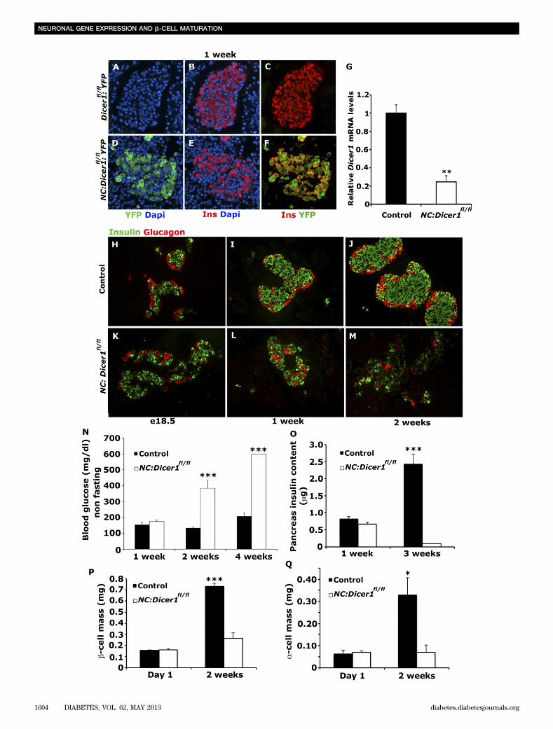
diabetes. The catalytic role of Dicer1 is central to thefunctional maturation of miRNAs from their precursors (9).To investigate in vivo the role of miRNAs during isletcell specification from endocrine precursor cells express-ing Neurogenin3 (Ngn3), the Dicer1 conditional allele(Dicer1fl/fl) (14) was deleted specifically from endocrineprecursor cells using NC-mediated excision (15) therebypreventing the generation of active miRNAs in these cells.In addition, an R26R-YFP reporter line (16) was crossedonto the NC:Dicer1fl/fl mice to heritably label any cellsundergoing recombination and deletion of Dicer1. Co-immunostaining of NC:Dicer1fl/fl pancreatic sections forinsulin/YFP (Fig. 1A–F) and glucagon/YFP (data not shown)showed a high degree of overlap of these markers in themutant islets, therefore confirming that a high percentage ofspecified islet cells were derived from endocrine precursorcells that had undergone Cre-mediated recombination. Real-time qPCR analysis revealed that the Dicer1 transcript wasreduced by 80% in the mutant NC:Dicer1fl/fl islets at 1 week(Fig. 1G), further confirming the deletion of Dicer1 froma majority of islet cells.
To determine whether loss of Dicer1 affected thespecification of islet cells, pancreatic tissue isolatedfrom control (heterozygous for the Dicer1 conditionalallele NC:Dicer1fl/1) and mutant NC:Dicer1fl/fl mice wereco-immunostained for insulin and glucagon at differentstages of neonatal development. Immunostaining of pan-creata from e18.5 NC:Dicer1fl/fl mice for insulin and glu-cagon revealed that a- and b-cells specified appropriatelyin the pancreas, and no morphological differences in isletarchitecture were apparent (Fig. 1H and K). However,pancreata from 1-week-old NC:Dicer1fl/fl mice exhibitedaltered islet organization, with many instances of a-cellsprevalent within the core of the islet rather than at theperiphery, an observation also confirmed by Pdx1 andglucagon staining at the same stage (Fig. 1I and L; Sup-plementary Fig. 1A and B). Pancreata from 2-week-old NC:Dicer1fl/fl mice displayed severe defects in morphology,and the expression of insulin and glucagon was severelydiminished (Fig. 1J and M). Quantification at 1 day afterbirth revealed similar b- and a-cell mass (Fig. 1P and Q).A similar comparison showed a dramatic reduction in en-docrine cell mass in the mutant NC:Dicer1fl/fl mice at 2weeks after birth. Consistent with the reduction in b-cellmass by 2 weeks (Fig. 1P), an almost total loss of pan-creatic insulin content was observed in the mutant NC:Dicer1fl/fl animals (shown at 3 weeks in Fig. 1O). However,only a very modest decrease in insulin content was no-ticeable in the mutant NC:Dicer1fl/fl animals at 1 week. Themutant NC:Dicer1fl/fl animals displayed an inability tometabolize glucose and developed hyperglycemia andfrank diabetes within 2 weeks of birth (Fig. 1N), consistentwith a nearly total loss of b-cells by that age. Taken to-gether, our results suggest that Dicer1 in the endocrineprogenitors was not required for specification of endocrinecells during embryogenesis but was required postnatally tomaintain the expression of hormones and the maintenanceof endocrine cell mass. These observations therefore un-derscore the key role miRNAs play during the neonatalperiod when endocrine cells became functionally matureand capable of maintaining blood glucose levels.
M.S. KANJI, M.G. MARTIN, AND A. BHUSHAN
diabetes.diabetesjournals.org DIABETES, VOL. 62, MAY 2013 1603
NEURONAL GENE EXPRESSION AND b-CELL MATURATION
1604 DIABETES, VOL. 62, MAY 2013 diabetes.diabetesjournals.org
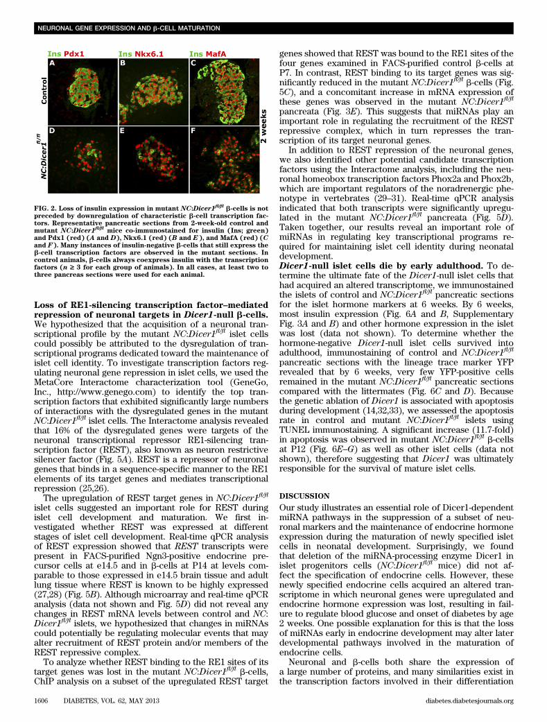
Endocrine cells from Dicer1-null mice maintainexpression of characteristic transcription factorsdespite losing hormone expression. Next, we askedwhether the loss of hormone expression in mutant NC:Dicer1fl/fl islet cells was due to any potential effect on keyislet transcriptional regulators upon ablation of miRNAs.We tested this hypothesis by assessing the expression ofcharacteristic b-cell transcription factors Pdx1, Nkx6.1,and MafA. Immunostaining of pancreata from 2-week-oldcontrol mice showed the expected coexpression of allthese transcription factors with insulin (Fig. 2A–C and datanot shown). In contrast, whereas the pancreata from mu-tant NC:Dicer1fl/fl littermates showed normal expressionlevels of Pdx1, Nkx6.1, and MafA in the islets, only a few ofthese cells were also positive for insulin, with the rest de-void of insulin expression (Fig. 2D–F). Similarly, 2-week-oldmutant NC:Dicer1fl/fl pancreatic sections showed a numberof cells that expressed MafB but not glucagon (Supple-mentary Fig. 1C and D). Furthermore, real-time qPCRanalyses of a set of key b-cell genes between control andmutant NC:Dicer1fl/fl pancreata revealed a significant re-duction in the expression of Insulin1 and Insulin2 mRNA,whereas no differences in the expression of transcriptionfactors markers was observed (Supplementary Fig. 1E andF and Supplementary Fig. 2A). These results suggested thatendocrine hormone expression was lost although the tran-scriptional regulatory genes that characterize endocrinecells were intact in the absence of Dicer1.Deletion of Dicer-1 upregulates neuronal genes inislet cells. To investigate the molecular changes in en-docrine cells of mutant NC:Dicer1fl/fl animals that could beresponsible for the loss of hormone expression in endo-crine cells, we performed genome-wide transcription pro-filing using microarrays on two independent pairs ofcontrol and mutant NC:Dicer1fl/fl pancreata isolated at P7.The microarray was performed at this stage, which pre-cedes any apparent physiological changes. Transcriptionprofiling analysis revealed 162 differentially expressedgenes with at least a twofold change at a false discoveryrate of 5%, of which 145 genes were upregulated and 17were downregulated. A surprisingly disproportionatenumber of neuronal genes were upregulated in the NC:Dicer1fl/fl pancreata, whereas the expression of transcrip-tion factors and other characteristic islet cell markers didnot exceed the twofold change threshold (Fig. 3A–C). Theupregulation of these neuronal genes in NC:Dicer1fl/fl
pancreata was further validated by performing real-timeqPCR analysis from the microarray samples (Fig. 3E) aswell as on a triplicate of samples independent of thoseused for the microarray analysis (Supplementary Fig. 2A).
Gene ontology analysis for biological processes associ-ated with the dysregulated genes revealed a significantenrichment of genes involved in nervous system de-velopment, synaptic transmission, transmission of nerveimpulse, and neurogenesis (Fig. 3D). The upregulated
genes included pan-neuronal markers such as Scg10,Stmn3, and the neurofilament markers Nefl and Nefm, aswell as molecular markers of noradrenergic neurons, suchas the neurotransmitter-synthesizing enzymes Th and do-pamine b hydroxylase (Dbh), the vesicular transport mole-cule–vesicular monoamine transporter 2 (Vmat2), and theplasma membrane transporter–norepinephrine transporter(Net). In fact, many components of the noradrenergicprogram were upregulated. A number of these neuronalgenes are expressed to some extent early during endocrinepancreas development, but their expression declines sig-nificantly by 3 to 4 weeks of age (24). For example, real-time qPCR analysis indicated that the expression ofneuropeptide Y (NPY), which is normally downregulatedupon maturation, was expressed at high levels in themutant NC:Dicer1fl/fl pancreata.
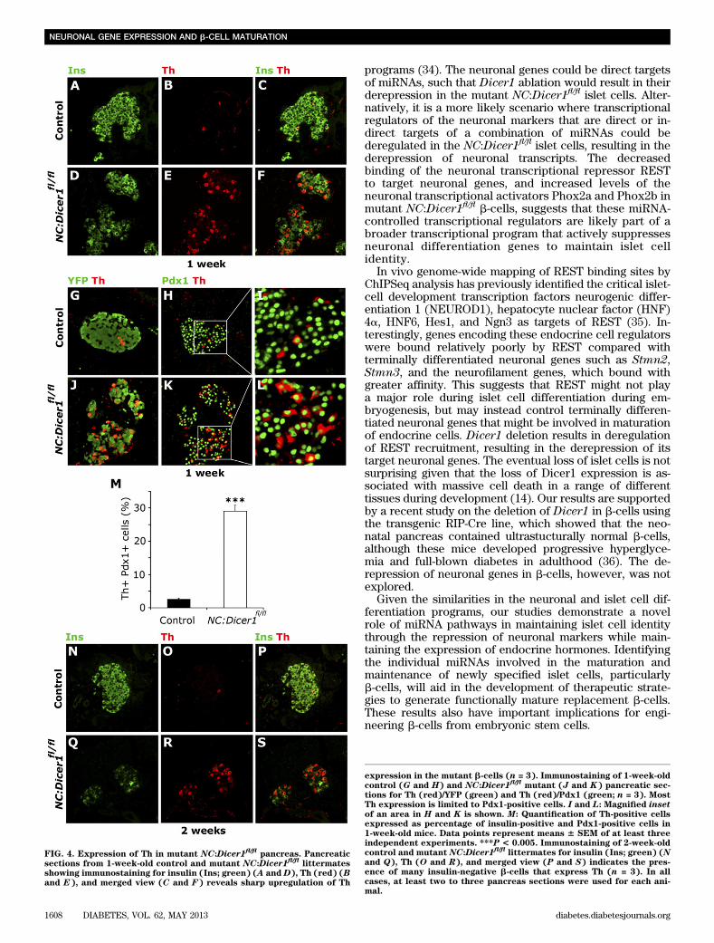
To test whether the increase in transcript levels of theneuronal genes was associated with a corresponding in-crease in protein levels, we immunostained 1 week controland NC:Dicer1fl/fl pancreatic sections for insulin and Th.Very few Th and insulin double-positive cells were ob-served in control sections (Fig. 4A–C). In contrast, themutant NC:Dicer1fl/fl sections displayed a larger percent-age of cells that coexpressed insulin and Th (Fig. 4D–F).Thus, the upregulation of mRNA of neuronal genes due tothe absence of Dicer1 also results in a concomitant in-crease at the protein level. To assess whether the upre-gulation of Th was limited to b-cells, we costained Th withPdx1. All Th-positive cells in the NC:Dicer1fl/fl islets alsocoexpressed Pdx1 (Fig. 4H–M), suggesting that b-cellsupregulated the neuronal marker Th in the absence ofDicer1. To verify the endocrine lineage of these Th-positivecells, we performed immunostaining for the lineage tracemarker YFP and Th in the mutant NC:Dicer1fl/flpancreata.All of the Th-positive cells were also positive for YFPstaining (Fig. 4G and J), therefore confirming that theTh-positive cells were derived from endocrine precursorcells that had undergone recombination and lost Dicer1expression.
We also examined Th expression in the mutant NC:Dicer1fl/fl islets at 2 weeks. Consistent with observations inislets from 1-week-old control mice, costaining of Th withinsulin (Fig. 4N–S) revealed that control islets from2-week-old mice also typically displayed only a few Th-positive cells that also stained for insulin (Fig. 4N–P). Incontrast, mutant NC:Dicer1fl/fl islets displayed a massiveupregulation of Th staining (Fig. 4Q–S). A few insulin andTh-copositive islet cells were evident, but most of the Th-positive cells did not show any insulin staining, suggestingthat neuronal gene upregulation persists in mutant isletcells that have already downregulated endocrine hor-mones. Taken together, our results suggest that miRNAslikely play an important role in suppressing a neuronalgene program during the maturation phase of endocrineislet cells.
FIG. 1. Mutant NC:Dicer1fl/fl
islet cells specify normally but lose hormone expression and develop hyperglycemia during neonatal development.Immunostaining of 1-week-old Cre negative (A–C) and mutant NC:Dicer1
fl/fl(D–F) pancreatic sections (n = 3) for insulin (red ) (B and E), YFP
that marks recombined cells (green) (A and D), and DAPI to visualize the nuclei (blue). C and F: Most of the insulin1 b-cells colocalize with YFP in
the mutant islets but not in the control islets. G: Transcript levels of Dicer1 determined by real-time qPCR using RNA isolated from islets ofcontrol and mutant NC:Dicer1
fl/flmice (n = 3) at P7. Expression levels in control were set as one arbitrary unit. Representative pancreatic sections
(n = 3 for each age and genotype) from e18.5 and 1- and 2-week-old control (H–J) and mutant NC:Dicer1fl/fl
(K–M) littermates were immunostainedfor insulin (green) and glucagon (red). N: Nonfasting blood glucose levels in neonatal control and mutant NC:Dicer1
fl/flmice at 1, 2, and 4 weeks
(n = 5 for each age group). NC:Dicer1fl/fl
mutant mice are hyperglycemic by 2 weeks.O: Total pancreatic insulin content in control and NC:Dicer1fl/fl
mice at 1 and 3 weeks reveals minimal pancreatic insulin content remaining in NC:Dicer1fl/fl
mice at 3 weeks (n ‡ 3). P–Q: b- and a-cell mass incontrol and mutant NC:Dicer1
fl/flmice at P1 and 2 weeks was assessed as described in RESEARCHDESIGNANDMETHODS (n = 4). The error bars represent
the SEM. *P < 0.05, **P < 0.01, ***P < 0.005. In all cases, at least two to three pancreas sections were used for each animal.
M.S. KANJI, M.G. MARTIN, AND A. BHUSHAN
diabetes.diabetesjournals.org DIABETES, VOL. 62, MAY 2013 1605
Loss of RE1-silencing transcription factor–mediatedrepression of neuronal targets in Dicer1-null b-cells.We hypothesized that the acquisition of a neuronal tran-scriptional profile by the mutant NC:Dicer1fl/fl islet cellscould possibly be attributed to the dysregulation of tran-scriptional programs dedicated toward the maintenance ofislet cell identity. To investigate transcription factors reg-ulating neuronal gene repression in islet cells, we used theMetaCore Interactome characterization tool (GeneGo,Inc., http://www.genego.com) to identify the top tran-scription factors that exhibited significantly large numbersof interactions with the dysregulated genes in the mutantNC:Dicer1fl/fl islet cells. The Interactome analysis revealedthat 16% of the dysregulated genes were targets of theneuronal transcriptional repressor RE1-silencing tran-scription factor (REST), also known as neuron restrictivesilencer factor (Fig. 5A). REST is a repressor of neuronalgenes that binds in a sequence-specific manner to the RE1elements of its target genes and mediates transcriptionalrepression (25,26).
The upregulation of REST target genes in NC:Dicer1fl/fl
islet cells suggested an important role for REST duringislet cell development and maturation. We first in-vestigated whether REST was expressed at differentstages of islet cell development. Real-time qPCR analysisof REST expression showed that REST transcripts werepresent in FACS-purified Ngn3-positive endocrine pre-cursor cells at e14.5 and in b-cells at P14 at levels com-parable to those expressed in e14.5 brain tissue and adultlung tissue where REST is known to be highly expressed(27,28) (Fig. 5B). Although microarray and real-time qPCRanalysis (data not shown and Fig. 5D) did not reveal anychanges in REST mRNA levels between control and NC:Dicer1fl/fl islets, we hypothesized that changes in miRNAscould potentially be regulating molecular events that mayalter recruitment of REST protein and/or members of theREST repressive complex.
To analyze whether REST binding to the RE1 sites of itstarget genes was lost in the mutant NC:Dicer1fl/fl b-cells,ChIP analysis on a subset of the upregulated REST target
genes showed that REST was bound to the RE1 sites of thefour genes examined in FACS-purified control b-cells atP7. In contrast, REST binding to its target genes was sig-nificantly reduced in the mutant NC:Dicer1fl/fl b-cells (Fig.5C), and a concomitant increase in mRNA expression ofthese genes was observed in the mutant NC:Dicer1fl/fl
pancreata (Fig. 3E). This suggests that miRNAs play animportant role in regulating the recruitment of the RESTrepressive complex, which in turn represses the tran-scription of its target neuronal genes.
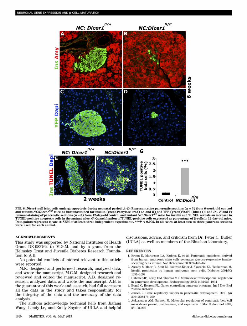
In addition to REST repression of the neuronal genes,we also identified other potential candidate transcriptionfactors using the Interactome analysis, including the neu-ronal homeobox transcription factors Phox2a and Phox2b,which are important regulators of the noradrenergic phe-notype in vertebrates (29–31). Real-time qPCR analysisindicated that both transcripts were significantly upregu-lated in the mutant NC:Dicer1fl/fl pancreata (Fig. 5D).Taken together, our results reveal an important role ofmiRNAs in regulating key transcriptional programs re-quired for maintaining islet cell identity during neonataldevelopment.Dicer1-null islet cells die by early adulthood. To de-termine the ultimate fate of the Dicer1-null islet cells thathad acquired an altered transcriptome, we immunostainedthe islets of control and NC:Dicer1fl/fl pancreatic sectionsfor the islet hormone markers at 6 weeks. By 6 weeks,most insulin expression (Fig. 6A and B, SupplementaryFig. 3A and B) and other hormone expression in the isletwas lost (data not shown). To determine whether thehormone-negative Dicer1-null islet cells survived intoadulthood, immunostaining of control and NC:Dicer1fl/fl
pancreatic sections with the lineage trace marker YFPrevealed that by 6 weeks, very few YFP-positive cellsremained in the mutant NC:Dicer1fl/fl pancreatic sectionscompared with the littermates (Fig. 6C and D). Becausethe genetic ablation of Dicer1 is associated with apoptosisduring development (14,32,33), we assessed the apoptosisrate in control and mutant NC:Dicer1fl/fl islets usingTUNEL immunostaining. A significant increase (11.7-fold)in apoptosis was observed in mutant NC:Dicer1fl/fl b-cellsat P12 (Fig. 6E–G) as well as other islet cells (data notshown), therefore suggesting that Dicer1 was ultimatelyresponsible for the survival of mature islet cells.
DISCUSSION
Our study illustrates an essential role of Dicer1-dependentmiRNA pathways in the suppression of a subset of neu-ronal markers and the maintenance of endocrine hormoneexpression during the maturation of newly specified isletcells in neonatal development. Surprisingly, we foundthat deletion of the miRNA-processing enzyme Dicer1 inislet progenitors cells (NC:Dicer1fl/fl mice) did not af-fect the specification of endocrine cells. However, thesenewly specified endocrine cells acquired an altered tran-scriptome in which neuronal genes were upregulated andendocrine hormone expression was lost, resulting in fail-ure to regulate blood glucose and onset of diabetes by age2 weeks. One possible explanation for this is that the lossof miRNAs early in endocrine development may alter laterdevelopmental pathways involved in the maturation ofendocrine cells.
Neuronal and b-cells both share the expression ofa large number of proteins, and many similarities exist inthe transcription factors involved in their differentiation
FIG. 2. Loss of insulin expression in mutant NC:Dicer1fl/fl b-cells is not
preceded by downregulation of characteristic b-cell transcription fac-tors. Representative pancreatic sections from 2-week-old control andmutant NC:Dicer1
fl/flmice co-immunostained for insulin (Ins; green)
and Pdx1 (red) (A and D), Nkx6.1 (red) (B and E), and MafA (red) (Cand F). Many instances of insulin-negative b-cells that still express theb-cell transcription factors are observed in the mutant sections. Incontrol animals, b-cells always coexpress insulin with the transcriptionfactors (n ‡ 3 for each group of animals). In all cases, at least two tothree pancreas sections were used for each animal.
NEURONAL GENE EXPRESSION AND b-CELL MATURATION
1606 DIABETES, VOL. 62, MAY 2013 diabetes.diabetesjournals.org
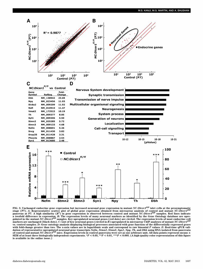
FIG. 3. Unchanged endocrine gene expression but increased neuronal gene expression in mutant NC:Dicer1fl/fl
islet cells at the presymptomaticstage (P7). A: Representative scatter plot of global gene expression obtained from microarray analysis of control and mutant NC:Dicer1
fl/fl
pancreas at P7. A high similarity (R2) in gene expression is observed between control and mutant NC:Dicer1
fl/flsamples. Red lines indicate
a twofold difference in expression. B: The expression levels of many neuronal markers as identified by the Gene Ontology database are upre-gulated in the mutant NC:Dicer1
fl/flsamples. Key upregulated neuronal genes (red dots) are circled. The expression levels of most endocrine cell
markers are unchanged (black dots). C: List of key neuronal genes (circled in B) upregulated in microarray ChIP analyses of mutant NC:Dicer1fl/fl
vs. control samples. D: Gene ontology analysis displaying biological processes associated with gene function of the differentially expressed geneswith fold-change greater than two. The x-axis values are in logarithmic scale and correspond to raw binomial P values. E: Real-time qPCR vali-dation of representative upregulated neuronal gene transcripts Nefm, Stmn2, Stmn3, Syn1, Npy, Th, and Dbh using RNA isolated from pancreataof control and mutant NC:Dicer1
fl/flmice. Expression levels in control pancreata were set as one arbitrary unit. All data points represent means 6
SEM of at least three biologically independent experiments. *P< 0.05, **P< 0.01, ***P< 0.005. (A high-quality color representation of this figureis available in the online issue.)
M.S. KANJI, M.G. MARTIN, AND A. BHUSHAN
diabetes.diabetesjournals.org DIABETES, VOL. 62, MAY 2013 1607
programs (34). The neuronal genes could be direct targetsof miRNAs, such that Dicer1 ablation would result in theirderepression in the mutant NC:Dicer1fl/fl islet cells. Alter-natively, it is a more likely scenario where transcriptionalregulators of the neuronal markers that are direct or in-direct targets of a combination of miRNAs could bederegulated in the NC:Dicer1fl/fl islet cells, resulting in thederepression of neuronal transcripts. The decreasedbinding of the neuronal transcriptional repressor RESTto target neuronal genes, and increased levels of theneuronal transcriptional activators Phox2a and Phox2b inmutant NC:Dicer1fl/fl b-cells, suggests that these miRNA-controlled transcriptional regulators are likely part of abroader transcriptional program that actively suppressesneuronal differentiation genes to maintain islet cellidentity.
In vivo genome-wide mapping of REST binding sites byChIPSeq analysis has previously identified the critical islet-cell development transcription factors neurogenic differ-entiation 1 (NEUROD1), hepatocyte nuclear factor (HNF)4a, HNF6, Hes1, and Ngn3 as targets of REST (35). In-terestingly, genes encoding these endocrine cell regulatorswere bound relatively poorly by REST compared withterminally differentiated neuronal genes such as Stmn2,Stmn3, and the neurofilament genes, which bound withgreater affinity. This suggests that REST might not playa major role during islet cell differentiation during em-bryogenesis, but may instead control terminally differen-tiated neuronal genes that might be involved in maturationof endocrine cells. Dicer1 deletion results in deregulationof REST recruitment, resulting in the derepression of itstarget neuronal genes. The eventual loss of islet cells is notsurprising given that the loss of Dicer1 expression is as-sociated with massive cell death in a range of differenttissues during development (14). Our results are supportedby a recent study on the deletion of Dicer1 in b-cells usingthe transgenic RIP-Cre line, which showed that the neo-natal pancreas contained ultrastucturally normal b-cells,although these mice developed progressive hyperglyce-mia and full-blown diabetes in adulthood (36). The de-repression of neuronal genes in b-cells, however, was notexplored.
Given the similarities in the neuronal and islet cell dif-ferentiation programs, our studies demonstrate a novelrole of miRNA pathways in maintaining islet cell identitythrough the repression of neuronal markers while main-taining the expression of endocrine hormones. Identifyingthe individual miRNAs involved in the maturation andmaintenance of newly specified islet cells, particularlyb-cells, will aid in the development of therapeutic strate-gies to generate functionally mature replacement b-cells.These results also have important implications for engi-neering b-cells from embryonic stem cells.
FIG. 4. Expression of Th in mutant NC:Dicer1fl/fl
pancreas. Pancreaticsections from 1-week-old control and mutant NC:Dicer1
fl/fllittermates
showing immunostaining for insulin (Ins; green) (A and D), Th (red) (Band E), and merged view (C and F) reveals sharp upregulation of Th
expression in the mutant b-cells (n = 3). Immunostaining of 1-week-oldcontrol (G and H) and NC:Dicer1
fl/flmutant (J and K) pancreatic sec-
tions for Th (red)/YFP (green) and Th (red)/Pdx1 (green; n = 3). MostTh expression is limited to Pdx1-positive cells. I and L: Magnified insetof an area in H and K is shown. M: Quantification of Th-positive cellsexpressed as percentage of insulin-positive and Pdx1-positive cells in1-week-old mice. Data points represent means 6 SEM of at least threeindependent experiments. ***P < 0.005. Immunostaining of 2-week-oldcontrol and mutant NC:Dicer1
fl/fllittermates for insulin (Ins; green) (N
and Q), Th (O and R), and merged view (P and S) indicates the pres-ence of many insulin-negative b-cells that express Th (n = 3). In allcases, at least two to three pancreas sections were used for each ani-mal.
NEURONAL GENE EXPRESSION AND b-CELL MATURATION
1608 DIABETES, VOL. 62, MAY 2013 diabetes.diabetesjournals.org
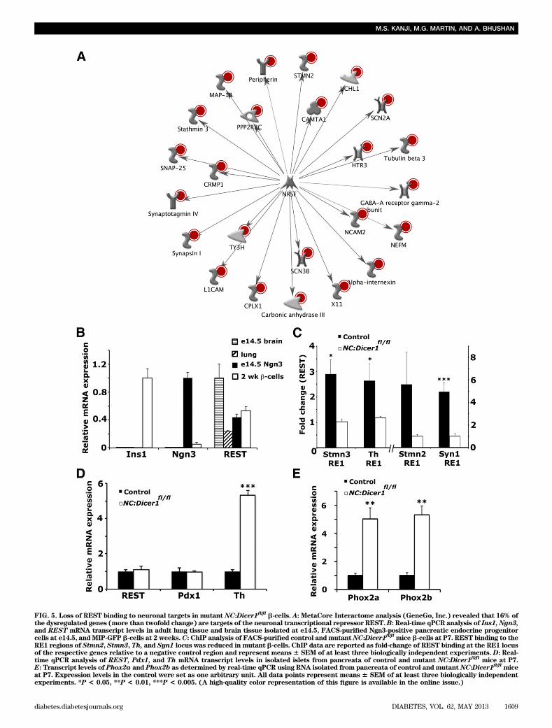
FIG. 5. Loss of REST binding to neuronal targets in mutant NC:Dicer1fl/fl b-cells. A: MetaCore Interactome analysis (GeneGo, Inc.) revealed that 16% of
the dysregulated genes (more than twofold change) are targets of the neuronal transcriptional repressor REST.B: Real-time qPCR analysis of Ins1, Ngn3,and REST mRNA transcript levels in adult lung tissue and brain tissue isolated at e14.5, FACS-purified Ngn3-positive pancreatic endocrine progenitorcells at e14.5, andMIP-GFP b-cells at 2 weeks.C: ChIP analysis of FACS-purified control andmutantNC:Dicer1
fl/flmice b-cells at P7. REST binding to the
RE1 regions of Stmn2, Stmn3, Th, and Syn1 locus was reduced in mutant b-cells. ChIP data are reported as fold-change of REST binding at the RE1 locusof the respective genes relative to a negative control region and represent means6 SEM of at least three biologically independent experiments. D: Real-time qPCR analysis of REST, Pdx1, and Th mRNA transcript levels in isolated islets from pancreata of control and mutant NC:Dicer1
fl/flmice at P7.
E: Transcript levels of Phox2a and Phox2b as determined by real-time qPCR using RNA isolated from pancreata of control and mutant NC:Dicer1fl/fl
miceat P7. Expression levels in the control were set as one arbitrary unit. All data points represent means 6 SEM of at least three biologically independentexperiments. *P < 0.05, **P < 0.01, ***P < 0.005. (A high-quality color representation of this figure is available in the online issue.)
M.S. KANJI, M.G. MARTIN, AND A. BHUSHAN
diabetes.diabetesjournals.org DIABETES, VOL. 62, MAY 2013 1609
ACKNOWLEDGMENTS
This study was supported by National Institutes of HealthGrant DK-083762 to M.G.M. and by a grant from theHelmsley Trust and Juvenile Diabetes Research Founda-tion to A.B.
No potential conflicts of interest relevant to this articlewere reported.
M.K. designed and performed research, analyzed data,and wrote the manuscript. M.G.M. designed research andreviewed and edited the manuscript. A.B. designed re-search, analyzed data, and wrote the manuscript. A.B. isthe guarantor of this work and, as such, had full access toall the data in the study and takes responsibility forthe integrity of the data and the accuracy of the dataanalysis.
The authors acknowledge technical help from JiafangWang, Lendy Le, and Emily Snyder of UCLA and helpful
discussions, advice, and criticism from Dr. Peter C. Butler(UCLA) as well as members of the Bhushan laboratory.
REFERENCES
1. Kroon E, Martinson LA, Kadoya K, et al. Pancreatic endoderm derivedfrom human embryonic stem cells generates glucose-responsive insulin-secreting cells in vivo. Nat Biotechnol 2008;26:443–452
2. Assady S, Maor G, Amit M, Itskovitz-Eldor J, Skorecki KL, Tzukerman M.Insulin production by human embryonic stem cells. Diabetes 2001;50:1691–1697
3. Habener JF, Kemp DM, Thomas MK. Minireview: transcriptional regulationin pancreatic development. Endocrinology 2005;146:1025–1034
4. Bonal C, Herrera PL. Genes controlling pancreas ontogeny. Int J Dev Biol2008;52:823–835
5. Jensen J. Gene regulatory factors in pancreatic development. Dev Dyn2004;229:176–200
6. Ackermann AM, Gannon M. Molecular regulation of pancreatic beta-cellmass development, maintenance, and expansion. J Mol Endocrinol 2007;38:193–206
FIG. 6. Dicer1-null islet cells undergo apoptosis during neonatal period. A–D: Representative pancreatic sections (n = 3) from 6-week-old controland mutant NC:Dicer1
fl/flmice co-immunostained for insulin (green)/amylase (red) (A and B) and YFP (green)/DAPI (blue) (C and D). E and F:
Immunostaining of pancreatic sections (n = 3) from 12-day-old control and mutant NC:Dicer1fl/fl
mice for insulin and TUNEL reveals an increase inTUNEL-positive apoptotic cells in the mutant mice. G: Quantification of TUNEL-positive cells expressed as percentage of b-cells in 12-day-old mice.Data points represent means 6 SEM of at least three independent experiments. ***P < 0.005. In all cases, at least two to three pancreas sectionswere used for each animal.
NEURONAL GENE EXPRESSION AND b-CELL MATURATION
1610 DIABETES, VOL. 62, MAY 2013 diabetes.diabetesjournals.org
7. Lagos-Quintana M, Rauhut R, Lendeckel W, Tuschl T. Identification ofnovel genes coding for small expressed RNAs. Science 2001;294:853–858
8. Ambros V. MicroRNA pathways in flies and worms: growth, death, fat,stress, and timing. Cell 2003;113:673–676
9. Denli AM, Tops BB, Plasterk RH, Ketting RF, Hannon GJ. Processing ofprimary microRNAs by the Microprocessor complex. Nature 2004;432:231–235
10. Han J, Lee Y, Yeom KH, Kim YK, Jin H, Kim VN. The Drosha-DGCR8complex in primary microRNA processing. Genes Dev 2004;18:3016–3027
11. Joglekar MV, Parekh VS, Hardikar AA. New pancreas from old: micro-regulators of pancreas regeneration. Trends Endocrinol Metab 2007;18:393–400
12. Lynn FC, Skewes-Cox P, Kosaka Y, McManus MT, Harfe BD, German MS.MicroRNA expression is required for pancreatic islet cell genesis in themouse. Diabetes 2007;56:2938–2945
13. Melkman-Zehavi T, Oren R, Kredo-Russo S, et al. miRNAs control insulincontent in pancreatic b-cells via downregulation of transcriptional re-pressors. EMBO J 2011;30:835–845
14. Harfe BD, McManus MT, Mansfield JH, Hornstein E, Tabin CJ. The RNa-seIII enzyme Dicer is required for morphogenesis but not patterning of thevertebrate limb. Proc Natl Acad Sci U S A 2005;102:10898–10903
15. Schonhoff SE, Giel-Moloney M, Leiter AB. Neurogenin 3-expressing pro-genitor cells in the gastrointestinal tract differentiate into both endocrineand non-endocrine cell types. Dev Biol 2004;270:443–454
16. Srinivas S, Watanabe T, Lin CS, et al. Cre reporter strains produced bytargeted insertion of EYFP and ECFP into the ROSA26 locus. BMC DevBiol 2001;1:4
17. Zhong L, Georgia S, Tschen SI, Nakayama K, Nakayama K, Bhushan A.Essential role of Skp2-mediated p27 degradation in growth and adaptiveexpansion of pancreatic beta cells. J Clin Invest 2007;117:2869–2876
18. Nishimura W, Kondo T, Salameh T, et al. A switch from MafB to MafAexpression accompanies differentiation to pancreatic beta-cells. Dev Biol2006;293:526–539
19. Georgia S, Bhushan A. Beta cell replication is the primary mechanism formaintaining postnatal beta cell mass. J Clin Invest 2004;114:963–968
20. Lee CS, Perreault N, Brestelli JE, Kaestner KH. Neurogenin 3 is essentialfor the proper specification of gastric enteroendocrine cells and the main-tenance of gastric epithelial cell identity. Genes Dev 2002;16:1488–1497
21. Hara M, Wang X, Kawamura T, et al. Transgenic mice with green fluo-rescent protein-labeled pancreatic beta-cells. Am J Physiol EndocrinolMetab 2003;284:E177–E183
22. Dahl JA, Collas P. A rapid micro chromatin immunoprecipitation assay(microChIP). Nat Protoc 2008;3:1032–1045
23. Downey T. Analysis of a multifactor microarray study using Partek ge-nomics solution. Methods Enzymol 2006;411:256–270
24. Blum B, Hrvatin SS, Schuetz C, Bonal C, Rezania A, Melton DA. Functionalbeta-cell maturation is marked by an increased glucose threshold and byexpression of urocortin 3. Nat Biotechnol 2012;30:261–264
25. Chong JA, Tapia-Ramírez J, Kim S, et al. REST: a mammalian silencerprotein that restricts sodium channel gene expression to neurons. Cell1995;80:949–957
26. Schoenherr CJ, Anderson DJ. The neuron-restrictive silencer factor(NRSF): a coordinate repressor of multiple neuron-specific genes. Science1995;267:1360–1363
27. Palm K, Belluardo N, Metsis M, Timmusk T. Neuronal expression of zincfinger transcription factor REST/NRSF/XBR gene. J Neurosci 1998;18:1280–1296
28. Kreisler A, Strissel PL, Strick R, Neumann SB, Schumacher U, Becker CM.Regulation of the NRSF/REST gene by methylation and CREB affects thecellular phenotype of small-cell lung cancer. Oncogene 2010;29:5828–5838
29. Pattyn A, Morin X, Cremer H, Goridis C, Brunet JF. The homeobox genePhox2b is essential for the development of autonomic neural crest de-rivatives. Nature 1999;399:366–370
30. Pattyn A, Goridis C, Brunet JF. Specification of the central noradrenergicphenotype by the homeobox gene Phox2b. Mol Cell Neurosci 2000;15:235–243
31. Apostolova LG, Thompson PM, Rogers SA, et al. Surface feature-guidedmapping of cerebral metabolic changes in cognitively normal and mildlyimpaired elderly. Mol Imaging Biol 2010;12:218–224
32. Harris KS, Zhang Z, McManus MT, Harfe BD, Sun X. Dicer function isessential for lung epithelium morphogenesis. Proc Natl Acad Sci U S A2006;103:2208–2213
33. O’Rourke JR, Georges SA, Seay HR, et al. Essential role for Dicer duringskeletal muscle development. Dev Biol 2007;311:359–368
34. Wilson ME, Scheel D, German MS. Gene expression cascades in pancreaticdevelopment. Mech Dev 2003;120:65–80
35. Johnson DS, Mortazavi A, Myers RM, Wold B. Genome-wide mapping of invivo protein-DNA interactions. Science 2007;316:1497–1502
36. Kalis M, Bolmeson C, Esguerra JL, et al. Beta-cell specific deletion ofDicer1 leads to defective insulin secretion and diabetes mellitus. PLoSONE 2011;6:e29166
M.S. KANJI, M.G. MARTIN, AND A. BHUSHAN
diabetes.diabetesjournals.org DIABETES, VOL. 62, MAY 2013 1611