full genome characterization of porcine circovirus type 3
TRANSCRIPT

RESEARCH Open Access
Full genome characterization of porcinecircovirus type 3 isolates reveals theexistence of two distinct groups of virusstrainsRobert Fux1* , Christina Söckler2, Ellen Kathrin Link1, Christine Renken2, Roman Krejci3, Gerd Sutter1,Mathias Ritzmann2 and Matthias Eddicks2
Abstract
Background: The occurrence of the novel porcine circovirus type 3 (PCV3) was reported from the Americas, Asiaand Europe. Although this virus was detected in association with various clinical syndromes in pigs, its role as possibleswine pathogen remains unclear. PCV3 was detected with high prevalence in Polish farms, but to date no genomesequences were available from European PCV3 strains.
Methods: We collected 1060 serum samples from piglets at the age of 20–24 weeks from 53 farms distributed all overGermany. PCV3 DNA was detected using a real-time PCR and subsequently complete PCV3 genome sequences wereobtained after multiply primed rolling circle amplification and sequencing of overlapping PCR products. Phylogeneticanalysis was performed by neighbor-joining method and maximum likelihood method.
Results: We obtained 15 complete PCV3 genome sequences as well as nine partial sequences including the putativeORFs 1, 2 and 3 from PCV3 viremic animals in German pig farms. Phylogenetic analysis of these German as well as 30full genome sequences received from GenBank divided the PCV3 strains into two main groups and several subclusters.Furthermore, we were able to define group specific amino acid patterns in open reading frame 1 and 2.
Conclusion: PCV3 is distributed with high prevalence in German pig industry. Phylogenetic analysis revealed twoclearly separated groups of PCV3 strains, which might be considered as PCV3 genotypes. Specific nucleotide andamino acid marker positions may serve for easy and fast intraspecies classification and genotyping of PCV3 strains. Nocorrelation between PCV3 variants with their geographical origin was evident. We found the same diversity of PCV3strains in Germany as in other countries. We hypothesize that PCV3 is not a newly emerging virus in the German pigpopulation. Future studies will have to show, if PCV3 genotype specific biological properties are evident.
Keywords: PCV3, Porcine circovirus, Swine pathogen, Emerging disease
BackgroundThe discovery of a new virus species in humans as wellas in animals always raises the same questions. What isits pathogenic potential? Are specific diseases or symp-toms triggered by the virus? What is the prevalence in thesusceptible population? Is it a newly emerging virus? Is ita homogeneous virus population or are there different
virus variants? Recently, a new type of porcine circoviruses(PCV) was described, and consequently the name porcinecircovirus type 3 (PCV3) was proposed. First discoveredin the USA [1, 2], there are now several reports orsequences available from China [3], South Korea [4],Poland [5] and Brazil [6]. The detection of PCV3 wasassociated with different clinical syndromes and diseasesin pigs of different ages. The porcine dermatitis andnephropathy syndrome [1] was noticed as well as repro-ductive problems [1, 3, 6], cardiac and multisystemicinflammation [1], respiratory diseases [7] and congenital
* Correspondence: [email protected] for Infectious Diseases and Zoonoses, LMU Munich, Veterinärstrasse13, 80539 Munich, GermanyFull list of author information is available at the end of the article
© The Author(s). 2018 Open Access This article is distributed under the terms of the Creative Commons Attribution 4.0International License (http://creativecommons.org/licenses/by/4.0/), which permits unrestricted use, distribution, andreproduction in any medium, provided you give appropriate credit to the original author(s) and the source, provide a link tothe Creative Commons license, and indicate if changes were made. The Creative Commons Public Domain Dedication waiver(http://creativecommons.org/publicdomain/zero/1.0/) applies to the data made available in this article, unless otherwise stated.
Fux et al. Virology Journal (2018) 15:25 DOI 10.1186/s12985-018-0929-3

tremors in neonatal pigs [8]. However, high PCV3 preva-lence was also reported from randomly selected farms ofdifferent health status from Poland without association ofPCV3 to specific clinical signs [5]. Similar results wereobtained by a study conducted in Korea [4].PCV1 was first identified as cell culture contaminant
in the 1980s and is considered as nonpathogenic for pigs[9]. In contrast, PCV2 is associated with several clinicaldiseases and syndromes and is responsible for majoreconomic losses in swine industry worldwide [10]. Allthree PCVs are small, non-enveloped viruses with a circu-lar, single-stranded DNA genome. The genome size ofPCV1 is about 1760 bp, the typical PCV2 genome varies,depending on the genotype, between 1767 bp (PCV2b, 2cand 2d) and 1777 bp (PCV2e) and PCV3 has the largestgenome with 2000 bp (see Fig. 1). Two major open read-ing frames (ORF) encode the replicase protein (ORF1)and the capsid protein (ORF2), respectively. In contrast toPCV1 and PCV2 a canonical start codon (ATG) is missingin ORF1 of PCV3. Here an alternative start codon wasdiscussed, as it has been proposed for some avian circo-viruses [2]. The PCV3 ORF2 is in opposite orientation toORF1 and encodes a 214 amino acid (aa) protein. ForPCV2 two further ORFs were characterized as apoptosis-inducing (ORF3) and apoptosis-suppressing gene (ORF4)(for review see [11]). Also for PCV3 a third putative ORFwas described. Similar to PCV3 ORF1 the start codonfor ORF3 remains unclear. An alternative initiation codon(TCG, nucleotide (nn) position 1900–1902) would result ina 231 aa protein (ORF3231), whereas a methionine codon(ATG, nn position 62–64) would yield a 177 aa protein(ORF3177) [2].
Based on phylogenetic analysis and the definition ofgenotype-specific marker positions four PCV2 genotypes(2a-2d) could be defined by Franzo and colleagues [12].Furthermore, a fifth genotype (2e) was reported from theUSA [13]. The correct taxonomical classification and geno-typing is important for molecular epidemiology. However,the precise classification of virus strains within the speciesPCV2 has been the occasion for several controversialdiscussions (for review see [12]). Because of the verylimited number of available PCV3 sequences or the useof small fragments of the PCV3 genome, results of per-formed phylogenetic analysis of PCV3 has to be consid-ered as preliminary [3, 4].In this study we report about (i) the first record of
PCV3 in Germany, (ii) the PCV3 prevalence in Germanswine farms, (iii) the phylogenetic analysis of GermanPCV3 sequences, which suggested a clear division ofPCV3 strains into two groups and (iv) the identificationof group specific marker codons.
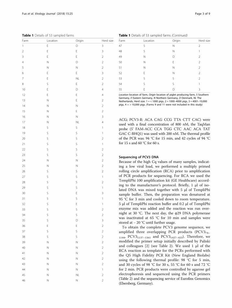
MethodsSample originThe examined sera were obtained within a serologicalporcine health screening (approved by the ethic committeeof the Centre for Clinical Veterinary Medicine, approvalnumber 73–31–05-2016) in 53 German fattening farmswith noticeable respiratory clinical signs from 2015 (Table 1).The age of all sampled pigs ranged between 20 and 24 weeks.On each farm 20 serum samples were collected. Indetail, 23 farms were located in Southern Germany (Bavariaor Baden-Wuerttemberg), 10 farms in Eastern Germany(Mecklenburg-West Pomerania, Brandenburg, Saxony-Anhalt or Thuringia) and 20 farms in Northern Germany(Lower Saxony, North Rhine-Westphalia or Schleswig-Holstein). Herd sizes varied between 400 and 28.000animals. Piglet producing farms were located in Germany,Denmark and The Netherlands.
PCV3 detectionPCV3 DNA was detected using a real-time PCR assaypublished previously by Palinski and colleagues [2]. First,serum pools (n = 5) were screened for PCV3. If a samplepool was PCV3 DNA positive, the included single sam-ples were tested subsequently. If two or more serumpools from one farm were tested positive, only the poolwith the lowest Cq (quantification cycle) value (indicat-ing highest viral load) was investigated further. In total,we tested 212 serum pools and 200 single samples.DNA from serum samples was isolated using the
NucleoSpin Virus Core Kit (Macherey-Nagel) and a Micro-lab Starlet workstation (Hamilton Robotics) according tothe manufacturer’s instruction. For the real-time PCR weused the QuantiTect probe PCR kit (Qiagen). Oligonucleo-tide primers (PCV3-F: 5′ AGT GCT CCC CAT TGA
Fig. 1 Schematic presentation of the PCV3 genome and its putativeopen reading frames (ORF)
Fux et al. Virology Journal (2018) 15:25 Page 2 of 9
ACG; PCV3-R: ACA CAG CCG TTA CTT CAC) wereused with a final concentration of 800 nM, the TaqManprobe (5’ FAM-ACC CCA TGG CTC AAC ACA TATGAC C-BHQ1) was used with 200 nM. The thermal profileof the PCR was: 94 °C for 15 min, and 42 cycles of 94 °Cfor 15 s and 60 °C for 60 s.
Sequencing of PCV3 DNABecause of the high Cq values of many samples, indicat-ing a low viral load, we performed a multiply primedrolling circle amplification (RCA) prior to amplificationof PCR products for sequencing. For RCA we used theTempliPhi 100 amplification kit (GE Healthcare) accord-ing to the manufacturer’s protocol. Briefly, 1 μl of iso-lated DNA was mixed together with 5 μl of TempliPhisample buffer. Then, the preparation was denatured at95 °C for 3 min and cooled down to room temperature.5 μl of TempliPhi reaction buffer and 0.2 μl of TempliPhienzyme mix was added and the reaction was run over-night at 30 °C. The next day, the φ29 DNA polymerasewas inactivated at 65 °C for 10 min and samples werestored at − 20 °C until further usage.To obtain the complete PCV3 genome sequence, we
amplified three overlapping PCR products (PCV374–1144, PCV31137–1561 and PCV31427–433). Therefore, wemodified the primer setup initially described by Paliskiand colleagues [2] (see Table 2). We used 1 μl of theRCA reaction as template for the PCRs performed withthe Q5 High Fidelity PCR Kit (New England Biolabs)using the following thermal profile: 98 °C for 5 min,and 30 cycles of 98 °C for 30 s, 55 °C for 60 s and 72 °Cfor 2 min. PCR products were controlled by agarose gelelectrophoresis and sequenced using the PCR primers(Table 2) and the sequencing service of Eurofins Genomics(Ebersberg, Germany).
Table 1 Details of 53 sampled farms
Farm Location Origin Herd size
1 E D 3
2 E E 3
3 E E 2
4 N D 2
5 N N 2
6 E E 3
7 E NL 2
8 S E 2
10 E D 4
12 E E 4
13 N E 2
14 N N 2
15 N E 2
16 N N 3
17 N NL 4
18 S S 1
19 S S 1
20 S S 2
21 S S 1
22 S S 2
23 S S 2
24 N N 2
25 N N 2
26 S S 2
27 S S 1
28 S S 2
29 S S 1
30 S E 3
31 S S 1
32 S E 1
33 S S 2
34 S S 2
35 S D 3
36 E E 2
37 S S 2
38 S S 2
39 N N 2
40 N N 2
41 N N 2
42 N N 2
43 N N 2
44 N N 1
45 N NL 2
46 N N 2
Table 1 Details of 53 sampled farms (Continued)
Farm Location Origin Herd size
47 S N 2
48 S N 1
49 N D 2
50 N E 2
51 N N 2
52 E N 2
53 S S 2
54 S S 1
55 E D 3
Location location of farm, Origin location of piglet producing farm, S SouthernGermany, E Eastern Germany, N Northern Germany, D Denmark, NL TheNetherlands, Herd size: 1 = < 1000 pigs, 2 = 1000–4000 pigs, 3 = 4001–10,000pigs, 4 = > 10,000 pigs. (Farms 9 and 11 were not included in this study)
Fux et al. Virology Journal (2018) 15:25 Page 3 of 9

Sequence assembly and analysisDNASTAR Lasergene and MEGA6 software was usedfor assembly, alignment and analysis of the sequences. AclustalW algorithm was used for alignments, and phylo-genetic trees (based on the full genome sequence or theORF2) were constructed using the neighbor-joining (NJ)method (p-distance model, 1000 bootstraps) and the max-imum likelihood method. For comparison with the GermanPCV3 sequences, all currently available complete PCV3genomes (from Brazil, China, South Korea and USA) weredownloaded from NCBI GenBank (see Additional file 1).
Statistic examinationFor the statistical analysis we used SPSS 23 for windows(IBM® SPSS Inc., USA). Each single farm or each sequencedstrain served as a statistical unit, respectively. The signifi-cance level of this investigation was 5% with a confidenceinterval of 95%. To evaluate a possible association betweenthe farm specific factors “origin of fattening pigs”, “locationof fattening farm” and “herd size of fattening farm”(Table 1) and the occurrence of PCV3 or an assumedPCV3-cluster on a farm, we used cross tables and Yates’chi-squared test.
ResultsPCV3 Prevalence in German pig farmsWe investigated 53 German farms of different locationand size for the occurrence of PCV3 viremic fatteningpigs. In 75% (40/53) of the farms we detected PCV3 DNAat least in one serum pool (Table 3). Cq values of poolsamples ranged from 26 to 39. Neither the location of thefarm, the origin of the piglets nor the herd size had a sig-nificant influence on virus occurrence on farm level.
Characterization of German PCV3 genome sequencesWe used selected single serum samples (with the lowestCq values) for amplification and sequencing of the PCV3genome. We obtained 15 full genome sequences and ninepartial sequences, including all three putative ORFs, repre-senting 24 of the 40 PCV3 positive farms (see Table 3). Asdescribed by Palinsky and colleagues (2017), in 13 cases
the amplified and sequenced PCV3 genome fragmentsassembled to a 2000 bp long circular genome. However,because of a deletion at nn position 1224 in the noncodingregion between ORF1 and ORF2, the genome of strainDE41.16 had a length of only 1999 bp. In contrast to thatstrain DE26.17 showed an insertion between nucleotide 6and 7 and a genome length of 2001 bp. This insertionwould result in a codon shift of the putative ORF3231starting at nn position 1900 and a stop codon at aa pos-ition 47. Additionally, the ORF3231 sequence of strainDE15.17 had a stop codon at aa position 21. Therefore,these both virus variants would not be able to induce thesynthesis of a functional ORF3231 protein. The alternativeORF3177, starting at nn position 62, would not be affectedby these mutations.
Phylogenetic analysis of German PCV3 genomesequencesA NJ tree based on the PCV3 full genome including 15German and 30 reference sequences is shown in Fig. 2.The sequences could be divided into two main groups (aand b). A phylogenetic tree constructed with the max-imum likelihood (ML) method displayed the same top-ology (data not shown). In contrast to group a, whichshowed high sequence identities (99.1–100%), group bwas further subdivided into three clusters and sequenceidentities were lower (97.3–100%) in this branch.A corresponding phylogenetic tree including 24 German
and 30 reference sequences was constructed based on thenucleotide sequence of the ORF2 (Fig. 3a). Again thetwo main groups were present. However, three sequences(KX458235, KY996344 and KY96345), which were assignedto group b in the full genome based tree, were now allo-cated together with the German sequence DE6.1 as a separ-ate subcluster in group a (a2). Interestingly, this subclusterwas split when the aa sequence of ORF2 was used foranalysis (NJ and ML, data not shown). Strains KX458235and KY996345 were located within subgroup a1, KY996344was clustered at the edge of that branch and strain DE6.1became member of group b.To support the definition of these two main groups
and further subgroups, we aligned the putative PCV3 ORFsand identified group specific marker codons (Fig. 3b andTable 4). Because of the overlap of the putative ORF3231with ORF1 and ORF2 (see Fig. 1) three nucleotide changes(nn position 579, 1901 and 1910) resulted in aa alterationsin ORF1/2 and ORF3 simultaneously.Summarizing the marker codons in ORF1 and ORF2
resulted in a specific aa pattern for group a (A V K S I;motif 1) and for group b (S A R S I; motif 2) (see Fig. 3b).However, the members of the above-mentioned subgroup(a2 in Fig. 3, clustering differently in the ORF2 and the fullgenome based phylogenetic analysis) demonstrated aa mo-tifs (S V/A K S I) that were between motif 1 and motif 2.
Table 2 Oligonucleotide primers used for amplification andsequencing PCV3 genome fragments
Primer Sequence 5′-3′ PCR product
PCV374 F CAC CGT GTG AGT GGA TAT AC 1072 bp
PCV31144 R CAC CCC AAC GCA ATA ATT GTA
PCV3910 Fa GAC AAT TCC CAC CCA AAC
PCV31137 F TTG GGG TGG GGG TAT TTA TT 425 bp
PCV31561 R ACA CAG CCG TTA CTT CAC
PCV31427 F AGT GCT CCC CAT TGA ACG 1007 bp
PCV3433 R CGA CCA AAT CCG GGT AAG Caonly used for sequencing
Fux et al. Virology Journal (2018) 15:25 Page 4 of 9

Additionally, a clearly separated cluster in group b (b2)showed a slight modification in motif 2 (S A R T L). There-fore, we could define two main groups a and b, whichcould be subdivided into two subgroups (a1 and a2; b1and b2), respectively. While nn sequence identities ofORF2 in subgroups a1, a2 and b2 were high (98.0–100%),subgroup b1 showed a higher divergence (sequence iden-tity 96.4–99.8%). None of the clusters was significantlycorrelated to a specific origin of the included samples.PCV3 sequences from Asia, America and Germany weremembers of all subgroups, respectively.
DiscussionRecently, several studies with varying approaches demon-strated the existence of PCV3 in Asia, Europe and theUSA. Specific specimens from diseased pigs from singlefarms were investigated as well as random samples from re-gional screening programs. Organs and tissue samples wereused as well as serum samples and oral-fluids. As basis forthe design of this study, we hypothesized PCV3 might showsimilar infection dynamics on farm level like PCV2. There-fore, we used serum samples of piglets after the decline ofmaternal antibodies, expecting higher rates of viremic ani-mals. This is consistent with studies performed by Kwonand colleagues [4] and Stadejek and colleagues [5], whichdemonstrated high PCV3 prevalence in weaned pigs. Inorder to obtain an authentic picture of the PCV3 situationin Germany, the origin of samples represented the mainareas with swine production in Germany. Also differentstructures of pig production in Germany (e.g. herd sizes)were considered. Actually, the sampling was performed toevaluate the seroprevalence of respiratory pathogens inGerman pig industry. However, our study is not suitableto correlate the presence of PCV3 with the appearance of
Table 3 PCV3 real-time PCR results and PCV3 sequences
Farm PCV3 positivepools
Range ofCq values
Obtained PCV3sequence
1 0 NA NA
2 3/4 33–36 DE2.8p
3 2/4 30–33 DE3.7c
4 2/4 31–33 DE4.3c
5 1/4 32 DE5.15p
6 3/4 32–36 DE6.1p
7 3/4 26–38 DE7.3c
8 1/4 32 NA
10 2/4 34–37 NA
12 3/4 27–35 DE12.19p
13 3/4 35–38 DE13.20c
14 3/4 33–36 DE14.15p
15 2/4 32–38 DE15.19p
16 0 NA NA
17 1/4 37 DE17.20p
18 2/4 31 DE18.2c
19 1/4 32 DE19.15c
20 4/4 33–38 NA
21 0 NA NA
22 1/4 31 NA
23 2/4 31–33 DE23.17c
24 2/4 37–38 NA
25 0 NA NA
26 3/4 27–35 DE26.17c
27 1/4 31 DE27.16c
28 3/4 33–37 DE28.12p
29 1/4 36 NA
30 1/4 38 NA
31 2/4 32–37 DE31.17p
32 0 NA NA
33 0 NA NA
34 2/4 32–35 DE34.5c
35 1/4 36 NA
36 0 NA NA
37 1/4 38 NA
38 0 NA NA
39 1/4 37 NA
40 1/4 39 NA
41 1/4 30 DE41.16c
42 0 NA NA
43 0 NA NA
44 3/4 32–36 NA
45 0 NA NA
Table 3 PCV3 real-time PCR results and PCV3 sequences(Continued)
Farm PCV3 positivepools
Range ofCq values
Obtained PCV3sequence
46 3/4 35–36 NA
47 0 NA NA
48 2/4 31–37 DE48.7c
49 2/4 33–38 NA
50 0 NA NA
51 1/4 34 NA
52 2/4 34–36 DE52.18c
53 3/4 28–34 DE53.8c
54 2/4 35–36 NA
55 2/4 34–37 DE55.1c
c complete PCV3 genome sequence, p partial PCV3 genome sequence (ORFs1, 2 & 3 complete), NA not adequate, (Farms 9 and 11 were not included inthis study)
Fux et al. Virology Journal (2018) 15:25 Page 5 of 9

respiratory diseases in swine. To our opinion, for thispurpose the same requirements would have to be ful-filled as for the definition of PCV associated diseases inthe context of PCV2: the explicit description of the clinicaldisease, combined with a (specific) pathohistological pictureand the detection (and if possible quantification) of thevirus in association with the lesions. To our opinion, thepresence of PCV3 and simultaneous absence of otherdetectable pathogens in a clinical case might indicate anetiological role of PCV3, but more detailed investigationswill be essential to identify its real pathogenic potential.In our study 79 sample pools from 40 farms were posi-
tive for PCV3 DNA. Therefore the virus prevalence was75% at herd level. Similar high values were reported byothers. In a study from Poland PCV3 was detected in 12of 14 farms (86%) [5], in South Korea 53 of 73 farms
(73%) were tested PCV3 positive [4] and from China aprevalence of 69% (24 of 35 farms) was reported [3].The herd size, the geographical location of the farm orthe piglet producing unit had no effect on the PCV3prevalence. Furthermore, no significant correlation betweenPCV3 detection and PCV2 prevalence and PCV2 vaccin-ation status of the herds was observed, respectively. There-fore, differences in PCV3 prevalence between farms may beattributed to individual factors like pig flow management,biosecurity, immunosuppressing effects or existing herd im-munity as it was discussed already by others [5].Although we performed no quantification of PCV3, the
high Cq values of most samples suggest that viral loads inthe blood of the viremic animals was moderate or low.Only in 18 (of 200) single tested samples we obtained Cqvalues < 30. This observation is in consistence with the
Fig. 2 Phylogenetic analysis of PCV3 based on the complete genome of 15 German PCV3 strains (●) and 30 PCV3 reference strains (GenBankaccession number, country and year of collection; more details are listed in Additional file 1). The tree was constructed using the neighbor joiningmethod (p-distance model; 1000 bootstraps; only bootstrap values above 50 are shown). The scale bar indicates nucleotide substitutions per site
Fux et al. Virology Journal (2018) 15:25 Page 6 of 9

Fig. 3 Phylogenetic analysis of PCV3 based on ORF2 and identification of group specific amino acid motifs. a Phylogenetic tree based on ORF2 of 24German PCV3 strains (●) and 30 PCV3 reference strains (GenBank accession number, country and year of collection; more details are listed in Table A1). Thetree was constructed using the neighbor joining method (p-distance model; 1000 bootstraps; only bootstrap values above 50 are shown). The scale barindicates nucleotide substitutions per site. b Amino acid alignments of the putative ORFs 1, 2 and 3 were used to identify group specific motifs
Table 4 Codon variants which allow PCV3 subtyping (in brackets the proportion of sequences with fitting marker position)
Codon PCV3 group a1 PCV3 group b1 PCV3 group b2
ORF1–122 GCG, alanine (16/16) TCG, serine (20/21) TCG, serine (13/13)
ORF2–24 GTC, valine (15/16) GCC, alanine (20/21) GCC, alanine (13/13)
ORF2–27 AAA, lysine (16/16) AGA/CGA, arginine (21/21) AGA/CGA, arginine (13/13)
ORF2–77 AGC, serine (15/16) AGC, serine (21/21) ACC, threonine (13/13)
ORF2–150 ATT, isoleucine (16/16) ATT, isoleucine (20/21) CTT/CTA, leucine (12/13)
ORF3231–1 TTT, phenylalanine (16/16) TCT/TCG, serine (21/21) TCT/TCG, serine (13/13)
ORF3231–4 GAC, aspartic acid (15/16) GGC, glycine (20/21) GGC, glycine (13/13)
ORF3231–227 GGC, glycine (16/16) GTC, valine (19/21) GTC, valine (13/13)
Fux et al. Virology Journal (2018) 15:25 Page 7 of 9

study from Poland [5]. For PCV2 it is generally accepted,that there is a strong correlation between viral loads inserum or tissues and the severity of virus-induced histo-pathological lesions (for review see [10]). In regard of thehigh PCV3 prevalence, the observed low virus loads inthis and other studies and the unclear role of PCV3 aspossible swine pathogen, the same might be true forPCV3 infections.Because of the low viral loads of many samples and the
moderate sensitivity of the used PCR, we performed RCAprior to amplification of larger fragments of the PCV3genome for sequencing. Thus, we were able to obtain 15complete PCV3 genome sequences. Additionally, fromother nine strains we were able to determine the sequencesof ORF1, ORF2 and ORF3231, respectively. Most sequencesmatched previous descriptions [1, 2]: a circular 2000 bpgenome with ORFs for a replicase protein (nn 216–1107)and the capsid protein (nn 1336–1980) and two putativevariants of ORF3 (ORF3231 nn 1900–595; ORF3177 nn 62–595). However, one sequence (DE41.16) had a deletion inthe noncoding region between ORF1 and ORF2, resulting agenome length of 1999 bp and another sequence (DE26.17)had a nucleotide insertion between nn position 6 and 7resulting in a 2001 bp genome sequence. Interestingly, thesecond mutation would cause a codon shift in the putativeORF3231 with a termination signal after aa 47. Additionally,the sequence DE15.17 possessed a stop codon in theamino-terminal part of ORF3231. Therefore, both virusvariants would be unable to synthesize a 231 aa ORF3protein. However, the translation of the shorter ORF3177would not be affected. This finding questions the meaningof the large ORF3 and might favor the existence of anORF3177.The intraspecific classification and genotyping of PCV2 is
an ongoing challenge. At the beginning the limited numberof published genome sequences was a major problem andnowadays the occurrence of a significant number of recom-binant sequences and different rates of evolution within thedifferent clades of the PCV2 phylogenetic tree poses prob-lems to the used PASC (pairwise sequence comparisons)analysis [12] (Franzo et al., 2015). Keeping this and the lownumber of available PCV3 genome sequences in mind, theauthors are aware that the performed phylogenetic analysiswill have some kind of preliminary character. However,using the full genome as well as the ORF2 sequences andcalculating phylogenetic trees with the neighbor-joiningmethod as well as with the maximum likelihood method,the PCV3 strains were clearly separated into two majorclades, which might be considered as two different geno-types of PCV3. Similar was reported by Ku and colleagues[3], however, one reference sequence (MO2015 from USA,KX778720) clustered differently in comparison to our ana-lysis. This might be explained by the small number (ten) ofincluded PCV3 full genome sequences in that work. By
analogy to the PCV2 field, we identified marker nucleotideand codon positions, which substantiate the definition ofthe two PCV3 subgroups. In ORF1 codon 122 and inORF2 codons 24, 27, 77 and 150 gave a typical aa motif forgroup a (A V K S I) and for group b (S A R S I), whichmight be helpful for the intraspecific classification. Interest-ingly, a small branch of three or four sequences (a2 in Fig.3) clustered differently in the ORF2 nn, ORF2 aa and thefull genome based phylogenetic tree. In addition, the signalmotif of these PCV3 viruses represented an intermedi-ate sequence (S A/V K S I) of pattern 1 and 2. Thesestrains, as well as sequence KY966337 (A V R S I),might represent some kind of evolutionary linker be-tween the two main groups or recombinant viruses, al-though this was not evident. The same might apply tothe sequences DE48.7, DE55.1 which show slightlymodified signal motifs and may represent some kind oflinker between subgroups b1 and b2. Sequence iden-tities in group b were lower than in group a and se-quences of a separated branch (b2 in Fig. 3)demonstrated a typical variance of the motif 2: S AR T L. Therefore, the main groups (especially group b)might be divided in several subgroups, however, morePCV3 sequences are needed to endorse this assumption.Future work has to demonstrate if these genetic differencescorrelate to specific biological properties of the PCV3groups. However, this might be a challenging task, be-cause attempts to isolate infectious PCV3 were unsuc-cessful up till now, and as for PCV2 it might bedifficult to establish appropriate animal models.One subject of our study was to identify a putative cor-
relation of PCV3 variants with their geographical origin.However, neither for Germany nor for the internationallyavailable sequences this was successful. The GermanPCV3 sequences were evenly distributed over the maingroups and subclusters of the phylogenetic trees and everygroup or subclade contained sequences from America,Europe and Asia. Although the number of included se-quences was limited, these findings indicate a uniform dis-tribution of different PCV3 strains worldwide. Theexample of the emerging PCV2 genotype 2d shows howfast the worldwide distribution of a new type of PCV canhappen [14, 15]. Nevertheless, the available phylogeneticand epidemiological data allow the speculation, that PCV3is not a newly emerging pathogen, but might be present inthe world’s swine population for a longer period.
ConclusionWe demonstrated that PCV3 is distributed with highprevalence in German pig industry. Phylogenetic analysisof German and international genome sequences revealedtwo clearly separated groups of PCV3 strains, which mightbe considered as PCV3 genotypes. Specific nucleotide andamino acid marker positions in ORF 1 and 2 should be
Fux et al. Virology Journal (2018) 15:25 Page 8 of 9

useful for intraspecies classification and genotyping ofPCV3 strains. To identify biological properties of putativePCV3 genotypes will be a task for future research. A cor-relation between PCV3 variants with their geographicalorigin was absent. In Germany the same diversity of PCV3strains was noticed as in other countries. We suggest thatPCV3 is not a newly emerging virus in the German pigpopulation.
Additional file
Additional file 1: PCV3 sequences used for phylogenetic analysis.(DOCX 15 kb)
Abbreviationsaa: Amino acid(s); bp: Base pair(s); Cq: Quantification cycle; NJ: Neighbor-joining;nM: Nanomolar; nn: Nucleotide(s); ORF: Open reading frame; PASC: Pairwisesequence comparisons; PCV: Porcine circoviruses; PCV3: Porcine circovirus type3; RCA: Rolling circle amplification
AcknowledgmentsWe gratefully acknowledge the excellent technical laboratory assistance fromElena Tsikoula.
FundingThis work was not funded by third parties.
Availability of data and materialsSequencing data was deposited and is available at NCBI GenBank with theaccession numbers MG014362 to MG014385.
Authors’ contributionsRF and ME conceived and designed the study, analyzed data andwrote the paper. CS and EL performed experiments and analyzed data.CR and RK contributed samples and epidemiological data. GS and MRconceived and designed the study. All authors read and approved thefinal manuscript.
Ethics approval and consent to participateThe study was approved by the ethic committee of the Centre for ClinicalVeterinary Medicine, approval number 73–31–05-2016.
Consent for publicationNot applicable.
Competing interestsThe authors declare that they have no competing interests.
Publisher’s NoteSpringer Nature remains neutral with regard to jurisdictional claims inpublished maps and institutional affiliations.
Author details1Institute for Infectious Diseases and Zoonoses, LMU Munich, Veterinärstrasse13, 80539 Munich, Germany. 2Clinic for Swine at the Centre for ClinicalVeterinary Medicine, LMU Munich, Sonnenstrasse 16, 85764Oberschleissheim, Germany. 3CEVA, La Ballastiere - BP 126, 33501 Libourne,France.
Received: 20 October 2017 Accepted: 12 January 2018
References1. Phan TG, Giannitti F, Rossow S, Marthaler D, Knutson TP, Li L, Deng X,
Resende T, Vannucci F, Delwart E. Detection of a novel circovirus PCV3 inpigs with cardiac and multi-systemic inflammation. Virol J. 2016;13:184.
2. Palinski R, Pineyro P, Shang P, Yuan F, Guo R, Fang Y, Byers E, Hause BM. Anovel porcine circovirus distantly related to known circoviruses is associatedwith porcine dermatitis and nephropathy syndrome and reproductivefailure. J Virol. 2017;91:e01879-16.
3. Ku X, Chen F, Li P, Wang Y, Yu X, Fan S, Qian P, Wu M, He Q. Identificationand genetic characterization of porcine circovirus type 3 in China.Transbound Emerg Dis. 2017;64:703–8.
4. Kwon T, Yoo SJ, Park CK, Lyoo YS. Prevalence of novel porcine circovirus 3in Korean pig populations. Vet Microbiol. 2017;207:178–80.
5. Stadejek T, Wozniak A, Milek D, Biernacka K. First detection of porcinecircovirus type 3 on commercial pig farms in Poland. Transbound EmergDis. 2017;64:1350–3.
6. Tochetto C, Lima DA, Varela APM, Loiko MR, Paim WP, Scheffer CM, HerpichJI, Cerva C, Schmitd C, Cibulski SP, Santos AC, Mayer FQ, Roehe PM. Full-genome sequence of porcine circovirus type 3 recovered from serum ofsows with stillbirths in Brazil. Transbound Emerg Dis. 2017. https://doi.org/10.1111/tbed.12735.
7. Shen H, Liu X, Zhang P, Wang L, Liu Y, Zhang L, Liang P, Song C. Genomecharacterization of a porcine circovirus type 3 in South China. TransboundEmerg Dis. 2017. https://doi.org/10.1111/tbed.12639.
8. Chen GH, Mai KJ, Zhou L, Wu RT, Tang XY, Wu JL, He LL, Lan T, Xie QM, SunY, Ma JY. Detection and genome sequencing of porcine circovirus 3 inneonatal pigs with congenital tremors in South China. Transbound EmergDis. 2017. https://doi.org/10.1111/tbed.12702.
9. Tischer I, Mields W, Wolff D, Vagt M, Griem W. Studies on epidemiology andpathogenicity of porcine circovirus. Arch Virol. 1986;91:271–6.
10. Segales J. Porcine circovirus type 2 (PCV2) infections: clinical signs, pathologyand laboratory diagnosis. Virus Res. 2012;164:10–9.
11. Lv QZ, Guo KK, Zhang YM. Current understanding of genomic DNA ofporcine circovirus type 2. Virus Genes. 2014;49:1–10.
12. Franzo G, Cortey M, Olvera A, Novosel D, Castro AM, Biagini P, Segales J,Drigo M. Revisiting the taxonomical classification of porcine circovirus type2 (PCV2): still a real challenge. Virol J. 2015;12(131)
13. Davies B, Wang X, Dvorak CM, Marthaler D, Murtaugh MP. Diagnosticphylogenetics reveals a new porcine circovirus 2 cluster. Virus Res.2016;217:32–7.
14. Opriessnig T, Xiao CT, Gerber PF, Halbur PG. Emergence of a novel mutantPCV2b variant associated with clinical PCVAD in two vaccinated pig farms inthe U.S. concurrently infected with PPV2. Vet Microbiol. 2013;163:177–83.
15. Eddicks M, Fux R, Szikora F, Eddicks L, Majzoub-Altweck M, Hermanns W,Sutter G, Palzer A, Banholzer E, Ritzmann M. Detection of a new cluster ofporcine circovirus type 2b strains in domestic pigs in Germany. VetMicrobiol. 2015;176:337–43.
• We accept pre-submission inquiries
• Our selector tool helps you to find the most relevant journal
• We provide round the clock customer support
• Convenient online submission
• Thorough peer review
• Inclusion in PubMed and all major indexing services
• Maximum visibility for your research
Submit your manuscript atwww.biomedcentral.com/submit
Submit your next manuscript to BioMed Central and we will help you at every step:
Fux et al. Virology Journal (2018) 15:25 Page 9 of 9