frontiers in neurodegenerative disordersvchak/lectureslides_201705.pdftau & tangles...
TRANSCRIPT

Frontiers in Neurodegenerative
DisordersMay 3, 2017
190 Kapoor Hall

Links to text books
• Basic and Clinical Pharmacology
http://accesspharmacy.mhmedical.com.gate.lib.buffalo.edu/book.aspx?bookid=388
Very little on neurodegenerative disease outside PD
• Goodman and Gilman’s The Pharmacological Basis of Therapeutics
http://accesspharmacy.mhmedical.com.gate.lib.buffalo.edu/book.aspx?bookid=374
Chapter 22 – Alzheimer’s, ALS, Huntington’s

Neurodegenerative Diseases
• Alzheimer’s disease
• Parkinson's disease
• Huntington’s disease
• Amyotrophic Lateral Sclerosis (ALS)
• Others:• Frontotemporal dementia with Parkinsonism
• Prion diseases
• Multiple sclerosis

Common features?
1. Loss of neurons and neurological function
2. Inherited (rare & typically early onset) and sporadic forms (more common, unclear cause)
3. More prevalent in older populations – problem is exacerbated as global population ages
4. Common pathological observations are the intracellular and extracellular protein aggregation and deposition of insoluble proteins.
5. Mitochondrial dysfunction?

Concepts
• Functional reserve• Threshold below which clinical signs appear
• Selective vulnerability• Genetic mutation in all cells causes dysfunction and death in subset of
neurons• PD – SNpc DA neurons• HD – medium spiny neurons in neostriatum• ALS – upper and lower motor neurons• AD – cholinergic neurons
• Common mechanisms?• Misfolding & protein accumulation
• PD – alpha-synuclein• AD – beta-amyloid and tau• HD – huntingtin• ALS – SOD1 and TDP-43
• Excitotoxicity/NMDA
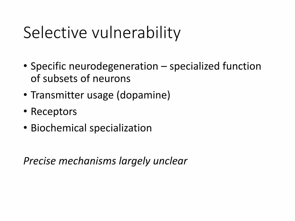
Selective vulnerability
• Specific neurodegeneration – specialized function of subsets of neurons
• Transmitter usage (dopamine)
• Receptors
• Biochemical specialization
Precise mechanisms largely unclear

Alzheimer’s disease
• Prevalence• USA: 5.5 million patients, with 350,000 new patients / yr
• Under 65 yr age, rare but increases with age• > 85 yr age, between 10-30% of population!• Most common cause of dementia (50-56%)
• Common genetic causes (1-2% familial)• Amyloid precursor protein (APP)• Presenilin 1 (PSEN1) – 11% of genetic cases• Presenilin 2 (PSEN2)• APOE4, an important risk factor

Clinical overview
• Medial temporal lobe – entorhinal cortex and hippocampus
• Anterograde episodic memory loss: repeated questions, misplaced items, missed appointments, and forgotten details of daily life. (termed mild-cognitive impairment)
• AD diagnosis requires dementia
• MCI progresses to AD at rate of 10% / yr
• Imaging is used to exclude other diagnosis
• Death: 3-9 years after diagnosis
• Definitive AD only possible post-mortem
Memory loss


Neurochemistry of ADthe cholinergic hypothesis• Profound deficiency of
acetylcholine (ACh)• Atrophy of subcortical
cholinergic neurons• Basal forebrain cholinergic
neurons• Noradrenergic neurons in
locus ceruleus• 5-HT neurons in raphe
• Cholinergic antagonists can induce similar ‘confused’ state to that observed in AD patients


Approved Alzheimer’s drugs –NONE MODIFY THE DISEASE• AChE inhibitors
• Donepezil• Rivastigmine• Galantamine• Tacrine (rarely used due to adverse side-effect profile)
• Non-competitive NMDAR antagonist• Memantine, acts on Mg2+ site to prevent excessive activation
• Combination of donepezil and memantine statistically significant benefit but of marginal effect size
• Treatment of behavioral systems in AD• Atypical antipsychotics, mood stabilizers, antidepressants
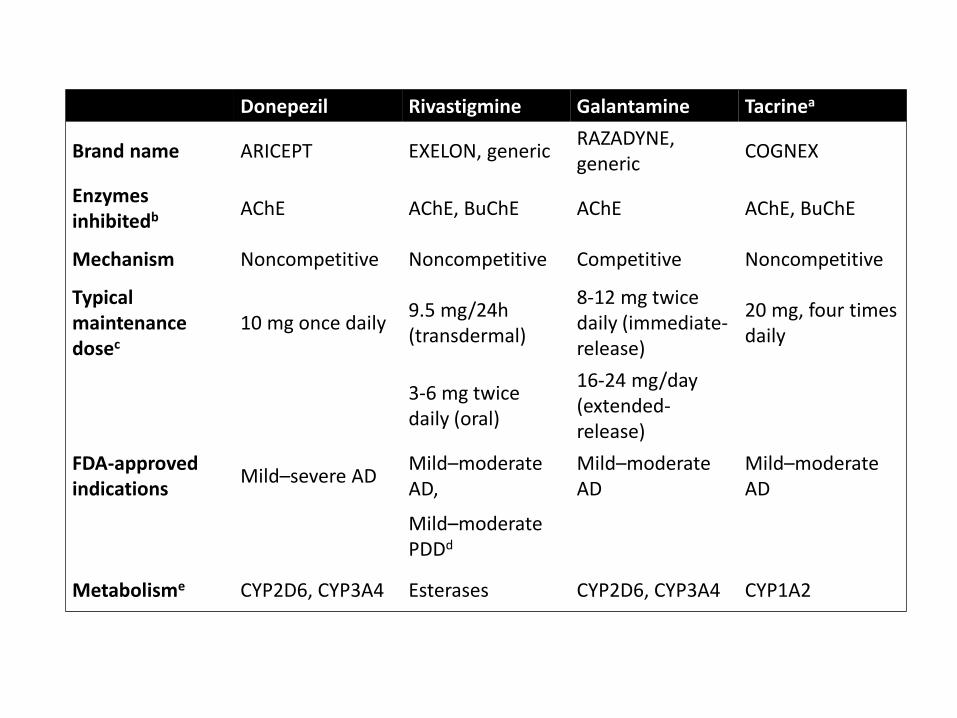
Donepezil Rivastigmine Galantamine Tacrinea
Brand name ARICEPT EXELON, genericRAZADYNE, generic
COGNEX
Enzymes inhibitedb AChE AChE, BuChE AChE AChE, BuChE
Mechanism Noncompetitive Noncompetitive Competitive Noncompetitive
Typical maintenance dosec
10 mg once daily9.5 mg/24h (transdermal)
8-12 mg twice daily (immediate-release)
20 mg, four times daily
3-6 mg twice daily (oral)
16-24 mg/day (extended-release)
FDA-approved indications
Mild–severe ADMild–moderate AD,
Mild–moderate AD
Mild–moderate AD
Mild–moderate PDDd
Metabolisme CYP2D6, CYP3A4 Esterases CYP2D6, CYP3A4 CYP1A2

Why do AChE inhibitors fail?
• Many other neuronal systems affected• Glutamatergic, 5-HT, neuropeptides
• Cortical and hippocampal atrophy in addition to cholinergic degeneration
• Disease is far more complex!

Familial AD• Mutations in APP -> early onset AD
• Mouse models show plaques not tangles
• Trisomy 21 – very early onset dementia like AD with massive AD-like pathology• Mouse models of APP over-production do not result in AD-like pathology
• Presenilin 1 & 2 – causes over-production of Ab

Pathological hallmarks of AD
• Cortical wasting – widening of sulci and loss of tissue
• Senile plaques - Beta-amyloid (Ab) accumulation• Soluble Ab is highly neurotoxic
• Neurofibrillary tangles• Comprised of hyper-phosphorylated Tau• Appears to be a consequence of Ab accumulation
• Characteristic pattern of changes• Early – temporal lobe in entorhinal cortex• Hippocampus• Later – other cortical areas• Consistent with idea of spread along known connections


Querfurth H, LaFerla F. N Engl J Med 2010;362:329-344

Ab & Amyloid Hypothesis
• Produced by abnormal processing of APP• BACE
• Gamma-secretase (4 subunit protein, contains PSEN1/2)
• Ab is 36-43 amino acid fragment of APP
• Accumulates, oligomerizes & forms insoluble plaques
• Ab42 is directly toxic to synapses
• Extracellular Ab can lead to excitotoxic cell death by mediating glutamate release

Synaptic dysfunction caused by Ab
Querfurth H, LaFerla F. N Engl J Med 2010;362:329-344

Tau & Tangles
• Abnormally phosphorylated tau
• Insoluble and associates to formed paired-helical filaments
• Tau is a microtubule associated protein binds to and stabilizes microtubules (MTs)
• Phosphorylation of tau reduces binding
• Likely secondary to Ab
PHF
Hyperphosphorylated Tau
Tangles

Oxidative stress and mitochondrial failure

New drugs
• Targeted at beta-amyloid• LY450139 – γ-secretase inhibitor (Phase 3 – failed in 2010)• Vaccines to elicit immune response against Ab
• AM-1792 – Phase 2a: stopped due to brain inflammation adverse events.• ACC-001 – Phase 2a. Better safety profile. Phase 3: ineffective.
• Antibody against Ab. Bapineuzumab. Also failed Phase 3.
• Neuroprotection• AL-108 – intranasal peptide – Phase 2a promising (2009). • PBT2 – Phase 2a promising (2010) – metal chaperone reduce free
divalent metal ions that result in ROS damage• Etanercept (Enbrel) – RA drug, small Phase 2 promising (2008)
• Tau• Methythionium chloride – methylene blue, reduces tau oxidation
and aggregation (TRx023 - ongoing Phase III trials)

AD pathogenesis

More complex interactions of Ab with neural environment

Does impaired clearance of Ab lead to neurovascular dysfunction?
YES! 60-90% patients have ischemic vascular disease, with high incidence of major infarctions. Many vascular dementia’s have pathological changes akin to AD.
Ab is toxic to endothelial and small muscle components of neurovascular compartment.

Future
• 1772 trials in ClinicalTrials.gov
• 523 open trials
• 54 Phase III
– Amyloid targeting vaccine, 2nd generation (CAD106, CNP520)
– BACE1 inhibitors (LY3314814, JNJ-54861911)
– TTP448 (antagonist of RAGE, interacts with Ab)
– Encenicline (α7-nAChR agonist)

Neurodegenerative Diseases
• Alzheimer’s disease
• Parkinson's disease
• Amyotrophic Lateral Sclerosis (ALS)
• Huntington’s disease
• Others:
– Frontotemporal dementia with Parkinsonism
– Prion diseases
– Multiple sclerosis

Amyotrophic Lateral Sclerosis
• Motor neuron disease (Lue Gehrig's disease)
• 5-10% cases have known genetic cause (i.e. familial)
• Rapidly fatal (1-5 years)
• Loss of upper (cortical) and lower (spinal) motor neurons

Epidemiology and genetics
• Prevalence – Much rarer than AD and PD– Age of onset between 40-60 yrs– Men > Women– USA: 6000-8000 people, 530 new cases / yr– 4.7 cases per 100,000 – Lifetime risk is 1 in 1000
• Genetic risk (90% are sporadic)– Superoxide dismutase (SOD1) – 15-20% of genetic cases– TAR DNA binding protein (TDP-43)– FUS/TLS
• Both bind DNA/RNA regulating transcription

Clinical overview
• Rapid progressive weakness, muscle atrophy and fasciculations (twitch), spasticity (stiffness), dysarthria (speech), dysphagia (eating), respiratory compromise.
• Sensory (non-motor) function is spared
• ALS usually is progressive and fatal. Most patients die of respiratory compromise and pneumonia after 2-3 years.

Pathological mechanisms?
SOD1 mutations in ALS identified in 1993
• Oxidative hypothesis model– SOD1 involved in converting superoxide radicals– Some SOD mutations can result in reduce function leading
to oxidative stress– Others do not effect enzyme function– SOD1 null mice do not develop ALS-like pathology– Suggests a toxic ‘gain of function’
• Aggregation?

SOD1 mutant aggregates

Why do SOD1 aggregates kill motor neurons?
• SOD1 misfolding is detected by the unfolded protein response (UPR), a cellular stress response.
• This should activate a cellular stress pathway in an attempt to restore homeostasis1. Increases proteolysis via proteasome (increase clean-up
of misfolded protein)
2. Increases chaperone expression (improve folding)
3. Reduces protein translation (reduce burden)
• High threshold for this stress response in motor neurons may contribute to selective vulnerability
• Failure of the UPR can result in stimulation of apoptosis
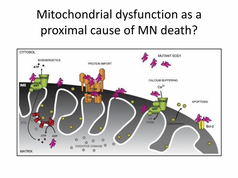
Mitochondrial dysfunction as a proximal cause of MN death?

Evolution of MN injury in ALS

Treatment of ALS
• ONLY Riluzole approved for treatmentMechanism of action is poorly characterized, effects include:
1. Inhibition of glutamate release2. Blockade of post-synaptic NMDA and kainate receptors3. Inhibition of Nav channels.4. GPCR target?
Modest effect on survival and well tolerated. Milestone but unclear how this will lead to future advances in drug therapy.
Spasticity – Balcofen (GABAB) and diazepam (GABAA)Dysphagia – Amitriptyline (TCA) – anticholinergic to prevent excess saliva production

Loss of upper MN input leads to spasticity GABA
agonists substitutes
for loss of inhibitory
input

Current trials on ClinicalTrials.gov
• 38 trials Phase III – 5 open– Olanzapine
• Aimed at treating appetite loss
– Tirasemtiv (Cytokimetics)• Skeletal muscle troponin activator – increases
sensitivity to Ca2+ thereby allowing fewer MN fibers to elicit larger effects, i.e. may delay loss of muscle function
– Masitinib (AB1010) – anti-cancer – inhibits RTKs• Reduce inflammation

Neurodegenerative Diseases
• Alzheimer’s disease
• Parkinson's disease
• Amyotrophic Lateral Sclerosis (ALS)
• Huntington’s disease
• Others:
– Frontotemporal dementia with Parkinsonism
– Prion diseases
– Multiple sclerosis

Huntington’s disease
• Incidence 4-10 / 100,000 new cases per year
• Genetically determined autosomal dominant
• Near 100% dominance with prominent anticipation– i.e. subsequent generations suffer from earlier onset
• Trinucleotide repeat disease (CAGn) in the huntington gene (Chr 4)– Normal = 9-34 triplets (median: 19)
– HD = 40-100+

Clinical overview
• Gradual onset of motor incoordination and cognitive decline
• Huntington’s chorea – movement disorder – brief jerk-like movements
• Fine-motor coordination and impairment of rapid eye movements are early symptoms
• Psychiatric symptoms (confusion, amnesia, psychosis) and cognitive dysfunction (dementia)
• Disease is fatal, typically over 15-20 years.

Basal Ganglia Pathology
• Massive loss of medium spiny neurons in neostriatum (up to 95%)– GABAergic inhibitory
interneurons that receive input from SNpC DA neurons
– Affects innervation of GPiand SNpr (indirect) before Gpe (direct pathway)

Reduced activity
Increased activity

NONE MODIFY THE DISEASE

Pathological mechanisms – two hypotheses
• Excitotoxicity– Animal model – infusion of excitotoxin into striatum can
produce similar motor symptoms and loss of MSNs– MSNs receive large excitatory input from neocortex
• Mitochondrial dysfunction– Ultrastructural evidence– PET – reduced glucose and O2 metabolism– Mitochondrial toxins into striatum cause similar pattern of
MSN loss with preservation of other interneurons• Which can be blocked by removal of cortical input or NMDA
antagonists
• Combination?

HD is a monogenic autosomal dominant disease
• One gene – when mutated – causes disease
• Identified in 1993 as Huntingtin (HTT)
• Mutation characterized as a CAG triplet repeat in HTT
Toxic N-term fragments

Key features of HD pathogenesis
1. Mutant HTT misfolds
2. Unfolded protein response is impaired
3. Mutant HTT is truncated and fragments are highly toxic
4. Post-translational modifications of HTT influence toxicity
5. Nuclear translocation of HTT contributes to toxicity
6. Cellular metabolism is impaired

HTT inclusion in medium spiny neurons

Intracellular pathogenesis in HD

Cell interactions and intercellular pathogenesis
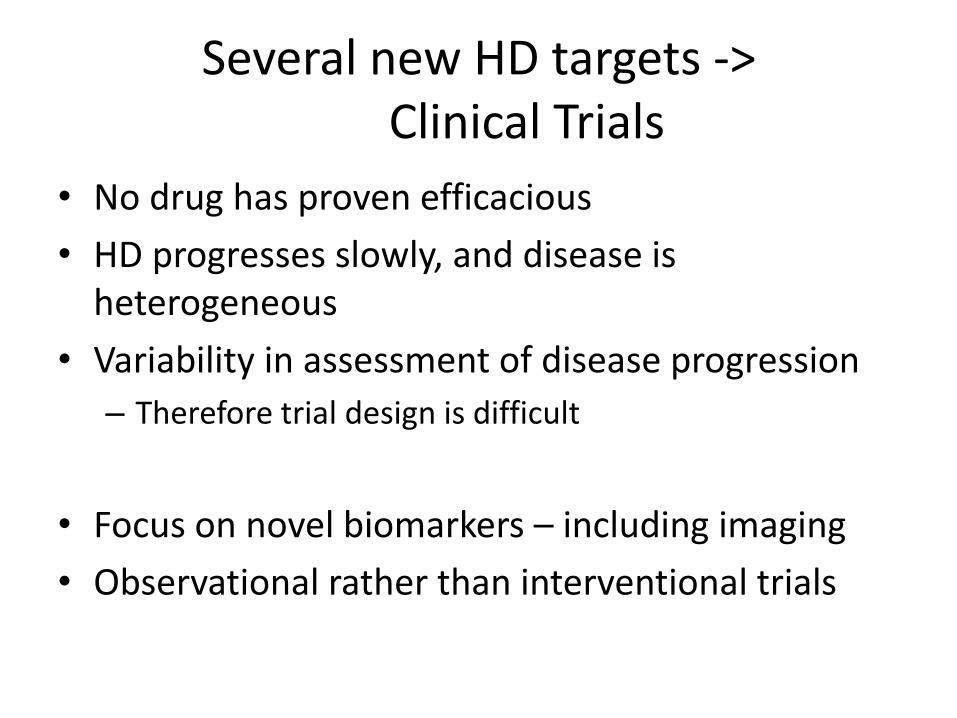
Several new HD targets -> Clinical Trials
• No drug has proven efficacious
• HD progresses slowly, and disease is heterogeneous
• Variability in assessment of disease progression
– Therefore trial design is difficult
• Focus on novel biomarkers – including imaging
• Observational rather than interventional trials

MRI identifies prodromal disease and may be used to identify HD patients
for improved intervention

Other triplet repeat disease
• HD part of a family of triplet repeat diseases characterized by polymorphic triplet repeats
• Show ‘anticipation’ – earlier onset in subsequent generations
• Can be in coding and non-coding regions, in coding regions they encode poly amino acid repeats
Huntington’s Disease CAG
Fragile X Disease CGG
Myotonic Dystrophy CTG
Spinocerebellar atrophy (type 1)
CAG
Spinal and bulbar muscularatrophy (Kennedy’s disease)
CAG

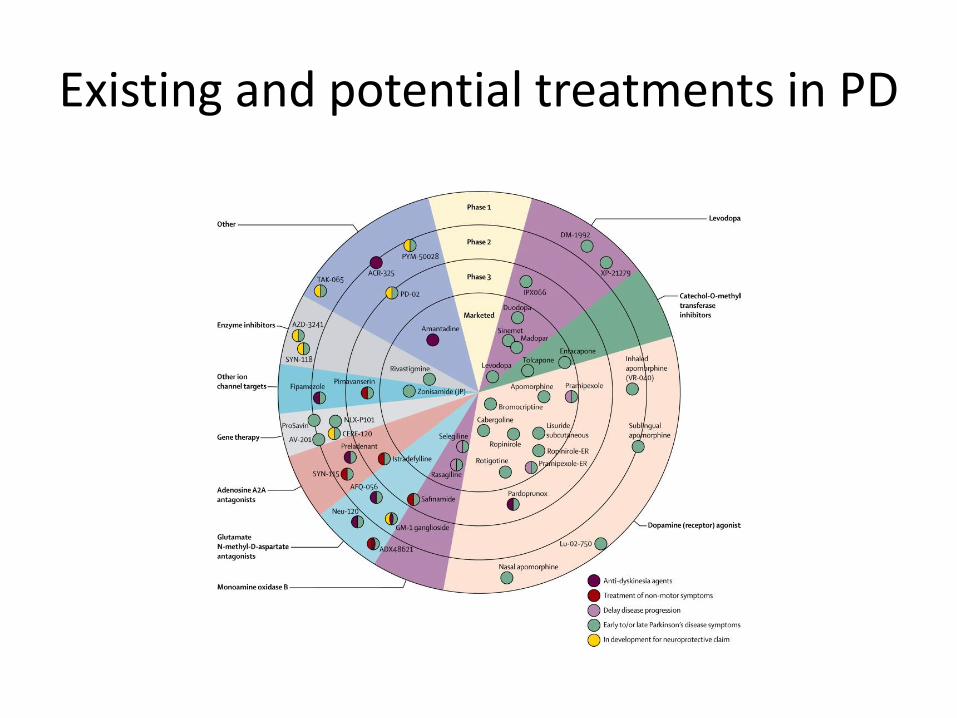
Existing and potential treatments in PD

Evolution of neurodegenerative diseases

Therapeutic strategies
Examples in PD

Strategies to prevent protein misfolding, aggregation and UPR stress
Examples in PD

Gene and cell therapy for neurodegenerative disease
• Cell replacement?
– PD – fetal mDA neurons, iPSCs, etc
– HD – MSNs
– ALS – MNs
– AD – unclear, too challenging
• Gene therapy?
– Viral vectors – likely AAV-based
– GDNF, BDNF, IGF, etc.

Cellular Transplantation
• Cellular source and preparation
• Placement of cells– Original site of damage (requires projection
though tissue)
– Target site (will not receive appropriate inputs)
• Immunologic rejection– CNS privileged site but immunosuppression will be
necessary for allogenic or xenografts
• Function

Transplantation in PD
• 6-OHDA model – apomorphine induce rotations due to R supersensitivity
• Inject DA neuron-containing graft• Assess rotational behavior
• Early clinical trials (Sweden, 1987) used chromaffin cells –failure
• Human fetal midbrain grafts– PET confirmation of DA release– 12 months to reach peak
• Stem cells: hES & iPSCs• Directed reprogramming: PMY516 recitation