free-standing nanoparticle superlattice sheets: from ...ion-permeability membranes. free-standing...
TRANSCRIPT

August 2017
EPL, 119 (2017) 48004 www.epljournal.orgdoi: 10.1209/0295-5075/119/48004
Focus Article
Free-standing nanoparticle superlattice sheets: From designto applications(a)
Wenlong Cheng(b)
Department of Chemical Engineering, Faculty of Engineering, Monash UniversityClayton 3800, Victoria, Australia andThe Melbourne Centre for Nanofabrication - 151 Wellington Road, Clayton 3168, Victoria, Australia
received 1 September 2017; accepted 6 October 2017published online 8 November 2017
PACS 81.16.Dn – Self-assemblyPACS 68.65.Cd – SuperlatticesPACS 81.07.Bc – Nanocrystalline materials
Abstract – Here I summarize my perspective in free-standing nanoparticle superlattice sheetswith regards to their fabrication, properties, and applications.
focus article Copyright c© EPLA, 2017
Why free-standing?– Nanoparticles have beencoined as “artificial atoms” due to their unique size/shape-dependent optical, electrical, magnetic and conductiveproperties. Encouraging progress made over the pastseveral decades allows for controlling nanoparticles sizesand shapes, enabling the formation of the so-called “ar-tificial periodic table” [1]. The crystallization of theseartificial atoms into highly-ordered arrays represents aunique strategy to design metamaterials with collectiveproperties different from those of bulk phase crystals,isolated nanocrystals and even disordered nanocrystal as-semblies [2–6]. These unusual properties include spinproperties [7], metal-insulator transition [2], mechanicalproperties [4,8], p-type conductivities [5], vibrational co-herence [3], and plasmonic properties [9–11]. In addition,diverse types of lattice structures have been found includ-ing face-centered cubic, body-centered cubic, hexagonalclosed-packed, diamond-like lattice, and lattice structuresnot existing in nature [12].
It has to be noted that the majority of these stud-ies focus on solid-substrate–supported nanoparticle super-lattices or solution-state nanoparticle superlattices. Incontrast, free-standing nanoparticle superlattice sheetsrefer to suspended ordered nanoparticle arrays withminimal contact with the solid substrate. Comparedto dominant solid-substrate–supported or solution-statesystems, free-standing nanoparticle superlattice sheets
(a)Contribution to the Focus Issue Self-assemblies of Inorganic andOrganic Nanomaterials edited by Marie-Paule Pileni.(b)E-mail: [email protected]
offer the advantages of facile transferring, manufacturableinto 1D or 3D structures, and application as ion-permeablemembranes.
Transferability. In comparison to their counter-parts in substrate-supported or solution-state system,free-standing nanoparticle superlattices offer the uniqueadvantage of facile transfer from one substrate to another.Figure 1(a) shows the transfer of Langmuir-deposited goldnanowire superlattice membranes from air/water interfacefrom PDMS substrate to PET substrate. One can see thatmonolayered nanowire membranes can be transferred ontoPET substrate with 100% transfer fidelity [13]. This faciletransfer was also demonstrated to be feasible for trans-ferring plasmene nanosheets onto paper-based Maylasianbanknote, Australian plastic banknote, and even topologi-cally complex rigid surfaces such as coins [14,15]. It is evenpossible to obtain free-standing bilayered nanoparticle su-perlattice sheets [16].
Manufacturability into 1D and 3D structures. An-other unique attribute of free-standing systems isthat they can be further manufactured into com-plex structurally well-defined one-dimensional and three-dimensional nanoarchitectures, which are often challeng-ing to obtain from substrate-supported or solution-statesystem. As an example, suspended polystyrene-basednanoparticle superlattices can be transformed into 1Dnanoribbons simply using “hard milling” by focused ionbeam (FIB) lithography (fig. 1(b)). The hard millingmeans that both organic ligand and inorganic corematerials are removed. On the other hand, soft milling
48004-p1

Wenlong Cheng
Fig. 1: (Color online) Transferability and manufacturabilityof free-standing superlattices. (a) Transfer of nanowire super-lattice membranes from PDMS to PET substrates; (b) free-standing Au@Ag nanocube superlattice ribbons; (c) schematicof fabrication of Au@Ag nanocube superlattice sheets intocube-like origami by focused-ion-beam lithography; (d) SEMimage of cube-like origami from Au@Ag nanocube buildingblocks. Reproduced from refs. [9,13].
Fig. 2: (Color online) Bilayered gold nanoparticle superlat-tice sheets as ionic transport membrane. (a) Optical image ofbilayered nanoparticle superlattice membranes; (b), (c): SEMimages of bilayered nanoparticle superlattice membranes undertwo different magnifications; (d) schematic of cone-like geome-try enabling directional regulation of ionic transports. Repro-duced from ref. [16].
involves the use of FIB in a mild condition (eitherweak beam currents and/or short dwelling time), whichcan be controlled to remove soft ligand materials only.Combination of hard and soft FIB milling at the pro-grammed locations allows for versatile nanomanufactureof 3D orgami [9,17]. Figure 1(c) shows the sequential softmilling applied to polystyrene-based nanoparticle super-lattice sheets. Note that site-specific soft ligand removalby soft FIB milling generates local stress. Along the stressrelaxation, the nanosheets can self-fold into well-definedorigami (fig. 1(d)), which has not yet been realized withentirely inorganic 2D materials.
Ion-permeability membranes. Free-standingnanosheets also offer the attribute of ion permeability
and/or regulation. Using bilayered nanoparticle super-lattice sheets, one can fabricate an artificial membranebased on an asymmetric structure, which can directunidirectional ionic transport similar to natural cellmembrane [16]. The asymmetric geometry can be con-trolled by using two different particle sizes, which leadsto cone-like nanochannels formed within the membranes(fig. 2). This results in an asymmetric ion transportbehavior, and diode-like behavior. Ionic rectificationratios can be controlled by adjusting relative particlesizes. The free-standing particle sheet-based design ofnanomembrane may lead to promising applications inbiosensor devices, energy conversion, biophotonics, andbioelectronics.
How to fabricate free-standing nanoparticlesuperlattice sheet?– The formation of free-standingnanoparticle superlattice sheets requires a balance be-tween attraction force and repulsion forces amongnanoparticles, which can be achieved either via entropy-driven strategy or enthalpy-driven approach.
Entropy-driven strategy. Drying-mediated self-assembly has demonstrated to be a powerful and generalstrategy to fabricate ordered assemblies of nanoparti-cles [2–5]. To achieve ordered structures, nanoparticlesneed to be highly repulsive. Slow evaporation of solventsis usually required to gradually restrict the availablevolume for each individual nanoparticles to controlordered nucleation and growth process. However, one-dimensionally confined physical environments are requiredfor two-dimensional nucleation and growth in order toobtained two-dimensional free-standing nanoparticlesuperlattice sheets [4,8]. Alkyl-capped gold nanopar-ticles and polystyrene-capped plasmonic nanoparticlesare hydrophobic, which have been successfully usedto generate giant nanosheets (i.e., nanoscale thicknessyet with macroscopic lateral dimensions, correspondingto large aspect ratio) from a two-stage drying process(fig. 3(a)) [4,9]. In the first-stage drying, organic solventsquickly evaporate leaving nanoparticle patches formed atthe air/water interface; in the second-stage drying, waterslowly evaporates offering strong surface tension causingcoalesce and fusion of small patches of nanoparticlesuperlattice sheets into giant ones.
Interestingly, hydrophilic nanoparticles can also be usedto generate free-standing nanoparticle superlattice sheets,however, strategies need to be devised to confine crys-tallization events to a two-dimensional plane. DNA-capped gold nanoparticles have been successfully usedto generate nanosheets through microhole-confined self-assembly [8]. When a droplet of DNA-nanoparticle solu-tion dries on a holey substrate, satellite microdroplets areformed and trapped in individual microholes owing to pin-ning of their contact lines onto the microhole edges. Thenthe microhole-trapped satellite microdroplets thinnes aswater evaporates, forming a two-dimensional water filmwhich confines crystallization of nanoparticles. Finally,
48004-p2
Free-standing nanoparticle superlattice sheets: From design to applications
Fig. 3: (Color online) Self-assembled nanosheets by entropyor enthalpy-driven processes. (a) Drying-mediated entropy-driven self-assembly of hydrophobic nanoparticles at theair/water interface; (b) Gibbs monolayered DNA-nanoparticlesheets by enthalpy-driven Watson-Crick base-pairing forces;(c) DNA-based multidomain self-assembled gold nanoparti-cle films by enthalpy-driven Watson-Crick base-pairing forces.Reproduced from refs. [9,18,19].
ordered nanoparticle superlattice sheets are suspended inthe microholes. This approach allows for simultaneousinternal structural control such as interparticle spacing.
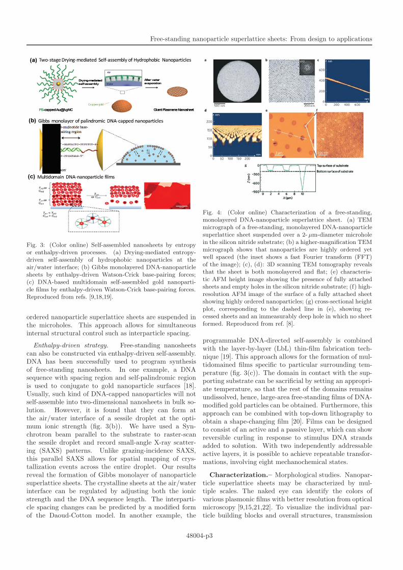
Enthalpy-driven strategy. Free-standing nanosheetscan also be constructed via enthalpy-driven self-assembly.DNA has been successfully used to program synthesisof free-standing nanosheets. In one example, a DNAsequence with spacing region and self-palindromic regionis used to conjugate to gold nanoparticle surfaces [18].Usually, such kind of DNA-capped nanoparticles will notself-assemble into two-dimensional nanosheets in bulk so-lution. However, it is found that they can form atthe air/water interface of a sessile droplet at the opti-mum ionic strength (fig. 3(b)). We have used a Syn-chrotron beam parallel to the substrate to raster-scanthe sessile droplet and record small-angle X-ray scatter-ing (SAXS) patterns. Unlike grazing-incidence SAXS,this parallel SAXS allows for spatial mapping of crys-tallization events across the entire droplet. Our resultsreveal the formation of Gibbs monolayer of nanoparticlesuperlattice sheets. The crystalline sheets at the air/waterinterface can be regulated by adjusting both the ionicstrength and the DNA sequence length. The interparti-cle spacing changes can be predicted by a modified formof the Daoud-Cotton model. In another example, the
Fig. 4: (Color online) Characterization of a free-standing,monolayered DNA-nanoparticle superlattice sheet. (a) TEMmicrograph of a free-standing, monolayered DNA-nanoparticlesuperlattice sheet suspended over a 2- µm-diameter microholein the silicon nitride substrate; (b) a higher-magnification TEMmicrograph shows that nanoparticles are highly ordered yetwell spaced (the inset shows a fast Fourier transform (FFT)of the image); (c), (d): 3D scanning TEM tomography revealsthat the sheet is both monolayered and flat; (e) characteris-tic AFM height image showing the presence of fully attachedsheets and empty holes in the silicon nitride substrate; (f) high-resolution AFM image of the surface of a fully attached sheetshowing highly ordered nanoparticles; (g) cross-sectional heightplot, corresponding to the dashed line in (e), showing re-cessed sheets and an immeasurably deep hole in which no sheetformed. Reproduced from ref. [8].
programmable DNA-directed self-assembly is combinedwith the layer-by-layer (LbL) thin-film fabrication tech-nique [19]. This approach allows for the formation of mul-tidomained films specific to particular surrounding tem-perature (fig. 3(c)). The domain in contact with the sup-porting substrate can be sacrificial by setting an appropri-ate temperature, so that the rest of the domains remainsundissolved, hence, large-area free-standing films of DNA-modified gold particles can be obtained. Furthermore, thisapproach can be combined with top-down lithography toobtain a shape-changing film [20]. Films can be designedto consist of an active and a passive layer, which can showreversible curling in response to stimulus DNA strandsadded to solution. With two independently addressableactive layers, it is possible to achieve repeatable transfor-mations, involving eight mechanochemical states.
Characterization.– Morphological studies. Nanopar-ticle superlattice sheets may be characterized by mul-tiple scales. The naked eye can identify the colors ofvarious plasmonic films with better resolution from opticalmicroscopy [9,15,21,22]. To visualize the individual par-ticle building blocks and overall structures, transmission
48004-p3

Wenlong Cheng
Fig. 5: (Color online) Schematic of Synchrotron-based Small-Angle X-ray Scattering (SAXS) setup for probing spatialcrystallization events of DNA-capped nanoparticles at theair/water interface. Reproduced from ref. [18].
electron microscope (TEM) is usually required (fig. 4(a)and (b)). By tilting substrates and recording a series oftwo-dimensional TEM images, a 3D tomographic TEMimage may be obtained (fig. 4(c), (d)). Morphologicalcharacterization can also be done with atomic force mi-croscopy (AFM). Figures 4(e)–(g) show that both intactand ruptured nanosheets can be identified and the high-resolution AFM image shows ordered nanoparticle arrays(fig. 4(f)).
Crystalline structures. Unlike conventional X-raytechniques, a high flux of X-ray from Synchrotron canenable acquisition of crystalline information almost in-stantaneously. Hence, Synchrotron-based SAXS hasdemonstrated to be a powerful tool to probe, in real-time,temporal and spatial crystallization events [18,23]. Whiletransmission SAXS have been used to probe crystalliza-tion events in bulk solutions [24], grazing-incidence SAXScan be used to monitor the formation of 2D nanoparticlesuperlattices [18]. As an example, using grazing-incidenceSAXS in a special configuration (parallel SAXS, orparSAXS), we can map the crystallization of DNA-cappednanoparticles by raster-scanning across a sessile droplet.This can be done in a humidity-controlled environment(fig. 5). The results reveal the formation of crystallineGibbs monolayers of DNA-capped nanoparticles at the air-liquid interface. By integrating original two-dimensionalSAXS patterns, one can obtain a one-dimensional pat-tern with well-defined Bragg peaks, which then allow foranalysis of crystalline lattice structures and other key pa-rameters such as nearest-neighbor spacing.
Properties.–
Plasmonic properties. Due to their size- andshape-dependent plasmonic properties, noble metalnanoparticles can be treated as “meta-atoms”. Whenthose meta-atoms are closely-packed in the extended2D arrays, both gap-mode plasmons and propagatingplasmons are present [9]. Figure 6(a) shows a typicalTEM image of Au@Ag nanocube plasmene nanosheet,which was imported into COMSOL for exact plasmonic
Fig. 6: (Color online) Properties of free-standing nanoparticlesuperlattice sheets. (a) Representative TEM image for Au@Agnanocube-based plasmene nanosheet; (b) simulated near-fielddistributions when excited with light having a free space wave-length of 490 nm; (c) experimental extinction spectrum of theAu@Ag nanocube-based plasmene sheet; (d) nano-indentationwith a nanoscale AFM probe; (e) a typical AFM force-indentation curve showing consecutive events of no contact,jump in, linear and nonlinear deformation of a free-standingnanoparticle superlattice sheet. Reproduced from refs. [9,26].
modelling. The modelling result is shown in fig. 6(b),where a strong gap-mode plasmon hot spot is evident. Thisgap-mode plasmons may be responsible for the sharp plas-monic peak observed (fig. 6(c)). This peak gets sharperas the silver coating thickness increases, which is in agree-ment with the COMSOL modelling results. On the topof the plasmene nanosheet, propagating plasmons arealso present as demonstrated theoretically and experimen-tally. It turns out that plasmene nanosheets support onlytransverse electric field, leading to the conversion of un-polarized light into polarized light.
Mechanical properties. Unlike traditional colloidalcrystals, free-standing ligand-linked nanoparticlesuperlattice sheets are elastic [4,8]. They can serveas drumhead or mechanical resonators [25]. The changeof soft ligand length enables the tuning of mechaniclproperties. As for the ultrathin two-dimensional materi-als, key micromechanical parameters such as the elasticmodulus, ultimate strength, and maximum elongation,can be in principle derived from mechanical tests, suchas bulge tests, analysis of buckling deformations, forcespectroscopy, and stretching ridge analysis [26]. Inpractice, AFM nanoindentation has been successfullyused for estimating Young’s modulus, spring constant,breaking strength [4,8]. Figure 6(d) illustrates the AFMindentation process. Note that commercial AFM probesare generally sharp, and therefore a blunting proceduremay be applied to avoid rupturing the nanosheet. Theforce spectroscopy typically features initial linear followedby nonlinear deformation (fig. 6(e)). For the linear region,bending stiffness or spring constant can be obtained.
48004-p4

Free-standing nanoparticle superlattice sheets: From design to applications
Fig. 7: (Color online) Representative applications of free-standing nanoparticle superlattice sheets. (a) Application assoft, semitransparent SERS substrate for direct real-world sur-face chemical identifications; (b) combination of plasmoniccodes (sizes and shapes) with molecular codes (fingerprintvibrational signals) to offer dually-coded security labels for en-cryption applications; (c) asymmetric ionic regulation via bi-layered nanoparticle superlattice sheets; (d) palindromic DNAsequences are responsive to ionic strength. Reproduced fromrefs. [16,18,21,29].
At larger indentations, nonlinear behavior is generallyobserved. The theory of a point load on a clamped thinfilm can predict its mechanical properties, which canbe used to model experimental force curve to determineelastic moduli and ultimate strength [27].
In addition, an interesting recent study shows that thegrazing-incidence SAXS can identify 0.6 nm difference inaverage ligand-shell thickness between two sides of alky-ligated nanoparticle superlattice membranes [28]. Hence,they called their membranes Janus-like structures whichwere also proven by SERS measurement using a moleculartracer. Upon exposure to electron beam, the detached free-standing membranes can self-fold into tubular structures.Because the air-facing side has higher ligand density andless strain, the folding is always towards the water-facingside which has lower ligand density and higher strain.
Applications.–
As soft surface enhanced Raman scattering (SERS)substrates. The commercially available Klarite SERSsubstrate is based on evaporated gold on lithographi-cally patterned silicon. It is rigid and opaque, preventingit from establishing conformal contact with topologicallycomplex real-world surfaces, such as banknotes and coins.Hence, it is impractical to use the Klarite substratefor direct identification of trace amount of chemicals.This limitation can be overcome by using soft plasmonicnanoparticle superlattice sheets (or plasmene nanosheets)
which renders it possible to directly “attach and detect”(fig. 7(a)) [14,29]. The elastic nature of nanosheets en-ables their conformal contact with topologically complexsurfaces to position plasmonic field in close affinity withsurface chemicals; their optical semitransparency enablesdirect spectral acquisition without the need of extractingtrace amount of chemicals into solution; the uniformparticle size/shape enables reliable spatial quantification.
Anticounterfeiting. Nanosheet-constituent buildingblocks can carry optical signatures specific to nanoparticlesize and shape [1]; simultaneously, SERS molecularfingerprint vibrational information can be built intothe nanosheets during synthesis [21]. Thus, plasmonicnanoparticle superlattice sheets can serve as a dual-codedsecurity label for banknotes (fig. 7(b)). As a proof of con-cept, we have designed nine different plasmonic codes fromgold nanospheres, gold rhombic dodecahedrals, and goldnanostars as building blocks. Each type of building blockshas three different sizes. In addition, we have chosen fiveadditional SERS fingerprint barcodes. This means that wehave generated 45 dually-coded security labels. Consider-ing the choices of nanoparticle building blocks and SERSmolecules, this approach can in principle offer virtuallyunlimited coding capacity.
Ionic gating. The interstices between nanoparticles inthe nanosheets may be used for ionic gating applications.Usually, larger particle size leads to larger interparticlespacing. When nanosheets with two different nanoparti-cle sizes are assembled into a bilayer configuration, asym-metric cone-like ionic channels form [16]. This geometricalasymmetry leads to diode-like I-V curves. Furthermore,a simulation map can be established to describe the rela-tionship between channel structures and ionic selectivity.By choosing different constituent particle size pairs, thecurrent rectifying ratio can be tuned (fig. 7(c)).
Stimuli-responsivity. Ligand-based nanoparticlesuperlattice sheets can possess soft materials properties,such as stimuli-responsive properties. Figure 7(d) showsthat the nearest-neighbor spacing in DNA-nanoparticlesheets is sensitive to the ionic strength [18]. Higherionic strength screens charges so that DNA strands tendto collapse and consequently the interparticle spacingreduces. This has been proven by Synchrotron-basedSAXS. In addition, plasmonic properties of ligand-basednanoparticle superlattice sheets may be sensitive tomechanical stimuli [22]. When the lattice is stretched,interparticle spacing may increase, reducing plasmoniccoupling, hence, causing spectral shift. This attributemay be used for strain gauge sensing applications.
Challenges and opportunities.– As is describedabove, free-standing nanoparticle superlattice sheets are aspecial type of materials in the family of self-assemblies ofnanocrystals. The mechanical flexibility endows this kindof 2D materials with novel properties and applications,which are difficult to achieve with substrate-supported
48004-p5

Wenlong Cheng
and solution-state counterparts. Soft adhesive-like SERSsubstrate, 3D origami and ion-gating nanomembranes arerepresentative examples.
Despite encouraging progress made so far, scalablefabrication technologies to achieve defect-free structuresremain the challenge. It is known that the nanoparticlesare extremely difficult to manipulate due to complexnanoscale forces occurring at different temporal andspatial scales. To date, success is limited to only a fewtypes of simple particle sizes and shapes; production ofhigh-quality nanosheets at the macroscopic scales remainchallenging. Soft capping ligands play crucial roles inregulating chemical potentials among nanoparticles inorder to obtain 2D nanosheet assemblies. However, itremains illusive to control ligand-to-ligand interactionsincluding steric hindrance, hydrogen bonding, electro-static attraction/repulsion and bio-recognition forces atthe desired temporal and spatial scales. Hence, thereremains lack of general design rules towards well-defined2D nanosheet assemblies.
Free-standing nanoparticle superlattice sheets may betreated as a nanoscale analogue of current 2D materi-als. However, they only arouse research interest from afew groups worldwide and only a few type of nanoparticlesize/shapes were demonstrated. There is a variety of otherparticle geometries, material and soft ligand composition,as well as hybrid binary or tertiary systems which remainto be explored. While I acknowledge it is a challengingtask, modern experimental techniques such as SAXS, 3Dstructural tomography, optical microscopy in conjunctionwith multiscale modeling and simulation (molecular dy-namics, coarse-grain model, Monte Carlo technique) mayallow for the establishment of useful design rules for thereliable fabrication of scalable nanoparticle superlatticesheets to complement and/or add new functions to thecurrent 2D materials family.
∗ ∗ ∗I acknowledge the financial support from the Aus-
tralian Research Council via Discovery Grant schemeDP140100052, DP170102208. I also express great thanksto Ms Qianqian Shi for her help in editing themanuscript.
REFERENCES
[1] Tan S. J., Campolongo M. J., Luo D. and Cheng W.
L., Nat. Nanotechnol., 6 (2011) 268.[2] Collier C. P., Saykally R. J., Shiang J. J.,
Henrichs S. E. and Heath J. R., Science, 277 (1997)1978.
[3] Courty A., Mermet A., Albouy P. A., Duval E. andPileni M. P., Nat. Mater., 4 (2005) 395.
[4] Mueggenburg K. E., Lin X. M., Goldsmith R. H.
and Jaeger H. M., Nat. Mater., 6 (2007) 656.[5] Urban J. J., Talapin D. V., Shevchenko E. V.,
Kagan C. R. and Murray C. B., Nat. Mater., 6 (2007)115.
[6] Pileni M. P., EPL, 109 (2015) 58001.[7] Black C. T., Murray C. B., Sandstrom R. L. and
Sun S. H., Science, 290 (2000) 1131.[8] Cheng W. L., Campolongo M. J., Cha J. J., Tan S.
J., Umbach C. C., Muller D. A. and Luo D., Nat.Mater., 8 (2009) 519.
[9] Si K. J., Sikdar D., Chen Y., Eftekhari F., Xu Z.,
Tang Y., Xiong W., Guo P., Zhang S., Lu Y., Bao
Q., Zhu W., Premaratne M. and Cheng W., ACSNano, 8 (2014) 11086.
[10] Shi Q., Si K. J., Sikdar D., Yap L. W., Premaratne
M. and Cheng W., ACS Nano, 10 (2016) 967.[11] Ng K. C., Udagedara I. B., Rukhlenko I. D., Chen
Y., Tang Y., Premaratne M. and Cheng W., ACSNano, 6 (2012) 925.
[12] Auyeung E., Cutler J. I., Macfarlane R. J., Jones
M. R., Wu J. S., Liu G., Zhang K., Osberg K. D.
and Mirkin C. A., Nat. Nanotechnol., 7 (2012) 24.[13] Chen Y., Zi O., Gu M. and Cheng W., Adv. Mater.,
25 (2013) 80.[14] Chen Y., Si K. J., Sikdar D., Tang Y., Premaratne
M. and Cheng W., Adv. Opt. Mater., 3 (2015) 919.[15] Si K. J., Guo P., Shi Q. and Cheng W., Anal. Chem.,
87 (2015) 5263.[16] Rao S., Si K. J., Yap L. W., Xiang Y. and Cheng W.,
ACS Nano, 9 (2015) 11218.[17] Si K. J., Chen Y. and Cheng W. L., Mater. Today, 19
(2016) 363.[18] Campolongo M. J., Tan S. J., Smilgies D. M., Zhao
M. Chen Y., Xhangolli I., Cheng W. L. and Luo D.,ACS Nano, 5 (2011) 7978.
[19] Estephan Z. G., Qian Z. X., Lee D., Crocker J. C.
and Park S. J., Nano Lett., 13 (2013) 4449.[20] Shim T. S., Estephan Z. G., Qian Z. X., Prosser J.
H., Lee S. Y., Chenoweth D. M., Lee D., Park S. J.
and Crocker J. C., Nat. Nanotechnol., 12 (2017) 41.[21] Si K. J., Sikdar D., Yap L. W., Foo J. K. K., Guo
P. Z., Shi Q. Q., Premaratne M. and Cheng W. L.,Adv. Opt. Mater., 3 (2015) 1710.
[22] Guo P., Sikdar D., Huang X., Si K. J., Su B., Chen
Y., Xiong W., Wei Yap L., Premaratne M. andCheng W., J. Phys. Chem. C, 118 (2014) 26816.
[23] Cheng W. L., Hartman M. R., Smilgies D. M., Long
R., Campolongo M. J., Li R. P., Sekar K., Hui C.
Y. and Luo D., Angew. Chem., Int. Ed., 49 (2010) 380.[24] Park S. Y., Lytton-Jean A. K. R., Lee B., Weigand
S., Schatz G. C. and Mirkin C. A., Nature, 451 (2008)553.
[25] Kanjanaboos P., Lin X. M., Sader J. E., Rupich S.
M., Jaeger H. M. and Guest J. R., Nano Lett., 13(2013) 2158.
[26] Cheng W., Campolongo M. J., Tan S. J. and Luo D.,Nano Today, 4 (2009) 482.
[27] Wan K. T., Guo S. and Dillard D. A., Thin SolidFilms, 425 (2003) 150.
[28] Jiang Z., He J. B., Deshmukh S. A., Kanjanaboos
P., Kamath G., Wang Y. F., Sankaranarayanan S.
K. R. S., Wang J., Jaeger H. M. and Lin X. M., Nat.Mater., 14 (2015) 912.
[29] Chen Y., Si K. J., Sikdar D., Tang Y., Premaratne
M. and Cheng W., Adv. Opt. Mater., 3 (2015) 918.
48004-p6