free radical biology & medicine - connecting repositories · 2016-12-07 · some indications...
TRANSCRIPT

Free Radical Biology & Medicine 51 (2011) 1717–1726
Contents lists available at SciVerse ScienceDirect
Free Radical Biology & Medicine
j ourna l homepage: www.e lsev ie r.com/ locate / f reeradb iomed
Original Contribution
Effects of heme oxygenase-1 on induction and development of chemically inducedsquamous cell carcinoma in mice
Halina Was a, Malgorzata Sokolowska a, Aleksandra Sierpniowska b, Paweł Dominik a, Klaudia Skrzypek a,Bozena Lackowska c, Antoni Pratnicki d, Anna Grochot-Przeczek a, Hevidar Taha a, Jerzy Kotlinowski a,Magdalena Kozakowska a, Andrzej Mazan a, Witold Nowak a, Lucie Muchova e, Libor Vitek e, Anna Ratajska d,Jozef Dulak a, Alicja Jozkowicz a,⁎a Department of Medical Biotechnology, Faculty of Biochemistry, Biophysics, and Biotechnology, Jagiellonian University, Gronostajowa 7, 30-387 Krakow, Polandb Department of Biophysics, Faculty of Biochemistry, Biophysics, and Biotechnology, Jagiellonian University, 30–387 Krakow, Polandc Department of Pathology, Oncology Center, Krakow, Polandd Department of Pathological Anatomy, Medical University of Warsaw, Warsaw, Polande Charles University, Prague, Czech Republic
⁎ Corresponding author. Fax: +48 12 664 6918.E-mail address: [email protected] (A. Jozkow
0891-5849 © 2011 Elsevier Inc.doi:10.1016/j.freeradbiomed.2011.07.025
Open access under CC BY
a b s t r a c t
a r t i c l e i n f oArticle history:Received 6 March 2011Revised 12 July 2011Accepted 27 July 2011Available online 10 August 2011
Keywords:Heme oxygenase-1CarcinogenesisSquamous cell carcinomaDMBAInflammationOxidative stressFree radicals
Heme oxygenase-1 (HO-1) is an antioxidative and cytoprotective enzyme, which may protect neoplastic cellsagainst anticancer therapies, thereby promoting the progression of growing tumors. Our aim was toinvestigate the role of HO-1 in cancer induction. Experiments were performed in HO-1+/+, HO-1+/−, and HO-1−/−mice subjected to chemical induction of squamous cell carcinomawith 7,12-dimethylbenz[a]anthraceneand phorbol 12-myristate 13-acetate. Measurements of cytoprotective genes in the livers evidenced systemicoxidative stress in the mice of all the HO-1 genotypes. Carcinogen-induced lesions appeared earlier in HO-1−/− and HO-1+/− than in wild-type animals. They also contained much higher concentrations of vascularendothelial growth factor and keratinocyte chemoattractant, but lower levels of tumor necrosis factor-α andinterleukin-12. Furthermore, tumors grew much larger in HO-1 knockouts than in the other groups, whichwas accompanied by an increased rate of animal mortality. However, pathomorphological analysis indicatedthat HO-1−/− lesions were mainly large but benign papillomas. In contrast, in mice expressing HO-1, mostlesions displayed dysplastic features and developed to invasive carcinoma. Thus, HO-1 may protect healthytissues against carcinogen-induced injury, but in already growing tumors it seems to favor their progressiontoward more malignant forms.
icz).
license.
© 2011 Elsevier Inc.Open access under CC BY license.
Hemeoxygenases (HOs) are the rate-limiting enzymes in catabolismof heme that convert it to the biologically active products carbonmonoxide (CO), biliverdin, and ferrous iron [1]. Two distinct variants ofHOs have been described in humans and rodents: HO-2, present at highlevels in the brain, testes, or endothelial cells [2], and HO-1, which isstrongly induced in many cell types by heme, heavy metals, inflamma-tory cytokines, UV irradiation, and reactive oxygen species [3].Expression of HO-1 is usually higher in cancer cells than in surroundinghealthy tissues, as shown for lymphosarcoma, prostate cancers, braintumors, adenocarcinoma, hepatoma, squamous carcinoma, glioblasto-ma, melanoma, Kaposi sarcoma, or pancreatic carcinoma [4–12]. HO-1can be directly induced by some oncogenes, such as viral G-protein-coupled receptor encoded by Kaposi sarcoma-associated herpes virus orBCR/ABL fusion kinase [10]. The potential meaning of such induction foroncogenesis has not been investigated yet.
Expression of HO-1 in tumors can be further elevated in responseto chemo-, radio-, or photodynamic therapies, possibly as a result ofoxidative stress [10,14,15]. There are several lines of evidenceshowing that HO-1 exerts potent and comprehensive protumoraleffects in growing tumors. Its upregulation improves the survival ofvarious tumor cell lines exposed to stressful agents both in vitro[8,16–20] and in vivo [21,22]. It may also influence the cell cycleprogression in neoplastic tissues. Downregulation of HO-1 has beenshown to inhibit the growth of a pancreatic tumor cell line [13],whereas overexpression of HO-1 in human and murine melanomacells increased significantly their proliferation [20]. These effects seemto be strongly cell-type-dependent, as HO-1 activity exhibited anti-proliferative actions in human and rat breast cancer cell lines [23]. HO-1can also promote tumor angiogenesis and metastasis as demonstratedin humans [11,24] and in animal models [20,22,25]. Its overexpressionin melanoma cells [20] and pancreatic cancer cells [26] increasedcolonization of lungs, whereas inhibition of HO activity completelyinhibited formation of pulmonary metastases [26].

1718 H. Was et al. / Free Radical Biology & Medicine 51 (2011) 1717–1726
In contrast to the well-known role of HO-1 in growing tumors, dataconcerning its possible influence on tumor induction are very limited.Some indications come from clinical studies on polymorphisms of theHO-1 promoter, in which the presence of less active allelic variantsseems to correlate with a higher incidence of lung cancer or squamouscell carcinoma (SCC) in smokers or areca chewers, respectively [27,28],suggestingaprotective role forHO-1 in cancer induction. This suppositionhas not been verified under controlled, experimental conditions.Therefore, our aim was to examine the effect of HO-1 on the inductionof squamous cell carcinoma in mice subjected to the two-step model ofchemical carcinogenesis.
Materials and methods
Reagents
7,12-Dimethylbenz[a]anthracene (DMBA), phorbol 12-myristate13-acetate (PMA), Triton X-100, phenylmethylsulfonyl fluoride,leupeptin, aprotinin, heparin, bicinchoninic acid protein assay kit,eosin, hematoxylin, and SYBR Green Jump Start Taq Ready Mix forHigh Throughput QPCRwere purchased from Sigma (Poznan, Poland).Acetone, chloroform, ethanol, and xylene were procured from PolskieOdczynniki Chemiczne (Gliwice, Poland). Qiazol was obtained fromQiagen (Hilden, Germany) and BrdU Cell Proliferation ELISA fromRoche Diagnostics (Warsaw, Poland). A total RNA extraction kit andreverse transcription system were obtained from Promega (Gdansk,Poland). ELISA kits for mouse vascular endothelial growth factor(VEGF), VEGF receptor-1 (VEGFR-1), tumor necrosis factor (TNF),sTNF-RI, keratinocyte attractant (KC), interleukin-1β (IL-1β), IL-6, IL-12, SDF-1α (stromal cell-derived factor-1α), and granulocyte colonystimulating factor (G-CSF) were procured from R&D Systems(Warsaw, Poland). Antibody for proliferating cell nuclear antigen(PCNA) detection was purchased from DAKO (Warsaw Poland) andFACS lysing solution from Becton–Dickinson (Gliwice, Poland).
Animals
Experiments were performed on HO-1−/− (N=11), HO-1+/−
(N=13), and HO-1+/+ (N=12) females of C57BL/6×FVB mice,generated from the HO-1+/− breeding pairs kindly gifted by Dr.Anupam Agarwal (Birmingham, AL, USA). The first treatment wasconducted onmice at the age of 9 weeks. All procedures were approvedby the Institutional Animal Care and Use Committee at JagiellonianUniversity.
Chemical carcinogenesis
A single dose of DMBA (100 nmol in 100 μl of acetone) was appliedon shaved dorsal skin. Applications of PMA (5 nmol in 100 μl ofacetone) started 2 weeks later and were repeated twice a week for28 weeks. All manipulations were performed under anesthesiainduced by inhaled isoflurane. Once a week the animals wereweighed, and tumor sizes and locations were documented. Tumorvolumes were determined by means of caliper and calculated usingthe following formula: V=D×d2×0.52 (V is the tumor volume, D isthe biggest dimension; d is the smallest dimension).
Hematoxylin–eosin (H&E) analysis
Skin tumors, collected at the end of the experiment (28thweek) or onthe day of death of particular animals (12–27 weeks), were fixed inneutral buffered formalin, embedded in paraffin, sectioned (6 μm), andstainedwith H&E. All slideswere investigated in a blinded fashion by twopathologists (A.R. and A.P.) and assessed for tumor architecture,keratinocyte differentiation, cytologic atypia, and inflammation. Tumorswere classified as follows: (i) typical papillomas—tumors showing
papillomatous architecture and consisting of well-differentiated kerati-nocytes without cytologic atypia, withmitotic figures limited to the basaland lower epidermis; (ii) atypical papillomas—tumors with similararchitecture, but comprising keratinocytes with cytologic atypia andaugmentedmitotic figures not limited to the lower epidermis; (iii) SCC insitu—tumors demonstrating keratinocyte cytologic atypia and mitoticfigures involving the entire epidermis; and (iv) invasive SCC—tumors inwhich the clear penetration or disruption of the basement membrane bymalignant keratinocytes was detected. Additionally, the semiquantitativeassessment of inflammatory infiltrate, erythrocyte extravasation, andnecrotic areas (0—no, 1—weak, 2—moderate, 3—strong reaction) wasperformed in a blinded fashion by two pathologists (A.R. and A.P.).
PCNA staining
Staining was performed on paraffin sections after heat-inducedepitope retrieval (0.01 M citric acid, pH6.0, 95 °C, 3×4 min). Specimenswere blocked (10% goat serum in TBS, room temperature, 60 min) andincubated with primary antibody (diluted 1:200 in TBS) overnight at4 °C. After beingwashed in TBS, the specimenswere incubated for 1 h atroom temperature with secondary antibody (diluted 1:200 in TBS)conjugated with Alexa Fluor-546. Then the specimens were embeddedin 4′,6-diamidino-2-phenylindole-containing medium. Negative con-trol was prepared with the primary antibody omitted.
Preparation of tissue lysates
Tumors and fragments of the liver isolated from each animal werehomogenized using an automatic tissue lyser (Qiagen) in ice-coldPBS containing 1% Triton X-100 and protease inhibitors (1 μg/mlphenylmethylsulfonyl fluoride, 1 μg/ml leupeptin, and 1 μg/mlaprotinin). Then the samples were incubated for 30 min on ice andcentrifuged (21,000g, 10 min, 4 °C). Clear supernatants were collect-ed and total protein concentrations were determined by thebicinchoninic acid protein assay kit, according to the vendor'sprotocol.
ELISA
Concentrations of proangiogenic and proinflammatory cytokineswere measured in blood sera or tumor lysates using colorimetricsandwich ELISA according to the vendor's instructions.
RNA isolation and real-time PCR
Fragments of liver isolated from each animal were homogenized in1 ml of Qiazol and mixed with 300 μl of chloroform. The obtained lysateswere vortexed, incubated on ice for 30 min, and centrifuged (30 min,10,000g, 4 °C). Then, an upper aqueous phasewas collected and subjectedto ethanol precipitation. The RNA pellet was dissolved in nuclease-freewater. Reverse transcriptionwas performed on 1 μg of total RNA for 1 h at42 °C using oligo(dT) primers and AMV reverse transcriptase. Real-timePCR was carried out using a Rotor Gene RG-3000 (Corbett Research) in amixture containing SYBR Green PCR Master Mix, specific primers, and30 ngof cDNA ina total volumeof 15 μl. PCRswereperformed induplicateusing a 5-min incubation at 94 °C followed by 40 three-step cycles of 30 sat 94 °C, 60 s at 60 °C, and 45 s at 72 °C. Relative gene expressionwas calculated with the ΔCT method, with the elongation factor-2(EF2) geneused asan internal control. The followingprimerswereused:5′-GACATCACCAAGGGTGTGCA (EF2 F), 5′-TCAGCACACTGGCATAGAGG(EF2 R); 5′-GTGGAGACGCTTTACGTAGTGC (HO-1 F), 5′-CTTTCA-GAAGGGTCAGGTGTCC (HO-1 R); 5′-TGACCGAGCAGAAAATACCC(HO-2, F), 5′-GAAGTAAAGTGCAGTGGTGGC (HO-2, R); 5′-CCCCAA-GATCCCGAACCTCT (biliverdin reductase, BVR F), 5′-TCAAGGCTCC-CAAGTTCTCGTC (BVR R); 5′-GCTAGAGAAGATGGTCGCCAAGCAG(thioredoxin-2, Thrx-2 F), 5′-TCCTCGTCCTTGATCCCCACAAACTTG
1719H. Was et al. / Free Radical Biology & Medicine 51 (2011) 1717–1726
(Thrx-2 R); 5′-TTTTTGCGCGGTCCTTTCC (superoxide dysmutase-1,SOD-1 F), 5′-ATGGACGTGGAACCCATGCT (SOD-1 R); 5′-AATCT-CAACGCCACCGAGGA (SOD-2 F), 5′-TCTCCTTTGGGTTCTCCACCA(SOD-2 R); 5′-CTTCGAGCCTGAGCCCTTTG (ferritin F), 5′-CAGGTT-GATCTGGCGGTTGA (ferritin R); 5′-TGACATGGTCTGGGACTTCTGG(catalase F), 5′-TTGATGCCCTGGTCGGTCTT (catalase R); 5′-CCCCTTTCCCTCTGCTGAAG (glutathione S-transferase-1, GSTA-1 F),5 ′ - TGCAGCTTCACTGAATCTTGAAAGC (GSTA-1 R) ; 5 ′ -GGCACTTGCGTGAATGTTGG (glutathione reductase, GR F), 5′-GGCATCCCTTTTCTGCTTGATG (GR R); 5′-GTCCAATCCTGGTGATGT(p21 F), 5′-GTTTTCGGCCCTGAGATG (p21 R); 5′-ACTACCTG-GACCGCTTCCTGT (cyclin D1 F), 5′-GATGAAATCGTGGGGAGTCATG(cyclin D1 R); 5′-ATTTCAAGTGCGTGCAGAAGGA (cyclin D2, F), 5′-GTCAGCGGGATGGTCTCTTT (cyclin D2, R); 5′-CTCCTACTTC-CAGTGCGTCAA (cyclin D3, F), 5′-CAATAGTCAGGGGCGTGGTTTC(cyclin D3 R).
Gene profile screening
An expression profile of genes regulating the cell cycle wasanalyzed using the GEArray Q series assay (SuperArray Biosciences,Bethesda, MD, USA) according to the vendor's protocol.
Isolation of primary fibroblasts
Analysis of gene expression in tumors is hampered by hetero-geneity of the tissue. Therefore, to investigate the influence of HO-1on the cellular response to DMBA, we isolated primary fibro-blasts from the skin of mice and incubated them in the presence ofDMBA (5 μM). Additionally, to investigate the role of HO-1 in celltransformation, we transduced the fibroblasts with c-myc, usingretroviral vectors.
Fragments of skin taken frommurinenewbornswere digested for 6–8 h at 37 °C with a mixture containing collagenase type II (1.5 mg/ml)dissolved in Dulbecco's modified Eagle's medium (DMEM) LG supple-mented with penicillin (100 U/ml), streptomycin (100 μg/ml), Gluta-max (2 mM), and 20% fetal bovine serum (FBS; for later passages andregular culture 10% FBS was applied). The obtained cell suspension wasgently mixed and centrifuged (1100g, 10 min, at room temperature).Supernatant was discarded, whereas cells were resuspended in culturemedium and plated. On the third day fibroblasts were washedwith PBSand cultured under standard conditions. Cells at passages 3 and 4 wereused for experiments.
Production of retroviral vectors and transduction of fibroblasts
Retroviral vectors were produced using the Phoenix-Eco HEK293packaging cell line. Cells were cotransfected with plasmids pBabeencoding the murine c-myc gene and pM13 harboring the gag and polgenes, using SuperFect, according to the vendor's protocol. Controlvectors were produced using pM13 and pLZRS plasmids bearing theenhanced yellow fluorescent protein (EYFP) gene. Media containingretroviruses were collected 48 h after cotransfection and frozen at−80 °C or immediately mixed with an equal volume of DMEMsupplemented with Polybrene (4 μg/ml) and used for transduction offibroblasts.
Primary fibroblasts were grown to ~60% confluency. Just beforetransduction thecellswerewashedwithPBSandoverlaidwith retroviralsolution for 3 days. Afterward viruses were removed, and the fibroblastswere cultured for the next 3 days and then subjected to RNA or proteinisolation. In each experiment the efficacy of transduction was ~30% forall genotypes as assessed in control cells treated with retroviral vectorsharboring the EYFP reporter gene.
Kinase activity assay
Measurement of phosphorylation of Akt and S6 ribosomal proteinwas done using the PathScanMultiplexWestern Cocktail I Detection Kit(Cell Signaling Technology,Warsaw, Poland) according to themanufac-turer's instructions. In short: untreated or c-myc-overexpressing cellswere seeded in six-well plates and grown to about 50% confluency. Thensome samples were stimulatedwith DMBA at the final concentration of5 μmol/L. Proteins were isolated after a 72-h incubation period.
Protein samples (15 μg) were diluted in dH2O to a final volume of33 μl, mixedwith 7 μl of Protein Loading Buffer, and denatured for 5 minat 95 °C. Then the proteins were separated by SDS–PAGE using 4%stacking and 12% running gels, at 100 and 200 V, respectively. Transferto the nitrocellulosemembranewas run at 4 °C for 1.5–2 h at 100 V. Themembranewas stainedwith Ponceau S for 2 min to visualize theproteinbands. Then, the stain was removed by triple washing with dH2O. Themembrane was blocked in 5% nonfat dry milk in 1× TBS with 0.05%Tween 20 for 1 h at room temperature,washed six times for 5 min in 1×TBS with 0.05% Tween 20, and incubated overnight with primary rabbitantibody cocktail diluted 1:200 in a dilution buffer (5% albumin in 1×TBSwith 0.05% Tween 20) at 4 °C. Next, themembranewaswashed andincubated for 1 h with horseradish peroxidase (HRP)-conjugatedsecondary antibody (1:1000) in a dilution buffer (5% nonfat dry milkin 1× TBS with 0.05% Tween 20) at room temperature. Then, themembranewaswashed and incubatedwith an HRP substrate for 5 min,followed by membrane development.
Cell proliferation assay
Proliferation of fibroblasts was measured using a bromodeoxyur-idine (BrdU) ELISA according to the vendor's protocol. Briefly: primaryfibroblasts (5000 cells/well) were seeded in 96-well plates, starved for24 h in the absence of FBS, and then stimulatedwithmediumcontaining10% FBS for the next 24 h. Unstimulated cells served as a controls. BrdU(final concentration of 10 μmol/L) was added to the cell cultures for thelast 2 h. The mediumwas removed, and the cells were dried, fixed withFixDenat (100 μl per well, 30 min), and incubated with anti-BrdUantibody (1:100, 1.5 h, room temperature). The cellswerewashed threetimes and incubated with substrate solution (15 min, incubation atroom temperature in darkness). The reaction was stopped with 1 MH2SO4 and absorbance was measured at the wavelength 450 nm.
Cell motility assay
Migration of fibroblasts was analyzed using a modified Boydenchamber (8-μm pore diameter). Cells (150,000 per well) were seededon the transwell insert in 300 μl of medium devoid of FBS butsupplemented with 5% bovine serum albumin. To the lower part,500 μl of the same medium with or without FBS (10%) was added.Migration was assessed 24 h later: cells on the lower surface of insertswere fixed and stained with crystal violet (CV) solution. Quantifica-tion of migration was performed by measurement of absorbance aftermethanol extraction of CV at the wavelength 570 nm.
Semiliquid agarose clonogenic test
Agarose (0.8 and0.4%)wasdissolved in sterilewater and autoclaved.Warmed 0.8% agarose was mixed with 2× DMEM (1:1 v/v) and 2 ml ofthemixture was placed in eachwell of a six-well plate. Cell suspensions(2×10⁴ cells/ml) were prepared in 2×DMEM,mixedwith 0.4% agarose(1:1 v/v), and gently poured on a 0.8% agarose layer (1 ml/well). Plateswere incubated under standard conditions for 7 days and then colonieswere counted under the microscope in 50 random fields of view. In allexperiments, B16(F10) murine melanoma cells were used as a positivecontrol, and untreated primary fibroblasts served as a negative control.

1720 H. Was et al. / Free Radical Biology & Medicine 51 (2011) 1717–1726
Measurements of carbon monoxide
Tumor cells were seeded on plates (100,000 per well) and culturedfor 48 h. Then, the cells were washed twice with PBS, scraped, andcentrifuged (10,000g, 10 min, 4 °C). Pellets of cells were snap-frozenin liquid nitrogen and production of CO was quantified using gaschromatography as described elsewhere [29].
Statistical analysis
Results are presented as means±SEM of at least three indepen-dent experiments. Statistical significance was determined usingStudent's t test or Fisher's exact test. Kaplan–Meier analysis wasused to compare the time course of observed effects.
Results
Effect of HO-1 expression on sensitivity to carcinogens
Treatment of mice with DMBA and PMA led to the development ofskin lesions resembling papilloma, which gradually progressed tosquamous cell carcinoma. This was confirmed by macroscopicobservations and by morphological analysis of paraffin-embeddedtissues stained with H&E, by which specimens were classified as
Fig. 1. Histological analysis of tumors. Lesions were classified as follows: (A) normal epithelium,Hematoxylin and eosin staining on paraffin-embedded sections. Original magnification: 170× (A)
normal epithelium (Fig. 1A), hyperplasia (Fig. 1B), papilloma (Fig. 1C),papilloma atypicum (Fig. 1D), or carcinoma (Fig. 1E). Metastaticinfiltration to the local lymph nodes was observed in mice with themost advanced tumors (Fig. 1F).
Experiments were performed in mice of different HO-1 genotypes.Importantly, the first lesions appeared 2 weeks later in the wild-typegroup than in their HO-1−/− and HO-1+/− counterparts (Fig. 2A).Such delay in induction of papilloma development was observed inanimals with a normal level of HO-1 during the whole time course ofthe experiment (Pb0.05 for comparison of HO-1+/+ and HO-1+/−
and P=0.061 for comparison of HO-1+/+ and HO-1−/−, Kaplan–Meier test). Thus, induction of lesions in 50% of the treated animalsrequired recurrent PMA applications for 9 weeks in the HO-1−/−, 8 inthe HO-1+/−, and 12 in the HO-1+/+ mice, whereas tumors appearedin all individuals respectively after 17, 25, and 26 weeks. We alsofound that lack of HO-1 expression was associated with reducedweight gain (Fig. 2B) and increased mortality of animals (Fig. 2C).Thus, survival rate at the end of the experiment was 82% in HO-1+/+,69.2% in HO-1+/−, and only 41.7% in HO-1−/− individuals (Pb0.05 forcomparison of HO-1+/+ and HO-1−/−, Kaplan–Meier test).
The volume of nodules in the wild-type animals increased muchmore slowly than in the other genotypes, and even at the last week ofexperiments the tumors remained small (Figs. 3A and B). Interestingly,there were apparent differences in lesion progression between HO-
(B) hyperplasia, (C) papilloma, (D) atypicum papilloma, (E) carcinoma, and (F) metastases., 340× (B, D, E), or 80× (F).

Fig. 2. Tumor development and survival of mice of different HO-1 genotypes treatedwithDMBA/PMA. (A) Time course of latency to the first tumor (Pb0.05 HO-1+/− vs HO-1+/+,P=0.061 HO-1−/− vs HO-1+/+). Each point represents themean±SEM. (B) Time courseof bodyweight increase (*Pb0.05HO-1−/− vsHO-1+/+). (C) Survival ofmice (Pb0.05HO-1−/− vs HO-1+/+).
Fig. 3. Sizes and numbers of DMBA/PMA-induced tumors in mice of various HO-1genotypes. (A) Representative photos of tumors at the end of the experiment. (B) Meanvolume of tumors. (C) Mean number of tumors (*Pb0.05 HO-1−/− vs HO-1+/+,$Pb0.05 HO-1+/− vs HO-1+/+). Each point represents the mean±SE.
1721H. Was et al. / Free Radical Biology & Medicine 51 (2011) 1717–1726
1+/− and HO-1−/− mice. Low levels of HO-1 expression in HO-1+/−
animals resulted in the generation of more numerous but still relativelysmall nodules (Figs. 3A, B, and C). A complete lack of HO-1 in HO-1−/−
individuals led to the formation of few (sometimes single) tumors, buttheir volume was large, much bigger than in the other experimentalgroups (Figs. 3A, B, and C). At the 28th week of treatment the averagetumor volume was 8.62±1.91 mm3 in HO-1+/+, 25.52±9.38 mm3 inHO-1+/− (Pb0.01, Student's t test), and 63.12±42.55 mm3 inHO-1−/−
mice (Pb0.05, Student's t test). Taken together, these observationsindicate that expression of HO-1 may protect animals from the DMBA/PMA-induced skin injury and development of lesions.
It should be kept in mind, however, that HO-1 is not the onlysource of HO activity. Measurement of CO production in cells isolatedfrom the tumors and cultured in vitro showed only an ~45% decreasein CO generation by HO-1−/− cells (10.2 and 5.7 nmol/h/mg protein inHO-1+/+ and HO-1−/− cells, respectively). This indicates relativelyhigh activity of HO-2. Accordingly, qRT-PCR analysis confirmed theexpression of HO-2 in tumors, at similar levels in HO-1+/+ and HO-1−/− samples (respectively 0.259±0.055 and 0.207±0.044, HO-2/EF2 relative expression).
Surprisingly, pathomorphological analysis of H&E-stained speci-mens evidenced that the nodules on HO-1−/− mice, although large,displayed features of benign papilloma. Namely, at the end of the
experiment, dysplastic foci in tumors were observed in only 1 of 6 HO-1−/− individuals (16.7%). In contrast, even low levels of HO-1 in lesions(in HO-1+/− animals) were associated with a higher grade of tumormalignancy, suggesting that expression of HO-1 may facilitate malig-nant transformation of growing tumors. Dysplastic foci or featurestypical of infiltrating squamous cell carcinomawere found in 8 of 9 HO-1+/− individuals (88.9%) and in all 10 HO-1+/+ mice. Differencesbetween genotypes were statistically significant (Pb0.001, Fisher'sexact test).
Effect of HO-1 on expression of inflammatory and angiogenic factors
To monitor the carcinogen-induced systemic inflammatory reac-tion of mice we periodically checked the frequency of major leukocytepopulations in the peripheral blood, using flow cytometric SSC/FSCdot-plot analysis (Figs. 4A–C). In healthy HO-1−/− animals thefrequency of monocytes was higher than in the two other groups(Fig. 4A). Similarly, 2 weeks after topical application of DMBAmonocytes constituted 4.7±0.6, 4.7±0.6, and 17.1±5.2% of periph-eral blood leukocytes in HO-1+/+, HO-1+/−, and HO-1−/− mice. Twoweeks after the first PMA treatment the level was raised to 27.2±5.9%in HO-1 knockouts, whereas it remained lower in heterozygous (7.3±1.1%) and wild-type mice (3.1±0.4%). Starting from the 10th week oftreatment the percentage of monocytes in HO-1-deficient micereturned to the initial value. Then it gradually rose until week 18,but again decreased at the end of the experiment (Fig. 4A). Therewereno differences in the frequency of leukocyte subpopulations betweenanimals that survived until the 28th week or died in the course of theexperiment.

Fig. 4. Percentages of (A) monocyte, (B) lymphocyte, and (C) granulocyte sub-populations in the blood of mice of various HO-1 genotypes treated with DMBA/PMA.FACS analysis of SSC/FSC dot-blots (*Pb0.05 HO-1−/− vs HO-1+/+, $Pb0.05 HO-1+/− vsHO-1+/+). Each point represents the mean±SE.
Fig. 5. Concentrations of (A) KC, (B) VEGF, (C) VEGF-R1, (D) TNFα, and (E) IL-12 intumor lysates measured using colorimetric ELISA at the end of the experiment (*Pb0.05vs HO-1+/+). Each bar represents the mean+SE.
1722 H. Was et al. / Free Radical Biology & Medicine 51 (2011) 1717–1726
We also measured the local concentrations of proinflammatoryand proangiogenic cytokines in tumors. Thus, the nodules growing inwild-type animals tended to produce less KC, VEGF, and VEGF-R1(Figs. 5A, B, and C) than HO-1+/− or HO-1−/− tumors. In contrast,they contained higher levels of TNFα and IL-12, proteins displayingsome antitumoral activities (Figs. 5D and E). Production of IL-6, SDF-1α, and G-CSF was similar in tumors of the various HO-1 genotypes(data not shown).
Despite the distinct production of proinflammatory cytokines,pathomorphological investigation did not show any apparent differ-ences in the inflammatory infiltrates. The semiquantitative assess-ment showed that the average grade of inflammatory infiltration was1.250±0.229, 1.0±0.365, and 1.670±0.333 in HO-1+/+, HO-1+/−,and HO-1−/− individuals, respectively. The infiltrates containedmononuclear cells, neutrophils, eosinophils, and mast cells and weresimilar in tumors of all genotypes.
Labeling of endothelial cells using isolectin binding has demon-strated that all lesions were well vascularized (Supplementary Fig.S1A). The results did not show, however, any genome-specificdifferences. Also in pathomorphological assessments no clear differ-ences in vascularization were noticed (data not shown). Nevertheless,semiquantitative evaluation of erythrocyte extravasation suggestedan HO-1-dependent trend, in which HO-1-deficient mice had thelower rate of intratumoral bleeding (the average extravasation gradewas 0.615±0.320, 0.250±0.207, and 0.200±0.211 for HO-1+/+, HO-1+/−, and HO-1−/− animals, respectively).
Unexpectedly, although HO-1-deficient tumors grew faster, wewere unable to find any differences in the number of proliferating,
PCNA-positive cells in HO-1+/+, HO-1+/−, and HO-1−/− mice(Supplementary Fig. S1). Also analysis of mitogenic figures inhistological specimens did not indicate the genotype-related trends(data not shown).
Additionally we performed semiquantitative histological analysisof necrotic areas. Here we found a trend toward the higher necrosisrate in HO-1-deficient animals: the average necrosis grade was 0±0,0.167±0.192, and 0.400±0.281 for HO-1+/+, HO-1+/−, and HO-1−/− mice, respectively. It is difficult to judge, however, if the morepronounced necrosis in HO-1−/− tumors was a direct consequence ofHO-1 deficiency and increased cell mortality or an indirect effect oflarger volumes of tumors.
Effect of DMBA/PMA treatment on expression of cytoprotective genes
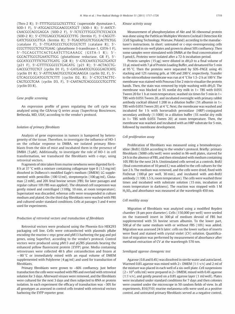
Because local application of DMBA/PMA can potentially induce asystemic oxidative stress, and HO-1-deficient mice are more proneto systemic oxidative challenge, we checked the expression ofantioxidative genes in the livers. Using qRT-PCR we compared theexpression patterns in healthy animals and in mice after the 28-week exposure to carcinogens. First, we found that the level of HO-1in healthy individuals was very low and similar in HO-1+/+ and HO-1+/−mice. In response to carcinogens it increased potently, with thestrongest reaction in wild-type and much weaker in heterozygousanimals (Fig. 6A). Carcinogens led also to significant upregulation ofSOD-1, SOD-2, GSTA-1, Thrx-2, and BVR, to similar extents in mice ofall HO-1 genotypes (Figs. 6B–F). On the other hand, expression of GRand catalase was higher in the healthy HO-1+/− group than in HO-1+/+ and HO-1−/− (Figs. 6G and H). In response to carcinogens itwas significantly augmented in the wild-type and knockout mice,but remained relatively stable in heterozygotes. Ferritin wassimilarly expressed before and after treatment in all experimentalgroups (Fig. 6I). These changes may indicate a systemic oxidativestress induced by carcinogens, which is not meaningfully influencedby the level of HO-1 expression. It is also suggested that the differencesobserved in thedevelopmentof tumorsdonot result fromdifferent levelsof systemic oxidative response in HO-1+/+, HO-1+/−, and HO-1−/−
animals.
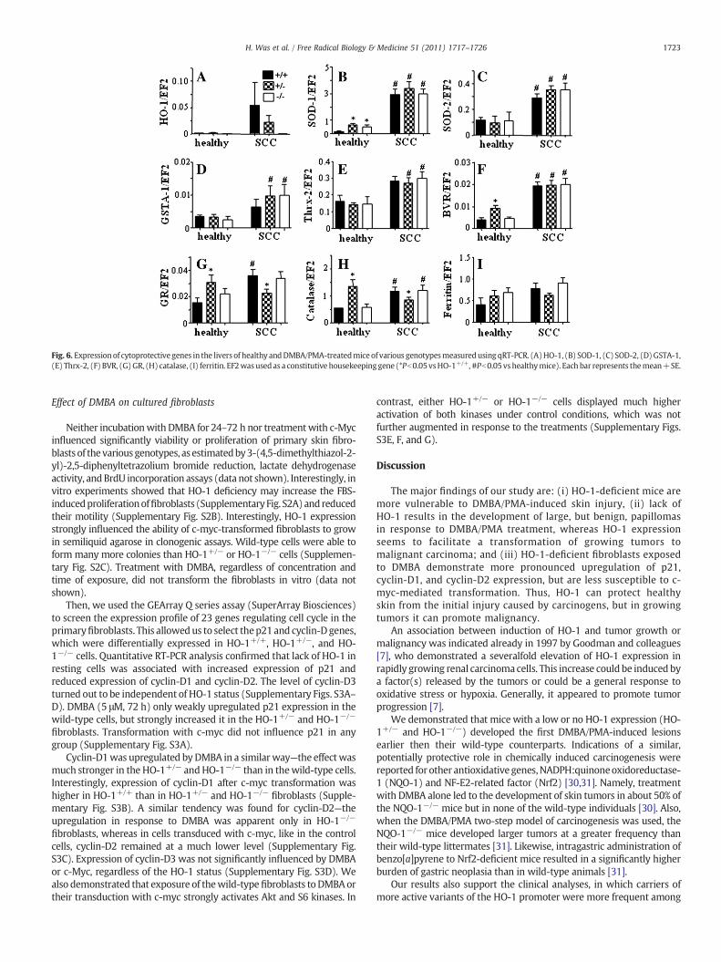
Fig. 6. Expressionof cytoprotective genes in the livers of healthy andDMBA/PMA-treatedmiceof various genotypesmeasuredusing qRT-PCR. (A)HO-1, (B) SOD-1, (C) SOD-2, (D)GSTA-1,(E) Thrx-2, (F) BVR, (G)GR, (H) catalase, (I) ferritin. EF2wasusedas a constitutive housekeeping gene (*Pb0.05vsHO-1+/+, #Pb0.05vshealthymice). Each bar represents themean+SE.
1723H. Was et al. / Free Radical Biology & Medicine 51 (2011) 1717–1726
Effect of DMBA on cultured fibroblasts
Neither incubationwith DMBA for 24–72 h nor treatmentwith c-Mycinfluenced significantly viability or proliferation of primary skin fibro-blasts of thevariousgenotypes, as estimatedby3-(4,5-dimethylthiazol-2-yl)-2,5-diphenyltetrazolium bromide reduction, lactate dehydrogenaseactivity, andBrdU incorporation assays (data not shown). Interestingly, invitro experiments showed that HO-1 deficiency may increase the FBS-inducedproliferationoffibroblasts (SupplementaryFig. S2A) andreducedtheir motility (Supplementary Fig. S2B). Interestingly, HO-1 expressionstrongly influenced the ability of c-myc-transformed fibroblasts to growin semiliquid agarose in clonogenic assays. Wild-type cells were able toform many more colonies than HO-1+/− or HO-1−/− cells (Supplemen-tary Fig. S2C). Treatment with DMBA, regardless of concentration andtime of exposure, did not transform the fibroblasts in vitro (data notshown).
Then, we used the GEArray Q series assay (SuperArray Biosciences)to screen the expression profile of 23 genes regulating cell cycle in theprimaryfibroblasts. This allowedus to select thep21and cyclin-Dgenes,which were differentially expressed in HO-1+/+, HO-1+/−, and HO-1−/− cells. Quantitative RT-PCR analysis confirmed that lack of HO-1 inresting cells was associated with increased expression of p21 andreduced expression of cyclin-D1 and cyclin-D2. The level of cyclin-D3turned out to be independent of HO-1 status (Supplementary Figs. S3A–D). DMBA (5 μM, 72 h) only weakly upregulated p21 expression in thewild-type cells, but strongly increased it in the HO-1+/− and HO-1−/−
fibroblasts. Transformation with c-myc did not influence p21 in anygroup (Supplementary Fig. S3A).
Cyclin-D1was upregulated by DMBA in a similarway—the effect wasmuch stronger in the HO-1+/− andHO-1−/− than in thewild-type cells.Interestingly, expression of cyclin-D1 after c-myc transformation washigher in HO-1+/+ than in HO-1+/− and HO-1−/−
fibroblasts (Supple-mentary Fig. S3B). A similar tendency was found for cyclin-D2—theupregulation in response to DMBA was apparent only in HO-1−/−
fibroblasts, whereas in cells transduced with c-myc, like in the controlcells, cyclin-D2 remained at a much lower level (Supplementary Fig.S3C). Expression of cyclin-D3 was not significantly influenced by DMBAor c-Myc, regardless of the HO-1 status (Supplementary Fig. S3D). Wealso demonstrated that exposure of thewild-typefibroblasts toDMBAortheir transduction with c-myc strongly activates Akt and S6 kinases. In
contrast, either HO-1+/− or HO-1−/− cells displayed much higheractivation of both kinases under control conditions, which was notfurther augmented in response to the treatments (Supplementary Figs.S3E, F, and G).
Discussion
The major findings of our study are: (i) HO-1-deficient mice aremore vulnerable to DMBA/PMA-induced skin injury, (ii) lack ofHO-1 results in the development of large, but benign, papillomasin response to DMBA/PMA treatment, whereas HO-1 expressionseems to facilitate a transformation of growing tumors tomalignant carcinoma; and (iii) HO-1-deficient fibroblasts exposedto DMBA demonstrate more pronounced upregulation of p21,cyclin-D1, and cyclin-D2 expression, but are less susceptible to c-myc-mediated transformation. Thus, HO-1 can protect healthyskin from the initial injury caused by carcinogens, but in growingtumors it can promote malignancy.
An association between induction of HO-1 and tumor growth ormalignancy was indicated already in 1997 by Goodman and colleagues[7], who demonstrated a severalfold elevation of HO-1 expression inrapidly growing renal carcinomacells. This increase could be induced bya factor(s) released by the tumors or could be a general response tooxidative stress or hypoxia. Generally, it appeared to promote tumorprogression [7].
We demonstrated that mice with a low or no HO-1 expression (HO-1+/− and HO-1−/−) developed the first DMBA/PMA-induced lesionsearlier then their wild-type counterparts. Indications of a similar,potentially protective role in chemically induced carcinogenesis werereported for other antioxidative genes,NADPH:quinoneoxidoreductase-1 (NQO-1) and NF-E2-related factor (Nrf2) [30,31]. Namely, treatmentwith DMBA alone led to the development of skin tumors in about 50% ofthe NQO-1−/− mice but in none of the wild-type individuals [30]. Also,when the DMBA/PMA two-step model of carcinogenesis was used, theNQO-1−/− mice developed larger tumors at a greater frequency thantheir wild-type littermates [31]. Likewise, intragastric administration ofbenzo[a]pyrene to Nrf2-deficient mice resulted in a significantly higherburden of gastric neoplasia than in wild-type animals [31].
Our results also support the clinical analyses, in which carriers ofmore active variants of the HO-1 promoter were more frequent among

1724 H. Was et al. / Free Radical Biology & Medicine 51 (2011) 1717–1726
healthy subjects than among patients suffering from oral squamous cellcarcinoma in betel chewers, lung adenocarcinoma in heavy smokers,and breast carcinoma in women subjected to iron supplementation[27,28,32–35]. This suggests a protective role for HO-1 at least againstcarcinogens associated with induction of oxidative stress.
Although mean latency to the first lesion is very similar in HO-1−/− and HO-1+/− animals, the resulting tumors are different: HO-1−/− mice develop a few, but large, lesions, and HO-1+/− mice muchsmaller but very numerous ones. The mechanism underlying thesedifferences is not clear. One can hypothesize that lack of HO-1 maylead to a higher apoptotic rate of affected cells in skin treated withcarcinogens and thereby to a reduced rate of initiation, whereas aftertransformation it may facilitate cell proliferation. This suppositionmight be supported by the observation of increased proliferation offibroblasts isolated from HO-1−/− mice, although it should be kept inmind that the influence of HO-1 on the cell cycle is tissue-specific [32].Additionally, we noticed the augmented activity of Akt kinase and itseffector S6 ribosomal protein in resting HO-1+/− and HO-1−/− cells.This pathway regulates proliferation, apoptosis, and growth in avariety of cell types [36]. Its inhibition contributes to the antineo-plastic effects of several drugs, such as dibenzoylmethane [37],enzastaurin [38], resveratrol [39], xanthorrhizol [40], or bromelain[41]. Expression of constitutively active Akt increases the susceptibil-ity of mice to the induction of mammary tumors of epithelial origin byDMBA [36]. Thus it seems possible that the increased activity of Akt inHO-1-deficient cells might facilitate the growth of tumors.
HO-1 knockout mice displayed the highest mortality and thesmallest body weight gain in the time course of the experiments.However, large tumors developed by HO-1−/− individuals remainedbenign papillomas, in contrast to the dysplastic lesions found in HO-1+/− and HO-1+/+ mice. It must be stressed that the low number ofHO-1−/− animals with malignant carcinoma at the end of theexperiment did not result from an earlier death of individuals withadvanced tumors in this group. Apart from tumors harvested from thesurviving animals at the end of the study (at week 28), we alsocollected tumors from animals that died earlier, starting from week13. The results (not shown) were the same as for tumors taken onweek 28: all HO-1−/− animals dying between weeks 13 and 28developed benign lesions, whereas the first carcinoma in a wild-typeindividual was found already at week 16. Therefore, the differencesbetween the proportions of malignant tumors in mice of differentgenotypes were not caused by preterm deaths of HO-1−/− animalswith more advanced tumors and survival of those with milder lesions.
A discrepancy between the growth and the malignancy of tumorswas also observed in p53-deficient mice. In this model, opposite toHO-1 deficiency, the p53 knockout mice developed smaller DMBA-induced papillomas than their wild-type counterparts. However, themalignant conversion of these small papillomas to squamous cellcarcinomas was markedly accelerated [42].
We suppose that increasedmortality of HO-1 knockouts observed inthe last part of the experiment might not be directly associated withtumormalignancy, but rathermight be caused by a higher sensitivity toinflammatory response to PMA treatment, especially in older animals.Indeed, Poss and Tonegawa have shown that HO-1-deficient micedevelop chronic inflammation and augmented sensitivity to oxidativeinjury, the effect increasing with age [43]. Here we demonstrated thatHO-1 deficiency was also associated with a higher proportion ofmonocytes in the blood of untreated animals and with a much strongerincrease in monocyte fraction in response to the first PMA treatment,which may indicate a stronger inflammatory response.
We also found that the local concentrations of VEGF were higher inHO-1-deficient than in wild-type individuals. This was surprising,because VEGF is positively regulated by HO-1 in many cell typescultured in vitro [3], and its upregulation canmediate the proangiogenicactivities of HO-1 in some cancers [11,20,24,26]. Possibly, the highertumoral concentration of VEGF in the HO-1-deficient mice is rather an
indirect effect, resulting, for example, from upregulation of someproinflammatory mediators. In contrast to the healthy tissues, in theinflamed organs HO-1 is known to inhibit VEGF production [44]. Onesuch mediator might be KC, the murine ortholog of human IL-8, whichdirectly upregulates VEGF expression in an NF-κB-dependent manner[45]. The KC concentration was much higher in HO-1−/− and HO-1+/−
than in HO-1+/+mice (Fig. 5A). Alternatively, larger tumors growing inHO-1-deficient mice could be more hypoxic and therefore mightproduce more VEGF. This supposition might be supported by a verysimilar increase in expression of VEGF-R1 (Fig. 5C), which is directlyupregulated by hypoxia [46,47].
It seems that production of proinflammatory cytokines is not asignificant factor influencing themalignant conversion of papilloma inour model. In HO-1+/− and HO-1−/− tumors, the local concentrationsof all cytokines studied were similar, whereas the frequency ofdysplastic foci wasmuch higher in heterozygotes. It has been reportedthat conversion of papilloma to SCC can be accelerated by oxidativestress, as antioxidants inhibit PMA-dependent promotion of carcino-genesis [48–49].We observed an upregulation of several antioxidativeenzymes in the livers of animals treated with DMBA/PMA, whichindicates the induction of systemic oxidative stress. However thisresponse was similar in all experimental groups, regardless of the HO-1 genotype. Thus, it seems that the antioxidative potential of HO-1does not play a major role in our system. Furthermore, a lack of HO-1did not facilitate but actually attenuated the malignant conversion.
Treatment with DMBA is known to induce p21 and cyclin-D1 incarcinoma [50]. Experiments performed on primary fibroblasts isolatedfrom the skin of mice of the various HO-1 genotypes confirmed theinduction of p21 and cyclin-D1 and demonstrated that this responsewas much stronger in HO-1+/− and especially in HO-1−/− cells than intheir wild-type counterparts. Thus, it seems that HO-1 may to someextent attenuate the effects of DMBA, which we also observed in thegrowing tumors in vivo. Interestingly, it was reported that p21deficiency in DMBA-treated mice resulted in the development of moreundifferentiated tumors,with a high frequency of anaplastic spindle cellcarcinomas, indicating the protective role of p21 against progressivemalignancy [51]. We found the lowest rate of dysplastic conversion inHO-1 knockouts, in which the fibroblastic expression of p21 was thehighest, whichmight suggest the involvement of this pathway in HO-1-dependent differences in DMBA/PMA carcinogenesis. However, incontrast to in vitro cell cultures, we did not find statistically significantdifferences in p21 expression in heterogeneous tumor tissue (data notshown).
We also compared the influence of HO-1 on the transformation ofprimary fibroblasts overexpressing c-myc after retroviral transduction.This proto-oncogene is a keyplayer in the cell cycle, actingamongothersthrough inhibition of p21 and upregulation of cyclins-D [52–54]. Incontrast tomodulation of DMBA activity, lack of HO-1 reduced the effectof c-myc overexpression on the cell-cycle-regulating genes. Thus, thelevels of cyclin-D1 and -D2were significantly lower in both resting andc-myc-overexpressing fibroblasts. Accordingly, the c-Myc-inducedtransformation and clonal growth in the semiliquid agarose werecompletely ineffective in theHO-1-deficient cells,which can support theobservation of reducedmalignant transformation in the HO-1 knockoutmice. Furthermore, the lack of HO-1 reduced migration capabilities offibroblasts. A significantpositive role forHO-1 in cellmotilitywas earlierobserved in endothelial progenitor cells [55] and keratinocytes [56].Thus, one can suppose that reduced motility of cells with a low HO-1expression may be associated with the lower infiltration of tissues andless malignant phenotype of cells.
In summary, DMBA/PMA treatment of HO-1 knockout mice inducedthe formationof skin lesions at the earliest, and resulting tumors grew thebiggest, which was accompanied by increased mortality of the animals.However, histological analysis showed that the tumors remained benignpapillomas. In contrast, tumors in the wild-type mice were smaller anddeveloped later, but much more effectively underwent malignant

1725H. Was et al. / Free Radical Biology & Medicine 51 (2011) 1717–1726
conversion. Heterozygous animals grew relatively small, but verynumerous, dysplastic tumors. Thus, HO-1 is protective against cancerinitiation, but then seems to facilitate clonal promotion. These data alsosuggest that patients with a low expression of HO-1 may be moresusceptible to development of SSC after exposure to carcinogens.
Supplementarymaterials related to this article can be found onlineat doi:10.1016/j.freeradbiomed.2011.07.025.
Acknowledgments
Thisworkwas supported byGrantsN301 08032/3156, N301 144336,347/N-INCA/2008, and 311/N-COST/2008 from Ministry of Science andHigher Education. A.J. was the recipient of a Wellcome Trust SeniorResearch Fellowship in Biomedical Science. H.W. is the recipient of aSTART fellowship from the Foundation for Polish Science. The Faculty ofBiochemistry, Biophysics, and Biotechnology of the Jagiellonian Univer-sity is a beneficiary of structural funds from the European Union (GrantsPOIG.02.01.00-12-064/08, 02.02.00-00-014/08, 01.02-00-109/09, and01.02.00-069/09).
References
[1] Maines, M. D. The heme oxygenase system: a regulator of second messengergases. Annu. Rev. Pharmacol. Toxicol. 37:517–554; 1997.
[2] Maines, M. D. Heme oxygenase: function, multiplicity, regulatory mechanisms,and clinical applications. FASEB J. 2:2557–2568; 1988.
[3] Loboda, A.; Jazwa, A.; Grochot-Przeczek, A.; Rutkowski, A. J.; Cisowski, J.; Agarwal, A.;Jozkowicz, A.; Dulak, J. Heme oxygenase-1 and the vascular bed: from molecularmechanisms to therapeutic opportunities. Antioxid. Redox Signal. 10:1767–1812; 2008.
[4] Schacter, B. A.; Kurz, P. Alterations in hepatic and splenic microsomal electrontransport system components, drug metabolism, heme oxygenase activity, andcytochrome P-450 turnover in Murphy–Sturm lymphosarcoma-bearing rats.Cancer Res. 42:3557–3564; 1982.
[5] Hara, E.; Takahashi, K.; Tominaga, T.; Kumabe, T.; Kayama, T.; Suzuki, H.; Fujita, H.;Yoshimoto, T.; Shirato, K.; Shibahara, S. Expression of heme oxygenase andinducible nitric oxide synthase mRNA in human brain tumors. Biochem. Biophys.Res. Commun. 224:153–158; 1996.
[6] Maines, M. D.; Abrahamsson, P. A. Expression of heme oxygenase-1 (HSP32) inhuman prostate: normal, hyperplastic, and tumor tissue distribution. Urology 47:727–733; 1996.
[7] Goodman, A. I.; Choudhury, M.; da Silva, J. L.; Schwartzman, M. L.; Abraham, N. G.Overexpression of the heme oxygenase gene in renal cell carcinoma. Proc. Soc. Exp.Biol. Med. 214:54–61; 1997.
[8] Doi, K.; Akaike, T.; Fujii, S.; Tanaka, S.; Ikebe, N.; Beppu, T.; Shibahara, S.; Ogawa, M.;Maeda, H. Induction of haem oxygenase-1 nitric oxide and ischaemia in experimentalsolid tumours and implications for tumour growth. Br. J. Cancer 80:1945–1954; 1999.
[9] Tsuji, M. H.; Yanagawa, T.; Iwasa, S.; Tabuchi, K.; Onizawa, K.; Bannai, S.; Toyooka,H.; Yoshida, H. Heme oxygenase-1 expression in oral squamous cell carcinoma asinvolved in lymph node metastasis. Cancer Lett. 138:53–59; 1999.
[10] Was, H.; Dulak, J.; Jozkowicz, A. Heme oxygenase in tumor biology and therapy.Curr. Drug Targets 11:1551–1570; 2010.
[11] Torisu-Itakura, H.; Furue, M.; Kuwano, M.; Ono, M. Co-expression of thymidinephosphorylase and heme oxygenase-1 in macrophages in human malignantvertical growth melanomas. Jpn. J. Cancer Res. 91:906–910; 2000.
[12] McAllister, S. C.; Hansen, S. G.; Ruhl, R. A.; Raggo, C. M.; DeFilippis, V. R.;Greenspan, D.; Fruh, K.; Moses, A. V. Kaposi sarcoma-associated herpesvirus(KSHV) induces heme oxygenase-1 expression and activity in KSHV-infectedendothelial cells. Blood 103:3465–3473; 2004.
[13] Berberat, P. O.; Dambrauskas, Z.; Gulbinas, A.; Giese, T.; Giese, N.; Kunzli, B.;Autschbach, F.; Meuer, S.; Buchler, M.W.; Friess, H. Inhibition of heme oxygenase-1 increases responsiveness of pancreatic cancer cells to anticancer treatment. Clin.Cancer Res. 11:3790–3798; 2005.
[14] Nowis, D.; Legat, M.; Grzela, T.; Niderla, J.; Wilczek, E.; Wilczynski, G. M.;Glodkowska, E.; Mrowka, P.; Issat, T.; Dulak, J.; Jozkowicz, A.; Was, H.; Adamek,M.; Wrzosek, A.; Nazarewski, S.; Makowski, M.; Stoklosa, T.; Jakobisiak, M.; Golab,J. Heme oxygenase-1 protects tumor cells against photodynamic therapy-mediated cytotoxicity. Oncogene 25:3365–3374; 2006.
[15] Kocanova, S.; Buytaert, E.; Matroule, J. Y.; Piette, J.; Golab, J.; deWitte, P.; Agostinis,P. Induction of heme-oxygenase 1 requires the p38MAPK and PI3K pathways andsuppresses apoptotic cell death following hypericin-mediated photodynamictherapy. Apoptosis 12:731–741; 2007.
[16] Chen, G. G.; Liu, Z. M.; Vlantis, A. C.; Tse, G. M.; Leung, B. C.; van Hasselt, C. A. Hemeoxygenase-1 protects against apoptosis induced by tumor necrosis factor-alphaand cycloheximide in papillary thyroid carcinoma cells. J. Cell. Biochem. 92:1246–1256; 2004.
[17] Liu, Z. M.; Chen, G. G.; Ng, E. K.; Leung, W. K.; Sung, J. J.; Chung, S. C. Upregulationof heme oxygenase-1 and p21 confers resistance to apoptosis in human gastriccancer cells. Oncogene 23:503–513; 2004.
[18] Mayerhofer, M.; Florian, S.; Krauth, M. T.; Aichberger, K. J.; Bilban, M.;Marculescu, R.; Printz, D.; Fritsch, G.; Wagner, O.; Selzer, E.; Sperr, W. R.;Valent, P.; Sillaber, C. Identification of heme oxygenase-1 as a novel BCR/ABL-dependent survival factor in chronic myeloid leukemia. Cancer Res. 64:3148–3154; 2004.
[19] Busserolles, J.; Megias, J.; Terencio, M. C.; Alcaraz, M. J. Heme oxygenase-1 inhibitsapoptosis in Caco-2 cells via activation of Akt pathway. Int. J. Biochem. Cell Biol. 38:1510–1517; 2006.
[20] Was, H.; Cichon, T.; Smolarczyk, R.; Rudnicka, D.; Stopa, M.; Chevalier, C.; Leger, J. J.;Lackowska, B.; Grochot, A.; Bojkowska, K.; Ratajska, A.; Kieda, C.; Szala, S.; Dulak, J.;Jozkowicz, A. Overexpression of heme oxygenase-1 inmurinemelanoma: increasedproliferation and viability of tumor cells, decreased survival of mice. Am. J. Pathol.169:2181–2198; 2006.
[21] Tanaka, S.; Akaike, T.; Fang, J.; Beppu, T.; Ogawa, M.; Tamura, F.; Miyamoto, Y.;Maeda, H. Antiapoptotic effect of haem oxygenase-1 induced by nitric oxide inexperimental solid tumour. Br. J. Cancer 88:902–909; 2003.
[22] Hirai, K.; Sasahira, T.; Ohmori, H.; Fujii, K.; Kuniyasu, H. Inhibition of hemeoxygenase-1 by zinc protoporphyrin IX reduces tumor growth of LL/2 lung cancerin C57BL mice. Int. J. Cancer 120:500–505; 2007.
[23] Hill, M.; Pereira, V.; Chauveau, C.; Zagani, R.; Remy, S.; Tesson, L.; Mazal, D.;Ubillos, L.; Brion, R.; Asghar, K.; Mashreghi, M. F.; Kotsch, K.; Moffett, J.; Doebis, C.;Seifert, M.; Boczkowski, J.; Osinaga, E.; Anegon, I. Heme oxygenase-1 inhibits ratand human breast cancer cell proliferation: mutual cross inhibition withindoleamine 2,3-dioxygenase. FASEB J. 19:1957–1968; 2005.
[24] Nishie, A.; Ono, M.; Shono, T.; Fukushi, J.; Otsubo, M.; Onoue, H.; Ito, Y.; Inamura,T.; Ikezaki, K.; Fukui, M.; Iwaki, T.; Kuwano, M. Macrophage infiltration and hemeoxygenase-1 expression correlate with angiogenesis in human gliomas. Clin.Cancer Res. 5:1107–1113; 1999.
[25] Marinissen, M. J.; Tanos, T.; Bolos, M.; de Sagarra, M. R.; Coso, O. A.; Cuadrado,A. Inhibition of heme oxygenase-1 interferes with the transforming activity ofthe Kaposi sarcoma herpesvirus-encoded G protein-coupled receptor. J. Biol.Chem. 281:11332–11346; 2006.
[26] Sunamura, M.; Duda, D. G.; Ghattas, M. H.; Lozonschi, L.; Motoi, F.; Yamauchi, J.;Matsuno, S.; Shibahara, S.; Abraham, N. G. Heme oxygenase-1 accelerates tumorangiogenesis of human pancreatic cancer. Angiogenesis 6:15–24; 2003.
[27] Kikuchi, A.; Yamaya, M.; Suzuki, S.; Yasuda, H.; Kubo, H.; Nakayama, K.; Handa, M.;Sasaki, T.; Shibahara, S.; Sekizawa, K.; Sasaki, H. Association of susceptibility to thedevelopment of lung adenocarcinoma with the heme oxygenase-1 gene promoterpolymorphism. Hum. Genet. 116:354–360; 2005.
[28] Lin, S. C.; Liu, C. J.; Yeh, W. I.; Lui, M. T.; Chang, K. W.; Chang, C. S. Functionalpolymorphism in NFKB1 promoter is related to the risks of oral squamous cellcarcinomaoccurringonoldermaleareca (betel) chewers.Cancer Lett.243:47–54;2006.
[29] Vreman, H. J.; Wong, R. J.; Kadotani, T.; Stevenson, D. K. Determination of carbonmonoxide (CO) in rodent tissue: effect of heme administration and environmen-tal CO exposure. Anal. Biochem. 341:280–289; 2005.
[30] Long, D. J.; Waikel, R. L.; Wang, X. J.; Roop, D. R.; Jaiswal, A. K. NAD(P)H:quinoneoxidoreductase 1 deficiency and increased susceptibility to 7,12-dimethylbenz[a]-anthracene-induced carcinogenesis in mouse skin. J. Natl. Cancer Inst. 93:1166–1170; 2001.
[31] Ramos-Gomez, M.; Kwak, M. K.; Dolan, P. M.; Itoh, K.; Yamamoto, M.; Talalay, P.;Kensler, T. W. Sensitivity to carcinogenesis is increased and chemoprotectiveefficacy of enzyme inducers is lost in nrf2 transcription factor-deficient mice. Proc.Natl. Acad. Sci. U.S.A. 98:3410–3415; 2001.
[32] Jozkowicz, A.; Was, H.; Dulak, J. Heme oxygenase-1 in tumors: is it a false friend?Antioxid. Redox Signal. 9:2099–2117; 2007.
[33] Chang,K.W.; Lee, T. C.; Yeh,W. I.;Chung,M.Y.; Liu, C. J.; Chi, L.Y.; Lin, S.C.Polymorphismin heme oxygenase-1 (HO-1) promoter is related to the risk of oral squamous cellcarcinoma occurring on male areca chewers. Br. J. Cancer 91:1551–1555; 2004.
[34] Lo, S. S.; Lin, S. C.; Wu, C. W.; Chen, J. H.; Yeh, W. I.; Chung, M. Y.; Lui, W. Y. Hemeoxygenase-1 gene promoter polymorphism is associated with risk of gastricadenocarcinoma and lymphovascular tumor invasion. Ann. Surg. Oncol. 14:2250–2256; 2007.
[35] Hong, C. C.; Ambrosone, C. B.; Ahn, J.; Choi, J. Y.; McCullough, M. L.; Stevens,V. L.; Rodriguez, C.; Thun, M. J.; Calle, E. E. Genetic variability in iron-relatedoxidative stress pathways (Nrf2, NQO1, NOS3, and HO-1), iron intake, andrisk of postmenopausal breast cancer. Cancer Epidemiol. Biomarkers Prev. 16:1784–1794; 2007.
[36] Blanco-Aparicio, C.; Perez-Gallego, L.; Pequeno, B.; Leal, J. F.; Renner,O.; Carnero,A.Miceexpressing myrAKT1 in the mammary gland develop carcinogen-induced ER-positivemammary tumors that mimic human breast cancer. Carcinogenesis 28:584–594; 2007.
[37] Khor, T. O.; Yu, S.; Barve, A.; Hao, X.; Hong, J. L.; Lin, W.; Foster, B.; Huang, M. T.;Newmark, H. L.; Kong, A. N. Dietary feeding of dibenzoylmethane inhibits prostatecancer in transgenic adenocarcinoma of themouse prostate model. Cancer Res. 69:7096–7102; 2009.
[38] Graff, J. R.; McNulty, A. M.; Hanna, K. R.; Konicek, B. W.; Lynch, R. L.; Bailey,S. N.; Banks, C.; Capen, A.; Goode, R.; Lewis, J. E.; Sams, L.; Huss, K. L.;Campbell, R. M.; Iversen, P. W.; Neubauer, B. L.; Brown, T. J.; Musib, L.;Geeganage, S.; Thornton, D. The protein kinase Cbeta-selective inhibitor,Enzastaurin (LY317615.HCl), suppresses signaling through the AKT pathway,induces apoptosis, and suppresses growth of human colon cancer andglioblastoma xenografts. Cancer Res. 65:7462–7469; 2005.
[39] Roy, P.; Kalra, N.; Prasad, S.; George, J.; Shukla, Y. Chemopreventive potentialof resveratrol in mouse skin tumors through regulation of mitochondrial andPI3K/AKT signaling pathways. Pharm. Res. 26:211–217; 2009.
[40] Chung, W. Y.; Park, J. H.; Kim, M. J.; Kim, H. O.; Hwang, J. K.; Lee, S. K.; Park, K. K.Xanthorrhizol inhibits 12-O-tetradecanoylphorbol-13-acetate-induced acute

1726 H. Was et al. / Free Radical Biology & Medicine 51 (2011) 1717–1726
inflammation and two-stage mouse skin carcinogenesis by blocking the expression ofornithine decarboxylase, cyclooxygenase-2 and inducible nitric oxide synthase throughmitogen-activatedprotein kinases and/or thenuclear factor-kappaB.Carcinogenesis28:1224–1231; 2007.
[41] Kalra, N.; Bhui, K.; Roy, P.; Srivastava, S.; George, J.; Prasad, S.; Shukla, Y. Regulation ofp53, nuclear factor kappaB and cyclooxygenase-2 expression by bromelain throughtargeting mitogen-activated protein kinase pathway in mouse skin. Toxicol. Appl.Pharmacol. 226:30–37; 2008.
[42] Kelly-Spratt, K. S.; Gurley, K. E.; Yasui, Y.; Kemp, C. J. p19Arf suppresses growth,progression, and metastasis of Hras-driven carcinomas through p53-dependent and-independent pathways. PLoS Biol. 2:E242; 2004.
[43] Poss, K. D.; Tonegawa, S. Reduced stress defense in heme oxygenase 1-deficient cells.Proc. Natl. Acad. Sci. U.S.A. 94:10925–10930; 1997.
[44] Bussolati, B.; Mason, J. C. Dual role of VEGF-induced heme-oxygenase-1 inangiogenesis. Antioxid. Redox Signal. 8:1153–1163; 2006.
[45] Martin, D.; Galisteo, R.; Gutkind, J. S. CXCL8/IL-8 stimulates vascular endothelial growthfactor (VEGF) expression and the autocrine activation of VEGFR2 in endothelial cells byactivating NFκB through the CMB (Carma3/Bcl10/Malt1) complex. J. Biol. Chem. 284:6038–6042; 2009.
[46] Nevo, O.; Soleymanlou, N.;Wu, Y.; Xu, J.; Kingdom, J.;Many, A.; Zamudio, S.; Caniggia, I.Increased expressionof sFlt-1 in in vivo and in vitromodels of humanplacental hypoxiaismediatedbyHIF-1.Am. J. Physiol. Regul. Integr. Comp. Physiol.291:R1085–R1093;2006.
[47] Hirakawa, S.; Kodama, S.; Kunstfeld, R.; Kajiya, K.; Brown, L. F.; Detmar, M. VEGF-Ainduces tumor and sentinel lymph node lymphangiogenesis and promotes lymphaticmetastasis. J. Exp. Med. 201:1089–1099; 2005.
[48] Shimizu, Y.; Kondo, S.; Shirai, A.; Furukawa, M.; Yoshizaki, T. A single nucleotidepolymorphism in the matrix metalloproteinase-1 and interleukin-8 gene promoterpredicts poor prognosis in tongue cancer. Auris Nasus Larynx 35:381–389; 2008.
[49] Kensler, T. W.; Egner, P. A.; Taffe, B. G.; Trush, M. A. Role of free radicals in tumorpromotion and progression. Prog. Clin. Biol. Res. 298:233–248; 1989.
[50] Letchoumy, P. V.; Mohan, K. V.; Prathiba, D.; Hara, Y.; Nagini, S. Comparativeevaluation of antiproliferative, antiangiogenic and apoptosis inducingpotential of black tea polyphenols in the hamster buccal pouch carcinogen-esis model. J. Carcinog. 6:19; 2007.
[51] Philipp, J.; Vo, K.; Gurley, K. E.; Seidel, K.; Kemp, C. J. Tumor suppression byp27Kip1 and p21Cip1 during chemically induced skin carcinogenesis. Oncogene18:4689–4698; 1999.
[52] Qi, Y.; Tu, Y.; Yang, D.; Chen, Q.; Xiao, J.; Chen, Y.; Fu, J.; Xiao, X.; Zhou, Z. Cyclin Abut not cyclin D1 is essential for c-myc-modulated cell-cycle progression. J. Cell.Physiol. 210:63–71; 2007.
[53] Claassen, G. F.; Hann, S. R. A role for transcriptional repression of p21CIP1 by c-Myc in overcoming transforming growth factor beta-induced cell-cycle arrest.Proc. Natl. Acad. Sci. U.S.A. 97:9498–9503; 2000.
[54] Coller, H. A.; Grandori, C.; Tamayo, P.; Colbert, T.; Lander, E. S.; Eisenman, R.N.; Golub, T. R. Expression analysis with oligonucleotide microarrays revealsthat MYC regulates genes involved in growth, cell cycle, signaling, andadhesion. Proc. Natl. Acad. Sci. U.S.A. 97:3260–3265; 2000.
[55] Deshane, J.; Chen, S.; Caballero, S.; Grochot-Przeczek, A.; Was, H.; Li Calzi, S.; Lach, R.;Hock, T. D.; Chen, B.; Hill-Kapturczak, N.; Siegal, G. P.; Dulak, J.; Jozkowicz, A.; Grant,M. B.; Agarwal, A. Stromal cell-derived factor 1 promotes angiogenesis via a hemeoxygenase 1-dependent mechanism. J. Exp. Med. 204:605–618; 2007.
[56] Grochot-Przeczek, A.; Lach, R.; Mis, J.; Skrzypek, K.; Gozdecka, M.; Sroczynska, P.;Dubiel, M.; Rutkowski, A.; Kozakowska, M.; Zagorska, A.; Walczynski, J.; Was, H.;Kotlinowski, J.; Drukala, J.; Kurowski, K.; Kieda, C.; Herault, Y.; Dulak, J.; Jozkowicz,A. Heme oxygenase-1 accelerates cutaneous wound healing in mice. PLoS One 4:e5803; 2009.