foxh1 role on left-right asymmetry -...
TRANSCRIPT
Vânia Filipa Lima Fernandes
2013 |
FoxH1 role on Left-Right
Asymmetry
Departamento de Biologia Animal
Master thesis on Biologia Evolutiva e do Desenvolvimento
Universidade de Lisboa | Faculdade de Ciências
This project was developed under the supervision of Sebastian Shimeld, PhD (University of Oxford, United Kingdom) and Solveig Thorsteinsdottir, PhD (Faculdade de Ciências da Universidade de Lisboa, Portugal) and submitted for a master degree title.
FoxH1 role on Left-Right
Asymmetry
2013 |
Vânia Filipa Lima Fernandes
Universidade de Lisboa | Faculdade de Ciências
Departamento de Biologia Animal
Master thesis on Biologia Evolutiva e do Desenvolvimento

Acknowledgments
I would like to express my gratitude to my supervisor, Dr Sebastian Shimeld, whose expertise, understanding and patience were beyond what I asked for and
were essential for my evolving as a scientist. I would also like to thank to the other members of my lab, Nathan, Erica, Atsuko and Cedric for the assistance
and guidance they provided during my project.
A very special thanks goes to Dr Solveig Thorsteindottir, who provided me with direction, understanding and kindness.
I must also thank my family and friends for the patience and love which they
have supported me during this last year.

Abstract
Left-Right (LR) asymmetry is a conserved feature across the Bilateria and is characterized by differential morphogenesis and positioning of internal organs across the LR axis. This pattern is established during early embryogenesis and involves a number of highly regulated developmental mechanisms. Based on the fact that initially all organisms appear morphologically symmetric (zygotes or early embryos), a major question emerged: how symmetry is broken internally and which is the first asymmetric event?
Studies performed on different model organisms suggest that distinct symmetric-breaking mechanisms may occur during their early development. In vertebrates, regulation of LR asymmetry downstream of symmetry-breaking seems to be conserved between different taxa and many reports show that it is established through the Nodal signalling molecule. Nodal starts being expressed symmetrically and then becomes asymmetrically expressed on the left side of lateral plate mesoderm at the early somite stage in chordates. However this is not true for Echinodermata, Nodal is always expressed asymmetrically and is found only on the right side. In the protostome models C. elegans and D. melanogaster Nodal has not been found, and hence it was thought that probably it evolved within the early deuterostome lineage. However, recently an orthologue of Nodal was found in snails (Mollusca) and it was shown that it also plays a role in left-right asymmetry, demonstrating it is of much earlier evolutionary origin. Pitx is a mediator of Nodal and it is involved in many processes related with the asymmetric growth. FoxH1 is a transcription factor that despite not being a canonical member of Nodal signalling pathway has been shown to modulate Nodal expression in vertebrates.
The main goal of my project is to characterise FoxH1, Nodal and Pitx expression pattern across three different animals, Patella, Amphioxus and Lamprey during early embryo development and address if FoxH1 plays a role on Nodal signalling in protostomes. Key-Words: Left-Right asymmetry, FoxH1, Nodal

Sumário
A assimetria esquerda-direita (ED) é uma característica que se mantém conservada e bem patenteada em animais do grupo dos Bilateria. Esta consiste na morfogénese e posicionamento diferencial de órgãos internos ao longo do eixo ED. Por exemplo, por norma em humanos esta padronização assimétrica manifesta-se pela presença do coração e do estômago do lado esquerdo e o fígado do lado direito. Trata-se de um traço que é estabelecido numa fase muito precoce do desenvolvimento embrionário e que envolve inúmeros mecanismos moleculares altamente regulados. Tendo em consideração que inicialmente todos os organismos são bilaterais e morfologicamente simétricos, impõe-se uma questão essencial: como é que a simetria é quebrada internamente e qual o primeiro evento assimétrico a ter lugar? Estudos realizados em diferentes modelos animais sugerem mecanismos de quebra de simetria distintos, sendo actualmente considerados três modelos. Estudos em embriões de ratinho sugerem que um fluxo assimétrico provocado pela rotação de cílios no nodo leva a uma expressão diferencial de genes em torno do organizador ED, o que se manifesta mais tardiamente em assimétricas morfológicas – modelo ciliar. Dentro deste modelo podem ser ainda distinguidas duas sub-teorias, uma defendendo que a motilidade ciliar é responsável pelo transporte unilateral dos morfogénios para o lado esquerdo e outra defendendo que a existência de dois tipos de cílios são responsáveis não só pela geração do fluxo de morfogénios, cílios móteis, como também pela percepção destes, cílios sensitivos. No entanto, outros estudos realizados em embrião de galinha reportam que a existência de junções gap na linha primitiva está envolvida em eventos assimétricos ainda antes da formação do nodo – modelo citoplasmático. Este modelo baseia-se na complexidade e na dinâmica do citoesqueleto para explicar a quebra da simetria e muitas experiências confirmam que este possui uma quiralidade inerente, por exemplo, durante as primeiras clivagens de embriões de rã, já é notória uma distribuição diferencial de canais de potássio e de bombas de protões, herdadas maternalmente, que contribuem para uma distribuição de iões assimétrica que juntamente com as junções gap culmina na disposição diferencial de morfogénios. Estes primeiros resultados a suportar ambas as teorias pareciam indicar que provavelmente o modelo ciliar estava restrito aos mamíferos, no entanto foi igualmente descoberta a acção de cílios na quebra de simetria em zebrafish. Um terceiro modelo – separação diferencial de cromatídeos – propõe ainda que eventos moleculares mais recentes dos citados acima poderão estar envolvidos na origem da simetria. Esse modelo consiste na segregação diferencial de cromatídeos aquando a primeira divisão logo após a fertilização, contudo poucos estudos foram feitos nesta vertente, sendo que a maioria foram feitos em levedura e caracóis pelo que é muito difícil extrapolar para outros animais de organização mais complexa. Independentemente do primeiro evento a despoletar a assimetria ED, é igualmente importante saber como esta é estabelecida e mantida ao longo do desenvolvimento. Inúmeros genes foram assim descritos, sendo o Nodal um dos mais bem estudados, bem como os seus genes alvo, Pitx e FoxH1. Em vertebrados, a assimetria é assegurada pela via e sinalização Nodal, uma proteína secretada encarregue de transferir sinais do nó para a mesoderme da placa lateral. O Nodal é uma proteína pertencente à família dos TGF-β, Transforming Growth Factor – beta, tendo também um papel importante em processos de diferenciação celular, formação da endoderme e mesoderme e estabelecimento da linha primitiva em ratinho, peixe-zebra e rã durante o desenvolvimento. A via de sinalização Nodal actua através da ligação deste a receptores transmembranares serina/treonina cinase tipo II que, quando fosforilados, activam os receptores do tipo I. Quando fosforilados, estes receptores fosforilam proteínas Smad2 que vão interagir com Smad4, formando um complexo que funciona como um factor de transcrição que ao entrar no núcleo da célula, activa determinados genes que incluem o Pitx e o próprio Nodal. Em animais cordados, o Nodal começa por ser expresso simetricamente e posteriormente, numa fase mais avançada do desenvolvimento (somitogénese), torna-se assimétrico,

Sumário
encontrando-se presente somente do lado esquerdo. Isto não se verifica no entanto para o táxon dos equinodermes, cuja expressão do Nodal começa por ser desde cedo assimétrica e presente unicamente do lado direito. Em modelos clássicos de protostómios, tais como C. elegans ou D. melanogaster, o Nodal nunca foi encontrado levando à especulação geral de que provavelmente este tinha surgido unicamente na linha dos deuterostómios. Contudo, foi publicado um artigo recentemente que dita a existência de um órtologo do Nodal em caracóis (moluscos) e que por sua vez também se encontra associado ao estabelecimento da assimetria ED. Esta descoberta representa a formulação de uma nova hipótese, a de que estes mecanismos estão mais conservados do que se pensava inicialmente. Uma peça importante no complexo de regulação via Smads é o FoxH1. Este factor de transcrição pertence à vasta família dos genes Fox, genes que contêm um domínio forkhead. Este encontra-se altamente conservado e espalhado ao longo dos cordados e embora não seja um membro canónico da via de sinalização Nodal, tem sido descrito como um modulador da expressão do Nodal. Estudos realizados em ratinho mostram que as expressões do Nodal e do FoxH1 se sobrepõem. Em embriões cujo FoxH1 estava mutado havia claros sinais de uma incorrecta padronização antero-posterior. Ainda em ratinho, foi demonstrado o importante papel do FoxH1 durante o desenvolvimento embrionário na formação de estruturas cruciais como o nó, a notocorda e a mesoderme lateral que se tornam essenciais na futura formação e posicionamento de órgãos. Este factor de transcrição aparece como um transdutor da sinalização Nodal necessário para o estabelecimento da assimetria ED, embora o Nodal possa actuar dependente e independentemente deste factor de transcrição. O Pitx, gene alvo da sinalização Nodal, pode igualmente ser activado pelo FoxH1 e consequentemente regular outros factores ou outras vias envolvidas na manutenção da assimetria.
A importância do FoxH1 na regulação da via de sinalização Nodal em vertebrados e a sua presença não só em deuterostómios mas também em protostómios, levanta a hipótese de que este gene está envolvido na mediação do Nodal e consequentemente no estabelecimento da assimetria de forma mais abrangente no grupo dos bilateria. Assim, este projecto foi desenhado com o propósito de averiguar o papel do FoxH1 na via de sinalização Nodal e na assimetria em protostómios e animais deuterostómicos que não pertencem ao grupo dos vertebrados e cujo gene não tenha sido devidamente descrito em termos de função. Numa primeira instância foram tidas em conta análises filogenéticas com o intuito de perceber melhor a relação evolutiva da família dos genes Fox e a possível origem do FoxH1 no grupo dos bilateria. Para estudos de expressão dos genes FoxH1, Nodal e Pitx, foram utilizadas quatro espécies, Patella vulgata, Branchiostoma floridae, Branchiostoma lanceolatum e Lampetra planeri, que por se encontrarem em posições de interesse na ‘árvore’ filogenética se tornam organismos chave para a compreensão da conservação de mecanismos a operar durante o desenvolvimento. Estes também foram sujeitos a comparação com modelos onde a expressão destes genes já está devidamente descrita. Estudos funcionais foram realizados para perceber se de facto o FoxH1 influenciava a expressão do Nodal e se sim de que forma. Estes foram feitos através da inibição do FoxH1 bem como do Nodal para perceber se o FoxH1 tinha um efeito regulador na via do Nodal ou se pelo contrário era afectado por este, ou mesmo pelo Pitx. Palavras-chave: Assimetria Esquerda-Direita, FoxH1, Nodal

Index
Introduction
Left-Right Asymmetry Breaking Symmetry Maintenance of Left-Right Asymmetry Aims and hypothesis
10 11 12 15 17
Methodology
19
Results Fox genes evolution and conservation across Bilateria FoxH1 in Lamprey Gene Expression Patterns during embryonic development Branchiostoma floridae Branchiostoma lanceolatum Patella vulgata
Discussion
24 25 25 29 29 30 32 34
Bibliography 37

Index of Figures
Figure 1 Asymmetrical disposition of organs in Human
11
Figure 2 Ciliary Model to explain Nodal flow in LR asymmetry
12
Figure 3 Representation of Cytoplasmic Model for LR asymmetry based on early development in Xenopus laevis
14
Figure 4 Nodal signalling pathway
16
Figure 5 Representation of the phylogenetic tree of Bilateria
18
Figure 6 Maximum likelihood tree of Fox amino acid sequences in selected Bilateria
26
Figure 7 Synteny of Fox genes within the deuterostome lineage
27
Figure 8 FoxH1 presence across early development in lamprey
28
Figure 9 Expression of Nodal, Pitx and FoxH1 in Branchiostoma floridae embryos during early development
30
Figure 10 Expression of Nodal, Pitx and FoxH1 in Branchiostoma lanceolatum embryos during early development.
31
Figure 11 Expression of Nodal Original, Nodal New, Pitx and FoxH1 in Patella vulgata embryos during early development
32

Introduction Is not birth, marriage, or death, but
gastrulation which is truly the most important time in your life.
Lewis Wolpert

11
Introduction
Figure 1. Asymmetrical disposition of organs in
Human: Situs solitus (A) and Situs inversus (B). Ventral view. Adapted from Saúde et al., 2000.
Left-Right Asymmetry
When looking into a mirror we all seem symmetrical but underneath we can clearly distinguish morphologically our left side from our right side. In humans, the internal organ disposition is differential, whereas the heart, the spleen and the stomach are located on the left side, the liver is located on the right – situs solitus. Usually this asymmetric disposition is specific and predominant for each species, however there are rare cases where this distribution is inversed – situs inversus (Figure 1) (Nakamura & Hamada 2012).
Asymmetric organ placement is not exclusive to humans, being also present across other vertebrates. In fact most animals, besides cnidarians, sponges and other basal groups, belong to a huge group: the Bilateria, and members of this group have bilateral symmetry. Their bodies are built and organized with anteroposterior (AP) and dorsovrentral (DV) axes and structures displayed around the midline (Boorman & Shimeld 2002c). Although LR
asymmetry expresses itself differently across taxa, it is crucial for correct patterning for a normal functioning and development of the organism as a whole.
Asymmetric organ placement and behaviour (i.e., handeness) have been wrongly associated: organ disposition and laterality preference are genetically determined through gene expression cascades but the genes involved in each one are different (Morgan 1991; Vandenberg & Levin 2010). It is possible to distinguish different types of asymmetry. LR asymmetries emerged from environmental changes that could occur during embryonic development, per example birth marks, are not heritable and are called fluctuating asymmetries (Harnad 1977). If the existence of an asymmetric feature is genetically stated but randomly determined at each generation it’s called antisymmetry (Palmer 1994). In case of directional asymmetries such as internal organ disposition in humans, these are fixed and heritable within a group or across members of a species. Actually, the first asymmetries arise much earlier during development than organ formation (Michael Levin et al. 1995).
Initially, scientists were concerned and focused on the identity of the molecule(s) responsible for such differences between left and right sides. The first mention of a possible candidate was made by Brown and Wolpert (1990) who conceived the F molecule. Such a molecule was never found at least until now, but many others already described take a part in LR asymmetry establishment and maintenance. One of the most known and studied is Nodal, a left sided protein found in mouse (Michael Levin et al. 1995) and present in all vertebrates. Experimental and functional studies have shown that this factor as well Pitx2, a downstream target of Nodal, act together on the left side of vertebrates patterning LR asymmetry. Additional studies also reinforce Nodal’s role on initial breaking of symmetry (Nonaka et al. 1998).
Since Nodal was described as present only in Deuterostomes, there was a general assumption that it probably had originated within this lineage. However, Grande & Patel (2009) recently published a study proving the existence of a Nodal orthologue in Protostomes, more precisely in snails (molluscs). The study also showed that this orthologue was involved in left-right asymmetry establishment. This recent

12
Introduction
Figure 2. Ciliary Model to explain Nodal flow in LR asymmetry: a – Morphogen hypothesis, in
which ciliary motility transports morphogens (and NPVs) to the left side and b – Two-cilia
Model, in which motile cilia generate a leftward-directed fluid flow that is sensed by the immotile cilia on the node. Nodal pit cells are depicted in light brown, whereas perinodal crown cells are in dark brown. The motile cilia are tilted posteriorly. Basal bodies are indicated with red dots and the Nodal Flow is shown with the black arrow: A – anterior; P – posterior; R – right; L – left. Adapted from Babu & Roy (2013).
b
discovery raises again the discussion about how conserved these mechanisms and features are across Bilateria and foments even more the search of Nodal in other non-deuterostome groups.
Breaking Symmetry
Based on the fact that initially all organisms appear morphologically symmetric as zygotes or early embryos, a major question emerged: how symmetry is broken internally and which is the first asymmetric event?
Studies performed in different model organisms seem to answer this question differently. Currently there are three consistent models: Ciliary Model, Cytoplasmic Model and Segregation of differentially-imprinted chromatids Model. Ciliary Model
The association of cilia with LR asymmetry started in the 1970’s, when a man was diagnosed with a very rare genetic disorder called Kartagener syndrome (KS) (Afzelius 1976). Patients with this condition normally have severe respiratory problems, renal dysfunction and also situs inversus. Respiratory and renal problems could be easily correlated with ultrastructural defects in their motile cilia but not with misplacement of visceral organs (Babu & Roy 2013; Afzelius 1976).
Cilia are microtubule-based hair-like organelles found on nearly all eukaryotic cells that play an important role in cellular motility, fluid transport and a huge variety of signal transduction pathways (Babu & Roy 2013). They are found at the node at gastrulation stage in vertebrate embryos where each node cell carries a single cilium (Sulik et al. 1994; Bellomo et al. 1996). Kartagener patients have defects in genes that encode several components of ciliary dynein (Olbrich et al. 2002; Bartoloni et al. 2002; Pennarun et al. 1999; Guichard et al. 2001). Dynein is a protein that changes its own conformation through ATP ligation and hydrolysis and it is responsible for cilia movement (Burgess et al. 2003). The first experimental evidence to relate cilia and left-right asymmetry came from studies using knockouts/mutations of dynein in mouse. The absence of dynein affects ciliary motility which ultimately affects LR asymmetry (Nonaka et al., 2002; Yoshiba et al. 2012). Studies performed on mouse embryos demonstrated that node cilia are motile and produce a leftward fluid flow using the extraembryonic fluid – Nodal flow (Nonaka et al. 1998). This flow may transport morphogens and specific molecules however, it doesn’t explain how the fluid is sensed (McGrath et al. 2003; Yoshiba et al. 2012).
The ciliary model comports some disagreements related with cilia function. While ‘morphogen hypothesis’ defends that ciliary motility is responsible for the unilateral transport of morphogens to the left side, ‘two-cilia model’ states that motile cilia generate a leftward-directed fluid flow that is
a

13
Introduction
sensed by the immotile cilia on the node (Figure 2) (Babu & Roy 2013; Vandenberg & Levin 2010; McGrath et al. 2003). This last theory could also explain why it is still possible to observe LR randomization of gene expression even when motile cilia are absent. Immotile cilia act independently of motile cilia and when they are the only ones present they can still sense the morphogens. This can also explain why, when both type of cilia are absent, genes such as Pitx2 are expressed bilaterally or not expressed at all (Nonaka et al. 1998; Marszalek et al. 1999; Takeda et al. 1999).
In spite of being one of the most popular models to explain LR asymmetry establishment and maintenance, the Ciliary model cannot be generalised. The node structure is not widely spread across Bilateria and many animals such as frog, sea urchin and plenty of protostomes break symmetry without it. Even some animals with this structure early on in development (chicken and pig) don’t depend on it for the initial break event since there is no nodal flow present (Levin 2005; Gros et al. 2009). The fact that many of these animal also present asymmetries as earlier as 4-cell-stage, which is the case of the frog, lead to dissatisfaction with this model and the subsequent integration of new ones (Levin & Mercola 1998; Levin, 2002). Cytoplasmic Model
The cytoskeleton forms important structures such as flagella, cilia and lamellipodia and plays a crucial role on intercellular transport and cellular division. It is constituted by three main filaments: actin filaments, intermediate filaments and microtubules (Frixione 2000). The cytoplasmic model relies on cytoskeleton complexity and dynamics to explain the first event of breaking LR asymmetry. This model derives from the consistent asymmetric orientation of an intracellular chiral component that distributes cytoplasmic ion transporter proteins in a biased manner resulting in early biophysical asymmetries at early developmental stages, such as initial cleavages (Levin & Palmer 2007). Cytoskeleton has been associated with an important architectural role for the correct LR asymmetry establishment (Yost 1991) and it is also known that, in some cases, the cytoskeleton has inherent chirality (Danilchik et al. 2006; Schaefer et al. 2008). Despite the difficult job concerning the cytoskeleton’s role during LR asymmetry, it has been easy to address its function on signalling polarisation (Qiu et al. 2005; Vignaud et al. 2012).
LR asymmetry through cytoskeleton action can be observed in frog embryos (Figure 3). During the first cleavages, the asymmetric cytoskeleton components drives the maternal protein cargo differently across the LR axis (Vandenberg & Levin 2010). These maternal proteins consist of proton pumps (Levin et al. 2002; Adams et al. 2006) and potassium channels (Schaefer et al. 2008; Morokuma et al. 2008). This asymmetrical ionic distribution can result in differential transmembrane potential gradients, which together with open gap junctions leads to a distribution of morphogens mainly on the right/ventral part of the embryo blastomere (Fukumoto et al. 2005). All these accumulated asymmetries ultimately induce LR differential gene expression and organ disposition (Levin 2006). This model doesn’t apply to frog only. Recent studies have shown that cytoskeleton is also required for LR asymmetry in sea urchins (Duboc et al. 2005), Ciona (Shimeld & Levin 2006), chick (Levin et al. 2002; Adams et al. 2006; Raya & Izpisúa Belmonte 2004), zebrafish (Adams et al. 2006), C.elegans (Chuang et al. 2007) and snails (Shibasaki et al. 2004). In these species, specific characteristics such as the special distribution of morphogens or timing can be different from one another. Studies using snails have reported that the physical alteration of the cell cleavage direction alters the cytoskeletal orientation that ultimately affects Nodal expression and thus LR asymmetry (Kuroda et al. 2009).
14
Introduction
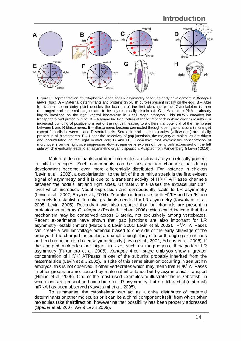
Figure 3. Representation of Cytoplasmic Model for LR asymmetry based on early development in Xenopus laevis (frog). A – Maternal determinants and proteins (in bluish purple) present initially on the egg; B – After
fertilization, sperm entry point decides the location of the first cleavage plane. Cytoskeleton is then rearranged and maternal cargo starts to be asymmetrically distributed; C – Maternal mRNA is already
largely localized on the right ventral blastomere in 4-cell stage embryos. This mRNA encodes ion transporters and proton pumps; D – Asymmetric localization of these transporters (blue circles) results in a
increased pumping of positive ions out of the rigt cell, leading to a differential potencial of the membrane between L and R blastomeres; E – Blastomeres become connected through open gap junctions (in orange)
except for cells between L and R ventral cells. Serotonin and other molecules (yellow dots) are initially present in all blastomeres; F – Under the selectivity of gap junctions, the majority of molecules are driven and accumulated on the right ventral cell; G and H – Somehow, that asymmetric concentration of
morphogens on the right side suppresses downstream gene expression, being only expressed on the left side which eventually leads to an asymmetric organ disposition. Adapted from Vandenberg & Levin ( 2010).
Maternal determinants and other molecules are already asymmetrically present in initial cleavages. Such components can be ions and ion channels that during development become even more differentially distributed. For instance in chicken (Levin et al., 2002), a depolarisation to the left of the primitive streak is the first evident signal of asymmetry and it is due to a transient activity of H+/K+ ATPases channels between the node’s left and right sides. Ultimately, this raises the extracellular Ca2+ level which increases Nodal expression and consequently leads to LR asymmetry (Levin et al., 2002; Raya et al., 2004). Zebrafish in turn uses both H+/K+ and Na+/K+ ion channels to establish differential gradients needed for LR asymmetry (Kawakami et al. 2005; Levin, 2005). Recently it was also reported that ion channels are present in protostomes such as C. elegans (Poole & Hobert 2006) which could indicate that this mechanism may be conserved across Bilateria, not exclusively among vertebrates. Recent experiments have shown that gap junctions are also important for LR asymmetry- establishment (Mercola & Levin 2001; Levin et al.,2002). H+/K+ ATPases can create a cellular voltage potential biased to one side of the early cleavage of the embryo. If the charged molecules are small enough they diffuse through gap junctions and end up being distributed asymmetrically (Levin et al., 2002; Adams et al., 2006). If the charged molecules are bigger in size, such as morphogens, they pattern LR asymmetry (Fukumoto et al. 2005). Xenopus 4-cell stage embryos show a greater concentration of H+/K+ ATPases in one of the subunits probably inherited from the maternal side (Levin et al., 2002). In spite of this same situation occurring in sea urchin embryos, this is not observed in other vertebrates which may mean that H+/K+ ATPases in other groups are not caused by maternal inheritance but by asymmetrical transport (Hibino et al. 2006). One of the most used examples to illustrate this is zebrafish, in which ions are present and contribute for LR asymmetry, but no differential (maternal) mRNA has been observed (Kawakami et al., 2005).
To summarise, the cytoskeleton can act as a chiral distributor of maternal determinants or other molecules or it can be a chiral component itself, from which other molecules take theirdirection, however neither possibility has been properly addressed (Spéder et al. 2007; Aw & Levin 2009).

15
Introduction
Segregation of differentially-imprinted chromatids Model
This model proposes that differential chromatid segregation during early development, probably right after fertilization, is the first asymmetric event taking place (Klar 1994; Armakolas et al. 2010; Armakolas & Klar 2007). This third model is intrinsically linked with Cytoplasmic model described above, as the cytoskeleton is involved in directional movements of chromatids that are asymmetrically distributed to daughter cells, as have been shown for maternal determinants (Vandenberg & Levin 2010). This process has been successfully studied in yeast (Armakolas et al., 2010) and some animals, such as snails, during early cleavages. It was shown that this differential chromatid segregation is determined by the asymmetric structure of a contractile ring (Meshcheryakov & Beloussov 1975). The recent discovery of the implication of dynein, a LR motor protein involved in asymmetric events in vertebrates, in this process suggests even more the fact that all three models are, in a basic sense, compatible.
Nevertheless, this is a really recent and preliminary model that currently takes into account only a small number of species. Further studies using animal models that are already well characterized are important.
Maintenance of left-right asymmetry
After the initial break of symmetry it is important to understand how asymmetry
is established and maintained during development. Numerous genes have been addressed, in which Nodal has appeared as one of the most studied as well as its downstream targets: Pitx and FoxH1. Since Nodal is widely spread across Bilateria, an interest has been increasing about how conserved its function is and how conserved are the mechanisms behind LR asymmetry. Nodal signalling
Nodal is a secretory protein, discovered in 1993 (Zhou et al., 1993), that
belongs to the transforming growth factor-beta (TGF-β) superfamily and it is involved in signal transfer from the node to lateral plate mesoderm (LPM) (Kawasumi et al., 2011). Nodal signaling is also involved in cellular differentiation, mesoderm and endoderm formation and primitive streak establishment during gastrulation in mouse, zebrafish and frog (Whitman 2001).
In spite of being conserved across vertebrates, Nodal has a different number of copies depending on the species, for instance in humans and other mammals there is one copy, in zefrafish three copies were found and five in Xenopus (Juan & Hamada 2001). Nodal starts being expressed symmetrically and then becomes asymmetrically expressed on the left side of LPM at the early somite stage in chordates (Vandenberg & Levin 2009). However this is not true for Echinodermata, Nodal is always expressed asymmetrically and is found only on the right side (Duboc et al. 2004). In the typical protostome models, C. elegans and D. melanogaster, nodal has not been found, and hence it was thought that probably it evolved within the early deuterostome lineage. However recently an orthologue of Nodal was found in snails (Mollusca) and it was shown that it also plays a role in left-right asymmetry, demonstrating it is of much earlier evolutionary origin (Grande & Patel, 2009).
b
16
Introduction
Figure 4. Nodal signalling pathway. Schier &
Shen, 2000.
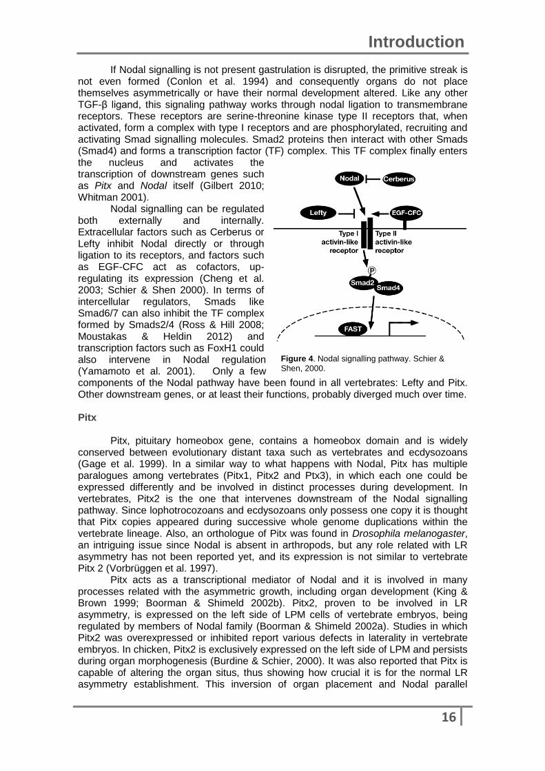
If Nodal signalling is not present gastrulation is disrupted, the primitive streak is not even formed (Conlon et al. 1994) and consequently organs do not place themselves asymmetrically or have their normal development altered. Like any other TGF-β ligand, this signaling pathway works through nodal ligation to transmembrane receptors. These receptors are serine-threonine kinase type II receptors that, when activated, form a complex with type I receptors and are phosphorylated, recruiting and activating Smad signalling molecules. Smad2 proteins then interact with other Smads (Smad4) and forms a transcription factor (TF) complex. This TF complex finally enters the nucleus and activates the transcription of downstream genes such as Pitx and Nodal itself (Gilbert 2010; Whitman 2001).
Nodal signalling can be regulated both externally and internally. Extracellular factors such as Cerberus or Lefty inhibit Nodal directly or through ligation to its receptors, and factors such as EGF-CFC act as cofactors, up-regulating its expression (Cheng et al. 2003; Schier & Shen 2000). In terms of intercellular regulators, Smads like Smad6/7 can also inhibit the TF complex formed by Smads2/4 (Ross & Hill 2008; Moustakas & Heldin 2012) and transcription factors such as FoxH1 could also intervene in Nodal regulation (Yamamoto et al. 2001). Only a few components of the Nodal pathway have been found in all vertebrates: Lefty and Pitx. Other downstream genes, or at least their functions, probably diverged much over time. Pitx Pitx, pituitary homeobox gene, contains a homeobox domain and is widely conserved between evolutionary distant taxa such as vertebrates and ecdysozoans (Gage et al. 1999). In a similar way to what happens with Nodal, Pitx has multiple paralogues among vertebrates (Pitx1, Pitx2 and Ptx3), in which each one could be expressed differently and be involved in distinct processes during development. In vertebrates, Pitx2 is the one that intervenes downstream of the Nodal signalling pathway. Since lophotrocozoans and ecdysozoans only possess one copy it is thought that Pitx copies appeared during successive whole genome duplications within the vertebrate lineage. Also, an orthologue of Pitx was found in Drosophila melanogaster, an intriguing issue since Nodal is absent in arthropods, but any role related with LR asymmetry has not been reported yet, and its expression is not similar to vertebrate Pitx 2 (Vorbrüggen et al. 1997).
Pitx acts as a transcriptional mediator of Nodal and it is involved in many processes related with the asymmetric growth, including organ development (King & Brown 1999; Boorman & Shimeld 2002b). Pitx2, proven to be involved in LR asymmetry, is expressed on the left side of LPM cells of vertebrate embryos, being regulated by members of Nodal family (Boorman & Shimeld 2002a). Studies in which Pitx2 was overexpressed or inhibited report various defects in laterality in vertebrate embryos. In chicken, Pitx2 is exclusively expressed on the left side of LPM and persists during organ morphogenesis (Burdine & Schier, 2000). It was also reported that Pitx is capable of altering the organ situs, thus showing how crucial it is for the normal LR asymmetry establishment. This inversion of organ placement and Nodal parallel

17
Introduction
expression was also observed in Pitx mutant mouse embryos (Ryan et al. 1998; Lin et al. 1999). FoxH1 (Fast1) FoxH1 is a winged-heliix transcription factor and it is a member of a big family of genes containing a forkhead box domain. It is widespread and conserved across chordates and despite not being a canonical member of Nodal signalling, FoxH1 has been shown to modulate Nodal expression through ligation to Smads. This FoxH1/Smads complex is formed in response to activins (Chen et al. 1996) and consequently activates transcription of left-sided specific genes such as Mix1 and goosecoid (Chen et al. 1996; Chen et al. 1997; Watanabe & Whitman 1999). The forkhead domain is usually present as a single copy within a Fox protein, that uses this domain to bind to specific DNA sequences (Shimeld et al. 2010). Although Fox genes are widely present across animals, FoxH has been only found in Bilateria, but this could be due to lack of information (Shimeld et al. 2010).
Studies performed in mouse have shown that Nodal and FoxH1 expression patterns overlap with each other, and mutant embryos, in which FoxH1 was mutated, show defects in antero-posterior patterning (Yamamoto et al. 2001). In mouse, FoxH1 starts being expressed bilaterally in the LPM then becoming restricted to the heart (Saijoh et al. 2000), suggesting a role during embryonic patterning. FoxH1 null mice appear without crucial structures during early development, such as the node, notocord and prechordal LPM. Besides midline structures, FoxH1 is also important for the embryonic development of endoderm (Hoodless et al. 2001), a structure that is also dependent on Nodal signalling. Actually, FoxH1 seems to be the major transcriptional transducer of Nodal signalling during early development in mice, playing multiples roles over that time. However, FoxH, Smads and Nodal mutants suggest that Nodal signalling can act in two ways: dependent or not dependent of FoxH1 (Yamamoto et al. 2001).
In Xenopus, FoxH1 appears to be related to mesodermal gene expression and with gastrulation movements (Watanabe & Whitman 1999). Although there was some controversy about FoxH1 function in pattern formation and Nodal signalling, the analyses of maternal contribution of FoxH1 confirmed indeed that this transcription factor plays a role on the development of axial structures and regulates Nodal expression (Kofron et al. 2004). In zebrafish, when FoxH1 is missing, embryos present various defects in axial structures, which may be a indicator of FoxH1 function in nodal signalling modulation (Pogoda et al. 2000).
Pitx is also activated by FoxH1 (Yoshida & Saiga 2008) and consequently up-regulates other transcription pathways involved in LR asymmetry processes (Hamada et al. 2002). Until now, FoxH1 was thought to be absent from protostomes, but recent reports have indicated that it is present in Patella vulgata (mollusc). Aims and hyphotesis Although it is obvious that deuterostome and protostome development is very different, recent studies show that genes like Nodal involved in the acquisition of asymmetric morphology in vertebrates, were also present and played a similar function in molluscs (Grande & Patel 2009). Due to their morphological diversity and lifestyle it has been a difficult job to reconstruct mollusc phylogeny. Patella vulgata, a common European limpet, is a species from the basal group of gastropod molluscs (Figure 5) (Haszprunar,1988; Ponder & Lindberg, 1997). In addition, the fact that Nodal, Pitx and FoxH1 are also present in this species makes Patella a decisive model to understand general mollusc phylogeny and conservation of LR asymmetry establishment mechanisms across Bilateria. During my project, I started to address Nodal, Pitx and

18
Introduction
Figure 5. Representation of the phylogenetic tree of Bilateria. Animals used in this project are written in red bold below the taxa name.
FoxH1 expression patterns at different developmental stages. After initial gene characterization, a further insight about FoxH1 role on Nodal signalling was also taken in account. If FoxH1 had a regulatory effect, that could be through its ligation to the Smads complex (intracellulary). It could also happen that FoxH1, even if present, does not have a direct regulatory role but could be affected by both Nodal and Pitx. All these possibilities should be considered during experimental work. The study was also extended to other species: Branchiostoma floridae, Branchiostoma lanceolatum (Amphioxus) and Lampetra planeri (Lamprey). Lampreys constitute the Agnatha taxon (vertebrates without jaw) which is known as the sister group of vertebrates with jaw (Figure 5). These groups are closely related, and both share many genes, pathways and features. It is known that Nodal and Pitx are present in lampreys as well as FoxH1, but there is no reliable data about their expression patterns and possible functions on LR asymmetry. A few questions are imperative to understand how conserved mechanisms are since the last split between vertebrates: Is
nodal expressed symmetrically and then asymmetrically on the left side as in other vertebrates? Or it is expressed on the right like sea urchins? Does FoxH1 mediate Nodal signalling and how it is done? On the other hand, amphioxus belongs to the
Cephalochordata (Invertebrates), a more distant group than lampreys but still within the Deuterostome lineage (Figure 5). Similar to what happens with lampreys, Nodal, Pitx and FoxH1 are also present in these animals but detailed information about their role in LR asymmetry is missing. Once again, the same
questions about gene function and mechanisms conservation are maintained. However, it is
already known that Nodal expression is present on the left side of the embryos, which is an intriguing feature, since all invertebrates described until now, such as sea urchin, have Nodal being expressed on the right side. In the end, it is expected a better understanding of how asymmetry establishment and maintenance is achieved and how well preserved it is among Bilateria.

Methodology We must revisit the idea that science is a
methodology and not an ontology.
Deepak Chopra
20
Methodology
Patella vulgata collection and In vitro fertilisation Male and female adults were collected from Plymouth and Portsmouth seas,
United Kingdom (UK). The animals were then maintained in tanks at 10ºC in the Zoology Department, University of Oxford (UK). In vitro fertilisations were performed as described in Hashimoto (2011) at room temperature (14ºC). Embryos were then collected and fixed (4%PFA in MOPS) at different stages: 0, 4, 10, 18, 24 and 48 hours post-fertilisation (hpf). They were stored at -20ºC in 100% EtOH.
Oligonucleotides FoxH1 and Nodals primers for Patella vulgata were already present on the lab (Shimeld S.): FoxH1_Forward (5’-3’): AGCCCGAAGCGAAAAGAAGA FoxH1_Reverse (5’-3’): GGAAGTGTCCGTGGCTGAAG NodalO_Forward (5’-3’): TAGACGCAACGGAGGAGAGT NodalO_Reverse (5’-3’): AAAGCACCGTGGGATATGAC
NodalNew_Forward (5’-3’): CCACTTAACCGCCAACAAAGA NodalNew_Reverse (5’-3’): TCGCATGATTTGTGGGTTTG
FoxH1, Nodal and Pitx primers for amphioxus genes were all design manually from the Branchiostoma floridae genome dataset present online (JGI).
Pitx_Forward (5’-3’): GCTTGGACCAACCTCACAGA Pitx_Reverse (5’-3’): AGTGTTGCTTGGCCTTCAG Since Nodal is a relatively long gene, primers were designed taking in account
the most conserved region. Forward (5’-3’): CAGAGTCTGAGCTCGGGGA Reverse (5’-3’): TTCTTCTGACGTCTCCTGTTTG In amphioxus, the forkhead domain of FoxH1 is present in two different exons.
This domain is the most conserved region and also the most identical between both species, so primers were designed using genomic DNA.
Forward (5’-3’): ACCGCGATCAGGACTTCGAC Reverse (5’-3’): CACCACGTCGTACCTGGGCAT GAPDH primers for lamprey used for RT-PCR were ordered based on a
published paper, in which this gene was successfully obtained ((Pancer et al. 2004)). Forward (5’-3’): GAACATCGGCATCAATGGGT Reverse (5’-3’): GAGGCCTTATCGATGGTGGT Pitx primers for lamprey were previously used in the lab (Shimeld S.). Forward (5’-3’): GGCAGCGGACCCATTTCAC Reverse (5’-3’): CTGGAGTGCTGCTTGGCTTT FoxH1 primers for lamprey were designed from a really short sequence from
Petromyzon marinus assembly data. Forward (5’-3’): CACTTGTTCGCAGAGAGGT Reverse (5’-3’): ACTACAACCACTACCACTACT
Polymerase Chain Reaction (PCR) and DNA extraction from agarose gels PCRs were performed using BioTaq™ DNA polymerase kit (Bioline) according to manufacturer instructions with thermocycler standard conditions (Table I).

21
Methodology
Table I. Thermocycling conditions for a routine PCR. Step Temperature Time
Initial denaturation 95ºC 30 seconds 35 cycles
95ºC 55ºC 68ºC
30 seconds 30 seconds 1 minute/kb
Final extension 60ºC 5 minutes Hold 4-10ºC ----
After checking final products through electrophoresis, bands were cut and
disposed in new tubes to purify the DNA. Illustra™GFX™ PCR DNA and Gel Band Purification Kit (GE Healthcare) and QIAquick® Gel Extraction Kit (QIAGEN) were used. In both cases DNA was eluted in 50 µl of RNAse-free water.
Double-stranded RNA synthesis and Microinjections of Patella vulgata embryos
Double-stranded RNAs (dsRNA) for Nodal original, Nodal new, FoxH1 and Pitx were synthesised using MEGAscript® RNAi Kit (Ambion®) and samples were analysed and concentration was assessed using the NanoDrop (ThermoScientific).
Microinjections were performed in embryos at 0-0.30 hpf according to the protocol of Zhang (2007), using an Eppendorf FemtoJet® Microinjector. The injection solution was prepared with 20% glycerol and dsRNA (100µgml-1). After filling the needle with this solution, 0-0.30hpf eggs were disposed on a glass chamber filled with sea water. After being injected, embryos were transferred for a new plate with fresh
sea water to grow in normal conditions until fixation.
Extraction of RNA Total RNA was extracted from adult amphioxus (Branchiostoma lanceolatum)
previously preserved in 100% EtOH. After tissue homogenization in Trizol (pH), using a rotor-stator homogenizer, chloroform was added. Samples were spun and the aqueous phase was transferred into new tubes. Samples were then precipitated in 100% EtOH and 3M Sodium acetate at -80ºC. The resultant pellet was washed with 70% EtOH, ressuspended and loaded on both mini and micro columns and spun. Final product was washed once again and digested with DNase to degradate possible residual genomic DNA. The enzyme was inhibited with successive washes with kit buffers and RNA was finally diluted in miliQ-H2O and kept at -80ºC.
Cloning of DNA (PCR fragments): Ligation of DNA into vectors and Transformation of Cells PCR products were inserted into the vector – pCRII (Invitrogen), and then plasmids were transformed into competent E.coli through heat shock treatment. Competent cells were put in SOC medium on a 37ºC waterbath for an hour and were then spread onto LB medium plates containing ampicilin, IPTG and X-gal and put to grow at 37ºC, overnight. Only colonies with the inserted plasmid (white colonies) were collected and put to grow in tubes containing LB medium overnight at 37ºC with shaking.
Plasmid purification from bacteria was performed using QIAprep Spin Miniprep Kit from QIAGEN®. To elute DNA, 50µl of RNAse-free water was used per sample. To make sure that the insert on the plasmid had the same size as the interested gene, a diagnostic digest was carried using EcoRI restriction enzyme. The digestion was confirmed on a 1% agarose gel.

22
Methodology
DNA sequencing
Sequencing protocol was performed with the Sanger method, using the plasmid miniprep product. Both T7 and SP6 primers were used and PCR reaction was set up according with the conditions in Table II).
Samples were then precipitated with 100% EtOH+NaAc 3M at -20ºC overnight. After precipitation samples were spun and the supernatant was discarded. EtOH 70% was used to wash the pellet, then discarded. The pellet was dried and then covered in foil and maintained at -20ºC until sequencing took place.
Table II. Thermocycling conditions for a PCR for DNA sequencing.
Step Temperature Time
Initial denaturation 95ºC 4 minutes 30-35 cycles
95ºC 50ºC 68ºC
30 seconds 30 seconds 60 seconds
Final extension 68ºC 4 minutes Hold 4-10ºC ----
In situ hybridisation Riboprobe synthesis In order to produce mRNA probes for in situ hybridisation, a standard PCR (see
Table III) was settled to produce the DNA fragments from the miniprep products. SP6 and T7 primers were used to acquire both sense and antisense products. All products were checked on a 1% agarose gel.
Table III. Thermocycling conditions for a PCR for Riboprobe synthesis. Step Temperature Time
Initial denaturation 94ºC 4 minutes 35 cycles
94ºC 53ºC 72ºC
30 seconds 30 seconds 90 seconds
Final extension 72ºC 5 minutes Hold 4-10ºC ----
PCR products were then incubated in a riboprobe synthesis mix containing
buffers, DIG-labelled UTPs and SP6/T7 RNA polymerases, at 37ºC for 2 hours. Final products were checked on a 1% agarose gel. RNA samples were then precipitated in DEPC water, LiCl 4M and 100% EtOH at -20ºC, overnight. After centrifugation, supernatant was discarded and pellet was washed with 70% EtOH. Supernatant was once again discarded and the pellet air-dried. RNA was then dissolved in DEPC water (50µl), confirmed through electrophoresis and stored at -80ºC.
Patella vulgata in situ hybridisation In situ hybridisation started with embryo’s rehydration in PBT, incubated with proteinase K (10mgml-1) for 5 minutes and refixed in 4% paraformaldehydein PBT for 30 minutes. After washing embryos in PBT they were rinsed in Hybridisation solution and put at 65ºC for 2 hours to pre-hybridise. The solution was renewed, the probe was added and hybridisation took place at 65ºC, overnight. Following washes were made with Ciona wash solution, then PBT, and embryos were put in blocking solution for 3 hours at 4ºC. The blocking solution was replaced by the antibody solution and incubated again at 4ºC, overnight. Embryos were later washed with PBT then APT and transferred to a multiwell dish with staining solution until revelation was complete. For long-term storage, embryos were washed in PBT and put in 80% Glycerol at 4ºC.
23
Methodology
Amphioxus in situ hybridisation In situ hybridisations were performed in both B. floridae and B. lanceolatum
species using an adaptation of Garcia’s protocol. During this phase were used amphioxus embryos previously stored in 100% Ethanol at -20ºC. Embryos were rehydrated with EtOH in PBT and then incubated in proteinase K (10mgml-1) for a few minutes. The following washes were done with Gycine 10%, PBT in Glycine and PBT. Embryos were then fix in 4% paraformaldehyde for 30 minutes and washed in Triethanolamine 0.1M, Acetic anhydride (2.5µM and 5µM) in PBT. PBT solution was replaced by fresh hybridisation solution and embryos were left to pre-hybridisation at 60ºC for 2 hours. Hybridisation solution was renewed, the probe (100mgml-1) was added and the embryos were then incubated at 60ºC overnight. Several washes were performed using firstly hybridisation solution, then wash solution and MABT, then embryos were incubated in MAB + Blocking 2% + Sheep Serum 10% for 1-2 hours at 4ºC. The last solution was then renewed and an antibody was added and embryos were again incubated for 2-4 hours at 4ºC. Following washes with MABT were done to eliminate residual blocking and antibody solution and the staining process was initiated using NBT+BCIP and embryos were maintained on the dark. After being stained, embryos were washed in MABT and refixed with 4% paraformaldehyde and kept in 80% Glycerol for storage.
Lamprey embryos collection Lamprey embryos were collected from Highland Water, a river in The New
Forest (UK), and transported and maintained in river water at room temperature (RT) until future experiments were undertaken. Embryos were used not only to extract RNA. Embryos were staged (Tahara 1988) and then fixed and stored in 100% methanol at -20ºC.
Reverse-Transcription PCR (RT-PCR) Total RNA was collected from lamprey embryos at different stages (ST 11/12,
13/14, 15/16, 17/18, 19/20, 21/22) as described above for amphioxus, using Trizol-Chloroform extraction. The final quality and concentration of samples were measured using a Nanodrop.
RT-PCR was performed using SuperScript® III Reverse Transcriptase (Life Technologies). As negative control for cDNA synthesis I used an additional tube with the same conditions (RNA+miliQ-H2O+dT/random primers+dNTPs+5Xfs Buffer+0.1M DTT+ Superasin+ SSIII) except for the addition of the enzyme SSIII which was omitted. After cDNA synthesis, a routine PCR reaction was performed (see Table I), this time using GAPDH primers as positive control, since they are expressed at any developmental stage.
Phylogenetic analyses Phylogenetic and molecular evolutionary analyses were conducted using MEGA
version 5 (Tamura, Peterson, Stecher, Nei and Kumar 2011), MAFFT version 7 (Katoh, Standley 2013) and BioEdit version 7.2.2 (Hall 1999). Gene synteny was addressed using Genomicus version 73.01(Louis et al. 2013).

Results Absence of evidence is not evidence of
absence.
Carl Sagan

25
Results
Fox genes evolution and conservation across Bilateria In order to trace the evolutionary origin of FoxH1 in the Metazoa and understand how well-conserved this gene is across this group, a phylogenetic tree was constructed using not only FoxH genes from different organisms but also a wide representation of other Fox genes. In Figure 6, FoxH1 genes in both protostome and deuterostome animals are sorrounded by a red rectangle. FoxH1 is only found within the Bilateria, and from the tree we can see that FoxH1 emerged quite late, sharing the most immediate ancestor with FoxQ1 (green circle). Since the divergence between FoxQ1 and FoxH1, FoxH1 has been separated in two major groups, one with branchiostoma FoxH1 and other one with all vertebrates and a mollusc FoxH1. This feature is intriguing, from all FoxH sequences only one is from the protostome group – patella – and that gene is closer to a vertebrate (zebrafish) than to a invertebrate. An overall look into the other Fox genes (FoxO, FoxJ, FoxG, FoxC and FoxF), shows that these relations between genes change.
FoxH1 in Lamprey
The current project aims to address Nodal, Pitx and FoxH1 expression patterns across early development in four species: Patella vulgata, Branchiostoma floridae, Branchiostoma lanceolatum and Lampetra planeri. During the search and gathering of the respective gene’s sequences to design the specific primers, a blast analysis was first performed to each sequence to confirm the gene identity.
For lamprey genes, we used gene sequences of Petromyzon marinus present on Ensembl database, as reference due to the high similarity between this species and Lampetra planeri. However, the sequence described on the database as FoxH1 revealed to be wrongly noted, being in fact a FoxQ1. This raises the question whether FoxH1 actually exists in lamprey or not.
In order to answer that question we proceeded with further analyses of FoxH1 synteny, which in other words means that we try to see if FoxH1 was conserved in the same chromosome, or surrounded by the same genes, in all organism selected. We addressed FoxH1 synteny across the deuterostome lineage (Figure 7 – A), trying to see if it was present in all vertebrates and if they were surround by the same flanking genes. As we can see on Figure 7 – A, FoxH1 is really well conserved in mammals (Human, Chimpanzee and Mouse), being not only present but also having many flanking genes in common. The next nearest group in terms of evolutionary divergence is Reptiles, in this figure represented by the lizard. Despite the reduced amount of information on this branch, we can clearly see that FoxH1 is present as well one of human FoxH1 flanking genes. The amphibian group representative is Xenopus laevis (a frog) which has 2 copies of FoxH1 gene. On the GL173015.1 scaffold, FoxH1 is flanked by different genes when compared with human FoxH1, but that genes are also present in human, chromosome 11. On the other scaffold, GL174256.1, FoxH1 is surrounded by different genes as well, but the one present on the left side is also present in Human chromosome 8. Although the data is scarce for platyfish, it seems that FoxH1 and its flanking genes are even better conserved than in reptiles or amphibians. If we look at zebrafish, is noticeable that the few flanking genes present are the same as platyfish/mammals. In addition, the genes present on chromosome 19 in zebrafish are pretty conserved and appears to have the same disposition as FoxH1 flanking genes in humans.
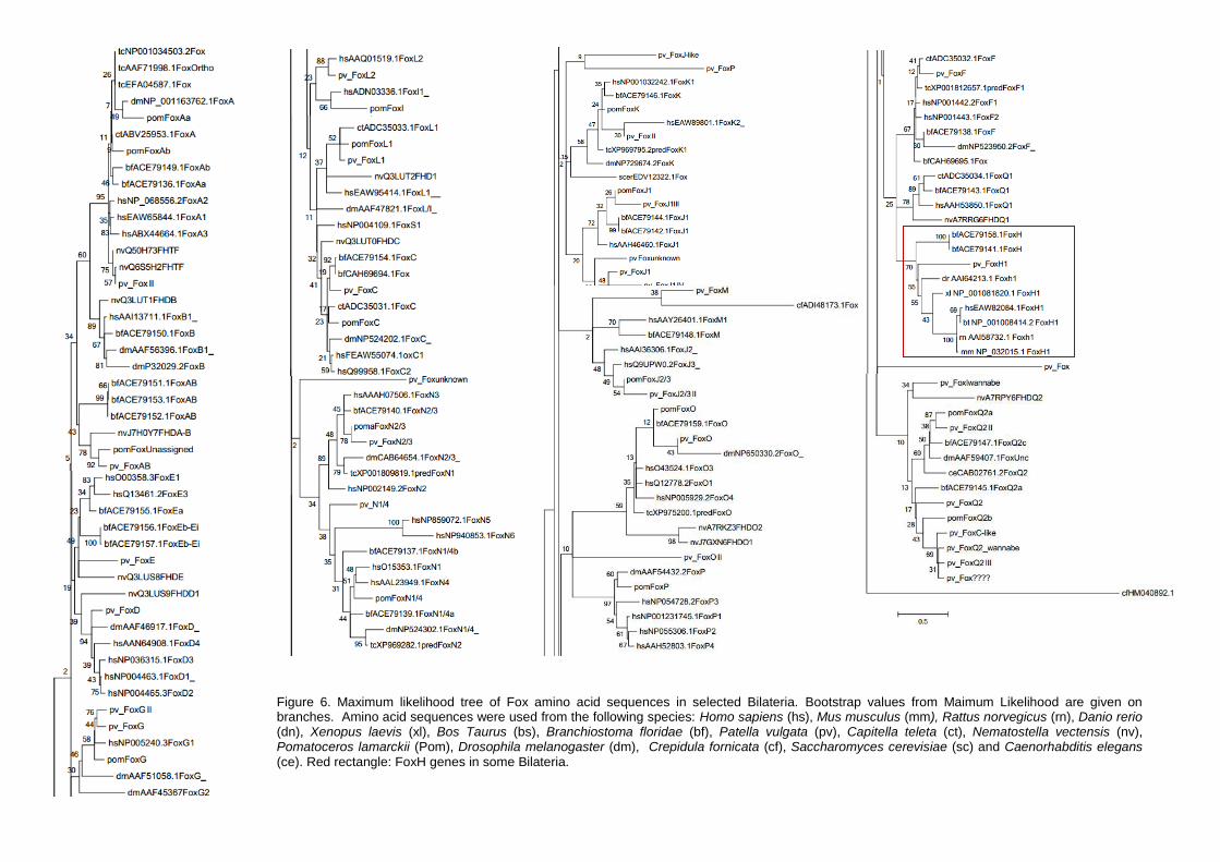
Figure 6. Maximum likelihood tree of Fox amino acid sequences in selected Bilateria. Bootstrap values from Maimum Likelihood are given on branches. Amino acid sequences were used from the following species: Homo sapiens (hs), Mus musculus (mm), Rattus norvegicus (rn), Danio rerio (dn), Xenopus laevis (xl), Bos Taurus (bs), Branchiostoma floridae (bf), Patella vulgata (pv), Capitella teleta (ct), Nematostella vectensis (nv), Pomatoceros lamarckii (Pom), Drosophila melanogaster (dm), Crepidula fornicata (cf), Saccharomyces cerevisiae (sc) and Caenorhabditis elegans (ce). Red rectangle: FoxH genes in some Bilateria.

27
Results
Figure 7. Synteny of Fox genes within the deuterostome lineage. (A) FoxH1 synteny is well patented in all jawed vertebrates analysed. (B) FoxQ1 synteny was also addressed due to similarity between these two Fox genes. Data gathered and selected from Genomicus version 73.01(Louis, A. Muffato, H. and Crollius, H.R., 2012).
The lamprey FoxH1 present in the Figure 7 – A is the one described on
Ensembl and it’s possible to see that the flanking genes present are not the same as for other organism (scaffold GL476587), but we must take in account that there isn’t much data to rely on. However, the genes present on GL476793 scaffold in lamprey, are also present in Human chromosome 8, not in the same order but still they show some conservation between species. Different blast analyses end up revealing that the gene wrongly noted is actually a FoxQ1. We decide then to check FoxQ1 synteny between human and lamprey (Figure 7 – C). In both species FoxQ1 is conserved in terms of chromosomal disposition. The flanking genes did not resemble at all the ones from FoxH1.
Recently a paper was published on Nature Genetics (Smith et al. 2013) with the lamprey’s entire genome sequenced as well as with RNA-seq datasets which is another useful way to see if FoxH1 is present in jawless vertebrates. We started to assemble the mRNA datasets from a wide range of early developmental stages: blastula, gastrula and neurula (SRX110029.2, SRX110030.2, SRX110031.2, SRX110032.2, SRX110033.2, SRX110034.2, SRX110035.2). After assembly all sequences we performed an analysis to check for fragmented or small sequences (<100base pairs). Using FoxH1 cDNA sequences from human, zebrafish and Xenopus, we ran a blast analysis. From each comparison between lamprey and other species, we selected the final 10 top hits, the 10 sequences with more similarity. These 10 hits
A
B
28
Results
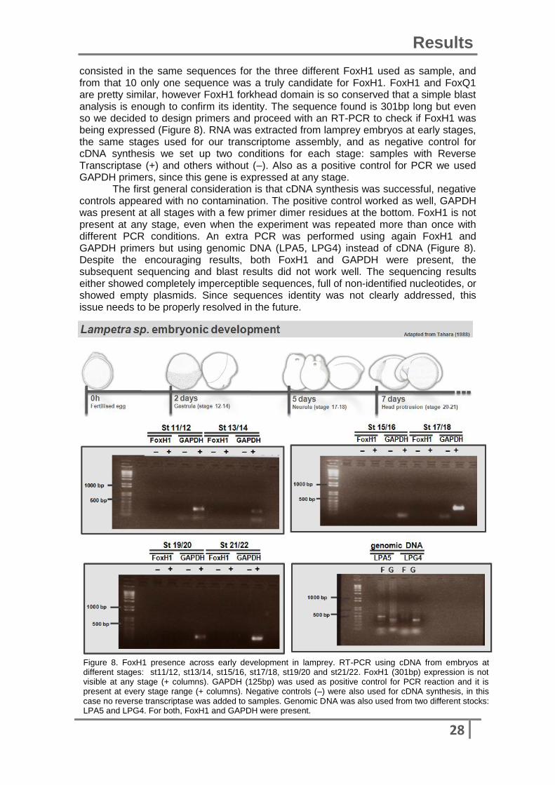
Figure 8. FoxH1 presence across early development in lamprey. RT-PCR using cDNA from embryos at different stages: st11/12, st13/14, st15/16, st17/18, st19/20 and st21/22. FoxH1 (301bp) expression is not visible at any stage (+ columns). GAPDH (125bp) was used as positive control for PCR reaction and it is present at every stage range (+ columns). Negative controls (–) were also used for cDNA synthesis, in this case no reverse transcriptase was added to samples. Genomic DNA was also used from two different stocks: LPA5 and LPG4. For both, FoxH1 and GAPDH were present.
consisted in the same sequences for the three different FoxH1 used as sample, and from that 10 only one sequence was a truly candidate for FoxH1. FoxH1 and FoxQ1 are pretty similar, however FoxH1 forkhead domain is so conserved that a simple blast analysis is enough to confirm its identity. The sequence found is 301bp long but even so we decided to design primers and proceed with an RT-PCR to check if FoxH1 was being expressed (Figure 8). RNA was extracted from lamprey embryos at early stages, the same stages used for our transcriptome assembly, and as negative control for cDNA synthesis we set up two conditions for each stage: samples with Reverse Transcriptase (+) and others without (–). Also as a positive control for PCR we used GAPDH primers, since this gene is expressed at any stage.
The first general consideration is that cDNA synthesis was successful, negative controls appeared with no contamination. The positive control worked as well, GAPDH was present at all stages with a few primer dimer residues at the bottom. FoxH1 is not present at any stage, even when the experiment was repeated more than once with different PCR conditions. An extra PCR was performed using again FoxH1 and GAPDH primers but using genomic DNA (LPA5, LPG4) instead of cDNA (Figure 8). Despite the encouraging results, both FoxH1 and GAPDH were present, the subsequent sequencing and blast results did not work well. The sequencing results either showed completely imperceptible sequences, full of non-identified nucleotides, or showed empty plasmids. Since sequences identity was not clearly addressed, this issue needs to be properly resolved in the future.
29
Results
Gene expression patterns during embryonic development
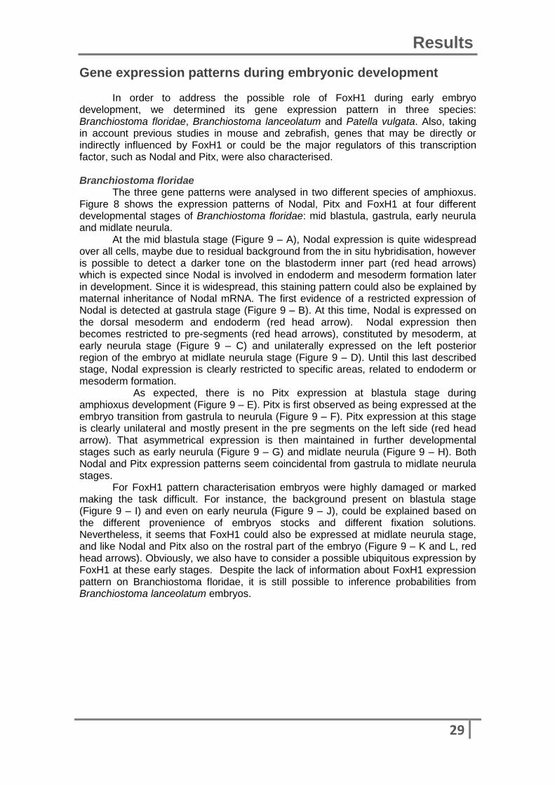
In order to address the possible role of FoxH1 during early embryo development, we determined its gene expression pattern in three species: Branchiostoma floridae, Branchiostoma lanceolatum and Patella vulgata. Also, taking in account previous studies in mouse and zebrafish, genes that may be directly or indirectly influenced by FoxH1 or could be the major regulators of this transcription factor, such as Nodal and Pitx, were also characterised. Branchiostoma floridae The three gene patterns were analysed in two different species of amphioxus. Figure 8 shows the expression patterns of Nodal, Pitx and FoxH1 at four different developmental stages of Branchiostoma floridae: mid blastula, gastrula, early neurula and midlate neurula. At the mid blastula stage (Figure 9 – A), Nodal expression is quite widespread over all cells, maybe due to residual background from the in situ hybridisation, however is possible to detect a darker tone on the blastoderm inner part (red head arrows) which is expected since Nodal is involved in endoderm and mesoderm formation later in development. Since it is widespread, this staining pattern could also be explained by maternal inheritance of Nodal mRNA. The first evidence of a restricted expression of Nodal is detected at gastrula stage (Figure 9 – B). At this time, Nodal is expressed on the dorsal mesoderm and endoderm (red head arrow). Nodal expression then becomes restricted to pre-segments (red head arrows), constituted by mesoderm, at early neurula stage (Figure 9 – C) and unilaterally expressed on the left posterior region of the embryo at midlate neurula stage (Figure 9 – D). Until this last described stage, Nodal expression is clearly restricted to specific areas, related to endoderm or mesoderm formation. As expected, there is no Pitx expression at blastula stage during amphioxus development (Figure 9 – E). Pitx is first observed as being expressed at the embryo transition from gastrula to neurula (Figure 9 – F). Pitx expression at this stage is clearly unilateral and mostly present in the pre segments on the left side (red head arrow). That asymmetrical expression is then maintained in further developmental stages such as early neurula (Figure 9 – G) and midlate neurula (Figure 9 – H). Both Nodal and Pitx expression patterns seem coincidental from gastrula to midlate neurula stages.
For FoxH1 pattern characterisation embryos were highly damaged or marked making the task difficult. For instance, the background present on blastula stage (Figure 9 – I) and even on early neurula (Figure 9 – J), could be explained based on the different provenience of embryos stocks and different fixation solutions. Nevertheless, it seems that FoxH1 could also be expressed at midlate neurula stage, and like Nodal and Pitx also on the rostral part of the embryo (Figure 9 – K and L, red head arrows). Obviously, we also have to consider a possible ubiquitous expression by FoxH1 at these early stages. Despite the lack of information about FoxH1 expression pattern on Branchiostoma floridae, it is still possible to inference probabilities from Branchiostoma lanceolatum embryos.
30
Results
Figure 9. Expression of Nodal, Pitx and FoxH1 in Branchiostoma floridae embryos during early development. (A – D) Nodal: (A) mid blastula; (B) gastrula, side view; (C) early neurula and (D) midlate neurula. (E – H) Pitx: (E) mid blastula, (F) midlate gastrula, side view; (G) early neurula and (H) midlate neurula. (I – L) FoxH1: (I) mid blastula, (J) early neurula (K and L) midlate neurula. Red head arrows indicate higher expression domains. bp= blastopore; d=dorsal; ps=pre-segment (mesoderm); a= anterior; p=posterior.
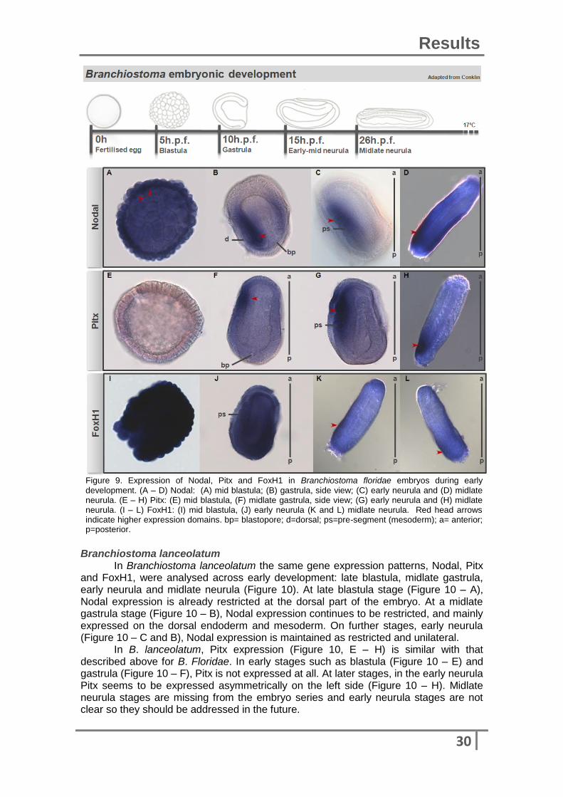
Branchiostoma lanceolatum In Branchiostoma lanceolatum the same gene expression patterns, Nodal, Pitx and FoxH1, were analysed across early development: late blastula, midlate gastrula, early neurula and midlate neurula (Figure 10). At late blastula stage (Figure 10 – A), Nodal expression is already restricted at the dorsal part of the embryo. At a midlate gastrula stage (Figure 10 – B), Nodal expression continues to be restricted, and mainly expressed on the dorsal endoderm and mesoderm. On further stages, early neurula (Figure 10 – C and B), Nodal expression is maintained as restricted and unilateral.
In B. lanceolatum, Pitx expression (Figure 10, E – H) is similar with that described above for B. Floridae. In early stages such as blastula (Figure 10 – E) and gastrula (Figure 10 – F), Pitx is not expressed at all. At later stages, in the early neurula Pitx seems to be expressed asymmetrically on the left side (Figure 10 – H). Midlate neurula stages are missing from the embryo series and early neurula stages are not clear so they should be addressed in the future.
31
Results
At late blastula stage, FoxH1 resembles Pitx by not being expressed so early in development (Figure 10 – I). However its expression comes earlier than Pitx, as at the midlate gastrula stage FoxH1 expression is already noticeable on the edges of blastopore lips (Figure 10 – J). At this point, FoxH1 expression seems to be tightly restricted to the endoderm (red head arrows). In spite of the unspecific staining in the early neurula stage (Figure 10 – K), FoxH1 specific expression still seems to endure in the endoderm from the anterior region (red head arrows).
Unfortunately, it was impossible to address FoxH1 role on later developmental stages, such as midlate neurula (Figure 10 – P). FoxH1 expression at these stages should be characterised in the future to better insight about this gene action area.
Figure 10. Expression of Nodal, Pitx and FoxH1 in Branchiostoma lanceolatum embryos during early
development. (A – D) Nodal: (A) late blastula; (B) midlate gastrula, blastopore view; (C) early neurula and (D) midlate neurula. (E – H) Pitx: (E) late blastula, (F) midlate gastrula, blastopore view; (G) early neurula and (H) midlate neurula. (I – L) FoxH1: (I) late blastula, (J) midlate gastrula, blastopore view, (K and L) midlate neurula. Red head arrows indicate higher expression domains. bp= blastopore; d=dorsal; ps=pre-segment (mesoderm); a= anterior; p=posterior.

32
Results
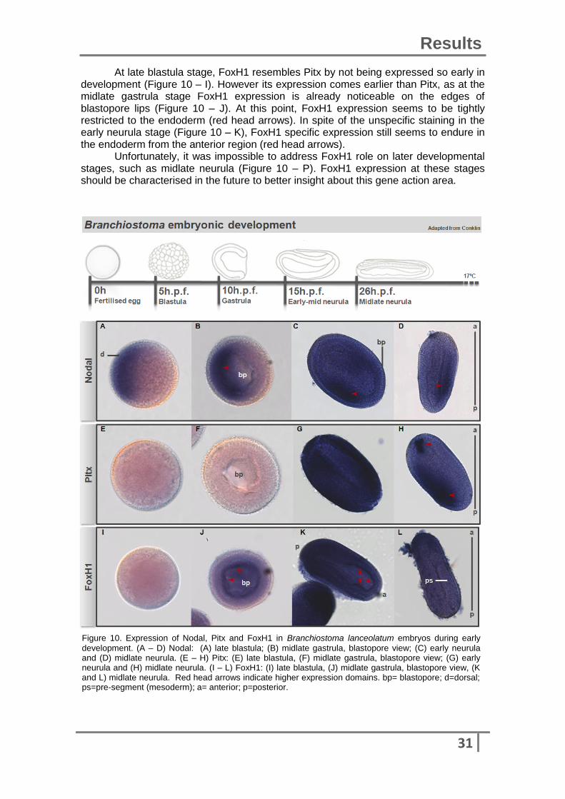
Figure 11. Expression of Nodal Original, Nodal New, Pitx and FoxH1 in Patella vulgata embryos during early development. (A – D) Nodal Original: (A) 4 h.p.f., (B); 10 h.p.f., (C) 18 h.p.f. and (D) 24 h.p.f. (E – H): (E) 4 h.p.f., (F) 10 h.p.f., (G) 18 h.p.f, posterior view and (H) 24 h.p.f. (I – L) Pitx: (I) 4 h.p.f., (J) 10 h.p.f., posterior view (K) 18 h.p.f and (L) 24 h.p.f. (M – N) FoxH1: (M) 4 h.p.f., (N) 10 h.p.f., (O) 18 h.p.f and (P) 24 h.p.f. a= anterior, p= posterior, Pr.tb= primary trochoblasts. Red head arrows indicate higher expression domains.
Patella vulgata For Patella vulgata 4 genes were characterized, FoxH1, Pitx and two Nodal copies, Nodal Original and Nodal New, across early development, 4hpf, 10hpf, 18hpf and 24hpf (Figure 11).

33
Results
As it is shown in Figure 11 (A – D), Nodal Original is not expressed at any stage. It could be not expressed at all during early development, or it could happen to be expressed at fertilization moment (maternal inheritance) or further on development than the stages analysed. Nodal New is expressed at all considered stages (Figure 11, E – H). At 4hpf (Figure 11, E) Nodal is expressed on 2/3 of the embryo. On a later stage, 10hpf (Figure 11, F) it is already noticeable a specific staining, probably by the primitive mesoderm cells (red head arrow). At 18hpf (Figure 11, G), Nodal is again present in primitive mesoderm cells but additionally, it seems to be slightly present at another region. What could be happening is that primitive mesoderm cells are initially aggregated but as development goes on, this aggregate becomes two and is then separated. At 24hpf (Figure11, H), Nodal New is seen on the posterior part of the embryo, located in what seems to be near to the blastopore. Pitx expression was already assessed in the lab, and it seems to be similar. At early stages, 4hpf (Figure 11, I), in spite of the unspecific staining observed, Pitx is not expressed. It only starts to be expressed at 6hpf stage. At 10hpf (Figure 11, J), Pitx expression is located on the posterior part of the embryo, being already asymmetrical towards the right side (red head arrow, posterior view). That asymmetric expression is then maintained at 18hpf stage (Figure 11, K). At 24hpf, despite the blurry staining, Pitx is seen as present on the right side of the embryo, in the mesodermal band, and also around the mouth. FoxH1 starts being already expressed on a restricted area at 4hpf stage (Figure 11, M), being present at the posterior region. At later stages, such as 10 and 18hpf, FoxH1 seems to be expressed in a very similar way to Nodal New, being present on the posterior part and also on regions were mesoderm primitive cells seem to be localised, close to the blastopore (Figure 11, N and O). At later stages, 24hpf (Figure 11, P) FoxH1 seems not be expressed at all, however future experiments should be done to confirm this short period of expression, and to address its expression at later stages during development.
These same genes were also addressed at 0, 6 and 48 hpf embryos, but the results were inconclusive, as embryos were too damaged or showed too much variability in gene expression (data not shown: by expression variability, I mean, variability that also could be background and not specific staining).
Functional studies were also performed, attempted by dsRNA microinjections, in which Nodal Original, Nodal New and FoxH1 were inhibited. However, these experiments weren’t successful as embryos ended up dead, probably due to poor quality of the eggs. Further microinjections and other functional studies were impossible to perform due to successive unsuccessful fertilizations of Patella. Different conditions, taking in account environmental factors, such temperature or water acidity, were tested but all resulted in dead embryos only just two hours after fertilisation.

Discussion Nothing in Biology makes sense except
in the light of evolution
Theodosius Dobzhansky
35
Discussion
If FoxH1 plays a role on LR asymmetry in various deuterostomes and possibly
in protostomes, it is important to trace the evolutionary origin and conservation of this gene in metazoans. Unlike other Fox genes which are present widely across all animals, FoxH1 seems to be only present in Bilateria. For instance, FoxQ1 is quite similar to FoxH1 but it is more widespread across metazoans, even in groups lacking both nodal and FoxH1 such as arthropods and nematodes. The fact that these both genes have not been described in Ecdysozoa doesn’t mean that they are not present, but taking the phylogenetic analysis in consideration, the most probable scenario is that FoxH1 probably originated within Bilateria and then was lost in a few groups. All gene families are organized into monophyletic groups, just like previous studies show, which supports viability of the data and reinforces the conclusions. FoxF,FoxQ and FoxH seem to share a common ancestor. The striking point in the phylogenetic analyses is the position that FoxH1 from Patella occupies in the tree, being more similar with vertebrates, than any one of them is with amphioxus. This could mean that FoxH1 expression could be more similar and probably the mechanisms involved may be more conserved as well. However, these results were obtained using a single protostome FoxH1 sequence and the addition of more FoxH1 sequences from protostomes and other invertebrates, would give a better insight about this and probably this relation would change.
Lamprey belongs to Agnatha group, the sister taxon to vertebrates with jaws, and is a key taxon for understanding the conservation of left-right asymmetric mechanisms or even genes related with it. Both Nodal and FoxH1 had been annotated in the genome of this animal however, during this study we determined realising that the gene described as FoxH1 was not a FoxH1, raising the hypothesis that this gene could be not present, maybe due to gene loss, or that it could be indeed present but not described. Using data from a recent paper (Smith et al. 2013), we were able to find a good candidate for FoxH1 in lamprey but the results from RT-PCR did not confirm it. This could be interpreted as absence of FoxH1 expression at early stages, something unlikely, or wrong primer design or even the fact that FoxH1 is present in such small concentration that it is not easy to amplify by PCR. The fact that the sequence found through assembly is too short (301bp) also ads difficulty to the process. There is a research group in Japan currently working with hagfish, another species from Agnatha, who found a FoxH1 gene on its genome (Personal communication from Juan Anaya). This raises our confidence that FoxH1 is present in Agnatha and future analyses of the assembled data and PCR with newly-designed primers should be performed in future experiments. Lamprey embryos were collected and a few were fixed while others were put to growth in medium with a Nodal inhibitor, SB431542, to be used in future functional studies. If FoxH1 is indeed present, studies for gene expression characterisation and gene function should be done.
Synteny analyses using different vertebrates were also performed in order to address FoxH1 presence and conservation across deuterostomes. In general, we can consider that FoxH1 is conserved across this lineage. In specific cases such as in frog and lamprey, FoxH1 synteny could be explained based on the existence of the same genes as the flanking genes in human. In lamprey these genes are not present in the same order as in human, probably due to gene deletion or inversion over time, but we still consider this a syntenic relation between them.
Amphioxus is a basal chordate that lacks extensive gene duplications but resembles vertebrates in having a dorsal neural tube, a notochord and somites. Nodal expression was already addressed on amphioxus as being firstly symmetrically expressed by the organizer and mesoderm and later on in development as being asymmetrically expressed on the left side of mesoderm and endoderm, on the blastopore lip (Yu et al., 2002; Langeland et al. 2006). This pattern is by itself interesting since outside of vertebrates Nodal expression is characterized as being always asymmetric and restricted to the right side, making amphioxus an important

36
Discussion
organism to understand how conserved some developmental processes, in particular LR asymmetry work.
The expression patterns of Nodal were similar in both amphioxus species used, but further experiments need to be repeated to make sure that every stage is covered for each species. Nodal expression was according to what was expected from previous studies in amphioxus and also from jawed vertebrates. In spite of not being expressed at such an early stage as Nodal, Pitx appears with the same expression domains, which is normal since this is a downstream target of Nodal signalling. FoxH1 apparently starts being expressed at the gastrula stage, even before than Pitx, and appears to be continually expressed on the edges of the endoderm and finally becomes expressed asymmetrically on the left side. This pattern is in part coincident with Nodal, which is also expressed in the endoderm, and may be indicative of a FoxH1 role during development, like it has in jawed vertebrates. However, these results are too preliminary, for Branchiostoma floridae for instance it is not clear if the widespread staining at blastula and early neurula stages is unspecific or is acquired through maternal inheritance. Functional studies recurring to inhibition of Nodal or FoxH1 would help to characterize FoxH1 role during development and would contribute to a better understanding of LR asymmetry conservation within the deuterostome lineage.
During this project, we tried to address FoxH1 role in LR asymmetry in protostomes using Patella vulgata. In this species there are two Nodals, Nodal Original and Nodal new, they are quite similar in sequence but it is not known whether their expression or function are coincident or someway dependent of each other. In all stages analysed, no expression of Nodal Original was detected, but this does not mean that it is not expressed during embryonic development. Unfertilised and fertilised eggs (0 hpf) have shown a wide range of staining patterns, which could be due to maternal inheritance or just background, making necessary repeat the experiments with earlier stages than 4 hpf. Despite the small differences in the expression patterns, Nodal new, Pitx and FoxH1 seem to be expressed in restricted zones of the embryo including endoderm and mesoderm. It is known that Nodal acts during early development on the formation of such tissues, so these expression patterns may be indicative of a possible role of FoxH1 and Pitx in the same processes. However functional studies should be considered in the future to confirm this. Each day, protostomes models seem to be the key to understand the conservation of LR asymmetry mechanisms across Bilateria.

37
Bibliography
Adams, D.S. et al., 2006. Early, H+-V-ATPase-dependent proton flux is necessary for consistent left-right patterning of non-mammalian vertebrates. Development (Cambridge, England), 133(9), pp.1657–1671.
Afzelius, B.A., 1976. A human syndrome caused by immotile cilia. Science, 193(4250), pp.317–319. Available at: http://www.ncbi.nlm.nih.gov/pubmed/1084576.
Armakolas, A. & Klar, A.J.S., 2007. Left-right dynein motor implicated in selective chromatid segregation in mouse cells. Science (New York, N.Y.), 315(5808), pp.100–101.
Armakolas, A., Koutsilieris, M. & Klar, A.J.S., 2010. Discovery of the mitotic selective chromatid segregation phenomenon and its implications for vertebrate development. Current opinion in cell biology, 22(1), pp.81–87.
Aw, S. & Levin, M., 2009. Is left-right asymmetry a form of planar cell polarity? Development (Cambridge, England), 136(3), pp.355–66. Available at: http://www.pubmedcentral.nih.gov/articlerender.fcgi?artid=2687587&tool=pmcentrez&rendertype=abstract [Accessed October 21, 2013].
Babu, D. & Roy, S., 2013. Left − right asymmetry : cilia stir up new surprises in the node Left – right asymmetry : cilia stir up new surprises in the node. Open Biology, 3(130052).
Bartoloni, L. et al., 2002. Mutations in the DNAH11 (axonemal heavy chain dynein type 11) gene cause one form of situs inversus totalis and most likely primary ciliary dyskinesia. Proceedings of the National Academy of Sciences of the United States of America, 99(16), pp.10282–10286.
Bellomo, D. et al., 1996. Cell proliferation in mammalian gastrulation: the ventral node and notochord are relatively quiescent. Developmental dynamics : an official publication of the American Association of Anatomists, 205(4), pp.471–485.
Boorman, C.J. & Shimeld, S.M., 2002a. Cloning and expression of a Pitx homeobox gene from the lamprey, a jawless vertebrate. Development Genes and Evolution, 212(7), pp.349–53. Available at: http://www.ncbi.nlm.nih.gov/pubmed/12185488 [Accessed August 2, 2013].
Boorman, C.J. & Shimeld, S.M., 2002b. Pitx homeobox genes in Ciona and amphioxus show left-right asymmetry is a conserved chordate character and define the ascidian adenohypophysis. Evolution & Development, 4(5), pp.354–65. Available at: http://www.ncbi.nlm.nih.gov/pubmed/12356265.
Boorman, C.J. & Shimeld, S.M., 2002c. The evolution of left-right asymmetry in chordates. BioEssays : news and reviews in molecular, cellular and developmental biology, 24(11), pp.1004–11. Available at: http://www.ncbi.nlm.nih.gov/pubmed/12386931 [Accessed November 11, 2012].
Brown, N.A. & Wolpert, L., 1990. The development of handedness in left/right asymmetry. Development (Cambridge, England), 109(1), pp.1–9.
Burgess, S.A. et al., 2003. Dynein structure and power stroke. Nature, 421(6924), pp.715–718.
Chen, J.N. et al., 1997. Left-right pattern of cardiac BMP4 may drive asymmetry of the heart in zebrafish. Development (Cambridge, England), 124(21), pp.4373–4382.
Chen, J.N. et al., 1996. Mutations affecting the cardiovascular system and other internal organs in zebrafish. Development (Cambridge, England), 123, pp.293–302.

38
Bibliography
Cheng, S.K. et al., 2003. EGF-CFC proteins are essential coreceptors for the TGF-beta signals Vg1 and GDF1. Genes & development, 17(1), pp.31–36.
Chuang, C.-F. et al., 2007. An innexin-dependent cell network establishes left-right neuronal asymmetry in C. elegans. Cell, 129(4), pp.787–799.
Conlon, F.L. et al., 1994. A primary requirement for nodal in the formation and maintenance of the primitive streak in the mouse. Development (Cambridge, England), 120(7), pp.1919–1928.
Danilchik, M. V, Brown, E.E. & Riegert, K., 2006. Intrinsic chiral properties of the Xenopus egg cortex: an early indicator of left-right asymmetry? Development (Cambridge, England), 133(22), pp.4517–4526.
Duboc, V. et al., 2005. Left-right asymmetry in the sea urchin embryo is regulated by nodal signaling on the right side. Developmental Cell, 9(1), pp.147–58. Available at: http://www.ncbi.nlm.nih.gov/pubmed/15992548 [Accessed August 2, 2013].
Duboc, V. et al., 2004. Nodal and BMP2/4 signaling organizes the oral-aboral axis of the sea urchin embryo. Developmental cell, 6(3), pp.397–410.
Frixione, E., 2000. Recurring views on the structure and function of the cytoskeleton: a 300-year epic. Cell motility and the cytoskeleton, 46(2), pp.73–94.
Fukumoto, T., Blakely, R. & Levin, M., 2005. Serotonin transporter function is an early step in left-right patterning in chick and frog embryos. Developmental neuroscience, 27(6), pp.349–363.
Gage, P.J., Suh, H. & Camper, S. a, 1999. Dosage requirement of Pitx2 for development of multiple organs. Development (Cambridge, England), 126(20), pp.4643–51. Available at: http://www.ncbi.nlm.nih.gov/pubmed/10498698.
Gilbert, S., 2010. Developmental Biology 9th ed., Sinauer Associates Incorporated.
Grande, C. & Patel, N.H., 2009. Nodal signalling is involved in left-right asymmetry in snails. Nature, 457(7232), pp.1007–11. Available at: http://www.pubmedcentral.nih.gov/articlerender.fcgi?artid=2661027&tool=pmcentrez&rendertype=abstract [Accessed October 8, 2012].
Gros, J. et al., 2009. Cell movements at Hensen’s node establish left/right asymmetric gene expression in the chick. Science (New York, N.Y.), 324(5929), pp.941–944.
Guichard, C. et al., 2001. Axonemal dynein intermediate-chain gene (DNAI1) mutations result in situs inversus and primary ciliary dyskinesia (Kartagener syndrome). American journal of human genetics, 68(4), pp.1030–1035.
Hall, T.A., 1999. BioEdit: a user-friendly biological sequence alignment editor and analysis program for Windows.
Hamada, H. et al., 2002. Establishment of vertebrate left-right asymmetry. Nature reviews. Genetics, 3(2), pp.103–113.
Harnad, S., 1977. Lateralization in nervous system, New York: Academic Press.
Hibino, T. et al., 2006. Ion flow regulates left-right asymmetry in sea urchin development. Development genes and evolution, 216(5), pp.265–276.
39
Bibliography
Hoodless, P.A. et al., 2001. FoxH1 ( Fast ) functions to specify the anterior primitive streak in the mouse. Genes & Development, 15, pp.1257–1271.
Juan, H. & Hamada, H., 2001. Roles of nodal-lefty regulatory loops in embryonic patterning of vertebrates. Genes to cells : devoted to molecular & cellular mechanisms, 6(11), pp.923–930.
Kawakami, Y. et al., 2005. Retinoic acid signalling links left-right asymmetric patterning and bilaterally symmetric somitogenesis in the zebrafish embryo. Nature, 435(7039), pp.165–171.
King, T. & Brown, N.A., 1999. Embryonic asymmetry: the left side gets all the best genes. Current biology : CB, 9(1), pp.R18–R22.
Klar, A.J., 1994. A model for specification of the left-right axis in vertebrates. Trends in genetics : TIG, 10(11), pp.392–396.
Kofron, M. et al., 2004. New roles for FoxH1 in patterning the early embryo. Development (Cambridge, England), 131(20), pp.5065–78. Available at: http://www.ncbi.nlm.nih.gov/pubmed/15459100 [Accessed August 2, 2013].
Kuroda, R. et al., 2009. Chiral blastomere arrangement dictates zygotic left-right asymmetry pathway in snails. Nature, 462(7274), pp.790–794.
Langeland, J.A. et al., 2006. An amphioxus LIM-homeobox gene, AmphiLim1/5, expressed early in the invaginating organizer region and later in differentiating cells of the kidney and central nervous system. International journal of biological sciences, 2(3), pp.110–116.
Levin, M et al., 1995. A molecular pathway determining left-right asymmetry in chick embryogenesis. Cell, 82(5), pp.803–814.
Levin, M. et al., 2002. Asymmetries in H+/K+-ATPase and cell membrane potentials comprise a very early step in left-right patterning. Cell, 111(1), pp.77–89.
Levin, Michael et al., 1995. Determining Left-Right Asymmetry in Chick Embryogenesis. Cell, 82(5), pp.803–814.
Levin, M., 2006. Is the early left-right axis like a plant, a kidney, or a neuron? The integration of physiological signals in embryonic asymmetry. Birth defects research. Part C, Embryo today : reviews, 78(3), pp.191–223.
Levin, M., 2005. Left-right asymmetry in embryonic development: a comprehensive review. Mechanisms of development, 122(1), pp.3–25. Available at: http://www.ncbi.nlm.nih.gov/pubmed/15582774 [Accessed September 17, 2013].
Levin, M. & Mercola, M., 1998. The compulsion of chirality: toward an understanding of left-right asymmetry. Genes & Development, 12(6), pp.763–9. Available at: http://www.ncbi.nlm.nih.gov/pubmed/9512510.
Levin, M. & Palmer, A.R., 2007. Left-right patterning from the inside out: widespread evidence for intracellular control. BioEssays : news and reviews in molecular, cellular and developmental biology, 29(3), pp.271–287.
Lin, C.R. et al., 1999. Pitx2 regulates lung asymmetry, cardiac positioning and pituitary and tooth morphogenesis. Nature, 401, pp.279–282.
40
Bibliography
Louis, A., Muffato, M. & Roest Crollius, H., 2013. Genomicus: five genome browsers for comparative genomics in eukaryota. Nucleic acids research, 41(Database issue), pp.D700–5. Available at: http://www.pubmedcentral.nih.gov/articlerender.fcgi?artid=3531091&tool=pmcentrez&rendertype=abstract [Accessed October 23, 2013].
Marszalek, J.R. et al., 1999. Situs inversus and embryonic ciliary morphogenesis defects in mouse mutants lacking the KIF3A subunit of kinesin-II. Proceedings of the National Academy of Sciences of the United States of America, 96(9), pp.5043–5048.
McGrath, J. et al., 2003. Two populations of node monocilia initiate left-right asymmetry in the mouse. Cell, 114(1), pp.61–73. Available at: http://www.ncbi.nlm.nih.gov/pubmed/12859898.
Mercola, M. & Levin, M., 2001. Left-right asymmetry determination in vertebrates. Annual review of cell and developmental biology, 17, pp.779–805. Available at: http://www.ncbi.nlm.nih.gov/pubmed/11687504 [Accessed August 2, 2013].
Meshcheryakov, V.N. & Beloussov, L. V., 1975. Asymmetrical rotations of blastomeres in early cleavage of gastropoda. Wilhelm Roux’s Archives of Developmental Biology, 177(3), pp.193–203. Available at: http://link.springer.com/10.1007/BF00848080 [Accessed October 23, 2013].
Morgan, M.J., 1991. The asymmetrical genetic determination of laterality: flatfish, frogs and human handedness. Ciba Foundation Symposium, 162, pp.234–247; discussion 247–250.
Morokuma, J. et al., 2008. Modulation of potassium channel function confers a hyperproliferative invasive phenotype on embryonic stem cells. Proceedings of the National Academy of Sciences of the United States of America, 105(43), pp.16608–16613.
Moustakas, A. & Heldin, C.-H., 2012. Role of Smads in TGFβ signaling. Cell and Tissue Research, 347(1), pp.21–36.
Nakamura, T. & Hamada, H., 2012. Left-right patterning: conserved and divergent mechanisms. Development, 139(18), pp.3257–3262. Available at: http://dev.biologists.org/cgi/doi/10.1242/dev.061606.
Nonaka, S. et al., 1998. Randomization of left-right asymmetry due to loss of nodal cilia generating leftward flow of extraembryonic fluid in mice lacking KIF3B motor protein. Cell, 95(6), pp.829–837.
Olbrich, H. et al., 2002. Mutations in DNAH5 cause primary ciliary dyskinesia and randomization of left-right asymmetry. Nature genetics, 30(2), pp.143–144.
Palmer, A.R., 1994. Waltzing with Asymmetry Is fluctuating asymmetry a powerful new tool for biologists or just an alluring new dance step ? BioScience, 46(7), pp.518–532.
Pancer, Z. et al., 2004. Somatic diversification of variable lymphocyte receptors in the agnathan sea lamprey. Nature, 430(6996), pp.174–80. Available at: http://www.ncbi.nlm.nih.gov/pubmed/15241406.
Pennarun, G. et al., 1999. Loss-of-function mutations in a human gene related to Chlamydomonas reinhardtii dynein IC78 result in primary ciliary dyskinesia. American journal of human genetics, 65(6), pp.1508–1519.

41
Bibliography
Pogoda, H.M. et al., 2000. The zebrafish forkhead transcription factor FoxH1/Fast1 is a modulator of nodal signaling required for organizer formation. Current Biiology, 10(17), pp.1041–9. Available at: http://www.ncbi.nlm.nih.gov/pubmed/10996071.
Poole, R.J. & Hobert, O., 2006. Early embryonic programming of neuronal left/right asymmetry in C. elegans. Current biology : CB, 16(23), pp.2279–2292.
Qiu, D. et al., 2005. Localization and loss-of-function implicates ciliary proteins in early, cytoplasmic roles in left-right asymmetry. Developmental dynamics : an official publication of the American Association of Anatomists, 234(1), pp.176–89. Available at: http://www.ncbi.nlm.nih.gov/pubmed/16059906 [Accessed October 2, 2013].
Raya, A. & Izpisúa Belmonte, J.C., 2004. Sequential transfer of left-right information during vertebrate embryo development. Current opinion in genetics & development, 14(5), pp.575–81. Available at: http://www.ncbi.nlm.nih.gov/pubmed/15380250 [Accessed October 8, 2012].
Ross, S. & Hill, C.S., 2008. How the Smads regulate transcription. The international journal of biochemistry & cell biology, 40(3), pp.383–408.
Ryan, a K. et al., 1998. Pitx2 determines left-right asymmetry of internal organs in vertebrates. Nature, 394(6693), pp.545–51. Available at: http://www.ncbi.nlm.nih.gov/pubmed/9707115.
Saijoh, Y. et al., 2000. Left-right asymmetric expression of lefty2 and nodal is induced by a signaling pathway that includes the transcription factor FAST2. Molecular cell, 5(1), pp.35–47.
Schaefer, A.W. et al., 2008. Coordination of actin filament and microtubule dynamics during neurite outgrowth. Developmental cell, 15(1), pp.146–162.
Schier, A.F. & Shen, M.M., 2000. Nodal signalling in vertebrate development. Nature, 403(6768), pp.385–389.
Shibasaki, M., Vogl, E.M. & Ohshima, T., 2004. Asymmetric Heck Reaction. Advanced Synthesis Catalysis, 346(13-15), pp.1533–1552. Available at: http://doi.wiley.com/10.1002/adsc.200404203.
Shimeld, S.M., Degnan, B. & Luke, G.N., 2010. Evolutionary genomics of the Fox genes: origin of gene families and the ancestry of gene clusters. Genomics, 95(5), pp.256–60. Available at: http://www.ncbi.nlm.nih.gov/pubmed/19679177 [Accessed August 2, 2013].
Shimeld, S.M. & Levin, M., 2006. Evidence for the regulation of left-right asymmetry in Ciona intestinalis by ion flux. Developmental dynamics : an official publication of the American Association of Anatomists, 235(6), pp.1543–1553.
Smith, J.J. et al., 2013. Sequencing of the sea lamprey (Petromyzon marinus) genome provides insights into vertebrate evolution. Supplementary material. Nature genetics.
Spéder, P. et al., 2007. Strategies to establish left/right asymmetry in vertebrates and invertebrates. Current opinion in genetics & development, 17(4), pp.351–358.
Sulik, K. et al., 1994. Morphogenesis of the murine node and notochordal plate. Developmental dynamics : an official publication of the American Association of Anatomists, 201(3), pp.260–278.
42
Bibliography
Tahara, Y., 1988. Normal Stages of Development in Lamprey, Lampetra reissneri (Dybowski). Zoological Science, 5(1), pp.109–118.
Takeda, S. et al., 1999. Left-right asymmetry and kinesin superfamily protein KIF3A: new insights in determination of laterality and mesoderm induction by kif3A-/- mice analysis. The Journal of cell biology, 145(4), pp.825–836.
Vandenberg, L.N. & Levin, M., 2010. Far from solved: a perspective on what we know about early mechanisms of left-right asymmetry. Developmental Dynamics, 239(12), pp.3131–46. Available at: http://www.ncbi.nlm.nih.gov/pubmed/21031419 [Accessed October 7, 2012].
Vandenberg, L.N. & Levin, M., 2009. Perspectives and open problems in the early phases of left-right patterning. Seminars in cell & developmental biology, 20(4), pp.456–463.
Vignaud, T., Théry, M. & Blanchoin, L., 2012. Directed cytoskeleton self-organization. Trends in Cell Biology.
Vorbrüggen, G. et al., 1997. Embryonic expression and characterization of a Ptx1 homolog in Drosophila. Mechanisms of development, 68(1-2), pp.139–147.
Watanabe, M. & Whitman, M., 1999. FAST-1 is a key maternal effector of mesoderm inducers in the early Xenopus embryo. Development (Cambridge, England), 126(24), pp.5621–34. Available at: http://www.ncbi.nlm.nih.gov/pubmed/10572039.
Whitman, M., 2001. Nodal signaling in early vertebrate embryos: themes and variations. Developmental Cell, 1(5), pp.605–17. Available at: http://www.ncbi.nlm.nih.gov/pubmed/11709181.
Yamamoto, M. et al., 2001. The transcription factor FoxH1 ( FAST ) mediates Nodal signaling during anterior-posterior patterning and node formation in the mouse. Genes & Development, 15(10), pp.1242–1256.
Yoshiba, S. et al., 2012. Cilia at the Node of Mouse Embryos Sense Fluid Flow for Left-Right Determination via Pkd2. Science, 338(6104), pp.226–231. Available at: http://www.ncbi.nlm.nih.gov/pubmed/22983710 [Accessed October 11, 2012].
Yoshida, K. & Saiga, H., 2008. Left-right asymmetric expression of Pitx is regulated by the asymmetric Nodal signaling through an intronic enhancer in Ciona intestinalis. Development genes and evolution, 218(7), pp.353–360.
Yost, H.J., 1991. Development of the left-right axis in amphibians. Ciba Foundation symposium, 162, pp.165–176; discussion 176–181.