formin-induced actin cables are required for polarized...
TRANSCRIPT

712 Author Correction
Formin-induced actin cables are required for polarized recruitment of theSte5 scaffold and high level activation of MAPK Fus3Maosong Qi and Elaine A. Elion
Journal of Cell Science 120, 712 (2007) doi:10.1242/jcs03398
There was an error published in J. Cell Sci. 118, 2837-2848 (2005).
Throughout the article and in the supplementary figures, the plasmid GFP-Ste5 was incorrectly referred to as Ste5-GFP.
The authors apologise for this error.
Jour
nal o
f Cel
l Sci
ence

IntroductionEukaryotic cells frequently respond to external stimuli withsignal transduction pathways that are activated by plasmamembrane receptor-linked G proteins and GTPases (Leof,2000). Often, scaffold proteins mediate the linkage of a Gprotein or GTPase to effectors and their targets. This generalregulatory device is used by cells to respond to a wide varietyof extracellular stimuli and is particularly crucial for cellpolarity and migration (Palmieri and Haarer, 1998; Garringtonand Johnson, 1999; Henrique and Schweisguth, 2003).Scaffold proteins play important roles in the activation ofmitogen-activated protein kinase (MAPK) cascades by Gproteins and GTPases and have been speculated to specify andintensify the activation of kinases and assemble them at sitesof stimulation (Elion, 1995; Burack and Shaw, 2000;Garrington and Johnson, 1999; Morrison and Davis, 2003). Anumber of MAPK scaffolds have been identified, includingSte5, Pbs2, the JIPs (JIP-1, JIP-2, JIP3, JSAP1), KSR and β-arrestin (Elion, 1995; Morrison and Davis, 2003). MAPKscaffolds can be asymmetrically enriched at plasma membranesites, suggesting their localization may be sensitive to spatialcues of cell polarity and they, in turn, may influence cellpolarity (Mahanty et al., 1999; Kelkar et al., 2000; Muller etal., 2001; Ge et al., 2003).
Many stimuli that activate MAPK cascades also stimulatechanges in cell morphology and motility through GTPase-
induced changes in the actin and microtubule cytoskeletons.For example, Rho-type GTPases, including Cdc42, play acentral role in mediating cell polarity by establishingasymmetry by binding a wide variety of effector proteins thatnucleate actin filaments and localize microtubules at leadingedges in eukaryotic cells (Erickson and Cerione, 2001). Keydownstream effectors of Rho-type GTPases include WASPfamily proteins and formin homology (FH) proteins thatnucleate higher-order actin structures (Takenawa and Miki,2001; Zigmond, 2004). Many intersections between regulatorsof the actin cytoskeleton and signaling pathways also exist. Forexample, Cdc42 anchors p21-activated kinases (PAKs) thatdirectly phosphorylate mitogen-activated protein kinase kinasekinase (MAPKKKs) (Bokoch, 2003), and binds to the Par6-Par3 complex and activates atypical protein kinase C (Etienne-Manneville and Hall, 2003). In mammalian T cells, the actincytoskeleton may function as a scaffold for signalingcomponents such as PKC-θ (Dustin and Cooper, 2000). Thecomplexity of these interactions makes it difficult to establishthe order of events.
The response to mating pheromone in budding yeastprovides a system to study the relationship between signaltransduction and the actin cytoskeleton. Cells respond tomating pheromone through a MAPK cascade that is activatedby a serpentine receptor coupled to a heterotrimeric G protein,which releases an inhibitory Gα subunit from a stimulatory βγ
2837
Little is known about how a mitogen-activated proteinkinase (MAPK) cascade is targeted to specific sites at theplasma membrane during receptor stimulation. In buddingyeast, the Ste5 scaffold is recruited to a receptor-coupled Gprotein during mating pheromone stimulation, allowing thetethered MAPK cascade to be activated by Ste20, a Cdc42-anchored kinase. Here we show that stable recruitment ofSte5 at cortical sites requires the formin Bni1, Bni1-induced actin cables, Rho1 and Myo2. Rho1 directsrecruitment of Bni1 via the Rho-binding domain, and Bni1mediates localization of Ste5 through actin cables andMyo2, which co-immunoprecipitates with Ste5 duringreceptor stimulation. Bni1 is also required for polarizedrecruitment and full activation of MAPK Fus3, which mustbind Ste5 to be activated, and polarized recruitment of
Cdc24, the guanine exchange factor that binds Ste5 andpromotes its recruitment to the G protein. In contrast, Bni1is not important for activation of MAPK Kss1, which canbe activated while not bound to Ste5 and does notaccumulate at cortical sites. These findings reveal that Bni1mediates the formation of a Ste5 scaffold/Fus3 MAPKsignaling complex at polarized sites, and suggests that apool of Ste5 may translocate along formin-induced actincables to the cell cortex.
Supplementary material available online athttp://jcs.biologists.org/cgi/content/full/118/13/2837/DC1
Key words: Ste5 scaffold, MAPK, Formin, Bni1, Actin cytoskeleton,Polarization
Summary
Formin-induced actin cables are required for polarizedrecruitment of the Ste5 scaffold and high levelactivation of MAPK Fus3Maosong Qi and Elaine A. Elion*Department of Biological Chemistry and Molecular Pharmacology, Harvard Medical School, MA 02115, USA*Author for correspondence (e-mail: [email protected])
Accepted 5 April 2005Journal of Cell Science 118, 2837-2848 Published by The Company of Biologists 2005doi:10.1242/jcs.02418
Research Article
Jour
nal o
f Cel
l Sci
ence

2838
dimer (Ste4/Ste18; Fig. 1A) (Dohlman and Thorner, 2001;Bardwell, 2004). Two MAPKs are activated, Fus3 and Kss1,however, Fus3 is the crucial kinase for cell cycle arrest,polarized morphological changes associated with matingdifferentiation and cell fusion to form a zygote (Elion et al.,1990; Elion et al., 1991; Farley et al., 1999). MAPK activationtriggers transcriptional activation of many genes, cell cyclearrest in G1 phase and polarized growth, resulting in a pear-shaped cell or shmoo. The polarized growth is chemotropic andinvolves localized enrichment of the receptor and G protein atsites of growth (Arkowitz, 1999). The Ste5 scaffold plays anessential role in these events by binding to the G protein βsubunit (Ste4) and a MAPK cascade consisting of Ste11(MAPKKK), Ste7 (MAPKK) and Fus3 (MAPK) (Elion, 2001).Pathway activation occurs through juxtaposition of the Ste5-multikinase complex with Ste20, a p21-activated kinase (PAK)that is anchored to Cdc42 at the plasma membrane and directlyphosphorylates Ste11. Fus3 must bind to Ste5 to be activated,whereas Kss1 is activated from the Ste5 scaffold (Anderssonet al., 2004). Once activated, it is thought that Fus3 is releasedfrom the scaffold complex to phosphorylate targets throughoutthe cell.
Cell polarity during pheromone signaling is dependent onthe actin cytoskeleton. Optimal pathway activation requires anumber of regulators of the cytoskeleton apart from Cdc42 andSte20, including the Cdc42 guanine exchange factor Cdc24(Zhao et al., 1995) and Bem1 (Lyons et al., 1996). The additionof latrunculin A to pheromone-treated cells blocks polarizedrecruitment of a number of cell polarity proteins includingCdc42, Rho1 and Bem1 and reduced expression of a pathwayreporter gene (Ayscough and Drubin, 1998). These findingssuggest that polarized recruitment of these factors by the actincytoskeleton could play a role in MAPK activation.
The localization of the Ste5 scaffold at the cell cortex is alsopolarized and requires prior nuclear shuttling, which is alsorequired for activation of Fus3 (Mahanty et al., 1999). In theabsence of pheromone, a small pool of Ste5 accumulates atcortical sites in G2/M and G1 phase cells through a processthat involves Cdc42, Cdc24 and Bem1, and is independent ofSte4 and the actin cytoskeleton (Wang et al., 2005). Cdc24shuttles through the nucleus (Gulli and Peter, 2001), binds toSte5 and promotes nuclear accumulation and recruitment ofSte5 (Wang et al., 2005). These, and other findings, suggestthat Ste5 is basally recruited by internal cues set up by Bem1,Cdc42 and Cdc24, which may shuttle through the nucleus andbe recruited with Ste5 as a complex. During pheromonestimulation, polarized recruitment of Ste5 becomes dependentupon the actin cytoskeleton in addition to Ste4 (Wang et al.,2005). Ste5 is detected at the cortex of G1 phase cells withinminutes after α factor addition and is found at the tips ofemerging shmoos (Pyciak and Huntress, 1998; Mahanty et al.,1999). Fus3 also localizes at the shmoo tip, but fluorescencerecovery after photobleaching (FRAP) analysis suggests itsresidency has a much shorter half-life than that of Ste5 (vanDrogen et al., 2001). Here, we show that during pheromonestimulation, the formin Bni1 is essential for Ste5 scaffoldrecruitment at cortical sites and controls Ste5 recruitmentthrough actin cables. Bni1 is also required for corticalrecruitment of Cdc24, efficient activation of Fus3 and stablerecruitment of Fus3 at the cell cortex. Myo2 is required forcortical localization of Ste5 and is found in Ste5 immune
complexes during pheromone-stimulation, raising thepossibility that a pool of Ste5 may translocate along actincables via Myo2.
Materials and MethodsYeast strains and plasmid constructionDetails can be found in supplementary material.
Quantitative mating and pheromone response assaysMating assays were performed as described previously (Sprague,1991). The percentages of unbudded and shmooed cells werequantitated after a 2-hour exposure to 50 nM α factor. FUS1transcription was assayed with a FUS1-lacZ reporter gene maintainedin a 2µ plasmid. β-galactosidase activity was quantitated as describedpreviously (Lyons et al., 1996). Halo assays were performed asdescribed previously (Lyons et al., 1996) using 5 µl of 2 mM or 0.1mM α factor in dimethylsulfoxide, depending on whether strains wereSST1 or sst1. ste5∆ is a null allele of STE5.
Yeast whole cell extracts and MAPK activation assayYeast strains were grown at 30°C in selective SC medium containing2% dextrose to an A600 of ~0.6. Cells were treated with α factor orinduced with 2% galactose. Whole cell extracts (WCE) were preparedas described previously (Elion et al., 1993) except that the modifiedH buffer contains 250 mM NaCl. Protein concentrations weredetermined with the Bio-Rad protein assay. Fus3 kinase assays weredone as described previously (Elion et al., 1993). To detect MAPKphosphorylation, 200 µg total protein was separated by 10% SDS-PAGE, then transferred onto a nitrocellulose membrane (Schleicherand Schuell, Keene, NH, USA). The membrane was blocked with 5%skim milk in Tris-buffered saline (TBS) containing 0.1% Tween 20,then in the same buffer containing rabbit anti-phospho-p44/p42antibody (1:1000; Cell Signaling, Beverly, MA, USA), followed bywashing and incubation in the same buffer containing horseradishperoxidase-coupled goat anti-rabbit antibody (1:10000, Bio-Rad).After the signal was visualized with ECL (Amersham), the membranewas stripped and reprobed with anti-Tcm1 monoclonal antibody (fromJ. Warner, Albert Einstein College of Medicine, Bronx, NY).
ImmunoprecipitationOne mg of WCE was mixed with 3 µg 12CA5 antibody (ascitesprovided by Harvard University antibody facility) in modified Hbuffer with 150 mM NaCl and incubated on ice for 45 minutes. 50 µlprotein A-Sepharose beads (Pharmacia) were added and samples wererotated at 4°C for 2 hours. The beads were washed three times withmodified H buffer and then boiled in protein loading buffer. Sampleswere separated with 8% SDS-PAGE and transferred ontonitrocellulose membrane. The membrane was then probed with 9E10(ascites from Harvard University antibody facility) or 12CA5monoclonal antibody.
Visualization of Ste5-Myc9 and GFP-tagged proteinlocalization Ste5, Ste20, Cdc24, Fus3 and Bni1 tagged with GFP were observedwith direct fluorescence. Cells were treated with α factor then fixedin 3.7% formaldehyde for 45 minutes. After washing with PBS andbrief sonification, cells were mounted in Vectashield medium (VectorLaboratories). Ste5 tagged with Myc9 was visualized by indirectimmunofluorescence as described previously (Mahanty et al., 1999)with some modifications. Cells were fixed with 3.7% formaldehydefor 45 minutes at room temperature, spheroplasted for 30 minutes in
Journal of Cell Science 118 (13)
Jour
nal o
f Cel
l Sci
ence

2839Formin-regulated actin cytoskeleton mediates scaffold recruitment
PBS containing 100 µg/ml lyticase, 3 µg/ml phenylmethylsulphonylfluoride and 2 µl/ml β-mercaptoethanol. Cells were then fixed in–20°C methanol for 5 minutes followed by 20 seconds in –20°Cacetone. After being air-dried, cells were permeabilized with 0.5%Triton X-100 for 5 minutes, washed with PBS three times, and thenblocked with 2% BSA at room temperature for 1 hour. To detect Myc-tagged Ste5, samples were incubated in a 1:1000 dilution of 9E10ascites fluid at room temperature for 1 hour or at 4°C overnight. Afterwashing with PBS, samples were exposed to a CY3-conjugatedsecondary antibody (1:1000, Zymed Laboratories, South SanFrancisco, CA, USA) and incubated in the dark for 1 hour. After fourwashes with PBS, the slides were mounted with Vectashield medium.All samples were observed on an Axioskop 2 microscope (Carl Zeiss,Thornwood, NY, USA) linked to a digital camera (C4742-95;Hamamatsu, Bridgewater, NJ, USA).
Temperature shift experiments, latrunculin A treatment andactin stainingCells were grown at room temperature then shifted to thenonpermissive temperature as follows: bni1TSbnr1∆ and tpm1TS
tpm2∆ cells were shifted to media that were pre-warmed to 34°C and34.5°C, respectively, and 5 µM α factor was added to the culture atthe same time. Cells were incubated at the nonpermissive temperaturefor 10 minutes, then fixed for immunostaining. rho1-2 and rho1-104cells were first shifted to 37°C for 1 hour before being treated with 5µM α factor for 30 minutes and myo2-66 cells were pre-cultured at37°C for 2 hours, then both were stimulated with 5 µM α factor for30 minutes before being fixed. Strains harboring GAL1-MYO2DNwere grown in 2% galactose for 1 hour and then incubated with 5 µMα factor for 15 minutes.
Latrunculin A (Molecular Probes) was dissolved indimethylsulfoxide (DMSO) at 5 mM and the stock solution was storedat –20°C. To treat the cells, latrunculin A was added to the mediumat a final concentration of 100 µM and DMSO was used as control.
To visualize actin, cells were fixed with 4% formaldehyde for 45minutes and then stained with Rhodamine-phalloidin (MolecularProbes; final concentration 5 U/ml) in the dark for 1 hour. Cells werewashed twice in PBS and resuspended in Vectashield mountingmedium. Actin was examined using a Axioskop 2 microscope.
ResultsBni1 is required for efficient pheromone-induced MAPKactivationWe identified BNI1 in a targeted screen for cytoskeletal andcell polarity genes required for efficient G1 arrest induced byα mating pheromone (C.M. and E.E., unpublished). AlthoughBni1 was known to be required for mating pheromone-inducedactin reorganization and cell polarization, it has not beenthought to play a direct role in signal transduction (Evangelistaet al., 1997; Buehrer and Errede, 1997). Since G1 arrestrequires the activation of MAPK Fus3, we investigated apossible relationship between actin cytoskeletonreorganization by Bni1 and MAPK signal transduction. Weused a previously published bni1 null mutant in the W303-1abackground (Evangelista et al., 1997) in which mating pathwayfunctions are strictly dependent upon the MAPKs Fus3 andKss1 (Elion et al., 1991). The bni1∆ mutant was severelydefective in α factor-induced shmoo formation (Fig. 1B) andmated at reduced frequency (Fig. 1D) as previously shown. Inaddition, the strain also underwent less efficient G1 arrest in ahalo assay (Fig. 1C), accumulated fewer unbudded cells aftertreatment with α factor (Fig. 1D) and expressed a FUS1-lacZ
reporter gene for the pathway less efficiently than the WTstrain (Fig. 1E). We note that the previous analysis of a
Fig. 1. bni1∆ mutant cells are defective in mating pathway responses.(A) Schematic diagram of yeast mating MAPK pathway. Binding ofmating pheromone (α factor) to its seven-transmembrane receptor(Ste2) induces dissociation of a heterotrimeric G protein, creating freeGβγ (Ste4/Ste18) dimer. Gβ associates with Ste5 scaffold allowingactivation of associated Ste11 (MAPKKK) by Ste20 (PAK), which isanchored at the plasma membrane through Cdc42 GTPase. Ste20 mayalso associate with and be stimulated by Gβ. Ste20 phosphorylatesSte11, allowing sequential phosphorylation of Ste7 (MAPKK) andFus3/Kss1 (MAPK). The MAPKs (mainly Fus3) activate Ste12(transcription factor) and Far1 (cyclin-dependent kinase inhibitor) totrigger growth arrest and shmoo formation. (B) Morphology of bni1∆ cells before and after α factor treatment.WT (EY699) and bni1∆ (EYL427) cells were grown to A600 ≈0.6 at30°C then treated with 5 µM α factor for 2 hours. The cells were fixedwith 4% formaldehyde, briefly sonicated and then observed under themicroscope. (C) bni1∆ cells do not undergo G1 arrest when exposedto α factor. Shown here is a halo assay of WT and bni1∆ SST1 strains.Photo was taken after 36 hours. (D) Quantification of bni1∆ celldefects in α factor-induced G1 arrest, shmoo formation and mating.For G1 arrest and shmoo formation, cells were treated with α factoras in B. Over 300 cells were counted. For quantitative mating, WTand bni1∆ cells were mated to a MATα strain for 6 hours on YPDplates. Diploid formation was quantitated as the percentage ofprototrophs formed between equal number of MATa and MATα cells.(E) α factor-activated transcription activation is suppressed in bni1∆cells. EY699 and EYL427 strains harboring FUS1-lacZ (EBL95)were treated with α factor for 1.5 hours. β-Galactosidase assays wereperformed as described. Values (Miller units of β-galactosidaseactivity) are means ± s.d. of triplicate transformants. Error bars aretoo small to be seen for the bni1∆ values.
Jour
nal o
f Cel
l Sci
ence

2840
requirement for Bni1 in the pheromone response pathway(Evangelista et al., 1997) was done in a strain that had an sst2∆mutation in the RGS protein that downregulates Gα (Gpa1)and has unnaturally high levels of free Gβγ (Siekhaus andDrubin, 2003). Our findings show that loss of Bni1compromises multiple outputs of the mating MAPK cascade,suggesting it interferes with MAPK activation.
Dose–response curves and fus3 mutant analysis suggest thatincreased Fus3 activation is required for transcriptionalactivation, G1 arrest and shmoo formation (Farley et al., 1999).Therefore, the partial defects in FUS1-lacZ expression and G1arrest in a bni1∆ strain could be explained by a reduction inthe level of Fus3 activation. Fus3 kinase activity was monitoredin wild-type (WT) and bni1∆ strains that lack the SST1/BAR1protease, which degrades α factor, allowing the use of muchlower amounts of α factor and bypassing the downregulatoryeffects of pheromone-induced expression of the Sst1/Bar1protease. The bni1∆ mutation still caused a reduction in G1
arrest and FUS1-lacZ expression in the sst1∆background, however a slightly smaller effect onFUS1-lacZ expression was detected (data notshown). A comparison of Fus3 activity in thesecells confirmed that bni1∆ cells do not efficientlyactivate the mating MAPK cascade. Fus3 activitywas lower in bni1∆ mutant cells than in WT cellsat all α factor concentrations in a dose–responseexperiment, including saturating concentrationsof α factor (25 nM and 125 nM; Fig. 2A).Furthermore, a time-course experiment showedthat the reduced Fus3 activity was not the resultof a delay in signal transduction. Fus3 wasactivated with similar kinetics in WT and bni1∆cells, however, the overall level of activation waslower at all time points in the bni1∆ cells (Fig.2B). Quantitation of multiple experiments bydensitometry revealed an approximate fourfoldreduction in Fus3 activity at various time pointsand α factor concentrations (Fig. S1 insupplementary material). Therefore, a bni1∆mutant is intrinsically defective in α factor-induced MAPK activation.
We determined whether Bni1 was alsorequired to activate Kss1, which does not need tobe bound to Ste5 to be activated by matingpheromone (Andersson et al., 2004). Theactivation of endogenous Fus3 and Kss1 in wholecell extracts was monitored with an antiphosphop42p44 antibody that recognizes the active formof ERK1 and ERK2 in mammalian cells andcross-reacts with active Fus3 and Kss1(Andersson et al., 2004). In the cells expressingwild-type BNI1, a stronger signal of active Fus3than Kss1 was detected after α factor stimulation(Fig. 2C, lanes 3, 4). Strikingly, the level of activeKss1 was only slightly reduced in the bni1∆strain, compared to a much larger reduction in thelevel of active Fus3 (Fig. 2C, lanes 1, 2). Thus,Bni1 is specifically required for high-levelactivation of Fus3.
Bni1 acts at, or upstream of, Ste5 in mating pathwaysignalingBni1 localizes at sites of polarized growth in dividing cells andin pheromone-stimulated cells (Evangelista et al., 1997; Ozaki-Kuroda et al., 2001), suggesting that it was likely to affect anaspect of Fus3 activation that takes place at the plasmamembrane, near the receptor, G protein or Ste20 steps of thepathway (Fig. 1A). To determine whether Bni1 acts upstreamor downstream of Ste20, we tested whether the bni1∆ mutationinterferes with activation of Fus3 by a constitutively activeform of MAPKKK Ste11, Ste11-4. Ste11-4 has a T596Imutation in subdomain VII (596TDFG599) of the catalyticdomain that renders it able to activate Fus3 in the absence ofthe receptor, G protein and Ste20. The Ste11-4 protein confersa dominant phenotype and is still constitutively active in thepresence of wild-type Ste11 (Stevenson et al., 1992).Expression of Ste11-4 at physiological levels partiallysuppressed the bni1∆ defect in Fus3 activation (Fig. 3A), such
Journal of Cell Science 118 (13)
Fig. 2. bni1∆ cells are defective in α factor-induced Fus3 activation. (A) Dosedependence. WT (EY957) and bni1∆ (EYL917) cells harboring FUS3-HA(PYEE1102) were treated with different concentrations of α factor for 15 minutesand frozen at –80°C. (B) Time course. Cells were treated with 25 nM α factor fordifferent lengths of time as indicated. Fus3 activation was monitored by in vitrokinase assay. Casein was used as a substrate. Upper bands are endogenous substratesthat co-precipitate with Fus3-HA. (C) Effect of bni1 deletion on Fus3 and Kss1activation. bni1∆ strain (EYL917) was transformed with CEN plasmids harboringWT BNI1 or bni1 domain deletion mutants. The deletions are the RBD (∆Rho), bothFH1 and FH2 domains (∆FH), the FH1 domain (∆FH1) and the FH2 domain(∆FH2). Empty vector was also transformed as control. Cells were stimulated with25 nM α factor for 15 minutes.
Jour
nal o
f Cel
l Sci
ence

2841Formin-regulated actin cytoskeleton mediates scaffold recruitment
that the levels of Fus3 activation in the bni1∆ mutantapproached that of the WT strain, suggesting that Bni1functions near the Ste11 step. Ste5 was a probable target pointbecause full activity of Ste11-4 is dependent upon recruitmentof Ste5 (Andersson et al., 2004).
We therefore asked whether Bni1 regulates Fus3 at orupstream of the Ste5 recruitment step that is essential for Ste11activation, by testing whether artificial recruitment of Ste5 tothe plasma membrane bypasses the bni1∆ defect in Fus3activation. This was done with Ste5-CTM, which localizes tothe plasma membrane through a carboxyl-terminaltransmembrane sequence and constitutively activates themating MAPKs through a process that requires activation ofSte11 by Ste20, but bypasses the need for binding of Ste5 tothe G protein (Pryciak and Huntress, 1998). This phenotype isdominant and detected in the presence of wild-type Ste5. WTand bni1∆ cells were shifted to galactose medium to induce theexpression of Ste5-CTM and Fus3 kinase activity wasmonitored. Ste5-CTM-activated Fus3 nearly identically inbni1∆ and WT cells (Fig. 3B). Therefore, Bni1 regulates Fus3activation at or upstream of Ste5 recruitment.
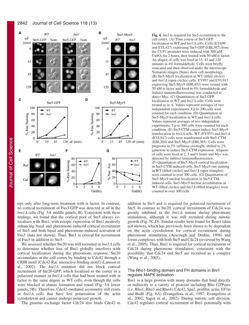
Bni1 is required for cortical recruitment of Ste5 duringpheromone stimulationWe determined whether bni1∆ mutants were defective inlocalizing Ste5 to cortical sites on the plasma membrane duringα factor stimulation in a time-course experiment. Corticalrecruitment of Ste5 was analyzed in live cells with a functionalSte5-GFP fusion expressed at the same level as wild-type Ste5using the CUP1 promoter (Mahanty et al., 1999). Therecruitment of Ste5-GFP was severely defective in bni1∆ cellsat all time points after α factor addition, including the 15minute time point that is well before shmoo formation (Fig.3A,C). In WT cells after 15 minutes of α factor treatment,Ste5-GFP could be detected at the plasma membrane (rimstaining) in ~12% of the population (Fig. 3C). These cells hadnot undergone shmoo formation and were still round. As thelength of α factor exposure increased, the number of cells thatexhibited Ste5 rim staining also increased, with more intense
staining at emergent shmoo tips. After 2 hours ofexposure to α factor, ~90% of the cells formed shmoos,and strong Ste5 rim staining was detected in 67% ofcells. In contrast, Ste5 recruitment was greatly reducedin bni1∆ cells at all time points. Similar results werefound by indirect immunofluorescence of a functionalSte5-Myc9 fusion in fixed cells (Fig. 3B,D). Ste5 wasrecruited to wild-type levels in a bnr1∆ strain lackingBnr1, the homolog of Bni1 (data not shown) and in shemutant strains (including she5, which is allelic to bni1)defective in polarized localization of specific mRNAs(Fig. S2 in supplementary material). Thus, Bni1 plays
a critical role in Ste5 recruitment that is not performed by Bnr1and is distinct from proteins involved in polarized mRNAlocalization.
Bni1 is still required for recruitment of Ste5 after theFus3 signaling defect is suppressedSte5 relocalizes from the nucleus to the plasma membrane inresponse to α factor stimulation of the associated MAPKcascade (Mahanty et al., 1999). Therefore, it was possible thatthe defect in Ste5 localization in the bni1∆ mutant was asecondary consequence of a defect in MAPK activation. To testthis possibility, we restored wild-type levels of Fus3 activationin a bni1∆ mutant by expressing Ste5-CTM from the GAL1promoter that is induced in the presence of galactose, andexamined whether Ste5-Myc9 could now be recruited to theplasma membrane. In WT cells, the expression of Ste5-CTMwas sufficient to induce translocation of Ste5-Myc9 from thenucleus to the emerging shmoo tip (Fig. 4F,G, percentagenuclear accumulation and percentage rim staining),demonstrating that activation of the MAPK cascade issufficient to induce Ste5 relocalization. (Note that it is easierto see the nuclear pool of Ste5 when cells are grown ingalactose containing medium than in glucose containingmedium; P. Maslo, R. McCully, E.A.E., unpublished data.)Ste5-CTM did not induce recruitment of Ste5-Myc9 in bni1∆cells; Ste5-Mcy9 remained predominantly nuclear despitenearly wild-type levels of Fus3 activation (Fig. 4E-G). Thus,Bni1 regulates recruitment of Ste5 by a mechanism that isindependent of MAPK activation. Furthermore, the retentionof Ste5 in nuclei of the bni1∆ cells indicates that Bni1 alsopromotes redistribution of Ste5 from the nucleus to thecytoplasm and cell cortex.
Bni1 is required for cortical recruitment of Fus3 andCdc24, but not Ste20In parallel experiments, we looked at the localization of Fus3,whose recruitment is dependent on Ste5. In WT cells, Fus3-GFP was nuclear and cytoplasmic and accumulated at shmoo
Fig. 3. bni1 functions at or upstream of Ste5 in Fus3activation. (A) Ste11-4-induced Fus3 activation of WT andbni1∆ cells. STE11-4 was expressed in a CEN plasmid(EBL284) and empty vector was used as control. (B) Ste5-CTM-induced Fus3 activation. Ste5-CTM (EBL204) wasexpressed from the GAL1 promoter. After shifting fromraffinose to galactose medium, cells were collected at 0, 1,2, 3 and 5 hours.
Jour
nal o
f Cel
l Sci
ence
2842
tips only after long-term treatment with α factor. In contrast,no cortical recruitment of Fus3-GFP was detected at all in thebni1∆ cells (Fig. 5A middle panels, B). Consistent with thesefindings, we found that the cortical pool of Ste5 always co-localizes with Bni1, with ectopic expression of Bni1 modestlyenhancing basal and pheromone-induced cortical recruitmentof Ste5 and both basal and pheromone-induced activation ofFus3 (data not shown). Thus, Bni1 is critical for recruitmentof Fus3 in addition to Ste5.
We assessed whether Ste20 was still recruited in bni1∆ cellsto determine whether loss of Bni1 globally interferes withcortical localization during the pheromone response. Ste20accumulates at the cell cortex by binding to Cdc42 through aCRIB motif (Cdc42-Rac interactive binding motif) (Lamson etal., 2002). The bni1∆ mutation did not block corticalrecruitment of Ste20-GFP, which localized to the cortex in apolarized manner in bni1∆ cells that had been treated with αfactor to the same degree as WT cells, even though the cellswere blocked in shmoo formation and round (Fig. 5A lowerpanels, 5B). Therefore, Cdc42-mediated asymmetry still existsin bni1∆ cells that have lost polarization of the actincytoskeleton and cannot undergo polarized growth.
The guanine exchange factor Cdc24 also binds Cdc42 in
addition to Ste5 and is required for polarized recruitment ofSte5. In contrast to Ste20, cortical recruitment of Cdc24 wasgreatly inhibited in the bni1∆ mutant during pheromonestimulation, although it was still recruited during mitoticgrowth (Fig. 5C,D). Similar results were found for Bem1 (datanot shown), which has previously been shown to be dependenton the actin cytoskeleton for cortical recruitment duringpheromone stimulation (Ayscough and Drubin, 1998) andforms complexes with both Ste5 and Cdc24 (reviewed by Wanget al., 2005). Thus, Bni1 is required for cortical recruitment ofCdc24 during pheromone stimulation, consistent with thepossibility that Cdc24 and Ste5 are recruited as a complex(Wang et al., 2005).
The Rho1-binding domain and FH domains in Bni1regulate MAPK activationBni1 is a large protein with many domains that bind directlyor indirectly to a variety of proteins including Rho GTPases(i.e. Rho1, Rho3 and Rho4), Cdc42, Spa2, profilin, actin, EF1αand Bud6 (Fig. 6A) (Evangelista et al., 1997; Evangelista etal., 2002; Sagot et al., 2002). During mitotic cell division,Cdc42 regulates cortical recruitment of Bni1 potentially with
Journal of Cell Science 118 (13)
Fig. 4. bni1 is required for Ste5 recruitment to thecell cortex. (A) Time course of Ste5-GFPlocalization in WT and bni1∆ cells. Cells (EY699and EYL427) expressing Ste5-GFP (EBL367) fromthe CUP1 promoter were induced with 500 µMCuSO4 for 2 hours, then treated with 50 nM α factor.An aliquot of cells was fixed at 15, 45 and 120minutes in 4% formaldehyde. Cells were brieflysonicated and then observed under the microscope.Nomarski images (Nom) show cell morphology. (B) Ste5-Myc9 localization in WT (filled circles)and bni1∆ (open circles) cells. EY957 and EYL917expressing Ste5-Myc9 (EBL453) were treated with50 nM α factor and fixed in 4% formaldehyde andindirect immunofluorescence was conducted todetect Myc. (C) Quantitation of Ste5-GFPlocalization in WT and bni1∆ cells. Cells weretreated as in A. Values represent averages of twoindependent experiments. Up to 200 cells werecounted for each condition. (D) Quantitation ofSte5-Myc9 localization in WT and bni1∆ cells.Values represent averages of two independentexperiments. Up to 300 cells were counted for eachcondition. (E) Ste5-CTM cannot induce Ste5-Myc9translocation in bni1∆ cells. WT (EY957) and bni1∆(EYL917) cells were transformed with Ste5-CTM(EBL204) and Ste5-Myc9 (EBL365). Cells werepregrown in 2% raffinose overnight, shifted to 2%galactose to induce Ste5-CTM expression. Aliquotsof cells were fixed at 2, 3 and 6 hours and Myc wasdetected by indirect immunofluorescence. (F) Quantitation of Ste5-Myc9 cortical localizationin Ste5-CTM-induced cells. Ste5-Myc9 rim stainingin WT (filled circles) and bni1∆ (open triangles)were counted in over 300 cells. (G) Quantitation ofSte5-Myc9 nuclear localization in Ste5-CTM-induced cells. Ste5-Myc9 nuclear accumulation inWT (filled circles) and bni1∆ (filled triangles) werecounted in over 300 cells.
Jour
nal o
f Cel
l Sci
ence

2843Formin-regulated actin cytoskeleton mediates scaffold recruitment
multiple Rho proteins (Jacquenoud and Peter, 2001; Dong etal., 2003). The control by Cdc42 is indirect compared to thatof Rho1, which is sufficient to stimulate plasma membranerecruitment of Bni1 (Jacquenoud and Peter, 2000). Bni1promotes actin cable formation through the FH1 and FH2domains and is thought to exist in an autoinhibited form viainteractions between the Rho-binding domain (RBD) and acarboxyl-terminal domain (DAD), by analogy to Diaphanous-related formin (Zigmond, 2004). The binding of Rho GTPaseis thought to relieve the autoinhibitory interaction and permitthe FH2 domain to nucleate actin polymerization (Evangelistaet al., 2002; Sagot et al., 2002).
We deleted these key functional domains of Bni1 andassessed their role in Fus3 activation (Fig. 2C) and matingoutputs (data not shown). As summarized in Table 1, deletionof the DAD that binds the RBD and Bud6 did not block matingresponses, whereas removing the RBD did. Loss of the RBDalso reduced Fus3 activation to the same degree as a bni1 nullstrain (Fig. 2C) and blocked shmoo formation (percentageshmoos after 2 hours in 50 nM α factor was 80% for WT,0.01% for bni1∆, 0.03% for bni1∆RBD; n=300 cells; see alsoFig. 6B). Furthermore, deletions that removed the FH1 andFH2 domains also inhibited Fus3 activation (Fig. 2C) andmating outputs. These findings suggest that Bni1 regulation ofSte5 recruitment requires a Rho GTPase and the formation ofactin cables.
To test this hypothesis, we determined whether the RBD wasrequired for cortical localization of Bni1. The RBD (aminoacid residues 90-343) has been defined as a region that bindsRho1-GTP directly in vitro and in two-hybrid assays (Kohnoet al., 1996). This region overlaps the region of Bni1 withhomology to the Diaphanous GTPase binding domain (amino
acids 93-286) (Marchler-Bauer et al., 2003). Bni1 lacking theRBD (bni1∆RBD-GFP) did not localize to bud tip and bud neckduring vegetative growth and was not recruited to the cellcortex during response to mating pheromone, even in cells thatexpressed wild-type Bni1 (Fig. 6B). Consistent with theabsence of cortical recruitment, bni1∆RBD-GFP failed tosupport shmoo formation in bni∆ cells (Fig. 6B), or mediatecortical recruitment of Ste5 (data not shown).
The requirement for the RBD for Bni1 localization andpheromone responses suggested that Bni1 is activated duringmating by a Rho GTPase. Five Rho GTPases (Rho1-Rho5) areimplicated in the regulation of Bni1, with varying roles duringthe cell cycle and under different environmental conditions(Dong et al., 2003). Although early work with a temperaturesensitive rho1-2 mutant suggested that Rho1 is not involved inlocalization of Bni1 (Ozaki-Kuroda et al., 2001), other workhas shown that Rho1 regulates the actin cytoskeleton duringG1 phase and shmoo formation (Kohno et al., 1996; Drgonovaet al., 1999), and binds to multiple effector proteins includingBni1, glucan synthase and protein kinase C (Evangelista et al.,1997; Saka et al., 2001). We compared Bni1 and Ste5localization in rho1-2 (E45V in Switch I) (Saka et al., 2001)and rho1-104 (D72N; C164Y) (Yamochi et al., 1994)temperature sensitive mutants that have different actincytoskeleton phenotypes and abilities to polarize myosin.Bni1-GFP was not recruited in a polarized manner to the cortexof dividing and pheromone-induced rho1-2 and rho1-104 cellsat nonpermissive temperature (Fig. 6C). To determine whetherRho1 was also required for Ste5 localization we usedTAgNLSK128T-Ste5-Myc9, which is more efficientlyreimported to the nucleus and recruited to the plasmamembrane than Ste5-Myc9, allowing its detection in a greater
Fig. 5. bni1 is required for Fus3, but not Ste20recruitment. (A) Recruitment of Fus3 andSte20 in bni1∆ strain. Upper panels: actinstaining. WT (EY957) and bni1∆ (EYL917)cells were treated with 50 nM α factor for 2hours, fixed, and then stained with Rhodamine-phalloidin. Middle panels: Fus3-GFP. Lowerpanels: Ste20-GFP. Strains expressing genomicFus3-GFP (EYL1096 and QMY27) or strains(EY957 and EYL917) transformed with GFP-Ste20 (EBL511) were treated with 50 nM αfactor for 2 hours, fixed with 4%formaldehyde, sonicated and then mounted formicroscopy. (B) Quantitation of Fus3-GFP andSte20-GFP localization in WT and bni1∆ cells.Values represent mean ± s.d. of twotransformants. (C,D) Bni1 is recruited forCdc24 recruitment during mating. WT(EY957) and bni1∆ (EYL917) strains weretransformed with CDC24-GFP (EBL664).Cells were pregrown overnight in mediumcontaining 0.19 mM methionine, then treatedwith 50 nM α factor for 2 hours. Note thatCdc24 localizes at bud tips (arrowheads) andshmoo tips (arrows). Over 200 cells werecounted. Values in D represent mean ± s.d. oftwo transformants.
Jour
nal o
f Cel
l Sci
ence

2844
number of cells after brief treatment with α factor (Mahanty etal., 1999). Strikingly, the rho1-2 and rho1-104 mutations bothdecreased Ste5 recruitment at nonpermissive temperature (Fig.6D). By contrast, Bni1 and Ste5 were still recruited to the siteof polarized growth in rho2, rho3, rho4 and rho5 null mutantsthat had been treated with α factor, and these cells were ableto form shmoos (Fig. S3 in supplementary material).Therefore, Rho1 specifically recruits Bni1 to cortical sitesduring mating and positively regulates recruitment of Ste5through control of Bni1.
Bni1-induced actin cables are required for Ste5recruitmentTo more stringently test for a primary role of Bni1-inducedcables in Ste5 recruitment, we used a bni1TS temperature-sensitive mutation within conserved residues of the FH2domain (R1528A and R1530A) that blocks actin cableformation (Sagot et al., 2002). Previous work has demonstratedthat inactivation of bni1TS after a brief 10-minute shift to 34°Cselectively disrupts actin cables while polarized actin patchesare maintained, although the effect is not complete. Because ofpartial functional overlap between Bni1 and Bnr1, the bni1TS
mutation was assessed in a bnr1∆ mutant as described(Evangelista et al., 2002; Sagot et al., 2002). At thenonrestrictive temperature, TAgNLSK128T-Ste5-Myc9 was
recruited to the plasma membrane in ~30% of WT and bni1TS
bnr1∆ mutant cells after α factor stimulation (Fig. 7A).Shifting the temperature to 34°C greatly inhibited therecruitment of TAgNLSK128T-Ste5-Myc9 in the bni1TSbnr1∆mutant, but did not reduce it in the WT control strain (Fig. 7A),consistent with a primary role for actin cables in Ste5recruitment.
To further confirm that actin cables regulate Ste5recruitment, we checked the effect of disrupting the TPM1 andTPM2 genes encoding tropomyosins, which bind to andstabilize actin cables and are not a component of actin patches(Pruyne et al., 1998). To assess the immediate importance of
Journal of Cell Science 118 (13)
Table 1. Phenotypes of bni1 mutantsGenotype Pheromone response
WT ++++bni1∆ +bni1∆RBD +bni1∆FH1/FH2 +bni1∆FH1 ++bni1∆FH2 ++bni1∆CT ++++
WT, wild type. bni1∆ cells were transformed with empty vector, or vectorexpressing different forms of the BNI1 gene. Sensitivity to α factor wasmonitored by Fus3 kinase assay and halo assay.
Fig. 6. Rho1 is involved in Bni1-mediated Ste5 recruitment. (A) Schematic structure of Bni1. RBD,Rho-binding domain; SBD, Spa2-binding domain; BBD, Bud6-bindingdomain. (B) Rho-binding domain isrequired for Bni1 polarization. WTcells were transformed withbni1∆RBD-GFP (QMB79) or BNI1-HA (EBL332); bni1∆ cells weretransformed with BNI1-GFP(EBL334) or bni1∆RBD-GFP(QMB79). Cells were treated with50 nM α factor for 2 hours, then directfluorescence of GFP was observed.Nomarski images (Nom) show cellmorphology. (C) Bni1 polarization isdefective in rho1-TS mutants. WT(QMY550), rho1-2 (QMY553) andrho1-104 (QMY551) cells expressingBni1-GFP were grown at roomtemperature. Cells were shifted to37°C for 1 hour, then induced with5 µM α factor at 37°C or 2 hours. (D) Ste5 recruitment is defective inrho1-TS mutants. WT, rho1-2 andrho1-104 cells expressingTAgNLSK128TSte5-Myc9 (EBL444)were treated as in C, except the αfactor stimulation time was 30minutes. Cells were fixed and Mycwas detected by indirectimmunofluorescence microscopy.Values represent mean ± s.d. of twotransformants.
Jour
nal o
f Cel
l Sci
ence

2845Formin-regulated actin cytoskeleton mediates scaffold recruitment
tropomyosin in Ste5 recruitment, we used the same rapidtemperature shift conditions used for the bni1TS mutant.Although TAgNLSK128T-Ste5-Myc9 was recruited to the cortexin tpm1TS tpm2∆ cells at nonrestrictive temperature, shifting tononpermissive temperature blocked its recruitment within afew minutes (Fig. 7B). Thus, actin cables generated by Bni1and stabilized by tropomyosin are required for recruitment ofSte5 to polarized sites at the plasma membrane.
Myo2 is required for recruitment of Ste5All of the known transport events along actin cables in S.cerevisiae require either of the type V myosin motor proteins,Myo2 and Myo4. Myo2 has been implicated in the transportof proteins (i.e. Kar9, Smy1) and organelles (i.e. secretoryvesicles and vacuoles) along cytoplasmic actin cables, whereasMyo4 is involved in transport of mRNAs to sites of polarizedgrowth (Bretscher, 2003; Vale, 2003). We used two approachesto test whether Myo2 is involved in Ste5 recruitment, a myo2-66 temperature-sensitive point mutation that maps to the actin-binding region of the motor domain of Myo2 (Lillie andBrown, 1994) and a dominant negative tail fragment of Myo2,MYO2DN that displaces Myo2 from actin cables (Reck-Peterson et al., 1999). Shifting the myo2-66 strain to 37°C for2 hours blocked recruitment of Ste5 (Fig. 7C). Since the myo2-66 mutation also induces actin depolarization in addition toinactivating Myo2, we examined the effect of MYO2DN underconditions of short-term overexpression from the GAL1promoter, which does not disrupt the actin cytoskeleton (Reck-
Peterson et al., 1999; Karpova et al., 2000). Shifting cells fromnoninducing raffinose medium to inducing galactose mediumfor 1 hour resulted in a fourfold inhibition of recruitment ofTAgNLSK128T-Ste5-Myc9 in the presence of α factor (Fig. 7D).Thus, Ste5 recruitment was blocked to nearly as great an extentby MYO2DN as it was by myo2-66. A comparative analysis ofSte5 recruitment in strains harboring mutations in theremaining myosin genes, MYO1, MYO3, MYO4 and MYO5 [ofwhich MYO3 and MYO5 are functional homologs (Bretscher,2003; Vale, 2003)] was also done. Deletion of MYO4, in eithera WT or myo2-66 background did not interfere with Ste5recruitment (Fig. S4A in supplementary material).Furthermore, null mutations in MYO1, MYO3, MYO5 anddouble null mutations in MYO3 MYO5 had no significant effecton Ste5 recruitment (Fig. S4B,C in supplementary material).Thus, Myo2 is specifically required for cortical recruitment ofSte5.
The actin cables and Myo2 could regulate Ste5 recruitmentindirectly by creating a polarized site of factors that captureand stabilize Ste5. Alternatively, it is possible that Myo2played a more direct role through translocation of Ste5 alongactin cables. To test the possibility that Myo2 might regulatetranslocation of Ste5 we determined whether Ste5 and Myo2form complexes in vivo in a co-immunoprecipitation assay.Ste5-Myc9 and Myo2-HA were co-expressed in a ste5∆ strainat physiological levels using low copy centromeric plasmidsand STE5 and MYO2 promoters to drive the expression ofSTE5-MYC9 and MYO2-HA genes, respectively. Strikingly,Myo2-HA associated with Ste5-Myc9 in cells that had been
Fig. 7. bni1 regulates Ste5 relocalization by controlling actincable formation. (A) The bni1TS mutant cannot recruit Ste5.WT (EYL1765) and bni1TS bnr1∆ (EYL1748) cells expressingTAgNLSK128TSte5-Myc9 were grown at room temperature.Cells were centrifuged, resuspended in pre-warmed medium(34°C) containing 5 µM α factor, and then incubated at 34°Cfor 10 minutes. (B) The tpm1TS mutant cannot recruit Ste5.TAgNLSK128TSte5-Myc9 was expressed in tmp2∆ (EYL1736)and tpm1TS/tmp2∆ (EYL1735) mutant cells. Cells were thentreated as in A, except at a nonpermissive temperature of34.5°C. Values in A and B represent mean of two independentexperiments. (C) Ste5 recruitment is inhibited in myo2-66 cellsat nonpermissive temperature. MYO2 (QMY458) or myo2-66(QMY459) cells expressing TAgNLSK128TSte5-Myc9(EBL444) were grown at room temperature, then incubated at37°C for 2 hours, treated with 5 µM α factor for 30 minutes,then fixed for indirect immunofluorescence. (D) Overexpression of Myo2 tail domain suppresses Ste5recruitment. Cells harboring MYO2DN (QMB75) andTAgNLSK128TSte5-Myc9 (EBL444) were pre-grown in mediumcontaining 2% raffinose overnight. The cells were shifted tomedium containing 2% galactose for 1 hour, treated with 5 µMα factor for 15 minutes, then fixed and Myc was detected byindirect immunofluorescence microscopy. (E) Ste5 co-immunoprecipitates with Myo2 in α factor-treated cells.EY1775 (ste5∆) expressing Ste5-Myc9 (from EBL358) and/orMyo2-HA (from QMB80), with or without control vectors,were grown to early logarithmic phase. Where indicated (+),cells were treated with 50 nM α factor for 45 minutes prior tocollecting cells for extract preparation. 12CA5 and 9E10monoclonal antibodies were used to detect Myo2-HA and Ste5-Myc9, respectively. Note the nonspecific binding of Ste5 toSepharose beads.
Jour
nal o
f Cel
l Sci
ence

2846
treated with α factor but not in vegetatively growing cells (Fig.7E), consistent with the dependence of polarized recruitmentof Ste5 on α factor stimulation. The ability to co-immunoprecipitate Ste5 and Myo2 was reproducible anddependent on expressing Myo2 at native levels, with nointeraction detected when Myo2 was overexpressed (data notshown). Collectively, these findings open the possibility thatSte5 translocates along actin cables through the action ofMyo2.
Ste5 does not need the Bni1 FH2 domain or actin tostay at the plasma membraneWe determined whether Bni1 or actin cables were required tomaintain Ste5 at the cell cortex once it has been recruited toGβ Ste4. To test this possibility, the effect of the bni1TS
mutation and latrunculin A treatment on a pool of Ste5 that hadbeen recruited to the cell cortex was assessed. bni1TS bnr1∆
mutant cells expressing TAgNLSK128T-Ste5-Myc9 were treatedwith α factor for 30 minutes at room temperature, then shiftedto 34°C for 10 minutes and 20 minutes. The treatment with αfactor induced Ste5 recruitment in 50% of the cells (Fig. 8A).The subsequent shift to 34°C blocked a further increase inrecruitment of Ste5 to the polarization sites, but did notdecrease the pool of Ste5 that had accumulated prior to thetemperature shift (Fig. 8A). Therefore, once Ste5 is recruitedto the plasma membrane, it no longer needs the FH2 domainof Bni1. The addition of latrunculin A prior to α factorinduction completely blocks the formation of actin patches andcables and α factor-induced actin polarization as well asrecruitment of Ste5 to the plasma membrane (Wang et al.,2005) (Fig. 8B). In sharp contrast, addition of latrunculin A toα factor-induced cells did not abolish the previouslyestablished Ste5-Myc9 recruitment (Fig. 8C), although it diddisrupt the actin cytoskeleton, further demonstrating that theactin cytoskeleton is not required to maintain Ste5 at theplasma membrane. Therefore, Bni1-induced actin cables arenot required to maintain Ste5 at the plasma membrane.
DiscussionWhile it is known that regulators of the actin cytoskeleton arerequired for signal transduction during MAPK signaling,specific functions of actin in signaling have not beendelineated. Here we present the first evidence of a specific rolefor a formin and Rho protein in assembly of a scaffold-kinasesignal transduction cascade at the plasma membrane inresponse to an external stimulus. This conclusion is based onthe selective requirement for Bni1 in high-level activation ofMAPK Fus3, but not MAPK Kss1 (Fig. 2), together with thecritical role for Bni1 in cortical recruitment of Ste5 and Fus3(Figs 4 and 5). Previous work suggests that Fus3 must beactivated while bound to Ste5 (Andersson et al., 2004) but Kss1can be activated from the scaffold by upstream kinases(Andersson et al., 2004; Maleri et al., 2004). In addition,plasma-membrane-bound Ste5-CTM does not recruit Kss1 tothe plasma membrane although it does recruit Fus3 (vanDrogen et al., 2001). Thus, Bni1 and cortical localization ofSte5 only play a significant role in the MAPK that co-localizeswith Ste5. Although Bni1 and actin cables are required forpolarized recruitment of Ste5 (Figs 4 and 7), they are notrequired for maintenance of Ste5 at the plasma membrane (Fig.8). This finding is consistent with the expectation that oncerecruited, Ste5 is able to bind to free Gβ (Ste4) at the plasmamembrane.
Our findings also reveal that Rho1 plays a specific role inmediating the recruitment of Bni1 and Ste5, acting through theRho Binding Domain of Bni1 (Fig. 6). This conclusion isconsistent with the ability of Rho1-GTP to bind to a Bni1fragment that overlaps the Rho binding domain (Kohno et al.,1996) and the lack of evidence of functional overlap from theother Rho proteins (Rho2, Rho3, Rho4, Rho5, Fig. S3 insupplementary material), which may regulate Bni1 during thebudding cycle (Dong et al., 2003). During the mitotic cycle,Rho1 also regulates the actin cytoskeleton indirectly throughthe protein kinase C pathway (Dong et al., 2003), raising thepossibility of an indirect link between Rho1 and Bni1 duringmating pheromone stimulation. However, the protein kinase Cpathway is activated by α factor much later than the mating
Journal of Cell Science 118 (13)
Fig. 8. Actin is not required to keep Ste5 at the plasma membrane.(A) Dynamics of Ste5 localization in bni1TS mutants. WT(EYL1765) and bni1TSbnr1∆ cells (EYL1748) expressingTAgNLSK128TSte5-Myc9 were treated with 5 µM α factor at roomtemperature for 30 minutes, and then shifted to 34°C for 10 minutesand 20 minutes. Values are means of two independent transformants.Two separate experiments gave comparable results. (B) Actinfilaments are required to recruit Ste5. WT (EY957) cells harboringTAgNLSK128TSte5-Myc9 were treated with 100 µM latrunculin A (LatA) for 15 minutes, then treated with 50 nM α factor for 15 minutes.(C) Ste5 dynamics in Lat A-treated cell. Cells as used in B weretreated with 50 nM α factor for 30 minutes, then Lat A was added tothe medium at a final concentration of 100 µM. The cells werefurther incubated for 15 minutes before fixed forimmunofluorescence. Values are mean ± s.d. from three independentexperiments.
Jour
nal o
f Cel
l Sci
ence
2847Formin-regulated actin cytoskeleton mediates scaffold recruitment
MAPK cascade, as a result of polarized growth (Buehrer andErrede, 1997; Zarzov et al., 1996), arguing against thispossibility. Interestingly, it was recently reported that Rho1associates with Ste4 in co-immunoprecipitates (Bar et al.,2003), raising the possibility that Gβγ recruits Rho1 to the siteof polarized growth during mating. Taken together with ourfindings, we speculate that Gβγ directs polarized growththrough dual recruitment of a Rho1-GTP-Bni1 complex, whichfacilitates polarized growth directly through actin cableformation, as well as through recruitment of Ste5.
A major question that still remains is how Ste5 is recruitedin a polarized manner. One possibility is that Ste5 is exportedfrom the nucleus either alone or in a complex with Cdc24 andsimply diffuses to the cortex where it is captured by cellpolarity factors such as Cdc42 and Bem1 whose localization isalso dependent on Bni1. The position of these factors could bedetermined by the asymmetry that is established from thereceptor and activated G protein and polarized actin thatassembles in response to the external pheromone stimulus.Such asymmetry could be built through an interaction betweenSte4 and Rho1, which binds to Bni1. Potential direct or indirectinteractions between Bni1 and Cdc42 (Evangelista et al., 1997;Jaquenoud and Peter, 2000) might serve to link the Bni1complex to Ste5-Cdc24. This possibility is consistent with ourability to co-immunoprecipitate Bni1 with Bem1 (M.Q. andE.A.E., data not shown).
Another interesting possibility that is consistent with ourfindings is that a pool of Ste5, either alone or with Cdc24,translocates along actin cables once it is exported from thenucleus. This possibility is consistent with the requirement forthe cargo domain of Myo2 in Ste5 recruitment and the abilityto co-immunoprecipitate Ste5 with Myo2. While it is possiblethat the interaction between Myo2 and Ste5 is indirect, it isnoteworthy that we can detect it, in light of the fact that we areunable to co-immunoprecipitate Bni1 with Ste5. Additionalsupport for the physiological relevance of this interactioncomes from the observation that the interaction is not detectedwhen Myo2 is overexpressed (data not shown) and that it isdependent upon pheromone signaling. Prior work hasimplicated Myo2 in the translocation of secretory vesicles,vacuoles and several cytoskeleton-associated proteins (i.e.Kar9 and Smy1) (Vale, 2003) but not the translocation of acytoplasmic protein. Additionally, the possibility that Ste5translocates along actin cables with Myo2 is still consistentwith a model in which Ste5 is captured at the cell cortex bycell polarity proteins. While further work is needed to knowwhether Ste5 is translocating along actin cables, thisinterpretation is attractive in that it provides a physical linkbetween nuclear export and events at a specific site at the cellcortex. Interestingly, concentration of JIP scaffolds at nerveterminals requires kinesin, suggesting they translocate alongmicrotubules via a direct interaction with kinesin (Verhey etal., 2001), although the reason for this phenomenon has notbeen established.
Finally, previous work has shown that Fus3 is required forcortical recruitment of Bni1 (Matheos et al., 2004). Ourunpublished findings are consistent with Fus3 being requiredfor cortical recruitment of Ste5 in G1 phase of the cell cycle(M.Q. and E.A.E., unpublished). This finding raises thepossibility that the formation of a stable signaling complex isa downstream event that occurs after initial pathway activation
by mechanisms that are dependent on the Bni1-induced actincytoskeleton. Further work is needed to clarify the relativeposition of MAPK activation with formation of a stable Ste5scaffold signaling complex and whether polarized recruitmentof Ste5 plays a specific role in events that take place at theshmoo tip, including polarized growth.
We are extremely grateful to: C. Moon, B. N. Lee and S. Mahanty,who made the initial observations that bni1∆ mutant is defective inG1 arrest, Fus3 activation and Ste5 recruitment, respectively. Wethank C. Boone, D. Pellman, A. Bretscher, R. Li, L. Weisman and A.Levchenko for yeast strains and plasmids used in this study. We thankD. Pellman and D. Lew for comments on the manuscript. Thisresearch was supported by National Institutes of Health grantGM46962, a Taplin Funds for Discovery Award, and American HeartGrant 0150175N to E.A.E.
ReferencesArkowitz, R. A. (1999). Responding to attraction: chemotaxis and
chemotropism in Dictyostelium and yeast. Trends Cell Biol. 9, 20-27.Andersson, J., Simpson, D. M., Qi, M., Wang, Y. and Elion, E. A. (2004).
Differential input by Ste5 scaffold and Msg5 phosphatase route a MAPKcascade to multiple outcomes. EMBO J. 23, 2564-2576.
Ayscough, K. R. and Drubin, D. G. (1998). A role for the yeast actincytoskeleton in pheromone receptor clustering and signalling. Curr. Biol. 8,927-930.
Bar, E. E., Ellicott, A. T. and Stone, D. E. (2003). Gbetagamma recruits Rho1to the site of polarized growth during mating in budding yeast. J. Biol. Chem.278, 21798-21804.
Bardwell, L. (2004). A walk-through of the yeast mating pheromone responsepathway. Peptides 25, 1465-1476.
Bokoch, G. M. (2003). Biology of the p21-activated kinases. Annu. Rev.Biochem. 72, 743-781.
Bretscher, A. (2003). Polarized growth and organelle segregation in yeast: thetracks, motors, and receptors. J. Cell Biol. 160, 811-816.
Buehrer, B. M. and Errede, B. (1997). Coordination of the mating and cellintegrity mitogen-activated protein kinase pathways in Saccharomycescerevisiae. Mol. Cell. Biol. 17, 6517-6525.
Burack, W. R. and Shaw, A. S. (2000). Signal transduction: hanging on ascaffold. Curr. Opin. Cell Biol. 12, 211-216.
Dohlman, H. G. and Thorner, J. W. (2001). Regulation of G protein-initiatedsignal transduction in yeast: paradigms and principles. Annu. Rev. Biochem.70, 703-754.
Dong, Y., Pruyne, D. and Bretscher, A. (2003). Formin-dependent actinassembly is regulated by distinct modes of Rho signaling in yeast. J. CellBiol. 161, 1081-1092.
Drgonova, J., Drgon, T., Roh, D. H. and Cabib, E. (1999). The GTP-bindingprotein Rho1p is required for cell cycle progression and polarization of theyeast cell. J. Cell Biol. 146, 373-387.
Dustin, M. L. and Cooper, J. A. (2000). The immunological synapse and theactin cytoskeleton: molecular hardware for T cell signaling. Nat. Immunol.1, 23-29.
Elion, E. A. (1995). Ste5: a meeting place for MAP kinases and theirassociates. Trends Cell Biol. 5, 322-327.
Elion, E. A. (2001). The Ste5p scaffold. J. Cell Sci. 114, 3967-3978.Elion, E. A., Grisafi, P. L. and Fink, G. R. (1990). FUS3 encodes a
cdc2+/CDC28-related kinase required for the transition from mitosis intoconjugation. Cell 60, 649-664.
Elion, E. A., Brill, J. A. and Fink, G. R. (1991). FUS3 represses CLN1 andCLN2 and in concert with KSS1 promotes signal transduction. Proc. Natl.Acad. Sci. USA 88, 9392-9396.
Elion, E. A., Satterberg, B. and Kranz, J. E. (1993). FUS3 phosphorylatesmultiple components of the mating signal transduction cascade: evidencefor STE12 and FAR1. Mol. Biol. Cell 4, 495-510.
Erickson, J. W. and Cerione, R. A. (2001). Multiple roles for Cdc42 in cellregulation. Curr. Opin. Cell Biol. 13, 153-157.
Etienne-Manneville, S. and Hall, A. (2003). Cell polarity: Par6, a PKC andcytoskeletal crosstalk. Curr. Opin. Cell Biol. 15, 67-72.
Evangelista, M., Blundell, K., Longtine, M. S., Chow, C. J., Adames, N.,Pringle, J. R., Peter, M. and Boone, C. (1997). Bni1p, a yeast formin
Jour
nal o
f Cel
l Sci
ence
2848
linking cdc42p and the actin cytoskeleton during polarized morphogenesis.Science 276, 118-122.
Evangelista, M., Pruyne, D., Amberg, D. C., Boone, C. and Bretscher, A.(2002). Formins direct Arp2/3-independent actin filament assembly topolarize cell growth in yeast. Nat. Cell Biol. 4, 32-41.
Farley, F., Satterberg, B., Goldsmith, E. A. and Elion, E. A. (1999). Relativedependance of different outputs of the Saccharomyces cerevisiae pheromoneresponse pathway on the MAP kinase Fus3. Genetics 151, 1425-1444.
Garrington, T. P. and Johnson, G. L. (1999). Organization and regulation ofmitogen-activated protein kinase signaling pathways. Curr. Opin. Cell Biol.11, 211-218.
Ge, L., Ly, Y., Hollenberg, M. and DeFea, K. (2003). A beta-arrestin-dependent scaffold is associated with prolonged MAPK activation inpseudopodia during protease-activated receptor-2-induced chemotaxis. J.Biol. Chem. 278, 34418-34426.
Gulli, M. P. and Peter, M. (2001). Temporal and spatial regulation of Rho-type guanine-nucleotide exchange factors: the yeast perspective. Genes Dev.15, 365-379.
Henrique, D. and Schweisguth, F. (2003). Cell polarity: the ups and downsof the Par6/aPKC complex. Curr. Opin. Genet. Dev. 13, 341-350.
Jaquenoud, M. and Peter, M. (2000). Gic2p may link activated Cdc42p tocomponents involved in actin polarization, including Bni1p and Bud6p(Aip3p). Mol. Cell. Biol. 20, 6244-6258.
Karpova, T. S., Reck-Peterson, S. L., Elkind, N. B., Mooseker, M. S.,Novick, P. J. and Cooper, J. A. (2000). Role of actin and Myo2p inpolarized secretion and growth of Saccharomyces cerevisiae. Mol. Biol. Cell11, 1727-1737.
Kelkar, N., Gupta, S., Dickens, M. and Davis, R. J. (2000). Interaction of amitogen-activated protein kinase signaling module with the neuronal proteinJIP3. Mol. Cell. Biol. 20, 1030-1043.
Kohno, H., Tanaka, K., Mino, A., Umikawa, M., Imamura, H., Fujiwara,T., Fujita, Y., Hotta, K., Qadota, H. and Watanabe, T. et al. (1996). Bni1pimplicated in cytoskeletal control is a putative target of Rho1p small GTPbinding protein in Saccharomyces cerevisiae. EMBO J. 15, 6060-6068.
Lamson, R. E., Winters, M. J. and Pryciak, P. M. (2002). Cdc42 regulationof kinase activity and signaling by the yeast p21-activated kinase Ste20. Mol.Cell. Biol. 22, 2939-2951.
Leof, E. B. (2000). Growth factor receptor signalling: location, location,location. Trends Cell Biol. 10, 343-348.
Lillie, S. H. and Brown, S. S. (1994). Immunofluorescence localization of theunconventional myosin, Myo2p, and the putative kinesin-related protein,Smy1p, to the same regions of polarized growth in Saccharomycescerevisiae. J. Cell Biol. 125, 825-842.
Lyons, D. M., Mahanty, S. K., Choi, K. Y., Manandhar, M. and Elion, E.A. (1996). The SH3-domain protein Bem1 coordinates mitogen-activatedprotein kinase cascade activation with cell cycle control in Saccharomycescerevisiae. Mol. Cell. Biol. 16, 4095-4106.
Mahanty, S. K., Wang, Y., Farley, F. W. and Elion, E. A. (1999). Nuclearshuttling of yeast scaffold Ste5 is required for its recruitment to theplasma membrane and activation of the mating MAPK cascade. Cell 98,501-512.
Maleri, S., Ge, Q., Hackett, E. A., Wang, Y., Dohlman, H. G. and Errede,B. (2004). Persistent activation by constitutive Ste7 promotes Kss1-mediated invasive growth but fails to support Fus3-dependent mating inyeast. Mol. Cell. Biol. 24, 9221-9238.
Marchler-Bauer, A., Anderson, J. B., DeWeese-Scott, C., Fedorova, N. D.,Geer, L. Y., He, S., Hurwitz, D. I., Jackson, J. D., Jacobs, A. R. andLanczycki, C. J. et al. (2003). CDD: a curated Entrez database of conserveddomain alignments. Nucleic Acids Res. 31, 383-387.
Matheos, D., M., Metodiev, M., Muller, E., Stone, D. and Rose, M. D.(2004). Pheromone-induced polarization is dependent on the Fus3p MAPKacting through the formin Bni1p. J. Cell Biol. 165, 99-109.
Morrison, D. K. and Davis, R. J. (2003). Regulation of MAP kinase signaling
modules by scaffold proteins in mammals. Annu. Rev. Cell Dev. Biol. 19,91-118.
Muller, J., Ory, S., Copeland, T., Piwnica-Worms, H. and Morrison, D. K.(2001). C-TAK1 regulates Ras signaling by phosphorylating the MAPKscaffold, KSR1. Mol. Cell 8, 983-993.
Ozaki-Kuroda, K., Yamamoto, Y., Nohara, H., Kinoshita, M., Fujiwara,T., Irie, K. and Takai, Y. (2001). Dynamic localization and function ofBni1p at the sites of directed growth in Saccharomyces cerevisiae. Mol. Cell.Biol. 21, 827-839.
Palmieri, S. J. and Haarer, B. K. (1998). Polarity and division sitespecification in yeast. Curr. Opin. Microbiol. 1, 678-686.
Pruyne, D. W., Schott, D. H. and Bretscher, A. (1998). Tropomyosin-containing actin cables direct the Myo2p-dependent polarized delivery ofsecretory vesicles in budding yeast. J. Cell Biol. 143, 1931-1945.
Pryciak, P. M. and Huntress, F. A. (1998). Membrane recruitment of thekinase cascade scaffold protein Ste5 by the Gbetagamma complex underliesactivation of the yeast pheromone response pathway. Genes Dev. 12, 2684-2697.
Reck-Peterson, S. L., Novick, P. J. and Mooseker, M. S. (1999). The tail ofa yeast class V Myosin, Myo2p, functions as a localization domain. Mol.Biol. Cell 10, 1001-1017.
Sagot, I., Klee, S. K. and Pellman, D. (2002). Yeast formins regulate cellpolarity by controlling the assembly of actin cables. Nat. Cell Biol. 4, 42-50.
Saka, A., Abe, M., Okano, H., Minemura, M., Qadota, H., Utsugi, T.,Mino, A., Tanaka, K., Takai, Y. and Ohya, Y. (2001). Complementingyeast rho1 mutation groups with distinct functional defects. J. Biol. Chem.276, 46165-46171.
Siekhaus, D. E. and Drubin, D. G. (2003). Spontaneous receptor-independentheterotrimeric G-protein signalling in an RGS mutant. Nat. Cell Biol. 5, 231-235.
Sprague, G. F., Jr (1991). Assay of yeast mating reaction. Methods Enzymol.194, 77-93.
Stevenson, B. J., Rhodes, N., Errede, B. and Sprague, G. F., Jr (1992).Constitutive mutants of the protein kinase STE11 activate the yeastpheromone response pathway in the absence of the G protein. Genes Dev.6, 1293-1304.
Takenawa, T. and Miki, H. (2001). WASP and WAVE family proteins: keymolecules for rapid rearrangement of cortical actin filaments and cellmovement. J. Cell Sci. 114, 1801-1809.
Vale, R. D. (2003). The molecular motor toolbox for intracellular transport.Cell 112, 467-480.
van Drogen, F., Stucke, V. M., Jorritsma, G. and Peter, M. (2001). MAPkinase dynamics in response to pheromones in budding yeast. Nat. Cell Biol.3, 1051-1059.
Verhey, K. J., Meyer, D., Deehan, R., Blenis, J., Schnapp, B. J., Rapoport,T. A. and Margolis, B. (2001). Cargo of kinesin identified as JIP scaffoldingproteins and associated signaling molecules. J. Cell Biol. 152, 959-970.
Wang, Y., Chen, W., Simpson, D. M. and Elion, E. A. (2005). Cdc24regulates nuclear shuttling and recruitment of the Ste5 scaffold to aheterotrimeric G protein in S. cerevisiae. J. Biol. Chem. 280, 13084-13096.
Yamochi, W., Tanaka, K., Nonaka, H., Maeda, A., Musha, T. and Takai,Y. (1994). Growth site localization of Rho1 small GTP-binding protein andits involvement in bud formation in Saccharomyces cerevisiae. J. Cell Biol.125, 1077-1093.
Zarzov, P., Mazzoni, C. and Mann, C. (1996). The SLT2 (MPK1) MAPkinase is activated during periods of polarized cell growth in yeast. EMBOJ. 15, 83-91.
Zhao, Z. S., Leung, T., Manser, E. and Lim, L. (1995). Pheromone signallingin Saccharomyces cerevisiae requires the small GTP-binding proteinCdc42p and its activator Cdc24. Mol. Cell. Biol. 15, 5246-5257.
Zigmond, S. H. (2004). Formin-induced nucleation of actin filaments. Curr.Opin. Cell Biol. 16, 99-105.
Journal of Cell Science 118 (13)
Jour
nal o
f Cel
l Sci
ence