bima, a tpr-containing protein required for mitosis, localizes to
TRANSCRIPT

BIMA, a TPR-containing Protein Required for Mitosis, Localizes to the Spindle Pole Body in Aspergillus nidulans Peter M. Mirabito and N. Ronald Morris Department of Pharmacology, University of Medicine and Dentistry of New Jersey, Robert Wood Johnson Medical School, Piscataway, New Jersey 08854-5635
Abstract. The Aspergillus nidulans bimA gene is re- quired for mitosis. Loss of function mutations in bimA cause cells to arrest growth with condensed chromatin and a short, metaphaselike mitotic spindle, bimA is a member of a gene family defined by a repeated motif called the Tetratrico Peptide Repeat (TPR), which is found in genes from bacteria, yeast and insects. Sev- eral yeast TPR genes are also required for mitosis, in- cluding Saccharomyces cerevisiae CDC27 and Schizo- saccharomyces pombe nuc2 § which appear to be functional homologs of bimA. We have developed an- tisera specific to the bimA protein (BIMA) and have characterized BIMA by western blot and immunocyto- chemical analyses. BIMA is heterogeneous in apparent molecular weight, consisting of a major 90-kD species
and at least two minor species of ,o105 kD. The results of BIMA localization by immunofluorescence microscopy depend on the level of BIMA expression. Overexpression of BIMA, which had no deleterious affect on growth or mitosis, resulted in localization of BIMA on or throughout most nuclei. Nuclear staining was granular, and overlapped but was not completely coincident with DNA staining by DAPI. In contrast, when expressed at normal levels, BIMA colocalized with the spindle pole body (SPB). BIMA localized to the SPB in a cell cycle independent manner. These results show that BIMA is either associated with or is a component of the SPB, and they suggest that BIMA functions at the spindle poles to promote the onset of anaphase.
M ITOSIS is one of the most fundamental processes in eukaryotic cells. Accordingly, the structure and gross composition of the mitotic apparatus have
been extensively studied. The role of spindle fibers and microtubule based motors in chromosome segregation are, in principle, understood (reviewed by Mclntosh and Hering, 1991). Nevertheless, the mechanisms regulating spindle as- sembly and function and coordinating chromosome move- ments are largely unknown. Genetic investigations of mitosis have been undertaken with the hope of uncovering these mechanisms and identifying the molecules involved.
Morris (1976) isolated mutations in several Aspergillus nidulans genes that caused mitotic defects. Conditionally le- thal mutations in one of these genes, bimA, caused cells to arrest growth with condensed chromatin and a short, metaphaselike mitotic spindle. Disruption of bimA by in- tegrative transformation is lethal and phenocopies the origi- nal bimA1 mutation, suggesting that the mutant phenotype is due to loss ofbimA function (O'Donnell et al., 1991). The bimA sequence predicts a 90-kD protein containing I0 cop- ies of a degenerate, 34 residue motif called the Tetratrico Peptide Repeat or TPR ~ (Sikorski et al., 1990; Hirano et
1. Abbreviations used in this paper: BIMA, bimA protein; PEM, 100 mM Pipes pH 6.9, 25 mM EGTA, 5 mM MGSO4; PIB, protein isolation buffer; SPB, spindle pole body; TPR, tetratrico peptide repeat.
al., 1990). TPR proteins are encoded by several other fungal genes required for mitosis (Saccharomyces cerevisiae CDC- 16, CDC23, CDC27, and Schizosaccharomyces pombe nuc2 + and cut9 +) as well as fungal genes involved in RNA biogen- esis (S. cerevisiae SSN6, SKI3, PRP6, and ST//) and mito- chondrial protein import (S. cerevisiae MAS70 and Neuro- spora crassa MOM72). Thus, the TPR motif itself is not specific for mitotic or nuclear functions. The identification of TPR genes in Drosophila and Synechococcus (a blue green bacterium) suggests that the TPR motif is of ancient evolutionary origin (Sikorski et al., 1991).
bimA is most highly related in structure and function to the TPR genes CDC27 and nuc2 +. Mutations in all three genes result in similar, metaphaselike, mitotic blocks (Goebl and Yanagida, 1991). The TPRs of bimA, CDC27, nuc2 + are positioned identically, with one TPR in the amino- terminus and nine tandem TPRs in the carboxy terminus (for example, see BIMA, Fig. 1). The sequences within TPR regions are ~50% identical in all three pairwise compari- sons. Moreover', expression of bimA in yeast complements deletion of CDC27, and creation of a cdc27 mutation that corresponds exactly to the original ts nuc2-663 lesion creates a ts cdc27allele (Sikorski et al., 1991). These data strongly suggest that bimA, nuc2 +, and CDC27 are functional homo- logs, and that they represent a central, evolutionarily con- served component or regulator of mitosis.
�9 The Rockefeller University Press, 0021-9525/93/02/959/10 $2.00 The Journal of Cell Biology, Volume 120, Number 4, February 1993 959-968 959
on March 19, 2018
jcb.rupress.orgD
ownloaded from

No specific biochemical function is known for any of the TPR proteins that are required for mitosis. Each TPR is predicted to form two, amphipathic helices, potentially in- volved in protein-protein interactions. Analyses of the TPR region of the nuc2 + protein by circular dichroism and pro- tease digestion support this prediction (Hirano et al., 1990), and Sikorski et al. (1991) demonstrated that the TPR proteins encoded by CDC23 and CDC27 are capable of forming homo- and hetero-dimers in vivo. Thus, physical interactions between TPR proteins may play a role in mitosis. There is also evidence that TPR proteins interact with a class of pro- teins related to the ~ subunit of transducin (Keleher et al., 1992; Williams et al., 1991; Goebl and Yanagida, 1991), a heterotrimeric G protein involved in signal transduction. We do not know how or if these interactions contribute to the function of mitotic TPR proteins.
Here we report the results of our characterization of the bimA protein (BIMA). We show that bimA encodes a 90-kD protein and several higher mol wt species that potentially represent posttranslationaUy modified forms of BIMA. The results of our immunocytochemical studies depend on the level of bimA expression. When overexpressed, BIMA colocalized with nuclei. The nuclear BIMA staining pattern was granular, and did not completely coincide with DNA staining by DAPI. When expressed at normal levels, BIMA localized to the spindle pole body (SPB). These results, to- gether with those from mutational analyses, provide evi- dence that BIMA, and perhaps other mitotic TPR proteins, may play a role in spindle pole function during the transition from mitotic metaphase to anaphase.
Materials and Methods
Strains and Microbiological Techniques
A. nidutans strains used in this study were R153 (wA2; pyroA4), SWI216 (yA2; methB3; nimT23), GR5 (pyrG89; wA2; pyroA4), TPMI03.15 (GR5 transformed with pPMI06), TPM200 (GR5 transformed with pPM104), TPM203 (GR5 t r a n s f o ~ with pKO40). TPMt03.tS, TPM200, and TPM203 each contain a single copy of the indicated plasmid integrated by homologous recombination at the bimA locus (data not shown). Integration of pPMI06 at the bimA locus of GR5 to make TPMI03.15 preserved the wild-type bimA gene (data not shown). Integration of pPM104 at the bimA locus of GR5 to make TPM200 disrupted the resident bimA gene and placed the full-length bimA coding sequence under control of the alcA promoter. Thus, TPM200 is bimA- on glucose medium (which represses the a/cA promoter), and is bimA + on ethanol medium (which induces the alcA pro- moter). Standard conditions were used for Aspergilhzs growth (IOfer, 1977), genetics (Pontecorvo et at., 1953), and transformation (Osmani et al., 1987). Standard molecular biological techniques were used for propagating and purifying plasmids (Sambrook et al., 1989).
PlasmM Constructions Plasmids were constructed using standard cloning procedures (Sambrook et ai., 1989) and all enzymes and reagents were used according to condi- tions specified by the supplier (New England Biolabs, Beverly, MA). The nucleotide position numbers referred to here correspond to those shown in Fig. 4 of O'Donoell et al. (1991). pPM132 was made by insertion of a 1619 bp HindIII bimA eDNA fragment (position 489 to 2108) into the HindIII site of pUR290 (Riither and Mniler-Hill, 1983; Sambrook et al., 1989). pPM118 was made by insertion of a 897 bp BamHI bimA germ fragment (po- sition 1296 to 2193) into the BamHI site of pATH2 (Koerner ea al., t991), pPM128 was constructed in two steps. First, a 1951 bp XhoI to SalI bimA eDNA fragment (position 359 to 2300) from pRS314G-bimA (Sikorski et al., 1991) was inserted into the SaiI site of pATH10 (Koerner et al., 1991) to yield plasmid pPM127. The XhoI site, which was inserted by oligonucle- otide mutagermsis via PCR, resulted in addition of CTCGAGAACC ira-
mediately 5' to the bimA ATG start codon, pPM127 was partially digested with BamHI to drop out the bimA BamHI fragment from position I296 to 2300, yielding pPM128. Thus, pPM128 contains bimA sequences encoding BIMA amino acid residues 1 through 314 artificially fused to residues 612 through 648. To simplify Fig. 1, BIMA residues 612 through 648 are not shown as part of pPMI28, pKO40 was made by inserting a •3,400 bp EcoRV fragment from pKO9 (O'Donnell et ai., 1991), containing the entire bimA genomic coding sequence plus 70 bp of 5' untranslated leader, into the Smal site of pAL30Varing et al., 1989). pPM104 was made by digestion of pKO40 with BamHI and retigation, which removed bimA sequences from position 1296 to the pAL3 BamHI vector site. The BamHI fragment from pKO40 that contains bimA sequences from position 2300 to the pAL3 vec- tor BamHI site was inserted into the BamHI site of pPM104 to yield pPM106.
Antibody Production and Purification
BIMA-containing fusion proteins were isolated as inclusion bodies from cultures of bacteria harboring pPM132, pPMllS, or pPM128 by using the procedures described by Koerner et al. (1991) except that for pPMI32 cul- tures, fusion protein ~pression was induced by 1 mM isopropyl B-o- gaiactopyranoside. BIMA-B-gelactosidase from pPM132 cultures was pu- rified by SDS-PAGE, electroeluted from gel slices, and used to immunize rabbits. Immunizations, serum collr and serum processing were per- formed by Hazleton Research Products, Inc. (Denver, PA). Antibodies specific to different regions of BIMA were affinity purified using pPM128- and pPMllS-derived BIMA-TRPE fusion proteins. Inclusion bodies con- raining predominantly BIMA-TPRE fusion proteins were sofubilized in 10 M urea containing 1 mM PMSF and then dialyzed against PBS plus PMSF at 4~ to remove the urea. Protein derived from bacteria harboring pPMI28 rerrmir~xt soluble in PBS after removal of urea, and was coupled to AFFI-GEL 15 by using procedures reeommeaxied by the supplier (Bic~ Pad Laboratories, Cambridge, MA). AFFI-GEL-coupled pPM128 fusion protein was then used to purify BIMA-specific antibodies by using standard procedures (Harlow and Lane, 1988). The antibodies were incubated with the affinity column material overnight at 4~ with gentle shaking, and the unbound antibodies were removed and saved. The column was washed ex- tensively with 10 mM Tris-HCl pH 7.5, 500 mM NaCI, and then the bound antibodies were eluted with 100 mM glycirm pH 2.5. The eluted antibodies were neutralized with one-quarter volume 1 M Tris-HC1 pH 8.0 and then frozen and stored at -80~
Protein derived from bacteria harboring pPMll8 was insoluble after removal of urea, therefore, we used 0.1% SDS to solubilize pPMll8 fusion protein and then bound the protein to strips of nitrocellulose. The mem- brarm strips were washed free of SDS using PBS, blocked with 3% BSA, and then used to purify BIMA-speciflc antibodies by the same procedure used with pPMt28 affinity columns, Several aliquots of whole serum were depleted of antibodies against both pPM128 and pPMII8 fusion proteins by several passages over pPM128 columns and pPMll8 membranes. The purified serum fractions used in this study gave absorbance values at 280 um of 0.48 for ANTI-128 and 0A4 for ANTI-118.
Protein Preparation and Western Blot Analysis
Aspergitlus strains were grown in appropriately supplemented minimal medium (Fdifer, 1977) containing either 50 mM glucose, 200 mM ethanol, or 200 mM ethanol plus 0.04% fructose as carbon sour~ (see below). For preparation of total protoplast protein (Fig. 2 a), minimal medium contain- ing ethanol plus fructose was inoculated with 5 • 106 spores/ml and in- cubated at 37~ for 16 h, The resulting myceaial culture was converted to protoptasts using the procedure of Yelton et aL (1984), which yields pro- toplasts essentially free of ceil wall debris. The isolated protoplasts were lysed by douncing in protein isolation buffer (PIB, see below), and the pro- toplast lysate was frozen and stored at -80~ For preparation of total mycelial protein (Fig. 2 b), minimal medium containing glucose was inocu- lated with 5 x 10⁶ spores/m1 and incubated at 370C for 12 h. The culture was harvested by filtration, washed with glucose-free medium, and then in- cubated in minimal medium containing ethanol at 37~ for 6 h. The cul- tures were harvested by filtration, washed with fresh medium, and then fro- zen in liquid nitrogen. The frozen tissue was lyophilized, ground to a fine powder using a mortar and pestle, and then was mixed with 4 vol of PIB and 1/2 volume glass beads on ice. To facilitate protein extraction, the mix- ture was vortexed three times for 30 s with 1 min intervals on ice between vortexings. Undisrupted cells and large cell fragments were removed by filtration through autoclaved miracloth (Calbiochem, La Jolla, CA) and the filtrate was frozen and stored at -80~
The Journal of Cell Biology, Volume 120, 1993 960
on March 19, 2018
jcb.rupress.orgD
ownloaded from

The Pierce BCA Protein Assay system (Pierce Chemical Co., Rockford, IL) was used to determine protein concentrations, and the relative pro- tein concentration of different samples was confirmed by SDS-PAGE mad Coomassie blue staining. PIB contains 50 mM Tris-HCt pHT.4, 300 mM NaCt, 5 mM EDTA, 5 mM EGTA, 0.1% NP-40, 0.2% Triton X-IO0, 60 mM a-glycerophosphate, 15 mM p-nitrophenylphosphate, 0.t mM NaF, 0.1 mM sodium vanadate, 1 mM DTT, 0.1 mg/ml Nc~-p-Tosyl-L-Arginine methyl ester, 0.3 mg/ml benzamidine, 0.01 mg/wi Na-p-Tosyl-L-Lysine chloromethyl ketone, 0.01 mg/ml soybean trypsin inhibitor, 0.01 mg/ml N-Tosyl-L-Pbenylaianine chloromethyl ketone, 0.01 mg/mi pepstatin, 0.01 mg/ml leupeptin, 0.01 mg/ml aprotinin, 5/,g/nil antipain, 0.01 mg/ml chy- mostatin, 4 mM o-phenanthroline, mad 1 mM diisopropyl fluorophosphate (all chemicals were from Sigma Chemical Co., St. Louis, MO).
SDS-PAGE and Western blotting were performed using standard proce- dures (Harlow and Lane, 1988). Blots were blocked by incubation for at least I h at room temperature in TBS containing 5% nonfat dry milk and 0.05% Tween-20. Primary and secondary antibodies were diluted in TBS containing 1% BSA, and antibody incubations with membranes were car- tied out at room temperature for at least 2 h. Membranes were washed using TBS containing 500 mM NaCI and 0.05 % Tween-20. The secondary anti- body was Goat anti-rabbit IgG conjugated to alkaline phosphatase (Sigma Chemical Co., St. Louis, MO) diluted 1:2,000.
Immunofluorescence Microscopy For cytological studies, cells were grown on coverslips in stationary liquid cultures in petri plates. Spores adhered to the coverslips during germina- tion, and then were processed on the coverslips as described below. For analysis of BIMA overexpression, spores were grown in minimal medium containing 200 mM ethanol as the sole carbon source. For analysis of wild- type strains in rich medium, synthetic complete medium was used (Kiifer, 1977). Cells were fixed and washed according to Oakl~. et al, (1990). For ceil wall removal, coverstips were incubated on I00/.d drops of 2% novo- zyme 234 (batch PPM 2934, Nox~a Industri A/S, Bngsvaerd, Denmark) in pembals (100 mM Pipes pH 6.9, 1 mM EGTA, 1 mM MgSO,, 1 M Sor- bitol, 1% BSA) at 280C for various times ranging from 15 to 90 rain (see below). For cells grown in minimal ethanol medium, 1% novozyme in pem- bals was used. After novozyme digestion, the cells were washed in PEM (I00 mM Pipes pH 6.9, 25 mM EGTA, 5 mM MgSO4) and then extracted with 0.2% NP-40, 10% DMSO in PEM for 45 to 60 s at room temperature. The cells were rapidly washed free of extraction buffer using PEM and then were stored in TBS containing 0.1% BSA at 4"C for up to 2 d without notice- able loss of BIMA or MPM2 staining.
Primary and secondary antibodies were diluted into TBS containing 1% BSA for use in staining cells. Anti-BIMA antibodies were used at 1:100 or t:250 and MPM2 antibody (from R N. Rao University of Texas, MD An- derson Hospital and "Ib.mor Institute, Houston, TX) was used at 1:800. In- cubations were carried out at 4~ for 16 to 24 h. The affinity purified sec- ondary antibodies, which were purchased from Jackson ImmunoResearch (West Grove, PA), were: [CY'3]-labeled, goat anti-rabbit IgG (used at 1:500); [CY3]-labeted, donkey anti-mouse IgG (used at 1:500); [DTAF]- labeled, donkey anti-rabbit IgG (used at 1:250); and [DTAF]-labeled, don- key anti-mouse IgG (used at 1:250). The [DTAF]-mati-rabbit antibody was preadsorhed to mouse serum proteins, and the ICY3]- and [DTAF]-mati- mouse antibodies were preadsorbed to rabbit serum proteins (Jackson Im- munoResearch). Incubations with secondary antibodies were carried out at 28"C for 1 to 2 h. Double staining of BIMA and MPM2 was done using two sequences of antibody treatments: (a) anti-BIMA; [CY3]-mati-rabbit; MPM2; [IYrAF]-mati-mouse, and (b) MPM2; [CW3]-mati-mouse; anti- BIMA; [DTAF]-anti-rabbit. anti-BIMA and MPM2 fluorescence colocai- ized using both procedures. No specific staining by [D'rAF]-anti.mouse was detected when MPM2 was omitted from double staining experiments using sequence a, nor was specific staining by [DTAFl-anti-rabbit detected when mati-BIMA was omitted from double staining experiments using sequence b.
The extent of novozyme digestion was a crucial parameter in the immu- nocytochemitait analysis of whole A. nidulans germlings. Insufficient diges- tion prevented antibody access to intracellalar structures and overdigestion destroyed the structures being studied. The optimum extent of novozyme digestion depended on the structure being studied. For example, cytoplas- mic microtubales were relatively sensitive to novozyme. They were best de- tected in samples digested for 15 rain and were undetectable in samples digested for 30-40 min, under the conditions used in this study. In contrast, MPM2 staining of the SPB was negative in samples digested for 15 rain, was optimum in samples digested from 40-50 win, mad was faint but detect- able in samples digested for 70-80 rain. Reproducible staining of SPBs with anti-128 was observed only in samples digested for 50-70 win. Under these
conditions, cytoplasmic and spindle microtubules were not preserved and DAPI staining of nuclei was diminished. For this reason, we could not show double staining with anti-BIMA and anti-ct-tubulin antibodies to demon- strate that the BIMA-stained mitotic SPBs were at the poles of the mitotic spindle. Shorter digestion times resulted in increased background fluores- cence and loss of SPB staining, and this staining pattern was the same whether maddd 28, anti-118, or anti-BIMA-depleted serum was used. No con- dition of novozyme digestion was found that gave specific staining of wild- type cells using anti-ll8 or whole serum depleted of anti-BIMA activity.
Results
BIMA is Heterogeneous in Apparent Molecular Weight To study BIMA, we raised rabbit antisera against a bacteri- ally produced fusion protein containing part of BFMA fused to Escherichia coli B-galactosidase (pPM132, Fig. 1). Most of the BIMA sequences present in the immunogen are unique to BIMA and are not highly conserved in other known TPR proteins. We affinity purified anti-BIMA-specific activity from whole serum using two different fusion proteins, each containing part of the BIMA region used in the immunogen, fused to the E. coli trpE protein (pPMll8 and pPM128, Fig. 1). These two affinity purified antisera, anti-118 and anti-128,
Figure L bimA-containing clones used for antisera production, im- munoaffinity purification, and for BIMA overexpression studies. (a) Schematic representation of BIMA polypeptides used in an- tisera preparation, showing TPR domains as cross-hatched boxes and indicating the region of BIMA encoded by each clone. BIMA is the full-length polypeptide, pPM132, pPMI28 and pPMll8 are bimA gene fusion plasmids encoding the indicated .portions of BIMA fused to either E. coli lacZ in pUR290 (pPM132) or to E. coil trpE in pATH2 (pPM128 and pPMllS). Anti-BIMA antisera was raised in rabbits against pPM132 derived fl-Galactosidase- BIMA fusion protein. Anti-BIMA activity was immunoaftinity purified using pPMt28 and pPMI18 derived TRPE-BIMA fusion proteins. (b) Schematic representation of BIMA sequences in alc.A::binul constructs used to make overexpression strains. TPM203 was constructed by transforming strain GR5 with pKO40 (alcA::bimA). TPM103.15 was constructed by transforming GR5 with pPM106 (alcA::bimAA106). TPM200 was constructed by transforming GR5 with pPM104 (see Materials and Methods). TPM200 contains alcA::bimA as its only functional bimA gene (the resident bimA locus is disrupted after amino acid residue 314), whereas TPM203 AND TPM103.15 also contain a wild-type bimA gene. The transforming plasmids integrated at the bimA locus in all three transformants,
Mirabito and Morris Characterization of B1MA, a Mitotic TPR Protein 96t
on March 19, 2018
jcb.rupress.orgD
ownloaded from

Figure 2. Characterization of BIMA by western blot analysis. (a) Total protein was isolated from protoplasm of several A, nidulans strains, which were prepared as described in the Materials and Methods. Equal amounts (13 #g) of protein were subjected to SDS- PAGE and transferred to nitrocellulose, and the blot was probed with a mixture of ANTI-118 (diluted 1:1,000) and ANTI-128 (diluted 1:1,000) affinity purified antisera. Lane 1, strain R153 (bimA wild type control); lane 2, strain TPM203 (bimA, alcA:: bimA); lane 3, strain TPM200 (alcA::bimA). (b) Total protein was isolated from mycelial cultures of several A. nidulans strains, as de- scribed in the Materials and Methods, Equal amounts (30 t~g) of protein were subjected to SDS-PAGE and transferred to nitrocellu- lose, and the blots were probed with affinity purified ANTI-118 antisera diluted 1:1,000 (lanes 1-3), ANTI-128 antisera diluted 1:1,000 (lanes 4-00, or a mixture of ANTI-118 plus ANTI-128 both diluted to 1:1,000 (lanes 7-9). Lanes 1, 4, and 7, strain R153; lanes 2, 5, and 8, strain TPM203; lanes 3, 6, and 9, strain TPM103.15 (bimA, alcA: :bimAA1000.
each bound to its respective purifying antigen on western blots but did not cross-react with the alternative antigen (data not shown). Thus, anti-ll8 and anti-128 bind specifically to BIMA, but recognize different, nonoverlapping BIMA epi- topes.
To facilitate the identification of BIMA in western blot and immunocytology experiments, we constructed Aspergillus strains containing the bimA coding sequence fused to the A. nidulans alcA promoter (alcA::bimA, Fig. 1 b). Transcrip- tion of alcA is repressed in medium containing glucose as carbon source and is highly induced in medium containing ethanol as carbon source (Lockington et al., 1985). There- fore, growth of alcA::bimA-containing strains in ethanol medium induces overexpression of BIMA. TPM203 con- tains a wild type bimA gene and one copy of alcA::bimA. TPM103.15 contains a wild-type copy of bimA and one copy of alcA fused to an internally truncated bimA gene (alcA::bimAAl06). In TPM200, the resident bimA gene is deleted after amino acid residue 314 and the only functional copy of bimA is alcA::bimA (see Materials and Methods). All alcA::bimA-contalning strains grow normally on ethanol medium, demonstrating that induction of alcA::bimA and hence overproduction of full-length or internally-truncated BIMA does not inhibit growth.
For Western blot analysis, we isolated protein from wild-
type and several BIMA overexpressing strains grown in medium that induces alcA::bimA. Fig. 2 a shows that a mix- ture of anti-118 and anti-128 bound to several distinct protein species, including a major band at 90 kD and minor bands from 100 to 105 kD. All of these proteins accumulated to higher levels in strains TPM203 and TPM200 that were in- duced to overexpress full-length BIMA (lanes 2 and 3). Fig. 2 b shows that both sera, when used individually, bound to the 90 kD and higher mol wt proteins (compare anti-I 18, lane 2 to anti-128, lane 5). In protein extracts of TPM103.15 cells expressing full-length and truncated BIMA, ANTI-128 se- rum bound to the 90-kD band and to a novel protein of the appropriate mol wt (Fig. 2 b, lane 6). anti-118 serum did not detect truncated BIMA (Fig. 2 b, lane 3) because the trunca- tion removed all the anti-ll8 epitopes (see Fig. 1). Anti-ll8 and anti-128 sera combined gave a stronger signal than either serum alone at the same antibody concentration (compare lanes 7, 8, and 9 to lanes 1-6), confirming that both sera recognize the same proteins. Whole serum depleted for anti- BIMA activity did not detect any of the above proteins in wild type or in BIMA overexpressing strains (data not shown). The lower mol wt proteins shown in Fig. 2 are presumably due to proteolysis of BIMA because they are overproduced in BIMA overexpressing strains. These data demonstrated that both antisera specifically recognize only BIMA in As- pergillus extracts.
The major product of in vitro transcription and translation of bimA eDNA migrates as a 90-kD protein on SDS gels (data not shown), which agrees with the predicted mol wt of BIMA (89.7 kD, O'Donnell et al., 1991). Numerous experi- ments using different culture conditions and/or different pro- tein extraction conditions gave results similar to those shown in Fig. 2. The 90-kD protein is, therefore, a primary bimA translation product and not a degradation product of the higher mol wt forms. Thus, BIMA is heterogenous in appar- ent mol wt, and consists of a major 90-kD form and minor forms from 100 to 105 kD, which potentially represent post- translationally modified forms of the 90-kD BIMA poly- peptide.
Localization of BIMA to Nuclear Regions
To determine the appropriate conditions for using our BIMA-specific antisera in immunolocalization experiments, we initially examined strains overexpressing BIMA. We in- oculated dormant, uninucleate spores into stationary liquid cultures and allowed them to germinate on coverslips for various times. During germination, each spore swells uni- formly, adheres to the coverslip, and then extends a germ tube. Closed mitotic divisions occur during spore swelling and germ tube extension, and the nuclei migrate down the germ tube (Morris and Enos, 1992).
Fig. 3 shows that overexpression of BIMA resulted in BIMA-specific staining on or throughout most nuclei. The nuclear staining pattern was granular, and overlapped but was not always identical to the pattern of DNA staining by DAPI. Most but not all nuclei of overexpressing strains stained brightly with anti-BIMA sera (compare Fig. 3, a and b), and some nuclei showed weak staining (see arrow in Fig. 3, a and b). The exact percentage of nuclei that stained with anti-BIMA sera varied from experiment to experiment, pre- sumably for technical reasons.
Anti-128 (Fig. 3, c and d) and anti-ll8 (Fig. 3, e a u d f )
The Journal of Cell Biology, Volume 120) 1993 962
on March 19, 2018
jcb.rupress.orgD
ownloaded from

Figure 3. In sire localization of BIMA in overexpressing strains. Photomicrographs of cells that were grown in minimal medium with ethanol as the sole carbon source. Cells were fixed and prepared for immunocytology as described in Materials and Methods, and then stained with anti-BIMA or control antisera to localize BIMA. a, c, e, g, and i show anti-BIMA or control immunofluorescence and b, d, f, h, andj show DAPI fluorescence. (a and b) TPM203 cells stained with a mixture of ANTI-118 plus ANTI-128 sera each diluted 1:250; (c and d) TPM203 cells stained with ANTI128 serum diluted 1:100; (e and f) TPM203 cells stained with ANTI-118 serum diluted 1:100; (g and h) R153 cells stained with a mixture of ANTI-118 and ANTI-128 sera each diluted 1:100; (i and j) TPM203 ceils stained with whole serum depleted of anti-BIMA activity diluted 1:100. The arrows in a and b show an example of a nucleus that stains weakly with anti-BIMA antisera. Bar, 20 pro.
on March 19, 2018
jcb.rupress.orgD
ownloaded from
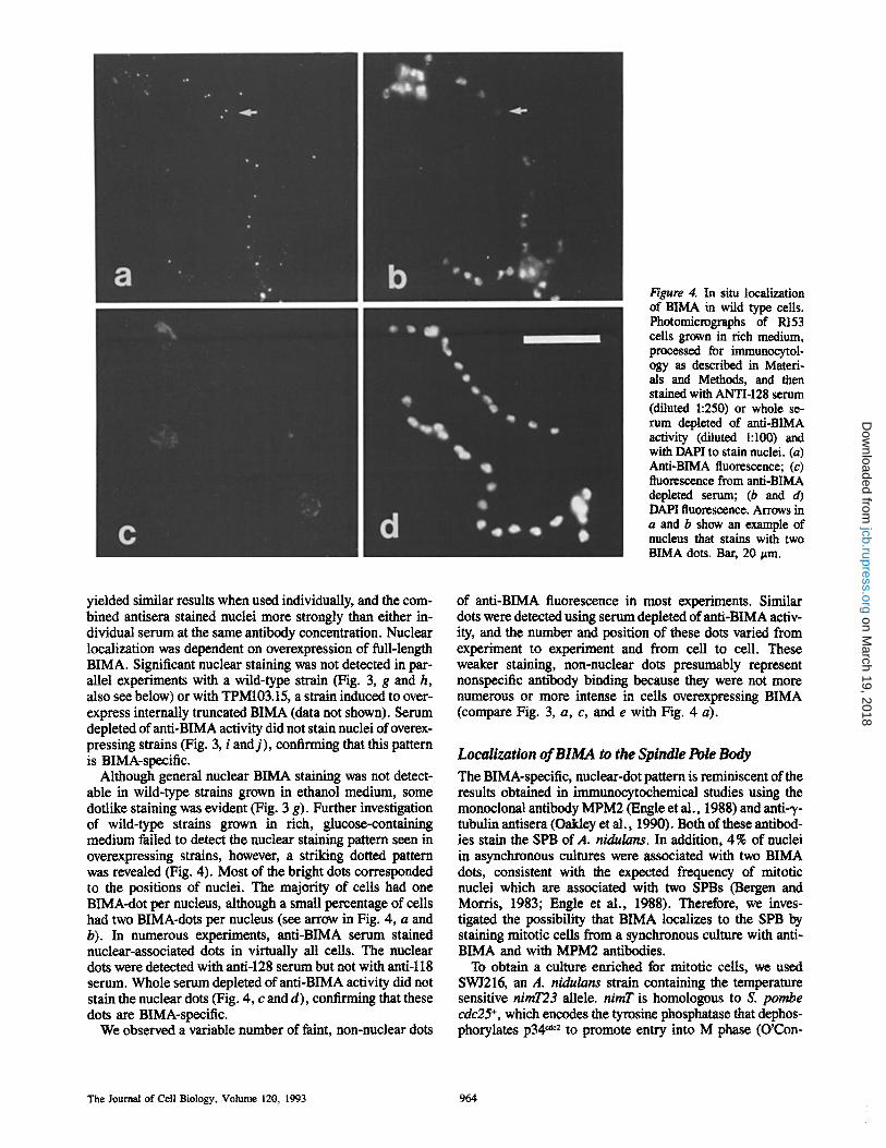
Figure 4. In situ localization of BIMA in wild type cells. Photomicrographs of R153 cells grown in rich medium, processed for immtmocytol- ogy as described in Materi- als and Methods, and then stained with ANTI-128 serum (diluted 1:250) or whole se- rum depleted of anti-BIMA activity (diluted 1:100) and with DAPI to stain nuclei. (a) Anti-BIMA fluorescence; (c) fluorescence from anti-BIMA depleted serum; (b and d) DAPI fluorescence. Arrows in a and b show an example of nucleus that stains with two BIMA dots. Bar, 20 ~m.
yielded similar results when used individually, and the com- bined antisera stained nuclei more strongly than either in- dividual serum at the same antibody concentration. Nuclear localization was dependent on overexpression of full-length BIMA. Significant nuclear staining was not detected in par- allel experiments with a wild-type strain (Fig. 3, g and h, also see below) or with TPM103.15, a strain induced to over- express internally truncated BIMA (data not shown). Serum depleted of anti-BIMA activity did not stain nuclei of overex- pressing strains (Fig. 3, i and j ) , confirming that this pattern is BIMA-specific.
Although general nuclear BIMA staining was not detect- able in wild-type strains grown in ethanol medium, some doflike staining was evident (Fig. 3 g). Further investigation of wild-type strains grown in rich, glucose-containing medium failed to detect the nuclear staining pattern seen in overexpressing strains, however, a striking dotted pattern was revealed (Fig. 4). Most of the bright dots corresponded to the positions of nuclei. The majority of ceils had one BIMA-dot per nucleus, although a small percentage of cells had two BIMA-dots per nucleus (see arrow in Fig. 4, a and b). In numerous experiments, anti-BIMA serum stained nuclear-associated dots in virtually all cells. The nuclear dots were detected with anti-128 serum but not with anti-ll8 serum. Whole serum depleted of anti-BIMA activity did not stain the nuclear dots (Fig. 4, c and d), confirming that these dots are BIMA-speeific.
We observed a variable number of faint, non-nuclear dots
of anti-BIMA fluorescence in most experiments. Similar dots were detected using serum depleted of anti-BIMA activ- ity, and the number and position of these dots varied from experiment to experiment and from cell to cell. These weaker staining, non-nuclear dots presumably represent nonspecific antibody binding because they were not more numerous or more intense in cells overexpressing BIMA (compare Fig. 3, a, c, and e with Fig. 4 a).
Localization of BIMA to the Spindle Pole Body
The BIMA-specific, nuclear-dot pattern is reminiscent of the results obtained in immunocytochemical studies using the monoclonal antibody MPM2 (Engle et al., 1988) and anti- T- tubulin antisera (Oakley et al., 1990). Both of these antibod- ies stain the SPB of A. nidulans. In addition, 4% of nuclei in asynchronous cultures were associated with two BIMA dots, consistent with the expected frequency of mitotic nuclei which are associated with two SPBs ~ r g e n and Morris, 1983; Engle et al., 1988). Therefore, we inves- tigated the possibility that BIMA localizes to the SPB by staining mitotic cells from a synchronous culture with anti- BIMA and with MPM2 antibodies.
To obtain a culture enriched for mitotic cells, we used SWI216, an A. nidulans strain containing the temperature sensitive nimT23 allele, nimT is homologous to S. pombe cdc25 +, which encodes the tyrosine phosphatase that dephos- phorylates p34 ~ to promote entry into M phase (O'Con-
The Journal of Cell Biology, Volume 120, 1993 964
on March 19, 2018
jcb.rupress.orgD
ownloaded from

Figure 5. Localization of BIMA to the spindle pole body. Pho- tomicrographs of synchronized SWJ216 cells grown as de- scribed below and processed for immunocytology as de- scribed in Materials and Meth- ods. The cells were stained with ANTI-128 diluted 1:250 to detect BIMA, with MPM2 diluted 1:800 to stain the spin- die pole bodies, and with DAPI to stain nuclei. SWJ216 spores were germinated at permissive temperature (25~ overnight, until most cells contained two to four nuclei. Cultures were then shifted to 44~ for 3 h to arrest nuclear division at the G:/M border. To release the cells into mitosis, the cultures were removed from 44~ placed at room temperature, and diluted with an equal vol- ume of 25~ medium. Sam- pies were harvested and fixed at 4 min intervals. Samples harvested 8 min after release consisted of 70 % mitotic cells as determined by c~-tubulin immunofluorescence, there- fore, 8 min samples were cho- sen for analysis with anti-BIMA and MPM2 antibodies. (a and e), Nomarski image; (b and f), DAPI fluorescence; (c and g), anti-BIMA fluorescence; (d and h), MPM2 fluorescence. Arrows in c and d show exam- pies of duplicated spindle pole bodies that stain with both anti-BIMA and with MPM2 antibodies. Arrows inf-h show clearly that BIMA, MPM2, and DAPI fluorescence colocalize. Bar, 20 #m.
Mirabito and Morris Characterization of BIMA, a Mitotic TPR Protein 965
on March 19, 2018
jcb.rupress.orgD
ownloaded from

nell et al., 1992; Dunphy and Kumagai, 1991; Gautier et al., 1991). Incubation of SWJ216 at 44~ resulted in synchroni- zation of cells at the G2 to M boundary, and shifting down to 28~ caused a rapid appearance of mitotic cells as judged by DAPI staining and anti-ot-tubulin immunofluorescence (data not shown, also see Osmani et al., 1991). Samples of the released culture that contained mostly mitotic cells (greater than 70% with condensed chromatin and bipolar mitotic spindles) were chosen for staining with anti-BIMA, with MPM2, or with both.
Anti-BIMA and MPM2 antibodies both stained 2 dots on most nuclei. In doubly stained samples, the MPM2 antigens and BIMA colocalized (Fig. 5). Under the conditions neces- sary to obtain double staining with ANTI-BIMA and MPM2 (see Materials and Methods), the mitotic spindle was not preserved and DAPI staining of nuclei was greatly dimin- ished. For example, Fig. 5, a-d, shows a cell positive for both BIMA and MPM2 SPBs but with almost no nuclear DAPI fluorescence above the background cytoplasmic fluo- rescence. The BIMA and MPM2 dots corresponded to the position of nuclei for all cells in which the nuclear morphol- ogy as revealed by DAPI fluorescence was preserved (for ex- ample, see arrows in Fig. 5, f-h). The apparent difference in distance between the spindle poles in the mitotic nuclei in Fig. 5, c and d, is due to different orientations of the spindles relative to the focal plane and is commonly seen in mitotic Aspergillus ceils (unpublished observations). These results demonstrated that BIMA, when expressed at normal level, localized to the SPB.
Discussion
The A. nidulans bimA gene is one of several fungal TPR genes required for the completion of mitosis. The S. pombe nuc2 + and S. cerevisiae CDC27 TPR genes appear to be functional homologs of bimA, suggesting that bimA repre- sents a fundamental, evolutionarily conserved mitotic com- ponent. We have developed two immunoattinity purified an- tisera specific for BIMA in order to study its structure and subcellular localization in hope of gaining new insight into BIMA function.
Our results demonstrated that BIMA is a 90-kD protein and they suggest that some BIMA polypeptides undergo post-translational modification which significantly increases their apparent mol wt. An internally deleted form of BIMA exhibited no such heterogeneity in relative mobility on SDS- PAGE (see Fig. 2 b, lane 6), suggesting that the deleted re- gion is required for these modifications, and is potentially the region of BIMA that is modified. In this regard it is worth noting that this region of BIMA contains several consensus recognition sites for serine, threonine and tyrosine kinases and one site for N-linked glycosylation.
BIMA localized to the SPB in wild-type cells and localized on or throughout most nuclei in cells overexpressing BIMA. SPB localization was detectable in essentially all cells of asynchronous cultures, arguing against cell cycle regulation of BIMA localization. It is possible that the different BIMA isoforms detected on western blots localize to different sub- cellular locations in situ. Because both of our antisera recog- nize the same set of BIMA proteins (see Fig. 2 b) we cannot rule out the possibility that some BIMA species localize to the SPB while others localize throughout the nucleus.
The two patterns of BIMA localization are similar to those reported for localization of the S. cerevisiae SPB protein en- coded by KAR/(Vallen et al., 1992). Moderate expression of certain KAR/fusion proteins resulted in SPB localization whereas high expression resulted in a general nuclear stain- ing as well as SPB staining. As suggested for KAR/, overex- pression of BIMA may saturate its binding site(s) at the SPB causing excess BIMA to accumulate in the nucleus. Alterna- tively, the nuclear and SPB localizations may both represent biologically significant locations for BIMA.
The functional and structural similarities of bimA and nuc2 + suggest that their gene products may reside in similar subcellular locations. The nuc2 + protein (NUC2) cofrac- tionates with nuclei (Hirano et ai., 1988) and localizes to the nucleus (Goebl and Yanagida, 1991). Those data, and the fact that NUC2 is insoluble in 2M NaC1, 2 % NP-40, and 25 mM lithium iodo-salicylate, suggested that NUC2 may be a component of the nuclear scaffold (Hirano et al., 1988). Those results are also consistent with the hypothesis that NUC2 localizes to the SPB because, like NUC2, BIMA shows nuclear localization when it is overexpressed and the S. pombe SPB is likely to cofractionate with nuclei and have solubility properties similar to NUC2.
The fact that NUC2 was not detected at the SPB in S. pombe could have been because of technical reasons. Al- though both of our purified serum fractions gave good sig- nals on Western blots of wild-type protein and good im- munofluorescence in BIMA overexpressing strains, only ANTI-128 stained SPBs in wild-type strains. ANTI-118, which binds to internal BIMA sequences (Fig. 1), gave no discernable staining pattern above background when used on wild-type cells (data not shown). The ANTI-118 epitopes are evidently inaccessible when BIMA is at the SPB, and it is possible that the epitopes recognized by the NUC2 antibod- ies are similarly occluded. Rout and Kilmartin (1990) also reported that several SPB epitopes were undetectable in whole ceils using formaldehyde fixation procedures and that some epitopes were more accessible to antibodies after chemical extraction removed microtubules and other SPB components. Indeed, our best staining of BIMA at the SPB is obtained under conditions in which cytoplasmic and spin- die microtubule staining is not preserved (unpublished ob- servations), suggesting that microtubules at the SPB could mask BIMA staining.
The phenotype of bimA mutations together with localiza- tion of BIMA to the SPB suggests that BIMA may play a role in SPB function during mitosis. If so, then bimA functions differently from most other genes thought to facilitate SPB function. For example, m/aations in the S. cerevisiae genes CDC31 (Byers, 1981), MPS1 and MPS2 (Winey et al., 1991), NDC1 (Thomas and Botstein, 1986) and KAR/ (Rose and Fink, 1987) all affect mitosis by perturbing SPB duplication. Inactivation of A. nidulans mipA, which encodes the SPB component "y-tubulin, disrupts mitosis by preventing spindle microtubule assembly (Oaldey et al., 1990). One gene somewhat similar to bimA in mutant phenotype and protein localization is KAR3, which encodes a kinesinlike protein re- quired for karyogamy in S. cerevisiae (Meluh and Rose, 1990). KAR3 protein localizes to cytoplasmic microtubules and the SPB, and inactivation of KAR3 causes 40% of mutant ceils to arrest with duplicated SPBs and a short mitotic spin- dle. For KAR3, localization to the SPB is apparently required
The Journal of Cell Biology, Volume 120, 1993 966
on March 19, 2018
jcb.rupress.orgD
ownloaded from

only for karyogamy because some mutations in KAR/that abolish karyogamy and SPB localization of the KAR3 protein do not affect mitosis (Vallen et al., 1992).
It seems likely that one aspect of BIMA function at the SPB will involve protein-protein interactions. Based on the results of biochemical and theoretical analyses of the NUC2 TPR domain, Hirano et al. (1990) proposed that each repeat associates with other repeats through hydrophobic interac- tions. The TPR proteins encoded by S. cerevisiae CDC23 and CDC27 form homo- and heterodimers in vivo (Sikorski et al., 1991), and they are coimmunoprecipitated by antibod- ies against the CDC23 protein (J. Lamb and P. Hieter, per- sonal communication). Although the TPR domains have not been proven to be responsible for the CDC23/CDC27 inter- actions, these results support the idea that TPR-containing polypeptides physically interact. It is possible that BIMA may interact with other TPR proteins as part of BIMA/s func- tion at the SPB. It follows that the BIMA TPR domain itself may specify localization of BIMA to the SPB via similar in- teractions.
In addition to interacting with other TPR genes, genetic studies in S. cerevisiae indicate that many TPR genes interact with members of the/~-transducin gene family. Examples in- clude PRP6/PRP4 (Dalrymple et al., 1989), SKI3/MAKI1 (Icho and Wickner, 1988), SSN6/TUP1 (Williams et al., 1991), and CDC20 with both CDC23 and CDC16 (D. Burke, personal communication). The best understood example is SSN6/TUP1, where biochemical and genetic experiments show that their gene products form a complex that acts as a general transcriptional repressor (Williams et al., 1991; Keleher et al., 1992). The fact that cdc20 and bimA mutants have similar phenotypes (Sethi et al., 1991; O'Donnell et al., 1991) is consistent with a role for/3-transducin-related pro- teins in mitosis. In fact, G-protein mediated signal transduc- tion could provide a mechanism for propagating a signal be- tween BIMA at the SPB and other parts of the mitotic apparatus, for example, the kinetochore.
Another way in which loss of BIMA function at the poles could be transmitted to the rest of the mitotic apparatus is through the activity of a diffusible regulatory protein such as p34 cat2. The SPB is the site of localization of p34 ~dc2 and cyclin B at metaphase in S. pombe (Alfa et al., 1990). bimA mutations could alter interactions between these regulators and the SPB by, for example, perturbing the process of cyclin B destruction. Degradation of cyclin B coincides with the on- set of anaphase A, and inactivation of the p34 ~2 kinase via turnover of cyclin B is necessary for the metaphase to anaphase transition (Murray et al., 1989; Forsburg and Nurse, 1991). Therefore, it is possible that mutations in bimA directly or indirectly prevent inactivation of p34 ~dc2, resulting in a failure to exit metaphase.
It is also possible that bimA mutations disrupt events at the poles that are directly involved in the metaphase to anaphase transition. An interesting example of such an event is kinetochore microtubule disassembly at the spindle poles during metaphase in mammalian cells (Mitchison et al., 1986; Mitchison, 1989; Mitchison and Sawin, 1990) and during both metaphase and anaphase-A in Newt lung cells (Mitchison and Salmon, 1992). Spindle microtubule disas- sembly is important for mitosis in Aspergillus because benA33, a mutation in the A. nidulans/3-tubulin gene, causes formation of hyperstable microtubules that block mitosis at
metaphase (Oaldey and Morris, 1981). Mutations in mipA, the gene encoding the SPB component 3,-tubulin, suppress the lethality and microtubule hyperstability of benA33 (Weil et al., 1986; Oakley and Oaldey, 1989; Oaldey et al., 1990), demonstrating that microtubule destabilization at the spindle poles is necessary for transition from metaphase to anaphase in Aspergillus. The phenotype of bimA mutations and local- ization of BIMA to the SPBs suggests that BIMA could play an important role in the dynamic interactions between kinetochore microtubules and spindle poles during mitosis.
We thank Herb Geller for use of his microscope and Kerry O'Donnell for construction of several bimA containing plasmids. We also thank Matthew O'Connell, Michelle Mischke, and Steven James for critical intellectual and technical contributions to this project.
This work was supported by National Institutes of Health grant GM- 34711-08 to N. R. Morris.
Received for publication 25 August 1992 and in revised form 10 November 1992.
References
Alfa, C. E., B. Ducommun, D. Beach, andJ. S. Hyams. 1990. Distinct nuclear and spindle pole body populations of cyclin-cdc2 in fission yeast. Nature (Lend.). 347:680-682.
Bergen, L. G., and N. R. Morris. 1983. Kinetics of the nuclear division cycle of Aspergillus nidulans. J. Bacteriol. 156:155-160.
Byers, B. 1981. Multiple roles of the spindle pole bodies in the life cycle of Saccharomyces cerevisiae. In Molecular Genetics in Yeast. Alfred Benzon Symposium. D. von Wettstein, J. Friis, M. Kielland-Brandt, and A. Sten- derap, editors. Munksgaard, Copenhagen. 119-131.
Dalrymple, M. A., S. Petersen-Bjorn, J. D. Friesen, and J. D. Beggs. 1989. The product of the PRP4 gene of S. cerevisiae shows homology to beta suhnnits of G proteins petter]. Cell. 58:811-812.
Dunphy, W. G., and A. Kumagai. 1991. The cdc25 protein contains an intrinsic phosphatase activity. Cell. 67:189-196.
Engle, D. B., J. H. Dconan, and N. R. Morris. 1988. Cell-cycle modulation of MPM-2-specific spindle pole body phosphorylation in Aspergillus nidu- lans. Cell Motil. Cytoskeleton. 10:432-437.
Forsburg, S. L., and P. Nurse. 1991. Cell cycle regulation in the yeasts Sac- charomyces cerevisiae and Schizosaccharomyces pomhe. Annu. Rev. Cell Biol. 7:227-256.
Gautier, J., M. J. Solomon, R. N. Booher, J. F. Bazan, and M. W. Kirsehner. 1991. cdc25 is a specific tyrosine phosphatase that directly activates p34 ~2. Cell. 67:197-211.
Goebl, M., and M. Yanagida. 1991. The TPR snap helix: A novel protein repeat motif from mitosis to transcription. TIBS (Trends Biochem. Sci.). 16:173- 177.
Harlow, E., and D. Lane. 1988. Antibodies: A Laboratory Manual. Cold Spring Harbor Laboratories, Cold Spring Harbor, New York.
Hirano, T., Y. Hiraoka, and M. Yanagida. 1988. A temperature-sensitive mu- tation of the Schizosaccharomyces pombe geae nuc2 + that encodes a nuclear scaffold-like protein blocks spindle elongation in mitotic anaphase. J. Cell Biol. 106:1171-1183.
Hirano, T., N. Kinoshita, K. Morikawa, and M. Yanagida. 1990. Snap helix with knob and hole, essential repeats in S. pombe nuclear protein nuc2 +. Cell. 60:319-328.
Icho, T., and R. B. Wickner. 1988. The MAKll protein is essential for cell growth and replication of M double-stranded RNA and is apparently a membrane-associated protein. J. Biol. Chem. 263:1467-1475.
Kiifer, E. 1977. Meiotic and mitotic recombination in Aspergillus nidulans and its chromosomal aberrations. Adv. Genet. 19:33-131.
Keleber, C. A., M. J. Redd, J. Schultz, M. Carlson, and A. D. Johnson. 1992. Ssnt-Tupl is a general represser of transcription in yeast. Cell. 68:709-719.
Koerner, T. J., J. E. Hill, A. M. Myers, and A. Tzagoloff. 1991. High- expression vectors with multiple cloning sites for construction of trpE fusion genes: pATH vectors. Methods Enzymol. 194:477-490.
Lockington, R. A., H. M. Scaly-Lewis, C. Scazzocchio, and R. W. Davies. 1985. Cloning and characterization of the ethanol utilization regulon in Aspergillus nidulans. Gene. 33:137-149.
Masuda, H., T. Hirano, M. Yanagida, and W. Z. Cande. 1990. In vitro reacti- vation of spindle elongation in fission yeast nuc2 mutant cells. J. Cell Biol. 110:417-425.
Mclntosh, J. R., and G. E. Hering. 1991. Spindle fiber action and chromosome movement. Annu. Revo Cell Biol. 7:403-426.
Meluh, P. B., and M. D. Rose. 1990. KAR3, a kinesin-related gene required
Mirabito and Morris Characterization of BIMA, a Mitotic TPR Protein 967
on March 19, 2018
jcb.rupress.orgD
ownloaded from

for yeast nuclear fusion. Cell. 60:1029-1041. Mitchison, T. 1989. Polewards microtubule flux in the mitotic spindle: evidence
from photoactivation of fluorescence. J. Cell Biol. 109:637-652. Mitehison, T. L, L. Evans, E. Sehulze, and M. Kirschner. 1986. Sites of
microtubule assembly and disassembly in the mitotic spindle. Cell. 45: 515-527.
Mitehison, T. J., and K. E. Sawin. 1990. Tubulin flux in the mitotic spindle: where does it come from, where is it going? CellMotil. Cytoskel. 16:93-98.
Mitchison, T. J., and E. D. Salmon. 1992. Poleward kinetochore fiber move- ment occurs during both metaphase and anaphase-A in Newt lung cell mito- sis. J. Cell Biol. 119:569-582.
Morris, N. R. 1976. Mitotic mutants of Aspergillus nidulans, Genet. Res. 26:237-254.
Morris, N. R., and A. Pallone Enos. 1992. Mitotic gold in a mold: Aspergillus genetics and the biology of mitosis. TIG (Trends Genet.). 8:32-37.
Murray, A. M., M. J. Solomon, and M. W. Kirschner. 1989. The role of cyclin synthesis and degradation in the control of maturation promoting factor ac- tivity. Nature (Lond.). 339:280-286.
O'Cormell, M. L, A. H. Osmani, N. R. Morris, and S. A. Osmani, 1992. An extra copy of n/mE ~l~ elevates pre-MPF levels and partially suppresses mutation of n/mT ~a~ in Aspergillus n/dutans. EMBO (Eur. Mol. Biol. Or- gan.) J. 11:2139-2149.
O'Donnell, K. L., A. H. Osmani, S. A. Osmani, and N. R. Morris. 1991. bimA encodes a member of the tetratrieopeptide repeat family of proteins and is required for the completion of mitosis in Aspergillus n/dulans. J. Cell Sci. 99:711-719.
Oakley, B. R., and N. R. Morris. 1981. A beta tubulin mutation in Aspergillus nidulans that blocks microtubule function without blocking assembly. Cell. 24:837-845.
Oakley, B. R., C. E, Oakley, Y. Yoon, and M. K. Jung. 1990. ?-Tubulin is a component of the spindle pole body that is essential for microtubule func- tion in Aspergillus nidulans. Cell. 61:1289-1301.
Oakley, C. E., and B. R, Oaldey. 1989. Identification of 7-tubulin, a new mem- ber of the tubulin superfamily encoded by mipA gene ofAspergillus n/dulans. Nature (Lond.). 338:662-664.
Osmani, A., S. L. McGuire, and S. A. Osmani. 1991. Parallel activation of the NIMA and p34 ~ cell cycle-regulated protein kinases is required to ini- tiate mitosis in A. nidulans. Cell. 67:283-291.
Osmani, S. A., G. S. May, and N, R. Morris. 1987. Regulation of the mRNA levels of n/mA, a gene required for the G2-M transition in Aspergillus n/du- lans. J. Cell Biol. 104:1495-1504.
Pontecorvo, G., J. A. Roper, L. M. Hemmons, K. D. MacDonald, and A. W. J. Bufton. 1953. The genetics ofAspergillus n/dulans. Adv. Genet. 5:141-238.
Rose, M. D., and G. R. Fink. 1987. KARl, a gene required for function of both intranuclear and extranuclear microtubules in yeast. Cell. 48:1047-1060.
Rout, M. P., and J. V. Kilmartin. 1990. Components of the yeast spindle and spindle pole body. J. Cell BioL 111:1913-1927.
Rfither, U., and B. Muller-Hill. 1983. Easy identification of cDNA clones. EMBO (Eur. Mol. Biol. Organ.) J. 2:1791-1794.
Samhrook, L, E. F. Fritsch, and T. Maniatis. 1989. Molecular cloning: a labo- ratory manual. Cold Spring Harbor Laboratories, Cold Spring Harbor, New York.
Sethi, N., M. C. Monteagudo, D. Koshland, E. Hogan, and D. L Burke, 1991. The CDC20 gene product of Saccharomyces cerevisiae, a ~-transducin homolog, is required for a subset of micrombule-dependent cellular processes. Mol. Cell. Biol. 11:5592-5602.
Sikorski, R. S., M. S. Boguski, M. Goebl, and P. Hieter. 1990. A repeating amino acid motif (TPR) in CDC23 defines a family of proteins and a new relationship among genes required for mitosis and RNA synthesis. Cell. 60:307-317.
Sikorski, R. S., W. A. Michaud, J. C. Woettun, M. S. Boguski, C. Cunnelly, and P. Hieter. t991, TPR proteins as essential components of the yeast cell cycle. Cold Spring Harbor Syrup. Quam. Biol. 56:663-673.
Thomas, J. H., and D. Botstein. 1986. A gene required for the separation of chromosomes on the spindle apparatus in yeast. Cell. 44:65-76.
Vallen, E. A., M. A. Hiller, T. Y. Scberson, and M. D. Rose. 1992. Separate domains of KARt mediate distinct functions in mitosis and nuclear fusion. J. Cell Biol. I17:1277-1287.
ValIen, E. A., T. Y. Scherson, T. Roberts, K. Van Zee, and M. D. Rose, 1992, Asymmetric mitotic segregation of the yeast spindle pole body. Cell. 69: 505-515.
Waring, R. B., G. S. May, and N. R. Morris. 1989. Characterization of an in- ducible expression system in Aspergillus nidulans using alcA and tubulin- coding genes. Gene. 79:119-130.
Well, C. F., C. E. Oakley, and B. R. Oakley. 1986. Isolation ofmip (microtu- bule interacting protein) mutations of Aspergillus n/dulans. Mol. Cell. Biol. 6:2963-2968.
Williams, F. E., U. Varanasi, and R. J. Trumbly. 1991. The CYC8 and TUP1 proteins involved in glucose repression in Saccharomyces cerevisiae are as- sociated in a protein complex. Mol. Cell. Biol. 11:3307-3316.
Winey, M., L. Goetsch, P. Baum, and B. Byers. 1991. MPS1 and MPS2: Novel yeast genes defining distinct steps of spindle pole body duplication. J. Cell BioL 114:745-754.
Yelton, M. M., J. E. Hamer, and W. E. Timborlake. 1984. Transformation ofAspergillus nidulans by using a trpC plasmid. Proc. Natl. Acad. Sci. USA. 81:1470-1474.
The Journal of Cell Biology, Volume 120, 1993 968
on March 19, 2018
jcb.rupress.orgD
ownloaded from