follow-up after acute pulmonary embolism
TRANSCRIPT

Hämostaseologie 1/2018 © Schattauer 2018
22 Review
KeywordsPulmonary embolism, CTEPH, post-PE syn-drome, quality of life
SummaryIn addition to among others major bleeding from anticoagulant therapy and recurrent ve-nous thromboembolism (VTE), patients who survived acute pulmonary embolism (PE) face an increased risk of chronic functional limitations and decreased quality of life. In recent years, this latter complications have been better framed within the evolving defi-
nition of “post-PE syndrome” of which chronic thromboembolic pulmonary hypertension (CTEPH) represents the most extreme presen-tation. The post-PE syndrome in all its aspects is a frequent and clinically relevant long-term complication of PE but -except for CTEPH- has been largely understudied. There is great need to better define and understand the natural course of acute PE, to predict the development of the post-PE syndrome and to evaluate the potential benefits evolving treatments such as cardiopulmonary rehabilitation.
SchlüsselwörterLungenembolie, CTEPH, chronisches Lungen-embolie-Syndrom, Lebensqualität
ZusammenfassungÜber Komplikationen wie größere Blutungen infolge der Antikoagulation oder rezidivie-rende venöse Thrombembolien (VTE) hinaus, haben Patienten, die eine akute Lungenem-bolie (LE) überlebt haben, ein erhöhtes Risiko für chronische funktionelle Einschränkungen und eine reduzierte Lebensqualität. Letztere werden neuerdings in dem aufkommenden Begriff des chronischen LE-Syndroms („Post-LE-Syndrom“) zusammengefasst, das seine extremste Ausprägung in der chronisch thromboembolischen pulmonalen Hypertonie (CTEPH) findet. Das chronische LE-Syndrom in all seinen Aspekten ist eine häufige und klinisch relevante Langzeitkomplikation der LE, wurde jedoch – mit Ausnahme der CTEPH – nur unzureichend untersucht. Es ist drin-gend erforderlich, den natürlichen Verlauf ei-ner akuten LE besser zu definieren und zu verstehen, um die Entwicklung eines chroni-schen LE-Syndroms vorherzusagen und den potenziellen Nutzen neuer Behandlungsfor-men, wie der kardiopulmonalen Rehabilitati-on, zu evaluieren.
Correspondence toF.A. KlokDepartment of Thrombosis and Hemostasis,LUMC,Albinusdreef 2,2300 RC LeidenEmail: [email protected]
Nachsorge bei akuter LungenembolieVorhersage einer chronisch thromboembolischen pulmonalen Hypertonie und eines chronischen Lungenembolie-SyndromsHämostaseologie 2018; 38: 22–32https://doi.org/10.5482/HAMO-17-06-0020received: June 20, 2017accepted in revised form: December 11, 2017
FundingFrederikus Klok reports research grants from Bayer, re-search grants from Bristol-Myers Squibb, research grants from Boehringer-Ingelheim, research grants from MSD and non-financial research support from Daiichi-Sankyo. Stefano Barco has received congress and travel payments from Daiichi-Sankyo and Bayer HealthCare. The work of Frederikus Klok and Stefano Barco is supported by the German Federal Ministry of Education and Research (BMBF 01EO1003 and 01EO1503).
Authorship statementBoth authors have contributed significantly to and ap-prove of this manuscript.
IntroductionAfter surviving acute pulmonary embolism (PE), patients face an increased risk of de-veloping major bleeding from anticoagu-lant therapy, recurrent venous thromboem-
bolism (VTE), arterial cardiovascular dis-eases, or chronic functional limitations (1–8). In recent years, this latter compli-cation has been better framed within the evolving definition of “post-PE syndrome”, which accounts for suboptimal cardiac
function, pulmonary artery flow dynamics, or pulmonary gas exchange at rest or dur-ing exercise, in combination with symp-toms of exercise intolerance, dyspnea, im-paired functional status, or worsened quality of life (8, 9). Chronic thromboem-
Follow-up after acute Pulmonary EmbolismPredicting chronic thromboembolic pulmonary hyperten-sion and post-pulmonary embolism syndrome
Frederikus A. Klok1,2; Stefano Barco1
1Center for Thrombosis and Hemostasis, University Hospital of the Johannes Gutenberg University Mainz, Mainz, Germany; 2Department of Thrombosis and Hemostasis, Leiden University Medical Center, Leiden, the Netherlands
Thi
s do
cum
ent w
as d
ownl
oade
d fo
r pe
rson
al u
se o
nly.
Una
utho
rized
dis
trib
utio
n is
str
ictly
pro
hibi
ted.

Hämostaseologie 1/2018 © Schattauer 2018
24 FA. Klok, S. Barco: Follow-up after acute pulmonary embolism
bolic pulmonary hypertension (CTEPH) represents the most severe manifestation of the post-PE syndrome (10–12). In the pres-ent review, we discuss the pathophysiology and frequency of CTEPH and post-PE syn-drome, as well as the strategies under de-velopment to predict, prevent, and treat long-term sequelae of acute PE.
Chronic thromboembolic pulmonary hypertensionPathophysiology of CTEPH
A commonly accepted mechanistic hy-pothesis is that CTEPH results from an incident episode of ´unresolved´ PE char-acterized by the persistence of residual thrombi despite anticoagulant therapy, which contributes to increase pulmonary vascular resistance and ultimately results in progressive right ventricular overload (13–15). The exact pathophysiological mechanism that may prevent from com-plete resolution of acute thrombus is how-ever not fully clarified yet, although pro-in-flammatory state, abnormal fibrinogen variants, aberrations in angiogenesis, and mesenchymal cell activation have been im-plicated in poorer pulmonary thrombus resolution (▶ Table 1) (13–21). Notably, whereas radiological signs of unresolved thrombotic material are necessary criteria for confirming a CTEPH diagnosis, these
do not always match with the severity of the clinical presentation of individual pa-tients and therefore cannot be accounted as an indicator for prognosis. In addition to chronic blood clots, CTEPH patients usually exhibit widespread pulmonary microvasculopathy, which resembles that of other pulmonary arterial hypertension subtypes (13, 22–24). Diffuse areas of the lung-affected or not by chronic thrombotic occlusions- similarly manifest microvascu-lopathy, indicating that higher share stress caused by chronic thrombi is not solely re-sponsible for arterial remodeling. One of the explanations that has been hypothes-ized is that microvascular changes may be caused by altered blood flow from multiple anastomoses between systemic and pul-monary circulation through hypertrophic bronchial arteries that are opened by the high pressure gradient which develops be-tween bronchial and pulmonary arteries af-fected by chronic blood clots (22).
Indeed, both the small vessel disease and the chronic proximal obstruction of the pulmonary arteries contribute to the progression of right ventricular overload. Initially, this outflow burden leads to right ventricular hypertrophy as a mechanism of the heart in order to increase the effective-ness of its pumping function, a process also referred to as ‘adaptive remodeling’.(25) However, adaptive remodeling is associated with a maintained right ventricular func-
tion for a limited period of time, after which inevitably ‘uncoupling’ occurs, caus-ing right ventricular dilatation and -es-pecially during exercise- insufficiency, which ultimately lead to terminal right ventricular failure and death (25, 26).
How often is CTEPH diagnosed after acute PE?
The incidence of CTEPH after sympto-matic acute PE has been reported to range from 0.1 % to 11.8 %, with most studies showing a frequency of 2–4 % (10, 27–36). Beyond geographic or historical variations, such wide range can be explained by differ-ences in patient selection and diagnostic criteria adopted by the investigators of published studies. For instance, most studies focused on specific subgroups of PE patients characterized by the absence of cardiopulmonary comorbidities and some did not apply right heart catheterization to confirm CTEPH.
To provide more clarity, a systematic re-view and meta-analysis of relevant studies and meeting abstracts has been recently published (37). Studies adopting invasive measurements to confirm the diagnosis of CTEPH were included in the main analysis and three patient subgroups were prede-fined to limit heterogeneity, namely i) ‘all comers’ (unselected consecutive PE pa-tients), ii) ‘survivors’ (PE patients who sur-vived the first three to six months), and iii) ‘survivors without comorbidities’ (surviv-ors further selected by excluding patients with cardiopulmonary, malignant and/or other severe comorbidities). A total of 4,047 PE patients were evaluated in the meta-analysis for pooled incidences in the predefined cohorts of 0.56 % (95 %CI 0.1–1.0), 3.2 % (95 %CI 2.0–4.4), and 2.8 % (95 %CI 1.5–4.1), respectively (▶ Figure 1) (37).
Notably, the comparable incidences of both ‘survivor’ cohorts indicated that the presence of other conditions that may ex-plain symptoms of functional impairment and dyspnea, such as COPD, do not appear to rule out the diagnosis of CTEPH, and adequate diagnostic tests for CTEPH should not be withheld in such patients. A final important observation of the meta-analysis regards the clear overdiagnosis of
Tab. 1 Summary of the pathophysiological mechanisms implicated for the transition of acute PE to CTEPH and clinical conditions which may “predispose” to CTEPH
Clinical conditions implicated with a higher risk of CTEPH
Thyroid replacement therapy
Malignancy
(Recurrent) Venous thromboembolism
Antiphospholipid antibodies
High FVIII
Non-O blood group
Ventriculo-atrial shunt
Infected pacemaker leads
Indwelling venous catheters and leads
Splenectomy
Chronic inflammatory disorders
Known mechanisms of thrombus nonresol-ution implicated with CTEPH pathophysiology
Defective angiogenesis
Systematic inflammation
Fibrinogen abnormalities/impaired fibrinolysis
Misguided immune response to venous clots
Hypoxia-induced endothelial and mesenchymalcell activation
Thi
s do
cum
ent w
as d
ownl
oade
d fo
r pe
rson
al u
se o
nly.
Una
utho
rized
dis
trib
utio
n is
str
ictly
pro
hibi
ted.

© Schattauer 2018 Hämostaseologie 1/2018
25
risk stratification in order to increase the expected rate of CTEPH in the study co-hort. Moreover, the absolute risk of CTEPH for patients presenting with these risk factors is unknown as they were often identified in case-control studies compar-ing subjects diagnosed with CTEPH and population controls or patients with class 1 PH, and not in cohorts of PE survivors (47). Finally, since their prevalence in PE cohorts is low (e.g. for splenectomy about 2 %), this approach appears unlikely to be sufficiently accurate.
A second strategy would be to study the added value of reassessing CTPA scans for detecting signs of chronic PE or pulmonary hypertension such as right ventricular hy-pertrophy, webs and bands, tortuous bron-chial arteries or mosaic perfusion (33). As discussed above, these signs may be very sensitive for prevalent CTEPH. Even so, the afore mentioned French study included only seven patients with CTEPH and does not provide sufficient evidence for strong recommendations for clinical practice.
Clinical decision rules and risk assess-ment models are widely used in clinical practice for diagnostic or prognostic pur-poses. A collaborative study between Ger-man, Polish and Dutch PE expert centers aimed at developing a score for prediction of CTEPH after PE (47). They performed a patient-level analysis of a total of 772 PE-survivors without major cardiopulmonary or oncological comorbidities at baseline, and fit multivariate regression models to identify clinical variables easily assessable at the time of index PE event that would in-dependently predict CTEPH. A score con-
CTEPH is not preceded by symptomatic VTE in 19–63 % of patients (39, 40). It re-mains purely speculative the hypothesis of these patients suffering from episodes of ‘silent PE’, or, alternatively, if they represent a different clinical scenario of CTEPH de-veloping from in situ thrombosis as a thrombogenic phenotype of class 1 pul-monary hypertension.
Can we predict CTEPH?
The ability to predict which PE patients will develop CTEPH will likely improve their prognosis by allowing physicians to better apply valuable resources to a specific high-risk group, while preventing un-necessary imaging tests to patients with a negligible risk of developing or having CTEPH. Unfortunately, evidence for strat-egies of risk prediction are scarce and this is largely due to the absolute low incidence of the disease and the unavailability of large databases of consecutive patients at risk of developing CTEPH (41, 42).
One possible strategy to overcome these limitations would be to focus on cohorts of PE patients with identified risk factors for CTEPH such as splenectomy, prior in-fected pacemaker leads, chronic inflamma-tory disease, antiphospholipid syndrome, known hypothyroidism, and ventriculo-at-rial shunt (▶ Table 1) (43–46). The first issue that may raise by adopting this ap-proach is represented by the fact that these variables have been identified as risk fac-tors associated with CTEPH, but not vali-dated as predictors of the disease and therefore cannot be used (yet) for a first
FA. Klok, S. Barco: Follow-up after acute pulmonary embolism
CTEPH in studies that did not apply right heart catheterization but echocardiography as the diagnostic standard for CTEPH with a 3-fold higher CTEPH incidence (9.1 %, 95 %CI 4.1–14), indicating that echocar-diography is not an accurate standalone test to confirm this diagnosis.
It must be noted however that the cases of CTEPH reported in the aforementioned follow-up studies likely represent a mix of incident and prevalent cases, therefore overestimating the actual figures of CTEPH incidence, since the diagnosis of CTEPH was often made within a few months after the index PE. In particular, a highly cited French study reported detailed reevaluation of the clinical PE presentation of the 7 (of 146) PE patients who were diagnosed with CTEPH (33). In five of seven patients, the authors described a no-table elevated (assessed via echocardio-graphy) systolic pulmonary arterial press-ure of 70 mmHg, while historical data sug-gested that acute PE can be associated with an increase in mean pulmonary artery pressure of up to 40 mmHg only with an extensive 70 % of the pulmonary artery tree occluded by blood clots (38). This observa-tion would suggest that these five patients may have developed pulmonary hyperten-sion before the PE diagnosis was confirm-ed. Indeed, after review of the initial CTPA images by an expert radiologist, all seven patients had at least two signs of the condi-tion at initial acute PE presentation, as did 20 % of patients who did not develop CTEPH during the median follow up of 26 months. Based on the latter observation, it could be hypothesized that preexisting (prevalent) CTEPH would have been mis-classified as acute PE in some or even all the patients eventually diagnosed with CTEPH over follow-up. Nevertheless, as long as it remains impossible to distinguish ‘true’ acute PE from CTEPH at a (sub)acute clinical presentation, the difference be-tween prevalent and incident cases of CTEPH after acute PE does not appear of clinical relevance for the initial treatment of these patients since all are candidate to extended anticoagulation, nor for deter-mining the proper level of awareness for CTEPH of PE caretakers.
To further reinforce this concept, sev-eral observations demonstrated that
Fig. 1 Pooled incidence of chronic thromboem-bolic pulmonary hyper-tension in three pre-defined patient sub-categories (reprinted with permission [37])
Thi
s do
cum
ent w
as d
ownl
oade
d fo
r pe
rson
al u
se o
nly.
Una
utho
rized
dis
trib
utio
n is
str
ictly
pro
hibi
ted.
Hämostaseologie 1/2018 © Schattauer 2018
26 FA. Klok, S. Barco: Follow-up after acute pulmonary embolism
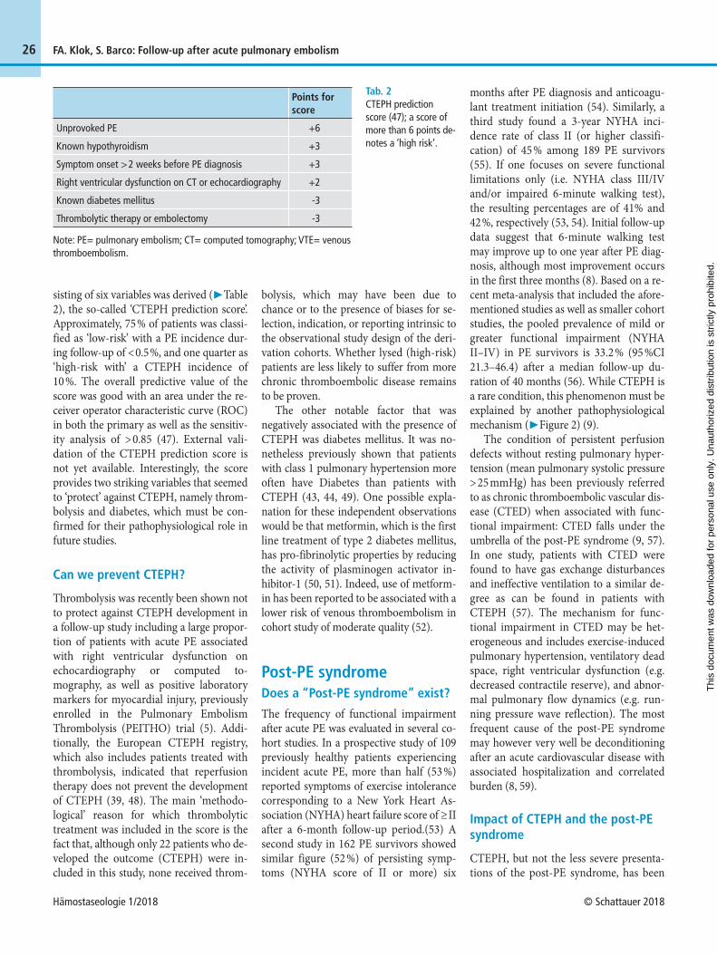
sisting of six variables was derived (▶ Table 2), the so-called ‘CTEPH prediction score’. Approximately, 75 % of patients was classi-fied as ‘low-risk’ with a PE incidence dur-ing follow-up of < 0.5 %, and one quarter as ‘high-risk with’ a CTEPH incidence of 10 %. The overall predictive value of the score was good with an area under the re-ceiver operator characteristic curve (ROC) in both the primary as well as the sensitiv-ity analysis of > 0.85 (47). External vali-dation of the CTEPH prediction score is not yet available. Interestingly, the score provides two striking variables that seemed to ‘protect’ against CTEPH, namely throm-bolysis and diabetes, which must be con-firmed for their pathophysiological role in future studies.
Can we prevent CTEPH?
Thrombolysis was recently been shown not to protect against CTEPH development in a follow-up study including a large propor-tion of patients with acute PE associated with right ventricular dysfunction on echocardiography or computed to-mography, as well as positive laboratory markers for myocardial injury, previously enrolled in the Pulmonary Embolism Thrombolysis (PEITHO) trial (5). Addi-tionally, the European CTEPH registry, which also includes patients treated with thrombolysis, indicated that reperfusion therapy does not prevent the development of CTEPH (39, 48). The main ‘methodo-logical’ reason for which thrombolytic treatment was included in the score is the fact that, although only 22 patients who de-veloped the outcome (CTEPH) were in-cluded in this study, none received throm-
bolysis, which may have been due to chance or to the presence of biases for se-lection, indication, or reporting intrinsic to the observational study design of the deri-vation cohorts. Whether lysed (high-risk) patients are less likely to suffer from more chronic thromboembolic disease remains to be proven.
The other notable factor that was negatively associated with the presence of CTEPH was diabetes mellitus. It was no-netheless previously shown that patients with class 1 pulmonary hypertension more often have Diabetes than patients with CTEPH (43, 44, 49). One possible expla-nation for these independent observations would be that metformin, which is the first line treatment of type 2 diabetes mellitus, has pro-fibrinolytic properties by reducing the activity of plasminogen activator in-hibitor-1 (50, 51). Indeed, use of metform-in has been reported to be associated with a lower risk of venous thromboembolism in cohort study of moderate quality (52).
Post-PE syndromeDoes a “Post-PE syndrome” exist?
The frequency of functional impairment after acute PE was evaluated in several co-hort studies. In a prospective study of 109 previously healthy patients experiencing incident acute PE, more than half (53 %) reported symptoms of exercise intolerance corresponding to a New York Heart As-sociation (NYHA) heart failure score of ≥ II after a 6-month follow-up period.(53) A second study in 162 PE survivors showed similar figure (52 %) of persisting symp-toms (NYHA score of II or more) six
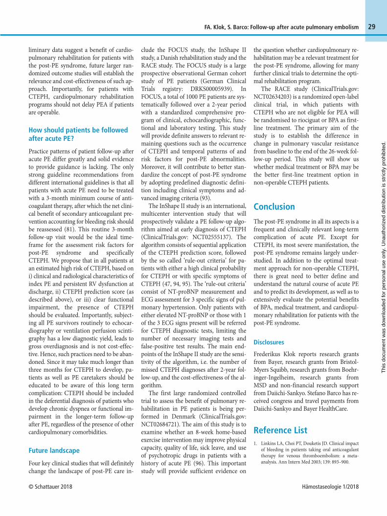
months after PE diagnosis and anticoagu-lant treatment initiation (54). Similarly, a third study found a 3-year NYHA inci-dence rate of class II (or higher classifi-cation) of 45 % among 189 PE survivors (55). If one focuses on severe functional limitations only (i.e. NYHA class III/IV and/or impaired 6-minute walking test), the resulting percentages are of 41% and 42 %, respectively (53, 54). Initial follow-up data suggest that 6-minute walking test may improve up to one year after PE diag-nosis, although most improvement occurs in the first three months (8). Based on a re-cent meta-analysis that included the afore-mentioned studies as well as smaller cohort studies, the pooled prevalence of mild or greater functional impairment (NYHA II–IV) in PE survivors is 33.2 % (95 %CI 21.3–46.4) after a median follow-up du-ration of 40 months (56). While CTEPH is a rare condition, this phenomenon must be explained by another pathophysiological mechanism (▶ Figure 2) (9).
The condition of persistent perfusion defects without resting pulmonary hyper-tension (mean pulmonary systolic pressure > 25 mmHg) has been previously referred to as chronic thromboembolic vascular dis-ease (CTED) when associated with func-tional impairment: CTED falls under the umbrella of the post-PE syndrome (9, 57). In one study, patients with CTED were found to have gas exchange disturbances and ineffective ventilation to a similar de-gree as can be found in patients with CTEPH (57). The mechanism for func-tional impairment in CTED may be het-erogeneous and includes exercise-induced pulmonary hypertension, ventilatory dead space, right ventricular dysfunction (e.g. decreased contractile reserve), and abnor-mal pulmonary flow dynamics (e.g. run-ning pressure wave reflection). The most frequent cause of the post-PE syndrome may however very well be deconditioning after an acute cardiovascular disease with associated hospitalization and correlated burden (8, 59).
Impact of CTEPH and the post-PE syndrome
CTEPH, but not the less severe presenta-tions of the post-PE syndrome, has been
Tab. 2 CTEPH prediction score (47); a score of more than 6 points de-notes a ‘high risk’.
Unprovoked PE
Known hypothyroidism
Symptom onset > 2 weeks before PE diagnosis
Right ventricular dysfunction on CT or echocardiography
Known diabetes mellitus
Thrombolytic therapy or embolectomy
Note: PE= pulmonary embolism; CT= computed tomography; VTE= venous thromboembolism.
Points for score
+6
+3
+3
+2
-3
-3
Thi
s do
cum
ent w
as d
ownl
oade
d fo
r pe
rson
al u
se o
nly.
Una
utho
rized
dis
trib
utio
n is
str
ictly
pro
hibi
ted.

Hämostaseologie 1/2018 © Schattauer 2018
28
of patients is considered inoperable and must be considered for the sole medical and support therapy. A trial of Riociguat, a soluble oral stimulator of guanylate cyclise, showed to improve 6-minute walk dis-tances and reduce pulmonary vascular re-sistance in inoperable CTEPH patients (78). Based on a growing number of obser-vation of its effectiveness, Riociguat is cur-rently the only approved medical therapy for inoperable CTEPH (78–81). Off-label use of other pulmonary hypertension drugs (e.g. bosentan, iloprost, and prosta-cyclin), or the use of riociguat as a thera-peutic bridge to PEA, is currently not rec-ommended mostly based on unavailable data (81). Similarly, its benefit in patients with post-PE syndrome have never been studied and therefore Riociguat remains contra-indicated, despite some rationale for its use in this setting.
A catheter-based approached, balloon pulmonary angioplasty (BPA), is being ex-plored at several centers for patients who do not match surgical suitability with PEA. In this procedure, small angioplasty bal-loons are introduced into segmental pul-monary artery branches and used to dilate webs and strictures (82–86). This pro-cedure was firstly performed in a Dutch patient in 1988, who developed non-lethal pulmonary edema after two procedures (87). In the past decades, the technique of BPA has been much improved in Japan and re-introduced in Europa and North-America in recent years. While preliminary results indeed are promising, solid ran-domized studies are needed to define BPA’s efficacy and the place of BPA in the treat-ment algorithm of CTEPH. As with medi-cal treatment, although patients with CTED may also benefit from BPA, BPA has never been systematically evaluated in this specific patient group.
Unlike patients with myocardial infarc-tion or other severe cardiopulmonary con-ditions, cardiopulmonary rehabilitation is not routinely offered to patients with a PE diagnosis, nor to patients with post-PE syndrome. However, available data -al-though scarce- suggest the effectiveness of such intervention in patients with PE (88, 89) or with an established CTEPH diag-nosis (90–92) in terms of exercise parame-ters and quality of life. Although these pre-
mortality and improves right circulation hemodynamics as well as exercise tolerance in prospective studies. During this surgical procedure, thromboembolic material is re-moved from the pulmonary artery tree after median sternotomy and during deep hypothermic circulatory arrest (75, 76). Postoperative hemodynamics become nor-mal or near normal in most patients after PEA with an average 65 % decrease in pul-monary artery resistance and a sharp de-crease in mean pulmonary artery pressure. In high-volume centers, the in-hospital mortality currently is lower than 5 % (75, 76). PEA is not an established therapeutic option for CTED since such death risk overcomes possible benefits: despite that, surgery is sometimes performed for this in-dication. In fact, a small cohort study in-cluding 42 patients undergoing PEA for CTED demonstrated a significant improve-ment in NYHA functional status, mean pulmonary artery pressure, pulmonary vascular resistance, 6-minute walk dis-tance, and quality of life at cost of major complications such as supraventricular ta-chycardia’s, re-surgery or re-intubation oc-curring in up to 40 % of patients (77). Hence, on a risk-benefit balance, PEA should not be routinely performed in CTED patients, but may be considered in individual cases based on the extend of the thrombotic lesions, gas exchange abnor-malities, severity of symptoms, and pa-tient´s preferences.
Due to extensive distal CTEPH or se-vere comorbidities, a not-negligible subset
FA. Klok, S. Barco: Follow-up after acute pulmonary embolism
associated with increased risk of death. Historical data indicate that 5-year survival after CTEPH diagnosis is as poor as 20 % in patients with pulmonary artery pressures > 50 mmHg if adequate treatment is not provided (60). More recent data in all-se-verity pulmonary hypertension patients showed a 5-year survival rate of 70 %, with marked better prognosis in those who underwent pulmonary endarterectomy (PEA), which is still indicated as the first-line treatment of CTEPH (39).
In general, patients with post-PE syn-drome have lower score measured by quality of life questionnaires compared with population controls or with PE pa-tients who report full recovery (7, 61, 62). Poorer quality of life has been demon-strated to be greatly dependent on func-tional impairment and correlates with poor physical performance on exercise testing. Other factors being possibly associated with worse quality of life include higher clot burden at index PE event, abnormal NT-proBNP at index PE event, persistent right ventricular dysfunction, as well as non-VTE comorbidities such as COPD obesity and cancer (8, 55, 59, 63–70). As it may be expected, the association between CTEPH and poor quality of life has been unequivocally established (71–74).
Treatment and prevention of CTEPH and the post-PE syndrome
The gold standard treatment for CTEPH is PEA, which has been shown to reduce
Thrombus resolution
Acute PE
Elevated PAP and/or right ventricular dysfunction
CTEPH
Post-PE Syndrome
“pulmonary arteriopathy and in situ thrombosis
Functional limitations and/or decreased
quality of life
• abnormal fibrinogen • endothelial cell abnormalities• impaired angiogenesis • infection• splenectomy• mesenchimal cell activation• […]
Persistent elevated PAP and/or right
ventricular dysfunction
Persistent thrombi
• inflammation• local vasoactive agents• cardiopulmonary comorbidity• […]
Fig. 2 Overview of post-pulmonary embolism syndrome, adjusted from (9) with permission.
Thi
s do
cum
ent w
as d
ownl
oade
d fo
r pe
rson
al u
se o
nly.
Una
utho
rized
dis
trib
utio
n is
str
ictly
pro
hibi
ted.
© Schattauer 2018 Hämostaseologie 1/2018
29FA. Klok, S. Barco: Follow-up after acute pulmonary embolism
liminary data suggest a benefit of cardio-pulmonary rehabilitation for patients with the post-PE syndrome, future larger ran-domized outcome studies will establish the relevance and cost-effectiveness of such ap-proach. Importantly, for patients with CTEPH, cardiopulmonary rehabilitation programs should not delay PEA if patients are operable.
How should patients be followed after acute PE?
Practice patterns of patient follow-up after acute PE differ greatly and solid evidence to provide guidance is lacking. The only strong guideline recommendations from different international guidelines is that all patients with acute PE need to be treated with a 3-month minimum course of anti-coagulant therapy, after which the net clini-cal benefit of secondary anticoagulant pre-vention accounting for bleeding risk should be reassessed (81). This routine 3-month follow-up visit would be the ideal time-frame for the assessment risk factors for post-PE syndrome and specifically CTEPH. We propose that in all patients at an estimated high risk of CTEPH, based on i) clinical and radiological characteristics of index PE and persistent RV dysfunction at discharge, ii) CTEPH prediction score (as described above), or iii) clear functional impairment, the presence of CTEPH should be evaluated. Importantly, subject-ing all PE survivors routinely to echocar-diography or ventilation perfusion scinti-graphy has a low diagnostic yield, leads to gross overdiagnosis and is not cost-effec-tive. Hence, such practices need to be aban-doned. Since it may take much longer than three months for CTEPH to develop, pa-tients as well as PE caretakers should be educated to be aware of this long term complication: CTEPH should be included in the deferential diagnosis of patients who develop chronic dyspnea or functional im-pairment in the longer-term follow-up after PE, regardless of the presence of other cardiopulmonary comorbidities.
Future landscape
Four key clinical studies that will definitely change the landscape of post-PE care in-
clude the FOCUS study, the InShape II study, a Danish rehabilitation study and the RACE study. The FOCUS study is a large prospective observational German cohort study of PE patients (German Clinical Trials registry: DRKS00005939). In FOCUS, a total of 1000 PE patients are sys-tematically followed over a 2-year period with a standardized comprehensive pro-gram of clinical, echocardiographic, func-tional and laboratory testing. This study will provide definite answers to relevant re-maining questions such as the occurrence of CTEPH and temporal patterns of and risk factors for post-PE abnormalities. Moreover, it will contribute to better stan-dardize the concept of post-PE syndrome by adopting predefined diagnostic defini-tion including clinical symptoms and ad-vanced imaging criteria (93).
The InShape II study is an international, multicenter intervention study that will prospectively validate a PE follow-up algo-rithm aimed at early diagnosis of CTEPH (ClinicalTrials.gov: NCT02555137). The algorithm consists of sequential application of the CTEPH prediction score, followed by the so called ‘rule-out criteria’ for pa-tients with either a high clinical probability for CTEPH or with specific symptoms of CTEPH (47, 94, 95). The ‘rule-out criteria’ consist of NT-proBNP measurement and ECG assessment for 3 specific signs of pul-monary hypertension. Only patients with either elevated NT-proBNP or those with 1 of the 3 ECG signs present will be referred for CTEPH diagnostic tests, limiting the number of necessary imaging tests and false-positive test results. The main end-points of the InShape II study are the sensi-tivity of the algorithm, i.e. the number of missed CTEPH diagnoses after 2-year fol-low-up, and the cost-effectiveness of the al-gorithm.
The first large randomized controlled trial to assess the benefit of pulmonary re-habilitation in PE patients is being per-formed in Denmark (ClinicalTrials.gov: NCT02684721). The aim of this study is to examine whether an 8-week home-based exercise intervention may improve physical capacity, quality of life, sick leave, and use of psychotropic drugs in patients with a history of acute PE (96). This important study will provide sufficient evidence on
the question whether cardiopulmonary re-habilitation may be a relevant treatment for the post-PE syndrome, allowing for many further clinical trials to determine the opti-mal rehabilitation program.
The RACE study (ClinicalTrials.gov: NCT02634203) is a randomized open-label clinical trial, in which patients with CTEPH who are not eligible for PEA will be randomised to riociguat or BPA as first-line treatment. The primary aim of the study is to establish the difference in change in pulmonary vascular resistance from baseline to the end of the 26-week fol-low-up period. This study will show us whether medical treatment or BPA may be the better first-line treatment option in non-operable CTEPH patients.
Conclusion
The post-PE syndrome in all its aspects is a frequent and clinically relevant long-term complication of acute PE. Except for CTEPH, its most severe manifestation, the post-PE syndrome remains largely under-studied. In addition to the optimal treat-ment approach for non-operable CTEPH, there is great need to better define and understand the natural course of acute PE and to predict its development, as well as to extensively evaluate the potential benefits of BPA, medical treatment, and cardiopul-monary rehabilitation for patients with the post-PE syndrome.
Disclosures
Frederikus Klok reports research grants from Bayer, research grants from Bristol-Myers Squibb, research grants from Boehr-inger-Ingelheim, research grants from MSD and non-financial research support from Daiichi-Sankyo. Stefano Barco has re-ceived congress and travel payments from Daiichi-Sankyo and Bayer HealthCare.
Reference List1. Linkins LA, Choi PT, Douketis JD. Clinical impact
of bleeding in patients taking oral anticoagulant therapy for venous thromboembolism: a meta-analysis. Ann Intern Med 2003; 139: 893–900.
Thi
s do
cum
ent w
as d
ownl
oade
d fo
r pe
rson
al u
se o
nly.
Una
utho
rized
dis
trib
utio
n is
str
ictly
pro
hibi
ted.

Hämostaseologie 1/2018 © Schattauer 2018
30 FA. Klok, S. Barco: Follow-up after acute pulmonary embolism
2. Klok FA, Zondag W, van Kralingen KW, van Dijk AP, Tamsma JT, Heyning FH, et al. Patient out-comes after acute pulmonary embolism. A pooled survival analysis of different adverse events. Am J Respir Crit Care Med 2010; 181: 501-506.
3. Prandoni P, Noventa F, Ghirarduzzi A, Pengo V, Bernardi E, Pesavento R, et al. The risk of recur-rent venous thromboembolism after discontinuing anticoagulation in patients with acute proximal deep vein thrombosis or pulmonary embolism. A prospective cohort study in 1,626 patients. Hae-matologica 2007; 92: 199–205.
4. den Exter PL, van der Hulle T, Lankeit M, Huis-man MV, Klok FA. Long-term clinical course of acute pulmonary embolism. Blood Rev 2013; 27: 185-192.
5. Konstantinides SV, Vicaut E, Danays T, Becattini C, Bertoletti L, Beyer-Westendorf J, et al. Impact of Thrombolytic Therapy on the Long-Term Out-come of Intermediate-Risk Pulmonary Embolism. J Am Coll Cardiol 2017; 69: 1536-1544.
6. Klok FA, Mos IC, Broek L, Tamsma JT, Rosendaal FR, de RA, et al. Risk of arterial cardiovascular events in patients after pulmonary embolism. Blood 2009; 114: 1484-1488.
7. Klok FA, van Kralingen KW, van Dijk AP, Heyning FH, Vliegen HW, Kaptein AA, et al. Quality of life in long-term survivors of acute pulmonary embol-ism. Chest 2010; 138: 1432-1440.
8. Kahn SR, Akaberi A, Granton JT, Anderson DR, Wells PS, Rodger MA, et al. Quality of Life, Dysp-nea, and Functional Exercise Capacity Following a First Episode of Pulmonary Embolism: Results of the ELOPE Cohort Study. Am J Med 2017.
9. Klok FA, van der Hulle T, den Exter PL, Lankeit M, Huisman MV, Konstantinides S. The post-PE syn-drome: a new concept for chronic complications of pulmonary embolism. Blood Rev 2014; 28: 221-226.
10. Pengo V, Lensing AW, Prins MH, Marchiori A, Da-vidson BL, Tiozzo F, et al. Incidence of chronic thromboembolic pulmonary hypertension after pulmonary embolism. N Engl J Med 2004; 350: 2257-2264.
11. Kim NH, Delcroix M, Jenkins DP, Channick R, Dartevelle P, Jansa P, et al. Chronic thromboem-bolic pulmonary hypertension. J Am Coll Cardiol 2013; 62: D92-D99.
12. Lang IM, Madani M. Update on chronic throm-boembolic pulmonary hypertension. Circulation 2014;130:508–18.
13. Simonneau G, Torbicki A, Dorfmuller P, Kim N. The pathophysiology of chronic thromboembolic pulmonary hypertension. Eur Respir Rev 2017; 26.
14. Morris TA. Why acute pulmonary embolism be-comes chronic thromboembolic pulmonary hy-pertension: clinical and genetic insights. Curr Opin Pulm Med 2013; 19: 422-429.
15. Lang IM, Dorfmuller P, Vonk NA. The Pathobiol-ogy of Chronic Thromboembolic Pulmonary Hy-pertension. Ann Am Thorac Soc 2016; 13 (Suppl. 3): S215-S221.
16. Bochenek ML, Rosinus NS, Lankeit M, Hobohm L, Bremmer F, Schutz E, et al. From thrombosis to fibrosis in chronic thromboembolic pulmonary hypertension. Thromb Haemost 2017; 117: 769-783.
17. Morris TA, Marsh JJ, Chiles PG, Auger WR, Fedul-lo PF, Woods VL, Jr. Fibrin derived from patients
with chronic thromboembolic pulmonary hyper-tension is resistant to lysis. Am J Respir Crit Care Med 2006; 173: 1270-1275.
18. Morris TA, Marsh JJ, Chiles PG, Magana MM, Liang NC, Soler X, et al. High prevalence of dysfi-brinogenemia among patients with chronic thromboembolic pulmonary hypertension. Blood 2009; 114: 1929-1936.
19. Marsh JJ, Chiles PG, Liang NC, Morris TA. Chronic thromboembolic pulmonary hyperten-sion-associated dysfibrinogenemias exhibit disor-ganized fibrin structure. Thromb Res 2013; 132: 729-734.
20. Quarck R, Wynants M, Verbeken E, Meyns B, Del-croix M. Contribution of inflammation and im-paired angiogenesis to the pathobiology of chronic thromboembolic pulmonary hypertension. Eur Respir J 2015; 46: 431-443.
21. Zabini D, Heinemann A, Foris V, Nagaraj C, Nier-lich P, Balint Z, et al. Comprehensive analysis of in-flammatory markers in chronic thromboembolic pulmonary hypertension patients. Eur Respir J 2014; 44: 951-962.
22. Dorfmuller P, Gunther S, Ghigna MR, Thomas dM, V, Boulate D, Paul JF, et al. Microvascular dis-ease in chronic thromboembolic pulmonary hy-pertension: a role for pulmonary veins and sys-temic vasculature. Eur Respir J 2014; 44: 1275-1288.
23. Moser KM, Bloor CM. Pulmonary vascular lesions occurring in patients with chronic major vessel thromboembolic pulmonary hypertension. Chest 1993; 103: 685-692.
24. Pietra GG, Capron F, Stewart S, Leone O, Humbert M, Robbins IM, et al. Pathologic assessment of vasculopathies in pulmonary hypertension. J Am Coll Cardiol 2004; 43: 25S-32S.
25. Vonk NA, Westerhof BE, Westerhof N. The Rela-tionship Between the Right Ventricle and its Load in Pulmonary Hypertension. J Am Coll Cardiol 2017; 69: 236-243.
26. Claessen G, La GA, Wielandts JY, Bogaert J, Van CJ, Wuyts W, et al. Exercise pathophysiology and sildenafil effects in chronic thromboembolic pul-monary hypertension. Heart 2015; 101: 637-644.
27. Fedullo PF, Auger WR, Kerr KM, Rubin LJ. Chronic thromboembolic pulmonary hyperten-sion. N Engl J Med 2001; 345: 1465-1472.
28. Klok FA, van Kralingen KW, van Dijk AP, Heyning FH, Vliegen HW, Huisman MV. Prospective car-diopulmonary screening program to detect chronic thromboembolic pulmonary hypertension in patients after acute pulmonary embolism. Hae-matologica 2010; 95: 970-975.
29. Held M, Hesse A, Gott F, Holl R, Hubner G, Kolb P, et al. A symptom-related monitoring program following pulmonary embolism for the early de-tection of CTEPH: a prospective observational registry study. BMC Pulm Med 2014; 14: 141.
30. Golpe R, Perez-de-Llano LA, Castro-Anon O, Vaz-quez-Caruncho M, Gonzalez-Juanatey C, Veres-Racamonde A, et al. Right ventricle dysfunction and pulmonary hypertension in hemodynamically stable pulmonary embolism. Respir Med 2010; 104: 1370-1376.
31. Klok FA, Tesche C, Rappold L, Dellas C, Hasenfuss G, Huisman MV, et al. External validation of a simple non-invasive algorithm to rule out chronic thromboembolic pulmonary hypertension after
acute pulmonary embolism. Thromb Res 2015; 135: 796–801.
32. Giuliani L, Piccinino C, D‘Armini MA, Manganiel-lo S, Ferrarotti L, Balbo PE, et al. Prevalence of un-diagnosed chronic thromboembolic pulmonary hypertension after pulmonary embolism. Blood Coagul Fibrinolysis 2014; 25: 649-653.
33. Guerin L, Couturaud F, Parent F, Revel MP, Gillai-zeau F, Planquette B, et al. Prevalence of chronic thromboembolic pulmonary hypertension after acute pulmonary embolism. Prevalence of CTEPH after pulmonary embolism. Thromb Haemost 2014; 112: 598–605.
34. Marti D, Gomez V, Escobar C, Wagner C, Zamarro C, Sanchez D, et al. [Incidence of symptomatic and asymptomatic chronic thromboembolic pulmon-ary hypertension]. Arch Bronconeumol 2010; 46: 628-633.
35. Pesavento R, Filippi L, Palla A, Visona A, Bova C, Marzolo M, et al. Impact of residual pulmonary obstruction on the long-term outcome of patients with pulmonary embolism. Eur Respir J 2017; 49.
36. Gall H, Hoeper MM, Richter MJ, Cacheris W, Hinzmann B, Mayer E. An epidemiological analy-sis of the burden of chronic thromboembolic pul-monary hypertension in the USA, Europe and Japan. Eur Respir Rev 2017; 26.
37. Ende-Verhaar YM, Cannegieter SC, Vonk NA, Delcroix M, Pruszczyk P, Mairuhu AT, et al. Inci-dence of chronic thromboembolic pulmonary hy-pertension after acute pulmonary embolism: a contemporary view of the published literature. Eur Respir J 2017; 49.
38. McIntyre KM, Sasahara AA. The hemodynamic response to pulmonary embolism in patients with-out prior cardiopulmonary disease. Am J Cardiol 1971; 28: 288-294.
39. Delcroix M, Lang I, Pepke-Zaba J, Jansa P, D‘Armi-ni AM, Snijder R, et al. Long-Term Outcome of Patients With Chronic Thromboembolic Pulmon-ary Hypertension: Results From an International Prospective Registry. Circulation 2016; 133: 859-871.
40. Lang IM. Chronic thromboembolic pulmonary hypertension--not so rare after all. N Engl J Med 2004; 350: 2236-2238.
41. (41) Ende-Verhaar YM, Huisman MV, Klok FA. To screen or not to screen for chronic thromboem-bolic pulmonary hypertension after acute pulmon-ary embolism. Thromb Res 2017; 151: 1–7.
42. Klok FA, Mos IC, van Kralingen KW, Vahl JE, Huisman MV. Chronic pulmonary embolism and pulmonary hypertension. Semin Respir Crit Care Med 2012; 33: 199–204.
43. Lang IM, Simonneau G, Pepke-Zaba JW, Mayer E, Ambroz D, Blanco I, et al. Factors associated with diagnosis and operability of chronic thromboem-bolic pulmonary hypertension. A case-control study. Thromb Haemost 2013; 110: 83–91.
44. Bonderman D, Wilkens H, Wakounig S, Schafers HJ, Jansa P, Lindner J, et al. Risk factors for chronic thromboembolic pulmonary hypertension. Eur Respir J 2009; 33: 325-331.
45. Bonderman D, Jakowitsch J, Adlbrecht C, Schemper M, Kyrle PA, Schonauer V, et al. Medical conditions increasing the risk of chronic throm-boembolic pulmonary hypertension. Thromb Haemost 2005; 93: 512-516.
Thi
s do
cum
ent w
as d
ownl
oade
d fo
r pe
rson
al u
se o
nly.
Una
utho
rized
dis
trib
utio
n is
str
ictly
pro
hibi
ted.

© Schattauer 2018 Hämostaseologie 1/2018
31FA. Klok, S. Barco: Follow-up after acute pulmonary embolism
46. Frey MK, Alias S, Winter MP, Redwan B, Stubiger G, Panzenboeck A, et al. Splenectomy is modifying the vascular remodeling of thrombosis. J Am Heart Assoc 2014; 3: e000772.
47. Klok FA, Dzikowska-Diduch O, Kostrubiec M, Vliegen HW, Pruszczyk P, Hasenfuss G, et al. Deri-vation of a clinical prediction score for chronic thromboembolic pulmonary hypertension after acute pulmonary embolism. J Thromb Haemost 2016; 14: 121-128.
48. Pepke-Zaba J, Delcroix M, Lang I, Mayer E, Jansa P, Ambroz D, et al. Chronic thromboembolic pul-monary hypertension (CTEPH): results from an international prospective registry. Circulation 2011; 124: 1973-1981.
49. Wong CL, Szydlo R, Gibbs S, Laffan M. Hereditary and acquired thrombotic risk factors for chronic thromboembolic pulmonary hypertension. Blood Coagul Fibrinolysis 2010; 21: 201-206.
50. Grant PJ, Stickland MH, Booth NA, Prentice CR. Metformin causes a reduction in basal and post-venous occlusion plasminogen activator in-hibitor-1 in type 2 diabetic patients. Diabet Med 1991; 8: 361-365.
51. Nagi DK, Yudkin JS. Effects of metformin on insu-lin resistance, risk factors for cardiovascular dis-ease, and plasminogen activator inhibitor in NIDDM subjects. A study of two ethnic groups. Diabetes Care 1993; 16: 621-629.
52. Lu DY, Huang CC, Huang PH, Chung CM, Lin SJ, Chen JW, et al. Metformin use in patients with type 2 diabetes mellitus is associated with reduced risk of deep vein thrombosis: a non-randomized, pair-matched cohort study. BMC Cardiovasc Dis-ord 2014; 14: 187.
53. Stevinson BG, Hernandez-Nino J, Rose G, Kline JA. Echocardiographic and functional cardiopul-monary problems 6 months after first-time pul-monary embolism in previously healthy patients. Eur Heart J 2007; 28: 2517-2524.
54. Kline JA, Steuerwald MT, Marchick MR, Hernan-dez-Nino J, Rose GA. Prospective evaluation of right ventricular function and functional status 6 months after acute submassive pulmonary embol-ism: frequency of persistent or subsequent elev-ation in estimated pulmonary artery pressure. Chest 2009; 136: 1202-1210.
55. Klok FA, van Kralingen KW, van Dijk AP, Heyning FH, Vliegen HW, Huisman MV. Prevalence and potential determinants of exertional dyspnea after acute pulmonary embolism. Respir Med 2010; 104: 1744-1749.
56. Sista AK, Miller LE, Kahn SR, Kline JA. Persistent right ventricular dysfunction, functional capacity limitation, exercise intolerance, and quality of life impairment following pulmonary embolism: Sys-tematic review with meta-analysis. Vasc Med 2017; 22: 37–43.
57. Held M, Kolb P, Grun M, Jany B, Hubner G, Grgic A, et al. Functional Characterization of Patients with Chronic Thromboembolic Disease. Respir-ation 2016; 91: 503-509.
58. Ribeiro A, Lindmarker P, Johnsson H, Juhlin-Dannfelt A, Jorfeldt L. Pulmonary embolism: one-year follow-up with echocardiography doppler and five-year survival analysis. Circulation 1999; 99: 1325-1330.
59. Kahn SR, Hirsch AM, Akaberi A, Hernandez P, Anderson DR, Wells PS, et al. Functional and Ex-
ercise Limitations After a First Episode of Pul-monary Embolism: Results of the ELOPE Prospec-tive Cohort Study. Chest 2017; 151: 1058-1068.
60. Riedel M, Stanek V, Widimsky J, Prerovsky I. Longterm follow-up of patients with pulmonary thromboembolism. Late prognosis and evolution of hemodynamic and respiratory data. Chest 1982; 81: 151-158.
61. van EJ, den Exter PL, Kaptein AA, Andela CD, Erkens PM, Klok FA, et al. Quality of life after pul-monary embolism as assessed with SF-36 and PEmb-QoL. Thromb Res 2013; 132: 500-505.
62. Hogg K, Kimpton M, Carrier M, Coyle D, Forgie M, Wells P. Estimating quality of life in acute ve-nous thrombosis. JAMA Intern Med 2013; 173: 1067-1072.
63. Lukas PS, Neugebauer A, Schnyder S, Biasiutti FD, Krummenacher R, Ferrari ML, et al. Depressive symptoms, perceived social support, and pro-thrombotic measures in patients with venous thromboembolism. Thromb Res 2012; 130: 374-380.
64. Tavoly M, Utne KK, Jelsness-Jorgensen LP, Wik HS, Klok FA, Sandset PM, et al. Health-related quality of life after pulmonary embolism: a cross-sectional study. BMJ Open 2016; 6: e013086.
65. Klok FA, Cohn DM, Middeldorp S, Scharloo M, Buller HR, van Kralingen KW, et al. Quality of life after pulmonary embolism: validation of the PEmb-QoL Questionnaire. J Thromb Haemost 2010; 8: 523-532.
66. Lubberts B, Paulino Pereira NR, Kabrhel C, Kuter DJ, DiGiovanni CW. What is the effect of venous thromboembolism and related complications on patient reported health-related quality of life? A meta-analysis. Thromb Haemost 2016; 116: 417-431.
67. Lukas PS, Krummenacher R, Biasiutti FD, Begre S, Znoj H, von KR. Association of fatigue and psy-chological distress with quality of life in patients with a previous venous thromboembolic event. Thromb Haemost 2009; 102: 1219-1226.
68. Klok FA, Tijmensen JE, Haeck ML, van Kralingen KW, Huisman MV. Persistent dyspnea complaints at long-term follow-up after an episode of acute pulmonary embolism: results of a questionnaire. Eur J Intern Med 2008; 19: 625-629.
69. Ma K, Kahn SR, Hirsch AM, Akaberi A, Anderson DR, Wells PS, et al. N-terminal of prohormone brain natriuretic peptide predicts functional limi-tation one year following pulmonary embolism: Results from the ELOPE study. Thromb Res 2017; 153: 47-49.
70. Krummenacher R, Lukas PS, Biasiutti FD, Begre S, Znoj H, von KR. Independent association of sleep quality, fatigue, and vital exhaustion with platelet count in patients with a previous venous throm-boembolic event. Platelets 2009; 20: 566-574.
71. Mathai SC, Ghofrani HA, Mayer E, Pepke-Zaba J, Nikkho S, Simonneau G. Quality of life in patients with chronic thromboembolic pulmonary hyper-tension. Eur Respir J 2016; 48: 526-537.
72. Roman A, Barbera JA, Castillo MJ, Munoz R, Escribano P. Health-related quality of life in a national cohort of patients with pulmonary arter-ial hypertension or chronic thromboembolic pul-monary hypertension. Arch Bronconeumol 2013; 49: 181-188.
73. Funabashi S, Kataoka M, Inami T, Kikuchi T, Ya-nagisawa R, Ishiguro C, et al. Depressive Status in Patients With Chronic Thromboembolic Pulmon-ary Hypertension. Circ J 2017.
74. Urushibara T, Tanabe N, Suda R, Kato F, Kasai H, Takeuchi T, et al. Effects of Surgical and Medical Treatment on Quality of Life for Patients With Chronic Thromboembolic Pulmonary Hyperten-sion. Circ J 2015; 79: 2696-2702.
75. Jenkins D, Madani M, Fadel E, D‘Armini AM, Mayer E. Pulmonary endarterectomy in the man-agement of chronic thromboembolic pulmonary hypertension. Eur Respir Rev 2017; 26.
76. Hoeper MM, Madani MM, Nakanishi N, Meyer B, Cebotari S, Rubin LJ. Chronic thromboembolic pulmonary hypertension. Lancet Respir Med 2014; 2: 573-582.
77. Taboada D, Pepke-Zaba J, Jenkins DP, Berman M, Treacy CM, Cannon JE, et al. Outcome of pulmon-ary endarterectomy in symptomatic chronic thromboembolic disease. Eur Respir J 2014; 44: 1635-1645.
78. Ghofrani HA, D‘Armini AM, Grimminger F, Hoeper MM, Jansa P, Kim NH, et al. Riociguat for the treatment of chronic thromboembolic pul-monary hypertension. N Engl J Med 2013; 369: 319-329.
79. Simonneau G, D‘Armini AM, Ghofrani HA, Grim-minger F, Jansa P, Kim NH, et al. Predictors of long-term outcomes in patients treated with rio-ciguat for chronic thromboembolic pulmonary hy-pertension: data from the CHEST-2 open-label, randomised, long-term extension trial. Lancet Re-spir Med 2016; 4: 372-380.
80. Simonneau G, D‘Armini AM, Ghofrani HA, Grim-minger F, Hoeper MM, Jansa P, et al. Riociguat for the treatment of chronic thromboembolic pul-monary hypertension: a long-term extension study (CHEST-2). Eur Respir J 2015; 45: 1293-1302.
81. Konstantinides SV, Torbicki A, Agnelli G, Danchin N, Fitzmaurice D, Galie N, et al. 2014 ESC guide-lines on the diagnosis and management of acute pulmonary embolism. Eur Heart J 2014; 35: 3033–3069k.
82. Olsson KM, Wiedenroth CB, Kamp JC, Breitheck-er A, Fuge J, Krombach GA, et al. Balloon pulmon-ary angioplasty for inoperable patients with chronic thromboembolic pulmonary hyperten-sion: the initial German experience. Eur Respir J 2017; 49.
83. Lang I, Meyer BC, Ogo T, Matsubara H, Kurzyna M, Ghofrani HA, et al. Balloon pulmonary angio-plasty in chronic thromboembolic pulmonary hy-pertension. Eur Respir Rev 2017; 26.
84. Akizuki M, Serizawa N, Ueno A, Adachi T, Hagi-wara N. Effect of Balloon Pulmonary Angioplasty on Respiratory Function in Patients With Chronic Thromboembolic Pulmonary Hypertension. Chest 2017; 151: 643-649.
85. Andreassen AK, Ragnarsson A, Gude E, Geiran O, Andersen R. Balloon pulmonary angioplasty in patients with inoperable chronic thromboembolic pulmonary hypertension. Heart 2013; 99: 1415-1420.
86. Fukui S, Ogo T, Goto Y, Ueda J, Tsuji A, Sanda Y, et al. Exercise intolerance and ventilatory inefficien-cy improve early after balloon pulmonary angio-plasty in patients with inoperable chronic throm-
Thi
s do
cum
ent w
as d
ownl
oade
d fo
r pe
rson
al u
se o
nly.
Una
utho
rized
dis
trib
utio
n is
str
ictly
pro
hibi
ted.

FA. Klok, S. Barco: Follow-up after acute pulmonary embolism
boembolic pulmonary hypertension. Int J Cardiol 2015; 180: 66-68.
87. Voorburg JA, Cats VM, Buis B, Bruschke AV. Bal-loon angioplasty in the treatment of pulmonary hypertension caused by pulmonary embolism. Chest 1988; 94: 1249-1253.
88. Noack F, Schmidt B, Amoury M, Stoevesandt D, Gielen S, Pflaumbaum B, et al. Feasibility and safety of rehabilitation after venous thromboem-bolism. Vasc Health Risk Manag 2015; 11: 397–401.
89. Lakoski SG, Savage PD, Berkman AM, Penalosa L, Crocker A, Ades PA, et al. The safety and efficacy of early-initiation exercise training after acute ve-nous thromboembolism: a randomized clinical trial. J Thromb Haemost 2015; 13: 1238-1244.
90. Nagel C, Prange F, Guth S, Herb J, Ehlken N, Fischer C, et al. Exercise training improves exer-cise capacity and quality of life in patients with in-operable or residual chronic thromboembolic pul-monary hypertension. PLoS One 2012; 7: e41603.
91. Inagaki T, Terada J, Tanabe N, Kawata N, Kasai H, Sugiura T, et al. Home-based pulmonary rehabili-tation in patients with inoperable or residual chronic thromboembolic pulmonary hyperten-sion: a preliminary study. Respir Investig 2014; 52: 357-364.
92. Fukui S, Ogo T, Takaki H, Ueda J, Tsuji A, Morita Y, et al. Efficacy of cardiac rehabilitation after bal-
loon pulmonary angioplasty for chronic throm-boembolic pulmonary hypertension. Heart 2016; 102: 1403-1409.
93. Konstantinides SV, Barco S, Rosenkranz S, Lankeit M, Held M, Gerhardt F, et al. Late outcomes after acute pulmonary embolism: rationale and design of FOCUS, a prospective observational multi-center cohort study. J Thromb Thrombolysis 2016; 42: 600-609.
94. Klok FA, Tesche C, Rappold L, Dellas C, Hasenfuss G, Huisman MV, et al. External validation of a simple non-invasive algorithm to rule out chronic thromboembolic pulmonary hypertension after acute pulmonary embolism. Thromb Res 2015; 135: 796–801.
95. Klok FA, Surie S, Kempf T, Eikenboom J, van Straalen JP, van Kralingen KW, et al. A simple non-invasive diagnostic algorithm for ruling out chronic thromboembolic pulmonary hypertension in patients after acute pulmonary embolism. Thromb Res 2011; 128: 21-26.
96. Rolving N, Brocki BC, Mikkelsen HR, Ravn P, Bloch-Nielsen JR, Frost L. Does an 8-week home-based exercise program affect physical capacity, quality of life, sick leave, and use of psychotropic drugs in patients with pulmonary embolism? Study protocol for a multicenter randomized clini-cal trial. Trials 2017; 18: 245.
Hämostaseologie 1/2018 © Schattauer 2018
Thi
s do
cum
ent w
as d
ownl
oade
d fo
r pe
rson
al u
se o
nly.
Una
utho
rized
dis
trib
utio
n is
str
ictly
pro
hibi
ted.