fmri methods lecture 12 – adaptation & classification
TRANSCRIPT
fMRI Methods
Lecture 12 – Adaptation & classification
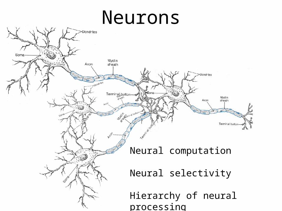
Neurons
Neural computation
Neural selectivity
Hierarchy of neural processing
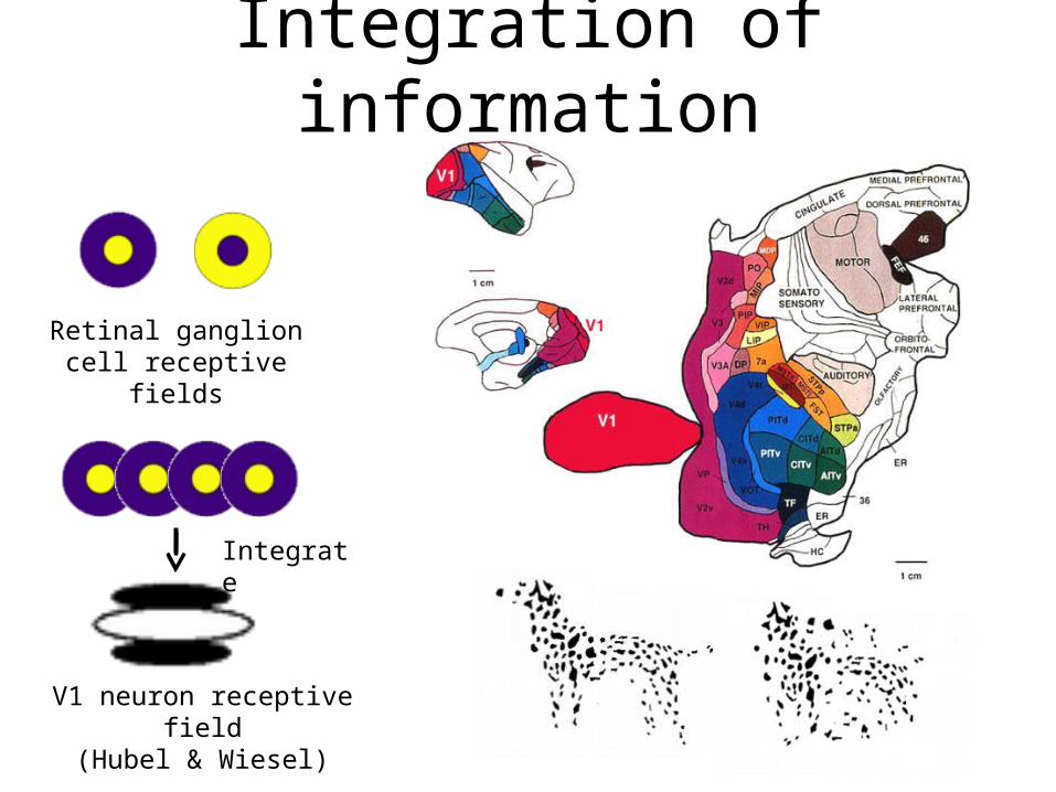
Integration of information
Retinal ganglion cell receptive fields
V1 neuron receptive field(Hubel & Wiesel)
Integrate

Grandma cell vs. distributed populationSparse codingNarrow selectivity

Grandma cell vs. distributed populationDistributed codingBroad selectivity
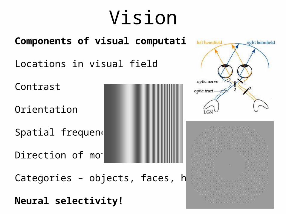
VisionComponents of visual computation:
Locations in visual field
Contrast
Orientation
Spatial frequency
Direction of motion
Categories – objects, faces, houses
Neural selectivity!
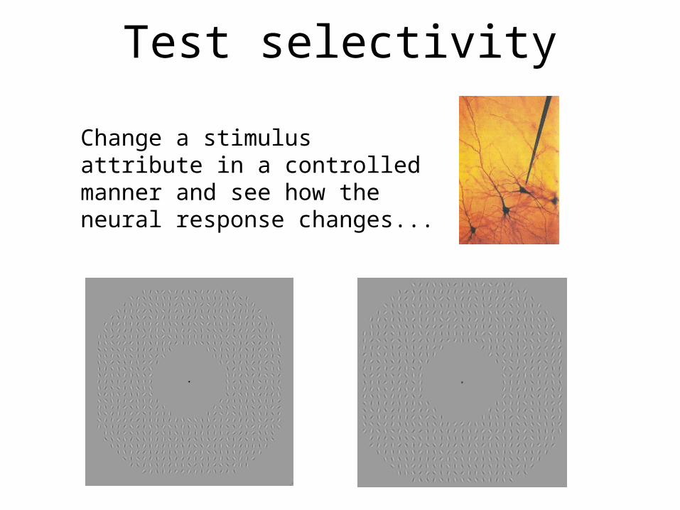
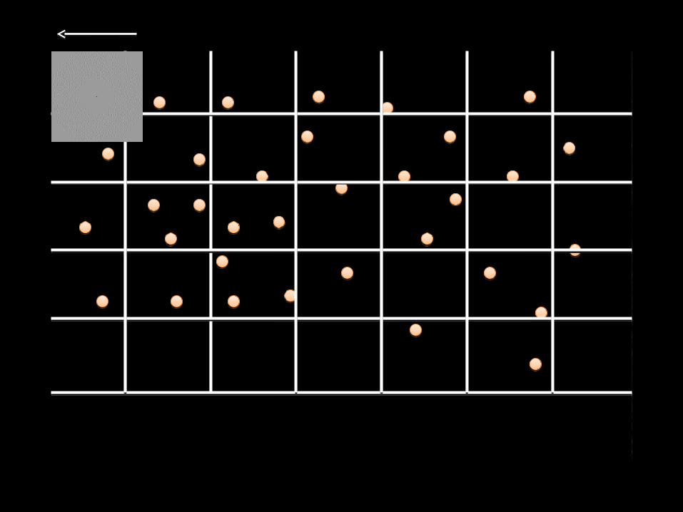
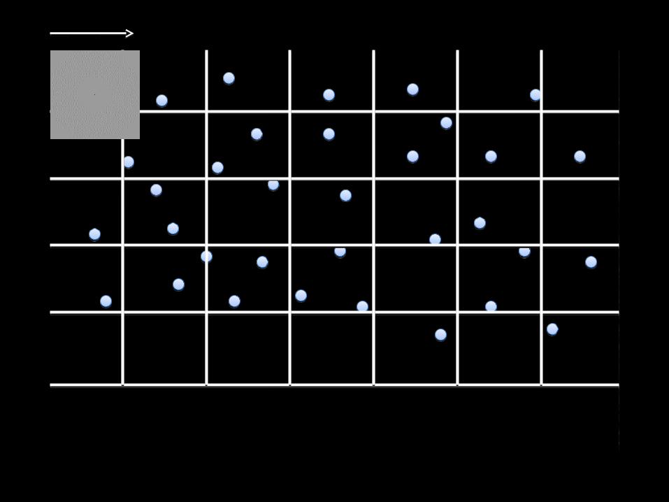
Test selectivity
Change a stimulus attribute in a controlled manner and see how the neural response changes...
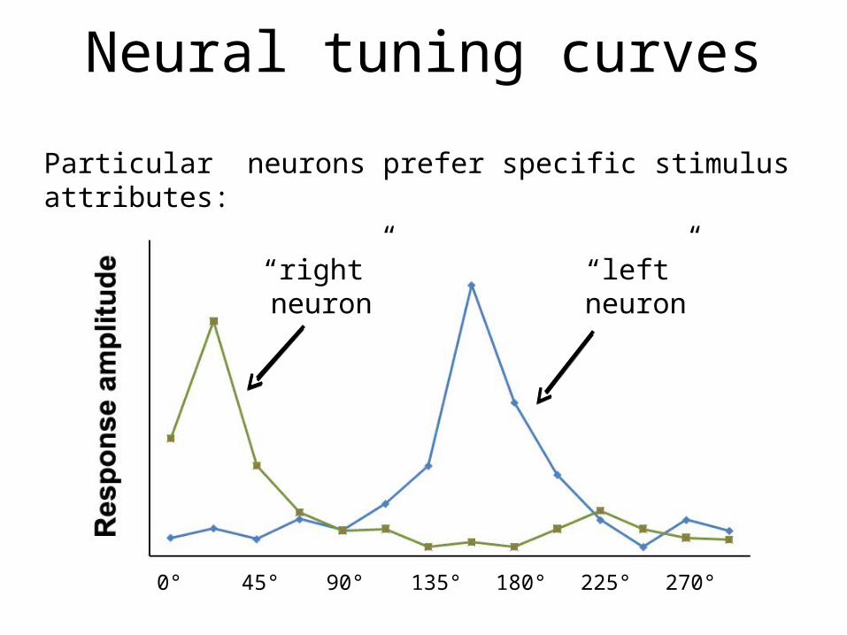
Neural tuning curves
Particular neurons prefer specific stimulus attributes:
“right” neuron “left” neuron
0° 45° 90° 135° 180° 225° 270°

Similar tuning within a column
Organization of orientation selective neurons in primary visual cortex.
200-300 µm wide
Cortex is 2-4 mm thick
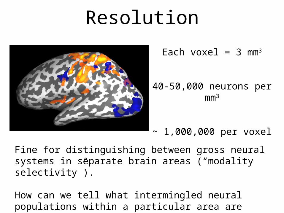
Each voxel = 3 mm3
40-50,000 neurons per mm3
~ 1,000,000 per voxel
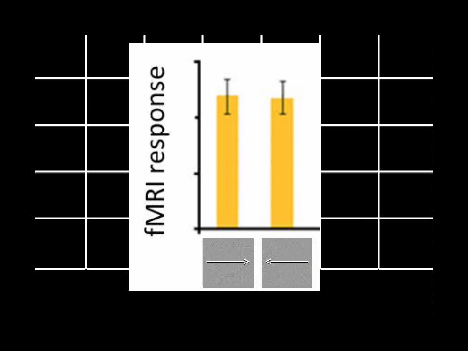
Resolution
Fine for distinguishing between gross neural systems in separate brain areas (“modality selectivity”).
How can we tell what intermingled neural populations within a particular area are selective for?

Adaptation & Classification
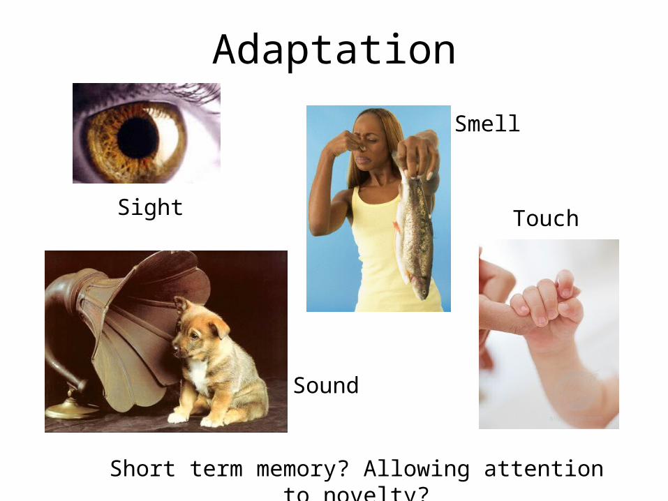
Adaptation
Sight Touch
Sound
Smell
Short term memory? Allowing attention to novelty?
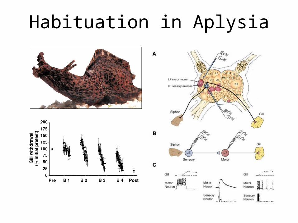
Habituation in Aplysia

Location of adaptation
Different forms of adaptation take place at different synaptic and cellular locations and have different time-scales…
There are multiple sensory neurons on the siphon, if adaptation is pre-synaptic will it generalize across locations?
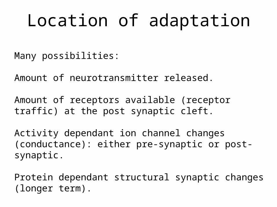
Location of adaptation
Many possibilities:
Amount of neurotransmitter released.
Amount of receptors available (receptor traffic) at the post synaptic cleft.
Activity dependant ion channel changes (conductance): either pre-synaptic or post-synaptic.
Protein dependant structural synaptic changes (longer term).

Membrane conductance change
Contrast adaptation in V1 neurons – 100’s of milliseconds
Carandini et. al. Science 1997
Tester strength (% contrast)
1.5% adapt
47% adapt
1.5% adapt
47% adapt
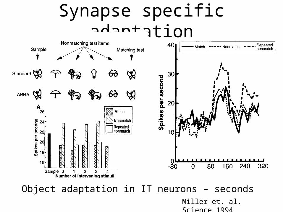
Synapse specific adaptation
Object adaptation in IT neurons – secondsMiller et. al. Science 1994

fMRI AdaptationFirst presentation Repeat
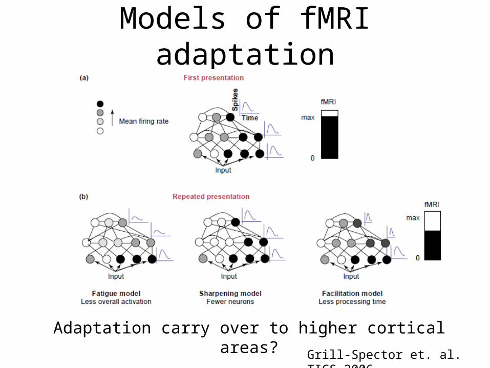
Models of fMRI adaptation
Grill-Spector et. al. TICS 2006
Adaptation carry over to higher cortical areas?
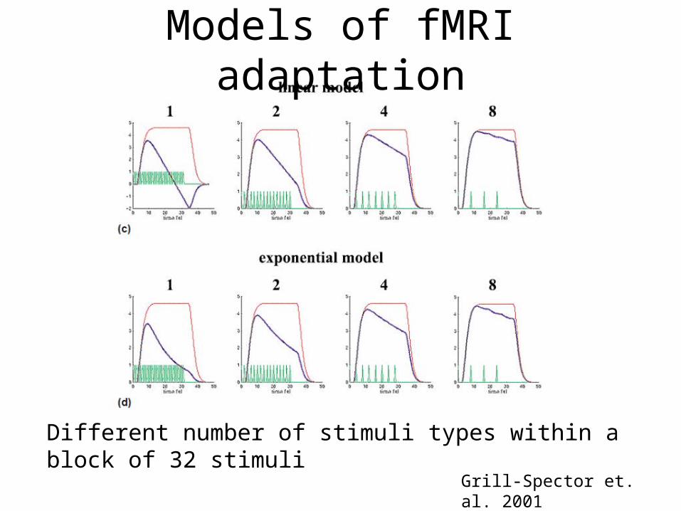
Models of fMRI adaptation
Grill-Spector et. al. 2001
Different number of stimuli types within a block of 32 stimuli
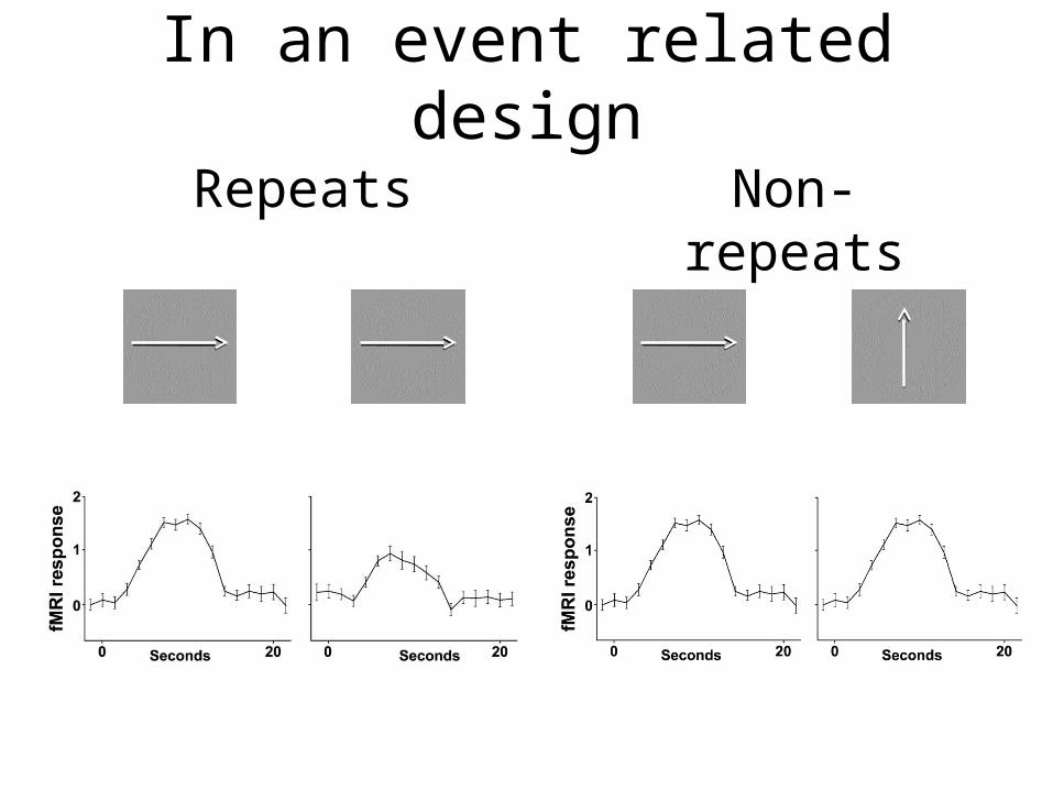
In an event related design
Repeats Non-repeats

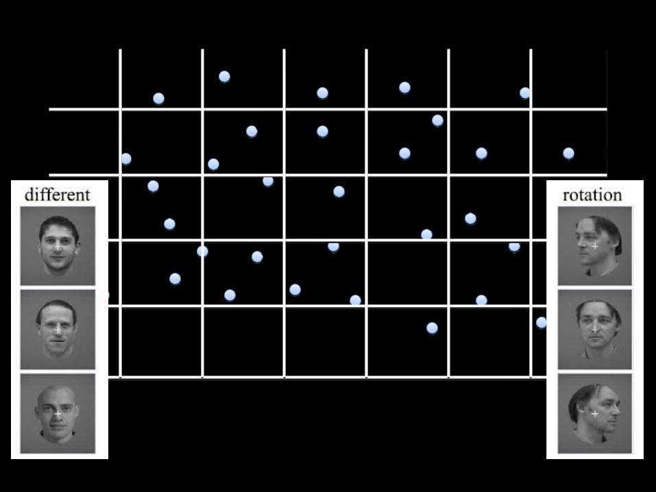
Selectivity, adaptation, & invariance
Grill-Spector et. al. 2001
A true test of “high level” selectivity would be invariance across “low level” changes…
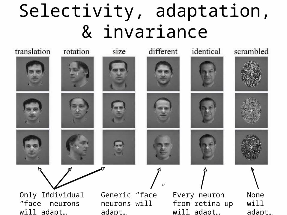
Selectivity, adaptation, & invariance
Every neuron from retina up will adapt…
None will adapt…
Generic “face” neurons will adapt…
Only Individual “face” neurons will adapt…
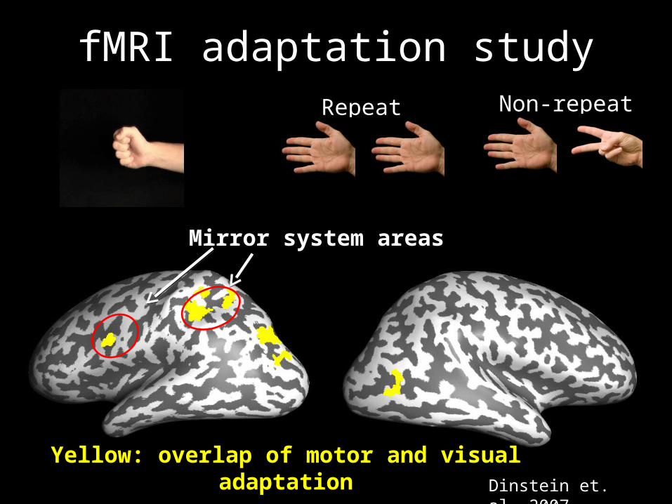
fMRI adaptation study
Yellow: overlap of motor and visual adaptation
Mirror system areas
Dinstein et. al. 2007
Repeat Non-repeat
Classification
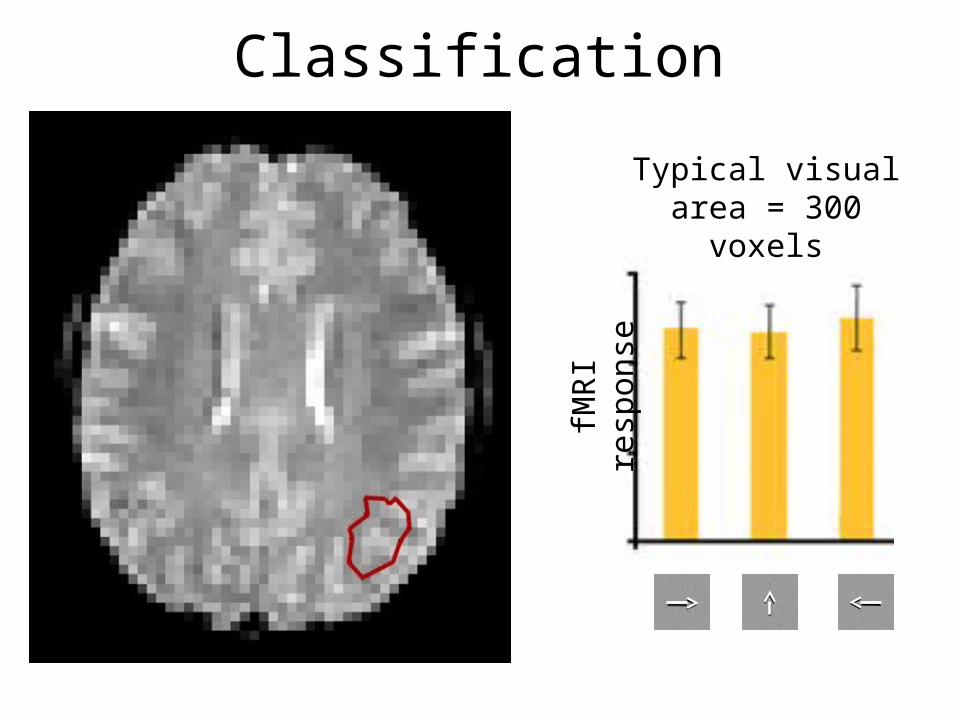
Classification
Typical visual area = 300 voxels
fMR
I re
spon
se

Classification
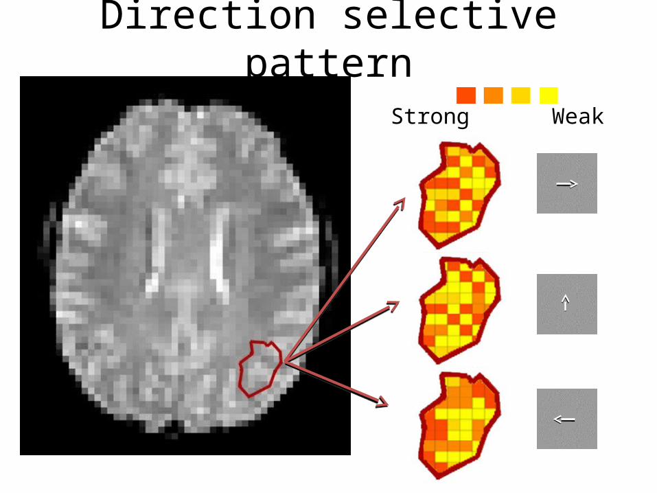
Direction selective pattern
Strong Weak
Strong Weak
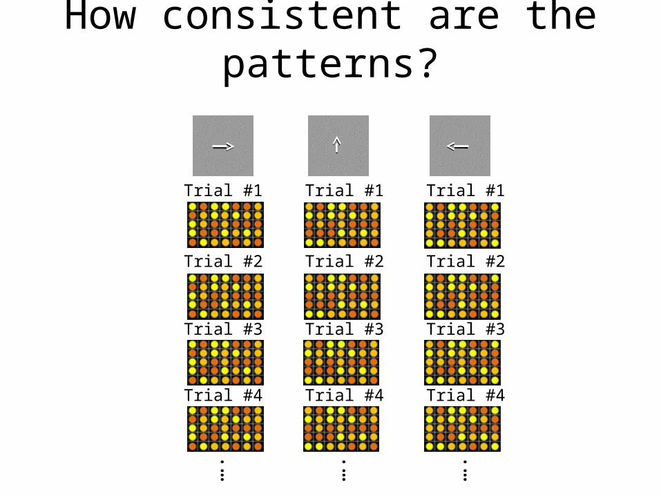
How consistent are the patterns?
Trial #1
….
….
….
Trial #2
Trial #3
Trial #4
Trial #1
Trial #2
Trial #3
Trial #4
Trial #1
Trial #2
Trial #3
Trial #4
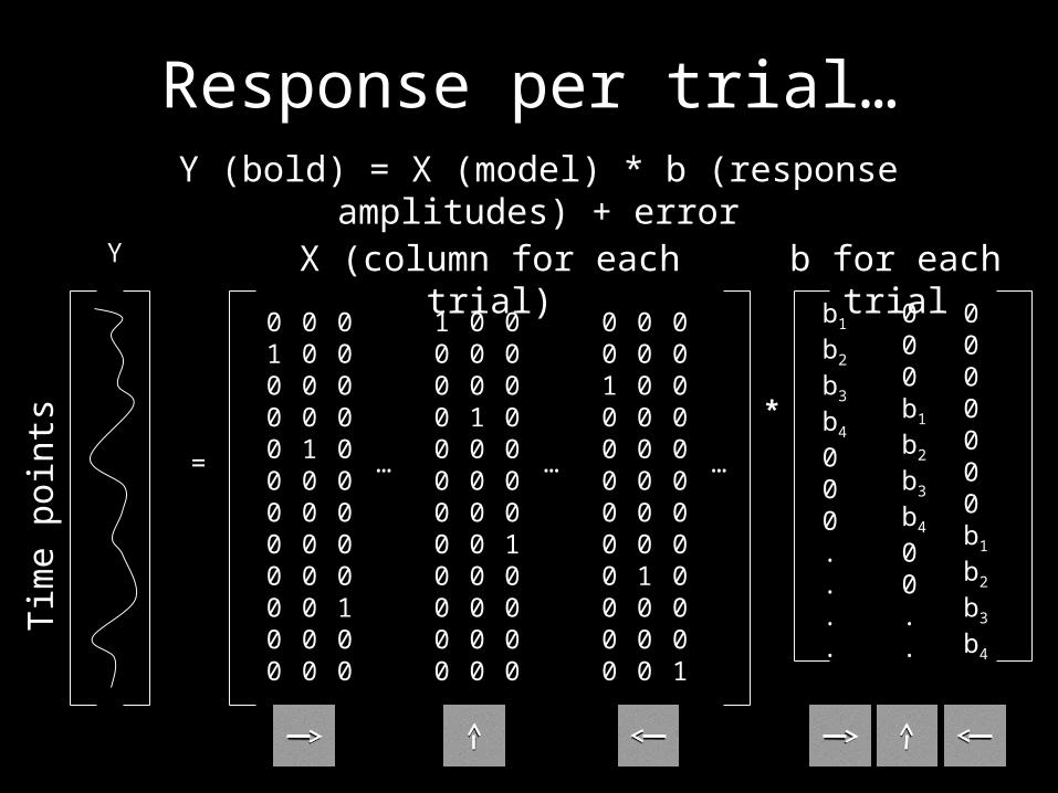
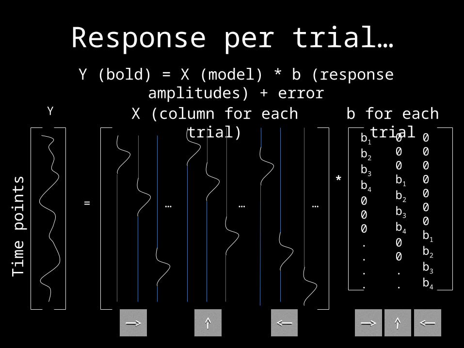
Response per trial…Y (bold) = X (model) * b (response amplitudes) + error
Y
=
Tim
e po
ints
X (column for each trial)
010000000000
000010000000
000000000100
100000000000
000100000000
000000010000
001000000000
000000001000
000000000001
… … …
b for each trial
*
b1
b2
b3
b4
000....
000b1
b2
b3
b4
00..
0000000b1
b2
b3
b4
Y
=
Tim
e po
ints
X (column for each trial)
… … …
b for each trial
*
b1
b2
b3
b4
000....
000b1
b2
b3
b4
00..
0000000b1
b2
b3
b4
Response per trial…Y (bold) = X (model) * b (response amplitudes) + error
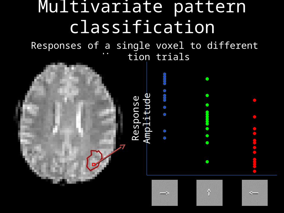
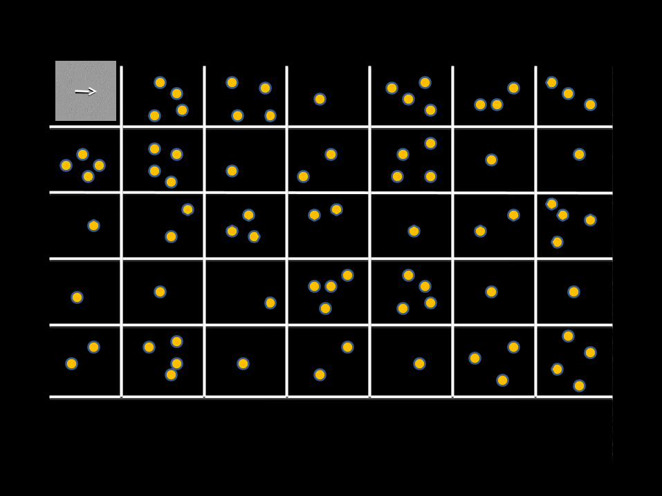
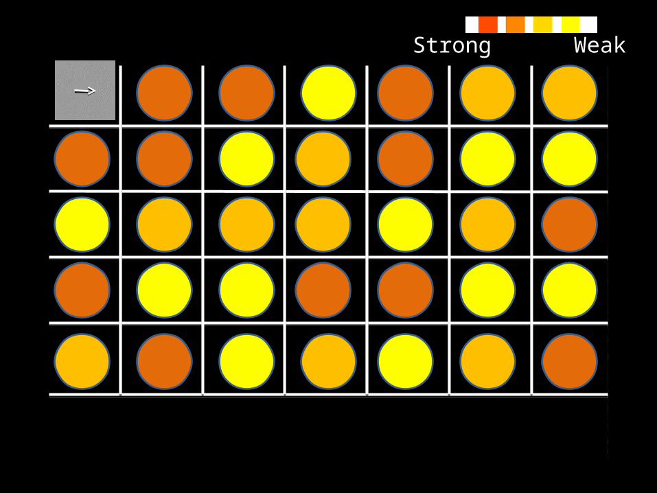
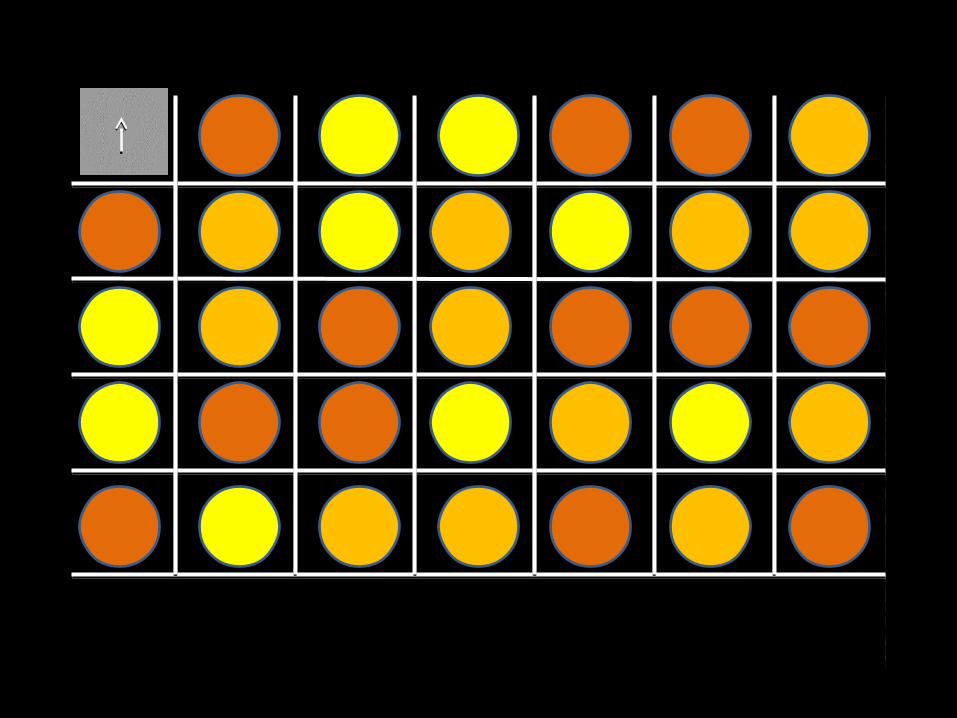
Multivariate pattern classificationResponses of a single voxel to different direction trials
Resp
onse
Am
plitu
de
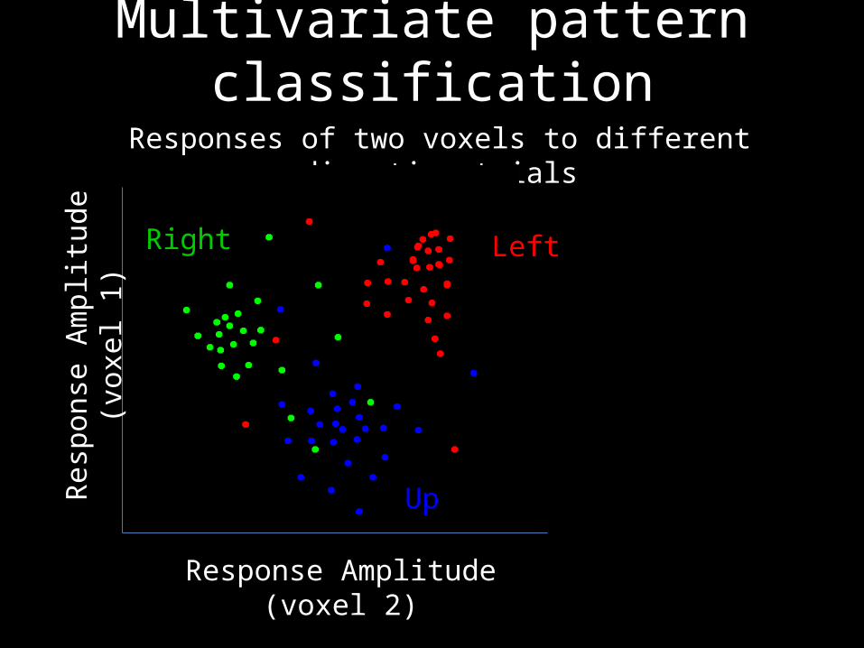
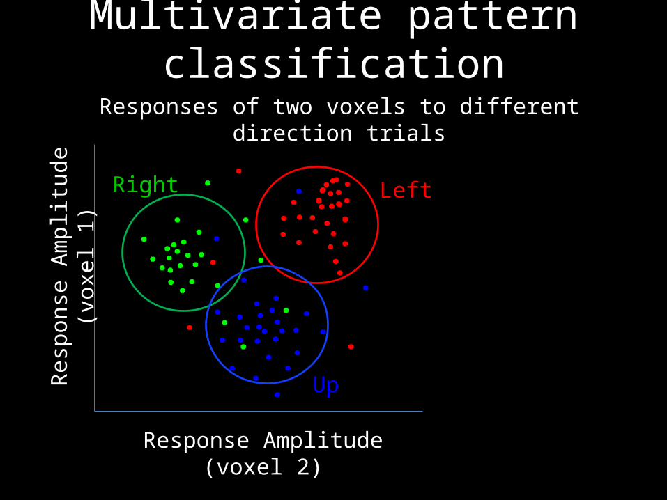
Multivariate pattern classificationResponses of two voxels to different direction trials
Res
pons
e A
mpl
itude
(vo
xel 1
)
Response Amplitude (voxel 2)
Left
Up
Right
Multivariate pattern classificationR
espo
nse
Am
plitu
de (
voxe
l 1)
Response Amplitude (voxel 2)
Left
Up
Right
Responses of two voxels to different direction trials

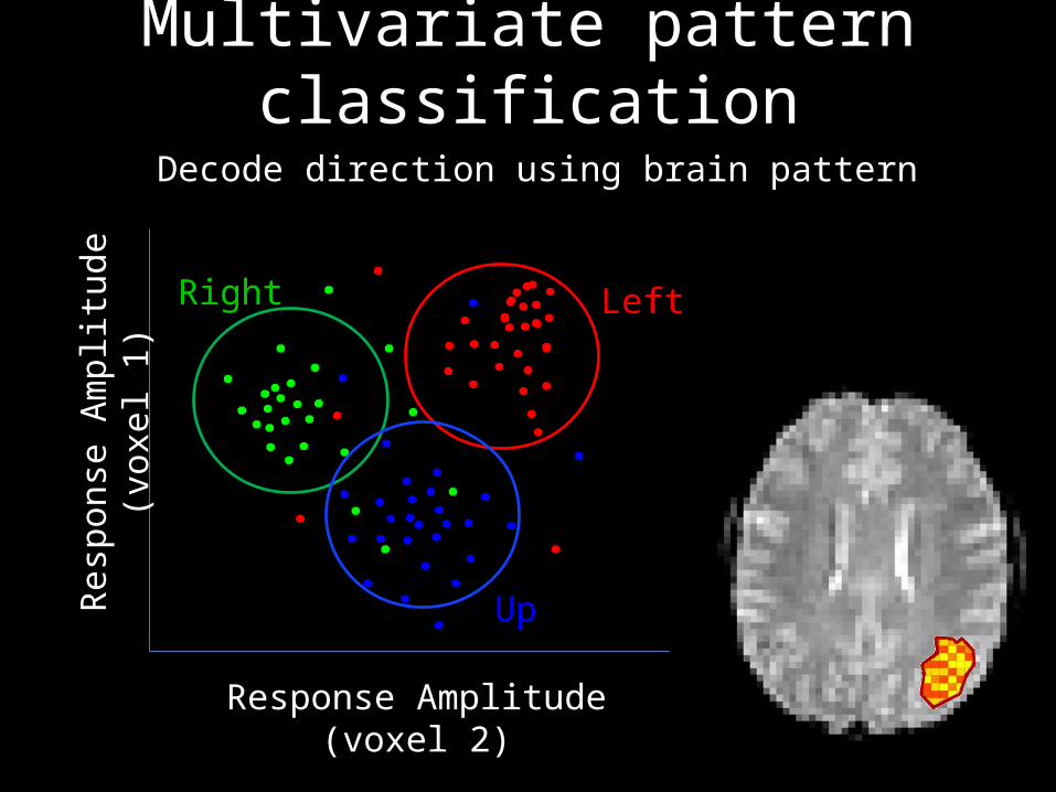
Multivariate pattern classificationDecode direction using brain pattern
Res
pons
e A
mpl
itude
(vo
xel 1
)
Response Amplitude (voxel 2)
Left
Up
Right
Left?
Multivariate pattern classificationDecode direction using brain pattern
Res
pons
e A
mpl
itude
(vo
xel 1
)
Response Amplitude (voxel 2)
Left
Up
Right
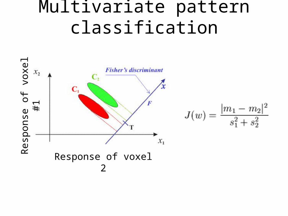
Multivariate pattern classificationR
espo
nse
of v
oxel
#1
Response of voxel 2

Decode trials (leave one out)
Trial #1
….
Trial #2
Trial #3
Trial #4
Trial #1
….
Trial #2
Trial #3
Trial #4
Trial #1
….
Trial #2
Trial #3
Trial #4

Decode direction trialsD
ecod
ing
accu
racy
Area MT Motor cortex
Chance

Decoding orientation in visual areas
Kamitani et. al. Nat. Neurosci 2005
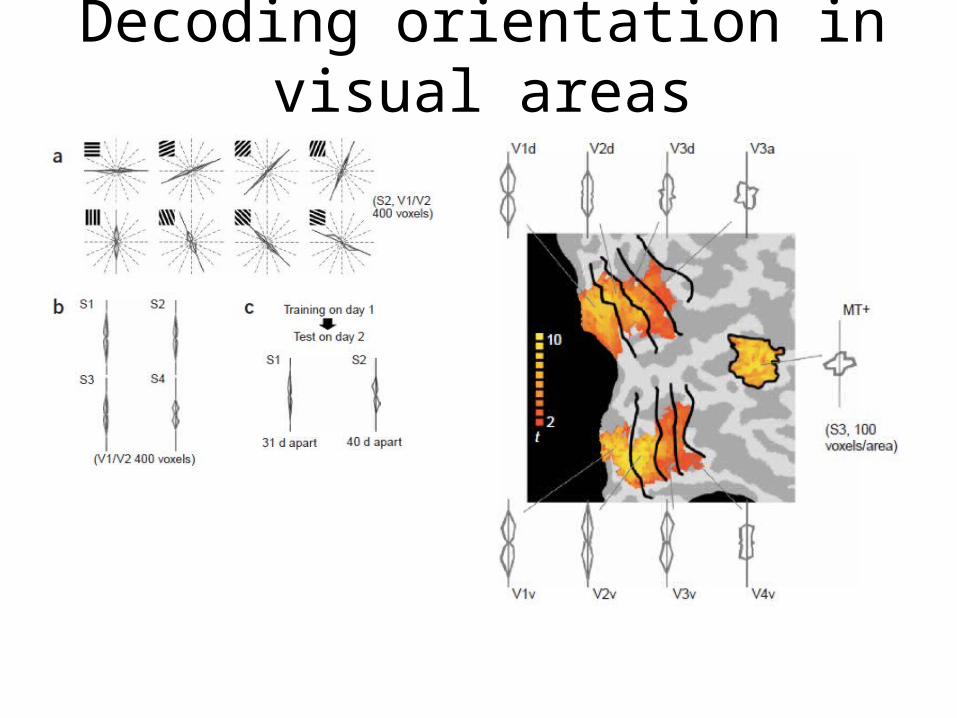
Decoding orientation in visual areas
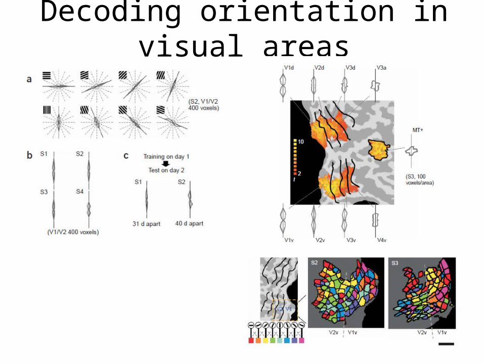
Decoding orientation in visual areas
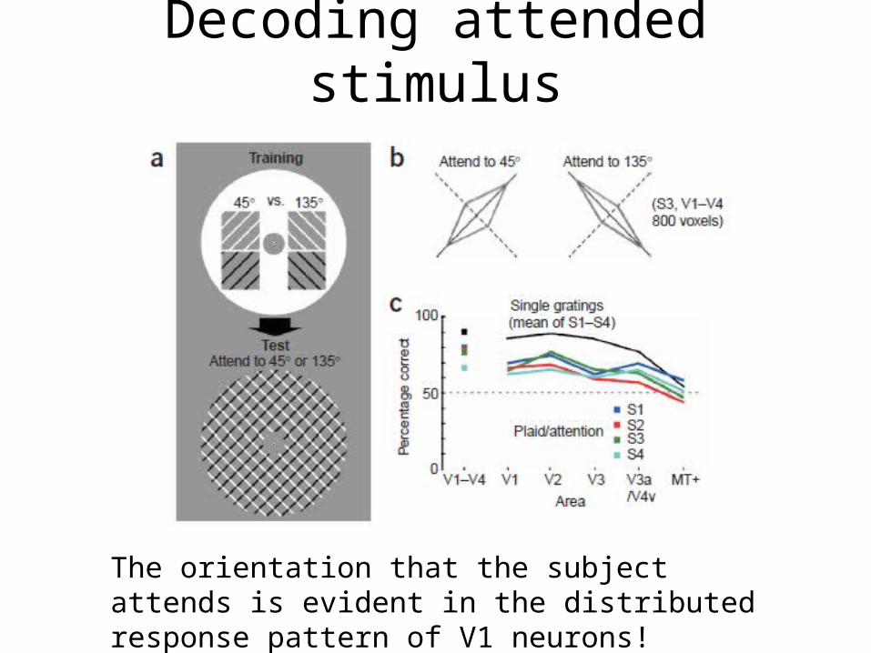
Decoding attended stimulus
The orientation that the subject attends is evident in the distributed response pattern of V1 neurons!

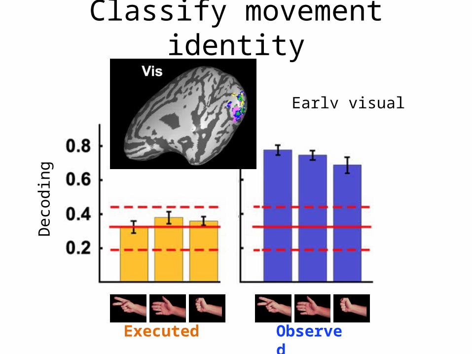
Classify movement identity
Dinstein et. al. 2008
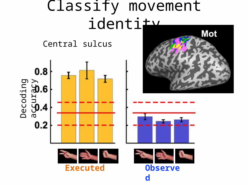
Classify movement identityD
ecod
ing
accu
racy
Central sulcus
Executed Observed
Classify movement identityD
ecod
ing
accu
racy
Early visual areas
Executed Observed
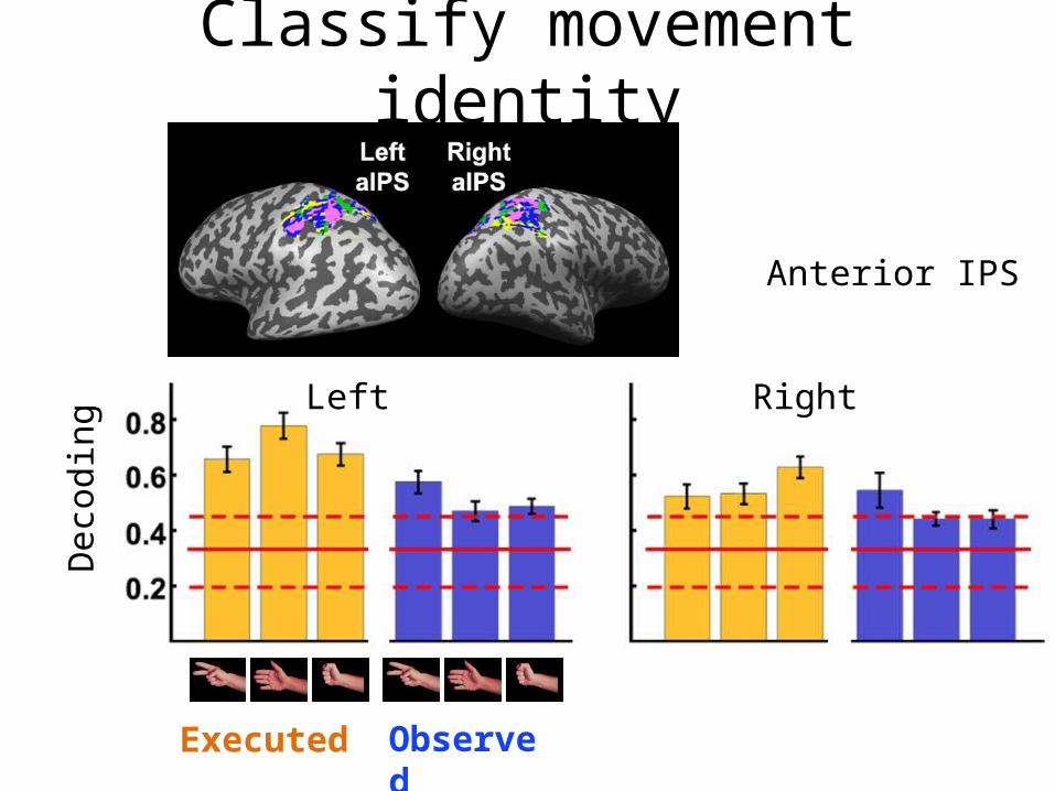
Classify movement identityD
ecod
ing
accu
racy
Anterior IPS
Executed Observed
Left Right

To the lab!