fluid flow increases membrane permeability to merocyanine 540 in human endothelial cells
TRANSCRIPT

ELSEVIER Biochimica et Biophysica Acta 1191 (1994) 209-218
Biochi f ic~a et Biophysica A~ta
Fluid flow increases membrane permeability to merocyanine 540 in human endothelial cells
Francois Berthiaume, John A. Frangos *
Department of Chemical Engineering, The Pennsyh,ania State University, University Park, PA 16802, USA
(Received 6 August 1993; revised manuscript received 24 November 1993)
Abstract
Fluid shear stress is a ubiquitous stimulus of mammalian cell metabolism; however, its signal transduction pathway is unknown. We hypothesized that shear stress may alter some physical properties of the cell membrane. Using primary human umbilical vein endothelial cells (HUVECs), we investigated the effects of shear on the cell membrane by monitoring flow-induced changes in the uptake of the amphipath merocyanine 540 (MC540). Under static conditions, MC540 was rapidly internalized by HUVECs at 37°C, and so was the membrane impermeant dye lucifer yellow, suggesting that the MC540 uptake was partly due to endocytosis. However, exposure to steady flow for 5 min at 37°C induced an increase in MC540 uptake while that of lucifer yellow was unchanged, suggesting that the flow-induced increase in MC540 uptake was not endocytosis-related. The increase in MC540 uptake was significant for levels of steady shear of 6 dyne/cm 2 and above. Pulsatile flow was more stimulatory than steady flow at 2 dyne/cm 2, but no significant difference between the two was seen at higher shear stress levels. We conclude that fluid shear stress enhanced the uptake of MC540 by a mechanism other than endocytosis, suggesting an increase in plasma membrane permeability during exposure of the cells to shear stress.
Key words: Flow; Shear stress; Membrane permeability; Merocyanine 540; Endothelial cell
1. Introduct ion
A variety of cell types, such as endothelial cells, osteoblasts, kidney epithelial cells, and fibroblasts have been shown to respond to fluid shear stress in vitro [1-6], suggesting that shear stress is a ubiquitous stimu- lus of mammalian cell metabolism. While shear stress is one of the environmental factors that cells can apparently detect in addition to hormonal signals and other physical forces, the mechanism by which shear stress stimulates ceils is unclear. In endothelial cells, there is evidence that activation of guanine nucleotide- binding proteins (G proteins) is implicated in flow-in- duced responses [7-8]; however, the primary mecha-
* Corresponding author. Fax: + 1 (814) 8657846. Abbreviations: BSA, bovine serum albumin; DPBSS, Dulbecco's modified phosphate-buffered saline; G proteins, guanine nucleotide- binding protein; HBSS, Hank's balanced salt solution; HUVEC, human umbilical vein endothelial cell; MC540, merocyanine 540.
0005-2736/94/$07.00 © 1994 Elsevier Science B.V. All rights reserved SSD! 0005-2736(93)E0391-M
nism by which fluid shear stress would activate these proteins has not been elucidated. It has been previ- ously suggested that a hyperpolarizing potassium chan- nel may be a potential flow sensor in endothelial cells [9]; however, whether this channel was directly sensi- tive to shear stress or activated as the result of earlier flow-induced events was not determined. Stretch- activated calcium channels have been reported in en- dothelial ceils, and the suggestion was made that they may be the primary flow sensors [10]; however, their role in flow-induced stimulation has not been thor- oughly investigated. Recently, several authors have suggested that at the onset of flow over an endothelial cell monolayer, a substantial increase in the levels of ATP (a component often present in the culture medium) occurs near the cell surface [11-15]. This increase appears to partially mediate a rapid flow-in- duced increase in intracellular calcium levels [11-15], and thus may be involved in other flow-dependent responses, given the role of intracellular calcium in agonist stimulation of endothelial cells (reviewed in [16]). Short-term effects induced by flow have been

210 F. Berthiaume, J.A. Frangos /Biochimica et Biophysica Acta 1191 (1994) 209-218
observed in ATP-free medium as well [8,13,17]. Recent evidence suggests that flow stimulates the release of endogeneous ATP from endothelial cells, which may be involved in the response of endothelial cells to flow observed in ATP-free solutions [18-20]. However, the primary mechanism of flow-induced ATP release from endothelial cells has not been elucidated. While ATP is known to be a potent agonist for endothelial cells, ATP receptors are also rapidly desensitized [21-22]. Thus an ATP-mediated mechanism may account for some of the short-term responses observed in endothe- lial cells, but less likely involved in the long term effects observed during chronic exposure to flow, in- cluding the reduction of endothelin and fibronectin secretion [23-25], and the increase in prostacyclin and tissue plasminogen activator synthesis [1,26,27].
Few investigators have studied the effect of physio- logical levels of fluid shear stress and other mechanical forces on the cell plasma membrane itself. Reports indicate that the passive permeability of erythrocytes to calcium ions is increased by shear stress [28], and gentle poking of the cell surface triggers intracellular calcium spikes in endothelial cells [29], suggesting that the plasma membrane of cells is sensitive to mechani- cal perturbations. The role of the phospholipid bilayer in these effects may be important as suggested by a study in a bacterial cell system showing that the gating force for stretch-activated channels could come from the surrounding lipids [30]. In addition, the permeabil- ity of phosphatidylcholine vesicles to calcium ions is increased by shear stress [31], which supports the no- tion that shear may have a direct effect on the lipid bilayer of cells.
We hypothesized that fluid shear stress may cause a perturbation in the cell membrane which would lead to the activation of membrane-bound proteins, thereby transducing the primary mechanical stimulus into a biochemical signal leading to a cellular response. Using endothelial cells as a model system, we first sought to detect a physical change in the plasma membrane during exposure to shear stress by measuring changes in plasma membrane permeability. Since endothelial cells possess intracellular stores of calcium and calcium channels, methods used elsewhere for erythrocytes and phospholipid vesicles could not be used. We therefore developed a new methodology using the anionic am- phipath merocyanine 540 (MC540), a membrane po- tential-sensitive fluorescent dye which has previously been shown to preferentially bind to loosely packed phospholipid membranes [32]. We found that although cellular uptake of the dye under static conditions was likely partially due to endocytosis, flow induced a rapid increase in uptake rate that was not endocytosis-medi- ated, suggesting that fluid shear stress caused an in- crease in the plasma membrane permeability of the membrane.
2. Materials and methods
Materials Dulbecco's modified phosphate buffered saline
(DPBSS), Hank's balanced salt solution (HBSS, Ca 2+ & Mg 2+ free) and Medium 199 were purchased from Irvine Scientific (Santa Anna, CA, USA). Defined fetal bovine serum was from Hyclone (Logan, UT, USA). Collagenase powder (type A) was obtained from Boehringer Mannheim (Indianapolis, IN, USA). c- Glutamine (200 mM in water), penicillin-streptomycin (respectively 5000 U/ml and 5 mg/ml in 0.9% NaCl), trypsin powder (1:250, from porcine pancreas), ethy- lene-diamine tetraacetic acid (EDTA, disodium-cal- cium salt), 2% gelatin solution in DPBSS, bovine serum albumin (BSA, fraction V), heparin, ionomycin, and Trypan blue stain were from Sigma (St. Louis, MO, USA). Sodium bicarbonate was obtained from Fisher Scientific (Fair Lawn, N J, USA). Endothelial mitogen was from Biomedical Technologies (Stoughton, MA, USA). Merocyanine 540 (MC540) was purchased from Eastman Kodak (Rochester, NY, USA), and lucifer yellow was from Molecular Probes (Eugene, OR, USA).
Cell culture Endothelial cells were obtained from human umbili-
cal cord veins treated with a 0.02% solution of collage- nase in DPBSS for 30 min at 25°C (adapted from [33]). The cells were resuspended in culture medium consist- ing of Medium 199 supplemented with 2 mM L-gluta- mine, 50 U/ml penicillin, 50/~g/ml streptomycin and 20% fetal bovine serum. Glass slides (75 × 38 mm, Corning, NY, USA), precoated with 0.2% gelatin in DPBSS, were each wetted with a meniscus of 1.5 ml of cell suspension. Cells harvested from 4-5 cords were plated on 8-10 slides, resulting in a plating density of 1.5-3.5.104 cells/cm 2. The cells were kept in the incubator at 37°C in air with 5% CO 2 for 24 h, after which the slides were rinsed with DPBSS and refed with 7.5 ml of culture medium each, and incubated until confluency (2-3 more days). Some slides were plated at a 3 fold less seeding density and fed with the same medium supplemented with 25 /.Lg/ml of en- dothelial mitogen and 5 U/ml of heparin until the cells reached confluency (6-7 more days).
Flow systems The cells were subjected to fluid flow as described
previously [26]. Briefly, a glass slide with a confluent monolayer of HUVECs attached to it was maintained on a plastic plate with a silicon gasket in between, thereby forming a rectangular channel (height: 250 /xm, width: 2.5 cm, length: 6 cm) above the cell mono- layer. Carefully designed entry and exit ports in the plastic plate allowed the channel to be perfused with culture medium under well-defined flow conditions.

F. Berthiaume, J.A. Frangos /Biochimica et Biophysica Acta 1191 (1994) 209-218 211
Flow was driven by a constant hydrostatic pressure head (generated by continuously pumping medium to the top of a reservoir above the flow chamber) to create steady flows with associated wall shear stresses of 6 dyne / c m 2 and above. Lower shear stresses were obtained by pumping the medium using a syringe pump (Harvard Apparatus, South Natick, MA, USA). Steady flow rates ranging from 2.8 to 40 ml /min were used. The corresponding Reynolds number was below 27, which is clearly in the laminar regime, and the wall shear stress could be determined from the equation
6/z r = h - ~ Q (1)
where /z is the viscosity of the medium (/x ~- 0.01 g / c m per s), h and b are the height and width of the channel, respectively (h = 0.025 cm, b = 2.5 cm), and Q is the flow rate.
Pulsatile flow was created by a system including a syringe driven by a motor and a set of one-way valves connected to the same flow chamber as used for steady flow experiments [34]. The flow rate was varied accord- ing to the following function of time (t):
Q = A s i n ( 2 ~ - t ) 0 < t < 0 . 5 s Q = 0 0.5 < t < ls (2)
where A is the flow amplitude, which was between 0.17 and 2.1 ml/s . This flow cycle was repeated every second. The Womersley number for an infinite parallel plate flow channel is
h w - v'7/,,, (3)
where h is the channel height, u is the kinematic viscosity of the perfusing medium, and w is the fre- quency of pulsation. Substituting the values of the parameters relevant to our flow system (h = 0.25 cm, ~,=0.01 cmZ/s, w = l s - l ) , we find W=0.25. Since W < 1, a pseudo-steady state may be assumed [35], and the instant value of the time-varying shear stress ~-(t) may be calculated from the flow rate Q(t) using equa- tion (1). The shear stress values for pulsatile flow experiments reported below represent the time-aver- aged wall shear stress calculated over the first half of the cycle, given by the equation
61z ~'a,g 7rh2b A (4)
Staining procedure The procedure for staining cells with MC540 was
adapted from [36] and [37]. To avoid any artefacts due light exposure, which is known to affect MC540 uptake by cells [36], all staining (for both static and sheared cells) was performed under low light conditions in a
temperature-controlled tent at 37°C, except for some monolayers were stained at 0°C by keeping the dish containing the cells on ice during exposure to the staining solution.
The cell monolayers were washed with DPBSS and then exposed to 20 ml of DPBSS containing 10 tzg/ml of MC540, 15 m g /m l lucifer yellow, and 0.15% BSA. Static controls were stained in regular Petri dishes, while the sheared cells were exposed to the staining solution in the appropriate shear apparatus. The cells were then rinsed 3 times with DPBSS and incubated with 0.2% Trypan blue in DPBSS for 5 min to quench the fluorescence from stained dead cells [36].
Stained monolayers could be visualized in an up- right Nikon Optiphot microscope. MC540 fluorescence was visualized using a 546/10 nm exciter, a 580 nm dichroic mirror and a 580 nm barrier filter, and lucifer yellow fluorescence was visualized using a 450-490 nm exciter, a 510 nm dichroic mirror and a 520 nm barrier filter. Pictures were taken using 1600 ASA color film. Camera exposure was set automatically except for pho- tography of MC540 fluorescence, where exposure time was limited by the rapid photobleaching of the dye (a few seconds). Fluorescence intensity distributions were obtained by feeding the cell suspensions (approxi- mately 10 6 cells/ml) to an Epics 753 flow cytometer (Coulter Electronics, Hialeah, FL, USA). The excita- tion light was provided by an argon laser tuned at 488 nm with a power output of 100 mW and passed through a 457-504 nm laser blocking filter and a 550 nm long pass dichroic filter. The lucifer yellow fluorescence was detected through a 525 nm band pass filter and the MC540 fluorescence through a 575 nm band pass filter and a 570 nm long pass filter. The high voltage gains used were 975 V with 10% subtraction and 925 V with 50% subtraction for lucifer yellow and MC540, respec- tively. With these settings, control cells stained with only one of the dyes showed fluorescence in only one channel at a time.
For quantitative measurements, the cells were ex- posed to 20 ml of DPBSS containing 10 /xg/ml of MC540 and 0.015% to 1.5% BSA for times ranging from 1 min to 10 min. They were then rinsed twice in HBSS and incubated with 1 ml of 0.05% trypsin and 0.02% ED TA in HBSS for 2 min. To stop the tryptic activity, 5 ml of serum-containing culture medium was added, the cells were pelleted and resuspended in 1 ml of DPBSS and kept on ice. 1 ml of 3% formaldehyde was added. After 15 min, the cells were centrifuged at 1700 rpm for 5 min at 4°C and resuspended in 1 ml of DPBSS. Fluorescence intensity was measured in a spectrofluorometer with excitation and emission wave- lengths set at 554 nm and 590 nm respectively. Ab- sorbance was measured at 540 nm in a Microplate Autoreader (Bio-Tek Instruments, Highland Park, VT, USA). All fluorescence intensities were normalized to
212 F. Berthiaume, ZA. Frangos /Biochimica et Biophysica Acta 119l (1994) 209-218
cel l a b s o r b a n c e and a u t o f l u o r e s c e n c e , as d e t e r m i n e d
f r o m u n s t a i n e d samples , was sub t r ac t ed . T h e ab-
s o r b a n c e was d i rec t ly p r o p o r t i o n a l to cel l n u m b e r and
was u n a f f e c t e d by the smal l p e r c e n t a g e o f T r y p a n b l u e
s t a i ned cei ls and the smal l a m o u n t o f c e l l - b o u n d MC540 .
T h e r e l a t i o n b e t w e e n the f l u o r e s c e n c e in tens i ty o f
s t a ined cel ls and ac tua l a m o u n t o f M C 5 4 0 in the cel ls
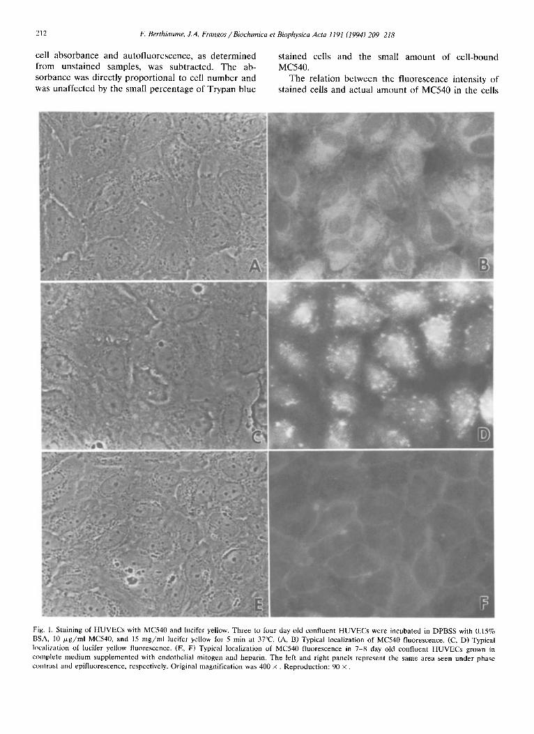
Fig. 1. Staining of HUVECs with MC540 and lucifer yellow. Three to four day old confluent HUVECs were incubated in DPBSS with 0.15% BSA, I0 p~g/ml MC540, and 15 mg/ml lucifer yellow for 5 min at 37°C. (A, B) Typical localization of MC540 fluorescence. (C, D) Typical localization of lucifer yellow fluorescence. (E, F) Typical localization of MC540 fluorescence in 7-8 day old confluent HUVECs grown in complete medium supplemented with endothelial mitogen and heparin. The left and right panels represent the same area seen under phase contrast and epifluorescence, respectively. Original magnification was 400 x . Reproduction: 90 x .

F. Berthiaume, J.A. Frangos /Biochimica et Biophysica Acta 1191 (1994) 209-218 213
was determined as follows. Several slides with HU- VECs were stained with MC540 as described above, but without the Trypan blue treatment. Each cell sus- pension was split in half, one half for measuring the fluorescence as usual, while the other half of the sample was extracted with n-pentanol, after which the fluorescence was measured. A standard curve of fluo- rescence intensity for MC540 in n-pentanol with con- centrations ranging between 0 and 10 txg/ml was used to convert fluorescent readings into pg MC540.
3. Results
Spatial localization of the fluorophores HUVECs were stained with MC540 and lucifer yel-
low for 5 min at 37°C under static conditions and observed microscopically. Primary HUVECs grown to confluence for 3 -4 days and stained with MC540 dis- played bright fluorescent areas in regions outlining the cell nuclei, suggesting that the dye entered the cells (Figs. 1 A,B). Dye internalization was confirmed by the absence of MC540 fluorescence quenching of viable cells by Trypan blue stain (not shown). The cell edges retracted slightly when exposed to the dye, but this was reversible upon removal of the staining solution and addition of culture medium (not shown). Lucifer yellow staining was visible as discrete bright spots, and a fluorescent halo was seen over most of the surface of the cells (Figs. 1 C,D). HUVECs seeded sparsely and grown for 7 -8 days in the presence of endothelial mitogen stained weakly with MC540 with the brighter fluorescence visible at the cell boundaries (Figs. 1 E,F).
Flow cytometry Cells were stained with both dyes under static and
flow conditions (the shear stress used was 25 dyne /cm 2) for 5 min. The results in Fig. 2 show that the fluores- cence distribution of the MC540 signal for the cells stained while under shear was shifted to the right when compared to that of cells stained under static condi- tions, thereby indicating an increase in dye uptake. The corresponding results for the lucifer yellow signal showed little difference between cells stained under static and flow conditions.
The effect of cold temperature on the cellular up- take of MC540 and lucifer yellow under static condi- tions is shown in Fig. 3. At 0°C, the rate of dye uptake was visibly reduced compared to that seen at 37°C, therefore to facilitate the comparison between dye uptake at both temperatures, cells stained at 0°C were exposed to the dye solution for 10 min, and cells stained at 37°C were exposed to the dye solution for 3 rain. Cells stained for 10 rain at 0°C displayed a peak in their MC540 fluorescence distribution that was two-fold less compared to cells stained for only 3 min at 37°C;
256
. a
E - j
Z
O)
192
128
64
0 10 100
MC540 Fluorescence
10 100 1000
Luc Yel Fluorescence
Fig. 2. Effect of flow on uptake of MC540 and lucifer yellow by HUVECs. Three to four day old confluent HUVECs were stained with MC540 and lucifer yellow for 5 min at 37°C under static conditions ( ) and flow conditions with a wall shear stress of 25 dyne/cm 2 ( . . . . . . ). The fluorescence intensity distributions of MC540 and lucifer yellow within the cell population are shown in the left and right panels, respectively.
however, most cells still stained with the dye. Most cells stained with lucifer yellow at 37°C, but there was a dramatic reduction in lucifer yellow uptake at 0°C since lucifer yellow staining was not detectable in 69% of the cells.
Quantitative analysis of MC540 uptake The time course of uptake of MC540 by HUVECs
under static conditions and flow conditions (shear stress = 25 dyne /cm 2) is shown in Fig. 4. In the ab- sence of flow, dye uptake was roughly linear for up to 5 min, after which this rate appeared to decrease. In the presence of flow, the uptake of MC540 under shear was not different from that of cells stained under static conditions for the first 3 rain, but was significantly higher after 5 and 10 min of exposure to the dye.
An increase in BSA concentration caused a marked increase in the fluorescence of staining solution, sug- gesting an interaction between MC540 and BSA (Fig.
256
192
E = 128
64
10 100
MC540 Fluorescence
10 100
Luc Yel Fluorescence
1000
Fig. 3. Effect of temperature on uptake of MC540 and lucifer yellow by HUVECs. Three to four day old confluent HUVECs were stained under static conditions with MC540 and lucifer yellow for 3 rain at 37°C ( ) or I0 min at 0°C ( . . . . . . ). The fluorescence intensity distributions of MC540 and lucifer yellow within the cell population are shown in the left and right panels, respectively.

214 F. Berthiaume, J.A. Frangos / Biochimica et Biophysica Acta 1191 (1994) 209-218
5A). The flow-induced increase in dye uptake as well as the uptake of MC540 under static conditions were inversely proportional to the albumin concentration in the staining solution (Fig. 5B). There was no significant enhancement in MC540 uptake due to flow at albumin concentrations of 0.5% and higher.
The effect of the level of shear stress used during staining is given in Fig. 6. MC540 uptake measured at levels of steady shear stress of 6 dyne /cm 2 and above was statistically significantly different from the control cell monolayers stained under static conditions. There was no statistically significant difference between the MC540 uptake observed at the different levels of shear stress between 2 dyne / cm 2 and 25 dyne /cm 2, how- ever, the shape of the dose response curve suggests a shear-dependent increase in MC540 uptake in the range of steady shear stresses from 0 to 13 dyne /cm 2. No further increase in MC540 uptake was observed beyond 13 dyne /cm 2. MC540 uptake for cells sub- jected to pulsatile flow peaked at average shear stresses of 2 and 25 dyne /cm 2. MC540 uptake at 2 dyne /cm 2 of pulsatile shear stress was significantly higher than that observed at the same level of steady shear stress.
To determine whether agonist stimulation affects the MC540 uptake of HUVECs, cells were stained under static conditions with MC540 for 5 min at 37°C in the presence of 1 /.~M of the calcium ionophore ionomycin. MC540 uptake in the presence of iono-
1 900- l *
oo. ::- 1
300 n ~ = s n=7
o i i g 8 io Time (rain)
Fig. 4. Time course of MC540 uptake. Three to four day old confluent HUVECs were incubated in DPBSS with 0.15% BSA and 10 p .g /ml MC540 at 37°C. Data shows total MC540 uptake under static (©) and flow conditions with a wall shear stress of 25 d y n e / c m z (D). Data pooled from experiments with three separate primary H U V E C isolates. Error bars represent the S.E. * : P < 0.05 by paired two-tailed t-test.
A 0.8
:~¢-.
o P
O.Ol . . . . . . . o'.1 . . . . . . . . i
B 8000- T
_ 800- , l ~
- ° 4 o o o . . =oo_ o o "- . . ;
\o.1 2000. n=5
O, 0.01 0~1 "1 [Albumin] (%w/v)
Fig. 5. Effect of albumin concentration on uptake of MC540 by HUVECs. Three to four day old confluent HUVECs were stained in DPBSS with 10 / zg /ml MC540 containing 0.015% to 1.5% BSA at 37°C for 5 min. (A) Fluorescence of staining solution (excitation = 554 nm, emission = 590 nm). Fluorescence of 10 ,~g /ml MC540 in n-pen- tanol, used as a reference standard, = 1. (B) MC540 uptake under static ( o ) and flow conditions with a wall shear stress of 25 d y n e / c m 2 (D). Inset shows detail of region between 0.15% and 1.5% BSA. Data pooled from experiments with 4 separate primary H U V E C isolates. Error bars represent the S.E.M. *: P < 0.05 by paired two-tailed t-test.
mycin was 725 _+ 70 pg/106 cells, and that for cells in the absence of any agonist was 780 + 144 pg/106 cells (n = 6; data pooled from experiments with three sepa- rate primary HUVEC isolates).
4. Discussion
Primary HUVECs cultured for 3 -4 days and stained with MC540 displayed distinct perinuclear fluores- cence, a staining pattern similar to that previously observed in leukemic lymphocytes, which were shown to internalize MC540 [36]. When HUVECs seeded more sparsely and grown for 7-8 days in the presence of endothelial mitogen until confluency were exposed to MC540, the cells lacked perinuclear fluorescence, suggesting that they did not significantly internalize MC540. While the reasons for this difference are un- clear, it nevertheless indicates that the entry of MC540 into the cells was not due to a nonspecific detergent-like effect of the fluorescent amphipath. Cells that stained

F. Berthiaume, J.A. Frangos / Biochimica et Biophysica Acta 1191 (1994) 209-218 215
_-- 1"8 I' n~5
l / ~ " =7
1/!--, 1 . 0 ~
0 5 1 '0 1 '5 2'0 2'5
Shear Stress (dy/cm 2)
Fig. 6. Effect of the level of steady and pulsatile shear stress on MC540 uptake. Three to four day old confluent HUVE C s were stained under steady (D) and pulsatile (z x) flow conditions with MC540 for 5 min at 37°C. The shear stress values for pulsatile flow represent the wall shear stress given by equation (4) t ime-averaged over the first half of the flow cycle. Data is expressed as ratios to static controls, which were cells stained with MC540 under stationary conditions for 5 min. Average MC540 uptake of static controls was 411+_33 pg MC540/106 cells (n = 18). Data shown is pooled from experiments with 9 separate primary H U V E C isolates. Error bars represent the S.E. *: P < 0.05 compared to static control by paired two-tailed t-test. **: P < 0 . 0 5 compared to steady flow-induced uptake with same mean shear stress by unpaired two-tailed t-test.
with MC540 also stained with the water soluble dye lucifer yellow. The localization of lucifer yellow was consistent with a entry mechanism involving at least partly endocytosis because fluorescence was seen in discrete spots, suggestive of endocytotic vesicles. Thus the cells were actively internalizing the medium con- tents, and it is likely that at least part of the MC540 uptake by HUVECs was also endocytosis-mediated. The staining pat tern of MC540, although different from that of lucifer yellow, was not inconsistent with an endocytosis-mediated mechanism. Endocytotic vesicles may fuse with internal membranes and release their content, thereby staining intracellular membranes.
Flow cytometry analysis of dye uptake by cells ex- posed to a staining solution containing both MC540 and lucifer yellow under static and flow conditions revealed an apparent flow-induced increase in MC540 uptake, while there was no change in the uptake of lucifer yellow. This suggests that the enhanced uptake of MC540 under flow conditions was not the result of a change in the rate of endocytosis. A previous study by Davies et al. had shown that a single step increase in
shear stress significantly increased the endocytotic rates of bovine aortic endothelial cells measured 30 min or later after the onset of flow [38]. Results obtained here with HUVECs suggest that flow-induced changes in endocytotic rates did not occur up to at least 5 min after the onset of flow. In the same study of Davies et al., bovine aortic endothelial cells subjected to re- peated step changes of shear stress between 3 and 13 d y n e / c m 2 with 5 min cycles did not exhibit any change in endocytotic rates when compared to controls. These findings, together with ours, suggest that the rate of endocytosis may not respond to flow changes occurring over time periods of 5 min or less.
If the flow-induced increase in MC540 was not endocytosis-mediated, it is also likely that the uptake of MC540 under static conditions was not be entirely due to endocytosis. In order to further confirm the existence of MC540 uptake mechanisms besides endo- cytosis, static HUVECs were stained at 0°C. There was a clear reduction in the uptake of lucifer yellow, which is consistent with the expected inhibition of endocy- totic vesicle formation at low temperatures. The up- take of MC540 was also reduced, but to a lesser extent. These results support the notion that endocytosis was not the only pathway of MC540 uptake by HUVECs, and that direct transport of the dye through the mem- brane was also occurring. The decrease in MC540 uptake at 0°C may reflect both the inhibition of endo- cytosis-mediated uptake and the effect of temperature on the physical properties of the plasma membrane of the cells.
The plasma membrane of many living cells is rela- tively impermeable to MC540, which is true in the case of erythrocytes, lymphocytes, monocytes and neu- trophils [39]. The polar sulfonate group present on the dye molecule presumably prevents it from going across lipid bilayers. In one previous study, leukemic lympho- cytes were found to be permeable to MC540 [36] and, as seen here, primary HUVECs cultured to confluency for 3 -4 d. The reasons which make some cells more permeable to MC540 than others are unclear at this time. The difference in permeability between normal and leukemic leukocytes was attributed to a failure of membrane maturat ion in leukemic cells [36]. Both in the latter case and in the present study, the preferred pathway of MC540 translocation through the mem- brane could not be determined. Studies on unilamellar phosphatidylcholine vesicles have suggested that mem- brane-bound MC540 monomers, after undergoing a dimerization, may go across a phospholipid membrane [40]; however, protein-lipid interfaces could also pro- vide routes of translocation in whole cells.
If the additional MC540 uptake due to flow was not the result of endocytosis, it suggests that dye uptake by permeat ion through the membrane was stimulated by flow. This could be a consequence of one or several of

216 F. Berthiaume, J.A. Frangos / Biochimica et Biophysica Acta 1191 (1994) 209-218
the following events: (1) enhanced transport of the dye to the surface of the membrane under flow conditions, (2) enhanced driving force for dye permeation across the membrane, which could be caused by an increased binding of the dye to the membrane or a change in membrane potential, and (3) increased 'porosity' of the membrane to the dye.
It may be argued that an increase in surface concen- tration of MC540 may occur at the onset of flow, and that increase may contribute to the enhanced uptake of MC540 under shear. For the static controls, measure- ments for total uptake of MC540 were of the order of 400-800 pg/106 cells after 5 min, which corresponds to an uptake rate of the order of 0.2 p g / s per cm 2, assuming 105 cel ls /cm 2. A standard diffusion-reaction model can be used to predict the concentration of MC540 at the cell surface as a function of time. With a constant uptake rate of 0.2 p g / s per cm 2, a diffusivity of 7 ' 10 -v cm2/s for albumin [41] (here we take the worst case of MC540 bound to albumin - free MC540 would have a higher diffusivity), and assuming an infi- nite height of liquid over the cells, the surface concen- tration of MC540 is predicted to decrease by 0.03 ~ g / m l after 5 rain (a solution of the analogous heat transfer problem is given in [42]), which is negligible in comparison with the initial bulk concentration of 10 ~g MC540/ml. Thus the surface concentration of MC540 was essentially uniform and constant during the time course of the experiment shown in Fig. 6, and not dependent on the presence of flow.
Two more phenomena may have occurred to explain the flow-induced increased rate of MC540 uptake: a change in membrane potential that would increase the driving force for dye entry, or a change in membrane physical properties that would favor an increased bind- ing of MC540 at the surface of the cell membrane a n d / o r an increased porosity of the membrane to MC540. The first mechanism, related to a flow-induced change in membrane potential, is not likely to be responsible for the increase in MC540 uptake because it has previously been shown that fluid shear stress hyperpolarizes the plasma membrane [9,43], which would tend to inhibit rather than favor the transloca- tion of the negatively charged MC540 to the inside of the cell. MC540 is known to bind preferentially to loosely packed phospholipid bilayers [32], and the per- meability of such bilayers to various small molecular weight compounds has been correlated to membrane fluidity, a physical property presumed to be dependent on the tightness of the phospholipid packing (reviewed in [44]). The data presented here could not distinguish whether there was increased MC540 binding or poros- ity of the membrane, but the overall permeability in- crease to MC540 induced by flow suggests that it was the result of a change in membrane physical proper- ties. Larsen et al. previously showed that physiological
levels of shear stress increased the passive permeability of red blood cells to calcium ions [30]. A similar flow- induced increase in membrane permeability has also been reported using phosphatidylcholine vesicles [31], suggesting that flow may have a general effect on cell membranes.
We found that a decrease in BSA concentration in the solution resulted not only in an increase in MC540 uptake under static conditions, but also in an enhance- ment of the relative effect of flow on MC540 uptake. Previous investigators have suggested that MC540 can bind to proteins, and that the latter can compete with cell membranes for MC540 molecules [45]. Addition of BSA to a solution of MC540 containing no protein results in a dramatic increase in the quantum yield of the dye, suggesting that BSA and MC540 do interact. It is expected that the amount of cell-bound MC540 decreases as the concentration of albumin is increased. This would decrease the driving force for dye perme- ation through the cell membrane, and therefore in- crease the relative importance of MC540 uptake via endocytosis. The apparent inhibition of the flow-in- duced increase in MC540 uptake at higher albumin concentration is consistent with a mechanism other than endocytosis-mediated. We cannot exclude the possibility that MC540 binding to the membrane itself may perturb the membrane in such a way that its leakiness to MC540 under static conditions and its sensitivity to flow would also be increased. MC540 and other amphipathic compounds have been found to act as membrane perturbants capable of inducing shape changes in erythrocytes and ion channel activation in bacterial cells [30,46-48].
A dose-dependent increase in MC540 uptake was observed in the range of shear stresses from 0 to 13 dyne /cm 2, which is in the physiologically relevant range [49]. Only a rough comparison between our results and other shear dose responses can be made because the sensitivity of endothelial cells to shear in vitro is strongly dependent on the signal measured. The shear dose response for flow-induced potassium currents in bovine aortic endothelial cells was found to cover a range from 0.2 to 17 dyne /cm 2 [9], and those for tissue-type plasminogen activator and prostacyclin production by HUVECs cover a range of 0-25 dyne /cm 2 and proba- bly beyond [25-26]. In contrast, the levels of platelet- derived growth factor messenger RNA A chain in HUVECs were maximized at 6 dyne /cm z [50]. The range of the shear dose response for MC540 uptake is comparable to that of these other flow-induced re- sponses in endothelial cells.
The shape of the dose response curve obtained with pulsatile flow was quite different from that of steady flow, particularly at the lowest non-zero shear stress used (2 dyne/cm2). There was a statistically significant difference between the two types of flow at a mean
IE Berthiaume, J.A. Frangos / Biochimica et Biophysica Acta 1191 (1994) 209-218 217
wall shear stress of 2 d y n e / c m 2, but not at higher shear stresses. The significance of this peak at low shear is unclear. There is evidence that, in certain cases, pulsatile flow is more stimulatory than steady flow. For example, HUVEC prostacyclin synthesis rates are higher when the cells are subjected to pulsatile shear stress between 8 and 12 dyne / cm 2 at a frequency of 1 Hz than if the cells are exposed to the same value of steady mean shear stress (10 dyne /cm 2) [1]. On the other hand, oscillatory shear stress between 3 and 13 dyne / c m 2 at a frequency of 1 Hz had no effect on pinocytotic rates compared to static controls while the same mean shear stress (8 dyne / cm 2) applied continu- ously caused a significant increase [38]. These apparent inconsistencies in the effect of pulsatile flow on en- dothelial cells may reflect differences in the sensitivity of endothelial cells with respect to these different responses, but may also be due to the fact there are no studies which used identical pulsatile flow wave func- tions.
We and others have previously shown that phospho- lipid turnover is stimulated in sheared endothelial cells [17,51,52], an indication that membrane-associated phospholipases become activated when endothelial cells are subjected to shear stress. It is possible that these enzymes, when activated, modify the physical proper- ties of the membrane. Since calcium ionophore had no effect on MC540 uptake, it suggests that the activation of calcium-sensitive phospholipases during shear is not likely to induce an increase in MC540 uptake; however, we cannot exclude the possibility that other phospho- lipases or enzymes play a role in the flow-induced increase in MC540 uptake.
The choice of MC540 as a dye to investigate flow-in- duced perturbations in the plasma membrane of cells was originally motivated by evidence suggesting that MC540 binding to cells is dependent on the plasma membrane lipid packing and organization [37,39,45]. We foresaw that changes in MC540 binding to the membrane could be monitored to detect alterations in the membrane physical state during exposure to flow. Since we found that HUVECs incubated with a solu- tion of MC540 internalize the dye rapidly, this pre- vented direct measurements of MC540 binding to the membrane. However, exposure to flow induced an in- crease in MC540 uptake within 5 min that was not mediated by endocytosis, suggesting that it was due to an increase in the plasma membrane permeability. This increase was dependent on the level of wall shear stress as well as the type of flow (e.g., steady or pulsatile). We suggest that changes in membrane per- meability to MC540 may be due to alterations in mem- brane physical properties of endothelial cells during short-term exposure to fluid shear stress, which is the first demonstration of such a phenomenon in anchor- age-dependent cells.
5. Acknowledgements
We thank Elaine Kunze for her excellent technical assistance in operating the flow cytometer. This work was supported in part by the National Heart, Lung, and Blood Institute Grant HL-40696. J.A.F. is the recipient of a National Science Foundation Presiden- tial Young Investigator Award.
6. References
[1] Frangos, J.A., Eskin, S.G., Mclntire, L.V. and Ives, C.L. (1985) Science 227, 1477-1479.
[2] Grabowski, E.F., Jaffe, E.A. and Weksler, B.B. (1985) J. Lab. Clin. Med. 103, 36-43.
[3] Dewey, C.F., Bussolari, S.R., Gimbrone, M.A., Jr. and Davies, P.F. (1981) J. Biomech. Eng. 103, 177-181.
[4] Reich, K.M., Gay, C.V. and Frangos, J.A. (1990) J. Cell. Physiol. 143, 100-104.
[5] Stathopoulos, N.A. and Hellums, J.D. (1985) Biotechnol. Bio- eng. 27, 1021-1026.
[6] Berthiaume, F. and Frangos, J.A. (1993) in Physical Forces and the Mammalian Cell (Frangos, J.A., ed.), pp. 139-192, Aca- demic, San Diego, CA.
[7] Hsieh, H.J., Li, N.Q. and Frangos J.A. (1993) J. Cell. Physiol. 154, 143-151.
[8] Berthiaume, F. and Frangos, J.A. (1992) FEBS Lett. 308, 277- 279.
[9] Olesen, S.P., Clapham, D.E. and Davies, P.F. (1988) Nature 331, 168-170.
[10] Lansman, J.B., Hallam, TJ. and Rink, TJ. (1987) Nature 325, 811-813.
[11] Mo, M., Eskin, S.G. and Schilling, W.P. (1991) Am. J. Physiol. 260, H1698-H1707.
[12] Dull, R. and Davies, P.F. (1991)Am. J. Physiol. 261, H149-H154. [13] Dull, R.O., Tarbell, J.M. and Davies, P.F. (1992) J. Vasc. Res.
29, 410-419. [14] Shen, J., Luscinkas, F.W., Connolly, A., Dewey, C.F., Jr. and
Gimbrone, M.A., Jr. (1992) Am. J. Physiol. 262, C384-C390. [15] Ando, J., Ohtsuka, A., Korenaga, R. and Kamiya, A. (1991)
Biochem. Biophys. Res. Commun. 19, 1192-1199. [16] Newby, A.C. and Henderson, A.H. (1990) Annu. Rev. Physiol.
52, 661-674. [17] Bhagyalakshmi, A., Berthiaume, F., Reich, K.M., Frangos, J.A.
(1992) J. Vasc. Res. 29, 443-449. [18] Hassessian, H., Bodin, P. and Burnstock, G. (1993) Br. J.
Pharmacol. 109, 466-472. [19] Ralevic, V., Milner, P., Kirkpatrick, K.A. and Burnstock, G.
(1992) Experientia 48, 31-34. [20] Bodin, P., Bailey, D. and Burnstock, G. (1991) Br. J. Pharmacol.
103, 1203-1205. [21] Toothill, V.J., Needham, L., Gordon, J.L. and Pearson, J.D.
(1988) Eur. J. Pharmacol. 157, 189-196. [22] Brock, T.A., Dennis, P.A., Griendling, K.K., Diehl, T.S. and
Davies, P.F. (1988) Am. J. Physiol. 255, C667-C673. [23] Kuchan, M.J. and Frangos, J.A. (1993) Am. J. Physiol. 264,
H150-H156. [24] Nollert, M.U., Diamond, S.L. and Mclntire, L.V. (1991)
Biotechnol. Bioeng. 38, 588-602. [25] Gupte, A. and Frangos, J.A. (1990) In Vitro Cell. Dev. Biol. 26,
57-60. [26] Frangos, J.A., Mclntire, L.V. and Eskin, S.G. (1988) Biotechnol.
Bioeng. 32, 1053-1060.
218 F. Berthiaume, J.A. Frangos / Biochimica et Biophysica Acta 1191 (1994) 209-218
[27] Diamond, S.L., Eskin, S.G. and Mclntire, L.V. (1989) Science, 243, 1483-1485.
[28] Larsen, F.L., Katz, S., Roufogalis, B.D. and Brooks, D.E. (1981) Nature, 294, 667-668.
[29] Goligorsky, M.S. (1988) FEBS Lett. 240, 59-64. [30] Martinac, B., Adler, J. and Kung, C. (1990) Nature 348, 261-263. [31] Chakravarthy, S.R. and Giorgio, T.D. (1992) Biochim. Biophys.
Acta 1112, 197-204. [32] Williamson, P., Mattocks, K. and Schlegel, R.A. (1983) Biochim.
Biophys. Acta 732, 387-393. [33] Gimbrone, M.A., Jr., Cotran, R.S. and Folkman, J. (1974) J.
Cell Biol. 60, 673-684 [34] Hillsley, M.V. (1990) The Effects of Fluid Shear Stress and
1,25-Dihydroxy-Vitamin D 3 on Collagen and Osteocalcin Pro- duction by Osteoblasts. M.S. Thesis, The Pennsylvania State University.
[35] Helmlinger, G., Geiger, R.V., Schreck, S. and Nerem, R.M. (1991) J. Biomech. Eng. 113, 123-131.
[36] Valinsky, J.E., Easton, T.G. and Reich, E. (1978) Cell, 13, 487-499.
[37] Schlegel, R.A., Reed, J.A., McEvoy, L., Lourdes, A., Williamson, P. (1987) Methods Enzymol. 149, 281-293.
[38] Davies, P.F., Dewey, C.F., Jr., Bussolari, S.R., Gordon, E.J. and Gimbrone, M.A., Jr. (1984) J. Clin. Invest. 73, 1121-1129.
[39] McEvoy, L., Schlegel, R.A., Williamson, P. and Del Buono, B.J. (1988) J. Leuk. Biol. 44, 337-344.
[40] Verkman, A.S. (1987) Biochemistry 26, 4050-4056. [41] Keller, K.H. (1969) in Biomaterials (Stark, L. and Agarwal, G.,
ads.), pp. 103-118. Plenum, New York, NY. [42] Bird, R.B., Stewart, W.E. and Lightfoot, E.N. (1960) Transport
Phenomena, p. 372, Wiley, New York, NY. [43] Nakache, M. and Gaub, H.E. (1988) Proc. Natl. Acad. Sci. USA,
85, 1841-1843. [44] Magin, R.L., Niesman, M.R. and Bacic, G. (1990) Membr.
Transp. Inform. Storage 4, 221-237 [45] Schlegel, R.A., Phelps, B.A., Waggoneer, A.S., Terada, L. and
Williamson, P. (1980) Cell, 20, 321-328. [46] Sheetz, M.P. and Singer, S.J. (1976) J. Cell Biol. 70, 193-203. [47] Deuticke, B. (1968) Biochim. Biophys. Acta, 163, 494-500. [48] Allan, D., Hagelberg, C., Kallen, K.-J. and Haest, C.W.M.
(1989) Biochim. Biophys. Acta, 986, 115-122. [49] Goldsmith, H.L., Turrito, V.T. (1986) Thromb. Haem. 55, 415-
435. [50] Hsieh, H.J., Li, N.Q. and Frangos, J.A. (1991) Am. J. Physiol.
29, H642-H646. [51] Nollert, M.U., Eskin, S.G. and Mclntire, L.V. (1990) Biochem.
Biophys. Res. Commun. 170, 281-287. [52] Bhagyalakshmi, A. and Frangos, J.A. (1989) Biochem. Biophys.
Res. Commun. 158, 31-37.