fluid and blood plasma - journal of nuclear medicine
TRANSCRIPT

JOURNAL OF NUCLEAR MEDICINE 6:632-644
Transfer Rates of Gamma Globulin Between CerebrospinalFluid and Blood Plasma (Results Obtained on A Series
of Multiple Sclerosis Patients)
Stuart W. Lippincott, M.D., Samuel Korman, M.D., Ph.D.,
Louis C. Lax,M.D. and CorneliaCorcoran,A.B.1'2-3
Winston-Salem, N.C., Brooklyn, N.Y., Los Angeles, Cal. and Upton, N.Y.
It has been known for many years (1,2) that in multiple sclerosis the gammaglobulin concentration usually becomes increased in the cerebrospinal fluid asthe disease develops, while it remains at a normal level in the blood. Frick andScheid-Seydel (3) investigated the transfer of gamma globulin in a single direction from blood to cerebrospinal fluid and concluded that in multiple sclerosis avariable portion of the cerebrospinal fluid gamma globulin probably arose denovo in the central nervous system. Our study of the kinetics of the elevatedgamma globulin in the cerebrospinal fluid in multiple sclerosis has extendedthe above observation by determining the transfer rates in two directions, fromblood to cerebrospinal fluid and from cerebrospinal fluid to blood. To investigate these rates in patients with multiple sclerosis, 1:311labeled gamma globulinwas injected intravenously, and the specific activity (the fraction of the injecteddose of ‘@‘Iper mg gamma globulin) was determined on timed serial blood serumand cerebrospinal fluid samples. At another time, the experiment was performedwith the same sampling procedure, except that the 131J labeled globulin was injected intrathecally. These same specific activity versus time data were utilized
‘From the Department of Pathology, Bowman Gray School of Medicine of Wake ForestCollege, Winston-Salem, North Carolina, the Department of Medicine, Jewish Chronic DiseaseHospital, Brooklyn, New York, the Departments of Surgery and Physiology, University of California School of Medicine, Los Angeles, California, and the Medical Department, BrookhavenNational Laboratory, Upton, New York.
‘Thisresearch was supported by the National Multiple Sclerosis Society, Grant No. 339,and by the United States Atomic Energy Commission.
@Presentedat the Twelfth Annual Meeting of the Society of Nuclear Medicine, Bal Harbour, Florida, June 19, 1965.
632
by on April 4, 2019. For personal use only. jnm.snmjournals.org Downloaded from

TRANSFER RATES OF GAMMA GLOBULIN 633
to calculate ( 1 ) the average time a molecule of gamma globulin resided in the
blood plasma and cerebrospinal fluid compartments, and ( 2 ) the rate of gammaglobulin replacement in these same compartments.
MATERIALS AND METhODS
Patients
Twenty-one patients with an established diagnosis of multiple sclerosis were
investigated. Both male and female patients were included in the group. The ages
ranged from 29 to 66 years. No patient had had reco;nizable symptoms for less
than five years and the maximum period was about 25 years. Immediately prior
to intravenous or intrathecal injection of the labeled globulin a sample of cerebrospinal fluid was taken for a cell count, total protein concentration and electrophoresis. Similar determinations were made on postinjection cerebrospinal
fluid samples. During the entire period of hospitalization the patients were carefully observed in the event that a reaction might occur with neurological manifestations. No “normal―healthy individuals were investigated because of the
inability to justify asking volunteers to be injected intrathecally with 1311 labeled
gamma globulin and to have one or more additional spinal taps. In addition, the
primary purpose of this study was to determine rates of transfer and catabolism
in multiple sclerosis, which required no other subjects.
Fractionation and lodination of Gamma Globulin
The gamma globulin was fractionated from the serum of normal donors,characterized and labeled with radioiodine as already described (4,5,6,7). The
amount of 131J gamma globulin injected intravenously varied from 1.2 to 4 mg
and the radioactivity from 29.1 to 152 ftC. By the intrathecal route the amount of131J gamma globulin injected varied from 0.5 to 2 mg and the radioactivity from
15 to 150 ,5C. The amount of radioactivity as the percent or fraction of the injected dose was determined for serum, urine and whole-body as in previous
studies (8,9). The cerebrospinal fluid samples were weighed and counted indried form in planchets in a Sharp low beta counter in order to minimize background counts in samples of low radioactivity. Specific activities were expressedas the fraction of the injected dose of ‘@‘Ilabeled gamma globulin/mg gammaglobulin in the sample.
Calculations
The tracer technique was the procedure chosen for this study chiefly because of its proven value as a physiological method wherever such quantitative
measurements are to be made (10). Such a study, however, could not be carried out without the availability of a suitable tracer material, or suitable analytical
and data processing methods. In all the studies to be reported in this paper, thetracer used was 1311labeled gamma globulin. The theoretical basis for the analysisof the specific activity vs time data involved in the determination of compartmental masses and their rates was described by Wrenshall (11). This approachto the determination of transfer rates has been shown to be free of many of the
by on April 4, 2019. For personal use only. jnm.snmjournals.org Downloaded from
634 LIPPINCOTr, KORMAN, LAX, AND CORCORAN
assumptions inherent in the development of similar formulations by other in
vestigators ( 12,13). The technique has, furthermore, not only been tested inmodel systems and found to be applicable under a variety of conditions such asnonsteady states and nonuniform mixing of the labeled and unlabeled materialin compartments ( 14,15 ), but has also been successfully applied in living systems to the study of multicompartmental phosphorus transfer rates with simul
taneous determination of compartmental contents of this metabolite ( 16 ) . Thenumerical techniques by which the radioisotope data were processed, fitted functions obtained, and transfer rates and compartmental masses derived have beendescribed elsewhere ( 17,18).
Rates of appearance and disappearance of gamma globulin in the cerebrospinal fluid compartment were determined from specific activity vs time datacollected on spinal fluid taps taken from patients with multiple sclerosis following the intrathecal administration of 1:311labeled gamma globulin. In addition,the mass of gamma globulin in the cerebrospinal fluid compartment was derived from these data by the application of the dilution principle. Serial samplingof the blood plasma in these same subjects permitted the simultaneous determination of the transfer rate of gamma globulin from the cerebrospinal fluid to theblood plasma compartment.
The entire foregoing procedures were then repeated in some of the samepatients and in some additional subjects with multiple sclerosis, except that thetracer dose of 1311 labeled gamma globulin was administered intravenously instead of intrathecally. This permitted the calculation of not only the rates ofappearance and disappearance of gamma globulin in the blood plasma compartment, but also the determination of the transfer rate of gamma globulin fromthe blood plasma to the cerebrospinal fluid compartment. The mass of gammaglobulin within the blood plasma was found from these data as well.
Since all calculated rates were derived in terms of basic units, (unitmass/unit time), and since the compartmental contents of gamma globulin forboth the cerebrospinal fluid and blood plasma compartment were determined aswell, other modes of expressing the metabolic activities of the two compartmentswith respect to the replacement of gamma globulin within them could be used.For example, by dividing the rate of appearance of gamma globulin in either ofthese two compartments by the respective mass of gamma globulin within them,the fraction of the compartmental content of gamma globulin replaced per unittime can be found. The reciprocal of this rate gives the average time spent bya molecule of gamma globulin in the respective compartment, otherwise knownas the “turnover time―, as defined by Zilversmit (19).
The formulae used to calculate the rates (11) are as follows:
Rate of appearance of gamma globulin in the cerebrospinal flu d compartment
following intrathecal injection of the 131! gamma globulin
(Mcgf)o (d_7c,f―\(RA, )°= —(@c,f)o \ dt Jo (1)
where (RAr,f)O is the rate of appearance of gamma globulin in the cerebrospinal
fluid compartnient (in mg/hour) at time t = 0.
by on April 4, 2019. For personal use only. jnm.snmjournals.org Downloaded from

TRANSFER RATES OF GAMMA GLOBULIN 635
(M@,1)0 is the content of gamma globulin in this compartment in mg attime t = 0.
(‘yc,f)o @5the specific activity of the gamma globulin in the cerebrospinal
fluid compartment (fraction injected dose/mg gamma globulin) at time t = 0.
(d@i@;si)isfoundbydifferentiatingthefittedspecificactivityvstimefunction and setting the value of t in the derivative equal to zero. The zero sub
scripts denote values found by extrapolation of experimental data to time t = 0.
This formula is a special case of the general one described by Wrenshall andapplies to the compartment into which the tracer is initially placed at zero time.By definition, at time t = 0 all of the tracer is in the compartment into which itwas initially introduced, so that incoming gamma globulin contains none of thelabel. This means that the specific activity of incoming gamma globulin at time
t = 0 is zero.
Rate of disappearance of gamma globulin from the cerebrospinal fluid com partment following intrathecal injection of the 1311labeled gamma globulin
(RD@,,)o = (RAC,, )° — (d M@i) (2)
where (RD@,,)o is the rate of disappearance of gamma globulin from the cerebro
spinal fluid compartment (in mg/hour) at time t = 0.
d (d M@,1―1— (M@)0 (@I[M@,1]@ 3an \ dt J@ — [M@,,@]0 \ dt i@@
where (d Mcsi) is the change/unit time in the total content of gamma globulindt@
in the cerebrospinal fluid compartment (in mg/hour) at time t = 0.
(M@,1)0 is as defined above.is the concentration of gamma globulin in the cerebrospinal fluid
at time t = 0.
(@_[fi@Li)isthechange/unittimeintheconcentrationofgammaglobulininthe cerebrospinal fluid compartment in mg/mi/hour at time t = 0.
Transfer rate of gamma globulin from the cerebrospinal fluid compartment tothe blood plasma following intrathecal injection of the 1311labeled gamma globulin
(R@,1 = (M@)0 (@) (4)(‘ycaf)o — (‘y@)@ dt o
where (R@,1—@ is the transfer rate of gamma globulin from the cerebrospinalfluid compartment to the blood plasma at time t = 0 (in mg gamma globulin/
hour).
(M@)0 is the content of gamma globulin in the blood plasma compartmentat time t = 0.
@ has already been defined.
by on April 4, 2019. For personal use only. jnm.snmjournals.org Downloaded from

636 LIPPINc@Orr, KORMAN, LAX, AND CORCORAN
(@)@is the specific activity of gamma globulin in the blood plasma compartment at time t = 0.
@ is the change/unit time in ‘ypat time t = 0.‘. dt io
Rate of appearance of gamma globulin in the blood plasma compartment following intravenous injection of 131!labeled gamma globulin is given by the followingexpression (in mg gamma globulin/hour):
(M@)0 •(RA)o = —(‘y@@ dt 10 (5)
Rate of disappearance of gamma globulin from the blood plasma compartmentfollowing intravenous injection of 1311 labeled gamma globulin is given by the
following expression (in mg gamma globulin/hour):
(RD@)o = (RA@)O —@\ dl /0 (6)
(dMP―) is determined from an expression analagous to that given by equa‘@ dl i@
tion (3).C....@Transfer rate of gamma globulin from the blood plasma to the cerebrospinal fluidcompartment following intravenous injection of 1311 labeled gamma globulin is
given by the following expression (in mg gamma globulin/hour):
wU,
20w $00tioUi-3z
UiC)
(R@ _-*csf)@ =@ /— @.‘Ycsf)o
1dy,,,\
@ dt )o
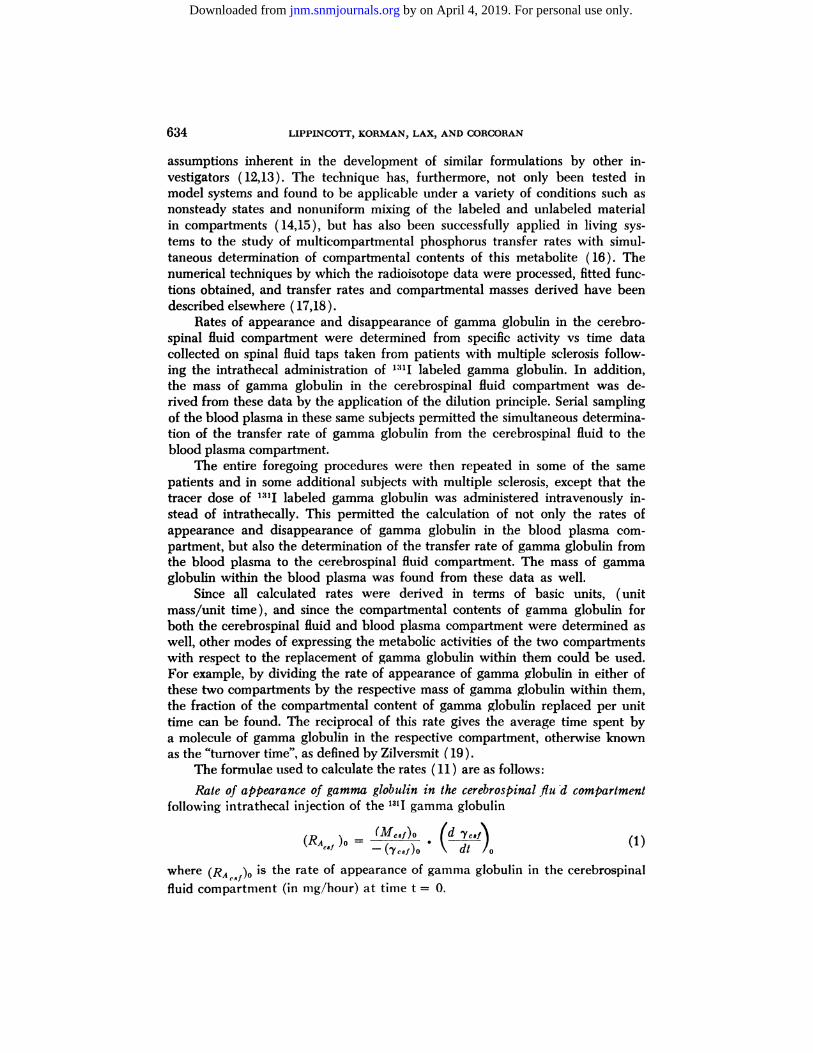
Fi9ure 1 Biological Half-Life (Tl) of Intravenously Injectedl1@'-LabeledGamma GlobulinDeterminedfromWhole-Body,Serumand Daily Urine Counts in a Patient with Multiple Sclerosis.
(7)
IC) P(RC@NT OF INJECTED DOSE / ML SERUM
T@= 19 Days
20 22 24 26 28TIME IN DAYS
by on April 4, 2019. For personal use only. jnm.snmjournals.org Downloaded from

TRANSFER RATES OF GAMMA GLOBULIN 637
RESULTS
An important requirement in using a radioactively labeled protein such asgamma globulin in a tracer study is that the protein should not be degraded dur
ing the fractionation nor during the iodination. Figure 1 contains data compiledfrom patient C. This ambulatory patient still retained control of his urinary bladder and so was a suitable subject. He received an intravenous injection of 142,zC of 1311labeled globulin ( 3 mg) and the biological half-life was determined tobe 21.5 days when he was counted in the whole-body gamma spectrometer, 21days .by counting 24 hour urinary collections in the same device, and 19 daysby counting samples of sera. These findings are in good agreement, and theglobulin preparation used was considered to be biochemically suitable for thisseries of studies.
The response of the cerebrospinal fluid concentration of gamma globulin andtotal protein to intravenous and intrathecal injection of tracer amounts of 1311
gamma globulin was determined in the patients with multiple sclerosis ( TablesI, II ). It appeared desirable, as a part of this overall study, to establish howsensitive such a response, if it occurred, might be as a basic observation in a dis
ease that may be associated with an autoimmune process. At periods varyingfrom six hours to 22 days, following intravenous injection of 1311 gamma globulin,there was no statistically significant change in the concentration of total proteinin the cerebrospinal fluid before and after the injection of the tracer material.At periods varying from six hours to 19 days, following intrathecal injection of 1311gamma globulin, there was a statistically significant rise in the total protein con
centration of cerebrospinal fluid as compared to the preinjection concentration.
Seventeen of the twenty-one patients injected intrathecally with 131! gamma gbbulin had negligible clinical reactions, while four had severe headache and twoof them had nuchal rigidity. It is possible that these reactions were due to thespinal tap alone but it is also conceivable that they were associated with theresponse to the injections of a presumed “tracer―amount of gamma globulin.
In multiple sclerosis it has been previously reported that during the course
of the disease the concentration of the serum gamma globulin may be within therange for normal subjects while the concentration in the cerebrospinal fluid isgreater than normal. Although it was anticipated that this also would be true in
our patients, it was thought that it would be best to establish the fact, since itwas desirable to compare the rate of gamma globulin replacement in serum andin cerebrospinal fluid. In eight patients the serum gamma globulin concentrationdetermined at two intervals from three to twenty-four months apart showed nosignificant difference. In nine patients the serum gamma globulin content measured as a percent of total protein concentration was compared with that in thecerebrospinal fluid. Both sets of values were obtained from chemical determinations and paper electrophoretic values. In seven of the nine patients the content ofgamma globulin, expressed as a percent of the total protein concentration, washigher in cerebrospinab fluid than in the serum. In one patient the value was the
same and in another the cerebrospinal fluid value was four percent less than theserum gamma globulin.
by on April 4, 2019. For personal use only. jnm.snmjournals.org Downloaded from

638 LIPPINCOTT, KORMAN, LAX, AND CORCORAN
A plot of the specific activity vs time for the cerebrospinal fluid followingintrathecal injection of the 1S1Ilabeled gamma globulin is shown in Fig. 2. This
is a composite curve made up of 18 points, for which specific activities were determined by using the fraction of the injected dose at the time of spinal tap,divided by the content of gamma globulin in the same sample, with each pointbeing obtained on a different individual. The solid line curve is given by the exponential function which was fitted by least squares iterative procedure programmed for the IBM 7090 in the FORTRAN language (18,20). This programnot only obtains the least squares fit to the given data in the form of a sum ofexponential terms, but it also calculates the mass of the compartment, the rateof appearance of the given metabolic factor within the compartment, and afteran elaborate calculation involving the inversion of the final matrix, which is used
Figure 2
CEREBROSPINALFLUID COMPOSITECURVE OF V»GAMMA GLOBULIN, INTRATHECALLYADMINISTERED
10t
•6
01, .ooa
.006
Fitted Function-.0579t -.00578t•>=0439e ».000121e
where t is the time in hours.
to fit the parameters in the exponential expression, it computes the standarddeviation of the rate of appearance. By means of equation (1), the overall transfer rate of gamma globulin into the cerebrospinal fluid compartment ( or the rateof appearance) and its standard deviation as determined by means of extrapolated values derived from the fitted function (11) was found to be 1.31 ±0.91mg gamma globulin/hour, or 31.2 mg/day. The rate of disappearance was calculated from the rate of appearance and the rate of change in the concentrationof gamma globulin in the cerebrospinal fluid compartment ( 16 ). It was found tobe equal to 1.28 mg gamma globulin/hour, or 30.7 mg/day. Since the rate ofchange in the content of gamma globulin in the cerebrospinal fluid was foundnot to differ significantly by Student's t-test from zero, the rate of appearance
can be considered to equal the rate of disappearance of gamma globulin from
by on April 4, 2019. For personal use only. jnm.snmjournals.org Downloaded from

TRANSFER RATES OF GAMMA GLOBULIN 639
the cerebrospinal fluid compartment, i.e. a dynamic steady state can be presumed to exist. The content of gamma globulin in the cerebrospinal fluid compartment was also determined from the fitted specific activity vs time functionusing a value found by extrapolation to time t = 0 and applying the dilutionprinciple (11,16). It was found to be equal to 22.7 mg gamma globulin.
If the rate of appearance of gamma globulin in the cerebrospinal fluid compartment is divided by the content of gamma globulin in this compartment, thefraction of the compartmental content of gamma globulin that is replaced perunit time can be calculated. This was found to be .0577 mg gamma glo-bulin/hr/mg gamma globulin in the compartment or hour"1. The reciprocal of
this value, 17.3 hour is the average time spent in the cerebrospinal fluid by amolecule of this protein.
Figure 3
SPECIFIC ACTIVITY OF SERUM I151 GAMMA GLOBULIN
AFTER INTRATHECAL INJECTION
8'
.6
.1.0806
.02-1
01 -I-008J
0061
•1-0000212e" -0000236e
where t is the time in hours.
=.0000245e
Fitted Functions
00365t -0000310e-.05061
-00335t -0143t0' Y = .0000287e -.0000289
where t is the time in hours
2 S 4 5 6 7 e 9 IO II 12 13 14 15 I 2
TIME IN DAYSIO II 12 13 14 19
Figure 3 shows the specific activity vs time data for the sera of four patientsfollowing intrathecal injection of the 1:!1Ilabeled gamma globulin. Similar data
were also obtained on a fifth subject. The transfer rate of gamma globulin fromthe cerebrospinal fluid to the blood plasma compartment was calculated fromthe data in Figs. 2 and 3 and was found to be 0.425 mg ±0.208 (S.D.) gammaglobulin/hour.
The four patients represented in Fig. 3 were also studied, together withfour more patients, by means of the intravenous injections with subsequent serialblood samples. The average rate of appearance of gamma globulin in the bloodplasma compartment was determined as 312 ±103 (S.D.) mg gamma globulin/hour. Since the serum gamma globulin concentration did not change significantly (by Student's t-test) throughout this study, the rate of disappearance of
by on April 4, 2019. For personal use only. jnm.snmjournals.org Downloaded from

640 LIPPINCOTT, KORMAN, LAX, AND CORCORAN
gamma globulin from the blood plasma compartment is assumed to equal therate of appearance, i.e. a dynamic steady state can be said to exist. From data oneight patients, the mean content of gamma globulin in the plasma compartmentwas calculated to be 22.5 ±3.6 (S.D.) gms. The mean value for the fraction ofthe gamma globulin content of the blood plasma being renewed per unit time is
312equal to 00 gnr>*-e- 0-0138 mg gamma globulin/hour/mg gamma globulin inthe compartment of hr1. The reciprocal of this value gives the average time
spent by a molecule of gamma globulin in the blood plasma compartmentas 72.4 hours. This has also been termed the "turnover time" ( 19 ) .
It is of interest to compare the rate at which gamma globulin is being replaced in the blood plasma compartment with that at which it is being replacedin the cerebrospinal fluid compartment. This can be done by means of the ratefigures which deal with the fraction of the compartment being replaced per unit
TABLE I. TOTAL PROTEINCONCENTRATIONIN CEREBROSPINALFLUID PRIOR TOANDFOLLOWINGINTRAVENOUSINJECTIONOF 131IGAMMAGLOBULIN
IN PATIENTSWITHMULTIPLESCLEROSIS
Number1234567891011PatientCodeFRS*M*AGHB*CETMg
Injected'"/LabeledGammaGlobulin4.02.62.82.04.03.02.00.54.01.24.03.03.04.02.01.5RadioactivityMicrocuries57.8152.098.8100.0106.0142.0131.315.0104.029.1104.0142.0142.0104.0131.3142.0Pretracer
InjectionCSFTotal
ProteinConcentrationmg/100
ml39.868.527.039.266.128.050.919.828.237.138.5Posttracer
InjectionCSFElapsed
TimefromInjection6
hours24hours24hours60hours24hours13days22
days8days4
days13days17days19days19days20
days4days22
daysTotal
ProteinConcentrationmg/100
ml39.767.823.723.636.240.041.868.325.944.623.818.322.634.133.735.5
*In patients S and B intravenous injections were given in two separate studies, in patient M
in three studies, while in the rest a single study was made.
by on April 4, 2019. For personal use only. jnm.snmjournals.org Downloaded from

NumberPatientCodeMg
Injected1311 Labeled
GammaGlobulinRadioactivity
MicrocuriesPretracer
Injection CSFPostt racer InjectionCSFTotal
ProteinConcentration
mg/100 mlElapsedTime
from InjectionTotal
ProteinConcentration
mg/100ml1
2
3
4567891011121314151617A*
B*
C@
DEFGHI
JKLMN0P
Q0.5
1.01.01.01.01.41.00.81.01.01.01.02.02.01.00.81.01.01.01.015.0
75.046.065.757.8
105.065.746.057.857.857.857.8150.0150.075.046.865.765.765.765.757.0
60.019.818.528.228.945.332.539.828.050.936.764.760.550.039.236.770.342.058.86hours
11 days18 hours24hours26 hoursl8days36 hours48hours72 hours4 days
ll3hoursl72hours8days12 days4days24hours102 hours126 hours144 hours156 hours66.1
72.460.040.585.844.649.351.945.465.9104.057.3141.397.085.025.420.555.834.335.7
TRANSFER BATES OF GAMMA GLOBULIN 641
time. From these values it can be seen that, on the average, the gamma globulin. . . . . 0.0577 .in the cerebrospmal fluid is being replaced o oi@ 4.18 times as fast as it
is in the plasma.A composite plot was made of the specific activity vs time in the cerebro
spinal fluid of eight subjects with multiple sclerosis in whom 1311labeled gammaglobulin was injected intravenously followed by a spinal tap. The function forthe fitted curve was obtained by the same techniques as already described. Thedata from the composite plot, together with that for the specific activity vs timein the blood plasma, were used to determine the transfer rats of gamma globulinfrom the blood plasma to the cerebrospinal fluid compartment. The mean value
TABLE II. TOTAL PROTEIN CONCENTRATION IN CEREBROSPINAL FLUID PRIOR TO
AND FOLLOWING INTRATHECAL INJECTION OF 131J GAMMA GLOBULIN
IN PATIENTS WITH MULTIPLE SCLEROSIS
4In patients A, B, and C intrathecal injections were given in two separate studies, while inthe remainder of the patients a single study was made.
by on April 4, 2019. For personal use only. jnm.snmjournals.org Downloaded from

642 LIPPINCOTr, KORMAN,LAX,ANDCORCORAN
found in eight subjects for this transfer rats was .0367 mg gamma globulin/hour.The transfer rate of gamma globulin from the cerebrospinal fluid to the blood
plasma was @1:, or 11.6 times as rapid as from the blood plasma to the
cerebrospinal fluid compartment.
DISCUSSION
Some of the properties of gamma globulin present in the cerebrospinal fluidof patients with multiple sclerosis have been reported recently. MacPherson andCosgrove (21 ) identified two immunologically different gamma globulins in“normal―and multiple sclerosis cerebrospinal fluids by immunoelectrophoreticanalyses employing rabbit antisera prepared against “normal―and multiple
sclerosis cerebrospinal fluids. The major gamma globulin of cerebrospinal fluid
was shown to be immunologically identical with the single gamma globulin of
human serum, but the minor gamma globulin of cerebrospinal fluid was identifled only in the latter. The major and minor gamma globulins of normal cere
brospinal fluid could not be distinguished from those of multiple sclerosis cerebrospinal fluid. These investigators felt that the presence of the gamma globulinpeculiar to cerobrospinal fluid confirmed the suggestion of Kabat, Freedman,
Murray and Kraub ( 1 ) that central nervous system tissue is capable of forminggamma globulin. They also thought it would be of interest to find out whetherany of these particular gamma globulins function as an antibody.
Tourtelbotte, Parker, Haerer, Harrell, Haerer, Gustafson and Dejong ( 22)raised the question in their study as to whether an elevation of a specific antibodycould be the reason for the increase in gamma@ globulin in the cerebrospinalfluid of patients with multiple sclerosis. They applied an ion exchange cellulosechromatography technique to the cerebrospinal fluid from 30 normal individuals,40 patients with multiple sclerosis, and 6 patients with neurosyphillis. A distinct
gamma globulin fraction found in the cerebrospinal fluid of seventy-five percentof the patients was named by them CM-il. The finding of this fraction (gammaglobulin) suggested to them that it might be related to the etiology of multiplesclerosis. Following the work of these two groups another study was reported byCaspary (23) who undertook a comparison of immunological specificityofgamma globulin in the cerebrospinal fluid in normal and multiple sclerosis subjects. The conclusion reached were contrary to those of MacPherson and Cosgrove. Caspary stated that antibodies prepared against the proteins of normalserum, and those of normal and multiple sclerotic cerebrospinal fluids indicatedtheimmune identityof gamma globulinfrom allthreesources.
In a pioneering study with radioisotopes, Frick and Scheid-Seydel (13)injected intravenously (but not intrathecally) @@‘1gamma globulin into 28 patients with various neurological disorders. Three to four days, or occasionallylater, after injection the specific activity was determined in samples of cerebrospinal fluid and blood. On the basis of this single route of exchange, in patientswith multiple sclerosis, it was concluded that a variable portion of the cerebrospinal fluid gamma globulin did not come from blood. The authors stated that thisassumption was based upon the fact that the specific activity was lower in the
by on April 4, 2019. For personal use only. jnm.snmjournals.org Downloaded from

TRANSFER RATES OF GAMMA GLOBULIN 643
cerebrospinal fluid than in the blood. Sites of production for the gamma globulinnot entering the cerebrospinal fluid from the blood were regarded as the meningesand the mesenchymal tissues of the central nervous system. Our transfer ratestudies indicate that the total amount of gamma globulin present in the cerebrospinal fluid could not be accounted for solely as a result of physical transferfrom the blood.
SUMMARY
13lJ gamma globulin was injected both intravenously and intrathecally into
patients with multiple sclerosis with subsequent sampling of blood and cerebrospinal fluid. Specific activity versus time curves were constructed from thedata obtained from these samples. The transfer rate of gamma globulin fromthe cerebrospinal fluid to the blood plasma was 11.6 times as rapid as from theblood plasma to the cerebrospinal fluid compartment. The average time a molecube of gamma globulin spent in the cerebrospinal fluid compartment was 17.3hours; whereas, in the blood plasma compartment it was 72.4 hours. The gammaglobulin in the cerebrospinal fluid compartment was found to be replaced 4.2times as fast as it was in the blood plasma compartment.
REFERENCES
1. KABAT,E. A., FREEDMAN,D. A., Muiui@y,J. P. ANDKRAUB,V. : A Study of the Crystalline Albumin, Gamma Globulin and Total Protein in the Cerebrospinal Fluid of One HundredCases of Multiple Sclerosis and in Other Diseases. Amer. J. Med. Sci. 219:55, 1950.
2. VAN SANDE,M., KARCHER,D. ANDLOWENTHAL,A. : ExarnensElectrophoretiquesdesProteins du Serum and du Liquide Cephalorachidien chez des Patients Atteints de Sclerose en
Plaques. Acta Neurol. Psychiat. Beig. 57:407, 1957.3. FRICK,E. ANDSCHEID-SEYDEL,L. : Untersuchungenmit Ii―Marldertem Gamma-Glob
ulin zur Frage der Abstammung der Liquoreiweisskorper. Kim. Wchschr. 36:857, 1958.4. NICHOL,J.C. AND DEUTSCH,H. F.:BiophysicalStudiesof BloodPlasmaProteins,
VII. Separation of Gamma Globulin from the Sera of Various Animals. J. Amer. Chem. Soc.70:80, 1948.
5. Ln@u@corr, S. W., KORMAN, S., FONG, C., STICKLEY, E., WOUNS, W. AND HUGHES,W. L.: Turnover of Labeled Normal Gamma Globulin in Multiple Myeloma. I. Cliii. Invest.
39:565, 1960.6. LIPPINc0Tr, S. W., KORMAN, S. AND HUGHES, W. L.: Turnover of Autologous and
Homologous Labeled Gamma Globulin in Multiple Myeloma. Arch. Path. 70:467, 1960.
7. HELMKAMP,R. %V.,GOODLAND,R. L., BALE, W. F., SPAR, I. L. ANDMUTSCHELER,S.:High Specific Activity Iodination of Gamma Globulin with Iodine-131 Monochloride. Cancer
Res. 20:1495, 1960.8. LIPPINC0TT, S. “N.,COHN, S. H., HAMEL, H., FINE, S. AND KORMAN, S.: Determination
of Radioactively Labeled Globulin Turnover by the Direct Whole-Body Counting Technique.
I.Clin.Invest.40:697,1961.9. LIPPINCOTr, S. W., COHN, S. H., ROBERTSON, J. S. AND FAiin, L. E.: In Vivo Measure
ment by the Whole-Body Spectrometer of the Degradation Rate of I―-Labeled Normal Al
bumin. Lab. Invest. 10:481, 1961.10. SHEPPARD, C. W.: Basic Principles of the Tracer Method. John Wiley and Sons, Inc.,
New York, New York, 1962.
11. WRENSH.ALL, G. A.: Working Basis for the Tracer Measurement of Transfer Rates ofa Metabolic Factor in Biological Systems Containing Compartments Whose Contents Do NotIntermix Rapidly. Canad. 1. Biochenl. Physiol. 33:909, 1955.
12. WRENSHALL, G. A.: Fact and Fancy Concerning Tracer-Determined Flux and Ac
cumulation of Substances in Vivo. Diabetes 11:236, 1962.
by on April 4, 2019. For personal use only. jnm.snmjournals.org Downloaded from

644 LIPPINC0Tr,KORMAN,LAX,ANDCORCORAN
13. WRENSHALL,G. A. AND HETENYI, G.: Tests of the Validity of Tracer-CalculatedAmounts and Transfer Rates of Substances in the Blood Plasma of Living Animal Systems.Ann. N. Y. Acad. Sci. 108:259, 1963.
14. LAX, L. C. ANDWRENSHALL,C. A.: Measurements of Turonver Rates in Systems ofHydrodynamic Pools Out of Dynamic Equilibrium. Nucleonlcs 11:18, 1953.
15. ScuAc1tI@, H.: Direct Versus Tracer Measurement of Transfer Rates in a Hydrodynamic System Containing a Compartment Whose Contents Do Not Intermix Rapidly. Canad.J. Blochem. Physlol. 33:940, 1955.
16. LAX, L. C., SIDLOFSKY,S. ANDWRENSHALL,G. A.: Compartmental Contents and Simultaneous Tranfer Rates of Phosphorus in the Rat. J. Physiol. 132:1, 1956.
17. WORSLEY,B. H., REm, D. B. W. ANDLAX, L. C.: Error Estimation in Transfer Ratesof Plasma Constituents. Proc. Second Conf. Computing and Data Processing Soc. Can. 158,Toronto, June, 1960. University of Toronto Press.
18. Wortsi.Ex, B. H. ANDLAX,L. C.: Selection of a Numerical Technique for AnalyzingExperimental Data of the Decay Type with Special Reference to the Use of Tracers in Biological Systems. Biochem. Biophys. Acta. 59:1, 1962.
19. ZILVERSMITH,D. B.: Design and Analysis of Tracer Experiments. Adv. Tracer Methodology 1:203, 1963.
20. Wostsi..icy, B. H.: Communicationa of the Assoc. of Computing Machinery 7:39, 1964.21. MAcPHERs0N, C. AND COSCROVE,J. B.: Immunochemical Evidence for a Gamma
Globulin Peculiar to Cerebrospinal Fluid. Caned. I. Biochem. and Physiol. 39:1567, 1961.22. TOURTELLOTTE,W. W., PAJ@XER,J. A., HAEREE, A. F., HARRELL,L. H., HAERER, H.
A., GUSTAFSON,K. A. ANDDEJ0NG, B. N.: Multiple Sclerosis Gamma Globulin. J. Neuropath.and Exper. Neurol. 23:193, 1964.
23. CASPARY,E. A.: Comparison of Immunological Specificity of Gamma Globulin in theCerebrospinal Fluid in Normal and Multiple Sclerosis Subjects. J. Neurol. Neurosurg. Psychiat.28:61, 1965.
by on April 4, 2019. For personal use only. jnm.snmjournals.org Downloaded from

1965;6:632-644.J Nucl Med. Stuart W. Lippincott, Samuel Korman, Louis C. Lax and Cornelia Corcoran (Results Obtained on A Series of Multiple Sclerosis Patients)Transfer Rates of Gamma Globulin Between Cerebrospinal Fluid and Blood Plasma
http://jnm.snmjournals.org/content/6/9/632This article and updated information are available at:
http://jnm.snmjournals.org/site/subscriptions/online.xhtml
Information about subscriptions to JNM can be found at:
http://jnm.snmjournals.org/site/misc/permission.xhtmlInformation about reproducing figures, tables, or other portions of this article can be found online at:
(Print ISSN: 0161-5505, Online ISSN: 2159-662X)1850 Samuel Morse Drive, Reston, VA 20190.SNMMI | Society of Nuclear Medicine and Molecular Imaging
is published monthly.The Journal of Nuclear Medicine
© Copyright 1965 SNMMI; all rights reserved.
by on April 4, 2019. For personal use only. jnm.snmjournals.org Downloaded from