fissure sealants - pdwgpdwg-ng.org/materials/fissure sealant.pdf · introduction •fissure...
TRANSCRIPT
Fissure sealants

Study objectives
• Discuss the historic development and current rationale for occlusal sealant use in children.
• Understand factors influencing enamel acid etching and mechanical bonding of the sealant.
• Discuss indications and contraindications for use of fissure sealants.
Introduction
• Fissure sealants are resin that are applied to the occlusal surfaces of teeth with the objective of sealing the pits and fissures from the oral environment. It is an effective caries reduction procedure when there is proper patient selection and application techniques.
Introduction - 2
• The sealants bind mechanically with the tooth and
prevent bacteria colonisation of the pits and fissures. It also prevents nutrient supplies to the bacteria present in the pits/fissures.
• Important because of the high incidence of pits and fissure caries even when there is a decrease in caries incidence with prevalence been as high as 70% by 17 years in USA.

History of pit and fissure caries control
• Chemical treatment of enamel with use of silver nitrate.
• Prophylaxis odontomy + restoration of fissures with amalgam.
• Sealing of fissures with black copper (found not to be retentive), cyanoacrylate (toxic), polyurethane, and GIC (its viscosity necessitates cavity preparation).
Current development
• With the development of Bis-GMA in 1971 by
Buonocore, the possibility of the current use of fissure sealant materials was feasible.
• Fissure sealants are currently autopolymerised or light cured resins.

Current development - 2
• Occlusal caries reach peak incidence 3 years after the eruption of new molars. The impact of systemic fluoride on the incidence of occlusal caries declines after 3 years following eruption of the molars thus the need and place for pits and fissure sealants. Systemic fluoride have greater impact on the prevention of smooth surface caries.

Effectiveness of sealants - 1
• The effectiveness of pits and fissure sealant therapy in reducing occlusal caries has been documented.
• Effectiveness is more for first molars and less so documented for second molars.
• Effectiveness range from 86% in 12 months to 57% in 48-54 months follow up.
• It is cost effective and of public health benefit.
Effectiveness of sealants - 2
• Recommended to be applied every 6 months if the need is indicated.
• Before the placement of a fissure sealant, it is important to first do a prophylaxis. After the placement of the sealant, then apply topical fluoride to the mouth every 6 months when needed. This method reduces caries incidence by 87.5% over 3 years with consistent use.
Effectiveness of sealants - 3
• In about 47.8% of cases, there might be the need for reapplication of the sealant over the 3 year period. loss of the sealant is often due to development of proximal caries.
Effectiveness of sealants - 4
• Over a 5 year period, 50% of the sealant remain intact, 30% need reapplication, 10% need 2 reapplication and another 10% need 3 reapplication.
• Fissure sealant is found to form an acceptable part of proven effective prevention measure for caries if properly applied even with a single application.
Indications for use of fissure sealants
• Deep, narrow pits and fissures. • Recently erupted teeth. • Where there are no caries in the proximal surfaces. • Patient with high caries risk though this increases
the tendency and need for reapplication.
Contraindications of use of fissure sealant
• Teeth with occluso-proximal lesions.
• Teeth with proximal lesions.
• Teeth that is caries free for 4years or more.
• Teeth with broad well coalesced pits and fissure.

Patient selection
This is important. the following criteria must be followed when selecting patient.
• Patient must be dependable and can make recall appointments.
• Patient must fall between 6-15 years.

Patient selection - 2
• Must be motivated and effective in caries control.
• Must have low caries activity to reduce rate of loss.
• Tooth must be a recently erupted tooth (within the last 3 years), caries free, permanent tooth with steep cuspal incline and deep pits.
• Little studies done on primary teeth.

Technique
• Material must be either filled or unfilled resin. There is no significant difference in the performance of either material.
• The material comes with an etchant also called a tooth conditioner. This contains acid of concentration varying from 35-50%. No evidence to demonstrate that the acid concentration within this range affects clinical performance.
Technique - 2
• Resins are either auto or light polymerised. Studies show that the autopolymerised have better results with respect to retention and caries protection.

Placement of sealant - 1
• Isolate the tooth to prevent saliva/fluid contamination
of etched enamel. Use of rubber dam more effective in contamination control especially when using UV light activated material.
Placement of sealant - 2
• Clean surface with pumice to remove plaque and debris. This may not be necessary if one uses phosphoric acid to etch as it removes the surface plaque, acquired pellicle and up to 10um of enamel. Retention can also be improved if occlusal surface is widened with round burs. Do follow manufacturer’s instructions. This is important.

Placement of sealant - 3
• Next etch by continuously dabbing the etchant gently on the enamel surface. This produces better result than rubbing.
Placement of sealant - 4
• After etching, wash and dry enamel surface using water for about 10-20 seconds. Dry for 10 seconds. Avoid the use of oil contaminated air sprays. On drying, the surface should appear dull, frosty or opaque. If not, re-etch. It is important to avoid moisture contamination at this stage as it affects the effectiveness of bond.

Placement of sealant - 5
• Apply sealant with the applicator supplied in the pack. A good sealant has low viscosity and high wetability thereby enhancing application. This allows for quick dispense of sealant off the groves on the teeth and excess can be removed with cotton tips prior to polymerisation.
Placement of sealant - 6
• After polymerisation, evaluate to ensure entire surface is covered. After material set, wipe surface with wet cotton rolls or pellet, and remove unpolymerised resin that accumulates after set. This results in a more pleasant after taste as well as provide for a clearer surface.
Placement of sealant - 7
• Next, use explorer to check for void or incomplete coverage. Where such exists, apply extra sealant.
• Check for occlusal interference. If unfilled resin is used, no need to bother to adjust as interference will quickly wear away with use. If filled resin, adjust with green stone.
Placement of sealant - 8
• Following adjustment, apply topical fluoride.
Sealants are often rechargeable as they absorb fluoride form the oral environment and slowly release them thereafter.
Placement of sealant - 9
• Finally, recall patient every 6months to re-examine for possible loss an replacement. If partially lost, test firmness of attachment of remnant using a sharp instrument. If firm, freshen sealant using fine stone or pumice, re-etch, expose area and repeat procedure. If not firm, then completely remove and repeat procedure.

Quiz 1
Fissure sealants
• Complements the use of systemic flouride
• More effective for preventing caries on second molars
• Best result is seen in deciduous dentition
• Not indicated for use in children with low risk for caries
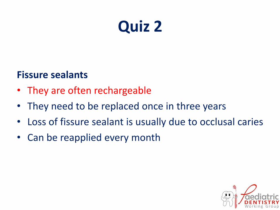
Quiz 2
Fissure sealants
• They are often rechargeable
• They need to be replaced once in three years
• Loss of fissure sealant is usually due to occlusal caries
• Can be reapplied every month
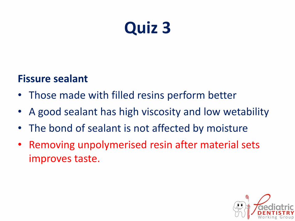
Quiz 3
Fissure sealant
• Those made with filled resins perform better
• A good sealant has high viscosity and low wetability
• The bond of sealant is not affected by moisture
• Removing unpolymerised resin after material sets improves taste.
Acknowledgement
• Slides were developed by Morenike Ukpong, Associate
Professor in the Department of Paediatric Dentistry, Obafemi Awolowo University, Ile-Ife, Nigeria.
• The slides was developed and updated from multiple materials over the years. We have lost track of the various references used for the development of the slides
• We hereby acknowledge that many of the materials are not primary quotes of the group.
• We also acknowledge all those that were involved with the review of the slides.