first principles calculation of pka values for 5 ... demonstrated antitumor and antiviral...
TRANSCRIPT
1
J. Phys. Chem.
First Principles calculation of pKa values for 5-substituted Uracils
Yun Hee Jang,# Lawrence C. Sowers,§ Tahir Cagin,# and William A. Goddard III,#*#Materials and Process Simulation Center, Beckman Institute (139-74)
California Institute of Technology, Pasadena, California 91125§Divisions of Pediatrics and Molecular Medicine, City of Hope National Medical Center,
1500 E. Duarte Road, Duarte, California 91010
Abstract
Oxidation of uracil (U) and thymine (5-Me-U) are believed to play a role in genetic
instability because of the changes these oxidations cause in the ionization constants (pKa values),
which in turn affects the base pairing and hence coding. However, interpretation of the
experimental evidence for the changes of pKa with substitution at U has been complicated by the
presence of two sites (N1 and N3) for ionization. We show that a procedure using first principles
quantum mechanics (density functional theory with generalized gradient approximation, B3LYP,
in combination with the Poisson-Boltzmann continuum-solvation model) predicts such pKa
values for a series of 5-substituted uracil derivatives in excellent correlation with experiment. In
particular, this successfully resolves which cases prefer ionization at N1 and N3. Such first
principles predictions of ionization constant should be useful for predicting and interpreting pKa
for other systems.
Keywords: oxidative DNA damage, pyrimidine bases, ionization constant (pKa), density
functional theory, Poisson-Boltzmann continuum-solvation model, Jaguar
* To whom correspondence should be sent, [email protected]
2
1. Introduction
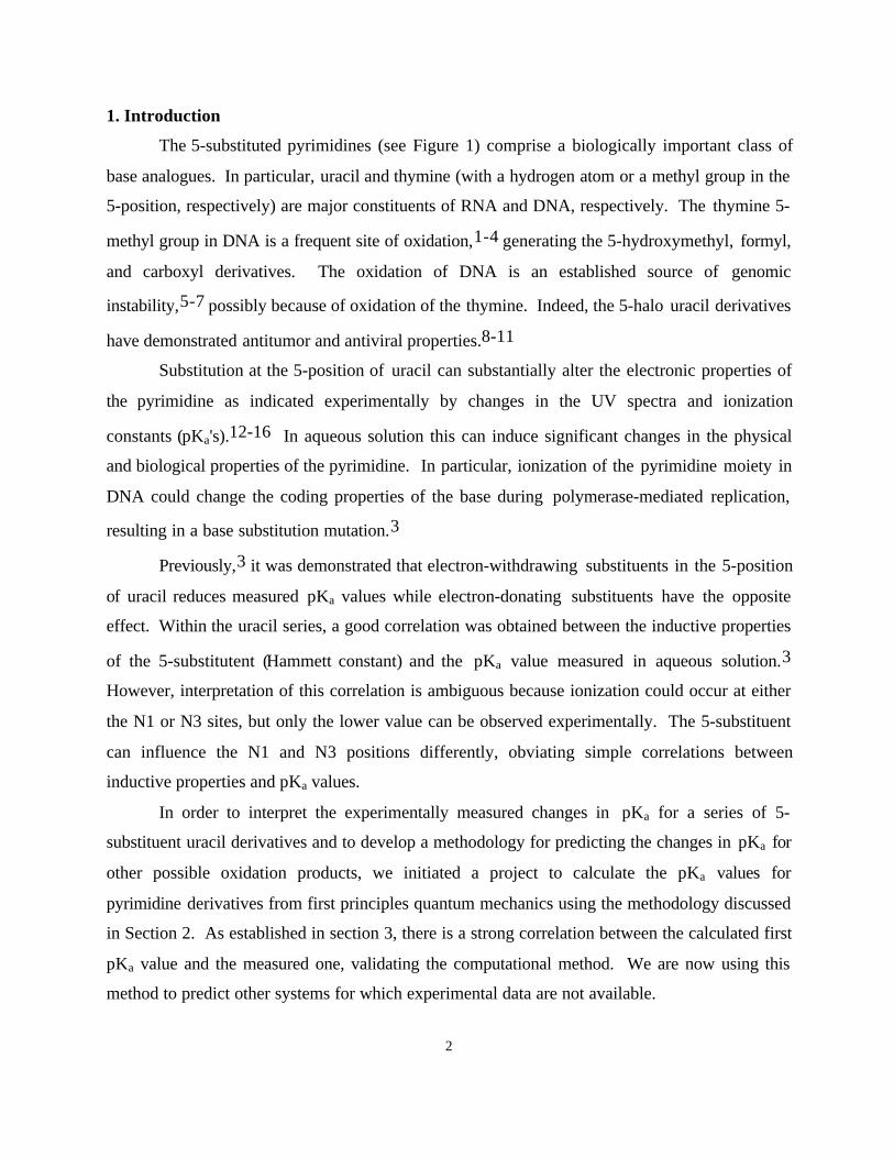
The 5-substituted pyrimidines (see Figure 1) comprise a biologically important class of
base analogues. In particular, uracil and thymine (with a hydrogen atom or a methyl group in the
5-position, respectively) are major constituents of RNA and DNA, respectively. The thymine 5-
methyl group in DNA is a frequent site of oxidation,1-4 generating the 5-hydroxymethyl, formyl,
and carboxyl derivatives. The oxidation of DNA is an established source of genomic
instability,5-7 possibly because of oxidation of the thymine. Indeed, the 5-halo uracil derivatives
have demonstrated antitumor and antiviral properties.8-11
Substitution at the 5-position of uracil can substantially alter the electronic properties of
the pyrimidine as indicated experimentally by changes in the UV spectra and ionization
constants (pKa's).12-16 In aqueous solution this can induce significant changes in the physical
and biological properties of the pyrimidine. In particular, ionization of the pyrimidine moiety in
DNA could change the coding properties of the base during polymerase-mediated replication,
resulting in a base substitution mutation.3
Previously,3 it was demonstrated that electron-withdrawing substituents in the 5-position
of uracil reduces measured pKa values while electron-donating substituents have the opposite
effect. Within the uracil series, a good correlation was obtained between the inductive properties
of the 5-substitutent (Hammett constant) and the pKa value measured in aqueous solution.3
However, interpretation of this correlation is ambiguous because ionization could occur at either
the N1 or N3 sites, but only the lower value can be observed experimentally. The 5-substituent
can influence the N1 and N3 positions differently, obviating simple correlations between
inductive properties and pKa values.
In order to interpret the experimentally measured changes in pKa for a series of 5-
substituent uracil derivatives and to develop a methodology for predicting the changes in pKa for
other possible oxidation products, we initiated a project to calculate the pKa values for
pyrimidine derivatives from first principles quantum mechanics using the methodology discussed
in Section 2. As established in section 3, there is a strong correlation between the calculated first
pKa value and the measured one, validating the computational method. We are now using this
method to predict other systems for which experimental data are not available.
3
(a)1
35
N
N
OH
OH
H
H3C
(b) 1
35
N
N
OH
OH
H
H
(c)1
35
N
N
OH
OH
H
F
(e)1
35
N
N
OH
OH
H
H
O
(d)1
35
N
N
OH
OH
H
O
H
(f)N
N
OH
OH
H
O2N
1
35
Figure 1. 5-Substituted Uracils. (a) Thymine, (b) Uracil, (c) 5-fluorouracil,
(d) trans 5-formyluracil, (e) cis 5-formyluracil, and (f) 5-nitrouracil.
2. Calculation Details
2.1 pKa calculations
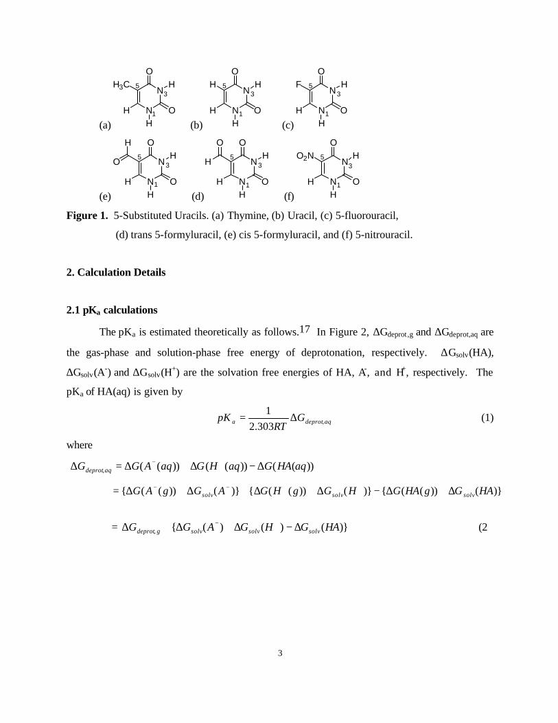
The pKa is estimated theoretically as follows.17 In Figure 2, ∆Gdeprot,g and ∆Gdeprot,aq are
the gas-phase and solution-phase free energy of deprotonation, respectively. ∆Gsolv(HA),
∆Gsolv(A-) and ∆Gsolv(H+) are the solvation free energies of HA, A-, and H+, respectively. The
pKa of HA(aq) is given by
aqdeprota GRT
pK ,303.21
∆= (1)
where
))(())(())((, aqHAGaqHGaqAGG aqdeprot ∆−∆+∆=∆ +−
)}())(({)}())(({)}())(({ HAGgHAGHGgHGAGgAG solvsolvsolv ∆+∆−∆+∆+∆+∆= ++−−
= )}()()({, HAGHGAGG solvsolvsolvgdeprot ∆−∆+∆+∆ +− (2)
4
HA(g)
HA(aq)
A-(g) + H+(g)
A-(aq) + H+(aq)
∆Gdeprot, g
∆Gdeprot, aq
∆Gsolv (A-)∆Gsolv (HA) ∆Gsolv (H+)
Figure 2. Thermodynamic cycle used in the calculation of pKa.17
2.2 Free energies
The Gibbs free energy of each state in gas-phase [∆G(HA(g)), ∆G(A-(g)), and ∆G(H+(g))]
is obtained by
KK GZPEEG 29800 →∆∆++=∆ . (3)
The total energy of the molecule at 0 K (E0K) is calculated at the optimum geometry from
quantum mechanics (QM). The zero-point energy (ZPE) and the Gibbs free energy change from
0 K to 298 K (∆∆G0→298K) are calculated from the vibrational frequencies calculated using QM.
We used ∆G[H+(g)] = 2.5 RT – T∆S = 1.48 – 7.8 = -6.3 kcal/mol from the literature.17
2.3 QM calculations
All QM calculations used the Jaguar v3.5 quantum chemistry software.18 To calculate
the geometries and energies of the various molecules, we used the B3LYP flavor of density
functional theory (DFT) which includes the generalized gradient approximation and a component
of the exact Hartree-Fock (HF) exchange. These calculations used the cc-pVTZ(-f)++ basis set
and started from the geometries optimized using the HF method with the 6-31G* basis set.
To obtain the vibrational frequencies required for the ZPE and free energy, we used the
HF method with the 6-31G* basis set, starting from the geometries optimized at this level. The
calculated frequencies were scaled down by 0.8953.19
2.4 Solvation Energies
The solvation free energy in water [∆Gsolv(HA) and ∆Gsolv(A-)] was calculated using the
continuum-solvation approach20-22 by solving the Poisson-Boltzmann (PB) equation
5
numerically.23 In this approach, the solute is described as a low-dielectric cavity (εQM = 1)
immersed in a high-dielectric continuum of solvent (εH2O = 80 for water24). The solute/solvent
boundary is described by the surface of closest approach as a sphere of radius 1.4 Å (probe
radius for water) is rolled over the van der Waals (vdW) envelope of the solute. The charge
distribution of the solute is represented by a set of atom-centered point charges, which are
determined by fitting to the electrostatic potential calculated from the wavefunction.
The procedure is as follows. A gas-phase calculation is carried out first to obtain the
electrostatic-potential fitted (ESP) charges (constrained to match the dipole moment from the
QM). Based on these charges, the PB equation is solved to obtain the reaction field of the
solvent (as a set of polarization charges located on the solute/solvent boundary surface). The
Fock-Hamiltonian for the HF calculation is then modified to include the solute-solvent
interaction due to the reaction field. This is solved to obtain a new wavefunction and a new set
of atom-centered ESP charges. This process is repeated self-consistently until convergence is
reached (to 0.1 kcal/mol in the solvation energy). This constitutes the electrostatic or "polar"
contribution to the solvation energy.
An additional "nonpolar" contribution due to creation of a solute cavity in the solvent is
accounted for by a term proportional to the solvent-accessible surface area of the solute.22
The solvation free energy calculation was done at the B3LYP/cc-pVTZ(-f) level.
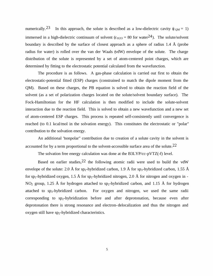
Based on earlier studies,22 the following atomic radii were used to build the vdW
envelope of the solute: 2.0 Å for sp2-hybridized carbon, 1.9 Å for sp3-hybridized carbon, 1.55 Å
for sp2-hybridized oxygen, 1.5 Å for sp2-hybridized nitrogen, 2.0 Å for nitrogen and oxygen in -
NO2 group, 1.25 Å for hydrogen attached to sp2-hybridized carbon, and 1.15 Å for hydrogen
attached to sp3-hybridized carbon. For oxygen and nitrogen, we used the same radii
corresponding to sp2-hybridization before and after deprotonation, because even after
deprotonation there is strong resonance and electron-delocalization and thus the nitrogen and
oxygen still have sp2-hybridized characteristics.
6
2.4 )( +∆ HGsolv
Calculating the pKa also requires the experimental solvation free energy of a proton in
water [ )( +∆ HGsolv ]. Unfortunately this value remains uncertain.17,25,26 “The precision of
)( +∆ HGsolv is limited by the fact that the standard hydrogen potential cannot be obtained by
measurement alone; some independent quantity is needed to determine an absolute half-cell
potential.”17,25 The )( +∆ HGsolv from the measurements of the standard hydrogen potential
range from -254 to -261 kcal/mol.17,25 From a set of cluster-ion solvation data, )( +∆ HGsolv
has been estimated to be -263.98 ± 0.07 kcal/mol.27
Because of these uncertainties, we chose )( +∆ HGsolv to minimize the RMS deviation
between the calculated and experimental ones pKa values for the 5-substituted Uracils. This
leads to a final value of –258.47 kcal/mol, which falls in the middle of the range of experimental
)( +∆ HGsolv .
2.5 dielectric constant
For the dielectric constant of water, we used the experimental value of εH2O = 80 (based
on T ~ 300 K).24
In principle we could take εQM = 1 for the dielectric constant of the region being
described in the QM. However, in this solvation model there are several limitations: (1) The
setting εQM = 1 assumes that the polarizability of the QM part is exact, but with the level of
wavefunction and basis set considered here it is likely that this is not the case; (2) the
experimental pKa was determined at a constant ionic strength of 0.1 M NaCl at room
temperature.14 The dielectric constant of this solution, especially at the interface with solute,
might be different from that of pure water; and (3) it might be appropriate to use different radii
for each atom after deprotonation due to the change of electron distribution, but we used the
same radii for each atom before and after deprotonation.
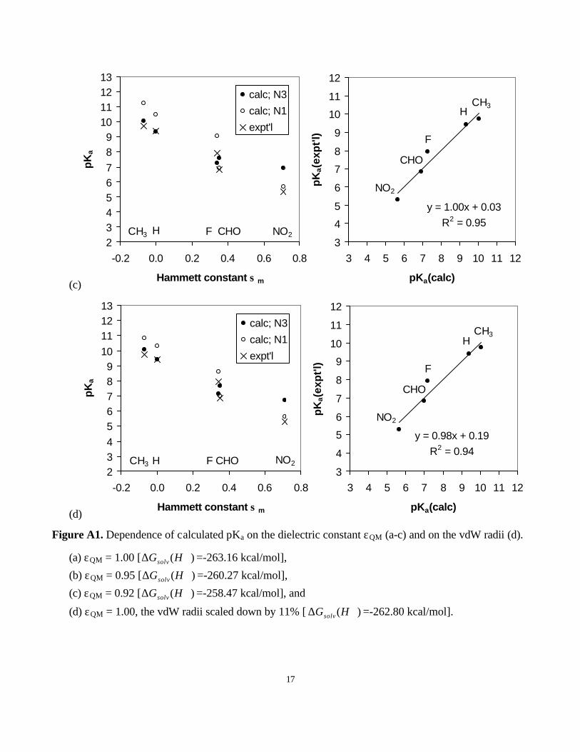
Consequently, we varied εQM to find the value that gives the same range of pKa values as
the experimental one (Figure A1). We derived to use the slightly lower value of εQM = 0.92.
7
With this choice, the )( +∆ HGsolv was determined to be -258.47 kcal/mol, which falls in the
middle of the experimental range.
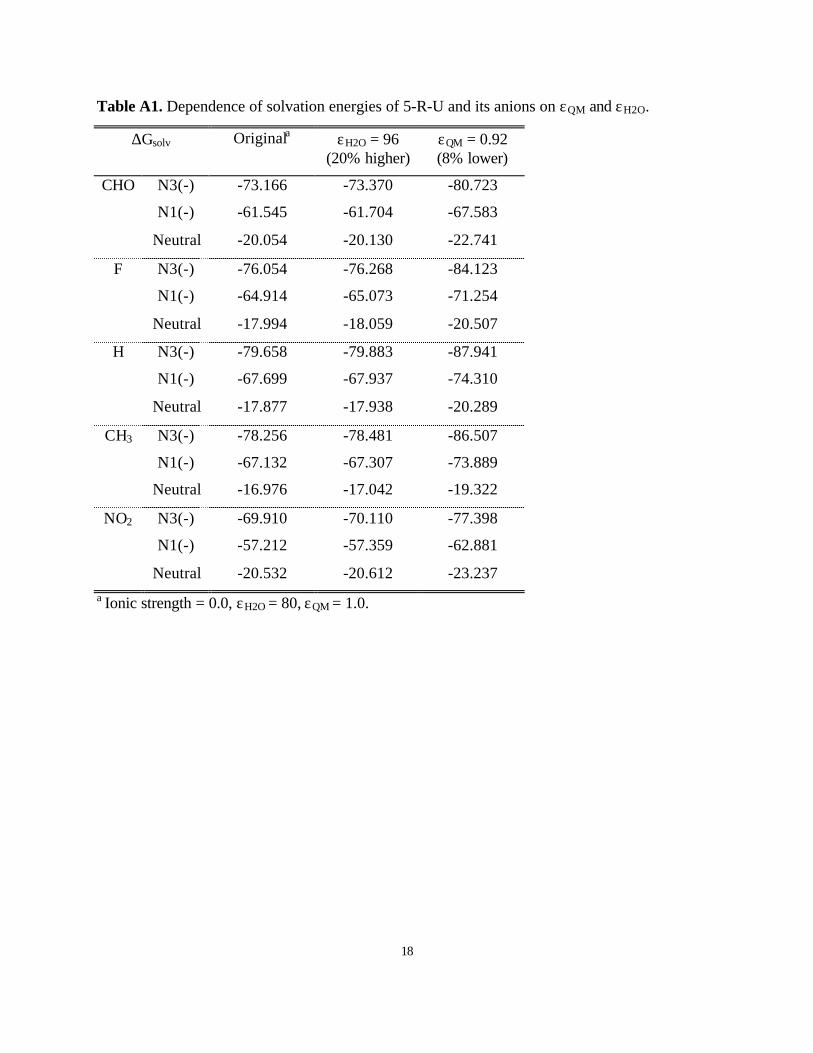
We also considered the effect of making εH2O larger than 80 and including for ionic
strength. However, as indicated in Table A1, the range of pKa is insensitive to these other
parameters.
The solvation model is sensitive to the atomic vdW radii used to estimate the boundary
between QM and continuum space. Keeping εQM = 1.0 but scaling down the atomic radii
uniformly by 11 % from the initial values, we also obtained the same range of pKa values as
experiment [Figure A1(d)].
3. Results
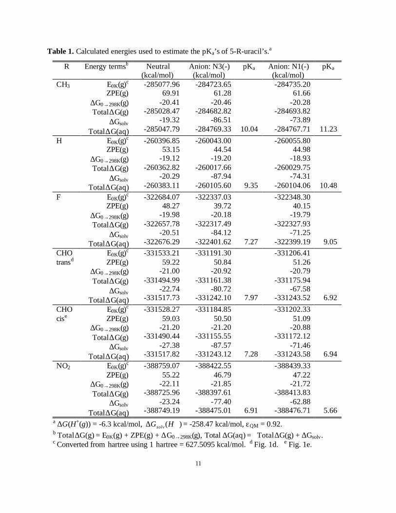
The detailed energy values used to calculate pKa are listed in Table 1.
For 5-formyluracil there are two conformational isomers shown in Fig. 1(d) and 1(e).
The calculations found that the trans conformation (Fig. 1(d)) is preferred by 4.5 kcal/mol in gas-
phase but has a 4.6 kcal/mol lower solvation energy, leading to similar energies for both
conformers in solution (the difference is less than 0.1 kcal/mol). Thus, to calculate the pKa value
for 5-formyluracil we include both conformers with the appropriate Boltzmann-average.
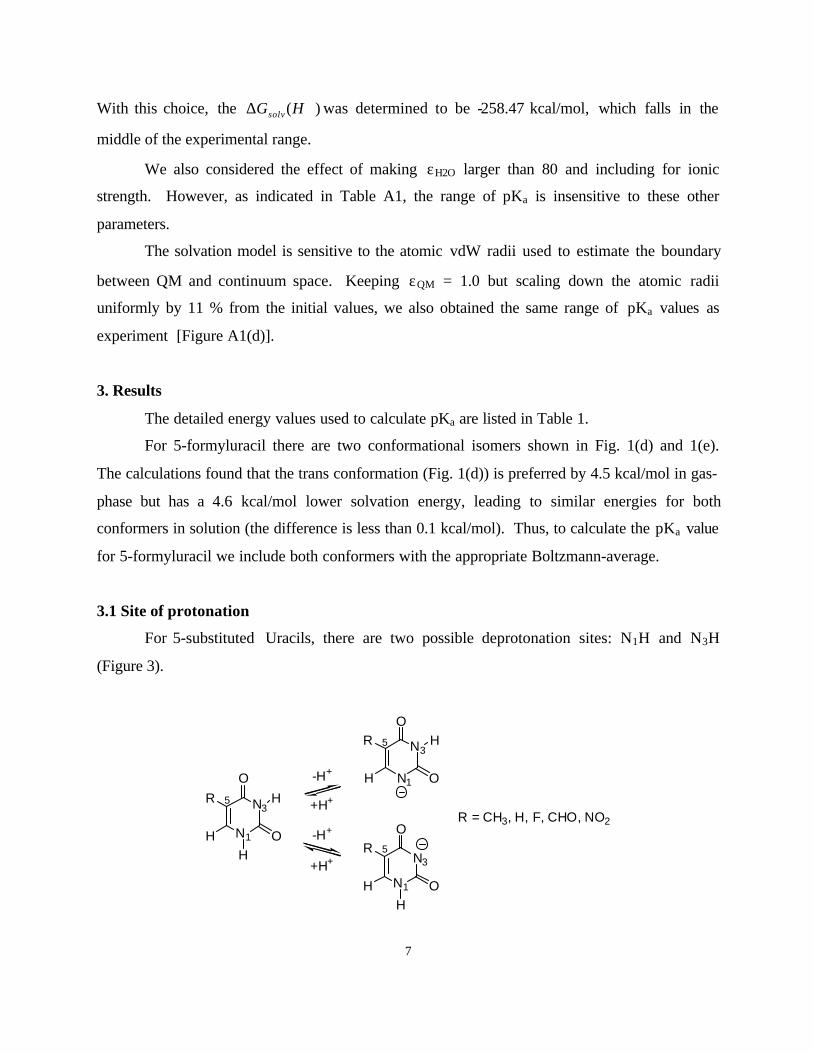
3.1 Site of protonation
For 5-substituted Uracils, there are two possible deprotonation sites: N1H and N3H
(Figure 3).
N1
N3
OH
OH
R
N1
N3
O
OH
H
R
R = CH3, H, F, CHO, NO2N1
N3
OH
OH
R
H
-H+
-H+
+H+
+H+
5
5
5
8
Figure 3. Two possible deprotonation sites of 5-substituted uracils: N1 and N3.
For thymine, uracil, and 5-fluorouracil, the pKa values of the N3 site are lower than for
the N1 site. In the gas phase, deprotonation from N1H of these species is 10~12 kcal/mol more
favorable than from N3H (calculated from gas-phase energy components in Table 1), as pointed
out in earlier calculations.28,29 However, the solvation of the N1(-) species is 13~14 kcal/mol
less favorable than for the N3(-) species (Table l). This is because N1(-) has greater charge
delocalization than N3(-). For example (see Table 2), the gas-phase dipole moment of the N1(-)
species of uracil is 2.15 D, whereas it is 7.36 D for the N3(-) species. Thus, the experimental pKa
values of thymine, uracil, and 5-fluorouracil correspond to the deprotonation from N3H.
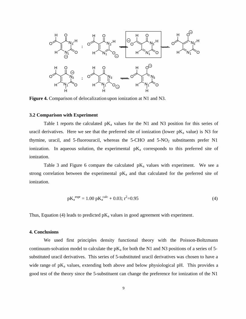
However, for 5-formyluracil and 5-nitrouracil, the solution-phase deprotonation from
N1H is more favorable than from N3H. The solvation of the N1(-) species is still 13~16 kcal/mol
less favorable than for the N3(-) species. However, in these cases the gas-phase deprotonation
from N1H is 15~17 kcal/mol more favorable than from N3H. This is plausible since these 5-
formyl and 5-nitro substituents can stabilize the N1(-) species by delocalizing negative charge
more extensively3 as indicated in Figure 4. This effect can be seen from the dipole moment and
geometry change (see Tables 2 and Figure 5). Thus, the experimental pKa values of 5-
formyluracil and 5-nitrouracil correspond to deprotonation from N1H.
This result is consistent with the experimental observation made on deoxyuridine where
the N1 is bonded to sugar ring so that deprotonation can occur only from the N3 site.3 The pKa
values of thymine, uracil, and 5-fluorouracil are very similar to those of the corresponding
deoxyuridines (9.75 vs. 9.69, 9.42 vs. 9.26, and 7.93 vs. 7.67), but the pKa of 5-CHO-U is
significantly lower than that of 5-CHO-deoxyuridine (6.84 vs. 8.12). (There is no experimental
observation on 5-NO2-deoxyuridine.)
9
N1
N3
OH
OH
H
O
N1
N3
O
OH
H N1
N3
O
OH
H N1
N3
O
OH
H
N1
N3
OH
OH
H
O
N1
N3
OH
OH
H
O
N1
N3
OH
OH
H
O:
:
H
O
H
O
H
O
Figure 4. Comparison of delocalization upon ionization at N1 and N3.
3.2 Comparison with Experiment
Table 1 reports the calculated pKa values for the N1 and N3 position for this series of
uracil derivatives. Here we see that the preferred site of ionization (lower pKa value) is N3 for
thymine, uracil, and 5-fluorouracil, whereas the 5-CHO and 5-NO2 substituents prefer N1
ionization. In aqueous solution, the experimental pKa corresponds to this preferred site of
ionization.
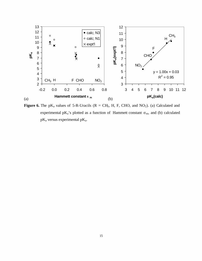
Table 3 and Figure 6 compare the calculated pKa values with experiment. We see a
strong correlation between the experimental pKa and that calculated for the preferred site of
ionization.
pKaexpr = 1.00 pKa
calc + 0.03; r2=0.95 (4)
Thus, Equation (4) leads to predicted pKa values in good agreement with experiment.
4. Conclusions
We used first principles density functional theory with the Poisson-Boltzmann
continuum-solvation model to calculate the pKa for both the N1 and N3 positions of a series of 5-
substituted uracil derivatives. This series of 5-substituted uracil derivatives was chosen to have a
wide range of pKa values, extending both above and below physiological pH. This provides a
good test of the theory since the 5-substituent can change the preference for ionization of the N1
10
and N3 positions. In aqueous solution, the more acidic proton will be lost first with increasing
solution pH, so that the experimental pKa value corresponds to the preferred ionization site.
Thus, the experimental pKa values must be compared with the pKa value calculated to be the
preferred site of ionization. This validates the computational method for predicting both the
preferred site of ionization and the pKa. We can now use this methodology to examine the
effects of additional modifications and derivatives including deoxynucleosides,
deoxynucleotides, and oligodeoxynucleotides.
Acknowledgments
This work was supported in part by the National Institutes of Health [HD36385 (WAG),
GM 41336 (LCW), and CA 33572(LCW)]. In addition, the facilities of the MSC are also
supported by DOE-ASCI, ARO-MURI, ARO-DURIP, National Science Foundation [CHE-95-
22179] Exxon Corp., Dow Chemical, 3M, Beckman Institute, Avery-Dennison, Chevron
Corporation, Seiko Epson, Asahi Chemical, and BP Amoco.
11
Table 1. Calculated energies used to estimate the pKa’s of 5-R-uracil’s.a
R Energy termsb Neutral(kcal/mol)
Anion: N3(-)(kcal/mol)
pKa Anion: N1(-)(kcal/mol)
pKa
CH3 E0K(g)c
ZPE(g)∆G0→298K(g)Total ∆G(g)
∆Gsolv
Total ∆G(aq)
-285077.96 69.91
-20.41-285028.47
-19.32-285047.79
-284723.6561.28
-20.46-284682.82
-86.51-284769.33 10.04
-284735.2061.66
-20.28-284693.82
-73.89-284767.71 11.23
H E0K(g)c
ZPE(g)∆G0→298K(g)Total ∆G(g)
∆Gsolv
Total ∆G(aq)
-260396.8553.15
-19.12-260362.82
-20.29-260383.11
-260043.0044.54
-19.20-260017.66
-87.94-260105.60 9.35
-260055.8044.98
-18.93-260029.75
-74.31-260104.06 10.48
F E0K(g)c
ZPE(g)∆G0→298K(g)Total ∆G(g)
∆Gsolv
Total ∆G(aq)
-322684.0748.27
-19.98-322657.78
-20.51-322676.29
-322337.0339.72
-20.18-322317.49
-84.12-322401.62 7.27
-322348.3040.15
-19.79-322327.93
-71.25-322399.19 9.05
CHOtransd
E0K(g)c
ZPE(g)∆G0→298K(g)Total ∆G(g)
∆Gsolv
Total ∆G(aq)
-331533.2159.22
-21.00-331494.99
-22.74-331517.73
-331191.3050.84
-20.92-331161.38
-80.72-331242.10 7.97
-331206.4151.26
-20.79-331175.94
-67.58-331243.52 6.92
CHOcise
E0K(g)c
ZPE(g)∆G0→298K(g)Total ∆G(g)
∆Gsolv
Total ∆G(aq)
-331528.2759.03
-21.20-331490.44
-27.38-331517.82
-331184.8550.50
-21.20-331155.55
-87.57-331243.12 7.28
-331202.3351.09
-20.88-331172.12
-71.46-331243.58 6.94
NO2 E0K(g)c
ZPE(g)∆G0→298K(g)Total ∆G(g)
∆Gsolv
Total ∆G(aq)
-388759.0755.22
-22.11-388725.96
-23.24-388749.19
-388422.5546.79
-21.85-388397.61
-77.40-388475.01 6.91
-388439.3347.22
-21.72-388413.83
-62.88-388476.71 5.66
a ∆G(H+(g)) = -6.3 kcal/mol, )( +∆ HGsolv = -258.47 kcal/mol, εQM = 0.92.b Total ∆G(g) = E0K(g) + ZPE(g) + ∆G0→298K(g), Total ∆G(aq) = Total ∆G(g) + ∆Gsolv.c Converted from hartree using 1 hartree = 627.5095 kcal/mol. d Fig. 1d. e Fig. 1e.
12
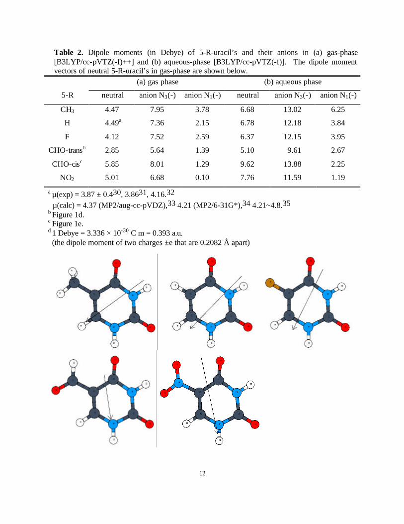
Table 2. Dipole moments (in Debye) of 5-R-uracil’s and their anions in (a) gas-phase[B3LYP/cc-pVTZ(-f)++] and (b) aqueous-phase [B3LYP/cc-pVTZ(-f)]. The dipole momentvectors of neutral 5-R-uracil’s in gas-phase are shown below.
(a) gas phase (b) aqueous phase
5-R neutral anion N3(-) anion N1(-) neutral anion N3(-) anion N1(-)
CH3 4.47 7.95 3.78 6.68 13.02 6.25
H 4.49a 7.36 2.15 6.78 12.18 3.84
F 4.12 7.52 2.59 6.37 12.15 3.95
CHO-transb 2.85 5.64 1.39 5.10 9.61 2.67
CHO-cisc 5.85 8.01 1.29 9.62 13.88 2.25
NO2 5.01 6.68 0.10 7.76 11.59 1.19
a µ(exp) = 3.87 ± 0.430, 3.8631, 4.16.32 µ(calc) = 4.37 (MP2/aug-cc-pVDZ),33 4.21 (MP2/6-31G*),34 4.21~4.8.35b Figure 1d.c Figure 1e.d 1 Debye = 3.336 × 10⁻³⁰ C m = 0.393 a.u. (the dipole moment of two charges ±e that are 0.2082 Å apart)
13
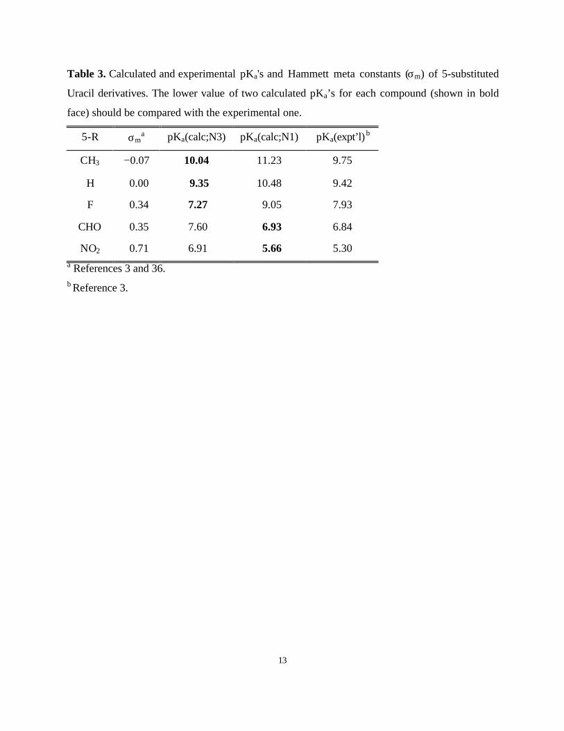
Table 3. Calculated and experimental pKa's and Hammett meta constants (σm) of 5-substituted
Uracil derivatives. The lower value of two calculated pKa’s for each compound (shown in bold
face) should be compared with the experimental one.
5-R σma pKa(calc;N3) pKa(calc;N1) pKa(expt’l)b
CH3 −0.07 10.04 11.23 9.75
H 0.00 9.35 10.48 9.42
F 0.34 7.27 9.05 7.93
CHO 0.35 7.60 6.93 6.84
NO2 0.71 6.91 5.66 5.30a References 3 and 36.b Reference 3.
14
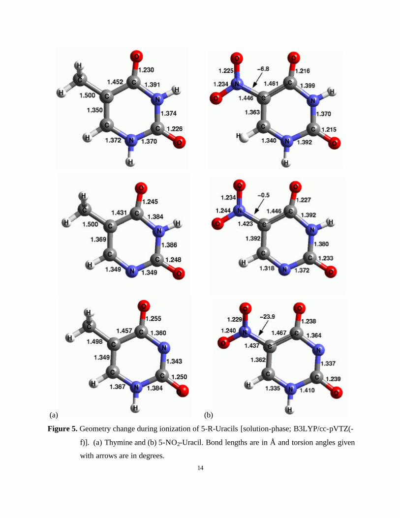
(a) (b)
Figure 5. Geometry change during ionization of 5-R-Uracils [solution-phase; B3LYP/cc-pVTZ(-
f)]. (a) Thymine and (b) 5-NO2-Uracil. Bond lengths are in Å and torsion angles given
with arrows are in degrees.
15
(a)
NO2CHOFHCH323456789
10111213
-0.2 0.0 0.2 0.4 0.6 0.8
Hammett constant σ m
pK
acalc; N3
calc; N1
expt'l
(b)
NO2
CHO
F
HCH3
y = 1.00x + 0.03R2 = 0.95
3
4
5
6
7
8
9
10
11
12
3 4 5 6 7 8 9 10 11 12
pKa(calc)
pK
a(ex
pt'l
)
Figure 6. The pKa values of 5-R-Uracils (R = CH3, H, F, CHO, and NO2). (a) Calculated and
experimental pKa’s plotted as a function of Hammett constant σm, and (b) calculated
pKa versus experimental pKa.
16
(a)
CH3 H F CHO NO223456789
10111213
-0.2 0.0 0.2 0.4 0.6 0.8
Hammett constant σ m
pK
acalc; N3
calc; N1
expt'l
CH3H
F
CHO
NO2
y = 0.67x + 2.62R2 = 0.99
3
4
5
6
7
8
9
10
11
12
3 4 5 6 7 8 9 10 11 12
pKa(calc)
pK
a(ex
pt'l
)
(b)
NO2CHOFHCH323456789
10111213
-0.2 0.0 0.2 0.4 0.6 0.8
Hammett constant σ m
pK
a
calc; N3
calc; N1
expt'l
CH3H
F
CHO
NO2
y = 0.86x + 1.13R2 = 0.98
3
4
5
6
7
8
9
10
11
12
3 4 5 6 7 8 9 10 11 12
pKa(calc)
pK
a(ex
pt'l
)
17
(c)
NO2CHOFHCH323456789
10111213
-0.2 0.0 0.2 0.4 0.6 0.8
Hammett constant σ m
pK
acalc; N3
calc; N1
expt'l
NO2
CHO
F
HCH3
y = 1.00x + 0.03R2 = 0.95
3
4
5
6
7
8
9
10
11
12
3 4 5 6 7 8 9 10 11 12
pKa(calc)
pK
a(ex
pt'l
)
(d)
CH3 H F CHO NO223456789
10111213
-0.2 0.0 0.2 0.4 0.6 0.8
Hammett constant σ m
pK
a
calc; N3
calc; N1
expt'l
CH3H
F
CHO
NO2
y = 0.98x + 0.19R2 = 0.94
3
4
5
6
7
8
9
10
11
12
3 4 5 6 7 8 9 10 11 12
pKa(calc)
pK
a(ex
pt'l
)
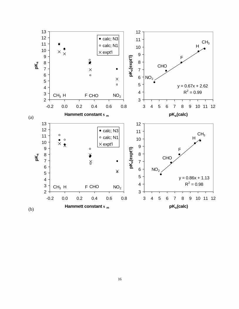
Figure A1. Dependence of calculated pKa on the dielectric constant εQM (a-c) and on the vdW radii (d).
(a) εQM = 1.00 [ )( +∆ HGsolv =-263.16 kcal/mol],
(b) εQM = 0.95 [ )( +∆ HGsolv =-260.27 kcal/mol],
(c) εQM = 0.92 [ )( +∆ HGsolv =-258.47 kcal/mol], and
(d) εQM = 1.00, the vdW radii scaled down by 11% [ )( +∆ HGsolv =-262.80 kcal/mol].
18
Table A1. Dependence of solvation energies of 5-R-U and its anions on εQM and εH2O.
∆Gsolv Originala εH2O = 96(20% higher)
εQM = 0.92(8% lower)
CHO N3(-) -73.166 -73.370 -80.723
N1(-) -61.545 -61.704 -67.583
Neutral -20.054 -20.130 -22.741
F N3(-) -76.054 -76.268 -84.123
N1(-) -64.914 -65.073 -71.254
Neutral -17.994 -18.059 -20.507
H N3(-) -79.658 -79.883 -87.941
N1(-) -67.699 -67.937 -74.310
Neutral -17.877 -17.938 -20.289
CH3 N3(-) -78.256 -78.481 -86.507
N1(-) -67.132 -67.307 -73.889
Neutral -16.976 -17.042 -19.322
NO2 N3(-) -69.910 -70.110 -77.398
N1(-) -57.212 -57.359 -62.881
Neutral -20.532 -20.612 -23.237a Ionic strength = 0.0, εH2O = 80, εQM = 1.0.
19
References
1)Myers, L. S.; Ward, J. F.; Tsukomato, W. T.; Holmes, D. E.; Julca, J. R. Science 1965, 148,
1234-1235.
2)Teebor, G. W.; Frenkel, K.; Goldstein, M. S. Proc. Natl. Acad. Sci. USA 1984, 81, 318-321.
3)Privat, E.; Sowers, L. Mutat. Res. 1996, 354, 151-156.
4)Privat, E.; Sowers, L. Chem. Res. Toxicol. 1996, 9, 745-750.
5)Imlay, J. A.; Linn, S. Science 1988, 240, 1302-1309.
6)Loeb, L. A. Cancer Res. 1989, 49, 5489-5496.
7)Ames, B. N.; Gold, L. S.; Willett, W. C. Proc. Natl. Acad. Sci USA 1995, 92, 5258-5265.
8)Heidelberger, C. Prog. Nucleic Acid Res. Mol. Biol. 1965, 4, 1-50.
9)Rowe, W. P.; Lowy, D. R.; Teich, N.; Hartley, J. W. Proc. Natl. Acad. Sci. USA 1972, 69,
1033-1035.
10)Sternglanz, H.; Bugg, C. E. Biochim. Biophys. Acta 1975, 378, 1-11.
11)Morris, S. M. Mutat. Res. 1993, 297, 39-51.
12)Saenger, W. Principles of Nucleic Acid Structure; Springer-Verlag: New York, 1983.
13)Lawley, P. D.; Brooks, P. D. J. Mol. Biol. 1962, 4, 216-219.
14)Sowers, L. C.; Shaw, B. R.; Veigl, M. L.; Sedwick, W. D. Mutat. Res. 1987, 177, 201-218.
15)Sowers, L. C.; Goodman, M. F.; Eritja, R.; Kaplan, B.; Fazakerley, G. V. J. Mol. Biol. 1989,
205, 437-447.
16)Yu, H.; Eritja, R.; Bloom, L. B.; Goodman, M. F. J. Biol. Chem. 1993, 268, 15935-15943.
17)Lim, C.; Bashford, D.; Karplus, M. J. Phys. Chem. 1991, 95, 5610-5620.
18)Jaguar 3.5; Schrodinger Inc.: Portland, OR, 1998; Greeley, B. H.; Russo, T. V.; Mainz, D. T.;
Friesner, R. A.; Langlois, J.-M.; Goddard III, W. A.; Donnelly, R. E.; Ringalda, M. N. J. Phys.
Chem. 1994, 101, 4028-4041.
19)Scott, A. P.; Radom, L. J. Phys. Chem. 1996, 100, 16502-16513.
20)Tannor, D. J.; Marten, B.; Murphy, R.; Friesner, R. A.; Sitkoff, D.; Nicholls, A.; Ringnalda,
M. N.; Goddard III, W. A.; Honig, B. A. J. Am. Chem. Soc. 1994, 116, 11875-11882.
21)Honig, B.; Nicholls, A. Science 1995, 268, 1144-1149.
22)Marten, B.; Kim, K.; Cortis, C.; Friesner, R. A.; Murphy, R. B.; Ringnalda, M. N.; Sitkoff,
D.; Honig, B. J. Phys. Chem. 1996, 100, 11775-11788.
20
23)Nicholls, A.; Honig, B. J. Comput. Chem. 1991, 12, 435-445.
24)Archer, D. G.; Wang, P. J. Phys. Chem. Ref. Data 1990, 19, 371-411.
25)Reiss, H.; Heller, A. J. Phys. Chem. 1985, 89, 4207-4213.
26)Marcus, Y. Ion Solvation; John Wiley and Sons, Ltd., 1985, pp 105-109.
27)Tissandier, M. D.; Cowen, K. A.; Feng, W. Y.; Gundlach, E.; Cohen, M. H.; Earhart, A. D.;
Coe, J. V.; Tuttle, T. R. J. Phys. Chem. A 1998, 102, 7787-7794.
28)Nguyen, M.; Chandra, A.; Zeegers-Huyskens, T. J. Chem. Soc. Faraday Trans. 1998, 94,
1277-1280.
29)Chandra, A.; Nguyen, M.; Zeegers-Huyskens, T. J. Phys. Chem. A 1998, 102, 6010-6016.
30)Boys, S. F.; Bernardi, F. Mol. Phys. 1970, 19, 553.
31)Brown, R. B.; Godfrey, P. D.; McNaughton, D.; Pierlot, A. P. J. Am. Chem. Soc. 1988, 110,
2329.
32)Kulakowska, I.; Geller, M.; Lesyng, B.; Wierzchowski, K. L.; Bolewski, K. Biochim.
Biophys. Acta 1975, 407, 420.
33)van Mourik, T.; Price, S.; Clary, D. J. Phys. Chem. A 1999, 103, 1611-1618.
34)Leszczynski, J. J. Phys. Chem. 1992, 96, 1649-1653.
35)Estrin, D. A.; Paglieri, L.; Corongiu, G. J. Phys. Chem. 1994, 98, 5653-5660.