first morphological and molecular identification of third
TRANSCRIPT

1© 2021 Authors. This is an Open Access article licensed under the Creative Commons CC BY 4.0 license, https://creativecommons.org/licenses/by/4.0/
JOURNAL OF NEMATOLOGYe2021-10 | Vol. 53Article | DOI: 10.21307/jofnem-2021-010
First morphological and molecular identification of third-stage larvae of Anisakis typica (Nematoda: Anisakidae) from marine fishes in Vietnamese water
Hoang Van Hien1, Bui Thi Dung1, Ha Duy Ngo1 and Pham Ngoc Doanh1, 2,*1Institute of Ecology and Biological Resources, Vietnam Academy of Science and Technology, Hanoi, Vietnam.2Graduate University of Science and Technology, Vietnam Academy of Science and Technology, Hanoi, Vietnam.
*E-mail: [email protected]
This paper was edited by Zafar Ahmad Handoo.
Received for publication November 24, 2020.
AbstractAnisakid nematodes are parasites of cetaceans, their larval stages live in marine fishes. The third-stage larvae of some Anisakis species are also the etiological agents of human anisakiasis caused by consumption of raw or undercooked infected fish. Thus, identification of Anisakis larvae at the species level is crucial for their ecology and epidemiology. In Vietnam, although Anisakis larvae have been reported, they have not been identified to the species level. The aim of this study was, therefore, to identify third-stage larvae of Anisakis collected from marine fishes in Vietnamese water, based on morphological characteristics and molecular analysis. All Anisakis larvae found in this study were morphologically similar to each other and identical to Anisakis typica. In addition, molecular analysis based on ITS1-5.8S-ITS2 sequences confirmed them as A. typica. Vietnamese A. typica population was genetically close to those from Asian countries and Australia. The third-stage larvae of A. typica were collected from eight fish species from three localities in the South of Vietnam. Among them, seven were recorded as new intermediate hosts of A. typica. This is the first identification of A. typica larvae in Vietnamese water with records of new fish hosts.
KeywordsAnisakid larvae, Intermediate fish host, Molecular analyzes, Morphology, Vietnam.
Nematodes of the genus Anisakis (Nematoda: Ani-sakidae) are parasites of marine organisms. The life cycle of these nematodes requires marine mammals, mainly cetaceans, as the definitive hosts, and crustaceans, fish, and cephalopods as intermediate/paratenic hosts (Klimpel and Palm, 2011). Humans are accidental hosts due to ingestion of raw or undercooked fish containing the third infective-stage larvae (L3). Human anisakiasis patients suffer from abdominal pain, nausea, vomiting, and/or diarrhea (Dorny et al., 2009). In addition, allergic reactions may occur due to exposure to the nematode antigens (Aibinu et al., 2019; Audicana et al., 2002). Given the influence on human health, Anisakis nematodes are
of interest. Anisakid larvae can be morphologically identified at the genus level by typical characteristics of anterior and posterior regions, and are classified into two types, type I and II, based on the length of the ventriculus and presence/absence of the tail spine (mucron): Anisakis type I larva has a longer ventriculus and a mucron, while type II larva has a shorter ventriculus and no mucron (Berland, 1961). Type I consists of A. simplex, A. pegreffii, A. typica, A. ziphidarum, and A. nascettii, while type II consists of A. paggiae, A. physeteris, and A. brevispiculata (Mattiucci and Nascetti, 2008). Previously, it was not easy to identify anisakid larvae at the species level, because there is a lack of distinct morphological characteristics

2
First identification of Anisakis typica larvae in Vietnam: Hien et al.
required for species identification (Farjallah et al., 2008). Recent studies indicated morphological differences between Anisakis species (Chen and Shih, 2015; Sonko et al., 2019; Tunya et al., 2020). In addition, molecular tools allow the accurate identification of anisakid larvae by using sequences of the internal transcribed spacer (ITS) region of ribosomal DNA (D’amelio et al., 2010; Mattiucci and Nascetti, 2008).
In Vietnam, data on Anisakis nematodes are scarce. There have been a few reports on Anisakis larvae without morphological description and identi-fication to species level (Arthur and Te, 2006; Ngo et al., 2009). During our recent comprehensive survey for parasites of marine fishes in Vietnamese water, we collected Anisakis larvae from eight fish species. The aim of the present study was to identify these Anisakis specimens from Vietnamese water by morphological and molecular approaches.
Materials and methods
Fish examination and larval collection
Marine fish were bought in 10 fish ports located in 10 provinces along the seashore of Vietnam where fishing vessels docked (Fig. 1). All fish specimens were placed on ice and transferred to the laboratory under good aeration. Fish were dissected, their body cavities and internal organs were examined under a stereomicroscope. Third-stage larvae were isolated from the body cavity and visceral organs. The larvae were washed in phosphate-buffered saline. For morphological identification, larvae were preserved in 4% formalin. Representative specimens were preserved in 70% ethanol for DNA isolation.
Morphological study
Anisakis larvae were soaked in a solution of glycerin-phenol-lactic acid-distilled water (2:1:1:1) for about 48 hr until the body parts were transparent. Then, the larvae were observed and measured under a light microscope (ECLIPSE H600 L Nikon). For scanning electron microscopy, Anisakis larvae were prepared according to Madden and Tromba (1976) and Morsy et al. (2017). Larvae were identified according to the reported references (Berland, 1961; Chen and Shih, 2015; Mattiucci et al., 2009; Sonko et al., 2019; Tunya et al., 2020).
Molecular and phylogenetic analysis
DNA of three representative larvae from three localities was extracted using QIAamp DNA stool
Minikit (Qiagen, Hilden, Germany). Two primers NC5-GTAGGTGAACCTGCGGAAGGATCATT (forward) and NC2-TTAGTTTCTTTTCCTCCGCT (reverse) were used in a polymerase chain reaction (PCR) to amplify the rDNA region of the first to the second internal transcribed spacer (ITS1-5.8S-ITS2) (Zhu et al., 2000). PCR products were electrophoresed in a 1.0% agarose gel and visualized by ethidium bromide staining. Positive PCR products were sent to Macrogen Company (Korea) for sequencing. The nucleotide sequences obtained in this study were deposited in GenBank under accession numbers LC592876-LC592878.
BLAST searches were performed at NCBI (http://blast.ncbi.nlm.nih.gov/Blast.cgi) to find sequence similarities. Sequences of Anisakis species available in GenBank were downloaded for analysis. The analysis involved 42 nucleotide sequences, including a sequence (KM491173) of Contracaecum osculatum as an out-group. The evolutionary history was inferred by using the Maximum Likelihood method based on the Kimura 2-parameter model in MEGA software v.7.0. (Kumar et al., 2016). Initial tree(s) for the heuristic search were obtained automatically by applying Neighbor-Join and BioNJ algorithms to a matrix of pairwise distances estimated using the Maximum Composite Likelihood approach, and then selecting the topology with superior log likelihood value. A discrete Gamma distribution was used to model evolutionary rate differences among sites (five categories (+G, parameter = 0.8044)). All positions containing gaps and missing data were eliminated. There were a total of 656 positions in the final dataset.
Results and discussion
A total of 3,775 fish of 138 species from 10 study sites were examined. Anisakis larvae were found from eight fish species from three localities, Khanh Hoa, Vung Tau, and Bac Lieu, in the South of Vietnam. Prevalences of infection ranged from 10 to 50% with intensity varied from 1 to 19 larvae/fish (Table 1), non-infected fish species were presented in the supplemental material.
All Anisakis larvae were morphologically similar to each other. The body of the larvae was cylindrical in shape, attenuated at both ends, and measured 17.2 to 20.3 (18.6 ± 1.1) mm long and 0.26 to 0.34 (0.29 ± 0.03) mm width (n = 30 larvae). The lips were inconspicuous, with a prominent boring tooth at the anterior extremity. The esophagus had an anterior muscular part and measured 1.52 to 1.58 (1.54 ± 0.02) mm long and a glandular ventriculus measured 0.58 to 0.82 (0.64 ± 0.04) mm long. Long intestinal caeca with clear demarcation were present. The body of

3
JOURNAL OF NEMATOLOGY
Figure 1: Study sites along the seashore of Vietnam. Three localities where fishes were infected with Anisakis larvae are print in bold.
larvae ended at a short cylindrical mucron measuring 0.021 to 0.030 (0.025±0.005) mm long (Figs. 2, 3). These characteristics of the third-stage larvae were identical to Anisakis larvae type I (Berland, 1961). It has previously been noted that it is difficult to distinguish between Anisakis species belonging to
type I because they look quite similar to each other (Farjallah et al., 2008). However, recent studies based on morphological and molecular approaches provided descriptions and microphotographs sho-wing differences between L3 larvae of A. pegreffii and A. typica (Chen and Shih, 2015; Sonko et al., 2019).

4
First identification of Anisakis typica larvae in Vietnam: Hien et al.
Table 1. Prevalence of Anisakis larvae infection in marine fishes in Vietnamese water.
LocalityNo. of fish examined
No. of fish species
Infected fish species
No. of infected/examined fish (%)
Density
Quang Ninh 615 62 0
Hai Phong 478 41 0
Nam Dinh 122 23 0
Nghe An 303 50 0
Quang Binh 520 75 0
Hue 211 28 0
Khanh Hoa 766 82 Dcapterus macarellus 10/20 (50.0) 1-19
Trichiurus lepturus 6/20 (30.0) 1-5
Sargocentron rubrum 1/10 (10.0) 1
Lutjanus johnii 1/10 (10.0) 1
Megalaspis cordyla 2/12 (16.7) 1; 3
Priacanthus hamrur 1/8 (12.5) 2
Pristipomoides filamentosus 3/10 (30.0) 1; 1; 1
Vung Tau 40 6 Megalaspis cordyla 1/5 (20.0) 6
Bac Lieu 390 58 Carangoides malabaricus 1/10 (10.0) 1
Kien Giang 330 63 0
Total 3775 138 8 1-19
In addition, Tunya et al. (2020) suggested that the protruded mucron of L3 larvae can be used to identify anisakid larvae at the species level: the protruded mucron of A. simplex was cone-shape, while that of A. typica was cylindrical-shape, which is narrower and longer than that of A. simplex. According to these, L3 larvae of Anisakis specimens found in this study were identified as A. typica.
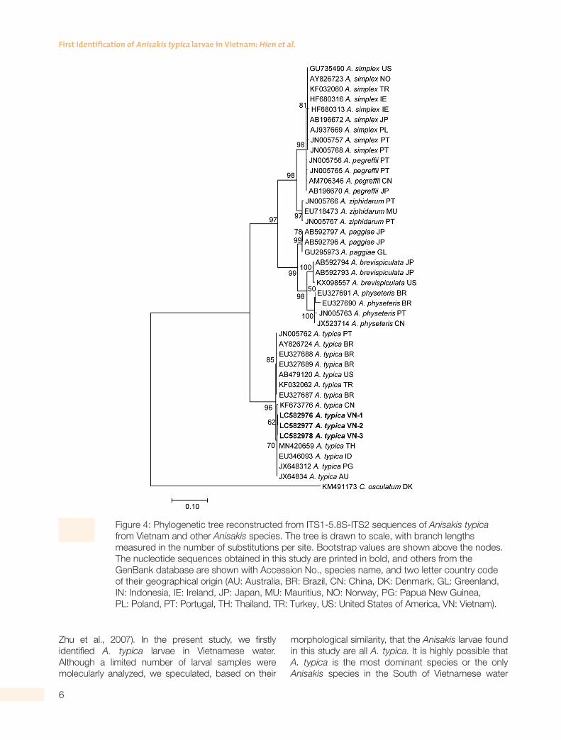
Because the Anisakis larvae collected in the present study were all morphologically similar to each other, three larvae representative for three locations were used for molecular analyses. Three ITS1-5.8S-ITS2 sequences obtained from three L3 larvae were 771 bp and completely identical (100%) with each other. In agreement with morphological identification, the BLAST searches revealed that the ITS1-5.8S-ITS2 sequences of Anisakis larvae from Vietnam showed the highest similarity (100%) with that of A. typica available in GenBank. The analysis of genetic distances demonstrated that inter-specific genetic distances between A. typica and other Anisakis species were: A. paggiae 17.2%; A. ziphidarum 17.3%; A. pegreffii 18.0%; A. simplex 18.0%; A. physeteris
18.4%; and A. brevispiculata 18.7%. In the phylogenetic tree (Fig. 4), A. typica made a distinct clade that was far distant from other Anisakis species. Vietnamese A. typica were genetically close to those from China, Thailand, Indonesia, Papua New Guinea, and Australia, to make a common group that was separated from another group of America, Brazil, Turkey, and Portugal. Our analysis is in agreement with a previous report that the separation of A. typica populations related to geographical origins (Tunya et al., 2020).
Molecular studies of Anisakis from various parts of the world Oceans confirmed the validity of nine Anisakis species (Klimpel and Palm, 2011). Most of them distribute in the Atlantic and the Mediterranean Sea with several records of some species in the South American, African, and Australian sea, and SW Pacific Ocean. A unique distribution pattern has been known for A. typica which has been reported in warmer temperate and tropical waters (Mattiucci and Nascetti, 2006). In Asian countries, A. typica larvae have been reported in Japan, Korea, China, Taiwan, Indonesia, and Thailand (Lee et al., 2016; Palm et al., 2008, 2017; Sonko et al., 2019; Tunya et al., 2020; Umehara et al., 2010;

5
JOURNAL OF NEMATOLOGY
Figure 2: Light micrographs of Anisakis typica larva. A. Whole larva; B. Anterior part of the body showing a long ventriculus; C. Anterior part of the body showing a boring tooth; D. Posterior end of the body showing a mucron.
Figure 3: Scanning electron micrographs of Anisakis typica larva. A. Anterior end showing a mouth and a boring tooth; B. Posterior end showing a mucron.
6
First identification of Anisakis typica larvae in Vietnam: Hien et al.
Figure 4: Phylogenetic tree reconstructed from ITS1-5.8S-ITS2 sequences of Anisakis typica from Vietnam and other Anisakis species. The tree is drawn to scale, with branch lengths measured in the number of substitutions per site. Bootstrap values are shown above the nodes. The nucleotide sequences obtained in this study are printed in bold, and others from the GenBank database are shown with Accession No., species name, and two letter country code of their geographical origin (AU: Australia, BR: Brazil, CN: China, DK: Denmark, GL: Greenland, IN: Indonesia, IE: Ireland, JP: Japan, MU: Mauritius, NO: Norway, PG: Papua New Guinea, PL: Poland, PT: Portugal, TH: Thailand, TR: Turkey, US: United States of America, VN: Vietnam).
Zhu et al., 2007). In the present study, we firstly identified A. typica larvae in Vietnamese water. Although a limited number of larval samples were molecularly analyzed, we speculated, based on their
morphological similarity, that the Anisakis larvae found in this study are all A. typica. It is highly possible that A. typica is the most dominant species or the only Anisakis species in the South of Vietnamese water

7
JOURNAL OF NEMATOLOGY
Table 2. Intermediate fish hosts of Anisakis typica in the World and in Vietnam.
No. Host species Localities References
1 Sotalia guianensis
2 Auxis thazard
3 Thunnus thynnus Brazil Coast
4 Pseudopercis numida
5 Trachurus picturatus
6 Scomber japonicus Portugal Mattiucci et al. (2002), Marques et al. (2006), Pantoja et al. (2015)
7 Platichthys flesus
8 Scomberomorus commerson
9 Euthynnus affinis
10 Sarda orientalis Somalia
11 Coryphaena hippurus
12 Stenella attenuata
13 Globicephala macrorhynchus Florida Mattiucci et al. (2005)
14 Scomber scombrus
15 Merluccius merluccius North Africa Farjallah et al. (2008)
16 Phycis phycis
Scomber japonicus Turkey Pekmezci et al. (2014)
17 Micromesistius poutassou
Trichiurus spp. Japan Umehara et al. (2010)
Scomber japonicus Suzuki et al. (2010)
18 Trichiurus lepturus Korea Lee et al. (2009)
19 Todarodes pacificus
20 Astroconger myriaster Cho et al. (2015)
21 Decapterus macarellus
22 Gerres oblongus
23 Pinjalo lewisi
24 Pinjalo pinjalo Papua New Guinea Koinari et al. (2013)
25 Selar crumenophthalmus
26 Scomberomorus maculatus
27 Thunnus albacares
28 Auxis rochei rochei Indonexia Palm et al. (2008)
29 Decapterus russelli
30 Nemipterus hexodon Thailand Tunya et al. (2020)
31 Nemipterus japonicus
32 Scomber australasicus Taiwan Umehara et al. (2010), Sonko et al. (2019)
Trichiurus lepturus
1 Carangoides malabaricus
2 Dcapterus macarellus

8
First identification of Anisakis typica larvae in Vietnam: Hien et al.
similar to reports in Thailand water where A. typica was the only species found in the Gulf of Thailand (Eamsobhana et al., 2018; Tunya et al., 2020).
Regarding intermediate fish hosts, the third-stage larvae of A. typica are found in various fish species. They differ from place to place depending on geographical locations. Outside of Vietnamese water, 32 fish species have been reported as intermediate hosts of A. typica (Table 2). In this study in Vietnam, A. typica larvae were found from eight fish species, Carangoides malabaricus, Dcapterus macarellus, Sargocentron rubrum, Lutjanus johnii, Megalaspis cordyla, Priacanthus hamrur, Pristipomoides filame ntosus, and Trichiurus lepturus. Among these, only T. lepturus has been previously reported as an intermediate host of A. typica in Korean and Taiwanese waters, the other seven species are reported as new hosts.
Conclusion
The present study firstly identified A. typica larvae, based on morphological characteristics and mole-cular analysis, from eight marine fish species in the South of Vietnamese water and recorded seven fish species as new intermediate hosts of this Anisakis nematode. Genetically, the ITS1-5.8S-ITS2 sequences of Vietnamese A. typica were close to those from Asian countries and Australia, to make a common group separated from another group from America and Europe.
Acknowledgments
This study was supported by National Program under code number VAST.DA47.DA.12/16–19. Conflict of interest: none.
ReferencesAibinu, I. E., Smooker, P. M., Lopata and A. L. 2019.
Anisakis nematodes in fish and shellfish from infection to allergies. International Journal for Parasitology: Parasites Wildlife 9:384–93.
Arthur, J. R. and Te, B. Q. 2006. Checklist of the parasites of fishes of Viet Nam FAO, Rome.
Audicana, M. T., Ansotegui, I. J., de Corres, L. F. and Kennedy, M. W. 2002. Anisakis simplex: dangerous – dead and alive?. Trends in Parasitology 18:20–5.
Berland, B. 1961. Nematodes from some Norwegian marine fishes. Sarsia 2:1–50.
Chen, H. Y. and Shih, H. H. 2015. Occurrence and prevalence of fish-borne Anisakis larvae in the spotted mackerel Scomber australasicus from Taiwanese waters. Acta Tropica 145:61–7.
Cho, J., Lim, H., Jung, B. K., Shin, E. H. and Chai, J. Y. 2015. Anisakis pegreffii Larvae in Sea Eels (Astroconger myriaster) from the South Sea, Republic of Korea. Korean Journal of Parasitology 53:349–53.
D’amelio, S., Busi, M., Ingrosso, S. and Paggi, L. 2010. “Anisakiasis”, In Liu, D. Y. (Ed.), Molecular detection of foodborne pathogens Taylor & Francis CRC Press, pp. 757–68.
Dorny, P., Praet, N., Deckers, N. and Gabriel, S. 2009. Emerging food-borne parasites. Veterinary Parasitology 163:196–206.
Eamsobhana, P., Yong, H. S., Song, S. L., Tungtrongchitr, A. and Roongruangchai, K. 2018. Genetic differentiation of Anisakis species (Nematoda: Anisakidae) in marine fish Priacanthus tayenus from Gulf of Thailand. Tropical Biomedicine 35:669–77.
Farjallah, S., Slimane, B. D., Busi, M., Paggi, L., Amor, N., Blel, H., Said, K. and D’Amelio, S. 2008. Occurrence and molecular identification of Anisakis spp. from the North African coasts of Mediterranean Sea. Parasitology Research 102:371–9.
Klimpel, S. and Palm, H. W. 2011. “Anisakid nematode (Ascaridoidea) life cycles and distribution: Increasing zoonotic potential in the time of climate change?”, In Mehlhorn, H. (Ed.), Progress in parasitology. Parasitology research monographs, Vol. 2 Springer, Berlin, pp. 201–22.
Koinari, M., Karl, S., Elliot, A., Ryan, U. and Lymbery, A. J. 2013. Identification of Anisakis species (Nematoda: Anisakidae) in marine fish hosts from Papua New Guinea. Veterinary Parasitology 193:126–133.
Kumar, S., Stecher, G. and Tamura, K. 2016. MEGA7: Molecular Evolutionary Genetics Analysis version 7.0. Molecular Biology and Evolution 33:1870–4.
Lee, M. H., Cheon, D. S. and Choi, C. 2009. Molecular genotyping of Anisakis species from Korean
3 Sargocentron rubrum
4 Lutjanus johnii Vietnam In this study
5 Megalaspis cordyla
6 Priacanthus hamrur
7 Pristipomoides filamentosus
Trichiurus lepturus

9
JOURNAL OF NEMATOLOGY
sea fish by polymerase chain reaction-restriction fragment length polymorphism (PCR-RFLP). Food Control 20:623–6.
Lee, W. J., Seo, D. J., Oh, H., Jeon, S. B., Jung, D. and Choi, C. 2016. Simultaneous detection and prevalence of allergens in Anisakis species isolated from marine fishes. Journal of Food Protection 79:789–94.
Madden, P. A. and Tromba, F. G. 1976. Scanning electron microscopy of the lip denticles of Ascaris suum adults of known ages. Journal of Parasitology 62:265–71.
Marques, J. F., Cabral, H. N., Busi, M. and D’Amelio, S. 2006. Molecular identification of Anisakis species from Pleuronectiformes off the Portuguese coast. Journal of Helminthology 80:47–51.
Mattiucci, S. and Nascetti, G. 2006. Molecular systematics, phylogeny and ecology of anisakid nematodes of the genus Anisakis Dujardin, 1845: an update. Parasite 13:99–113.
Mattiucci, S. and Nascetti, G. 2008. Advances and trends in the molecular systematics of anisakid nematodes, with implications for their evolutionary ecology and host-parasite co-evolutionary processes. Advance in Parasitology 66:47–148.
Mattiucci, S., Paoletti, M. and Webb, S. C. 2009. Anisakis nascettii n. sp. (Nematoda: Anisakidae) from beaked whales of the southern hemisphere: mor-phological description, genetic relationships between congeners and ecological data. Systematic Parasitology 74:199–217.
Mattiucci, S., Nascetti, G., Dailey, M., Webb, S. C., Barros, N. B., Cianchi, R. and Bullini, L. 2005. Evidence for a new species of Anisakis Dujardin, 1845: morpho-logical description and genetic relationships between congeners (Nematoda: Anisakidae). Systematic Parasi-tology 61:157–71.
Mattiucci, S., Paggi, L., Nascetti, G., Santos, C. P., Costa, G., Di Beneditto, A. P., Ramos, R., Argyrou, M., Cianchi, R. and Bullini, L. 2002. Genetic markers in the study of Anisakis typica (Diesing, 1860): larval identification and genetic relationships with other species of Anisakis Dujardin, 1845 (Nematoda: Anisa-kidae). Systematic Parasitology 51:159–70.
Morsy, K., Badr, A. M., Abdel-Ghaffar, F., Deeb, S. E. and Ebead, S. 2017. Pathogenic potential of fresh, frozen, and thermally treated Anisakis spp. type II (L3) (Nematoda: Anisakidae) after oral inoculation into Wistar rats: a histopathological study. Journal of Nematology 49:427–36.
Ngo, H. D., Ha, N. V., Tu, N. D. and Thanh, N. V. 2009. Preliminary study on the parasitic helminth fauna on marine fishes in the coastal waters of Hai Phong province. Vietnam Journal of Biology 31:1–8.
Palm, H. W., Damriyasa, I. M., Linda and Oka, I. B. M. 2008. Molecular genotyping of Anisakis Dujardin,
1845 (Nematoda: Ascaridoidea: Anisakidae) larvae from marine fish of Balinese and Javanese waters, Indonesia. Helminthologia 45:3–12.
Palm, H. W., Theisen, S., Damriyasa, I. M., Kusmintarsih, E. S., Oka, I. B., Setyowati, E. A., Suratma, N. A., Wibowo, S. and Kleinertz, S. 2017. Anisakis (Nematoda: Ascaridoidea) from Indonesia. Diseases of Aquatic Organisms 123:141–57.
Pantoja, C. S., Borges, J. N., Santos, C. P. and Luque, J. L. 2015. Molecular and morphological characterization of Anisakid nematode larvae from the sandperches Pseudopercis numida and Pinguipes brasilianus (Perciformes: Pinguipedidae) off Brazil. Journal of Parasitology 101:492–99.
Pekmezci, G. Z., Onuk, E. E., Bolukbas, C. S., Yardimci, B., Gurler, A. T., Acici, M. and Umur, S. 2014. Molecular identification of Anisakis species (Nematoda: Anisakidae) from marine fishes collected in Turkish waters. Veterinary Parasitology 201:82–94.
Sonko, P., Chen, S. C., Chou, C. M., Huang, Y. C., Hsu, S. L., Barčák, D., Oros, M. and Fan, C. K. 2019. Multidisciplinary approach in study of the zoonotic Anisakis larval infection in the blue mackerel (Scomber australasicus) and the largehead hairtail (Trichiurus lepturus) in Northern Taiwan. Journal of Microbiology, Immunology and Infection 53:1021–9.
Suzuki, J., Murata, R., Hosaka, M. and Araki, J. 2010. Risk factors for human Anisakis infection and association between the geographic origins of Scomber japonicus and anisakid nematodes. International Journal of Food Microbiology 137:88–93.
Tunya, R., Wongsawad, C., Wongsawad, P. and Chai, J. Y. 2020. Morphological and molecular characteristics of Anisakis typica larvae in two species of threadfin bream, Nemipterus hexodon and N. japonicus, from the Gulf of Thailand. Korean Journal of Parasitology 58:15–25.
Umehara, A., Kawakami, Y., Ooi, H. K., Uchida, A., Ohmae, H. and Sugiyama, H. 2010. Molecular identification of Anisakis type I larvae isolated from hairtail fish off the coasts of Taiwan and Japan. Inter-national Journal of Food Microbiology 143:161–5.
Zhu, X., D’Amelio, S., Paggi, L. and Gasser, R. B. 2000. Assessing sequence variation in the internal transcribed spacers of ribosomal DNA within and among members of the Contracaecum osculatum complex (Nematoda: Ascaridoidea: Anisakidae). Para-sitology Research 86:677–83.
Zhu, X. Q., Podolska, M., Liu, J. S., Yu, H. Q., Sonko, H. H., Lin, Z. X., Luo, C. B., Song, H. Q. and Lin, R. Q. 2007. Identification of anisakid nematodes with zoonotic potential from Europe and China by single-strand conformation polymorphism analysis of nuclear ribosomal DNA. Parasitology Research 101:1703–7.