firmicutes’ contains a diverse array of taxa that are not
TRANSCRIPT

DISSIMILATORY IRON-REDUCING AND ENDOSPORULATING BACTERIA
by
ROB UCHE ONYENWOKE
(Under the Direction of Juergen Wiegel)
ABSTRACT
This dissertation represents a diversified study of the biochemical, physiological, and
genetic traits of members of the low G+C subdivision of the Gram-type positive bacteria, also
known as the ‘Firmicutes’. The phylum ‘Firmicutes’ contains a diverse array of taxa that are not
easily separated into coherent phylogenetic groups by any one physiological trait, such as
endospore-formation or dissimilatory iron reduction. This dissertation considers numerous
contemporary, and highly convergent in providing breadth and scope of the subject matter,
methods of study. The principle aim was to examine the lineage ‘Firmicutes’ by a) a genomic
study of the occurrence or absence of endosporulation genes in numerous members of the
lineage, b) classic biochemical studies of the enzymes responsible for biotic iron reduction, and
c) culture-dependent studies and isolations of various ‘Firmicutes’ to both identify new iron-
reducers and better resolve the taxonomy of the lineage. The work with endosporulation genes
has shown there might not be a distinct set of “endosporulation-specific” genes. This raises
several new questions about this exceptionally complex process. The work described here on
“ferric reductases” suggests there are enzymes capable of iron reduction that also have additional
activities. The isolation of novel bacteria presented here have added to the diversity of the
‘Firmicutes’ but have also added to the phylogenetic and taxonomic complexity of this group.

Traditional boundaries for families and genera have been weakened or shown to be in need of
further studies.
INDEX WORDS: Gram-type positive bacteria, Firmicutes, Thermophiles, Endospores,
Dissimilatory iron reduction, Quinones, Oxidative stress, The University of Georgia

THE PHYSIOLOGY OF THE FIRMICUTES: NOVEL DISSIMILATORY IRON-REDUCING
BACTERIA, OXIDOREDUCTASE ENZYMES, AND THE ENDOSPORULATING
BACTERIA
by
ROB UCHE ONYENWOKE
B.S., The University of Georgia, 2000
A Dissertation Submitted to the Graduate Faculty of The University of Georgia in Partial
Fulfillment of the Requirements for the Degree
DOCTOR OF PHILOSOPHY
ATHENS, GEORGIA
2006

© 2006
Rob Uche Onyenwoke
All Rights Reserved

THE PHYSIOLOGY OF THE FIRMICUTES: NOVEL DISSIMILATORY IRON-REDUCING
BACTERIA, OXIDOREDUCTASE ENZYMES, AND THE ENDOSPORULATING
BACTERIA
by
ROB UCHE ONYENWOKE
Major Professor: Dr. Juergen Wiegel
Committee: Dr. Michael W. W. Adams Dr. Harry A. Dailey Dr. Robert J. Maier Dr. William B. Whitman
Electronic Version Approved: Maureen Grasso Dean of the Graduate School The University of Georgia December 2006

iv
ACKNOWLEDGEMENTS
First of all, I would like to express my gratitude to all of my family, friends, and members
of the University of Georgia/Athens community who have made my matriculation such a rich
and fulfilling period of my life. I thank my parents for their continued support of me as I have
progressed forward in my life. I thank Juergen Wiegel, my advisor and friend, for his guidance
and support, and my committee members for all of their thoughtful suggestions and willingness
to foster my academic development. Finally, I would like to say that I have been privileged to
have had the opportunity to work with so many wonderful people in the Department of
Microbiology.
v
TABLE OF CONTENTS
Page
ACKNOWLEDGEMENTS........................................................................................................... iv
LIST OF TABLES......................................................................................................................... ix
LIST OF FIGURES ....................................................................................................................... xi
CHAPTER
1 INTRODUCTION AND LITERATURE REVIEW .....................................................1
Thermophiles.............................................................................................................1
The ‘Firmicutes’........................................................................................................2
Isolation and characterization of ‘Firmicutes’ ..........................................................8
Biotic metal reduction .............................................................................................10
Iron transport, binding, and acquisition...................................................................12
Dissimilatory Fe(III) reduction ...............................................................................14
Possible mechanisms for Fe(III) reduction .............................................................17
Iron reductases.........................................................................................................21
Cellular localization of iron reductases ...................................................................22
Cytochromes............................................................................................................25
Other possible mechanisms for Fe(III) reductases ..................................................27
2 THE GENUS THERMOANAEROBACTERIUM.........................................................45
Abstract ...................................................................................................................46

vi
3 THE GENUS THERMOANAEROBACTER ................................................................71
Abstract ...................................................................................................................72
4 RECLASSIFICATION OF THERMOANAEROBIUM ACETIGENUM AS
CALDICELLULOSIRUPTOR ACETIGENUS COMB. NOV. AND
EMENDATION OF THE GENUS DESCRIPTION ...........................................119
Abstract .................................................................................................................120
Results and discussion...........................................................................................120
5 SPORULATION GENES IN MEMBERS OF THE LOW G+C GRAM-TYPE
POSITIVE BRANCH (FIRMICUTES).................................................................132
Abstract .................................................................................................................133
Introduction ...........................................................................................................133
Materials and methods...........................................................................................136
Results and discussion...........................................................................................139
Acknowledgments .................................................................................................145
6 CHARACTERIZATION OF A SOLUBLE OXIDOREDUCTASE WITH AN FE(III)
REDUCTION ACTIVITY FROM CARBOXYDOTHERMUS FERRIREDUCENS.........163
Abstract .................................................................................................................164
Introduction ...........................................................................................................164
Materials and methods...........................................................................................166
Results ...................................................................................................................172
Discussion .............................................................................................................177
Acknowledgements ...............................................................................................180

vii
7 IRON (III) REDUCTION: A NOVEL ACTIVITY OF THE HUMAN NAD(P)H
OXIDOREDUCTASE...........................................................................................208
Abstract .................................................................................................................209
Introduction ...........................................................................................................209
Experimental procedures.......................................................................................211
Results ...................................................................................................................213
Discussion .............................................................................................................215
Acknowledgements ...............................................................................................218
9 CONCLUSION .........................................................................................................242
REFERENCES ............................................................................................................................244
APPENDICES .............................................................................................................................300
A NOVEL CHEMOLITHOTROPHIC, THERMOPHILIC, ANAEROBIC BACTERIA
THERMOLITHOBACTER FERRIREDUCENS GEN. NOV., SP. NOV. AND
THERMOLITHOBACTER CARBOXYDIVORANS SP. NOV...............................300
Abstract .................................................................................................................301
Introduction ...........................................................................................................302
Materials and methods...........................................................................................304
Results ...................................................................................................................312
Discussion .............................................................................................................319
Description of Thermolithobacteria classis nov ...................................................322
Description of Thermolithobacterales ord. nov. ...................................................322
Description of Thermolithobacteraceae fam. nov. ...............................................322
Description of Thermolithobacter gen. nov. .........................................................323

viii
Description of Thermolithobacter ferrireducens sp. nov. ....................................323
Description of Thermolithobacter carboxidivorans sp. nov. ...............................324
Acknowledgements ...............................................................................................326
References .............................................................................................................327
B FE(III) REDUCTION BY NOVEL CHEMOLITHOTROPHIC STRAINS OF
GLYCOLYTIC THERMOPHILES ......................................................................350
Abstract .................................................................................................................351
Introduction ...........................................................................................................351
Materials and methods...........................................................................................352
Results and discussion...........................................................................................358
Description of ‘Caloramator celere’ strain JW/JH-1............................................362
Description of Clostridium thermobutyricum strain JW/JH-Fiji-1 .......................363
References .............................................................................................................364

ix
LIST OF TABLES
Page
Table 1.1: Energetics of various compounds used as electron acceptors ......................................32
Table 1.2: Examples of the taxa found within the three classes (i.e. the ‘Clostridia’, the ‘Bacilli’,
and the Mollicutes) of the phylum ‘Firmicutes’............................................................34
Table 1.3: Fe(III)-reducing, thermophilic bacteria ........................................................................36
Table 2.1: Comparison of physiological traits of the Thermoanaerobacterium species ...............67
Table 3.1: Comparison of physiological traits of the Thermoanaerobacter species ...................115
Table 4.1: Differential characteristics of Caldicellulosiruptor acetigenus X6BT, Caldicell-
ulosiruptor kristjanssonii I77R1BT and Caldicellulosiruptor lactoaceticus 6AT. ......128
Table 5.1: Bacterial species experimentally tested for the presence of sporulation-specific genes
spo0A, ssp, and dpa (A/B) ...........................................................................................146
Table 5.2: Presence and absence of sporulation genes (with sequence similarity to Bacillus
subtilis genes) in genomes of Bacillus and Geobacillus species.................................149
Table 5.3: Presence and absence of sporulation genes (with sequence similarity to B. subtilis
genes) in genomes of Clostridium and Desulfitobacterium species............................152
Table 5.4: Gene sequences with similarity to sporulation genes observed in genomes of Gram-
type-positive microorganisms that do not form endospores .......................................154
Table 5.5: Gene sequences with similarity to sporulation genes observed in genomes of Gram-
type-negative microorganisms that do not form endospores.......................................156
Table 5.6: Spore-specific genes observed in Bacillus and Clostridium and related species .......159
x
Table 6.1: Purification of the soluble oxidoreductase .................................................................181
Table 6.2: Enzymatic activities associated with the soluble oxidoreductase...............................183
Table 6.3: Kinetic parameters of the soluble oxidoreductase ......................................................185
Table A.1: Differentiation of JW/KA-2T from other Fe(III)-reducing thermophilic
microorganisms ...........................................................................................................333
Table A.2: A comparison of the rates of Fe(III) reduction by Thermolithobacter ferrireducens
strain JW/KA-2T to other iron-reducing bacteria ........................................................336
Table B.1: Substrates utilized by strains JW/JH-Fiji-1, Clostrium thermobutyricum JW171KT
(Wiegel et al. 1989), JW/JH-1, and Thermobrachium celere JW/YL-NZ35T (Engle et
al. 1996).......................................................................................................................367

xi
LIST OF FIGURES
Page
Figure 1.1: The simplified universal phylogenetic tree of life.......................................................39
Figure 1.2: Schematic (unrooted) representation of relationships within the ‘Firmicutes’ and
other taxa .......................................................................................................................41
Figure 1.3: Phylogenetic tree of the thermophilic, iron-reducing bacteria ....................................43
Figure 2.1: Phylogenetic tree of the Thermoanaerobacterium species .........................................69
Figure 3.1: Phylogenetic tree of the Thermoanaerobacter species .............................................117
Figure 4.1: Neighbour-joining tree showing the estimated phylogenetic relationships of
Caldicellulosiruptor acetigenus X6BT based on 16S rRNA gene sequence data with
maximum-likelihood correction for synonymous changes. ........................................130
Figure 5.1: Phylogenetic tree constructed from the 16S rRNA gene with maximum likelihood
correction for synonymous changes using the Fitch algorithm...................................161
Figure 6.1: Proposed model in which an electron shuttle serves to reduce insoluble Fe3+ oxides187
Figure 6.2: Time course showing the linearity of the NAD(P)H-dependent Fe3+ reduction activity
of the crude C. ferrireducens soluble (cytoplasmic) protein fraction .........................189
Figure 6.3: The effects of pH (A) and temperature (B) on the NAD(P)H-dependent Fe3+
reduction activity of the CFOR ...................................................................................191
Figure 6.4: Initial velocity kinetics of the Fe3+ reduction activity of the CFOR .........................194
Figure 6.5: The plot of the effect of [AQDS] on the AQDS reduction activity of the CFOR.....196
Figure 6.6: The plot of the effect of [Cr6+] on the Cr6+ reduction activity of the CFOR.............198

xii
Figure 6.7: Product inhibition patterns for Fe3+ reduction by the CFOR ....................................200
Figure 6.8: Proposed mechanisms of substrate reduction by the CFOR .....................................204
Figure 7.1: Schematic representation (ribbon diagram) of the human NQO1 homodimer .........219
Figure 7.2: Initial velocity kinetics of the iron reduction activity of human NQO1 ...................221
Figure 7.3: The combined, double reciprocal replot of the effect of [Fe(III) citrate] on the iron
reduction activity of human NQO1 .............................................................................223
Figure 7.4: Product inhibition patterns for the reaction catalyzed by human NQO1 ..................225
Figure 7.5: The kinetic scheme for a reversible enzyme inhibitor ..............................................229
Figure 7.6: The combined data from two (2) NQO1 complexes showing the superposition of
cofactor (FAD), inhibitor (Cibacron blue), and substrate (duroquinone) ...................231
Figure 7.7: The proposed mechanism of quinone reduction by NQO1.......................................233
Figure 7.8: The proposed mechanism for the obligatory two-electron reduction of a quinone
(benzoquinone = Q) by NQO1 ....................................................................................236
Figure 7.9: Proposed mechanism of Fe3+ reduction by the NQO1 ..............................................238
Figure A.1: Electron micrograph of JW/KA-2T...........................................................................338
Figure A.2: Growth and (A) Fe(II) formation by JW/KA-2T and (B) CO utilization/ H2
production by strain R1T..............................................................................................340
Figure A.3: Influence of incubation (A) temperature and (B) pH on growth of JW/KA-2T, and the
influence of incubation (C) temperature and (D) pH on Fe(III) reduction by resting
cells of JW/KA-2T .......................................................................................................343
Figure A.4: Phylogenetic tree ......................................................................................................346
Figure A.5: Phylogenetic tree of higher taxa ...............................................................................348
Figure B.1: Phase-contrast images of: (A) JW/JH-Fiji-1 and (B) JW/JH-1 ................................369

xiii
Figure B.2: Influence of incubation (A) temperature and (B) pH on growth of JW/JH-1. .........371
Figure B.3: Influence of incubation (A) temperature and (B) pH on growth of JW/JH-Fiji-1....374
Figure B.4: Fitch tree showing the estimated phylogenetic relationships of strains JW/JH-1 and
JW/JH-Fiji-1 based on 16S rRNA gene sequence data with Jukes-Cantor correction for
synonymous changes...................................................................................................377

CHAPTER 1
INTRODUCTION AND LITERATURE REVIEW
Thermophiles
A thermophile is generally defined as a microorganism with an optimum growth temperature
between 50ºC and 75ºC (Wiegel 1998b). Extreme thermophiles and hyperthermophilic bacteria
and archaea have temperature optima above 75ºC and up to 105ºC (Wiegel 1998b).
Even though the study of thermophiles dates back nearly a century, laboratory studies
focused on a relatively small subset of organisms (e.g., Geobacillus stearothermophilis and the
actinomycetes) up until the last four decades (Miehe 1907; Morrison and Tanner 1922; Emoto
1933; Brock 1967; Campbell and Pace 1968; Cross 1968; Brock and Freeze 1969). Knowledge
of thermophiles has increased tremendously over the last two decades (Stetter 1986; Wiegel
1992). Two well-studied examples of the multitude of problems which play a part in determining
the upper temperature limit for microbial growth are: the maintenance of a stable and fluid
membrane and the stability of protein components within the cell at elevated tempertures.
A general thermophilic modification to elevated temperatures is the increased saturation
of fatty acids to maintain membrane fluidity (Brock 1978). The increase in saturated fatty acid
composition in the membrane corresponds to a more rigid (stable) structure of lipids (Brock
1978). In addition, the archaea have a unique membrane structure that may contribute to
thermostability. Archaeal lipid composition consists of phytanyl chains which are linked via
ether bonds rather than ester bonds as in the bacteria (Brock 1978; Konings et al. 2002). The acyl
2
chains of archaeal lipids are usually fully saturated isoprenoids (Brock 1978; Konings et al.
2002). Most archaea growing under moderate conditions contain a lipid bilayer membrane just as
their bacterial and eukaryal counterparts (Brock 1978; Konings et al. 2002). However, in extreme
thermophilic archaea, a monolayer in which the lipids span the whole membrane is formed
(Brock 1978; Konings et al. 2002).
Studies have shown that thermophilic enzymes are not only heat tolerant but also
function optimally under elevated temperature conditions (Hibino et al. 1974; Wedler and
Hofman 1974; Brock 1978). However, thermophilic enzymes are only marginally different from
their mesophilic counterparts when only primary sequence is considered (Jaenicke 2000). It is
clear that a number of factors (e.g., localized packing of the polypeptide chain, secondary and
supersecondary structural elements, domains and subunits, etc.) contribute to protein stability at
elevated temperatures (Jaenicke 2000).
As is evident from the above-described thermophilic traits, very few generalizations can
be made about thermophiles. Thermophiles have been isolated and described from a diverse
array of environments and are represented by a multitude of distant taxa within the archaea and
bacteria (Brock 1978; Stetter 1986; Wiegel and Adams 1998; Wiegel 1992, 1998b; Wiegel et al.
2004).
The ‘Firmicutes’
The low G+C subdivision of the Gram-type (Wiegel 1981) positive bacteria, or phylum BXIII
‘Firmicutes’, are divided into three classes: the ‘Clostridia’, Mollicutes, and ‘Bacilli’; whereas
other Gram-type positive bacteria, such as Corynebacterium, are in the phylum ‘Actinobacteria’,
the second phylum containing Gram-type positive bacteria (Gibbons and Murray 1978; Garrity et

3
al. 2002; Table 1.2.; Figs. 1.1., 1.2., and A.5.). However, 16S rRNA cataloging has demonstrated
there is considerable heterogeneity among the aerobic and anaerobic, endospore-forming and
non-endospore-forming bacilli of the ‘Firmicutes’ (Ash et al. 1991; Farrow et al. 1992;
Wisotzkey et al. 1992; Collins et al 1994; Nazina et al. 2001). The ‘Bacilli’ alone contain at least
six highly divergent, taxonomic lineages (Ash et al. 1991; Farrow et al. 1992; Wisotzkey et al.
1992; Nazina et al. 2001). Accordingly, it was no surprise when Sokolova et al. (2006) recently
described the genus Thermolithobacter as a member of a novel distinct lineage of the
‘Firmicutes’. The levels of 16S rRNA gene sequence similarity were less than 85% between the
lineage containing the Thermolithobacter and the well-established members of the three classes
of the ‘Firmicutes’ and warranted the creation of a new class within the ‘Firmicutes’,
Thermolithobacteria (Sokolova et al. 2006).
Exemplary of the diversity found within the phylum ‘Firmicutes’ are the genera
Thermoanaerobacterium and Thermoanaerobacter (Onyenwoke and Wiegel, in press; see
Chapters 2 and 3, this dissertation). Both genera are members of the ‘Thermoanaerobacteriales’,
(order II of the class ‘Clostridia’) with the Thermoanaerobacterium belonging to the family
‘Thermoanaerobacteriaceae’ and the Thermoanaerobacter belonging to the family
‘Thermoanaerobacteriaceae’ (Garrity et al. 2002). However, differentiating between members of
the Thermoanaerobacterium and the Thermoanaerobacter is quite difficult if only using
physiological traits, such as endospore formation, growth temperature and pH ranges, and
thiosulfate reduction, are considered.
Although all members of both genera have a Gram-type positive cell wall, the Gram-
staining reaction is highly variable among the member species. Endospore formation has been
observed for some species of both genera, and the presence or absence of endospores had, in the

4
past, typically been used as a defining characteristic among the member species. However, the
formation of endospores is no longer regarded as a strong taxonomic property as several species
of bacteria never shown to produce endospores have been demonstrated to contain characteristic
spore specific genes (Brill and Wiegel 1997; Onyenwoke et al. 2004). Thus, species of
Thermoanaerobacterium and Thermoanaerobacter for which no spores have been observed may
now be regarded as asporogenic, i.e., containing several characteristic spore specific genes but
never shown to produce endospores, as well as endospore-forming and non-endospore-forming
(Onyenwoke and Wiegel, in press; this dissertation Chapter 5). Growth temperature and pH
ranges are extremely broad, and many species exhibit a temperature span for growth over 35°C.
However, temperature versus growth rate plots, which exhibit a biphasic curve, indicate changes
in the rate limiting steps. Interestingly, Wiegel (1990) suggested the members of these genera
contain, for some critical metabolic steps, two enzymes: one for the lower temperature of the
growth range and one for the higher. This could have an evolutionary relevance (Wiegel 1990;
Wiegel 1998a). Reduction of thiosulfate to elemental sulfur (S0) by all members of the
Thermoanaerobacterium had previously been used as a differentiating characteristic between the
Thermoanaerobacterium and the Thermoanaerobacter, which reduce thiosulfate to H2S (Lee et
al. 1993d), until the isolation of: Thermoanaerobacterium species capable of reducing
thiosulfate to sulfide (Collins et al. 1994), Thermoanaerobacterium species incapable of
thiosulfate reduction (Cann et al. 2001), and Thermoanaerobacter species cabable of reduceing
thiosulfate to either both H2S and S0, or only S0 (Kozianowski et al. 1997; Mona Dashti, M. S.
thesis, The University of Georgia; Lee et al. in preparation).

5
Taxonomy
Because of taxa such as the Thermoanaerobacterium and the Thermoanaerobacter, a natural
system of taxonomy/classification, e.g., 16S rRNA based, is typically the method employed for
systematic nomenclature (Woese et al. 1975; Stackebrandt and Woese 1984; Woese 1987).
Because of the use of 16S rRNA gene sequence data for classification, phylogenetic relationships
can be systematically inferred using sequence analysis tools instead of simply relying only upon
physiological similarities. Such tools include: BLAST (BLASTN), CLUSTAL_X (Thompson et
al. 1997), the phylogeny inference package (PHYLIP) software (Felsenstein 1989), the software
suite ARB (Ludwig et al. 2004), and the neighbour-joining algorithms (Saitou and Nei 1987), to
name a few. In addition, the use of natural classification, e.g., 16S rRNA based, can often clear
up incorrect taxonomy, as was the case with Thermoanaerobium acetigenum (Nielsen et al.
1993).
Thermoanaerobium acetigenum strain X6BT is, based upon many characteristics, i.e., a
Gram-type positive (Wiegel 1981), low-G+C content rod, a typical member of the ‘Firmicutes’.
Based on its physiological properties alone, it was placed in the genus Thermoanaerobium, the
type species of which was Thermoanaerobium brockii (Zeikus et al. 1979). However, the 16S
rRNA gene sequence for Thermoanaerobium acetigenum was not determined when originally
described by Nielsen et al. (1993). Therefore, the classification of Thermoanaerobium
acetigenum was based only on some physiological similarities, i.e., not natural classification.
Later the type species of Thermoanaerobium, Thermoanaerobium brockii, would be reclassified
as Thermoanaerobacter brockii by Lee et al. (1993d) and, subsequently, as Thermoanaerobacter
brockii subsp. brockii by Cayol et al. (1995). Thermoanaerobium acetigenum X6BT was not
transferred to the genus Thermoanaerobacter, or reclassified at all, because of the lack of 16S
6
rRNA gene sequence analysis (Wiegel and Ljungdahl 1981). Onyenwoke et al. (2006; this
dissertation Chapter 4) would later reassign Thermoanaerobium acetigenum to the genus
Caldicellulosiruptor; a member of the order Clostridiales, and not to the genus
Thermoanaerobacter; as Caldicellulosiruptor acetigenus, based on 16S rRNA gene sequence,
DNA–DNA hybridization analysis and retesting of its properties (Garrity et al. 2002).
Endosporualtion: taxonomic and phylogenetic importance
Endosporulation is known to occur only among the bacteria belonging to the ‘Firmicutes’
(Errington 1993; Brill and Wiegel 1997; Nicholson et al. 2000; Byrer et al. 2000; Stragier 2002)
and until recently was used as a mandatory characteristic for inclusion within, and differentiation
among, genera of the ‘Firmicutes’, such as: Bacillus, Desulfotomaculum, Clostridium,
Thermoanaerobacterium, and Thermoanaerobacter (Sneath 1984; Hippe et al. 1992; Slepecky
and Hemphill 1992; see also above in The ‘Firmicutes’). Based on 16S rRNA gene analysis,
species forming endospores do not truly form easily-defined, coherent groups, e.g., the Bacillus
and Clostridium are not coherent genera and are interspersed with genera partly or exclusively
consisting of species for which endospore formation has not been observed (Collins et al. 1994).
The juxtaposition of closely related, non-endospore-forming species among endospore-forming
species suggests endosporulation is an unsuitable taxonomoic trait/marker. However, the
phylogenetic intermingling of non-endospore-forming and endospore-forming species does raise
interesting issues as to how the process of endosporulation evolved.
Most of the processes of endosporulation investigated to date appear to be highly similar
among all endospore-forming species, and thus it is usually assumed that all endospore-forming
species most likely arose from the same sporulating ancestor (Errington 1993; Gerhardt and

7
Marquis 1989; Nakamura et al. 1995; Sauer et al. 1994, 1995). However, the process is complex;
requiring intricate networks of temporal and compartmental regulation as well as possibly more
than 150 different gene products, of which about 75 must act sequentially (Errington 1993;
Gould 1984; Grossman 1995; Ireton and Grossman 1994; Nicholson et al. 2000; Paidhungat et
al. 2001; Setlow 1995, 2001). The complexity makes endosporulation vulnerable to disruption,
i.e., if a single, required component functions incorrectly, endospore formation will not be
observed. Subsequently, a non-endospore-forming phenotype would easily evolve, even though
many functional sporulation genes would still be present.
Based on this hypothesis, Brill and Wiegel (1997) attempted to develop a fast method to
separate novel isolates that do not form endospores into asporogenic, i.e., bacteria with an
impaired sporulation process but containing the majority of sporulation genes, or non-spore-
forming, i.e., absence of sporulation specific genes such as in Gram-type-negative Escherichia
coli or Pseudomonas spp. Brill and Wiegel (1997) went on to describe a PCR and Southern-
hybridization-based assay to distinguish between asporogenic and non-spore-forming species by
employing probes directed against specific sporulation genes. This PCR and Southern-
hybridization-based assay system was used, and expanded on to include genome analyses, by
Onyenwoke et al. (2004; Chapter 5 this dissertation). By searching for sequences with similarity
to endosporulation-related genes identified from the genome of the endosporulation model
microorganism Bacillus subtilis, they were able to show 1) several endosporulating species
lacked sequences with significant similarity to those of B. subtilis, and 2) gene sequences with
weak similarity to genes thought to be endosporulation-specific could be identified in non-spore-
forming bacteria outside of the low G+C Gram-type-positive phylogenetic branch and in the
archaea (Stragier 2002). The obtained results raised interesting questions regarding the evolution

8
of sporulation among the ‘Firmicutes’. It might be that drastic changes in the genes could have
rendered them so different from the original genes that they are no longer recognizable when
compared to the B. subtilis genes. The other possibility is that the B. subtilis genes themselves
might have been changed from their original state.
Isolation and characterization of ‘Firmicutes’
Descriptions of ‘Firmicutes’ isolations are extremely diverse. Members of this taxa have been
found distributed in a wide array of environments; too wide to adequately describe in the context
and scope of this literature review (refer to Ljungdahl and Wiegel 1986; Wiegel 1992, 1998b;
Wiegel and Adams 1998; Garrity et al. 2002; and material cited therein for thorough reviews).
Even the known habitats and points of isolation for the thermophilic Thermoanaerobacterium
and Thermoanaerobacter are very diverse and include: alkaline and neutral hot springs and
pools, organic waste piles, sediments of acid springs, various soils, tartrate infusion of grape
residue, fruit juice waste products, pond sediment, thermal volcanic algal-bacterial mats, high
temperature petroleum reservoirs and other deep subsurface environments, etc. (Onyenwoke and
Wiegel, in press; this disseratation Chapters 2 and 3).
Thermoanaerobacterium and Thermoanaerobacter have commonly been isolated by the
direct supplementation of glucose and either yeast extract, peptone, tryptone or casamino acids to
media, i.e., chemoorganoheterotrophic type metabolism (Grassia et al. 1996). However, there
have been other reports of Thermoanaerobacter strains capable of coupling H2 oxidation directly
to growth, i.e., chemolithotrophic metabolism (Fardeau et al. 1993, 1994). Slobodkin et al.
(1999a, 1999b), Zhou et al. (2001) and Roh et al. (2002) have further reported on the isolation
and characterization of chemolithotrophic metal-reducing (specifically iron-reducing)

9
Thermoanaerobacter strains from deep subsurface environments. This type of metabolism,
referred to as chemolithotrophic, dissimilatory metal reduction, has become a topic of interest
due to its possible role in biogeochemical cycling and potential importance in the evolution of
microbial life (Lovley 1991; Liu et al. 1997). Of particular interest are the thermophilic, iron-
reducing, chemolithoautotrophs (i.e., able to grow with CO2, H2, and amorphous iron oxides
alone), which include several ‘Firmicutes’ (Slobodkin et al. 1997, 1999b; Sokolova et al., in
press; Figure 1.3.; this dissertation Appendix A and Appendix B). Despite observations dating
back nearly 80 years that bacteria were capable of biotic iron reduction (Harder 1919;
Pringsheim 1949), it had simply been assumed that the reduction of iron, Fe(III) to Fe(II), was an
abiotic reaction. However, mesophilic iron-reducing bacteria have been well-documented and
studied in recent years (Lovley and Longergan 1990; Longergan et al. 1996); whereas
thermophilic iron-reducers have been less frequently described (Boone et al. 1995; Slobodkin et
al. 1997, 1999b; Greene et al. 1997; Kashefi et al. 2003; Sokolova et al., in press; this
dissertation Appendix A and Appendix B). A thermophilic iron-reducer was not even described
until Bacillus infernus (Boone et al. 1995). Slobodkin and Wiegel (1997) showed that several
different Fe(III)-reducing microorganisms able to grow at temperatures up to 90oC must exist
through their work with Fe(III)-reducing enrichments at elevated temperatures.
Of the first described iron-reducers, all required organic carbon sources, i.e., were
heterotrophs. Slobodkin et al. (1997) first demonstrated chemolithoautotrophic growth with
Carboxydothermus ferrireducens and later with Thermoanaerobacter siderophilus (Figure 1.3.;
Slobodkin et al. 1999b, 2006). Thermophilic, autotrophic, iron reduction has gained interest
because it 1) is under-represented by current culture collections (not a routine characterization
step for novel microorganisms and therefore assumed to be much more prevalent than currently

10
reported), 2) may exist in biosphere pockets deep within the Earth (and possibly other planets)
(Gold 1992), and 3) may have been involved in low temperature, banded iron formation. In
addition, iron reduction can impact environmental systems in a number of ways: organic matter
oxidation, aromatic degradation, and inhibition/stimulation of other microbial populations
(Lovley 1995a).
Biotic metal reduction
An appreciation for the impact of biotic metal reduction has begun to flourish in recent years
with the discovery of microbes capable of the reduction of numerous transition metals, e.g., iron,
manganese, uranium, and arsenic (Boone et al. 1995; Caccavo et al. 1996b; Greene et al. 1997;
Kieft et al. 1999; Slobodkin et al. 1997; Myers and Nealson 1988a; Lovley and Phillips 1988b).
But the microbially-mediated reduction of metals is a phenomenon that was first explored
decades ago (Adeney 1894; King and Davis 1914; Harder 1919; Allison and Scarseth 1942;
Wachstein 1949; Terai et al. 1958; Tucker et al. 1962; Johnson and Stokes 1966).
Fe(III) and Mn(IV) have become the central players in the study of metal reduction
because of 1) their relative abundance (though iron is usually 5-10 times more abundant than
manganese) in anaerobic sediments, and 2) the number of microbes discovered that can reduce
them for the generation of energy, i.e., dissimilatory metal-ion-reducing microbes (DMRM)
(Myers and Nealson 1988a, 1988b; Myers and Nealson 1990; Nealson and Saffarini 1994;
Guerinot 1994). As might be inferred from their similar redox potentials, Fe(III) and Mn(IV)
reduction often occur in close spatial proximity with areas of Mn reduction activity always
occurring above areas of Fe reduction activity in stratified environments (Nealson and Myers
1992). The fact of the matter is any organism capable of Fe reduction is a potential indirect Mn
11
reducer via indirect chemical reduction (Nealson and Myers 1992). The activity of biotic iron
reduction plays an important role in geochemical iron cycling that is not, altogether, fully
understood.
Iron is one of the most abundant elements in the earth’s crust (2nd most abundant metal
after aluminum and 3rd most abundant element overall [Howard 1999; McGeary and Plummer
1997]) but was probably even more abundant in prebiotic times (Egami 1975; Cox 1994). Its
flexible redox potential (+300 mV in a-type cytochromes to -490 mV in certain iron-sulfur
proteins) has been exploited by incorporation into numerous proteins, i.e., assimilatory iron
reduction [the incorporation of Fe(II), the biologically active form of iron, into enzymes]
(Guerinot 1994; Andrews et al. 1999). Such proteins bind oxygen and/or are involved in electron
transfer (Egami 1975; Payne 1993; Cox 1994; Andrews et al. 1999). The redox potential of the
ferrous/ferric couple is pH related (Straub et al. 1996) and is most positive at extremely acidic
pH [+770 mV at pH 2, the oxygen/water couple is estimated to be between +820 and +830 mV at
circum-neutral pH (Thauer et al. 1977; see also Table 1.1; Johnson and Bridge 2002)]. Iron exists
predominately in its ferric form in an oxic environment and is highly insoluble, and therefore
biologically unavailable, at a neutral pH (approximately 10-18-10-17 M, far below the optimal
required for microbial growth [estimated to be 10-8 to 10-6 M]), as compared to a value of 100
mM for free ferrous iron (Guerinot 1994; Andrews et al. 1999). Fe(III) and Mn(IV) form a
variety of oxide and oxyhydroxide phases leading to varying mineral formations, which
complicate this energetic picture as various oxides can have different redox potentials (Nealson
and Myers 1992). Adding to the complexity of iron cycling, excess iron is toxic, possibly by the
interaction of reduced iron with oxygen creating hydroxyl radicals (Dancis 1998).

12
Manganese
Additionally, as stated above, manganese reduction is also an important process in nature
because of its abundance and intertwined nature with iron. Manganese reduction is thought to be
especially important in the deep sea and deep sea sediments. The microbially-mediated reduction
of manganese (IV) oxide and the enzymatic machinery of the reaction have been extensively
studied (Ehrlich 1963, 1966, 1970, 1971, 1974; Ghiorse and Ehrlich 1976; Myers and Nealson
1988a, 1988b, 1990). For example, Mn is substituted for Fe in many cases in Lactobacillus
plantarum. L. plantarum contains a Mn cofactored catalase instead of heme groups and
millimolar concentrations of non-enzymatic Mn(II) take on the role of the superoxide dismutase
(SOD) for this microorganism (Archibald 1986). Myers and Nealson (1988a, 1988b, 1990) have
demonstrated that respiratory proton translocation can be coupled to the anaerobic respiration of
manganese. Mn oxidation activity, i.e., Mn(II) to insoluble Mn(IV) oxide, has even been shown
for supposedly inactive bacterial endospores (van Waasbergen et al. 1993, 1996; Francis and
Tebo 2002). A Bacillus sp. strain SG-1 is capable of Mn(II) oxidation over a wide range of:
temperatures (3-70ºC), Mn(II) concentrations (less than nM to more than mM), and ionic
strengths (van Waasbergen et al. 1996; Francis and Tebo 2002).
Iron transport, binding, and acquisition
Before progressing any further on the subject of biotic iron reduction, it is worthwhile to briefly
review what is currently known about iron transport and acquisition in some of the best-studied,
microbial models where many proteins, and the genes encoding for those proteins, have already
been identified and well-characterized.

13
Iron acquisition, transport, and storage have been well-studied in many genera of fungi.
The use of these eukaryotic microorganisms to elucidate many of the mechanisms of iron
utilization in a biological setting has produced many physiologically relevant results. Perhaps the
yeast Saccharomyces cerevisiae is the best-studied. S. cerevisiae makes use of an NADPH-
dependent, plasma membrane-localized ferric reductase (Fre1) for both reduction of ferric iron
and for its assimilation (Anderson et al. 1992; Dancis et al. 1992; Kaplan and O’Halloran 1996;
Eide and Guerinot 1997). The budding yeast in general has both a low (Km= 40 µM) and high
(Km= 0.15 µM) affinity transport system for Fe (Kaplan and O’Halloran 1996). The low affinity
transporter, fet4 gene product, can also transport other metals, such as manganese and cadmium
(Dix et al. 1994; Kaplan and O’Halloran 1996) while the high affinity transporter, fet3 gene
product, is specific for and regulated by Fe (Askwith et al. 1994; Kaplan and O’Halloran 1996).
Fet3 is a multicopper oxidase that couples the oxidation of ferrous iron back to ferric iron with
the reduction of molecular oxygen to water (Askwith et al. 1994; De Silva et al. 1995; Kaplan
and O’Halloran 1996). Together with a permease component (Ftr1), Fet3 makes up the high
affinity Fe transport system (Ftr1-Fet3) (Stearman et al. 1996; Kaplan and O’Halloran 1996;
Severance et al. 2004). Ferrous iron uptake is independent of fre1 (Dancis et al. 1992).
It is also likely the production of siderophores plays some role in accessing insoluble,
extracellular stores of Fe in aerobic and facultative organisms (Guerinot 1994; Luu and Ramsay
2003). Strict anaerobes and the lactic acid bacteria have never been shown to produce
siderophores. Wide structural variation exists among siderophores. However, they may be
generally classified as either hydroxamates or phenolates/catecholates. Typically, the iron is
bound to the siderophore by the O2 atoms of these functional groups. Then, the iron-siderophore

14
complex binds a specific membrane-localized receptor. Finally, the siderophore releases the iron
into the cell (Luu and Ramsay 2003).
Work on siderophores has focused on the Gram-type negative bacteria, but work on
Bacillus subtilis, and in particular its catechol siderophore (Grossman et al. 1993) which has
similarity to enterobactin, suggests similarities also exist between the Gram-type positive and
negative bacteria (Guerinot 1994). These compounds bind with high affinities (Km) for Fe, e.g.,
10-52 M in E. coli, and are well-known for their stability (Brickman and McIntosh 1992). The Fe-
bound siderophore is then transported across the membrane through an interaction with a protein
thought to be able to couple the membrane electrochemical potential to active transport, the
TonB protein in E. coli (Hancock and Braun 1976; Bradbeer 1993; Postle1993; Larsen et al.
2003). The release of the Fe might then be mediated by a decrease in affinity of the siderophore
for the Fe. In the case of enterochelin, Fe release is mediated by an esterase that cleaves the ester
backbone of the siderophore, which in turn leads to a decrease in affinity of the siderophore for
Fe, 10-52 M to 10-8 M (Brickman and McIntosh 1992).
It is plausible some of the above-mentioned transport molecules/siderophores and ferric
reductases, or analogs of either type, are utilized for the transport and reduction of highly
insoluble forms of Fe(III) by the ‘Firmicutes’.
Dissimilatory Fe(III) reduction
Dissimilatory iron (Fe) reduction, the use of Fe(III) as the terminal electron acceptor in electron
transport, may be the most important chemical change that takes place in anaerobic soils and
sediments (Ponnamperuma 1972; Lovley 1991; Longergan et al. 1996; Das and Caccavo 2000).
Until recently, the reduction of Fe(III) to Fe(II) had been regarded as a primarily abiotic,

15
chemical, process (Fenchel and Blackburn 1979; Ghiorse 1988; Zehnder and Strumm 1988). The
isolation of microorganisms capable of metal reduction may have remained elusive because of 1)
culturing techniques using highly crystalline oxides that are difficult for microorganisms to
reduce, and 2) the use of glucose as the carbon source during isolations, which led to acidic
fermentation end-products and low pH conditions that reduced the metals abiotically (Nealson
and Saffarini 1994). However, the discovery of a diverse number of microorganisms capable of
Fe(III) reduction has proven the preconceived notion of Fe(III) reduction being primarily abiotic
to be a falsehood (Balashova and Zavarzin 1980; Obuekwe et al. 1981; Semple and Westlake
1987; Lovley and Phillips 1988; Lovley et al. 1993; Boone et al. 1995; Caccavo et al. 1996b;
Bowman et al. 1997; Slobodkin et al. 1997, 1999a, 1999b; Greene et al. 1997; Kieft et al. 1999;
Coates et al. 2001; Roh et al. 2002) and has led to the suggestion that the last common ancestor
of all extant life on Earth may have been an Fe(III)-reducing microorganism (Walker 1987;
Wiegel and Adams 1998; Kieft et al. 1999; Lovley and Coates 2000).
The ability to reduce Fe(III) is a highly conserved characteristic among many
microorganisms, both archaea and bacteria (Huber et al. 1987; Lovley 1991; Boone et al. 1995;
Slobodkin et al. 1997, 1999b; Vargas et al. 1998; Kieft et al. 1999; Kashefi and Lovley 2000;
Kashefi et al. 2002a, 2002b, 2003). The first Fe(III)-reducer discovered in the modern era shown
to link growth to the reduction of Fe(III) oxides was a Pseudomonas species [probably a member
of the group Shewanella putrefaciens] (Balashova and Zavarzin 1980; Obuekwe et al. 1981;
Semple and Westlake 1987). Among the classes of bacteria that include Fe(III)-reducers are: the
Proteobacteria, overwhelmingly the gamma and delta subclasses (Balashova and Zavarzin 1980;
Obuekwe et al. 1981; Semple and Westlake 1987; Lovley and Phillips 1988a, 1988b; Lovley et
al. 1989; Gorby and Lovley 1991; Myers and Nealson 1991; Caccavo et al. 1992, 1994; Roden

16
and Lovley 1993; Lovley et al. 1993, 1995; Coates et al. 1996, 2001), and the ‘Firmicutes’
(Boone et al. 1995; Slobodkin et al. 1997, 1999b; Kieft et al. 1999; Roh et al. 2002; Sokolova et
al., in press; this dissertation Chapter 6 and Appendix A). Fe(III)-reducers are also both
mesophiles, typically the Gram–type negative bacteria (Lovley and Phillips 1988b; Lovley et al.
1989, 1993, 1995; Gorby and Lovley 1991; Myers and Nealson 1991; Caccavo et al. 1992, 1994;
Roden and Lovley 1993; Coates et al. 1996, 2001), and (hyper)thermophiles, typically the
‘Firmicutes’ and archaea (Figure 1.3.; Boone et al. 1995; Slobodkin et al. 1997, 1999b; Roh et al.
2002; Kashefi et al. 2002a; Kashefi and Lovley 2000). Though thermophilic Fe(III)-reducers are
known outside of these taxa, e.g., the Gram-type negative Flexistipes, Deferribacter
thermophilus (Greene et al. 1997); the Proteobacteria, ‘Geothermobacterium ferrireducens’
(Kashefi et al. 2002b) and Geothermobacter ehrlichii (Kashefi et al. 2003); the Thermatogales,
Thermotoga maritima (Huber et al. 1986; Vargas et al. 1998); and the Deinococcus-Thermus
clade, Thermus scotoductus (Balkwill et al. 2004).
However, the ability of Gram-type positive bacteria to reduce Fe(III) at high temperatures
(above 60°C in some instances) has not been well-described or studied (Boone et al. 1995;
Slobodkin and Wiegel 1997; Slobodkin et al. 1997, 1999b; Table 1.2.). General differences that
exist between the ‘Firmicutes’ and the well-studied Gram-type negative microorganisms raise
questions regarding the differences which must exist between their respective mechanisms of
Fe(III) reduction. For example, owing to the fact that the ‘Firmicutes’ have a cell wall with a
significantly different structural architecture than that of Gram-type negative microorganisms, it
would be expected that the mechanisms for Fe(III) reduction would differ between the lineages.
Also, many ‘Firmicutes’ have elevated temperature optima for Fe(III) reduction in comparison to
Gram-type negative microorganisms.
17
In dealing with diverse thermal environments, psychrophilic, mesophilic, and
thermophilic (hyperthermophilic) are the categories of microorganisms immediately identified
(Wiegel 1990). In general, however, thermophiles and psychrophiles have been less studied.
Thermophiles, for instance, are typically more useful than their mesophilic counterparts for
developing strategies for the bioremediation efforts of thermally-heated waters contaminated
with uranium, technetium, chromium, and other toxic metals (Lovley 1995; Lovley and Coates
1997; Kashefi and Lovley 2000; Roh et al. 2002). To summarize, studies focusing on the
diversity of Fe(III)-reducers using different sources of Fe(III), including various minerals and
minerals of varying crystallinity, would provide useful data for comparisions of differing
methodologies used for microbial Fe(III) reduction at the very least.
Possible mechanisms for Fe(III) reduction
Direct cell contact required for the reduction of insoluble Fe(III) (hydr)oxides
Fe(III) (hydr)oxide minerals, as previously stated above, are highly insoluble but are the most
abundant form of available iron in terrestrial, aerobic habitats. They exist in the form of minerals
such as: ferrihydrite, lepidocrocite, maghemite, magnetite (Fe(II)Fe(III)2O4), hematite
(Fe(III)2O3), and goethite (α · Fe(III)OOH) (Lovley 1991; Phillips et al. 1993; Fredrickson and
Gorby 1996; Hernandez and Newman 2001). The presence of high abundances of such highly
insoluble Fe(III) oxides would suggest that direct cell contact to these minerals is the most
feasible way for the electron transfer to occur. Work performed by Munch and Ottow (1977,
1980, 1983), Arnold et al. (1988), Myers and Nealson (1988a, 1988b), and Myers and Myers
(1992) is also suggestive of this case, i.e., direct cell contact to insoluble Mn(IV) and Fe(III)

18
oxides is required for metal reduction. In particular, Shewanella putrefaciens has been shown to
have Fe (hydr)oxides (ferrihydrite, geothite, and hematite) tightly attached to and penetrating its
outer membrane and peptidoglycan layer (Glauser et al. 2001). Furthermore, cells of many
dissimilatory Fe(III)-reducers, in general, have been frequently observed attached to particles of
Fe(III) oxides, as it has been observed with Shewanella putrefaciens, Carboxydothermus
ferrireducens, Shewanella alga BrY, and Thermolithobacter ferrireducens (Arnold et al. 1988;
Lovley and Phillips 1988b; Slobodkin et al. 1997, 2006; Das and Caccavo 2000, 2001; Glasauer
et al. 2001, Sokolova et al., in press; this dissertation Appendix A). Interestingly, the adhesion
appears to be dictated by the surface chemistry of the cells and the oxides and not due to the
crystallinity (available surface area) of the particular Fe(III) oxide (Das and Caccavo 2001).
The cellular machinery, or at least parts of the machinary, required to allow for direct cell
contact and adhesion to insoluble Fe(III) oxides has recently begun to be elucidated. Shewanella
putrefaciens might make use of a type II protein secretion system, encoded by ferE, for the
transport of a 91 kDa heme-containing protein to its outer membrane (DiChristina et al. 2002).
This heme-containing protein could potentially have a role in the direct reduction of Fe(III) and
Mn(IV) (DiChristina et al. 2002). Geobacter metallireducens accesses insoluble Mn(IV) and
Fe(III) oxides by producing flagella and pili, which are not observed when the bacterium is
grown on soluble Fe(III) citrate (Childers et al. 2002).
Other evidence has been generated using cell-free filtrates and semipermiable barriers,
i.e., dialysis membranes and alginate beads, to support the direct cell contact model for Fe(III)
reduction. This data argue against the electron shuttle theory for Fe(III) reduction, i.e., the major
opposing theory of direct cell contact that proposes a soluble, iron-reducing intermediate/shuttle
is being produced to carry reducing equivalents/electrons from inside cells out to the insoluble

19
Fe(III) oxides. Using the cell-free filtrates from cultures of G. metallireducens, Nevin and
Lovley (2000) showed no compound was produced capable of reducing Fe(III) oxides, i.e., no
production of an extracellular, electron-shuttling compound. These filtrates did not stimulate the
reduction of the Fe(III) minerals (Nevin and Lovley 2000). However, the addition of the
commonly tested quinone analog 9,10-anthraquinone 2,6-disulfonic acid (AQDS), a proposed
electron shuttle, did stimulate the accumulation of Fe(II). In addition, if a semipermiable barrier
(300 kDa dialysis membrane; allowing movement of low molecular weight compounds such as
quinones) was placed between G. metallireducens or Shewenella cells and insoluble Fe(III)
oxides, no iron reduction was observed (Munch and Ottow 1977, 1980, 1983; Arnold et al. 1988;
Lovley and Phillips 1988b; Caccavo et al. 1992). Nevin and Lovley (2000) showed a similar lack
of Fe(III) reduction by G. metallireducens when the Fe(III) oxides were entrapped within
alginate beads. This experiment was repeated with a soluble source of Fe(III) by solubilizing the
Fe(III) oxides with nitrilotriacetic acid (Nevin and Lovley 2000). Again, no Fe(III) reduction was
observed. This evidence would support the other side of the story. This data supports the idea
direct cell contact is required for Fe(III) oxide reduction and argues against the electron shuttle
theory of Fe(III) reduction (Nevin and Lovley 2000), specifically for G. metallireducens. Other
bacteria, such as Shewanella alga (Caccavo et al. 1992) and Geothrix fermentens (Coates et al.
1999), might be a different story.
The growth of Shewanella alga and Geothrix fermentens on Fe(III) oxides was shown to
require solubilzation of the Fe(III) with nitrilotriacetic acid (Nevin and Lovley 2000).
Shewanella oneidensis (Newman and Kolter 2000) and Geothrix fermentens (Nevin and Lovley
2002) both apparently produce an electron-shuttling compounds, as well as possible Fe(III)
solubilization compounds (Nevin and Lovley 2002), thereby eliminating their need for direct cell

20
contact to insoluble Fe(III) oxides. Apparently attachment may not even be essential for Fe(III)
reduction by Shewanella alga as attachment deficient S. alga cells still reduce Fe(III) oxides and
the rate of Fe(III) oxide reduction in culture is not even lowered (Caccavo et al. 1997).
The shuttle mechanism, i.e., extracellular compounds may be produced to carry reducing
equivalents to insoluble Fe(III) oxides
It has been proposed some microorganisms can produce secreted, extracellular compounds that
reduce insoluble Fe(III) oxides (Lovley et al. 1996; Greene et al. 1997; Lovley and Blunt-Harris
1999; Hernandez and Newman 2001). This theory is backed by evidence linking the ability of all
Fe(III)-reducers to make use of quinones, or quinone analogs such as anthraquinone-2,6-
disulfonate (AQDS), as terminal electron acceptors (Lovley et al. 1998, 2000; Coates et al. 1998;
Lovley and Coates 2000; Newman and Kolter 2000; Hernandez and Newman 2001; Straub et al.
2001). Quinones typically act as intermediates in electron transport in many electron transport
chains in natural habitats (Lovley et al. 1996). The quinone moieties of humic substances
function as the electron shuttle to Fe(III) oxides (Lovley and Blunt-Harris 1999; Hernandez and
Newman 2001). A similar phenomenon has been demonstrated by McKinlay and Zeikus (2004)
with neutral red mediating iron reduction in fermentative, not anaerobically respiring,
Escherichia coli. This enzymology would also seem to be coupled to hydrogenase activity, i.e.,
hydrogen oxidation, as hydrogen was consumed during iron reduction (McKinlay and Zeikus
2004).
Work with Fe(III)-reducers, such as Thermus isolate SA-01, has demonstrated the
importance of electron shuttles, such as quinones. Thermus isolate SA-01 is able to reduce
(soluble) Fe(III) complexed to citrate or NTA, but the amount of hydrous ferric oxide reduced in

21
the absence of AQDS is relatively small, i.e., 0.025 mmol reduced without AQDS and 0.5 mmol
reduced with AQDS (Kieft et al. 1999). Another interesting study involving AQDS was
performed by Fredrickson et al. (2001), who showed nickel substitution in hydrous ferric oxides
inhibited dissimilatory iron reduction by Shewanella putrefaciens strain CN32 only in the
absence of AQDS. This result might suggest an additional role for AQDS, i.e., facilitating the
immobilization of Ni within the crystal structure of biogenic magnetite (Fredrickson et al. 2001).
The work with semipermeable barriers described above in “Direct cell contact required
for the reduction of insoluble Fe(III) (hydr)oxides” may not be applicable to all Fe(III)-
reducers as well. Luu et al. (2003) was able to achieve the reduction of Fe(III) oxides placed in
either dialysis membranes or alginate beads when soil and NTA were present in the medium.
They theorized the NTA solubilized some component of the soil, not the Fe(III) but possibly
humic material, thereby facilitating Fe(III) reduction (Luu et al. 2003). Unfortunately, this piece
of work was performed using an enrichment, and not a pure, culture.
Iron reductases
One definition for a ferric reductase would be any protein with the ability to transfer electrons
directly to ferric iron and reduce it to ferrous iron. The most frequently reported proteins
involved in dissimilatory and assimilatory iron reduction are: cytochromes or protein complexes
containing cytochromes (Roden and Lovley 1993; Magnuson et al. 2000; Assfalg et al. 2002;
Barton et al. 2003; see also “Cytochromes” in this literature review), and flavin-containing
proteins, also known as flavoproteins (Fontecave et al. 1987; Halle and Meyer 1992; Mazoy and
Lemos 1996; Vadas et al. 1999; Mazoy et al. 1999; Mazoch et al. 2004; see also “Flavin
reductases” this literature review), respectively. However, this fact has greatly complicated the

22
study of ferric iron reduction as both classes of proteins are highly promiscuous enzymes, i.e.,
they can reduce other compounds besides iron. In particular, cytochromes have been shown to
play a functional role in the reduction of several electron acceptors, e.g., elemental sulfur, iron,
and manganese, in addition to ferric iron (Roden and Lovley 1993; Assfalg et al. 2002).
Therefore, it has been quite difficult in many instances to prove iron is the true substrate for the
presumed ferric reductase beyond demonstrations of kinetic parameters that would indicate this
is the case, i.e., high specific activities for iron reduction and high substrate affinities for iron
(Vadas et al. 1999; Mazoy et al. 1999; Magnuson et al. 2000; Kaufmann and Lovley 2001;
Mazoch et al. 2004). Onyenwoke et al. (in preparation; this dissertation Chapter 6) report partly
on this issue in their characterization of a presumed ferric reductase (better described as a highly
promiscuous oxidoreductase) from the ‘Firmicutes’ Carboxydothermus ferrireducens. This
oxidoreductase is capable of the reduction of chromium, AQDS, and numerous other quinones
and metals, in addition to ferric iron.
Cellular localization of iron reductases
Membrane-bound iron reductases
It has been hypothesized, and it is tempting to think, dissimilatory iron reductases should be
membrane-localized for direct contact to occur. The knowledge that direct cell contact with
insoluble Fe(III) oxides may be necessary for the Fe(III) reduction in some microorganisms, e.g.,
Geobacter species but possibly not in Shewanella species, (see above in “Possible mechanisms
for Fe(III) reduction”; Arnold et al. 1988; Lovley and Phillips 1988b; Caccavo et al. 1996a;
Slobodkin et al. 1997; 1999b; Das and Caccavo 2000, 2001; Chiu et al. 2001; Glasauer et al.

23
2001). Indeed, the initial studies with the Fe(III)-reducers Geobacter metallireducens,
Shewanella putrifaciens, and Geobacter sulfurreducens have confirmed iron reductases are
found within the content of membrane fractions (Gaspard et al.1998; Gorby and Lovley 1991;
Myers and Myers 1993; Magnuson et al. 2000, 2001; Childers et al. 2002). These findings fit
very well into the Mitchell theory of chemi-osmotic electron transport phosphorylation, i.e., the
coupling of an ATPase to electron and hydrogen flow which requires a charge impermeable
membrane for the tight coupling of the electrochemical gradient of H+ ions across the membrane
(Mitchell 1961). This theory points to the essential need for a (charge impermeable) membrane
to generate a proton motive force which would essentially fuel the energy production of the cell.
This line of reasoning is suggestive of the hypothesis that an Fe(III) reductase would be
membrane-bound to localize the energy generating machinery of the cell to a common location.
Soluble iron reductases
It should be noted the majority of reported Fe(III) reductases reside in the cytoplasm (Mazoch et
al. 2004). The vast majority of studied Fe(III) reductases probably play a role in assimilatory
processes (Moody and Dailey 1985; Fontecave et al. 1987; Halle and Meyer 1992; Mazoy and
Lemos 1996; Vadas et al. 1999; Mazoy et al. 1999; Mazoch et al. 2004). In addition, the fact
remains some environments contain chelated (soluble) ferric iron as the dominant species, and
there have been reportings of Fe(III) reductases isolated from dissimilatory iron-reducers that
were localized to cytoplasmic cell fractions as well (Luther et al. 1996; Fortin et al. 2000).
An Fe(III) reductase was isolated from the cytoplasmic fraction of the dissimilatory iron-
reducer Geobacter sulfurreducens (Kaufmann and Lovley 2001). This reductase was unusual in
its strict preference for NADPH as electron donor over NADH, which was not utilized at all
24
(Kaufmann and Lovley 2001). This preference might be viewed as surprising as NADPH has
been dogmatically viewed as an electron donor only for assimilatory processes, such as
photosynthesis, even though recent studies, such as acetate oxidation in Geobacter species being
dependent on NADPH as an intermediate of the tricarboxcylic acid cycle, have demonstrated
otherwise (Champine et al. 2000; Galushko and Schink 2000). These traits were found to be
echoed by Pyrobaculum islandicum when an iron reductase was characterized from this iron-
reducing member of the archaea, i.e., the activity of the iron reductase was localized primarily to
the cytoplasm and NADPH was the preferred electron donor over NADH (Childers and Lovley
2001). The hyperthermophilic archaeon Archaeoglobus fulgidus also contains a ferric reductase
(FeR) localized to the cytoplasmic frasction, but either NADPH or NADH can serve as the
electron donor (Vadas et al. 1999; Chiu et al. 2001). Even though dissimilatory iron reduction
has never been shown for A. fulgidus, 1) the reduction of Fe(III) by H2 and FeR, and 2) the
relative abundance of the enzyme in the soluble cell fraction (~0.75%) suggests a catabolic role
for the enzyme (Vadas et al. 1999; Chiu et al. 2001).
However, the role of these soluble enzymes in vivo has been difficult to assess as they
could function in either assimilatory or dissimilatory processes. The G. sulfurreducens soluble
iron reductase was found to be constitutively expressed, i.e., found with or without iron in the
growth media as the electron acceptor, as is also true for P. islandicum (Kaufmann and Lovley
2001; Childers and Lovley 2001). Currently, no work has been performed to establish the effect
of growth conditions (an abundant or low iron concentration in growth media) on the abundance,
or activity, of FeR in A. fulgidus.

25
Cytochromes
The most frequently reported proteins involved in metal reduction are the cytochromes (Barton
et al. 2003). Cytochromes, especially c-type cytochromes, play some role in Fe(III) reduction as
either membrane-associated electron carriers or terminal Fe(III) reductases (Lojou et al. 1998a,
1998b; Gaspard et al. 1998; Magnuson et al. 2000, 2001; Leang et al. 2003; Lloyd et al. 2003;
Butler et al. 2004; DiDonato et al. 2004; Mehta et al. 2005). Lovley (2000) has proposed a model
for Fe(III) reduction for Geobacter sulfurreducens involving cytochromes in which a 41 kDa
cytochrome, a 9 kDa cytochrome, and an 89 kDa cytochrome are positioned in the outer
membrane, periplasm, and cytoplasmic membrane, respectively. The oxidation of cytoplasmic
compounds leads to a cascade/chain of events in which electrons are passed through the cell wall
and membranes along this cytochrome-linked “bridge” to the awaiting Fe(III).
In summarizing what is currently known about the correlation between Fe(III) reduction
and cytochromes, it should be noted cytochromes tend to localize to either the periplasmic space
or membrane of cells, as might be expected when growth is contingent upon the reduction of an
insoluble Fe(III) oxide (Myers and Myers 1992, 1993 1997, 2000). The cytochrome content of
the well-described Fe(III)-reducer Shewenella putrefaciens is markedly localized to the outer
membrane when S. putrefaciens is grown anoxically (Myers and Myers 1992, 1997). Secondly,
the production of c-type cytochromes tends to be induced during growth on ferric compounds
(Dobbin et al. 1999). As example, a 63.9 kDa periplasmic tetrahaem flavocytochrome c3 (Ifc3) is
expressed when Shewenella frigidimarina NCIMB400 is grown anaerobically with ferric citrate
or ferric pyrophosphate (Dobbin et al. 1999). Disruption of the ifcA chromosomal gene leads to
no significant rate of Fe(III) reduction but does lead to an increased production of other c-type
cytochromes. Also, the Fe(III)-reducer Geobacter sulfurreducens produces a c-type cytochrome

26
involved in Fe(III) reduction (Seeliger et al. 1998). This cytochrome was found in the membrane
fraction, the periplasmic space, and the surrounding medium in equal amounts (an interesting
combination of the proposed theories for Fe(III) reduction have been discussed here in “Possible
mechanisms for Fe(III) reduction”) (Seeliger et al. 1998). The described cytochrome can be
oxidized by both soluble Fe(III) citrate and Fe(III)-NTA and insoluble Fe(III) oxides (Seeliger et
al. 1998). This scenario offers the alternatives that this cytochrome may have activity as either a
membrane-localized protein or as a secreted, extracellular carrier protein. However, the work of
Seeliger et al. (1998) has been called into question by Lloyd et al. (1999), who concluded the
cytochrome described by Seeliger et al. (1998) was not involved in Fe(III) reduction.
Additionally, it has been shown that Geobacter metallireducens GS-15 mediates electron transfer
during growth on insoluble Fe(III) via a type b cytochrome and membrane-bound Fe(III)
reductase (Gorby et al. 1988; Gorby and Lovley 1991). The evidence linking cytochromes to
Fe(III) reduction is both convincing and plentiful; however, it is known, and comes as no
surprise, that cytochromes are highly promiscuous enzymes and serve multiple cellular functions
in respect to bioenergetics (see “Iron reductases”).
It is apparent that low-potential multiheme cytochromes, e.g., cyt c7 and cyt c3, interface
with numerous electron acceptors as nonspecific metal dehydrogenases (Barton et al. 2003).
Multiheme cytochromes may play a role in the reduction of elemental sulfur, iron, and
manganese (Roden and Lovley 1993; Assfalg et al. 2002) in Desulfuromonas acetoxidans. Since
G. metallireducens and G. sulfurreducens both have triheme cyt c7, it is appropriate to consider
these electron carriers also function in metal reduction in a manner similar to that reported for the
cyt c7 of Desulfuromonas acetoxidans (Seeliger et al. 1998; Afkar and Fukumori 1999;
Champine et al. 2000; Barton et al. 2003). But different methodologies for Fe(III) reduction must

27
exist. For instance, Pyrobaculum islandicum does not contain c-type cytochromes (Childers and
Lovley 2001), but it can conserve energy via dissimilatory Fe(III) oxide reduction (Kashefi and
Lovley 2000; see also “Possible mechanisms for Fe(III) reduction”).
Other possible mechanisms for Fe(III) reductases
Quinones and quinone reduction
In many (an)oxic soils and sediments, it is the quinone moieties of humic substances that
function as the electron shuttles to the highly prevalent Fe(III) oxides (Lovley et al. 1996;
Hernandez and Newman 2001). In addition, the importance of quinones to bacterial respiration
has been described. For instance, it has been shown Shewanella oneidensis requires
menaquinone (MK) during growth on several electron acceptors, including Mn(IV), Fe(III),
fumarate, nitrate, nitrite, thiosulfate, dimethyl sulfoxide (DMSO), and AQDS (Myers and Myers
1993b, 1994, 2004, 2000; Newman and Kolter 2000; Schwalb et al. 2003). In addition, mutants
of Shewanella putrefaciens strain MR-1 deficient in fumarate, iron, and nitrate respiration also
lack menaquinone. The addition of menaquinone restored respiration on the aforementioned
electron acceptors (Myers and Myers 1994). Based on the above information, it is obvious
quinones play some role in dissimilatory iron reduction. Therefore, it is not inconceivable to
argue that the role of “ferric iron reductases” may be intertwined with quinone reduction
schemes as well.
It is well-known from work done with E. coli and other Proteobacteria that quinones are
involved in respiration; specifically it has even been generalized that ubiquinones (UQs), i.e.,
benzoquinone isoprenologues, are essential components of oxygen and nitrate respiration,

28
whereas menaquinones (MKs), i.e., naphthquinone isoprenologues, are more functional in other
anaerobic respirations (Polglase et al. 1966; Gennis and Stewart 1996; Sohn et al. 2004; White et
al. 2005). Indeed, differences in midpoint potentials between UQ/reduced UQs (UQH2) [Em +113
mV] and MK/MKH2 [Em -74 mV] would dictate MKs are more suitable for a respiratory chain
utilizing lower-potential electron acceptors, e.g., fumarate, whereas UQs are better suited for
oxygen and nitrate respiration (Gennis and Stewart 1996). However, this scheme may be
oversimplified. UQ’s also function as the electron carriers between b-type cytochromes and
various terminal oxidases (Sùballe and Poole 1998) and have been found as the dominate
quinone species (and in unusually large amounts that equate to 5- to 20-fold greater than
aerobically grown E. coli) in strains of the strictly anaerobic bacteria Dehalococcoides (White et
al. 2005) and Carboxydothermus ferrireducens (Onyenwoke et al., in preparation; this
dissertation Chapter 7). Still, MKs, and not UQs, might still be the actual electron acceptors for
anaerobic respirers, such as Dehalococcoides sp. and C. ferrireducens, with the highly abundant
UQs [60-85 and 70 mol% in Dehalococcoides sp. (White et al. 2005) and C. ferrireducens,
respectively (Onyenwoke et al., in preparation; this dissertation Chapter 6)] playing some other
role in these strains.
Apart from their role in respiration, reduced UQs (UQH2) have been shown to scavenge
lipid peroxyl radicals and thereby prevent a chain reaction of oxidative damage to the
polyunsaturated fatty acids of biological membranes, a process known as lipid peroxidation
(Forsmark-Andrée et al. 1995). Therefore, the amount of UQH2 and other antioxidants, such as
vitamin E, present in low-density lipoprotein is of vital importance for the prevention of
oxidative, cellular damage (cytotoxic effects) and even diseases such as atherosclerosis
(Forsmark-Andrée et al. 1995). Pools of UQH2, and other reduced quinones are presumably

29
maintained by soluble quinone oxidoreductases, e.g., DT-diaphorase, also known as NAD(P)H:
quinone oxidoreductase, (EC 1.6.99.2); lipoamide dehydrogenase; the quinone oxidoreductases
MdaB and ChrR of Helicobacter pylori and Pseudomonas putida, respectively; and possibly the
Fe(III)-reducing enzyme described by Onyenwoke et al. (in preparation; this dissertation Chapter
6; see also “Iron reductases” in this literature review) (Beyer et al. 1996; Siegel et al. 2004;
Olsson et al. 1999; Wang and Maier 2004; Gonzalez et al. 2005). Thus, pools of UQH2 protect
against cytotoxic and carcinogenic effects. Therefore, any inhibition in the quinone reductase
activity would result in an increase in free radical damage (Beyer et al. 1996). The role of
protection from oxidative stress has already been theorized by White et al. (2005) by their work
on Dehalococcoides strains. These authors speculated UQs, specifically UQ-8, play some role in
oxidative stress management. Further evidence from Abhilashkumar et al. (2001) also suggests
UQ-8 is an antioxidant for the cattle filarial parasite Setaria digitata.
An iron and a quinone reductase?
As described above, there is a relationship between iron and quinone reduction. This duality
makes sense when it is realized most proteins termed “iron reductases” might more aptly be
referred to as “flavin reductases” (see “Iron reductases” and “Flavin reductases”) and are
generally constitutively expressed, i.e., high or low iron concentrations do not affect ferric
reductase activity. One notable exception is the ferric reductase of Magnetospirillum
magnetotacticum (Noguchi et al. 1999; Schroder et al. 2003). This terminology is validated by
the work of Magnuson et al. (2000) and Onyenwoke et al. (in preparation; this dissertation
Chapter 6) in the isolations of ferric reductases from Geobacter sulfurreducens and
Carbxoydothermus ferrireducens, respectively. The quinone reductase activity was even

30
significantly higher with a quinone rather than iron as substrate for the G. sulfurreducens
enzyme. This line of reasoning would ultimately lead Onyenwoke and Wiegel (submitted; this
dissertation Chapter 7) to examine the inverse proposal: If ferric reductases are highly
promiscuous enzymes that reduce several substrates, including quinones, are quionone
reductases similarly promiscuous, i.e., reducing several different electron acceptors, and possibly
iron?
For this particular inquiry, the well described quinone reductase NAD(P)H:quinone
oxidoreductase (NQO1), also known as DT- diaphorase (Ernster 1958, 1967, 1987), was chosen
for study (this dissertation Chapter 7). NQO1 is a widely-occurring dimeric flavoprotein,
contains one (1) catalytically essential, non-covalently bound FAD prosthetic group (Ernster et
al. 1962; Prochaska and Talalay 1986; Prochaska 1988; Smith et al. 1988; Faig et al. 2000;
Cavelier and Amzel 2001), and typically catalyzes a two electron transfer (Iyanagi and Yamazaki
1969, 1970; Powis and Appel 1980). This two electron transfer presumably plays a role in
protecting cells from reactive oxygen species that can be formed when one electron transfer
products, e.g., semiquinones, are produced in the presence of oxygen (Ross 2004). It has already
been shown NQO1 is active upon numerous substrates including various quinones and quinone
analogs, azo dyes, superoxide, chromium, and various other electron acceptors (Ernster and
Navazio 1958; Cui et al. 1995; Petrilli and de Flora 1988; Aiyar et al. 1992; Siegel et al. 2004).
Therefore, NQO1 is, by definition, a highly promiscuous and functionally diverse protein.
NQO1 was found to be capable of the enzymatic reduction of Fe(III) to Fe(II) via the
oxidation of the cosubstrate NADH and studied to obtain kinetic data by Onyenwoke and Wiegel
(this dissertation Chapter 7). However, the exact implications of this iron reduction activity by
NQO1 are still unclear. As decribed above, the highly promiscuous nature of NQO1 makes it

31
difficult to assess whether Fe(III) is a true substrate for the enzyme. This observed Fe(III)
reduction activity, and the broad substrate range of NQO1, may be further allusion to the true
activity of NQO1: a flavin reductase.
Flavin reductases
An Fe(III) reductase could actually function as an FAD reductase (Fontecave et al. 1994). As
previously mentioned, many presumed ferric reductases are flavin-containing proteins, or
flavoproteins (see also “Iron reductases” in this literature review). Estimates of total cellular
FAD and free FAD in microorganisms are estimated to be 51 µM and 13 µM, respectively
(Bochner and Ames 1982; Ohnishi et al. 1994). Flavin reductases are chromophore-less enzymes
which catalyze the reduction of these free flavins, i.e., FAD, FMN, or riboflavin, with the
concomitant oxidation of NADPH or NADH (Fontecave et al. 1994; Pierre et al. 2002). Here, the
product of the reaction is actually the reduced flavin which is released into the cytoplasm. In
terms of iron reduction, the reduced flavin might then chemically reduce the Fe(III)
adventitiously, a possible rationale for the observed extremely broad substrate specificity of
Fe(III) reductases.

32
Table 1.1. Energetics of various compounds used as electron acceptors. Abbreviations: DMSO,
dimethyl sulfoxide; DMS, dimethyl sulfide; TMAO, trimethylamine N-oxide; TMA,
trimethylamine. (Adapted from Thauer et al. 1977).

33
Electron acceptor Reduction half reaction
Number of
electrons
transferred
E0' (mV)
Oxygen O2/H2O 2 +818
Soluble Fe(III) Fe3+/Fe2+ 1 +772
Nitrate NO3-/NO2
- 2 +433
Mn(IV) MnO2/Mn2+ 2 +380
Nitrite NO2-/NO 1 +350
DMSO DMSO/DMS 2 +160
TMAO TMAO/TMA 2 +130
Fumarate Fumarate/Succinate 2 +33
Tetrathionate S4O62-/2S2O3
2- 2 +24
Sulfite HSO3-/HS- 6 -116
Carbon dioxide CO2/CH4 8 -244
Sulfate SO42-/ SO3
2- 2 -480
Sulfate SO42-/HSO3
- 2 -516

34
Table 1.2. Examples of the taxa found within the three classes (i.e. the ‘Clostridia’, the ‘Bacilli’,
and the Mollicutes) of the phylum ‘Firmicutes’. 1Examples of species within the indicated class
and orders. 2Orders Bacillales and ‘Lactobacillales’ are equivalent to ‘Bacilli’ I and ‘Bacilli’ II,
respectively, in Figure 1.2.

35
Class Order Examples1
Clostridiales Clostridium butyricum, Peptostreptococcus anaerobius, Peptococcus niger
‘Thermoanaerobacteriales’ Thermoanaerobacterium thermosulfurigenes, Thermoanaerobacter ethanolicus, Thermoanaerobacter tengcongensis
‘Clostridia’
Halanaerobiales Halanaerobium praevalens, Halobacteroides halobius
Bacillales2 Bacillus subtilis subsp. subtilis, Alicyclobacillus acidocaldarius, Bacillus anthracis ‘Bacilli’
‘Lactobacillales’ Lactobacillus delbrueckii, Enterococcus faecium, Leuconostoc mesenteroides, Streptococcus pyogenes
Mycoplasmatales Mycoplasma pneumoniae, Mycoplasma genitalium
Entomoplasmatales Entomoplasma ellychniae, Spiroplasma citri
Acholeplasmatales Acholeplasma laidlawii
Mollicutes
Anaeroplasmatales Anaeroplasma abactoclasticum

36
Table 1.3. Fe(III)-reducing, thermophilic bacteria. Abbreviations: ND, not determined.
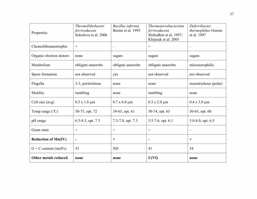
37
Properties
Thermolithobacter ferrireducens Sokolova et al. 2006
Bacillus infernus Boone et al. 1995
Thermoterrabacterium ferrireducens Slobodkin et al. 1997; Khijniak et al. 2005
Deferribacter thermophilus Greene et al. 1997
Chemolithoautotrophic + - + -
Organic electron donors none sugars sugars sugars
Metabolism obligate anaerobe obligate anaerobe obligate anaerobe microaerophilic
Spore formation not observed yes not observed not observed
Flagella 2-3, peritrichous none none monotrichous (polar)
Motility tumbling none tumbling none
Cell size (avg) 0.5 x 1.8 µm 0.7 x 6.0 µm 0.3 x 2.0 µm 0.4 x 3.0 µm
Temp range (°C) 50-75, opt. 72 39-65, opt. 61 50-74, opt. 65 50-65, opt. 60
pH range 6.5-8.5, opt. 7.3 7.3-7.8, opt. 7.3 5.5-7.6, opt. 6.1 5.0-8.0, opt. 6.5
Gram stain + + + -
Reduction of Mn(IV) - + - +
G + C content (mol%) 53 ND 41 34
Other metals reduced none none U(VI) none
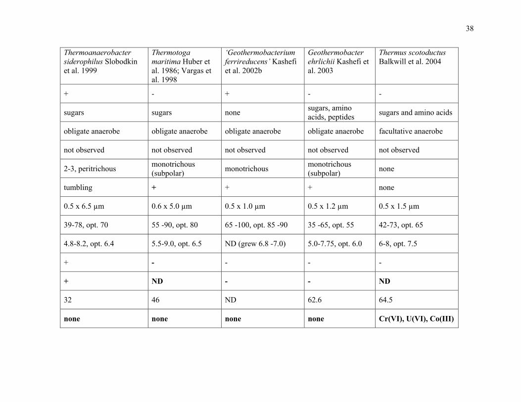
38
Thermoanaerobacter siderophilus Slobodkin et al. 1999
Thermotoga maritima Huber et al. 1986; Vargas et al. 1998
‘Geothermobacterium ferrireducens’ Kashefi et al. 2002b
Geothermobacter ehrlichii Kashefi et al. 2003
Thermus scotoductus Balkwill et al. 2004
+ - + - -
sugars sugars none sugars, amino acids, peptides sugars and amino acids
obligate anaerobe obligate anaerobe obligate anaerobe obligate anaerobe facultative anaerobe
not observed not observed not observed not observed not observed
2-3, peritrichous monotrichous (subpolar) monotrichous monotrichous
(subpolar) none
tumbling + + + none
0.5 x 6.5 µm 0.6 x 5.0 µm 0.5 x 1.0 µm 0.5 x 1.2 µm 0.5 x 1.5 µm
39-78, opt. 70 55 -90, opt. 80 65 -100, opt. 85 -90 35 -65, opt. 55 42-73, opt. 65
4.8-8.2, opt. 6.4 5.5-9.0, opt. 6.5 ND (grew 6.8 -7.0) 5.0-7.75, opt. 6.0 6-8, opt. 7.5
+ - - - -
+ ND - - ND
32 46 ND 62.6 64.5
none none none none Cr(VI), U(VI), Co(III)

39
Fig. 1.1. The simplified universal phylogenetic tree of life. The numbers on the branch tips
correspond to the following groups. The bacteria: 1, the Thermotogales; 2, the flavobacteria and
relatives; 3, the cyanobacteria; 4, the purple bacteria; 5, the Gram-type positive bacteria=
‘Firmicutes’; and 6, the green nonsulfur bacteria. The archaeon: kingdom Crenarchaeota:7, the
genus Pyrodictium; and 8, the genus Thermoproteus; and the archaeon kingdom Euryarchaeota:
9, the Thermococcales; 10, the Methanococcales;11, the Methanobacteriales; 12, the
Methanomicrobiales; and 13, the extreme halophiles. The Eucarya: 14, the animals; 15,
theciliates; 16, the green plants; 17, the fungi; 18, the flagellates; and 19, the microsporidia.
Taken from Woese et al. 1990.
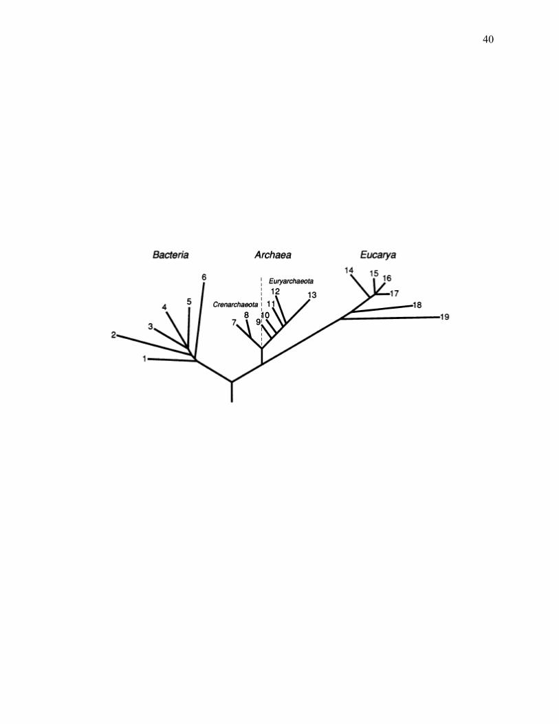
40

41
Fig. 1.2. Schematic (unrooted) representation of relationships within the ‘Firmicutes’ and other
taxa. ‘Firmicutes’ are indicated in bold. The orders of the ‘Clostridia’ and the Mollicutes have
been omitted for clarity. Modified from Wolf et al. 2004.
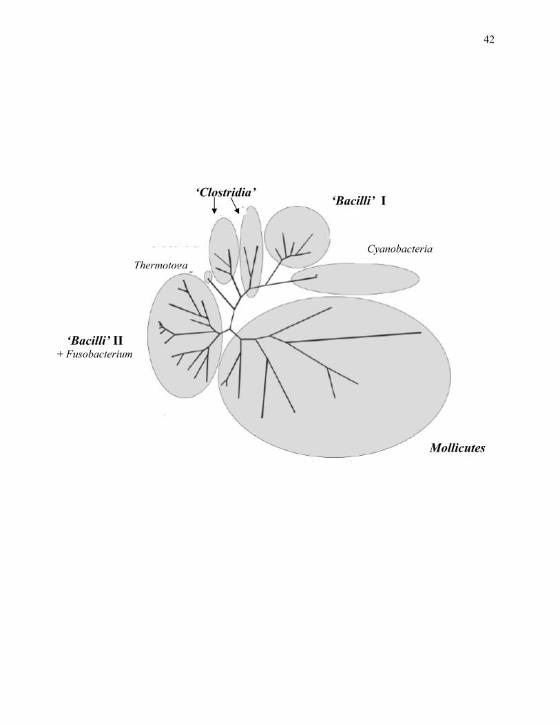
42
Mollicutes
‘Bacilli’ II + Fusobacterium
‘Bacilli’ I
Cyanobacteria Thermotoga
‘Clostridia’

43
Fig. 1.3. Phylogenetic tree of the thermophilic, iron-reducing bacteria. Fitch tree showing the
estimated phylogenetic relationships of thermophilic, iron-reducing bacteria based on 16S rRNA
gene sequence data with Jukes-Cantor correction for synonymous changes. Numbers at nodes
indicate bootstrap support percentages for 100 replicates. Bar, 0.02 nucleotide substitutions per
site. GenBank accession numbers are indicated after the strain identifier. The superscript “T”
denotes the strain is the type strain for the species. The thermophilic, iron-reducing bacteria are
indicated in bold.

44

45
CHAPTER 2
THE THERMOANAEROBACTERIUM 1
____________________________
1Onyenwoke, R. U., and J. Wiegel. Genus I. Thermoanaerobacterium. Submitted to Bergey’s
Manual of Systematic Bacteriology.

46
Abstract
Ther.mo.an.ae.ro.bac’te.ri.um. Gr. n. thermos, hot; Gr. pref. an, not; Gr.n. aer, air; Gr.n.
bacterion, a small rod; M.L. neut. n. Thermoanaerobacterium, rod which grows in the absence of
air at high temperatures.
Cells are Gram-type positive (Wiegel 1981), but stain Gram-negative (besides T.
polysaccharolyticum which is Gram-stain variable). Cells are rod-shaped and motile by
peritrichous flagella. All are obligate anaerobes, have thermophilic growth temperature optima
(between 60 to 70°C) and catalase negative. However, the temperature growth range for the
members of the genus is extremely broad and ranges from 35 to 75°C (for discussion see Wiegel
1990, 1998). The pH range for growth is equally broad (3.8 to 8.5). The pH optima vary from a
low of 5.2 for T. aotearoense (the lowest pH optimum for anaerobic, thermophilic bacteria) to an
alkaline high of 7.8-8.0 for T. thermosaccharolyticum. All members are chemoorganotrophic
with yeast extract stimulating growth for most species. Endospores have been observed for some
species, and their presence or absence has, in the past, typically been used as a defining
characteristic of the member species from one and other. However, the observation of
sporulation or not sporulating is no longer regarded as a strong taxonomic property (Onyenwoke
et al. 2004).The configuration of quinone systems, cellular fatty acids and diaminopimelic acids
in Thermoanaerobacterium species have been described by Yamamoto et al. (1998). Members
of the genus include some well- characterized, such as: T. thermosaccharolyticum (McClung
1935; Prévot 1938; Collins et al. 1994), and some recently characterized “canned food spoilers”
(McClung 1935; Dotzauer et al. 2002).

47
G+C content: 29 to 46 mol%
Type species: Thermoanaerobacterium thermosulfurigenes (Basonym: Clostridium
thermosulfurogenes, Corrig. Schink and Zeikus, 1983, 896VP) Lee Y.E., M.K. Jain, C. Lee, S.E.
Lowe and J.G. Zeikus. 1993, 48VP
Further descriptive information
Reduction of thiosulfate to elemental sulfur was used as the differentiating characteristic of the
genus Thermoanaerobacterium from Thermoanaerobacter (Lee et al. 1993d) until: the isolation
and characterization of T. polysaccharolyticum (thiosulfate is reduced to sulfide) and T. zeae
(thiosulfate is not reduced) (Cann et al. 2001), T. thermosaccharolyticum (thiosulfate is reduced
to sulfide) was added to the genus Thermoanaerobacterium (Collins et al. 1994) and
‘Thermoanaerobacter sulfurigignens’ was described to reduce thiosulfate to elemental sulfur.
(Dashti, M., M.S. thesis, The University of Georgia).
Thermoanaerobacterium species produce thermostable enzymes that have, and will have,
important industrial uses: alpha-galactosidase (King et al. 2002), pullulanases, glucoamylases,
alpha-glucosidases (Ganghofner et al. 1998; Hyun and Zeikus 1985a; Madi et al. 1987; Haeckel
and Bahl 1989; Burchhardt et al. 1991; Matuschek et al. 1994), endoxylanase (Shao et al. 1995a;
Liu et al. 1996b), beta-xylosidase, a novel acetyl xylan esterase (Shao and Wiegel 1995; Lorenz
and Wiegel 1997) and D-xylose ketol-isomerase (Kim et al. 2001; Liu et al. 1996c; Meng et al.
1993a). Glucose-isomerases in relation to saccharidase synthesis and development of single-step
processes for sweetener production have been studied by Lee et al. (1990) and Lee and Zeikus
(1991). Further work with Thermoanaerobacterium species has included: metabolic engineering
to eliminate the production of organic acids by thermophilic bacteria and to increase yields of

48
desired fermentation end-products (e.g. ethanol) (Klapatch et al. 1994; Simpson and Cowan
1997; Cameron et al. 1998; Altaras et al. 2001; Desai et al. 2004), characterization of β-D-
xylosidases (Lee and Zeikus 1993; Vocadlo et al. 2002a; Vocadlo et al. 2002b; Yang et al. 2004)
and glucuronidases (Shao et al. 1995b; Bronnenmeier et al. 1995) and the development of these
bacteria as possible alternate expression hosts, especially for genes with a thermophilic origin
(Mai and Wiegel 1999; Mai and Wiegel 2001).
Enrichment and isolation procedures
Known habitats include: various hot springs and pools, organic waste piles, sediments of acid
springs, various soils, tartrate infusion of grape residue, pond sediment, thermal volcanic algal-
bacterial mats and fruit juice waste products.
Thermoanaerobacterium have been isolated from high temperature petroleum reservoirs by the
direct supplementation of production waters with glucose and either yeast extract, peptone,
tryptone or casamino acids (Grassia et al. 1996).
Taxonomic comments
The genus Thermoanaerobacterium is a member of the ‘Thermoanaerobacteriales’, (order II of
the class ‘Clostridia’) and belong to the family ‘Thermoanaerobacteriaceae’ (Garrity et al.
2002). Cellular polyamine distribution profiles have been investigated for this genus and show
some differences from the phylogenetically related thermophilic anaerobes Moorella,
Dictyoglomus, and Thermoanaerobacter (Hamana et al. 2001).

49
List of species in the genus Thermoanaerobacterium
Thermoanaerobacterium aotearoense Liu S.Y., F.A. Rainey, H.W. Morgan, F. Mayer and J.
Wiegel. 1996, 395VP
ao.te.a’ro.en.se. Maori n. ao cloud; Maori adl. tea, white; Maori adj. roa, long white cloud,
referring to the native Maori name for New Zealand, Aotearoa, Land of the Long White Cloud.
This description is based on that of Liu et al. (1996a), and on study of the type strain
JW/SL-NZ613.
Rods (0.7 to 1.0 by 2.1 to 14.3 µm) that are typically peritrichous exhibiting a tumbling
motility. Cells stain Gram-negative but have a Gram-type positive cell wall covered with
hexagonal S-layer lattices. Oval - terminal endospores (1.4 to 2.1 by 2.8 to 2.9 µm) in slightly
swollen sporangia are produced by 5-15% of cells.
Growth characteristics
An obligate anaerobe with a temperature range for growth at pH 5.2 from 35 to 66°C (optimum
temperature 60-63°C). The pH25ºC range for growth at 60°C was about pH 3.8 to 6.8 (pH
optimum 5.2). Substrates that are utilized in the presence of 0.055% yeast extract included: L-
arabinose, galactose, cellobiose, glucose, mannose, fructose, lactose, pectin, sucrose, xylose,
maltose, starch, rhamnose, pectin, xylan, N-acetylglucosamine and salacin (some strains utilized
ribose and raffinose). Thiosulfate is reduced to elemental sulfur with sulfur deposition within the
cell and also in the medium. No dissimilatory sulfate reduction with glucose, acetate, or lactate
as electron donor.

50
Fermentation products: glucose ethanol + acetic acid + 0.5 lactic acid 2 CO2 + 2 H2
Enrichment and habitat
T. aotearoense has only been isolated from various hot springs in New Zealand (North Island).
The type strain was isolated from a small pool in the Weimangu thermal valley at the Warbick
terrace. Strain was cultured under anaerobic conditions at 60°C (pH 4.5) on 0.1% yeast extract
and 0.5% xylose (Liu et al. 1996a).
G+C content: 34.5 to 35 mol%
Available strains: JW/SL-NZ 604, JW/SL-NZ 606 = DSM 8692, JW/SL-NZ 611, JW/SL-NZ
613, JW/SL-NZ 614, JW/SL-NZ 625 (Liu et al., 1996a)
Type strain: JW/SL-NZ613 = DSM 10170
Gene Bank accession number (16S rRNA): X93359
Thermoanaerobacterium polysaccharolyticum Cann I.K.O., P.G. Stroot, K.R. Mackie, B.A.
White and R.I. Mackie. 2001, 299VP
poly. sac.cha.ro.ly’ti.cum. Gr. n. polysacchar, many sugars; Gr. adj. lyticus, dissolving; N.L.
neut. adj. polysaccharolyticum, many sugars dissolving.
This description is based on that of Cann et al. (2001), and on study of the type strain
KMTHCJ.
Cells are straight rods occurring singly or sometimes in pairs with tumbling motility by
flagella. Gram stain is variable but the ultrastructure determined by electron microscopy is
Gram-type positive. Spores have not been observed. A surface layer-like protein has been

51
observed. Cells are catalase negative. Major fatty acids were determined to be iso 15:0 (70.6%),
iso 17:0 (19.2%) and straight chain 16:0 (10.1%).
Growth characteristics
An obligate anaerobe that exhibits tolerance to air exposure, but the bacterium will grow in
sealed tubes only after the reduction of an oxidized medium. Growth is only seen in the
anaerobic region of a stab culture. Yeast extract is not required for growth. A wide variety of
complex and simple carbohydrates, which include: melibiose, raffinose, arabinose, galactose,
lactose, maltose, mannose, rhamnose, sucrose, trehalose, xylose, cellobiose and melezitose, are
fermented but not cellulose, starch, xylan, cracked corn, pectin, sorbose, mannitol, malate,
fumarate, citrate, glycerol or H2/CO2. Common end products of glucose fermentation are:
ethanol and carbon dioxide with hydrogen, acetate, formate (more acetate than formate), and
lactate being produced in lesser amounts. Glucose and xylose are used simultaneously when
present in media in equal proportions. Doubling times at 68°C with glucose, raffinose, and
melibiose as the carbon source are 2.1, 3.4 and 5.5 h, respectively. Thiosulfate is reduced to
sulfide. Nitrate, sulfate and sulfur are not reduced. Indole is not produced. The optimum
temperature for growth is between 65-68°C at pH 6.8 on glucose (max. is 70°C and growth does
not occur below 45°C). The pH range for growth was between 5.0 and 8.0 (optimum pH was
between 6.8-7.0 at 65°C).
Enrichment and habitat
Isolated from an organic waste pile from a canning factory in Hoopeston, IL, USA by the use of
raffinose as the sole added carbon source at an incubation temperature of 60°C.

52
Industrial applications
T. polysaccharolyticum produces a highly active enzyme exhibiting both mannanase and
endoglucanase activities (Cann et al. 1999). The thermostable alpha-galactosidase produced by
the bacterium is also useful for pretreating food ingredients that can elict gastrointestinal
disturbances if not modified by this enzyme (King et al. 2002). The utility of the CAK1-derived
phagemid for use in the development of a gene transfer system for the clostridia was evaluated
by using it to examine heterologous expression of manA derived from T. polysaccharolyticum in
E. coli (Li et al. 2002). This is the second system available for thermophilic, anaerobic
Firmicutes (Mai and Wiegel 1999).
G+C content: 46 mol%
Type strain: strain KMTHCJ = ATCC BAA-17 = DSM 13641 (Cann et al. 2001)
Gene Bank accession number (16S rRNA): U40229
Thermoanaerobacterium saccharolyticum Lee Y.E., M.K. Jain, C. Lee, S.E. Lowe and J.G.
Zeikus. 1993, 49VP
sac.cha.ro.ly’ti.cum. Gr. n. sacchar, sugar; Gr. adj. lyticus dissolving; N.L. neut. adj.
saccharolyticum, sugar dissolving.
This description is based on that of Lee et al. (1993d), and on study of the type strain
B6A-RI.
Cells appear as rods (~ 0.8 to 1.0 by 3.0 to 15 µm; some cells as long as 30 µm) occurring
in chains of varying lengths. Morphology is elongated during nutrient limitation or stationary
phase. Cells are motile with peritrichous flagella, Gram-type positive but Gram-stain negative

53
and catalase negative. Cell wall contains three electron dense layers that are 5 nm thick and
alternate with electron-light layers of similar thickness. Colonies on agar plates are soft, tan,
circular, and convex with hollow centers (“donut”- shaped). Colony diameters range from 0.5-
4.0 mm after 4 days at 55°C. Spores have not been observed. Cells contain a 1.5 MDa plasmid.
Growth characteristics
Bacterium is an obligate anaerobe existing chemoorganotrophically with growth stimulated by
the presence of yeast extract. Growth is observed with xylan and starch but not cellulose, pectin,
ribose, melibiose, melezitose, xylitol, or sorbitol. Hespell (1992) has further described the
fermentation of xylans by strain B6A while Weimer (1985) described the fermentation of
hemicellulose and hemicellulose-derived aldose sugars. Other complex and simple carbohydrates
fermented include: maltose, lactose sucrose, cellobiose, glucose, xylose, galactose, mannose,
fructose, trehalose, rhamnose, raffinose and mannitol. Landuyt et al. (1995) have also
demonstrated growth on paraffin oil. Fermentation products from either glucose or xylan
include: acetic acid and ethanol in approximately equal amounts, as well as H2, lactic acid and
CO2. L-rhamnose fermentation yields equimolar amounts of 1,2-propandiol and a mixture of
ethanol, acetic acid, lactic acid, H2, and CO2. No growth occurs in the absence of a fermentable
carbohydrate. Thiosulfate is reduced to elemental sulfur which is deposited on the cell and in the
media. The pH range for growth is 5.0 to less than 7.5 (the optimum pH is about 6.0). The
temperature range for growth is a min. of 45°C to a max. of 68 to 70°C (60°C is the optimum).
Cells can survive heating at 85°C for 15 min. but not 90°C for 5 min.

54
Growth inhibition
Growth is inhibited by penicillin G (200 µg/ml), 100 µg/ml of either chloramphenicol or
neomycin, or O2 (0.2 atm [ca. 20.26 kPa]). Growth occurs in the presence of up to 2% NaCl.
Enrichment and habitat
Isolation from geothermal sites in Yellowstone National Park and the Thermopolis areas, WY,
USA and the Steamboat area, NV, USA. The type strain was isolated from sediments of the
Frying Pan thermal acid spring in Yellowstone National Park. Isolation and enrichment of T.
saccharolyticum was achieved using xylan as the carbon source (Lee et al. 1993d).
Industrial applications
T. saccharolyticum strain B6A-RI is currently being actively studied due to its production of a β-
D-xylosidase (Lee and Zeikus 1993; Armand et al. 1996; Vocadlo et al. 2002a; Vocadlo et al.
2002b; Yang et al. 2004) and glucuronidases (Bronnenmeier et al. 1995). Lee et al. (1993a,
1993b) have cloned, sequenced, and biochemically characterized the gene for the endoxylanase
of B6A-RI and are working on the regulation schemes of the xylanolytic enzymes. Saha et al.
(1990) characterized an endo-acting amylopullulanase while Ramesh et al. (1994) cloned and
sequenced the apu gene from B6A-RI and purified and characterized the amylopullulanase from
E. coli The abundance of xylan in nature (the 2nd most abundant polymer in nature), cost and
environmental considerations have caught the attention of the pulp and paper industry toward the
application of this enzyme.
T. saccharolyticum was also used for the cloning of the L-lactate dehydrogenase and the
subsequent elimination of lactic acid as a fermentation product (Desai et al. 2004). The results

55
demonstrated progress toward metabolic engineering to eliminate the production of organic acids
by thermophilic bacteria and to increase yields of desired fermentation end-products (e.g.
ethanol). The xylose isomerase gene from T. saccharolyticum was cloned by complementation
into an E. coli mutant (Lee et al. 1993c). New vectors constructed in order to express
heterologous hydrolytic enzymes in T. saccharolyticum indicate this bacterium may function as
an alternate expression host, especially for genes with a thermophilic origin (Mai and Wiegel
2001).
G+C content: 36 mol%
Type strain: strain B6A-RI = ATCC 49915 = DSM 7060 (Lee et al. 1993d)
Gene Bank accession number (16S rRNA): L09169
Thermoanaerobacterium thermosaccharolyticum (Basonym: Clostridium thermosaccharo-
lyticum, McClung, 1935, 200AL Other synonyms: ("Therminosporus thermosaccharolyticus”
(McClung, 1935) Prévot, 1938; Clostridium tartarivorum, (Hollaus and Klaushofer 1973;
Matteuzzi et al. 1978)) Collins M.D., P.A. Lawson, A. Willems, J.J. Cordoba, J. Fernandez-
Garayzabal, P. Garcia, J. Cai, H. Hippe and J.A.E. Farrow. 1994, 824VP
ther.mo.sac.cha.ro.ly’ti.cum. Gr. adj. thermos, hot. Gr.n. sacchar, sugar; Gr. adj. lytikos,
dissolving; N.L. neut. adj. thermosaccharolyticum, referring to thermophilic and sugar-
dissolving.
This description is based on that of McClung (1935) and Hollaus and Sletyr (1972), and
on study of the type strain LMG 2811.

56
Cells in PYG broth culture are Gram-stain negative (though Gram-type positive),
typically motile, peritrichous and catalase negative. The T. thermosaccharolyticum S-layer
represents the first observation of a bacterial S-layer glycan without a core region connecting the
carbohydrate moiety with the polypeptide portion (Cejka and Baumeister 1987; Sara et al. 1988;
Altman et al. 1990; Altman et al. 1995; Altman et al. 1996; Schaffer et al. 2000). The molecular
mass of the monomeric subunit of the major S-layer protein from strain E207-71 was determined
to be ~ 75 kDa (Allmaier et al. 1995). Cells appear to have an intracellular network of fibrils
which could function as a cytoskeleton-like structure preserving cell shape (Mayer et al. 1998).
Cells are rods (0.4 to 0.7 by 2.4 to 16 µm) occurring singly or in pairs but never in chains. Spores
are observed as round or oval (1.3-1.5 µm) and located terminally distending the cell (Pheil and
Ordal 1967; Campbell and Ordal 1968; Hsu and Ordal 1970). Sporulation is enhanced by several
carbon sources that slow the growth rate of the cells (α- or β-methylglucoside, cellobiose,
galactose, salicin and starch) (Hsu and Ordal 1969a, 1969b, 1970). Pheil and Ordal (1967)
showed sporulation is enhanced by the addition of L-xylose or L-arabinose to a basal media
(optimum pH for sporulation was 5.0-5.5). Cells become shorter and thicker in media containing
glucose and do not sporulate when cultured on glucose. van Rijssel et al. (1992) isolated a
lithotrophic Clostridium strain with extremely thermoresistant spores from a pectin-limited
continuous culture of T. thermosaccharolyticum.
Colonies on pea-infusion agar are 2-4 mm, granular with indistinct feathered edges and
are greyish-white with an often slightly raised center.

57
Growth characteristics
Obligate anaerobes with growth not occurring in the absence of a fermentable carbohydrate. Cell
growth optimization experiments have been performed by Baskaran et al. (1995a). The optimum
temperature for growth is between 55-62°C (some growth at 37°C, poor if any at 30°C, and no
growth at 70°C). Tanaka et al. (1973) and Wilder et al (1963) have reported on the heat stable
ferredoxins of T. thermosaccharolyticum. The pH for growth was between 6.5-8.5 (optimum pH
7.8) (Liu et al. 1996; Mosolova et al. 1991). Mosolova et al. (1991) has studied the effects of:
pH, temperature and antibiotics on the growth and metabolism of T. thermosaccharolyticum.
Dextrin, pectin (Hollaus and Sleytr 1972), arabinose, fructose, galactose, glucose,
mannose, xylose, cellobiose, lactose, maltose, sucrose, trehalose, glycogen, starch, xylan and
salicin are fermented but not rhamnose, pyruvate, inulin or mannitol. However, it should be
noted that cells that were originally cultured on pyruvate will ferment it. It is cells that were
originally cultured with glucose that are unable to ferment pyruvate (Lee and Ordal 1967).
Diauxic growth is observed when cells are cultured on glucose and xylose (Aduseopoku and
Mitchell 1988). van Rijssel et al. (1993) showed the involvement of an intracellular
oligogalacturonate hydrolase in the metabolism of pectin.
Common fermentation end-products from growth on PYG (peptone-yeast extract-
glucose) broth include: acetic, butyric, lactic and succinic acids, ethanol (ethanol tolerance
(Baskaran et al. 1995b) and production (Saddler and Chan 1984; Vancanneyt et al. 1987; Mistry
and Cooney 1989) have been investigated in detail) and H2 (Nikitina et al. 1993). Lynd et al.
(2001) showed that ethanol, rather salt accumulation resulting from base added for pH control,
was not the limiting factor for the growth of strain HG-8 at elevated feed xylose concentrations.
Hill et al. (1993) have investigated end-product regulation during xylose fermentation under

58
nutrient limitations using continuous and batch cultures. Butanol formation at a neutral pH has
been demonstrated by Freier-Schroder et al. (1989).
Nitrate is reduced and, sulfite and thiosulfate are reduced to H2S. Meyer et al. (1990)
have studied a rubredoxin from T. thermosaccharolyticum that might have a role in cellular
sulfur metabolism. Nitrogen fixation and ammonia assimilation by cells has been studied by
Clarke (1949) and Bogdahn and Kleiner (1986).
Enrichment and habitat
Cultured from: strains obtained from the National Canners’ Research Laboratories in
Washington, DC, USA and from original soil isolates by McClung (1935), tartrate infusion of
grape residue (Mercer and Vaughn 1951) and pond sediment from the Netherlands (van Rijssel
and Hansen 1989).
Industrial applications
The application of metabolic engineering for the development of new fermentation processes is
an important technology for the conversion of renewable resources to chemicals (Saddler and
Chan 1984).
T. thermosaccharolyticum has been used in microbial fermentations as an important
organism for the production of 1,2-propanediol (1,2-PD) (Sanchezriera et al. 1987; Altaras et al.
2001; Cameron et al. 1998). T. thermosaccharolyticum is also of considerable interest as a
producer of thermostable amylolytic enzymes. A thermoactive glucoamylase was purified and
characterized by Specka et al. (1991). The bacterium has a soluble amylolytic enzyme system
which was fractionated into a pullulanase, a glucoamylase and an alpha-glucosidase by

59
Ganghofner et al. (1998) and Feng et al. (2002). These enzymes are currently being further
investigated (Aleshin et al., 2003).
Plasmids from strain DSM 571 have been mapped and further characterized by
Belogurova et al. (1991, 1992, 2002) and Delver et al. (1996). Klapatch et al. (1996a, 1996b)
have done some further molecular characterizations with strain HG-8 looking at: the specific
endonuclease activity of the cell extract and electrotransformation of the cells.
G+C content: 29-32 mol% (Liu et al. 1996)
Available strains: DSM 571, ATCC 31925 = HG-6, ATCC 31960 = HG-8 (McClung 1935;
Hollaus and Klaushofer 1973; Cameron and Cooney 1986), DSM 572 = ATCC 27384 = T9-1
(Clostridium tartarivorum), DSM 573 (Mercer and Vaughn 1951; Matteuzzi et al. 1978), DSM
869 = ATCC 25773 (Prévot 1938), DSM 7416 (van Rijssel and Hansen 1989), FH1 (Hoster et al.
2001)
Type strain: ATCC 7956 = DSM 571 = HAMBI 2225 = LMG 2811
Gene Bank accession number (16S rRNA): M59119
Thermoanaerobacterium thermosulfurigenes (Basonym: Clostridium thermosulfurogenes,
Corrig. Schink and Zeikus, 1983b, 896VP) Lee Y.E., M.K. Jain, C. Lee, S.E. Lowe and J.G.
Zeikus. 1993, 48VP
ther.mo.sul.fur.i’ge.nes. Gr. adj. thermos, hot. L.n. sulfur, brimstone; Gr. suff. genes, born from;
N.L. neut. adj. thermosulfurigenes, releasing sulfur in heat.
Type species of Thermoanaerobacterium.
This description is based on that of Schink and Zeikus (1983a), and on study of the type
strain 4B.

60
Cells are straight rods (0.5 by >2 µm), vary in length (from single cells 2 µm to
filamentous chains greater than 20 µm, motile and peritrichous. Stains Gram-negative but
electron micrographs reveal a double-layered wall without the presence of an outer membranous
layer (Gram-type positive). Distinctive features include: double-layered wall architecture,
numerous internal membranes that appeared vesicular, a thin cell wall, no outer cell wall and
large and electron-dense cytoplasmic granules inside cells. The components of the cell envelope
of strain EM1 were isolated, and the S-layer protein was purified and characterized by Brechtel
et al. (1999). In strain EM1, the S-layer homology domains do not attach to the peptidoglycan
(Brechtel and Bahl 1999). Significant differences in the cell envelope structure could be
observed when the cells were grown in continuous culture under glucose limitation as compared
to cells grown under starch limitation (Antranikian et al. 1987; Mayer et al. 1998). More than
likely the phenomenon is due to the increase in production of α-amylase and pullulanase during
starch limitation (Antranikian et al. 1987; Mayer and Gottschalk 2004). Agar embedded colonies
are fluffy (0.5-1.5 mm in diameter with no pigment). Terminal, spherical, refractile, white spores
(free spores rare) with distinctly swollen sporangia are observed with xylose or pectin as the
energy source (but never with glucose). Catalase is not present, and cytochromes are not
detected.
Growth characteristics
An obligate thermophile and anaerobe that grows chemoorganotrophically with yeast extract and
tryptone enhancing growth. The optimum temperature was near 60°C (max. below 75°C and
min. above 35°C). Narberhaus et al. (1994) has studied the synthesis of heat shock proteins in

61
strain EM1. The pH optimum for growth on glucose was 5.5-6.5 (with growth not observed
below 4.0 or above 7.6).
Fermentable carbohydrates include: pectin, starch, polygalacturonic acid, amygdalin,
aesculin, salicin, D-xylose, galactose, glucose, inositol, mannitol, melibiose, rhamnose,
trehalose, mannose, cellobiose, maltose, L-arabinose and sucrose but no growth on H2/CO2,
cellulose, arabinogalactan, galacturonate, citrate, pyruvate, lactate, tartrate, lactose, melezitose,
raffinose, D-ribose, sorbitol, methanol, or glycerol. β-amylase is expressed with growth on
maltose but not glucose or sucrose (Hyun and Zeikus 1985b). Doubling time on glucose or pectin
was approximately 2 h. Glucose fermentation yields H2/CO2, ethanol, acetate, and lactate
(whereas methanol and isopropanol were also produced during pectin fermentation). Ethanol is
produced as the major, soluble, reduced end-product of growth and not butyrate or acetate.
Fermentation products: glucose (0.5 %) 207 ethanol + 152 acetic acid + 113 lactic acid + 317
CO2 + 231 H2 (µmol)
Thiosulfate is reduced to elemental sulfur with no sulfide or sulfite formed. Sulfate and
nitrate are not reduced. Sulfur droplets were found localized in the cytoplasm (Liu et al. 1996).
Addition of sulfite inhibited growth with no production of elemental sulfur. Without the presence
of thiosulfate, growth occurred without the formation of inorganic sulfur metabolites. Gelatin is
liquefied but does not produce indole, acetylmethylcarbinol or hydrogen sulfide.
Growth inhibition
Cell growth is inhibited by: 100 µg/ml of penicillin, streptomycin, cycloserine, tetracycline or
chloramphenicol; 500 µg/mL sodium azide; 2% sodium chloride; O2 (0.203x10⁵ Pa); 2% NaCl or
sulfite. No growth inhibition was observed due to the presence of hydrogen.

62
Enrichment and habitat
Isolated from a thermal (55-65°C), volcanic, algal-bacterial community (Octopus Spring) in
Yellowstone National Park, WY, USA via enrichment with pectin as the energy source (Schink
and Zeikus 1983a) and fruit juice waste products in Germany (Madi et al. 1987).
Industrial applications
T. thermosulfurigenes was found to have very active pectinases, a high ethanol/ lactate ratio
during growth on pentoses and a pectin methylesterase and polygalacturonase hydrolase activity.
T. thermosulfurigenes has also been studied because of the activity of its starch degrading
enzymes (pullulanases and amylases), which are desirable due to their elevated temperature
optima (temperature being an exploited property to improve the solubility of starch and to
prevent bacterial contamination) (Hyun and Zeikus 1985a; Madi et al. 1987; Haeckel and Bahl
1989; Burchhardt et al. 1991; Matuschek et al. 1994; Sahm et al. 1996). Matuschek et al. have
characterized the genes from strain EM1 that encode: two glycosyl hydrolases (1996) and a
novel ABC transporter (1997). Huber et al. (1996) have investigated the structure and some of
the kinetic properties of the β-galactosidase from strain EM1.
Lloyd et al. (1994) have performed the crystallization and preliminary X-ray diffraction
studies of the xylose isomerase from strain 4B while Sriprapundh et al. (2000) reported on the
thermal stability and activity of the isomerase. The D-xylose ketol-isomerase of T.
thermosulfurigenes has also been studied for applications: in the food industry for the production
of sweeteners (can use D-glucose as a substrate to yield D-fructose) (Kim et al. 2001; Meng et al.
1993a; Meng et al. 1993b) and as an effective selection agent of transgenic plant cells (using D-
xylose as the selection agent) (Haldrup et al. 1998).

63
Knegtel et al. (1996) resolved the crystal structure of the cyclodextrin glycosyltransferase
of strain EM1 at 2.3 A, and Leemhuis et al. (2003) and Wind et al. (1995, 1998) have done
further work and characterization of this enzyme. Strain EM1 was used for studies of the three-
dimensional structure of polypeptides and proteins, in particular beta-galactosidases (Zolotarev
et al. 2003).
G+C content: 32.6 mol%
Available strains: 4B (Schink and Zeikus 1983a), DSM 3896 = EM1 (Madi et al. 1987)
Type strain: 4B = ATCC 33743 = DSM 2229
Gene Bank accession number (16S rRNA): L09171
Thermoanaerobacterium xylanolyticum Lee Y.E., M.K. Jain, C. Lee, S.E. Lowe and J.G.
Zeikus. 1993, 48VP
xy.lan.o.ly’ti.cum. Gr. n. xylanosum, xylan; Gr. adj. lyticus, dissolving; N.L. adj. xylanolyticum,
xylan dissolving.
This description is based on that of Lee et al. (1993d), and on study of the type strain LX-
11.
Cells are Gram-stain negative but Gram-type positive, motile, short rods (0.8 to 1.0 by
2.0 to 7.0 µm) that occur singly or in pairs. An elongated morphology does not develop during
nutrient limitation or stationary phase (unlike T. saccharolyticum), and the cytoplasm is much
less granular. Terminal, spherical spores are formed with free spores seen rarely. Surface
colonies on agar are circular, cloudy to white in color, 2 to 5 mm in diameter (rough surface
texture) with smooth edges.

64
Growth characteristics
Bacterium is an obligate anaerobe that grows on xylan and starch but not cellulose, ribose,
melibiose, melezitose, xylitol, pectin or lactate. Yeast extract stimulates growth. Other fermented
carbohydrates include: maltose, lactose sucrose, cellobiose, glucose, xylose, galactose, mannose,
fructose, rhamnose and mannitol. Fermentation products from either glucose or xylan include:
acetic acid and ethanol in approximately equal amounts, as well as H2 and CO2 but no lactic acid.
The pH range for growth is 5.0 to 7.5 (optimum pH is about 6.0) with a temperature range for
growth from 45 to 70°C (60°C is the optimum). Thiosulfate is reduced to elemental sulfur, which
is deposited on the cell and in the medium.
Growth inhibition
Cell growth is inhibited by penicillin G (200 µg/ml); 100 µg/ml of either neomycin sulfate,
ampicillin, streptomycin sulfate, rifampin, polymyxin B, erythromycin, tetracycline, or acridine
orange; or by 1% NaCl.
Enrichment and habitat
Isolated from geothermal sites (sediments of the Frying Pan thermal acid spring) in Yellowstone
National Park, WY, USA using xylan as the carbon source (Lee et al. 1993d).
G+C content: 36.1 mol%
Type strain: LX-11 = ATCC 49914 = DSM 7097 (Lee et al. 1993d)
Gene Bank accession number (16S rRNA): L09172

65
Thermoanaerobacterium zeae Cann I.K.O., P.G. Stroot, K.R. Mackie, B.A. White and R.I.
Mackie. 2001, 300VP
za.ae. Gr. n. zeae of corn, describing the use of corn as a substrate for growth.
This description is based on that of Cann et al. (2001), and on study of the type strain
mel2.
Cells are motile by peritrichous flagella, straight rods, Gram-type positive, even though
cells stain Gram variable, and occur singly or sometimes in pairs. Spore formation has never
been observed. Growth will occur on solid media with the production of catalase negative cells.
Growth characteristics
Cells are obligately anaerobic but neither nitrate, thiosulfate, sulfate nor sulfur is reduced. Indole
is not produced. A wide variety of complex and simple carbohydrates are fermented, which
include: cracked corn, starch, xylan, glucose, melibiose, raffinose, arabinose, galactose, lactose,
maltose, mannose, rhamnose, salicin, sucrose, trehalose, xylose, cellobiose, melizitose and
pyruvate but not cellulose, pectin, sorbose, mannitol, malate, fumarate, citrate, glycerol or
H2/CO2. The end products of glucose fermentation are ethanol and carbon dioxide with
hydrogen, acetate, formate also been produced in lesser quantities (with more formate than
acetate produced. The optimum temperature for growth (when glucose is used as the substrate)
is between 65-70°C at pH 6.8 (max. is 72°C and growth does not occur below 37°C). The pH
range for growth is 3.9-7.9. The fastest doubling time on glucose observed was 1.8 h at the
temperature optimum.

66
Growth inhibition
Growth is completely inhibited by tetracycline, rifampicin, kanamycin, and erythromycin (50
µg/ml for all). However, growth does occur in the presence of concentrations of 300 mM NaCl,
450 mM KCl and 150 mM NH4Cl.
Enrichment and habitat
Isolated from an organic waste pile from a canning factory in Hoopeston, IL, USA by the use of
melibiose as the sole added carbon source at an incubation temperature of 60°C.
G+C content: 42 mol%
Type strain: mel2 = ATCC BAA-16 = DSM 13642 (Cann et al. 2001)
Gene Bank accession number (16S rRNA): U75993

67
Table 2.1. Comparison of physiological traits of the Thermoanaerobacterium species. All
species are: motile and produce acetic acid, ethanol, H2 and CO2 when fermenting glucose.
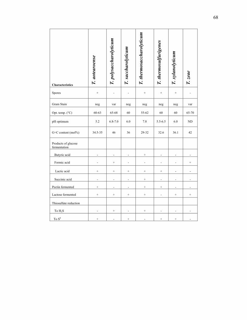
68
Characteristics T.
aot
earo
ense
T. p
olys
acch
arol
ytic
um
T. sa
ccha
roly
ticum
T. th
erm
osac
char
olyt
icum
T. th
erm
osul
furi
gene
s
T. x
ylan
olyt
icum
T. ze
ae
Spores
+
-
-
+
+
+
-
Gram Stain
neg
var
neg
neg
neg
neg
var
Opt. temp. (°C)
60-63
65-68
60
55-62
60
60
65-70
pH optimum
5.2
6.8-7.0
6.0
7.8
5.5-6.5
6.0
ND
G+C content (mol%)
34.5-35
46
36
29-32
32.6
36.1
42
Products of glucose fermentation
Butyric acid
-
-
-
+
-
-
-
Formic acid
-
+
-
-
-
-
+
Lactic acid
+
+
+
+
+
-
-
Succinic acid
-
-
-
+
-
-
-
Pectin fermented
+
-
-
+
+
-
-
Lactose fermented
+
+
+
+
-
+
+
Thiosulfate reduction
To H2S
-
+
-
+
-
-
-
To S0
+
-
+
-
+
+
-

69
Figure 2.1. Phylogenetic tree of the Thermoanaerobacterium species. Fitch tree showing the
estimated phylogenetic relationships of Thermoanaerobacterium species based on 16S rRNA
gene sequence data with Jukes-Cantor correction for synonymous changes. The 16S rRNA gene
data used represent Escherichia coli DSM30083T nucleotide positions 105–1450. Numbers at
nodes indicate bootstrap support percentages for 100 replicates. Bar, 0.05 nucleotide
substitutions per site. GenBank accession numbers are indicated after the strain identifier. The
superscript “T” denotes the strain is the type strain for the species.

70

71
CHAPTER 3
THE THERMOANAEROBACTER 1
____________________________
1Onyenwoke, R. U., and J. Wiegel. Genus VIII. Thermoanaerobacter. Submitted to Bergey’s
Manual of Systematic Bacteriology.
72
Abstract
Ther.mo.an.ae.ro.bac’ter Gr. adj. thermus, hot; Gr. pref. an not; Gr. n. aer, air; M. L. bacter
masc. equivalent of Gr. neut. n. bacterion rod staff; M. L. muse. n. thermoanaerobacter, rod
which grows in the absence of air at higher temperatures.
All species have a Gram-type positive cell wall (Wiegel 1981), but Gram-stain reaction is
variable among the member species. The Gram-stain reaction is even variable for some of the
individual species of the genus. Most species are motile by a peritrichous arrangement of
flagella. Cells are rod-shaped and occur in various arrangements that vary by species. Many of
the Thermoanaerobacter species, as first described for the type species (Wiegel and Ljungdahl
1981), form pleomorphic cells at the late exponential and stationary growth phases, assembling
through non-symmetric cell division (forming coccoid cells frequently in chains alternating rod-
shaped + coccoid shaped-cells). Other pleomorphic rods are formed, assumingly through
weakening of the cell wall, through the possible mode of action of lysozyme-like enzymes that
are the remnants of the sporulation process.
Further descriptive information
Endospore formation has been observed for all species except: T. acetoethylicus, T. ethanolicus,
and T. sulfurophilus. However, for several species the presence of the characteristic spore
specific genes have been demonstrated, thus the species for which no spores have been observed
are regarded as asporogenic (Onyenwoke et al. 2004). Spore formation in some species is rarely
observed (e.g. T. brockii subsp. brockii (Cook et al. 1991)), whereas strains of T.
thermohydrosulfuricus and T. kivui easily form spores with above 50% of vegetative cells
sporulating. Growth temperature optima range from 55- 75°C with growth ranges of 35- 78°C.

73
Many species exhibit a wide temperature span for growth over 35°C, however, the temperature
versus growth rate plots, which exhibit a biphasic curve, indicate changes in the rate limiting
steps. Wiegel (1990) suggested that these bacteria contain, for some critical metabolic steps, two
enzymes: one for the lower temperature of the growth range and one for the higher. This could
also have an evolutionary relevance (Wiegel 1990; Wiegel 1998). Several species also exhibit a
wide pH optimum over 3 pH units without a specific peak (e.g. T. ethanolicus JW 200). The pH
growth range is also quite wide among species ranging from 4.0- 9.9 with pH optima between
4.8- 8.5.
One of the previous characteristics of the genus was the reduction of thiosulfate to H2S
by all of its member species (Lee et al. 1993d) until T. italicus (Kozianowski et al. 1997) and ‘T.
sulfurigignens’ (Dashti, M., S.Y. Liu, F. Rainey, F. Mayer, and J. Wiegel, thesis, The University
of Georgia) were characterized and shown to produce: both H2S and S0, and only S0,
respectively. All species are capable of chemoorganotrophic growth with a requirement of yeast
extract and a fermentable carbohydrate for many species. An exception is T. wiegelii which has
no requirement for yeast extract (Cook et al. 1996). In addition, T. kivui (Leigh et al. 1981) and
T. siderophilus (Slobodkin et al. 1999) are also capable of autotrophic growth. There are also
reports of other Thermoanaerobacter strains that are capable of coupling H2 oxidation to growth
(Fardeau et al. 1993; Fardeau et al. 1994). Common fermentation end-products are: acetic acid,
ethanol (Kannan and Mutharasan 1985), lactic acid, H2 and CO2.
Information about peptide and amino-acid oxidation in the presence of thiosulfate by
members of the genus has been reported by Faudon et al. (1995). Cellular polyamine distribution
profiles have been investigated for this genus and show some differences from the
phylogenetically related thermophilic anaerobes Moorella, Dictyoglomus, and

74
Thermoanaerobacterium (Hamana et al. 2001). The configuration of quinone systems, cellular
fatty acids and diaminopimelic acids in Thermoanaerobacter species have been described by
Yamamoto et al. (1998). Chaperonins from Thermoanaerobacter species have been discussed by
Scopes and Truscott (1998) and Truscott and Scopes (1998). Members of the genus include
“canned food spoilers” (McClung 1935; Dotzauer et al. 2002). Subbotina et al. (2003a, 2003b)
have developed specific oligonucleotide probes for the detection of Thermoanaerobacter
species.
Isolation and enrichment
Thermoanaerobacter have been isolated from high temperature petroleum reservoirs by the
direct supplementation of production waters with glucose and either yeast extract, peptone,
tryptone or casamino acids (Grassia et al. 1996) and identified by 16S rDNA gene cloning and
sequencing (Leu et al. 1998; Orphan et al. 2000). Szewzyk et al. (1994) isolated glucose- and
starch-degrading strains related to Thermoanaerobacter from a deep borehole in granite.
Slobodkin et al. (1999a, 1999b), Zhou et al. (2001) and Roh et al. (2002) have reported on the
isolation and characterization of metal-reducing Thermoanaerobacter strains from deep
subsurface environments.
G+C content: 30-38 mol%
Type species: Thermoanaerobacter ethanolicus (Wiegel and Ljungdahl 1981) Wiegel and
Ljungdahl 1982, 384VP

75
Phylogenetic information
The genus Thermoanaerobacter is a member of the ‘Thermoanaerobacteriales’, (order II of the
class ‘Clostridia’) and belong to the family ‘Thermoanaerobacteriaceae’ (Garrity et al. 2002).
Thermoanaerobacter subterraneus (Fardeau et al. 2000), Thermoanaerobacter tengcongensis
(Xue et al. 2001) and Thermoanaerobacter yonseiensis (Kim et al. 2001) have been reassigned to
the genus Caldanaerobacter by Fardeau et al. (2004) and renamed Caldanaerobacter
subterraneus, Caldanaerobacter subterraneus subsp. tengcongensis and Caldanaerobacter
subterraneus subsp. yonseiensis, respectively. This reassignment was based on phylogenetic and
metabolic differences (e.g. production of L-alanine as a major fermentation product from growth
on glucose and growth at higher temperatures (up to 80°C)) between these 3 species and the
other Thermoanaerobacter (Fardeau et al. 2004).
Industrial applications
Thermoanaerobacter species have been used in studies to understand and exploit the huge array
of enzymes that these bacteria produce to breakdown carbohydrates (Alister et al. 1990; Zeikus
et al. 1991; Svensson 1994; Fardeau et al. 1996; Kuriki and Imanaka 1999) and because of the
typical thermostable nature of the enzymes themselves (Zamost et al. 1991; Vieille and Zeikus
2001). Lind et al. (1989) identified a beta-galactosidase from a strain of Thermoanaerobacter,
and Mitchell et al. (1982) characterized a beta-1, 4- glucosidase activity. The general mechanism
for regulation of glucoamylase and pullulanase synthesis has been studied (Hyun and Zeikus
1985a; Mathupala et al. 1993; Lin and Leu 2002). Xylose and pectin metabolism have also been
further elucidated by the studies of Erbeznik et al. (1998a, 1998b) and Kozianowski et al. (1997),
respectively. Wynter et al. (1996, 1997) have described a novel thermostable dextranase from a
76
Thermoanaerobacter species. Keratinoltytic activity has been shown for a species of
Thermoanaerobacter, ‘Thermoanaerobacter keratinophilus’, by the demonstration of hydrolysis
of feather meal (Riessen and Antranikian 2001). An industrial application was shown with
production of ethanol in pretreated hemicellulosic hydrolysates from wheat straw (Ahring et al.
1996; Ahring et al. 1999; Sommer et al. 2004). High ethanol tolerances also make these bacteria
useful for industrial production schemes (Wiegel and Ljungdahl 1981; Lovitt et al. 1984; Wiegel
1992; Larsen et al. 1997). The effects of temperature on the stereochemistry of enzymatic-
reactions, with particular detail to alcohol dehydrogenases, has been studied by Phillips (1996,
2002b) and Phillips et al. (1994). Efficient and thermostable isomerases have also been
characterized and studied (Jorgensen et al. 2004). Jorgensen et al. (1997) also identified and
cloned the nucleotide sequence of a thermostable cyclodextrin glycosyltransferase from a
Thermoanaerobacter species. Kim et al. (1997) also report on the production of cyclodextrin
using raw corn starch and a cyclodextrin glycosyltransferase from Thermoanaerobacter. Martin
et al. (2001, 2003, 2004) have used Thermoanaerobacter cyclodextrin glycosyltransferases as
immobilized biocatalysts for the synthesis of oligosaccharides. Nakano et al. (2003) have
reported on the synthesis of glycosyl glycerol by a cyclodextrin glucanotransferases. The effects
of temperature on the kinetics of mesophillic and thermophilic (a Thermoanaerobacter species)
3-phosphoglycerate kinases has been described by Thomas and Scopes (1998).

77
List of species in the genus Thermoanaerobacter
Thermoanaerobacter acetoethylicus (Basonym: Thermobacteroides acetoethylicus, Ben-Bassat
and Zeikus, 1981) Rainey and Stackebrandt 1993, 857VP
a.ce.to.e.thy.licus. L. n. acetum vinegar. M. L. n. ethylicus ethyl alcohol.
This description is based on that of Ben-Bassat and Zeikus (1981), and on study of the
type strain HTB2.
Cells are motile rods (0.6 by 1.5-2.5 µm) that occur singly or pairs. Flagella are arranged
peritrichously, and the cells have never been observed to form spores. The cells have a multi-
layered Gram-type positive (though Gram-stain negative) cell wall with no outer wall membrane.
Colonies are uniformly round, flat, mucoid and nonpigmented with a diameter of 3 mm after 48
h. Cytochrome pigment and catalase are both absent.
Growth characteristics
The optimum temperature for growth is 65°C (max. <80°C and min. >40°C). The pH range for
growth is 5.5 to 8.5 (at 65°C). Cells are obligate anaerobes and chemoorganotrophic with a
requirement for yeast extract. The glycolytic Embden-Meyerhof -Parnas pathway is used.
Fermented carbohydrates include: glucose, mannose, cellobiose, maltose, sucrose, lactose and
starch. The major end products of glucose fermentation were: acetic acid and ethanol (in equal
amounts), as well as isobutyric acid, butyric acid, H2 and CO2 but not lactic acid. Thiosulfate
(but not sulfate) is reduced to hydrogen sulfide.

78
Fermentation products (µmol): 130 glucose 102 ethanol + 126 acetic acid + 157 CO2 + 165 H2
Growth inhibition
Growth is inhibited by: air; 2% NaCl; and 10 µg/ml of tetracycline, streptomycin, penicillin G,
vancomycin and neomycin. H2 at 2 atm. does not inhibit growth.
Enrichment and habitat
Isolated from an Octopus Spring algal-bacterial mat at Yellowstone National Park, WY, USA by
the use of trypticase peptone-yeast extract glucose (TYEG) medium (pH 7.2).
G+C content: 31 mol%
Strains available: HTD1 (Zeikus et al. 1979; Zeikus et al. 1980).
Type strain: HTB2 = HTB2/W = ATCC 33265 = DSM 2359 (Zeikus et al. 1980).
Gene Bank accession number (16S rRNA): L09163
Thermoanaerobacter brockii subsp. brockii (Basonym: Thermoanaerobium brockii, Zeikus,
Hegge and Anderson 1983, 673VP) (Thermoanaerobacter brockii, Lee, Jain, Lee, Lowe and
Zeikus 1993, 49AL), emend. Cayol, Ollivier, Patel, Ravot, Magot, Ageron, Grimont and Garcia
1995, 787VP
brock’i.i. M.. L. gen. n. brockii, of Brock, named for Thomas Dale Brock, who performed
pioneering studies on the physiological ecology of extreme thermophiles.

79
This description is based on that of Zeikus et al. (1979), and on study of the type strain
HTD4.
Cells are: Gram-type positive (Gram-stain positive), short rods (1.0 by 2 to 20 µm). Cell
size varies frequently by length (minicells common), and cells arrangement occurs in pairs,
chains, and filaments. Round, heat-resistant terminal spores are observed. Cytochrome pigment
and catalase are absent. Colonies on agar are circular (0.2 to 0.3 mm in diameter), flat, mucoid
and nonpigmented. AGram-type positive cell wall architecture without an outer wall membrane
is present.
Growth characteristics
Cells are chemoorganotrophic with yeast extract and a fermentable carbohydrate required for
growth. The optimum temperature for growth is thermophilic (65 to 70°C with a temperature
range of >35°C to <85°C). The doubling time at the optimum temperature is ~ 1 h. The pH range
is 5.5 to 9.5 (with the optimum at 7.5). Cells are obligate anaerobes and ferment a wide variety of
substrates including: glucose, maltose, sucrose, lactose, cellobiose, starch and pyruvate but not
xylose, cellulose, arabinose, mannose, lactate, tartrate, ethanol, tryptone, casamino acids or
pectin. Common end products of glucose fermentation include: acetic acid, lactic acid, ethanol,
H2 and CO2 but not butyric acid. Exogenous H2 addition (0.4 to 1.0 atm) to a cellobiose
fermentation increased the ethanol/acetate ratio (Lamed and Zeikus 1980).
Fardeau et al. (1997) has reported on the utilization of serine, leucine, isoleucine, and
valine by the bacterium. Maruta et al. (2002) identified the gene encoding a trehalose
80
phosphorylase. Thiosulfate is reduced to hydrogen sulfide. No reduction of oxygen, sulfate,
nitrate or fumarate is observed. Also no protein hydrolysis occurs.
Growth inhibition
Cell growth is inhibited by: penicillin, cycloserine, streptomycin, tetracycline and
chloramphenicol (100 µg/ml); exposure to air (21% O2); and 2% NaCl .
Enrichment and habitat
Isolation of type strain originally from Washburn pool B spring sediment at Yellowstone
National Park, WY, USA using TYEG media (pH 7.2-7.4).
Industrial applications
The alcohol dehydrogenases of this bacterium have been characterized and studied by numerous
groups which include: Peretz and Burstein 1989; Bogin et al. 1997; Yang et al. 1997; Bogin et al.
1998a, 1998b; Korkhin et al. 1998; Peretz et al. 1997a, 1997b; Korkhin et al. 1999; Li et al.
1999; Kleifield et al. 2000; McMahon and Mulcahy 2002; Miroliaei and Nemat-Gorgani 2002;
Kleifield et al. 2003a, 2003b, 2004. Rabinkov et al. (2000) investigated the disulfide
modification and antioxidant properties of S-allylmercaptoglutathione using the alcohol
dehydrogenase of T. brockii subsp. brockii as one of disulfide-containing test enzymes.
T. brockii subsp. brockii is of interest because of its thermostable chaperone proteins
(chaperonin 60 and chaperonin 10 in particular) (Truscott et al. 1994; Todd et al. 1995). The
bacterium has also been used for studies on the reduction of β-oxoesters (Seebach et al. 1984).
Breves et al. (1997) identified the genes encoding two different beta-glucosidases.
Oligosaccharides synthesis and structural analysis has been performed by Okada et al. (2003).

81
G+C content: 30-31 mol%
Available strains: HTA1, HTD4, HTD6, HTR1 = DSM 2599, ATCC 35047 (Zeikus et al. 1979)
Type strain: HTD4 =ATCC 33075 = ATCC 53556 = DSM 1457
Gene Bank accession number (16S rRNA): L09165
Thermoanaerobacter brockii subsp. finnii (Basonym: Thermoanaerobacter finnii, Schmid et al.
1986) Cayol, Ollivier, Patel, Ravot, Magot, Ageron, Grimont and Garcia 1995, 788VP
fin’ni.i. M. L. gen. n. finnii, of Finn, named for Robert K. Finn, who made important
contributions to the development of the ethanol vacuum fermentation process.
This description is based on that of Schmid et al. (1986), and on study of the type strain
AKO-1.
Short rods (0.4 to 0.6 by 1 to 4 µm), occurring singly, in pairs and in short chains
(occasionally coccoid cells), which are motile. Cells are Gram-type positive (Gram-stain
variable) and form heat-resistant terminal spores. Colonies are circular (1 to 3 mm in diameter)
smooth, white and round. The cell wall contains peptidoglycan of the meso-diaminopimelic acid
type.
Growth characteristics
Cells are thermophilic (optimum temperature of 65°C) with a temperature range from 40°C to
75°C. The optimum pH is between 6.5-6.8. An obligate anaerobe with a chemoorganotrophic

82
growth requirement describes T.brockii subsp. finnii. Growth requires the presence of yeast
extract and a fermentable carbohydrate. Fermented sugars are numerous and include: glucose,
fructose, galactose, mannose, cellobiose, maltose, melibiose, sucrose, lactose, xylose, ribose,
mannitol and pyruvate. The end products of glucose and xylose fermentation are ethanol and
CO2, as well as minor amounts of acetic acid and L-lactic acid. Thiosulfate is reduced to
hydrogen sulfide.
Fermentation products (µmol/mL): 12.1 glucose 20.1 ethanol + 3.2 acetic acid + 4.6 lactic
acid + 20.1 CO2 + 3.0 H2
Growth inhibition
Growth inhibition was by penicillin G and tetracycline (6 µg/ml).
Enrichment and habitat
Isolated from sediment sludge from Lake Kivu in East Africa using M-1 media (which includes
lactic acid, glucose and yeast extract) at 60°C.
Industrial applications
This bacterium was used to develop an assay to determine pyridine nucleotide levels in cell
extracts as low as 1 pmol (Schmid et al. 1989). T. brockii subsp. finnii has also been used in
studies aimed at understanding the breakdown of carbohydrates found in sulfide-, elemental
sulfur- or sulfate-rich thermal hot springs and oil fields (Fardeau et al. 1996). Antranikian (1989)

83
reported on the formation of an extracellular, thermoactive amylase and pullulanase in batch
culture by T. brockii subsp. finni.
G+C content: 32 mol%
Type strain: AKO-1 = ATCC 43586 = DSM 3389 (Schmid et al. 1986)
Gene Bank accession number (16S rRNA): L09166
Thermoanaerobacter brockii subsp. lactiethylicus Cayol, Ollivier, Patel, Ravot, Magot, Ageron,
Grimont and Garcia 1995, 788VP
lac.ti.e.thy’li.cus. L. n. lacticum, lactic acid; M. L. n. ethylicus, ethyl alcohol; lactiethylicus,
referring to the production of both lactic acid and ethanol.
This description is based on that of Cayol et al. (1995), and on study of the type strain
SEBR 5268.
Cells are straight rods (0.5 by 2 to 3 µm), motile by means of peritrichous flagella and
occur singly or in pairs in young cultures. Pleomorphic filamentous cells (length of 15 µm)
appear in older cultures. Cells are Gram-type positive (Gram-stain positive) and form spores in a
medium containing D-xylose as an electron donor and thiosulfate as an electron acceptor (but not
if D-xylose is replaced with glucose). Colonies in a roll tube are: 4 mm in diameter after 2 days
of incubation at 60°C, smooth, uniformly round, mucoid, nonpigmented and flat.

84
Growth characteristics
This obligate anaerobe is thermophilic (optimum temperature 55-60°C) with a temperature range
between 40°C and 75°C. The pH range is 5.6 to 8.8. Yeast extract is required for fermentation of
carbohydrates. The doubling time with glucose as the carbon source is 2h at 60°C. Fermentable
substrates are: glucose, fructose, galactose, mannose, D-ribose, D-xylose, cellobiose, lactose,
maltose, sucrose, mannitol, starch, yeast extract and pyruvate but not L-arabinose, cellulose, L-
rhamnose, glycerol, ribitol, galactitol, sorbose or melibiose. End products of glucose
fermentation are acetic acid, lactic acid, ethanol, H2 and CO2. Thiosulfate, sulfite and elemental
sulfur are reduced to hydrogen sulfide. No reduction of sulfate, nitrate or fumarate occurs.
Growth inhibition
A concentration of 4.5% NaCl is tolerated (the optimum NaCl concentration is 1%).
Enrichment and habitat
Type strain isolated from geothermally heated (92°C) French, oil samples through the incubation
of samples in an anaerobic, glucose-based medium at 60°C.

85
Industrial applications
T. brockii subsp. lactiethylicus has been studied, along with T. brockii subsp. finnii, in an attempt
to understand the breakdown of carbohydrates found in sulfide-, elemental sulfur- or sulfate-rich
thermal hot springs and oil fields (Fardeau et al. 1996).
G+C content: 35 mol%
Available strains: SEBR 7311, SEBR 7312 (Cayol et al. 1995)
Type strain: SEBR 5268 = DSM 9801 (Cayol et al. 1995)
Gene Bank accession number (16S rRNA): U14330
Thermoanaerobacter brockii subsp. ‘pseudoethanolicus’ (Basonym: Clostridium
thermohydrosulfuricum 39E (Zeikus et al. 1980); Thermoanaerobacter ethanolicus 39E (Lee,
Jain, Lee, Lowe and Zeikus 1993, 47VP))
This description is based on that of Zeikus et al. (1980), and study of strain 39E. Based
on the 16S rRNA sequence analysis (Fig. 1), it became obvious that strain 39E does not belong
to the species ethanolicus. Thus, it is described here as a new subspecies pseudoethanolicus
strain 39 E of T. brockii, naming is based on the 16S rRNA and its T. ethanolicus JW 200- like
properties. The genome sequence of 39E will be available in the near future.
Cells are rod-shaped and sporulate when grown with xylose as substrate. Gram-stain
reaction is variable, but the cell wall is Gram-type positive. The cells are motile and reduce
thiosulfate.

86
Growth characteristics
Bacterium utilizes the Embden-Meyerhof-Parnas Pathway and the pentose-phosphate pathway,
respectively, for the catabolism of hexoses and pentoses to pyruvate (Zeikus et al. 1981).
Fermented carbohydrates include: xylose, cellobiose, starch, glucose, maltose and sucrose.
Glucose is consumed in preference to cellobiose as an energy source for growth (Ng and Zeikus
1982.). A coculture of T. brockii subsp. pseudoethanolicus and Clostridium thermocellum is
stable and leads to an increased yield of ethanol during cellulose fermentations (Ng et al. 1981).
Jones et al. have studied high-affinity maltose (2000, 2002b) and xylose (2002a) binding and
transport in strain 39E. No growth was observed using CO2 /H2. The temperature optimum is
65°C. The synthesis of very long bifunctional fatty acid species, making up about 40% of fatty
acyl components of the membrane, is part of the microorganism’s adaptation for optimum
growth at extremely high temperatures (Jung et al. 1994). The generation time at 65°C is 75 min.
Fermentation products (µmol): glucose 549 ethanol + 31 acetic acid + 50 lactic acid + 31 H2
Growth inhibition
Ethanol tolerance is not temperature dependent in the wild-type strain (39E) (Burdette et al.
2002) but was shown to be temperature dependent in mutants. Glucose is not fermented at an
ethanol concentration of 2.0% by wild-type 39E, but mutants have been shown to ferment
glucose at 8.0% ethanol at 45°C (3.3% at 68°C) (Lovitt et al. 1984).

87
Enrichment and habitat
Isolated from the Octopus Spring algal-bacterial mat in Yellowstone National Park, WY, USA
with TYEG media, or modified TYEG media that contained 5% xylose instead of glucose, at
65°C.
Industrial applications
The general mechanism for regulation of glucoamylase and pullulanase synthesis has been
studied in strain 39E (Hyun and Zeikus 1985a, 1985b), as well as some studies on the active site,
and characterization, of the amylopullulanase (Mathupala et al. 1993, 1994; Mathupala and
Zeikus 1993; Lin and Leu 2002). Podkovyrov et al. (1993) provided analysis of the catalytic
center of cyclomaltodextrinase. Xylose metabolism has also been further elucidated by the
cloning and characterization of genes encoding: a xylose isomerase, a xylulose kinase (Erbeznik
et al. 1998a, 1998b), a xylose ABC (ATP-binding cassette) transport operon (Erbeznik et al.
2004) and xylose binding protein, XylF (Erbeznik et al. 1998c).
The histidine biosynthesis pathway was identified in this bacterium making it the first
thermophilic, anaerobic bacterium with identified his anabolic genes (Erbeznik et al. 2000).
Other studied and characterized enzymes include: a lactate dehydrogenase (LDH), studied
because of a link between an increase in the initial concentration of glucose and an increase in
the activity of the LDH (Germain et al. 1986); a secondary-alcohol dehydrogenase; a primary-
alcohol dehydrogenase and an acetaldehyde dehydrogenase (Pham et al. 1989; Pham and Phillips
1990; Phillips 1992, 2002a; Zheng and Phillips 1992; Zheng et al. 1992., 1994; Burdette and
Zeikus 1994a, 1994b; Burdette et al. 1995, 1997, 2000, 2002; Arni et al. 1996; Secundo and

88
Phillips 1996; Tripp et al. 1998; Heiss and Phillips 2000; Heiss et al. 2001a, 2001b). The
secondary-alcohol dehyrogenase (Burdette et al. 1996) and a thermostable beta-galactosidase
(Fokina and Velikodvorskaia 1997) have been cloned and expressed in E. coli.
G+C content: mol%: not determined
Available strains: 39E = DSM 2355 = ATCC 33223 (Zeikus et al. 1980)
Gene Bank accession number (16S rRNA): L09164
Thermoanaerobacter ethanolicusT (Wiegel and Ljungdahl 1981) Wiegel and Ljungdahl 1982,
384VP
e.tha.no.li.cus. IVS. n. ethanol. corresponding alcohol of ethane (ethane+ol) M. L. masc. adj.
ethanolicus, indicating the production of ethanol. Type species of Thermoanaerobacter.
This description is based on that of Wiegel and Ljungdahl (1981), and on study of the
type strain JW 200.
Cells in early logarithmic growth are 0.3-0.8 by 4 -8 µm (rods) often with pointed ends.
In late logarithmic growth, cells are long filamentous cells (up to 100 µm). Cells have 1-12
peritrichously situated flagella (up to 50µm in length) and 4-8 pili. Cell division may lead to long
chains of bacteria. Coccoid cells (0.8-1.5 µm) are frequently observed, but spores have never
been observed. The Gram-stain is variable; however, the cell wall is Gram-type positive (meso-
diaminopimelic acid type), as observed by electron microscopy. Peteranderl et al. (1993)
demonstrated that strain JW200 is able to regenerate to a walled form after autolysis was induced
to form stable, cell wall-free cells. Deep agar (2%) colonies at 60°C are 0.5-1.0 mm in diameter,

89
lenticular and white. After 40 h at 60°C, colonies are 2-4 mm in diameter, white, smooth, round
to irregular and flat. In old cultures, the colonies turn brown, but no pigment has ever been
identified.
Growth characteristics
Cells are obligate anaerobes that grow from pH 4.4-9.9 (optimum pH 5.8-8.5). The growth
temperature is from 37-78°C with an optimum temperature at 69°C. Yeast extract and a
fermentable carbohydrate are required for growth. Lacis and Lawford (1985) have studied the
growth efficiencies of thermophilic and mesophilic anaerobes with an emphasis on strain JW
200. Glucose, fructose, galactose, mannose, ribose, xylose, cellobiose, lactose, maltose, sucrose,
starch and pyruvate are fermented but not cellulose, raffinose, rhamnose, fucose, m-erythritol, m-
inositol, xylitol, glycerol, mannitol, sorbitol, trehalose, melezitose, melibiose, niacinamide or
amygdalin. The major end products of glucose fermentation are: ethanol (1.8 mol/mol of
glucose) and CO2, with minor amounts of acetic acid, lactic acid, and H2 also produced.
Variations in end-products are observed. For instance, a decrease in the yeast extract to
fermented carbohydrate ratio leads to a decrease in ethanol production and an increase in acetic
acid and lactic acid concentrations (Hild et al. 2003). Lacis and Lawford (1988a, 1988b, 1989,
1991, 1992) have reported on ethanol yields from fermentations involving both glucose and
xylose. Wiegel et al. (1983) has described the production of ethanol from bio-polymers.
Esculin and gelatin are hydrolyzed, and cells are negative for: catalase, indole production
and lipase (or cellulase activity).
90
Fermentation products (mol): 1.0 glucose + 0.1 H2O 1.8 ethanol + 0.1 acetic acid + 0.1 lactic
acid + 1.9 CO2 + 0.2 H2
Growth inhibition
Cells from late logarithmic growth lyse on exposure to 100 µg/ml of polymyxin B for 30 min at
37°C or 50°C (pH 7.2). (This was not true of early logarithmic growth cells.) Cells are inhibited
by chloramphenicol but are resistant to erythromycin, tetracycline and penicillin-G. H2 is
inhibitory at 10% (growth does not occur at 75% or higher concentrations). Lactic acid, acetic
acid and ethanol also become inhibitory above 100 mM, 200 mM and 500 mM (65°C),
respectively. Type strain can be adapted to growth on 8% ethanol.
Enrichment and habitat
Type strain isolated from water and mud samples (pH ~8.8, temp. 45-50°C) that were collected
from an alkaline hot spring in White Creek (opposite the Great Fountain Geyser located at Fire
Hole Lake Drive) in Yellowstone National Park, WY, USA.
Industrial applications
T. ethanolicus strain JW 200 produces an extensive number of enzymes that have been studied
and well-characterized: the beta-xylosidase (Shao and Wiegel 1992) and alcohol dehydrogenases
(Bryant and Ljungdahl 1981; Bryant et al. 1988, 1992; Holt et al. 2000) are examples. Also, the

91
gene for the bifunctional xylosidase-arabinosidase (xarB) from T. ethanolicus has been cloned,
sequenced, and expressed in Escherichia. coli (Mai et al. 2000).
G+C content: 32 mol%
Available strains: JW 200, JW 201 (Wiegel and Ljungdahl 1981)
Type strain: JW 200 = ATCC 31550 = DSM 2246
Cell wall type: m-DAP
Gene Bank accession number (16S rRNA): L09162
Thermoanaerobacter italicus (Kozianowski et al. 1997) Kozianowski, Canganella, Rainey,
Hippe and Antranikian 1998, 1084VP
i.ta.li.cus. L. n. italia Italy; M. L. masc. adj. italicus pertaining to Italy, where the organism was
isolated.
This description is based on that of Kozianowski et al. (1997), and on study of the type
strain Ab9.
Cells are rod-shaped (0.4-0.75 by 2 -6 µm) and chains up to 50 µm when grown on
glucose. These cells are non-motile, Gram-stain negative and Gram-type positive (meso-
diaminopimelic acid type in cell wall). Spherical, terminal spores are produced with xylose as
substrate. Colonies on a glucose-containing agar are 2-3 mm in diameter, round with entire
margins, greyish-white and opaque with a glassy surface.
92
Growth characteristics
Bacterium is thermophilic (optimum growth temperature around 70°C with growth occurring
from 45 to 78°C), anaerobic, and a chemoorganotroph. The pH optimum is around 7.0. Doubling
time at 70°C with pectin is 3 h (compared to 2.1 h with glucose). Fermentable carbohydrates
include: amygdalin, arabinose, cellobiose, esculin, fructose, glucose, galactose, lactose, maltose,
mannose, melezitose, melibiose, mannitol, raffinose, sucrose, trehalose, starch, xylan, glycogen,
D-glucosamine, saccharose, inulin, pectin and xylose but not cellulose. End products of glucose
fermentation (0.5%) are: ethanol (26 mM), lactic acid (12.4 mM), acetic acid (2.2 mM),
succinate (0.3 mM), CO2 and H2 (end products of pectin or pectate fermentation are ethanol,
lactic acid, acetic acid, CO2 and H2). Thiosulfate is reduced to both elemental sulfur and sulfide.
Accumulation of sulfur in the media and in the cells can be observed.
Growth inhibition
1% NaCl did not inhibit growth; however, 10 µg/ml of cephalosporin, erythromycin, kanamycin
or rifampicin totally inhibits growth.
Enrichment and habitat
Isolated from water and mud samples (40-70°C) in thermal spas collected from the north of Italy
(Abano, Terme, Calzignano Terme, Montegrotto Terme, Battaglia Terme, Sirmione and Agano
Terme) with the use of pectin and pectate (polygalacturonic acid) as substrates. The isolation of
type strain occurred from medicinal mud (fango) of Abano Terme, Italy.
93
Industrial applications
Thermoactive xylanolytic (temperature optimum 70-75°C) enzymes and amylolytic and
pullulolytic (temperature optimum 80-85°C) enzymes are produced. Two thermoactive pectate
lyases were isolated after growth on pectin (pectate was the preferred substrate but higher
enzyme yields were obtained with pectin).
G+C content: 34.4 mol%
Type strain: Ab9 = DSM 9252 (Kozianowski et al. 1997)
Cell wall type: m-DAP
Gene Bank accession number (16S rRNA): no sequence
Thermoanaerobacter ‘keratinophilus’ Riessen and Antranikian 2001
ke.ra.ti.no’phil.us. Gr. n. keras keratin; Gr. adj. philos loving; M. L. adj. keratinophilus keratin-
loving.
This description is based on that of Riessen and Antranikian (2001), and on study of the
type strain 2KXI.
The cells are rod-shaped, 0.2-0.3 by 1-3.0 µm in the exponential growth phase and occur
singly, in pairs or in short chains. Cells are Gram-stain negative regardless of growth phase but
have a Gram-type positive cell wall. Stationary growth phase cells are pleomorphic, including a

94
filamentous form up to 10 or 15 µm in length. Spores were never observed. The rods are slightly
curved and commonly appear as coccoid-shaped.
Growth characteristics
The temperature optimum is 70°C, with growth occurring from 50°C to just below 80°C. The pH
optimum for growth is 7.0 with a pH range of 5.0-9.0. The optimal NaCl concentration is 5-10
g/l with a range of 0-30 g/l. Growth is fermentative and strictly anaerobic.
Utilized substrates include: casein, bactopeptone, yeast extract, tryptone, collagen,
gelatin, starch, pectin, glucose, fructose, galactose, mannose, pyruvate, maltose and cellobiose
but not xylan, cellulose, pullulan, xylose, arabinose, lactose or olive oil. Yeast extract and
tryptone (0.05% (wt/vol) of both) are required for growth on saccharolytic substances. The
generation time of the type strain when grown with 5 g/l yeast extract and 5 g/l tryptone was 67
min. Growth can be achieved with merino wool and chicken feathers solely. Thiosulfate and
sulfate can both serve as electron acceptors. Thiosulfate stimulates growth to approximately
threefold that of sulfate but is not required for growth. Ampicillin, kanamycin and streptomycin
inhibit growth at 10 µg/ml.

95
Enrichment and habitat
Isolation of the type strain was from hydrothermal vents, 74°C and pH 6.0, in the area of Furnas
on the Azorean island São Miguel using merino wool and chicken feathers as sustrates at an
incubation temperature of 70°C.
Industrial applications
Strain 2KXI possesses an intracellular protease and an extracellular keratinolytic enzyme with
different properties. The ability to degrade keratin is a unique characteristic among the known
Bacteria and Archaea (Friedrich and Antranikian 1996).
G+C content: 37.6 mol%
Type strain: 2KXI = DSM 14007 (Riessen and Antranikian 2001)
Gene Bank accession number (16S rRNA): AY278483
Thermoanaerobacter kivui (Basonym: Acetogenium kivui, Leigh and Wolfe 1983, 886AL)
Collins, Lawson, Willems, Cordoba, Fernandez-Garayzabal, Garcia, Cai, Hippe and Farrow
1994, 824VP
ki’vui. M. L. adj. kivuus pertaining to Kivu, named for its source, Lake Kivu.
This description is based on that of Leigh et al. (1981), and on study of the type strain
LKT-1.

96
Cells grown on H2 and CO2 are rod-shaped (0.7 by 2 -3.5 µm) but are longer, 0.7-0.8 by
5.5 -7.5 µm, when grown on glucose. Cells occur in pairs or chains and are non-flagellated. The
Gram-stain is negative, but Gram-type positive cell wall is present. Cell wall is covered by a
hexagonal S-layer that is composed of a single 80 kDa subunit (Lupas et al. 1994). Endospores
are not observed. Colonies on agar plates are: convex, circular, entire, translucent, tan in color,
and have a smooth, shiny surface (2 mm diameter after 1 week). Cells are catalase negative.
Growth characteristics
The temperature optimum is 66°C, with growth occurring from 50 to 72°C. Physiology is
obligate anaerobic growth not occurring without a reducing agent such as cysteine-sulfide.
However, small amounts of O2 in semisolid and liquid media caused a lag phase but did not alter
the ability of the bacterium to synthesize acetate via the acetyl coenzyme A pathway (Karnholz
et al. 2002). The pH optimum is 6.4 with a pH range of 5.3-7.3. Fermentable carbohydrates
include: glucose, mannose, fructose, and pyruvate, which yield acetic acid. However, galactose,
maltose, raffinose, ribose, sucrose, lactose, trehalose, cellobiose, cellulose, pectin, starch,
mannitol and inositol cannot serve as substrates. The bacterium oxidizes H2 and reduces CO2 to
produce acetic acid as the sole product of metabolism (Ryabokon et al. 1995). Acetic acid
formation has been studied by Kevbrina and Pusheva (1996) and Kevbrina et al. (1996). The
doubling time on H2 and CO2 is 1.75-2.5 h at 60°C. Poor growth is observed on formate while
yeast extract and trypticase increase cell yields.
2 CO2 + 4 H2 acetic acid + 2 H2O

97
Enrichment and habitat
Isolated, so far, only from sediments of Lake Kivu, Africa, enrichment was done at 60°C with H2
and CO2 (67 and 33%, respectively).
G+C content: 38 mol%
Type strain: LKT-1= ATCC 33488 = DSM 2030 (Leigh et al. 1981)
Gene Bank accession number (16S rRNA): L09160
Thermoanaerobacter mathranii (Larsen et al. 1997) Larsen, Nielsen and Ahring 1998, 327VP
ma.thra.ni.i. M. L. gen. n., of Mathrani, in honor of the late Indra M. Mathrani, who contributed
greatly to our understanding of thermophilic anaerobes from hot springs during his short career.
This description is based on that of Larsen et al. (1997) and Sonne-Hansen et al. (1993),
and on study of the type strain A3.
Cells are Gram-stain variable (Gram-type positive), straight and rod-shaped occurring
singly and when under under suboptimal conditions in long chains. The vegetative cells in
exponential growth phase are 0.7 by 1.8 -3.9 µm, motile and spore-forming (terminal, spherical
spores that swell cells). Cells are typically longer when either sporulated (6.4-8.2 µm) or grown
under suboptimal conditions (e.g. a temp. of 75°C). Colonies are white and irregular with a 1
mm diameter after growth on beechwood xylan agar for 7 days. The surface of the colonies is
granulated with top formation.
98
Growth characteristics
The optimum growth temperature of T. mathranii is between 70-75°C (no growth at 47 or 78°C).
The pH optimum is 6.8-7.8 (4.7 and 8.8 are the extreme pH values for growth). Cells are catalase
negative. The doubling time on xylose was 74 min. at 69°C (pH 7.0). Sulfide is produced from
casein-peptone, sulfate or thiosulfate. Yeast extract is required for growth along with a
fermentable carbohydrate but does not serve as a sole carbon/energy source. Carbohydrates used
for growth include: amygdalin, L-arabinose, cellobiose, D-fructose, D-glucose, glycogen,
lactose, maltose, D-mannitol, mannose, melezitose, melibiose, raffinose, D-ribose, sucrose,
trehalose, xylan and D- xylose but not avicel, casein-peptone, cellulose, D-galactose, glycerol,
inulin, pectin, L-rhamnose, salicin, sorbitol or yeast extract. The end-products of D-xylose
fermentation are: ethanol (Ahring et al. 1999), lactic acid, acetic acid, CO2 and H2.
Fermentation products: xylose + H2O 1.1 ethanol + 0.4 acetic acid + 0.06 lactic acid + 1.81
CO2 + 0.9 H2
Growth inhibition
Growth is inhibited by 100 mg/l of tetracycline, chlorampenicol, penicillin G, neomycin or
vancomycin. Growth is still seen in the presence of 10mg/l chlorampenicol and neomycin. T.
mathranii is also insensitive to: 50.66 kPa overpressure of H2, 2% NaCl and 5% ethanol. When
T. mathranii was grown with a wet-oxidized wheat straw hydrolysate, inhibitory effects were
also seen due to aromatic monomers (e.g. phenol aldehydes and to a lesser extent by phenol
ketones) (Klinke et al. 2001).
99
Enrichment and habitat
Isolated from a biomat and sediment from a slightly alkaline (pH 8.5) hot spring (70°C) in
Hverðagerdi-Hengil, Iceland (Sonne-Hansen et al. 1993). Beechwood xylan at 68°C (pH 8.4)
was the substrate used for isolation and the subsequent enrichment of the bacterium.
Industrial applications
An industrial application was shown with production of ethanol in pretreated hemicellulosic
hydrolysates from wheat straw (Ahring et al. 1996). Also the high tolerance to ethanol (up to
5%) makes this bacterium useful. One of the most efficient enzymes for converting D-galactose
into D-tagatose was found in T. mathranii and subsequently produced heterologously in E. coli
and characterized (Jorgensen et al. 2004). T. mathranii has also been used in the implementation
of an Upflow Anaerobic Sludge Blanket (UASB) purification reactor step for the detoxification
process of water derived from bioethanol production (Torry-Smith et al. 2003).
G+C content: 37 mol%
Type strain: A3 = DSM 11426 (Sonne-Hansen et al. 1993; Larsen et al. 1997)
Gene Bank accession number (16S rRNA): Y11279
Thermoanaerobacter siderophilus Slobodkin, Tourova, Kuznetsov, Kostrikina, Chernyh and
Bonch-Osmolovskaya 1999, 1477VP

100
si.de.ro’phil.us. Gr. n. sideros iron; Gr. adj. philos loving; M. L. adj. siderophilus iron-loving.
This description is based on that of Slobodkin et al. (1999a), and on study of the type
strain SR4.
Cells are straight to curved rods (0.4-0.6 by 3.5-9.0 l µm), Gram-type positive (Gram-
stain positive) and occur singly or in short chains. Round, refractile, heat-resistant spores are
formed in terminally swollen sporangia. Maximum sporulation was observed when growth was
with 9,10-anthraquione 2,6-disulfonic acid (AQDS). Colonies in agar-shake cultures were:
uniformly round, 0.5-1.0 mm in diameter and white. Cells exhibit slight tumbling motility due to
peritrichous flagellation.
Growth characteristics
Bacterium is an anaerobe with a thermophilic growth optimum (69-71°C) and temperature
growth range from 39-78°C. The pH range for growth is from 4.8 to 8.2 (with an optimum at
6.3-6.5). Substrates utilized in the presence, as well in the absence, of Fe(III) as an electron
acceptor include: peptone, yeast extract, beef extract, casein, starch, glycerol, pyruvate, glucose,
sucrose, fructose, maltose, xylose, cellobiose and sorbitol but not formate, acetate, lactate,
methanol, ethanol, propanol, isopropanol, butanol, propionate, n-butyrate, succinate, malate,
maleate, glycine, alanine, arginine, L-arabinose, olive oil, xylan or cellulose. Molecular
hydrogen and CO2 can be utilized in the presence of Fe(III) for growth. Fermentation products
from glucose are: ethanol, lactate, H2 and CO2. Electron acceptors reduced include: amorphous
iron(III) oxide, AQDS, sulfite, thiosulfate, elemental sulfur and MnO2. The products of

101
amorphous iron(III) oxide reduction are magnetite and siderite. Sulfite, thiosulfate and elemental
sulfur are reduced to hydrogen sulfide. Nitrate, sulfate and O2 cannot be used for growth.
Growth inhibition
Growth is inhibited by chloramphenicol, neomycin, polymyxinB and kanamycin (100 µg/ml) but
not by penicillin, ampicillin, streptomycin or novobiocin (100 µg/ml). Growth occurs with NaCl
concentrations from 0-3.5%. It was established that Fe(III) reduction in T. siderophilus is carried
out to relieve the inhibitory effect of hydrogen (Gavrilov et al. 2003).
Enrichment and habitat
Isolated from hydrothermal (70-94°C) vents in the area of Karymsky volcano on the Kamchatka
peninsula, Russia using amorphous iron(III) oxide and peptone in an anaerobic media at pH 6.8-
6.9 (70°C).
G+C content: 32 mol%
Type strain: SR4 = DSM 12299 (Slobodkin et al. 1999)
Gene Bank accession number (16S rRNA): AF120479

102
Thermoanaerobacter ‘sulfurigignens’ (Dashti, M., S.Y. Liu, F. Rainey, F. Mayer, Y.J. Lee and
J. Wiegel, thesis, The University of Georgia)
sul.fu.ri.gig’nens. L. n. sulfur, sulfur, brimstone; L. v. gignere, to produce, sulfurigignens
relating to the formation of sulfur droplets from thiosulfate.
This description is based on that of Dashti, thesis, The University of Georgia, and on
study of the type strain JW/SL- NZ826.
Cells are motile by peritrichous flagella, rods (0.3 to 0.8 by 1.2 to 4.0 µm during
exponential growth), non-pigmented and form spores (round, terminal spores 0.46 to 0.83 µm in
diameter usually seen during the late exponential or early stationary growth phase). Cells tend to
be longer during the stationary phase with lengths up to 35 µm observed. Elemental sulfur is
deposited on the cell surface and in the medium during growth on thiosulfate. The Gram-stain
reaction is negative but the cell wall is Gram-type positive. Colonies are creamy white, circular
(1 to 2 mm in diameter).
Growth characteristics
The temperature range for the growth at pH 6.5 is 34-74°C (with an optimum from 63-65°C).
The pH range for growth at 60°C is from 4.0 to 8.0 (with an optimum of 4.8-6.5). Xylose,
glucose, starch, lactose, galactose, maltose, fructose, sucrose, mannose, cellibiose, raffinose,
pyruvate, methanol and mannitol are fermented in the presence of 0.3% yeast extract. Ribose,
arabinose, dextran, xylan, cellulose, glycerol, xyliotol, formate and gluconate are not used as
103
substrates. The addition of 0.5% peptone, tryptone, casein hydrolysate or casamino acids in the
presence of 0.3% yeast extract does not increase cell growth. Weak growth is observed with
fumarate and succinate. Sulfate and sulfite are not reduced. Thiosulfate is reduced to elemental
sulfur inside the cells. The sulfur inclusion droplets are released into the media due to lysis of the
cells. ‘T. sulfurigignens’ can tolerate up to 800mM thiosulfate. Cells are catalase and indole
negative.
Fermentation products (in the presence of 0.3% yeast extract): glucose (or xylose) 0.7 mM
ethanol + 0.4 mM acetic acid + 0.9 mM lactic acid + 1.1 mM CO2 + 0.8 mM H2
Growth inhibition
Growth is inhibited by neomycin and chloramphenical at concentrations of 100 µg/ml and
gramicidin at 10µg/ml. Cells are resistant to vancomycin, bacitracin, tetracycline and ampicillin
at 10µg/ml and kanamycin, streptomycin, cycloheximide and cycloserine at 100 µg/ml.
Enrichment and habitat
Isolated and enriched from an acidic volcanic stream outlet on White Island, New Zealand at
60°C (pH 5.0) using anaerobic media containing: 20 mM thiosulfate, 0.5% yeast extract and
0.5% xylose.

104
G+C content: 34.5 mol%
Available strains: JW/SL- NZ824 and JW/SL- NZ826
Type strain: JW/SL- NZ826 = DSM 13515 = ATCC 700320
Gene Bank accession number (16S rRNA): AF234164
Thermoanaerobacter sulfurophilus (Bonch-Osmolovskaya et al. 1997) Bonch-Osmolovskaya,
E. A., M. L. Miroshnichenko, N. A. Chernykh, N. A. Kostrikina, E. V. Pikuta, and F. A. Rainey
1998, 631VP
sul.fu.ro.phi’lus. L. masc. adj., sulfurophilus, of L. n. sulfur- sulfur and GK. masc. adj. philos-
liking: sulfurophilus-liking elemental sulfur.
This description is based on that of Bonch-Osmolovskaya et al. (1997), and on study of
the type strain L-64.
Cells are curved, Gram-type positive rods (0.5 by 3-7 µm) with mini-cells common. Cells
occur in long wound chains in older, or nutrient deficient, cultures. Spores have never been
observed. Motility is through the use of peritrichous flagella.

105
Growth characteristics
The temperature range for the growth is 44-75°C with an optimum from 55-60°C. The pH range
for growth is from 4.5 to 8.0 with an optimum at 6.8-7.2. Bacterium has a obligately anaerobic
metabolism. Fermentable carbohydrates include: glucose, fructose, lactose, rhamnose, arabinose,
xylose, sorbitol, inositol, mannitol, sucrose, cellobiose, maltose, starch, pectin, pyruvate and
lactate, but no growth is observed on cellulose, succinate, citrate, formate, acetate, propionate,
butyrate, methanol, ethanol or H2 (in the presence or absence of elemental sulfur as the electron
acceptor). Growth with glucose yields: H2, CO2, and acetate, minor amounts of lactate and
ethanol (production of H2S, no lactate and higher amounts of ethanol when elemental sulfur was
also present). Elemental sulfur and thiosulfate are reduced to sulfide (addition of elemental sulfur
also stimulates growth). Nitrate, sulfate and sulfite are not reduced.
Fermentation products: lactate (µmol/mL of product) 3.0 H2S + 0.14 acetic acid + 0.20 H2
Enrichment and habitat
Isolated and enriched from a sulfur-containing cyanobacterial mat occurring along the rim of a
hot (53-58°C) pond from the Uzon caldera, Kamchatka by the use of a peptone, glucose, lactate
medium (incubation at 55°C with a media pH of 7.0) (Bonch-Osmolovskaya et al. 1997).
G+C content: 30.3 mol%
Available strains: P-82, G-1, and L-64 (Bonch-Osmolovskaya et al. 1997)

106
Type strain: L-64 = DSM 11584
Gene Bank accession number (16S rRNA): Y16940
Thermoanaerobacter thermocopriae (Basonym: Clostridium thermocopriae, Jin, Yamasato, and
Toda 1988, 280AL) Collins, Lawson, Willems, Cordoba, Fernandez-Garayzabal, Garcia, Cai,
Hippe and Farrow 1994, 824VP
ther.mo.co’pri.ae. Gr. n. thermos, heat; Gr. n. kopria, compost; M. L. gen. n. thermocopriae of
heat compost.
This description is based on that of Jin et al. (1988), and on study of the type strain JT3-3.
Cells are straight rods (0.5-0.7 by 2.2-6.0 µm) that produce terminal, spherical spores
with a diameter of 1.2-1.6 µm which swell the cell. The Gram-type positive cells are motile and
stain Gram negative.
Growth characteristics
The temperature range for growth of T. thermocopriae is 47-74°C with an optimum at 60°C. The
pH range for growth is from 6.0 to 8.0 with an optimum at 6.5-7.3. The bacterium is an obligate
anaerobe fermenting numerous carbohydrates: cellulose, hemicellulose, cellobiose, glucose,
fructose, maltose, arabinose, lactose, trehalose, glycogen, starch, amygaldin, xylan and mannose
but not melibiose, pectin, inulin or mannitol. The end products of carbohydrate fermentation are:

107
acetic acid, butyric acid, lactic acid, ethanol, H2 and CO2 but not propionic or valeric acid. Indole
is not produced, esculin is hydrolyzed and gelatin is not digested. No nitrate reduction occurs.
Fermentation products: 1% cellibiose (100 mL culture) (meq) 0.8 to 4.1 butyric acid + 0.3 to
1.7 acetatic acid + less than 0.8 lactic acid + 2 to 5 ethanol
Enrichment and habitat
Isolated from compost of camel feces (type strain), soil and a hot spring in Japan (The University
of Tokyo and Kinugawa) using cellobiose or cellulose in anaerobic culture at 60°C (pH 7.1) (Jin
and Toda, Abstr. Annu. Meet. Soc. Ferment. Technol. Jpn. 1987, p.134; Jin et al. 1988).
Industrial applications
An extracellular cellulose is produced by this bacterium.
G+C content: 37.2 mol%
Available strains: JT1, JT2, JT3-1, JT3-2, JT3-3, JT5 (Jin and Toda, Abstr. Annu. Meet. Soc.
Ferment. Technol. Jpn. 1987, p.134; Jin et al. 1988)
Type strain: JT3-3 = ATCC 51646 = IAM 13577 = JCM 7501
Gene Bank accession number (16S rRNA): L09167
108
Thermoanaerobacter thermohydrosulfuricus (Basonym: Clostridium thermohydrosulfuricum,
Klaushofer and Parkkinen 1965, 448AL (Skerman et al. 1980)) Lee, Jain, Lee, Lowe and Zeikus
1993, 49VP
ther.mo.hy.dro.sul.fur’i.cus. M. L. masc. n. thermos, hot; M. L. masc. adj. hydrosulfuricum,
pertaining to hydrogen sulfide; M. L. masc. adj. thermohydrosulfuricum, indicating that the
organism grows at high temperatures and reduces sulfite to H2S.
This description is based on that of Hollaus and Sleytr (1972) and Wiegel et al. (1979),
and on study of the type strain E100-69.
Cells occur singly, in short chains or (in some strains) in long filamentous groups. Cells
are 0.3 to 0.6 by 2.0 to 13.0 µm and motile by peritrichous flagella. As shown by fracture
planes, flagella appear to have empty cores that could possibly have a use for transport of
flagellin molecules during flagellar assembly (Sleytr and Glauert 1973). Spores are spherical
and terminal, and the sporangia swell the cells. Sporulating cultures tend to contain thinner, more
elongated cells. The Gram-stain is variable but a Gram-type positive cell wall composed of two
layers is present. The cell wall contains meso-diaminopimelic acid and is covered by an S-layer
(Sára and Sleytr 1996a, 1996b). The S-layer shows hexagonal symmetry, a center-to-center
spacing of the morphological units of 14.2 nm (Crowther and Sleytr 1977; Bock et al. 1994;
Messner et al. 1995) and is composed of glycoprotein subunits with a molecular weight of
~20,000 (Christian et al. 1988; Sára et al. 1989). Peteranderl et al. (1993) demonstrated that
strain JW102 is able to regenerate to a walled form after autolysis was induced to form stable,
cell wall-free cells. The cells are catalase negative.
109
Growth characteristics
Anaerobic conditions are required for cell growth. Growth occurs at pH 5.5 to 9.2, and the
optimum pH for growth is 6.9 to 7.5. The optimum growth temperature is between 67 and 69°C
with no growth occurring at 76 to 78°C. Growth at 37°C is poor and no growth is observed at
25°C. H2 in the gas phase inhibits growth as does lactate.
Fructose, galactose, glucose, mannose, xylose, cellobiose, maltose, sucrose, trehalose,
pectin, esculin and salicin are fermented. Cook et al. has studied the phenomena of glucose
(1993) and xylose (1994) uptake by this bacterium in detail. Fermentation of dextrin, potato
starch, mannitol, dulcitol and sorbitol and coagulation of litmus milk are variable. Inositol,
erythritol, glycerol, lactate, tartrate and cellulose are not fermented. When both glucose and
xylose (Patel et al. 1988) or starch and glucose (Parkkinen 1986) are present in media, they are
used simultaneously. Extracellular enzymatic starch degradation in relation to the bacterium’s
constant specific growth rate has been further investigated by Heitmann et al. (1996). Mori
(1995) showed that strain YM3 required yeast extract to grow unless it was grown in co-culture
with Clostridium thermocellum strain YM4, or the cell free broth of YM4. Growth with PYG
media yields: H2, CO2, acetic acid, lactic acid and ethanol (formic, butyric, isovaleric and
isocaproic acids, propanol and isopropanol may be detected). Cook and Morgan (1994) have
studied ethanol production under hyperbolic growth conditios. Mayer et al. (1995) isolated
mutants that were defective in acetate kinase and/or phosphotransacetylase in order to block
acetate production by cells. (L-lactate was the main fermentation product.) H2 and CO2 are
produced in media containing liver infusion. Methanol is the major metabolic end-product of
pectin fermentation.
110
glucose + 0.5 H2O ethanol + 0.5 acetic acid + 0.5 lactic acid + 1.5 CO2 + H2
Sulfite and thiosulfate are reduced to hydrogen sulfide, but sulfate is not reduced. H2S is
produced from tryptophan, peptone and yeast extract in the growth media. Nitrate, but not nitrite,
is reduced. Acetyl methyl carbinol and indole are not produced.
Enrichment and habitat
Isolated from: extraction juices from beet sugar factories (type strain) (Klaushofer and Parkkinen
1965; Hollaus and Klaushofer 1973), mud and soil (Wiegel et al. 1979; Klingeberg et al. 1990),
hot springs in Utah and Wyoming, USA and a sewage plant in Georgia, USA (Wiegel et al.
1979), and a sugar refinery in Germany (Klingeberg et al. 1990).
Industrial applications
The S-layer glycoprotein of T. thermohydrosulfuricus strain L111-69 has been investigated for
its possible use for carrying cell wall fragments (Sára and Sleytr 1996a, 1996b; Sleytr et al.
1999) (also macromolecules such as ferritin and invertase (Sára and Sletyr 1989)) as
‘microparticles’ for immunoassays: human IgG immobilization (Kupcu et al. 1995, 1996; Weber
et al. 2001) and recombinant major birch pollen allergen Bet v 1 immobilization (Jahn-Schmid et
al. 1996). Protein phosphorylation, as a regulatory mechanism, was studied using T.
thermohydrosulfuricus to extend the limited range of knowledge on this subject to include a
thermophile (Londesborough 1986). Sha et al. (1997) purified and characterized a thermostable

111
DNA polymerase from T. thermohydrosulfuricus. Thermostable lactate dehydrogenases
(Turunen et al. 1987) and α-amylases and pullulanases (Antranikian et al. 1987; Melasniemi
1987; Melasniemi and Paloheimo 1989) have also been purified to homogeneity and
characterized from the bacterium. An alpha-glucosidase exhibiting maltase, glucohydrolase and
'maltodextrinohydrolase' activity was isolated and purified from culture supernatants of T.
thermohydrosulfuricus (Wimmer et al. 1997).
G+C content: 35-37 mol%
Available strains: E100-69, L 110-69 = DSM 568 (Klaushofer and Parkkinen 1965; Hollaus and
Klaushofer 1973), L 77-66 = DSM 569, S 100-69 = DSM 570 (Hollaus and Klaushofer 1973),
JW102 = DSM 2247 (Wiegel et al. 1979), DSM 7021, and DSM 7022 (Klingeberg et al. 1990),
YM3 (Mori 1990)
Type strain: E100-69 = ATCC 35045 = DSM 567 = LMG 6659 = NCIB (now NCIMB) 10956
Cell wall type: m-DAP
Gene Bank accession number (16S rRNA): L09161
Thermoanaerobacter wiegelii Cook, Rainey, Patel and Morgan 1996, 126VP
wie.gel’i.i. M.L. gen. n. wiegelii, of Wiegel, in recognition of Juergen Wiegel’s contributions to
the study of thermophilic anaerobes.

112
This description is based on that of Cook et al. (1996), and on study of the type
strain Rt8.B1.
Cells grown on solid trypticase peptone-yeast extract glucose (TYEG) medium produced
nonpigmented colonies (0.5 to 2.0 mm) that were smooth and uniformly round. Cells obtained
from isolated colonies were Gram-stain negative rods (Gram-type positive cell wall). Cells
occurred singly, in pairs, or (less frequently) in chains and were 0.4 to 0.6 µm wide by 4 to 10
µm long. Electron micrographs of the cell wall revealed a two-layer structure. The inner layer,
which was adjacent to the cytoplasmic membrane, stained intensely, whereas the outer layer was
less dense. Cells were sluggishly motile by peritrichous flagella. Cells that were grown on TYEG
medium did not sporulate. However, when the organisms were grown in a minimal medium at
65°C, the cells were long and filamentous and spores were produced. Spores are round and
terminal, distend the cells and are brightly refractile. Spores survived more than 80 min of
exposure at 115°C, which confirmed their heat resistance.
Growth characteristics
Anaerobic conditions are required for cell growth. Growth occurs at pH 5.5 to 7.2, but not at pH
5.0 or 7.25, and the optimum pH for growth is 6.8. The temperatures for growth range from 38 to
78°C with an optimum growth temperature between 65 and 68°C; no growth at 34 or 80°C.
Yeast extract or trypticase is not required for growth, and both of these growth supplements can
be replaced by vitamin-free casamino acids and vitamins. However, no growth occurs in
trypticase peptone-yeast extract medium in the absence of a fermentable carbon source.

113
The utilized carbohydrates include: glucose, xylose, maltose, lactose, cellobiose,
raffinose, glucosamine, galactose, fructose, mannose, sucrose, glycerol, soluble starch, pectin
and chitin. Sorbitol, mannitol and trehalose are also fermented, but ethanol, DL-lactate, sodium
citrate, sodium succinate, transaconitate, malonate, glutamate, glutamine, sodium pyruvate, 2-
deoxyglucose, α -methyl-glucoside, L-arabinose, α -L-rhamnose, dulcitol, m-inositol, ribose, α-
L-fucose and L-sorbose are not. Growth with glucose yields: H2, CO2, acetic acid, lactic acid and
ethanol (up to 1.1 mol per mol of sustrate). Propionate is formed during growth on xylose or
cellibiose. The doubling time on glucose is 72 min., and growth studies demonstrated that
glucose and xylose were used simultaneously when they were supplied together at nonlimiting
concentrations (similar to T. ethanolicus) (Carreira et al. 1983).
Sulfite and thiosulfate are reduced to hydrogen sulfide. Nitrate, oxygen, sulfate and sulfur
are not reduced. Indole is not produced and esculin and gelatin are not hydrolyzed. Cells do not
accumulate anthrone-reactive material when they are grown with glucose.
Growth inhibition
Cephalosporin C, erythromycin, bacitracin, tetracycline or polymyxin B completely inhibited
growth at 10 mg/ml. Trimethoprim, rifampin, amphotericin B, D-cycloserine, penicillin G,
streptomycin sulfate, chloramphenicol and ampicillin did not inhibit growth at concentrations up
to 100 mg/mL. The metabolic inhibitors monensin (100 mM), 2,4-dinitrophenol (500 mM),
tetrachlorosalicylanilide (10 mM), N,N-dicyclohexylcarboiimide (500 mM) and iodoacetate (500
mM) all inhibited growth when they were added to cultures that were growing exponentially on
glucose. Sodium azide, sodium fluoride, potassium cyanide, and sodium arsenate completely
114
stopped growth at 65°C when they were added to a final concentration of 5 mM. Oxygen
completely inhibits growth.
Enrichment and habitat
Isolated from neutral to alkaline freshwater in a geothermally (56 to 69°C) heated water source
in Government Gardens, Rotorua, New Zealand. The primary enrichment cultures were prepared
by adding 0.2 ml of pool water to 10 ml of prereduced TYEG medium under an N2 atmosphere
(incubation at 70°). Purification involved the use of TYEG agar deeps.
Industrial applications
T. wiegelii was used to study the effect of the external media pH on ATP synthesis rates by
monitoring the proton motive force and membrane potential of the bacterium over its pH growth
range (Cook 2000).
G+C content: 35.6 mol%
Type strain: Rt8.B1 = DSM 10319
Gene Bank accession number (16S rRNA): X92513

115
Table 3.1. Comparison of physiological traits of the Thermoanaerobacter species.

116
Characteristics T. a
ceto
ethy
licus
T. b
rock
ii su
bsp.
bro
ckii
T.br
ocki
i sub
sp. f
inni
i
T. b
rock
ii su
bsp.
la
ctie
thyl
icus
T. b
rock
ii su
bsp.
ps
eudo
etha
nolic
us
T. e
than
olic
us
T. it
alic
us
T. k
ivui
T. m
athr
anii
T. si
dero
philu
s
‘T. s
ulfu
rigi
gnen
s’
T. su
lfuro
philu
s
T. th
erm
ocop
riae
T. th
erm
ohyd
rosu
lfuri
cus
T. w
iege
lii
Spores - + + + + - + - + + + - + + + Motility + - + + + + - - + + + + + + + Inhibition by H2 - ND ND ND ND - ND ND - + ND ND ND + ND Inhibition by 2 % NaCl + + ND - ND ND ND ND - - ND ND ND ND ND Gram stain neg pos var pos var var neg neg var pos neg ND neg var neg Opt. temp. (°C) 65 65-70 65 55-60 65 69 70 66 70-75 69-71 63-67 55-60 60 67-69 65-68 G+C content (mol%) 31 30-31 32 35 ND 32 34.4 38 37 32 34.5 30.3 37.2 35-37 35.6 Products of fermentation Acetic acid + + + + + + + + + - + + + + + Butyric acid + - - - - - - - - - - - + + - Ethanol + + + + + + + - + + + + + + + Isobutyric acid + - - - - - - - - - - - - - - Lactic acid - + + + + + + - + + + + + + + Succinate - - - - - - + - - - - - - - - H2 + + + + + + + - + + + + + + + CO2 + + + + + + + - + + + + + + + Pectin fermented - - ND ND ND + + - - ND ND + - + + Mannose fermented + - + + ND + + + + ND + ND + + + Xylose fermented - - + + + + + ND + + + + ND + + Pentoses fermented - ND + + + + + ND + + + + ND + + Thiosulfate reduction To H2S + + + + ND + + ND + + - + + + + To S0 ND - - - ND - + ND - - + - - - -

117
Figure 3.1. Phylogenetic tree of the Thermoanaerobacter species. Fitch tree showing the
estimated phylogenetic relationships of Thermoanaerobacter species based on 16S rRNA gene
sequence data with Jukes-Cantor correction for synonymous changes. The 16S rRNA gene data
used represent Escherichia coli DSM30083T nucleotide positions 107–1450. Numbers at nodes
indicate bootstrap support percentages for 100 replicates. Bar, 0.05 nucleotide substitutions per
site. GenBank accession numbers are indicated after the strain identifier. The superscript “T”
denotes the strain is the type strain for the species.

118
119
CHAPTER 4
RECLASSIFICATION OF THERMOANAEROBIUM ACETIGENUM AS
CALDICELLULOSIRUPTOR ACETIGENUS COMB. NOV. AND EMENDATION OF THE
GENUS DESCRIPTION1
____________________________
1Onyenwoke, R. U., Y.-J. Lee, S. Dabrowski, B. Ahring, and J. Wiegel. 2006. International
Journal of Systematic and Evolutionary Microbiology. 56:1391-1395.
Reprinted here with permission of the publisher.

120
Abstract
Although the type species of the genus Thermoanaerobium, Thermoanaerobium brockii, was
transferred to Thermoanaerobacter, Thermoanaerobium acetigenum was not transferred.
Therefore, Thermoanaerobium acetigenum should be reclassified. Based on 16S rRNA gene
sequence analysis and re-examination of physiological properties of the type strain, X6BT
(=DSM 7040T=ATCC BAA-1149T), we propose that Thermoanaerobium acetigenum should be
reclassified as Caldicellulosiruptor acetigenus comb. nov. Strain X6BT contains two separate
16S rRNA genes bracketing another species in the phylogenetic 16S rRNA gene-based tree.
Results and discussion
Thermoanaerobium acetigenum strain X6BT, an anaerobic, thermophilic bacterium, was isolated
by Nielsen et al. (1993) using xylan as the substrate. This bacterium, a Gram-type positive
(Wiegel 1981), low-G+C content rod, has many characteristics of a typical member of the
Firmicutes (Gibbons and Murray 1978). Based on its physiological properties alone, it was
placed in the genus Thermoanaerobium, the type species of which is Thermoanaerobium brockii
(Zeikus et al. 1979).
Because the 16S rRNA gene sequence for Thermoanaerobium acetigenum X6BT had not
been determined previously, the classification of Thermoanaerobium acetigenum X6BT was
therefore based only on some physiological similarities. Although the type species of
Thermoanaerobium, Thermoanaerobium brockii, was reclassified as Thermoanaerobacter
brockii by Lee et al. (1993d) and, subsequently, as Thermoanaerobacter brockii subsp. brockii
121
(type strain HTD4T) by Cayol et al. (1995), Thermoanaerobium acetigenum X6BT was not
transferred to the genus Thermoanaerobacter (Wiegel and Ljungdahl 1981) because of the lack
of 16S rRNA gene sequence analysis. Here we report on the assignment of the type strain of
Thermoanaerobium acetigenum to the genus Caldicellulosiruptor as Caldicellulosiruptor
acetigenus comb. nov., based on 16S rRNA gene sequence, DNA–DNA hybridization analysis
and retesting of its properties. Special attention was given to cellulose degradation, as all other
presently known Caldicellulosiruptor species are cellulolytic, whereas strain X6BT has been
described as being non-cellulolytic.
Strain X6BT was obtained as a freeze-dried culture of strain DSM 7040T from the DSMZ
(Braunschweig, Germany). To determine the 16S rRNA gene sequence, Thermoanaerobium
acetigenum DSM 7040T was grown under anaerobic conditions (Ljungdahl and Wiegel 1986;
Angelidaki et al. 1990). A basal salts medium (final pH 7.3–7.4) was prepared as described by
Nielsen et al. (1993). Strain DSM 7040T was grown in basal salts medium supplemented with
yeast extract (0.3%), tryptone (1.0%) and glucose (0.5%), and subjected to two rounds of
isolation of single colonies using yeast extract, tryptone, glucose salts medium solidified with
2.2% Gelrite (colonies became visible after incubation at 65°C for 48–72 h). Because the initial
16S rRNA gene sequence analysis yielded two different 16S rRNA species, which bracketed
another Caldicellulosiruptor species, it became necessary to confirm the purity of the culture
further. Therefore, strain DSM 7040T was grown using three different media (substrate
conditions as described below), and each culture was then subjected to three subsequent rounds
of single-cell colony isolation. To establish three lines of cultures, strain DSM 7040T was grown
in the above-described basal salts medium, supplemented with yeast extract, tryptone, glucose
and brain heart infusion (0.2%) (termed BYTG medium). From this culture, three parallel

122
cultures were inoculated (0.1% inoculum) using the following media: (i) basal salts plus 0.2%
arabinose medium, (ii) basal salts plus 0.2 % raffinose medium and (iii) BYTG medium.
Arabinose- and raffinose-supplemented basal salts media were used because the closest
Caldicellulosiruptor species to strain X6BT on the phylogenetic tree (Fig. 4.1.) are unable to use
these substrates (Table 4.1.). After checking microscopically that the cultures were suspensions
of individual cells and did not contain any clumps or associations of cells, each of the above
cultures was used to inoculate dilution series of Gelrite shake-roll tubes (Ljungdahl and Wiegel
1986), with 2.2% (w/v) Gelrite, to obtain single-cell colonies. The Gelrite shake-roll tubes were
incubated at 65°C for 48–72 h before colonies became visible. Colonies were picked in an
anaerobic chamber (Coy Products) and resuspended in a tube containing 0.3–0.4 ml of the
corresponding medium, which was then used to inoculate the next round of Gelrite shake-roll
tubes. This process of colony picking was repeated for three rounds of colony isolation with three
colonies being picked from each of the arabinose, raffinose and BYTG media after the third and
final round. Each of the picked final colonies was reinoculated into a fresh tube of the medium
from which it was isolated, resulting in nine cultures: three with the arabinose medium, three
with the raffinose medium and three with BYTG medium.
Subsequent extraction of DNA from the nine cultures was performed using a DNeasy
Tissue kit (Qiagen). The DNA was then amplified using a bacterial domain-specific primer set
for 16S rRNA, 27 forward and 1492 reverse (Lane 1991). PCR was carried out as described
previously (Lee et al. 2005). The PCR products were purified using a QIAquick PCR Purification
kit (Qiagen) and sequenced by Macrogen (Seoul, Korea). PCR products from the colonies were
cloned using a TOPO TA Cloning kit (Invitrogen). Clones were randomly chosen, from which
plasmid DNA was extracted by using an Eppendorf FastPlasmid Mini kit (Brinkman). The DNA
123
was subsequently amplified, purified and sequenced. The sequence similarities were determined
using Sequencher v4.1.4 (Gene Codes). Ten clones were sequenced, resulting in two similar sets
of sequences (Fig. 4.1.; termed T6 and T4, GenBank accession nos AY772477 and AY772476,
respectively). Three clones with the same 16S rRNA gene sequence were never obtained from a
single culture, suggesting that an even distribution of clones with the different sequences existed.
Analysis of the 16S rRNA gene sequence using nucleotide to nucleotide BLAST (BLASTN) at
NCBI (http://www.ncbi.nlm.nih.gov/blast/) to retrieve the most significant homologues of the
query 16S rRNA gene sequence revealed that the most significant sequence alignments of
Thermoanaerobium acetigenum X6BT were with Caldicellulosiruptor species (Sissons et al.
1987; Rainey et al. 1994), with Caldicellulosiruptor lactoaceticus 6AT (Mladenovska et al. 1995)
and Caldicellulosiruptor kristjanssonii I77R1BT (Bredholt et al. 1999) as the closest relatives
(Fig. 1). Subsequently, 16S rRNA gene sequence-based phylogenetic trees were generated using
CLUSTAL_X (Thompson et al. 1997) for sequence alignments, and phylogeny inference
package (PHYLIP) software (Felsenstein 1989) and neighbour-joining algorithms (Saitou and
Nei 1987) to look at differing tree constructions and to generate distance matrices. TreeExplorer
(Kumar et al. 1994), a supplemental program of MEGA, was used to view the tree. The
phylogenetic trees generated (the neighbor-joining tree is shown in Fig. 4.1.) showed clearly that
Thermoanaerobium acetigenum X6BT belongs to the clade of Caldicellulosiruptor and not to the
genus Thermoanaerobacter or Thermoanaerobacterium. Each of the repurified strains exhibited
the two different 16S rRNA sequences. In contrast to other reported cases, e.g., Clostridium
paradoxum (Rainey et al. 1996), the two sequences were not juxtaposed, but rather separated by
a sequence of another species, Caldicellulosiruptor kristjanssonii. A comparable situation has
been reported by Amann et al. (2000), i.e., single-cell-derived pure cultures contained two

124
different 16S rRNA genes with about 5% inferred difference in substitutions and bracketing a
different species. The relative distance between the two identified Thermoanaerobium
acetigenum X6BT 16S rRNA gene sequences was about 2% and the distance to the closest
neighbours Caldicellulosiruptor lactoaceticus 6AT and Caldicellulosiruptor kristjanssonii
I77R1BT was around 1%. Therefore, DNA–DNA hybridization experiments were performed
using the method described by De Ley et al. (1970) and modified by Huß et al. (1983).
Chromosomal DNA for DNA–DNA hybridization was extracted and purified using a Maxi
Genomic [0]DNA Prep kit (A&A Biotechnology).
DNA–DNA hybridization values between Thermoanaerobium acetigenum DSM 7040T
and Caldicellulosiruptor owensensis OLT, Caldicellulosiruptor lactoaceticus 6AT and
Caldicellulosiruptor kristjanssonii I77R1BT were 34.3, 50.9 and 53.1%, respectively. These
values are all significantly below the 70% relatedness mark that would indicate a relationship at
the species level (Wayne et al. 1987) and clearly distinguish Thermoanaerobium acetigenum
X6BT from these three Caldicellulosiruptor species that are its closest neighbours in the 16S
rRNA gene-based phylogenetic tree (Fig. 4.1.). The repeated isolation of single-cell colonies after
growth in different media, the observed homogeneity of the colony and cell morphologies with
and among cultures (using microscopy) and the fact that all isolated colonies gave rise to the two
different 16S rRNA sequences indicate that it is unlikely that the two sequences are due to a
mixed culture having been analysed. These results indicate that Thermoanaerobium acetigenum
X6BT belongs in the genus Caldicellulosiruptor, a member of the order Clostridiales, and not in
the genus Thermoanaerobacter, order ‘Thermoanaerobacteriales’ (Garrity et al. 2002).

125
Members of the genus Caldicellulosiruptor have the characteristic trait of coupling
cellulose degradation to growth (Rainey et al. 1994). However, Thermoanaerobium acetigenum
X6BT was characterized previously (Nielsen et al. 1993) as being incapable of cellulose
degradation.
Thermoanaerobium acetigenum DSM 7040T was retested for the ability to degrade
cellulose using Whatman no. 1 filter paper and carboxymethylcellulose (1.0% w/v, CMC 7LT or
7M; Hercules). In addition, cellulase activity was determined by the use of the reducing sugar
assay employing p-hydroxybenzoic acid hydrazide and glucose as a standard (Lever 1973).
Thermoanaerobium acetigenum DSM 7040T was incapable of degrading Whatman no. 1 filter
cellulose with or without 0.05% (w/v) yeast extract, but utilized CMC, exhibiting moderate
growth with 1.0% of the low substitution (substitution level 0.7 of 3) form Hercules 7LT or 7M.
Eleven and 10 µmol · ml–1, respectively, of reduced sugar residues was released from the cultures
after 4 days of incubation, with a requirement for yeast extract (0.05%, w/v) for growth. Growth
was not observed with only CMC 7LT/7M present. More highly substituted (e.g. 1.2 out of 3)
CMCs (Hercules 12M or 12L) did not serve as substrates.
The substrate utilization spectrum of Thermoanaerobium acetigenum DSM 7040T, as
performed by Nielsen et al. (1993), was re-examined by adding various carbohydrates (to a final
concentration of 2 g · l–1) from autoclaved stock solutions (pyruvate was filter-sterilized) to the
basal media. Cultures were incubated at 73°C for 48–72 h. Growth of cultures with insoluble
substrates was determined by cell counts (Olympus model Vanox microscope with a Petroff-
Hausser counting chamber). The results confirmed the previously published data.

126
The 16S rRNA gene sequence analysis, CMC-cellulase activity and growth observed on
low-substituted CMC indicate that Thermoanaerobium acetigenum belongs to the genus
Caldicellulosiruptor, and we propose the name Caldicellulosiruptor acetigenus comb. nov.
Emended description of the genus Caldicellulosiruptor Rainey et al. 1995.
The description is the same as that given by Rainey et al. (1994) with the addition that some
members do not possess the capacity to degrade crystalline cellulose or filter paper and cannot
use cellulose as a carbon and energy source, but can hydrolyse CMC.
Description of Caldicellulosiruptor acetigenus comb. nov.
Caldicellulosiruptor acetigenus (a.ce.ti.ge'nus. L. n. acetum vinegar; L. v. genere, gignere to
produce; N.L. masc. adj. acetigenus vinegar- or acetic acid-producing).
Basonym: Thermoanaerobium acetigenum Nielsen et al. 1994.
The description is based mainly on that given by Nielsen et al. (1993). Cells stain Gram-negative
but have a Gram-type positive cell wall structure, occur singly or in pairs, and are about 3.6–5.9
by 0.7–1.0 µm in size. Sometimes occur as chains of up to eight cells. On solidified xylan-
containing medium, off-white, milky-coloured colonies are observed. Strictly anaerobic
chemoorganoheterotroph. At pH 7.0, growth occurs between 50 and 78°C (optimum 65–68°C).
Growth occurs at pH 5.2–8.6 (optimum 7.0). Doubling time under optimal conditions is

127
approximately 4 h. Arabinose, cellobiose, fructose, D-galactose, D-glucose, lactose, maltose,
mannose, raffinose, soluble starch, sucrose, trehalose, D-xylose and xylan support growth.
Growth and CMC-cellulase activity is observed when grown on carboxymethylcellulose
(Hercules CMC, 7LT or 7M) in the presence of traces of yeast extract, but not with filter paper or
crystalline (Avicel) cellulose. Acetate, CO2, H2, ethanol and traces of isobutyric acid (but not
lactate) are formed during growth with glucose or D-xylose. The DNA G+C content of the type
strain is 35.7±0.8 mol% (chromatographic method).
The type strain is X6BT (=DSM 7040T=ATCC BAA-1149T), which was isolated from a
combined biomat and sediment sample taken from a slightly alkaline hot spring at Hverðagerdi,
Iceland.

128
Table 4.1. Differential characteristics of Caldicellulosiruptor acetigenus X6BT, Caldicell-
ulosiruptor kristjanssonii I77R1BT and Caldicellulosiruptor lactoaceticus 6AT. Strains: 1,
Caldicellulosiruptor acetigenus X6BT; 2, Caldicellulosiruptor kristjanssonii I77R1BT; 3,
Caldicellulosiruptor lactoaceticus 6AT. *Determined at: a, pH 7.0 with xylose as substrate; b,
pH 7.0 with cellobiose as substrate. Determined at: a, 68°C with xylose; b, 70°C with cellobiose.
Substrate concentrations were 2.0 g l–1. NaCl concentrations tested were 0.2% for strains X6BT
and I77R1BT and 0.5 % for strain 6AT.

129
Characteristic 1 2 3
Temperature range for growth (optimum) (°C)*
50–78 (65–68)a
50–82 (78)b 50–78 (68)b
pH range for growth (optimum) 5.2–8.5 (7.0)a 5.8–8.0 (7.0)b
5.8–8.2 (7.0)b
Substrate utilization :
Arabinose + – –
Fructose + + –
D-Galactose + + –
D-Glucose + + –
Mannose + + –
Raffinose + – –
Sucrose + + –
Trehalose + + –
Avicel – + +
Growth inhibition:
H2 (0.5 atm) + – –
NaCl + – –
Lactate as a major fermentation product – + +

130
Fig. 4.1. Neighbour-joining tree showing the estimated phylogenetic relationships of
Caldicellulosiruptor acetigenus X6BT based on 16S rRNA gene sequence data with maximum-
likelihood correction for synonymous changes. The 16S rRNA gene data used represent
Escherichia coli DSM30083T nucleotide positions 42–1424. Numbers at nodes indicate bootstrap
support percentages for 1000 replicates. Bar, 0.02 nucleotide substitutions per site. GenBank
accession numbers are given in parentheses.

131

132
CHAPTER 5
SPORULATION GENES IN MEMBERS OF THE LOW G+C GRAM-TYPE POSITIVE
BRANCH (‘FIRMICUTES’)1
____________________________
1Onyenwoke, R. U., J. A. Brill, K. Farahi, and J. Wiegel. Submitted to the Archives of
Microbiology.

133
Abstract
Endospore formation is a specific property found within bacteria belonging to the Gram-type-
positive low G+C mol% branch (‘Firmicutes’) of a phylogenetic tree based on 16S rRNA genes.
Within the Gram-type-positive bacteria, endospore-formers and species without observed spore
formation are widely intermingled. In the present study, a previously reported experimental
method (PCR and Southern hybridization assays) and analysis of genome sequences from 52
bacteria and archaea representing sporulating, non-spore-forming, and asporogenic species were
used to distinguish non-spore-forming (void of the majority of sporulation-specific genes) from
asporogenic (contain the majority of sporulation-specific genes) bacteria. Several sporulating
species lacked sequences similar to those of Bacillus subtilis sporulation genes. For some of the
genes thought to be sporulation specific, sequences with weak similarity were identified in non-
spore-forming bacteria outside of the Gram-type-positive phylogenetic branch and in archaea,
rendering these genes unsuitable for the intended classification into sporulating, asporogenic, and
non-spore-forming species. The obtained results raise questions regarding the evolution of
sporulation among the ‘Firmicutes’.
Introduction
Endosporulation is known to occur only among the bacteria belonging to the phylogenetic
branch of Gram-type-positive bacteria (Wiegel 1981; Wiegel and Quandt 1982), also called
‘Firmicutes’, within the prokaryotic domain Bacteria (Gibbons and Murray 1978; Woese 1987).
Garrity et al. (2003) narrowed the term ‘Firmicutes’ to the status of a phylum, containing only
the classes ‘Clostridia’, Mollicutes, and ‘Bacilli’, whereas other Gram-type-positive bacteria,
such as Corynebacterium, are in the phylum ‘Actinobacteria’, the second phylum containing

134
Gram-type-positive bacteria. Being one of the most complex developmental processes in
prokaryotes, endosporulation has been shown to require intricate networks of temporal and
compartmental regulation in Bacillus subtilis and involves more than 150 different gene
products, of which about 75 must act sequentially (Errington 1993; Gould 1984; Grossman 1995;
Ireton and Grossman 1994; Nicholson et al. 2000; Paidhungat et al. 2001; Setlow 1995, 2001).
This complexity makes the process of endosporulation vulnerable to disruptions. For instance, if
not all the components that are required in the sequential process function correctly, spore
formation will not be observed. Subsequently, an asporogenic phenotype can easily evolve, even
though many functional sporulation genes still will be present. Most of the processes of
endosporulation investigated to date appear to be highly similar among all endospore-forming
species, and thus it is usually assumed that all endospore-forming species most likely arose from
the same sporulating ancestor (Errington 1993; Gerhardt and Marquis 1989; Nakamura et al.
1995; Sauer et al. 1994, 1995).
Until recently, the ability to form endospores was used as a mandatory characteristic to
include novel isolates into the genera Bacillus, Desulfotomaculum, and Clostridium (Hippe et al.
1992; Slepecky and Hemphill 1992; Sneath 1984). However, according to phylogenetic analyses
based on 16S rRNA genes, Bacillus and Clostridium are not coherent genera and are interspersed
with genera partly or exclusively consisting of species for which endospore formation has not
been observed (Fig. 5.1.; Ash et al. 1991; Collins et al. 1994). Consequently, the genera Bacillus
and Clostridium have been redefined and subdivided into novel families and genera (P. deVos,
Universiteit Gent, in preparation; Garrity and Holt 2001; Wiegel et al. 2004). Several of the
newly formed genera contain both endospore-forming species and species for which no
endosporulation has been observed. This has led to the speculation that in a large number of

135
species that do not form endospores, the ability to sporulate has been lost due to an interruption
of the sequential sporulation process.
Brill and Wiegel (1997) attempted to develop a fast method to separate novel isolates into
asporogenic and non-spore-forming species on the basis of the presence and absence of
sporulation genes, respectively. [The term asporogenic has lately been used, including by Brill
and Wiegel (1997), in contrast to the use in the older literature and in several recent sporulation-
related publications. To avoid further confusion, we have decided to use the term in agreement
with the traditional use: “asporogenic” = bacteria with an impaired sporulation process but
containing the majority of sporulation genes and “non-spore-forming” = absence of sporulation
specific genes such as in Gram-type-negative Escherichia coli or Pseudomonas spp.] Another
term for asporogenic bacteria would be cryptic spore-formers since the restoration of one or two
gene products would presumably lead to spore formation. Brill and Wiegel (1997) described a
PCR and Southern-hybridization-based assay to distinguish between asporogenic and non-spore-
forming by employing probes directed against three representative and at the time assumed
specific sporulation genes: spo0A, ssp α/β -type, and dpaA/B. We applied this assay but also
extended our study to include analysis of available (up to February 2004) genome sequences. By
searching for sequences with similarity to 66 sporulation related genes from the sporulation
model microorganism B. subtilis (Stragier 2002), we sought to validate our results that some of
the asporogenic members of the low G+C group of the ‘Firmicutes’ do not contain “sporulation
specific” genes. In the present study, we analyzed genes that are similar to B. subtilis
sporulation-related genes using complete genome sequences from a multitude of prokaryotes.

136
Materials and methods
Organisms and growth conditions
The tested bacteria and their source are given in Table 5.1. and were cultivated as previously
described (Brill and Wiegel 1997).
Genomic DNA isolation, PCR, Southern hybridizations, and sequencing
Genomic DNA isolation, PCR, Southern hybridizations, and Sequencing were carried out as
previously described (Brill and Wiegel 1997).
Sequence retrieval and phylogenetic analysis
The following B. subtilis sequences served as the query sequences when searching against the
databases of complete genome sequences.
– Preseptation: minC, spo0A, spo0B, spo0H (sigma factor), rapA, spoVG, spoIIAA (anti-sigma
factor antagonist), spoIIAB (anti-sigma factor), spoIIAC (sigma factor), spoIIB, spoIID, spoIIE,
spoIIGA, spoIIGB (sigma factor precursor), spoIIM, spoIIP, spoIIQ, spoIIR;
– Postseptation: gerM, spoIIIA (A, B, C, D, E, F, G, H), spoIVA, spoIIID, spoVB, spoVK, cotE;
– Postengulfment: spoVID, gerPA, spoIIIG (sigma factor), spoIVB, spoIVCB (sigma factor
precursor), spoVA (A, B, C, D, E, F), spoVM, dpaA, dpaB, hep1, sspA, sspE, gerA (A, B, C),
gerD, gpr, splB, sleB, cwlD, cwlJ, cotD, and yqfC, yqfD, yabP, yabQ, spmA, spmB.
These sequences were obtained by screening of the amino acid sequences of the
SwissProt/TrEMBL databases (http://us.expasy.org/sprot/). Non-redundant GenBank

137
(http://www.ncbi.nlm.nih.gov/, reference sequences are indicated for GenBank), SwissProt, and
ERGO (ERGO-derived sequences are indicated by superscript 1) databases were used to obtain
the gene assignments for the comparisons of the amino acid sequences. Basic Local Alignment
Search Tool (BLAST) [(http://www.ncbi.nlm.nih.gov/BLAST)] searches were done on each
open reading frame (ORF) using a basic protein-to-protein blast search of amino acid similarities
to sequences in the GenBank, SwissProt, and ERGO non-redundant databases in February 2004.
The results of all of these searches were used to provide a putative identication of probable ORF
sequences with similarity to the B. subtilis query sequences. Probable similarity was based on the
combination of identity and probability (p) scores, and e-values from BLAST analyses. Final
assignments of similarity, as illustrated in Tables 2, 3, 4, 5, were based on comparisons of the e-
value of the query B. subtilis sequence to the similar sequence that was delivered by the BLAST
search. A (++) designation in the previously mentioned tables indicated a difference of less than
20-fold of the similar sequence e-value to the B. subtilis sequence. The (+) designation was a
difference greater than 20-fold but less than or equal to 40-fold. The (+/–) designation indicated a
similar sequence existed but with an e-value greater than 40-fold different from that of the B.
subtilis query. The (–) designation indicated no similar sequence was present or that the similar
sequence had an e-value greater than or equal to 0.001, which was used as the cut-off value for
this work. The genomes of the following bacteria and archaea were used in the comparison,
superscript T indicates it is the type strain: Staphylococcus aureus MSSA strain 4761,
Enterocococcus faecalis1, Enterococcus faecium DO(JGI)1, Lactococcus lactis subsp. lactis1 and
reference sequence: NC_002662T, Listeria innocuaT, Listeria monocytogenes EGD-e1 and
NC_003210, Streptococcus agalactiae 2603V/R NC_004116, Streptococcus equi1 and
NC_002955, Streptococcus mutans UA1591 and NC_004350 Streptococcus pneumoniae

138
TIGR41, Streptococcus pyogenes SF 370-M11, Streptococcus salvarius subsp. thermophilus1,
Mycoplasma pneumoniae M1291, Streptomyces coelicolor A3(2)1, Haemophilus
actinomycetemcomitans NC_002924 and HK 16511, Caulobacter crescentus CB15, NC_002696,
Escherichia coli K12, NC_000913 Escherichia coli O157:H7, NC_002695 Helicobacter pylori
J991 and NC_000921, Myxococcus xanthus1 and NC_004802, Salmonella enterica subsp.
enterica serovar typhi NC_003198, Salmonella typhimurium LT21 and NC_003197, Borrelia
burgdorferi B311 T and NC_004971, Synechococcus spp. PCC70021 and WH8102 NC_005070,
Prochlorococcus marinus subsp. pastoris str. CCMP 1378 NZ_AAAW00000000, Anabaena
spp. PCC71201, Chloroflexus aurantiacusT, Fusobacterium nucleatum spp. nucleatum ATCC
25586T, Deinococcus radiodurans R1T, Cytophaga hutchinsonii DSM 4304T, Archaeoglobus
fulgidus DSM 43041 T and NC_000917, Halobacterium spp. NRC-1 NC_002607,
Methanocaldococcus jannaschii DSM 2661T, Pyrococcus furiosus DSM 3638T, Aeropyrum
pernix1 and K1T, Bacillus anthracis strain Ames NC_003997, Bacillus cereus ATCC 14579T,
Bacillus firmus1, Bacillus halodurans C-1251 and NC_002570, Bacillus licheniformis1, Bacillus
megaterium1, Bacillus sphaericus1, Geobacillus stearothermophilus 101 and NC_002926,
Bacillus subtilis subsp. subtilis strain 1681 and NC_000964, Bacillus thuringiensis1, Bacillus
thuringiensis subsp. israelensis ATCC 35646 IG-591, Clostridium acetobutylicum ATCC824T,
Clostridium botulinum A NC_003223, Clostridium difficile 6301 and NC_002933, Clostridium
perfringens strain 13124T, Desulfitobacterium hafniense NZ_AAAW00000000.
Tree Building
The Phylogeny Inference Package (PHYLIP) and Molecular Evolutionary Genetics Analysis
(MEGA2) software contained all of the programs used for inferring phylogenies. The 16S rRNA

139
gene trees were generated using different algorithms [Fitch (Fig. 5.1.), neighbor-joining (data not
shown), and UPGMA (data not shown)] to examine differing tree constructions as a factor in
determining the interrelatedness of the representative microorganisms and to generate the
distance matrixes. The ClustalX alignment tool Tree Explorer was used for tree viewing.
Results and discussion
Experimentally analyzed species
Based on the assumption that sporulation evolved only once, and thus all or the majority of
sporulation-specific genes exhibit sequence similarity, we applied the method of Brill and
Wiegel (1997) to analyze 28 ‘Firmicutes’ species from 22 genera and to 16 strains of
Thermobrachium celere (Engle et al. 1996; Table 5.1.). These asporogenic species included pairs
of closely related (98% 16S rRNA sequence similarity) species representing isolates from a great
variety of mesobiotic and thermobiotic environments. The selected and representative species
covered eight different ‘Firmicutes’ clusters as defined by Collins et al. (1994) as well as lactic
acid bacteria and genera related to Bacillus (Table 5.1.). Many other species or genera from the
same or other clusters, such as clusters 16–20 (Engle et al. 1996), could have been chosen. For
most of the tested strains, the method of Brill and Wiegel (1997) yielded positive results, i.e., at
least three of the five assays indicated the presence of the representative sporulation genes
(Table 5.1.). The results suggested that this method provides a relatively fast experimental means
to differentiate between non-spore-forming and asporogenic bacteria (Wiegel 1992), especially
in species and strains for which no genome sequences are available. The data also support the
notion that differentiation between some of the “clostridial” species and species of the genera

140
described as lacking sporulation should be reevaluated. As an example, the separation of
Clostridium hastiforme from the Tissierella species based on sporulation appears to be no longer
justified on the basis of the high 16S rRNA sequence similarity of these bacteria (Fig. 5.1.)
combined with the presented data that the type species Tissierella praeacuta is asporogenic, i.e.,
contains sequences with similarity to sporulation genes from B. subtilis and other clostridia (Brill
and Wiegel 1997). Thus, the genus Tissierella should include sporulating species such as C.
hastiforme.
However, a few species did not yield indications for the presence of sporulation genes
(Table 5.1.). These species would now be regarded as non-spore-forming. As evident from the
phylogenetic tree (Fig. 5.1.), the non-spore-forming, Gram-type-positive bacteria identified here
do not cluster all together, but belong to different sub-branches, e.g., the Peptostreptococcus
species, or the Enterococcus/Carnobacterium/Gemella/Staphylo-coccus group, possibly
constituting a non-spore-forming sub-branch of the otherwise spore-forming
Bacillales/Lactobacillales.
Genomic analysis among Bacillus spp.
Due to some indications that the experimental test did not give unequivocal results (E.
Stackebrandt, Deutsche Sammlung von Mikroorganismen und Zellkulturen GmbH-
Braunschweig, Germany, personal communication) and the comparison of sequences of
sporulation genes done by Stragier (2002), we used available (February 2004) genome sequences
to challenge our findings and to test for the assumed similarities of sporulation genes. Fifty-two
bacteria and archaea, representing diverse phyla, were selected in order to search for the
occurrence of 65 assumed “spore-specific” genes that were chosen because they are a

141
representative sampling of the genes required for each stage of sporulation: preseptation,
postseptation, and postengulfment (Stragier 2002). The sequence identity matches were based on
identity to the B. subtilis sequences (as explained in the “Materials and methods”), which were
used as reference sequences. Among the chosen microorganisms were: 17 sporogenic bacteria
(‘Firmicutes’), 29 bacteria that do not form spores but belong to ‘Firmicutes’, and as negative
controls Proteobacteria (representing 4 of the 5 classes), spirochetes, cyanobacteria, green non-
sulfur bacteria, Fusobacteria, Deinococcus, Bacteroides, and Actinobacteria, and five archaea
(see “Materials and methods”). The choice of microorganisms was somewhat random but did
depend on the availability of at least greater than 95% complete genome sequence as a
prerequisite. Unfortunately, no genome sequences were available (February 2004) for most of
the above-described experimentally tested bacterial species (Table 5.1.).
Occurrence and absence of sporulation specific genes in completed genome sequences
Based on the similarity of the predicted protein sequence, the analysis of the complete genome
data indicated that, with a high probability, some “spore-specific” genes are in the genomes of
additional ‘Firmicutes’ that have never been shown to produce endospores (Table 5.4.). This
includes several Streptococcus species, two Enterococcus species, Lactococcus lactis, and most
numerously in the studied Listeria species. Listeria monocytogenes and Listeria innocua contain
17 supposedly “spore-specific” genes (Table 5.4.), which include many sigma-factor-related
genes. In contrast to the microorganisms listed in Table 5.4., the analysis of several Bacillus spp.
clearly demonstrates that the sporulating species B. licheniformis, B. firmus, B. sphaericus, and
B. thuringiensis apparently have quite different or modified sporulation genes, showing no or
low levels of similarity with the genes from B. subtilis (Table 5.2.). This finding is in agreement

142
with unpublished data from E. Stackebrandt (Deutsche Sammlung von Mikroorganismen und
Zellkulturen GmbH-Braunschweig, Germany, personal communication) using the experimental
assay of Brill and Wiegel (1997). One strain of Geobacillus stearothermophilus revealed fewer
genes with similarity to the B. subtilis genes than to the second analyzed strain that contained
many genes with similar sequences [It needs to be pointed out that for many strains the
assignment to G. stearothermophilus prior to the use of 16S rRNA sequence analysis is
questionable (Ash et al. 1991)]. Stragier (2002), based on the comparison of the few Bacillus
spp. and Clostridium spp. genome sequences available at that time of analysis, suggested already
to some extent that the inferred diversity in the sequences of sporulation genes needs to be
further analyzed and quantified in the future to further elucidate the phylogeny.
Phylogenetic ramifications
We failed to detect sequences similar to those of many sporulation genes of B. subtilis in
genomes of sporulating species, e.g., members of the order Clostridales tested were shown to
lack sequences similar to sspE. In additon, all of the Clostridales tested, except Clostridium
difficile and Desulfitobacterium hafniense, lacked sequences similar to dpaA/B of B. subtilis.
Even members of the order Bacillales were found to lack sequences similar to sporulation genes
of type species B. subtilis: All of the Bacillales tested, besides B. halodurans, B. megaterium, B.
subtilis, and G. stearothermophilus, lacked at least one ssp and all, besides B. anthracis, B.
cereus, B. halodurans, G. stearothermophilus 10, B. subtilis, and B. thuringiensis subsp.
israelensis, lacked dpaA/B (Table 5.2.). The lack of many identifiable similar sequences in B.
firmus and B. licheniformis (besides those found also in the Gram-type-negative species) was
surprising since these two species are closely related to B. subtilis (Fig. 5.1.). In contrast, G.

143
stearothermophilus is relatively distantly related. These findings lead us to several broader issues
related to phylogeny and the presence or absence of genes, as discussed below.
1) The phylogenetic differences are relatively small (e.g., depicted by short branches), so the
placement of the proposed non-spore-forming genera among the sporogenic taxa could be a
product of the tree-building algorithm and the selection of included species, which also led to
low boot strap values. It is possible that these taxa branched off from other ancestors of the
Bacillus/Clostridium branches before sporulation evolved in the orders Bacillales, Clostridales,
and Thermoanaerobacteriales, respectively (Garrity et al. 2003). However, the different tree-
building methods [e.g., Fitch (Fig. 5.1.) and neighbor-joining (data not shown)] yielded similar
tree morphology making this less likely. The placement of the non-spore-forming genera such as
Streptococcus, Listeria, and Enterococcus in the phylogenetic branch of the ‘Firmicutes’ has
recently been confirmed by using DNA-dependent RNA polymerase phylogeny (Morse et al.
2002);
2) Heavy horizontal gene transfer or loss of the sporulation genes may have occurred among the
early ‘Firmicutes’. However, these scenarios are doubtful as an explanation owing to the fact that
genes involved in complex developmental processes are typically widely dispersed over the
entire chromosome, as based on the arrangement of sporulation genes on the B. subtilis
chromosome or the large amount and distribution of genes involved with multicellular
developmental processes and regulation in the Streptomyces coelicolor genome (Bentley et al.
2002; Stragier 2002; Stragier and Losick 1996);
144
3) Our sequence similarity searches were mainly limited to using the B. subtilis gene sequences
as reference because it is the model organism for studying endospore formation. Even the
sequences from C. acetobutylicum, which were used partly as a reference set, were originally
identified by similarities to those from B. subtilis. Despite this limitation, the data imply that
either sporulation developed more than once or early sporulation genes underwent major changes
during the evolution to the genes of present-day species such as B. subtilis and C.
acetobutylicum.
Selection of genes involved solely in sporulation processes
In seeking to tabulate those genes that have one singular involvement in cells, namely
sporulation, we arrived at a significantly modified table (Table 5.6.) than the one presented by
Stragier (2002). Due to the much larger database of available genome sequences, our table is
both drastically more restricted and smaller. It is unclear at this stage whether the observed
division into genes present in Bacillus and genes present in Clostridum is artificial or due to
phylogenetic differences. The presented analysis demonstrates clearly that there is a need to
obtain a detailed analysis of sets of “sporulation-specific” genes from aerobic and anaerobic
species lacking sequences with unequivocal similarities to the B. subtilis genes. It has been
speculated that the anaerobic clostridial lineage is more ancient than the one of the Bacillales.
Thus, the Bacillus and Clostridium species might have quite differently modified sporulation
genes. Currently, there is not enough knowledge about the sporulation processes in clostridia to
attempt such analysis, which should also include unusual sporulation systems such as the ones
from Epulopiscium and Metabacterium (Angert et al. 1996; Siunov et al. 1999).
145
Acknowledgments
We thank Mary Ann Moran, for the universal eubacterial primers, and Phil Youngman for help
with the spo0A primers. We are indebted to Phyllis Pienta from the ATCC for support of the
early stages of this research and P. Stragier for providing us with a copy of his manuscript. We
thank Ross Overbeek for the access to sequences in ERGO, and Erko Stackebrandt for sharing
with us his unpublished results on our assay.

146
Table 5.1. Bacterial species experimentally tested for the presence of sporulation-specific genes
spo0A, ssp, and dpa (A/B). Clusters are as assigned by Collins et al. (1994) and Farrow et al.
(1995). Correct lengths of PCR-products are 100 bp for ssp and 300 bp for spo0A. For sequence
of the selected strains see Brill and Wiegel (1997). ND Not determined. aSome strains did not
yield unequivocal PCR-products for ssp1. Others did not yield positive hybridization bands with
the dpa probe but four out of the five tests were always positive. bNo unequivocal PCR product
for ssp1 obtained.

147
Bacteria Clusters
PCR products for ssp and spo0A and positive Southern hybridizations for all ssp, spo0A, and dpa probes
PCR-control Universal eubacterial 16S rRNA gene probe
Thermobrachium celere DSM 8682
Between II and III + +
16 isolates of Thermobrachium celere +a ND
Thermoanaerobacter ethanolicus DSM 2246 V + ND
Thermobrachium acetoethylicus ATCC 2359 V + ND
Thermosyntropha lipolytica DSM 11003 VIII +b +
Megasphaera cerevisiae ATCC 43254 IX + +
Veillonella parvula ATCC 10790 IX + ND
Selenomonas ruminantium ATCC 12561 IX + +
Selenomonas sputigena ATCC 35185 IX + +
Anaerobranca horikoshii DSM 11003 Closest to IX + ND
Desulfitobacterium dehalogenans DSM 9161 Closest to IX + ND
Peptostreptococcus anaerobius ATCC 27337 XI – +
Clostridium thermoalkaliphilum DSM 7309
XI + +
Eubacterium tenue ATCC 25553 XI + +
Tissierella praeacuta ATCC 25539 XII + +
Peptostreptococcus magnus ATCC 15 XIII – +
Eubacterium limosum ATCC 8486 XV +b +
Leuconstoc mesenteroides ssp. mesenteroides ATCC 8293
Lactobacillales – +

148
Bacteria Clusters
PCR products for ssp and spo0A and positive Southern hybridizations for all ssp, spo0A, and dpa probes
PCR-control Universal eubacterial 16S rRNA gene probe
Lactobacillus delbrueckii ssp. delbrueckii ATCC 9649
Lactobacillales – +
Lactococcus lactis ssp. lactis ATCC 19435 794 Lactobacillales – +
Streptococcus agalactiae ATCC 13813 Lactobacillales – +
Streptococcus pyogenes ATCC 12344 Lactobacillales – +
Carnobacterium divergens ATCC 35677 Lactobacillales – +
Gemella haemolysans ATCC 10379 Lactobacillales – +
Enterococcus faecalis ATCC 19433 Bacillales – +
Staphylococcus saprophyticus ATCC 15305
Bacillales – +
Staphylococcus aureus ATCC 12600 Bacillales – +
Listeria monocytogenes ATCC 15313 Bacillales – +
Brochothrix thermosphacta ATCC 11509 Bacillales – +

149
Table 5.2. Presence and absence of sporulation genes (with sequence similarity to Bacillus
subtilis genes) in genomes of Bacillus and Geobacillus species. Designations are based on p-
values and e-scores obtained from BLAST searches to identify relative similarity of gene
sequence to the B. subtilis query gene sequences (see Materials and methods section
Sequence retrieval and phylogenetic analysis for specific details of the analysis. (++) Scores
identify sequence as similar to the B. subtilis query sequence. (+) Some similarity over parts of
the sequence exists to the B. subtilis query sequence. (+/–) Score is too low to allow for a
definitive classification of the sequence relative to the B. subtilis query sequence. (–) Score
indicates that little to no similarity exists relative to the B. subtilis query sequence. The genes
that are in bold represent similar sequences found in species outside the Firmicutes (Table 5).
Asterisk Indicates the number of genes not found were too numerous to list and include those not
otherwise indicated in the other columns. aDifference between the two Geobacillus
stearothermophilus strains refers to the source of the genome data, see “Materials and methods”.

150
Bacillales (++) (+) (+/–) (–)
Bacillus subtilis All genes present (reference set)
Bacillus anthracis
spoVG, spoIIA(A, B), spoIID, spoIIE, spoIIGB, spoIIP, gerM, spoIIIAE, spoIVA, spoIIID, spoVB, spoVID, spoIIIG, spoIVB, spoVA(D, F), sspA, gerA(A, C), gpr, splB, yqfC, yqfD, yabP
spo0A, spo0H, spoIIAC, spoIIIA(B, D), cotE, gerPA, spoVA(B, C), dpaB, cwlJ, spmA, spmB
minC, rapA, spoIIGA, spo0B, spoIIB, spoIIM, spoIIQ, spoIIR, spoIIIA(A, C, F, G, H), spoVK, spoIVCB, spoVA(A, E), dpaA, hep1, gerAB, gerD, sleB, cwlD, cotD, yabQ
spoVM, sspE
Bacillus cereus
spoVG, spoIIA(A, B), spoIID, spoIIE, spoIIGB, spoIIP, gerM, spoIIIAE, spoIVA, spoIIID, spoVB, spoVID, spoIIIG, spoIVB, spoVA(D, F), sspA, gerA(A, C), gpr, splB, yqfC, yqfD, yabP
spo0A, spo0H, spoIIAC, spoIIIA(B, D), cotE, gerPA, spoVA(B, C), dpaB, cwlJ, spmA, spmB
minC, spo0B, rapA, spoIIB, spoIIGA, spoIIM, spoIIQ, spoIIR, spoIIIA(A, C, F, G, H), spoVK, spoIVCB, spoVA(A, E), dpaA, hep1, gerAB, gerD, sleB, cwlD, cotD, yabQ
spoVM, sspE
Bacillus firmus sspA spoVK *
Bacillus halodurans
spoVG, spoIIAA, spoIID, spoIIE, spoIIGB, spoIIP, gerM, spoIIIAE, spoIVA, spoIIID, spoVB, spoVID, spoIVB, spoVAF, spoVM, sspA, gerAA, gpr, splB, yqfD, yabP
spo0A, spo0H, spoIIA(B, C), spoIIIA(B, D), cotE, spoIIIG, spoVA(C, D), dpaB, sspE, gerAC, cwlJ, yqfC, spmA, spmB
minC, spo0B, rapA, spoIIB, spoIIGA, spoIIM, spoIIQ, spoIIR, spoIIIA(A, C, F, G, H), spoVK, spoIVCB, spoVAE, dpaA, hep1, gerAB, gerD, sleB, cwlD, cotD, yabQ
gerPA, spoVA(A, B)
Bacillus licheniformis
spo0H, spoIIA(A, B, C)
rapA, yqfC, yqfD
spoIIB, spoIIE, spoIIGB, spoIIIG, spoIVCB, cwlD
*
Bacillus megaterium
spoVG, spoIIAA, spoIIE, spoIIP, spoVID, sspA, gerAA, gpr, yqfC, yqfD
spo0H, spoIIA(B, C), sspE, gerAC
spo0A, spoIIG(A, B), spoIIIG, spoIVCB, spoVAF, gerAB, splB, cwlJ
*

151
Bacillales (++) (+) (+/–) (–)
Bacillus sphaericus spoIIAB spo0A, spoIIA(A, C), spoIIGB, spoIIIG, spoIVCB, dpaA
*
Bacillus thuringiensis spoIIGB, spoIIID spo0A
spo0H, rapA, spoIIAC, spoIIGA, spoIIIG, spoIVCB, spoVAF
*
Bacillus thuringiensis subsp. israelensis
spoVG, spoIIA(A, B), spoIIE, spoIIGB, spoIIP, gerM, spoIIIAC, spoIIID, spoVB, spoVID, spoIVB, spoVAC, sspA, gpr, splB, yqfC, yqfD, yabP
spo0A, gerA(A, C), spoIIAC, spoIIIA(B, D, E), cotE, gerPA, spoIIIG, spoVAB, cwlJ, spmB
minC, spo0B, spo0H, rapA, spoIIB, spoIID, spoIIGA, spoIIM, spoIIQ, spoIIR, spoIIIA(A,F, G, H), spoIVCB, spoVA(A, D, E, F), spoVK, dpaA, dpaB, hep1, sleB, cwlD, cotD, yabQ, spmA, gerAB
spoIVA, spoVB, spoVM, sspE, gerA(A, C)
Geobacillus stearothermophilusa spoIIAA, sspA, sspE spo0A,
spoIIA(B, C)
rapA, spoIIGB, spoIIIG, hep1, spoIVCB, dpaA
*
Geobacillus stearothermophilus 10
spoVG, spoIIA(A, B), spoIID, spoIIE, spoIIGB, spoIIP, gerM, spoIIIA(C, E), spoIIID, spoIVA, spoVB, spoVID, gerPA, spoIIIG, spoIVB, spoVAF, gerAA, gpr, splB, yqfC, yqfD, yabP, spmA
spo0A, spoIIAC, spoIIIA(B, D), cotE, spoVA(B, C, D), cwlJ, spmB
minC, spo0B, spo0H, rapA, spoIIGA, spoIIM, spoIIQ, spoIIR, spoIIIA(A,F, G), spoVK, spoIVCB, spoVA(A, E), dpaA, dpaB, hep1, gerA(B, C), gerD, sleB, cwlD, yabQ
spoIIB, spoIIIAH, spoVM, sspA, sspE, cotD

152
Table 5.3. Presence and absence of sporulation genes (with sequence similarity to B. subtilis
genes) in genomes of Clostridium and Desulfitobacterium species. Designations are based on p-
values and e-scores obtained from BLAST searches to identify relative similarity of gene
sequence to the B. subtilis query gene sequences. (++) Scores identify sequence as similar to the
B. subtilis query sequence. (+) Some similarity exists over parts of the sequence to the B. subtilis
query sequence. (+/–) Score is too low to allow for a definitive classification of the sequence
relative to the B. subtilis query sequence. (–) Score indicates that little to no similarity exists
relative to the B. subtilis query sequence. The genes that are in bold represent similar sequences
found in species outside the Firmicutes (Table 5). Asterisk indicates the number of genes not
found were too numerous to list and include all those not otherwise indicated in the other
columns

153
Clostridiales (++) (+) (+/–) (–)
Clostridium acetobutylicum spoIVA
spoVG, spoIIE, spoIIGB, spoIIID, spoIIIG, sspA, gerA(A, C), yqfC
minC, spo0A, spo0H, rapA, spoIIA(A, B, C), spoIID, spoIIM, spoIIP, spoIIQ, spoIIR, spoIIIA(A, B, C, D, E, F, G, H), spoVB, spoVK, spoIVB, spoIVCB, spoVA(C, D, E, F), gerAB, gpr, splB, sleB, cwlD, cwlJ, yqfD, yabP, yabQ, spmA, spmB
spo0B, spoIIB, spoIIGB, gerM, cotE, spoVID, gerPA, spoVA(A, B), spoVM, dpaA, dpaB, hep1, sspE, gerD, cotD
Clostridium botulinum gerAC spoVAF, gerA(A, B) *
Clostridium difficile spoIVA
spoVG, spoIIA(A, B), spoIIE, spoIIIA(C, D), spoIIID, spoIIIG
minC, spo0A, spo0H, rapA, spoIIAC, spoIID, spoIIGA, spoIIGB, spoIIM, spoIIP, spoIIQ, spoIIR, spoIVB, spoIVCB, spoIIIA(E, G, H), spoVB, spoVK, spoVA(C, D, E), dpaA, dpaB, gpr, splB, sleB, cwlD, yqfC, yqfD, yabP, yabQ, spmA, spmB
spo0B, spoIIB, gerM, spoIIIA(A, B, F), spoIVA, cotE, spoVID, gerPA, spoVA(A, B, F), spoVM, hep1, sspA, sspE, gerA(A, B, C), gerD, cwlJ, cotD
Clostridium perfringens spoIVA
spoVG, spoIIA(A, B), spoIID, spoIIE, spoIIGB, spoIIID, spoIIIG, sspA, yqfC
minC, spo0A, spo0H, spoIIAC, spoIIGA, spoIIM, spoIIP, spoIIQ, spoIIR, spoIIIA(A, B, C, D, E, G, H), spoVB, spoVK, spoIVB, spoIVCB, spoVA(C, D, E, F), gerAA, gpr, splB, sleB, cwlD, yqfD, yabP, yabQ, spmA, SpmB
spo0B, rapA, gerM, spoIIB, spoIIIAF, cotE, spoVID, gerPA, spoVA(A, B), spoVM, dpaA, dpaB, hep1, sspE, gerA(B, C), gerD, cwlJ, cotD
Desulfitobacterium hafniense
spoVG, spoIVA, spoIIID, spoIIIG, sspA, gerA(A, C), yqfC
minC, spo0A, spo0H, spoIIA(A, B, C), spoIID, spoIIG(A, B), spoIIP, spoIIQ, spoIIR, spoIIIA(A, B, D, E, G), spoVB, spoVK, spoIVB, spoIVCB, spoVA(C, D, E, F), dpaA, dpaB, gerAB, gpr, splB, sleB, cwlD, cwlJ, yqfD, yabP, spmA, spmB
spo0B, rapA, spoIIB, spoIIE, spoIIM, gerM, spoIIIA(C, F, H), cotE, spoVID, gerPA, spoVA(A, B), spoVM, hep1, sspA, sspE, gerD, cotD, yabQ

154
Table 5.4. Gene sequences with similarity to sporulation genes observed in genomes of Gram-
type-positive microorganisms that do not form endospores. (++) Scores identify a sequence as
being similar to the B. subtilis query sequence. (+) Some similarity over parts of the sequence to
the B. subtilis query sequence. (+/–) Score is too low to allow for a definitive classification of the
sequence relative to the B. subtilis query sequence. The genes that are in bold represent similar
sequences found in species outside the Firmicutes (Table 5.5.). The genes underlined represent
similar sequences not found in species outside the Firmicutes (Tables 5.2.-5.4.).
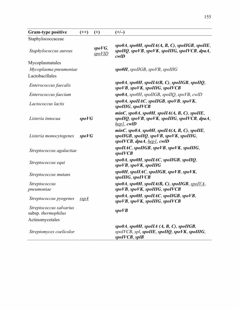
155
Gram-type positive (++) (+) (+/–) Staphylococcaceae
Staphylococcus aureus spoVG, spoVID
spo0A, spo0H, spoIIA(A, B, C), spoIIGB, spoIIE, spoIIQ, spoVB, spoVK, spoIIIG, spoIVCB, dpaA, cwlD
Mycoplasmatales Mycoplasma pneumoniae spo0H, spoIIGB, spoVB, spoIIIG Lactobacillales
Enterococcus faecalis spo0A, spo0H, spoIIA(B, C), spoIIGB, spoIIQ, spoVB, spoVK, spoIIIG, spoIVCB
Enterococcus faecium spo0A, spo0H, spoIIGB, spoIIQ, spoVB, cwlD
Lactococcus lactis spo0A, spoIIAC, spoIIGB, spoVB, spoVK, spoIIIG, spoIVCB
Listeria innocua spoVG minC, spo0A, spo0H, spoIIA(A, B, C), spoIIE, spoIIQ, spoVB, spoVK, spoIIIG, spoIVCB, dpaA, hep1, cwlD
Listeria monocytogenes spoVG minC, spo0A, spo0H, spoIIA(A, B, C), spoIIE, spoIIGB, spoIIQ, spoVB, spoVK, spoIIIG, spoIVCB, dpaA, hep1, cwlD
Streptococcus agalactiae spoIIAC, spoIIGB, spoVB, spoVK, spoIIIG, spoIVCB
Streptococcus equi spo0A, spo0H, spoIIAC, spoIIGB, spoIIQ, spoVB, spoVK, spoIIIG
Streptococcus mutans spo0H, spoIIAC, spoIIGB, spoVB, spoVK, spoIIIG, spoIVCB
Streptococcus pneumoniae spo0A, spo0H, spoIIA(B, C), spoIIGB, spoIVA,
spoVB, spoVK, spoIIIG, spoIVCB
Streptococcus pyogenes sspA spo0A, spo0H, spoIIAC, spoIIGB, spoVB, spoVB, spoVK, spoIIIG, spoIVCB
Streptococcus salvarius subsp. thermophilus spoVB
Actinomycetales
Streptomyces coelicolor spo0A, spo0H, spoIIA (A, B, C), spoIIGB, spoIVCB, spl, spoIIE, spoIIQ, spoVK, spoIIIG, spoIVCB, splB

156
Table 5.5. Gene sequences with similarity to sporulation genes observed in genomes of Gram-
type-negative microorganisms that do not form endospores. (++) Scores identify sequence as
similar to the B. subtilis query sequence. (+) Some similarity over parts of the sequence to the B.
subtilis query sequence. (+/–) Score is too low to allow for a definitive classification of the
sequence relative to to the B. subtilis query sequence

157
Gram-type negative (++) (+) (+/–) Proteobacteria Haemophilus actinomycetemcomitans spo0A, spoIIAC, spoIIQ, spoVK, spoIIIG, cwlD
Caulobacter crescentus spoIIQ, spoIVCB, dpaA, sleB, cwlD
Escherichia coli spo0A, spo0H, spoIIA(B, C), spoIIGB, spoIIQ, spoIIIAA, spoVK, spoIIIG, spoIVCB, cwID
Escherichia coli O157:H7 spoIVCB, cwlD, spo0A, spo0H, spoIIAB, spoIIGB, spoIIQ, cwID
Helicobacter pylori J99 spo0A, spoIIAC, spoIIGB, spoIIQ, spoVK, spoIIIG, spoIVCB, dpaA, cwlD
Myxococcus xanthus spo0A, spo0H, spoIIA(B,C), spoIIGB, spoIIIG, spoIVCB, sleB, dpaA, cwlD
Salmonella typhi spo0A, spo0H, spoIIAC, spoIIGB, spoIIQ, spoVK, spoIIIG, spoIVCB, cwlD
Salmonella typhimurium spo0A, spo0H, spoIIAC, spoIIGB, spoIIQ, spoVK, spoIIIG, spoIVCB, cwlD
Spirochaetes
Borrelia burgdorferi spo0A, spo0H, spoIIAC, spoIIGB, spoVG, spoIIQ, spoVK, spoIIIG, spoIVCB
Cyanobacteria
Synechococcus spp. minC, spoIID, spoIIQ, spoIIIAA, spoVK, spoIVCB, cwID
Prochlorococcus marinus spoIVCB, cwlD, minC, spoIID, spoIIQ, spoIIIAA, spoVK, spoIVCB, cwlD
Anabaena spp. minC, spo0A, spoIIA(A, C), spoIID, spoIIQ, spoIIIAA, spoVK, sleB, cwlD
Chloroflexi Chloroflexus aurantiacus minC, spoIIAB, spoIIE, spoIIIAA, spoIVCB Fusobacteria
Fusobacterium nucleatum spoVG minC, spo0A, spo0H, spoIIAA, spoIID, spoIIE, spoIIQ, spoVK, spoIVCB, splB, cwlD
Deinococcus-Thermus Deinococcus radiodurans minC, spoIIAB, spoIID, spoIIQ, spoVK, cwlD Bacteroidetes Cytophaga hutchinsonii spoIIE, spmA, spmB Archaea Euryarchaeota Archaeoglobus fulgidus spoVG spo0A, spoVB, spoVK, dpaB Halobacterium spp. NRC-1 spo0A, spoVK, dpaB

158
Gram-type negative (++) (+) (+/–) Methanocaldococcus jannaschii spoVB, spoVK
Pyrococcus furiosus spoVB Crenarchaeota Aeropyrum pernix spoVK

159
Table 5.6. Spore-specific genes observed in Bacillus and Clostridium and related species.

160
Classification Genes Present in some Bacillus spp. and some Clostridium spp., but no similar sequences observed in Gram-type-negative Proteobacteria or Cyanobacteria species
rapA, spoIIGA, spoIIM, spoIIP, spoIIR, spoIIIA (B, C, D, E, F, G, H), spoIVA, spoIIID, spoIVB, spoVA (C, D, E, F), sspA, gerA (A, B, C), gpr, cwlJ, yqfC, yqfD, yabP, yabQ, spo0B
Present in some Bacillus spp. only and absent from Clostridium spp.
spoIIB, gerM, cotE, spoVID, gerPA, spoVA (A, B), spoVM, dpaA, hep1, sspE, gerD, cotD

161
Fig. 5.1. Phylogenetic tree constructed from the 16S rRNA gene with maximum likelihood
correction for synonymous changes using the Fitch algorithm. Strain designations were omitted
for simplicity of the tree but are included in Materials and methods . Sporogenic species are
indicated in black. Asporogenic species, as determined by PCR and Southern-hybridization-
based assay are indicated in green. Assignments as asporogenic species based on genome
sequence analysis are indicated in blue. Non-spore-forming species are indicated in red for PCR
and Southern-hybridization-based assay and in orange for genome analysis. Numbers at nodes
indicate bootstrap support values for 100 replicates. Scale bar denotes number of nucleotide
substitutions per site. Using the neighbor-joining algorithm (figure not shown), a very similar
tree was obtained, except the relative position of Geobacillus stearothermophilus and Bacillus
brevis were closer to the position of Bacillus subtilis, whereas Clostridium innocuum was closer
to Mycoplasma pneumoniae.

162
163
CHAPTER 6
CHARACTERIZATION OF A SOLUBLE OXIDOREDUCTASE WITH AN FE(III)
REDUCTION ACTIVITY FROM CARBOXYDOTHERMUS FERRIREDUCENS 1
____________________________
1Onyenwoke, R. U., R. Geyer, and J. Wiegel. To be submitted to Applied and Environmental
Microbiology.
164
Abstract
An NAD(P)H-dependent oxidoreductase has been purified approximately 40-fold from the
soluble protein fraction of the dissimilatory iron-reducing, anaerobic, thermophilic bacterium
Carboxydothermus ferrireducens. The enzyme has a broad substrate range and reduces Fe3+,
Cr6+, and the quinone analog AQDS with rates of 0.31, 3.3, and 3.3 U · mg-1 protein, calculated
turnover numbers for NADH oxidation of 0.25, 2.5, and 2.5 s-1, respectively.
Introduction
Dissimilatory iron reduction is the use of Fe3+ as the terminal electron acceptor in anaerobic
respiration (Myers and Myers 1992). This phenomenon has important environmental
implications, particularly in anaerobic soils and may be related to the evolution of microbial life
(Slobodkin et al. 1997). Dissimilatory iron-reducing bacteria, as a group, are phylogenetically
diverse with members among many distinct taxa, e.g., the Gram-type negative bacteria
(especially the ∆-Proteobacteria) and the Gram-type positive bacteria (Myers and Myers 1992;
Coates et al. 2001; Slobodkin et al. 1997).
Fe3+ most commonly exists in the environment as a general class of insoluble compounds
known collectively as ‘Fe3+ oxides’. For this reason, as well as due to work with Shewanella and
Geobacter (Lovley 1991; Myers and Myers 1992; Magnuson et al. 2000), it has been suggested
that direct cell contact (and therefore a membrane-localized system of proteins) is required for
the reduction of ‘Fe3+ oxides’. However, solublized Fe3+ is important and in relatively high
abundances in environments rich in organic chelating agents (Ratering and Schnell 2000), and
soluble ‘Fe3+ reductases’ have been characterized (Kaufmann and Lovley 2001; Chiu et al.
2001). The role of these soluble enzymes is presently unknown but may be linked to Fe3+

165
reduction via a soluble electron carrier, such as a quinone (Lovley et al. 1996; Fig. 6.1.). This
hypothesis is strengthened by the reported ability of all described Fe3+-reducers to make use of
the quinone analog 9,10-anthraquinone-2,6-disulfonate (AQDS) as a terminal electron acceptor
of the electron transport chain. Quinones typically act as intermediates in cellular, electron
transport chains. In many (an)aerobic soils and sediments, it is the quinone moieties of humic
substances that function as the electron shuttles to ‘Fe3+ oxides’ (Lovley et al. 1996; Hernandez
and Newman 2001). In addition, the importance of quinones to bacterial respiration has been
described. Shewanella oneidensis requires menaquinone (MK) during growth on several electron
acceptors, including Mn4+, Fe3+, fumarate, nitrate, nitrite, thiosulfate, DMSO, and AQDS (Myers
and Myers 1993b; Myers and Myers 1994; Myers et al. 2004; Myers and Myers 2000; Newman
and Kolter 2000; Schwalb et al. 2003).
Here we report on an NAD(P)H-dependent oxidoreductase purified from the cytoplasmic
(soluble) fraction of the Gram-type positive, anaerobic, thermophilic, dissimilatory iron-reducing
bacterium Carboxydothermus ferrireducens (basonym: Thermoterrabacterium ferrireducens)
(Slobodkin et al. 1997, 2006) capable of Fe3+ and quinone reduction. Insoluble ‘Fe3+ oxides’,
chelated forms of Fe3+ (e.g., Fe3+ citrate and Fe3+-EDTA), AQDS, and fumarate serve as electron
acceptors for this bacterium (Slobodkin et al. 1997). Thus, the C. ferrireducens oxidoreductase
(CFOR) may play some role in reduction-oxidation type reactions in this microorganism;
possibly bridging the energetic gap between the quinone electron intermediates (shuttles) and the
Fe3+ terminal electron acceptors.

166
Materials and methods
Materials
Unless otherwise noted, all chemicals were obtained from Sigma Chemical Co., St. Louis, MO,
USA. A 1 mM stock of FADH2 was prepared by adding solid sodium dithionite to FAD and
following the loss of absorbance at 450 nm, i.e., the reduction of FAD to FADH2 (Louie et al.
2003). Recombinant human (rh)NQO1, purified from Escherichia coli (Beall et al. 1994), was
the generous gift of Dr. David Siegel, University of Colorado Health Sciences Center, Denver,
Colorado, USA. The purified NQO1 was at a final protein concentration of 3.3 mg/ml in 25 mM
Tris-HCL (pH 7.4) containing 250 mM sucrose and 5 µM FAD. Lyophilized guinea pig zeta-
crystallin protein was the generous gift of Dr. J. Samuel Zigler, National Eye Institute (NIH),
Bethesda, Maryland, USA.
Cell growth
Carboxydothermus ferrireducens (DSM 11255) was obtained from the authors’ laboratory
culture collection and cultivated under anoxic conditions [N2 headspace (Ljungdahl and Wiegel
1986)] in a minimal medium, described by Slobodkin et al. (1997), containing 40 mM glycerol
and 10 mM fumarate in a 100 l fermentor at the University of Georgia’s Fermentation Facility
(Athens, GA). Cells (50 to 60 g wet weight) were: harvested by continuous centrifugation
(13,000 g), washed and resuspended in 20 mM Tris buffer containing 10% (v/v) glycerol (pH
8.5) to a final concentration of 1.0 g cell mass/ml, and stored at -80ºC for further use.

167
Enzyme assays
Unless otherwise noted, all enzyme assays were performed in duplicate under anoxic conditions
(N2-sparged Hungate tubes) at 50°C for 15 min. The assay was linear for at least 30 min., and the
data indicated ~15% of the substrate (Fe3+) had been consumed in 15 min. (Fig. 6.2.). The
reaction mixture (final volume 1.0 ml) contained: buffer [20 mM MES, 20 mM Tris, 20 mM
MOPS, 20 mM TAPS, and 40 mM MgCl2 (pH 6.5)], 0.01-10 mM NADH (or NADPH), 1.0-10
µM FAD, (FMN or riboflavin), 0.2 mM ferrozine, 0.01-500 µM Fe3+ citrate, and 0.5-50 µg of
protein. The reaction was initiated by the addition of protein. Activity was monitored on a
Beckman DU-64 spectrophotometer by either 1) following the reduction of Fe3+ to Fe2+ by the
use of the Fe2+ capture reagent ferrozine (3-(2-pyridyl)-5,6-bis(4-phenyl-sulfonic acid)-1,2,4-
triazine) at 562 nm [molar extinction coefficient (ε562) of 27.9 mM-1cm-1 (Dailey and Lascelles
1977)], or 2) observing the rate of NADH oxidation at 340 nm [ε340 = 6.22 mM-1cm-1 (Ernster et
al. 1962)]. One milliunit (mU) is the amount of enzyme catalyzing the release of 1 nmol of
product per min. The maximal pH for Fe3+ reduction activity was determined by adjusting the pH
of the buffer (see above) using either 5.0 M HCl or 5.0 M NaOH.
Product inhibition studies were performed with the purified CFOR employing a reaction
mixture containing 1) 0-50 µM NAD+ with 100 nM Fe3+ citrate, 5.0 µM FAD, and 0-50 µM
NADH; 2) 0-50 µM FADH2 with 100 nM Fe3+ citrate, 50 µM NADH, and 0-50 µM FAD; or 3)
0-10 µM ferrous sulfate heptahydrate with 50 µM NADH and 5 µM FAD, and 0-10 µM Fe3+
citrate. Reaction was initiated by the addition of protein. Inhibition studies employing EDTA,
NTA, or metals (CuSO4 · 5H2O, ZnSO4 · 7H2O, NiCl2 · 6H2O, Na2CrO4, MnO2, Na2SeO3, or Co-
EDTA) were conducted using 100 nM ferric citrate, 5 µM FAD, 50 µM NADH, and 2.5 µg
protein.

168
For testing the reduction of other metals by the purified CFOR (added to the assay
mixture to give a final concentration of 50 µM), iron and ferrozine were omitted from the
reaction mixture. Manganese (Mn4+) reduction to Mn2+ was determined using a formaldoxime-
ammonia reagent (Brewer and Spencer 1971; Greene et al. 1997). The increase in absorbance at
450 nm (A450) due to the production of Mn2+ was followed and compared to Mn2+ standards.
Mn2+ standards were prepared by reducing a potassium permanganate solution with concentrated
sulfuric acid. Cobalt (Co3+) reduction was measured at 535 nm by observing the loss of Co3+
over time compared to sodium dithionite reduced standards (Caccavo et al. 1996b). Chromium
(Cr6+) reduction was determined by observing the increase in A540 using s-diphenylcarbazide and
sodium dithionite reduced chromium (Cr6+) standards (Urone 1955). Arsenic (As5+) reduction to
As3+ was determined using a molybdate color reagent (Johnson 1971). The decrease in A885 due
to the loss of As5+ was monitored and compared to chemically reduced As3+ standards.
Alternatively, the rate of NADH oxidation was also followed to determine all rates of metal
reduction. NADH oxidation was used as the exclusive method to follow selenate (Na2Se6+O4)
reduction. The reduction of quinones (all at 50 µM) involved the same assay system in which
metals were omitted for the appropriate quinone, and the rate of NADH oxidation was observed.
The reduction of 9,10-anthraquinone-2,6-disulfonate (AQDS) was monitored by following the
increase in A450 [ε450 = 3.5 mM-1cm-1] (Lovley et al. 1996).
The mechanism of quinone reduction was determined using cytochrome c as a trap for
semiquinone generation (Gonzalez et al. 2005). The purified CFOR was tested along with two
controls: guinea pig zeta-crystallin protein (Rao et al. 1992.) and human NAD(P)H:quinone
oxidoreductase, also known as DT-diaphorase (Ernster 1987). The increase in A550 due to

169
reduction of cytochrome c [ε550 = 31.2 mM-1cm-1] by the semiquinone was monitored in an assay
containing 50 µM 1,2-naphthoquinone.
Quinone analysis (Lipid extraction and fractionation)
Whole cell quinone analysis was performed as described by Sokolova et al. (in press). Briefly,
duplicate samples of C. ferrireducens (0.5 g cell mass/ml) were extracted using a modified Bligh
and Dyer (1959) extraction method (White et al. 2005). Lipids were separated on a silicic acid
column with chloroform, acetone, and methanol into neutral lipid, glycolipid, and polar lipid
fractions (Guckert et al. 1985). Solvents were removed under a gentle stream of nitrogen, and the
fractions were stored at -20°C. The neutral lipid fraction was resolved in methanol and analyzed
for respiratory quinones by HPLC-tandem mass spectrometry (Geyer et al. 2004).
Purification of a soluble oxidoreductase from C. ferrireducens with an Fe3+ reduction activity
All purification steps were performed aerobically at room temperature (~25ºC) unless otherwise
noted. A cell lysate was prepared from approximately 20 ml of cell suspension (~20 g cell mass)
by passage twice through a French pressure cell at 5500 kPa. The cell lysate was then
centrifuged at 10,000 g for 30 min. (4ºC) to pellet cell debris and ultra-centrifuged [105,000 g for
1h (4ºC)] to pellet the membrane fraction from the soluble (cytoplasmic) fraction. The membrane
fraction was discarded, and the soluble fraction was retained for the subsequent purification
protocol.
For the following procedures, all buffers contained 10 µM FAD. In addition, the
precaution of wrapping all columns, buffer-containing bottles, and containers used for dialysis in
aluminum foil was taken to prevent the degradation of the FAD due to exposure to light. The
170
soluble fraction (~20 ml), prepared as described above, was applied to a Q-Sepharose Fast Flow
column [1.0 x 30 cm (20 ml bed volume); Amersham Biosciences, Piscataway, NJ] that had been
equilibrated with 20 mM Tris buffer (pH 8.5) containing 10% (v/v) glycerol (buffer A). The
protein was eluted using buffer A + 1.0 M NaCl along a linear gradient of 0 to 0.6 M NaCl (total
volume 100 ml) with a flow rate of 1.5 ml/min (4.0 ml fractions collected). Fractions containing
Fe3+ reduction activity were eluted between 0.3 and 0.6 M NaCl and were pooled. The pooled
fractions (~45 ml) were dialyzed using Spectra/Por 12-14 kDa cut-off dialysis tubing (Spectrum
Laboratories, Rancho Dominguez, CA) overnight (2 exchanges for fresh buffer) against 400 ml
of 20 mM sodium phosphate buffer (pH 7.0) containing 10% (v/v) glycerol (buffer B). The
pooled fractions were then loaded onto a HiTrap Blue HP column [1.6 x 2.5 cm (5 ml bed
volume); Amersham Biosciences, Piscataway, NJ] that had been equilibrated using buffer B.
After loading of the protein onto the HiTrap column, at least 5 column volumes of buffer B were
used to remove any unbound protein before a step-wise elution (2.0 ml fractions collected) with
buffer B + 1.0 mM NADH (30 ml used for elution) . The flow rate was maintained at 2.0 ml/min.
The fractions containing the protein eluted by NADH were pooled (~30 ml) and dialyzed against
400 ml of buffer B (2 exchanges for fresh buffer) overnight. The pooled fractions were
concentrated to ~5 ml using an Amicon 8400 ultra-filtration stirred cell (400 ml capacity;
Millipore, Bedford, MA) equipped with a 10 kDa cut-off ultra-filtration membrane (Amicon,
Danvers, MA) and applied to a Sephacryl S-300 HR (Sigma Chemical Co., St. Louis, MO)
column [1.6 x 50 cm (100 ml bed volume); Amersham Biosciences, Piscataway, NJ] that had
been equilibrated with buffer B + 0.15 M NaCl at 0.4 ml/min. After application of the sample,
the protein was eluted using 120 ml of the equilibration buffer, and fractions (2.0 ml) were
collected.

171
Determination of native molecular weight
The native molecular weight (Mr) of the enzyme was determined using gel filtration on the
Sephacryl S-300 HR column. Apoferritin (443 kDa), alcohol dehydrogenase (150 kDa), and
bovine serum albumin (66 kDa) were the molecular weight standards.
Gel electrophoresis
Sodium dodecyl sulfate polyacrylamide-gel electrophoresis (SDS-PAGE) was carried out
according to the method of Laemmeli (1970). A Bio-Rad broad range molecular marker kit
[(Hercules, CA); myosin (Mr 200, 000), β-galactosidase (Mr 116,000), phosphorylase b (Mr
97,000), bovine serum albumin (Mr 66,000), ovalbumin (Mr 45,000), carbonic anhydrase (Mr
30,000), trypsin inhibitor (Mr 20,000), lysozyme (Mr 14,000), and aprotinin (Mr 6500)] was used
as standards. Proteins were separated on 12% polyacrylamide gels and stained using the Pierce
(Rockford, IL) GelCode stain reagent.
Determination of flavin and metal content
The purified CFOR was analyzed for flavin content, after perchloric acid extraction, by high-
pressure liquid chromatography (HPLC) using a C-18 reversed phase column (25 x 4.6 cm) with
a 20 mM ammonium acetate mobile phase (pH 6.0) containing 21% acetonitrile (v/v) at a flow
rate of 0.6 ml/min (Hausinger et al. 1986). Various concentrations of FAD (retention time = 4.88
min) and FMN (retention time = 5.71 min) were prepared as standards to identify, and to
determine the amount of, any possible bound flavin.
Metal content was determined using both the iron quantitation method of Fish (1988)
with Fe3+ citrate standardsand inductively coupled plasma mass spectrometry (ICP-MS) analysis
172
(Chemical Analysis Laboratory, UGA, Athens, GA). Total protein was determined using a Bio-
Rad protein assay kit (Richmond, CA) with a bovine serum albumin standard. The cofactor
content of the CFOR was calculated based upon a molecular mass of 45 kDa.
Determination of N-terminal amino acid sequence
For N-terminal amino acid sequencing, the purified CFOR was separated by SDS-PAGE (12%)
and transferred to a polyvinylidene difluoride membrane (Bio-Rad, Hercules, CA). The
membrane was stained using 1% Amido black, and the selected bands were excised using a clean
razor blade. The amino acid sequence was determined by automated Edman degradation (J. Pohl,
Microchemical and Proteomics Facility, Emory University, Atlanta, GA).
Kinetic data analysis
Data were expressed as the double-reciprocal plots. Nonlinear regression analysis was performed
with the kinetic software package Aca-Stat 5.1.25.
Results
Localization of the CFOR
Lysates prepared from cells grown on fumarate exhibited similar Fe3+ reduction rates, using the
ferrozine assay, to cells grown on Fe3+ citrate, as has been previously described (Kaufmann and
Lovley 2001). When the cell lysate was partitioned into soluble and membrane fractions, Fe3+
reduction activity was found to be equally localized to both fractions (Gavrilov et al. 2003, in
press). The activity in the soluble fraction was chosen for further study.

173
Purification of the CFOR
Oxygen did not serve as a substrate (i.e., no NAD(P)H-O2 activity ), and NAD(P)H-dependent
Fe3+ reduction activity was not inactivated by exposure to oxygen, i.e., the activity was
equivalent whether purification occurred under aerobic or anaerobic conditions, as has also been
previously observed with the strict anaerobe Shewanella putrefaciens strain MR-1 (Myers and
Myers 1993a). Therefore, purification was performed under aerobic conditions. In addition,
theactivity was stimulated by the addition of FAD. Therefore, purification of the CFOR was
performed in the presence of 10 µM FAD (see Materials and methods). A three-step,
chromatographic procedure (Table 6.1.) was then used to purify the CFOR to apparent, gel-
electrophoretic purity with an enzymatic yield of ~25% and a specific activity of 560 mU · mg -1
protein.
Physical and biochemical characteristics
The native molecular mass of the CFOR was determined to be ~200 kDa by gel filtration and
native, gradient (4-16%) PAGE. SDS-PAGE analysis showed a single subunit with a relative
molecular mass of ~45 kDa. Taken together, these data suggest an enzyme oligomer, possibly a
tetramer or (less likely) a pentamer.
ICP-MS of the purified CFOR, which was dialyzed against distilled water to remove any
cofactors not tightly associated, yielded an Fe content of 0.96±0.04 Fe per 45 kDa subunit of
enzyme. Furthermore, an Fe content of 0.98±0.05 Fe per 45 kDa subunit was determined by the
use of ferrozine to calculate the iron content spectrophotometrically (Fish 1988). HPLC analysis
of the purified CFOR after perchloric acid extraction indicated each 45 kDa subunit contained
0.9±0.09 FAD.
174
The CFOR exhibited maximal activity around a pH of 6.5 and a temperature of 50ºC
(Fig. 6.3.A. and 6.3.B., respectively). EDTA inhibited enzymatic activity by 25, 60, and 75% at
50, 100, and 1000 µM, respectively. NTA inhibited enzymatic activity by 25 and 50% at 10 and
100 µM, respectively. When added to the ferrozine assay (measuring rate of Fe(III) reduction),
nickel (NiCl2 · 6H2O) inhibited enzymatic iron reduction activity by 60% at 100 µM. The
addition of 100 and 1000 µM manganese (MnO2) increased enzymatic activity by 60 and 100%,
respectively. The addition of chromium (Na2CrO4), selenium (Na2SeO3), cobalt (Co-EDTA),
copper (CuSO4 · 5H2O), and zinc (ZnSO4 · 7H2O) [up to 1 mM of each tested] had no effect on
enzymatic activity.
Enzymatic activities and kinetic studies
The CFOR reduced Cr6+, 1,2-naphthoquinone and AQDS at rates comparable to or greater than
the rate of Fe3+ reduction. Exogenous FAD resulted in higher activities, e.g., the Fe3+ reduction
rate was 560 mU · mg-1 protein with 5 µM FAD, and 34 mU · mg-1 protein when exogenous FAD
was absent. The CFOR reduced FAD when other substrates were absent but only at a rate of 14
mU · mg-1 protein. As a control, the abiotic Fe3+ reduction rate with chemically reduced FAD,
i.e., no CFOR present, was determined. The Fe3+ reduction rate with 0.5 µM of reduced FAD,
and no CFOR, in the assay was calculated as 0.28 mU, as compared to 2.8 mU with no reduced
FAD and 5.0 µg NQO1, i.e., the ferrozine assay as described above. Other quinones and metals
were also reduced but at lower rates than the above described activities (Table 6.2.).
Kinetic studies were performed using a three substrate, steady-state analysis according to
the procedure of Segel (1975). Briefly, initial velocity studies were carried out by varying the
concentrations of the electron acceptor, (i.e., Fe3+, Cr6+, or AQDS), FAD, and NADH.

175
(Onyenwoke and Wiegel, submitted; Chapter 7, this dissertation). As illustrated in Figs. 6.4.-6.6.,
the double-reciprocal plots with the patterns of lines intersecting the x-axis to the left of the y-
axis were used for calculations of apparent Michaelis-Menten constants (Kmapp) and apparent
maximum velocities (Vmaxapp). The determined parameters [Km
app, Vmaxapp, and turnover number
(kcat)] for the enzyme during the reduction of Fe3+, as well as Cr6+ and AQDS, are shown in
Table 6.3.
The best fit plot for NADH saturation in the presence of various concentrations of Fe3+
citrate yields an intersection at a point close to the x-axis (data not shown). The inverse plot, i.e.,
Fe3+ citrate saturation in the presence of a series of NADH concentrations, (Fig. 6.4.) also,
roughly, intersects the x-axis at a single point. This data suggests Fe3+ citrate and NADH are
binding independently [random sequential in the nomenclature of Cleland (1977)]. A similar
result was obtained on examination of the effect of FAD with a series of [Fe3+ citrate], i.e., based
on interpretation of these kinetic data FAD and Fe3+ citrate also bind independently of each other
(data not shown). Figs. 6.5. and 6.6. show the best fit plots for AQDS and Cr6+ saturation,
respectively, in the presence of various concentrations of NADH. Again the plots are indicative
of a sequential reaction mechanism, though the plot for Cr6+ saturation indicates an ordered
sequential mechanism (Cleland 1977). In order to obtain some further data on the proposed
reaction mechanism (and to possibly determine if substrate binding and product release are
random or ordered if the mechanism is indeed sequential), the product (ferrous sulfate, FADH2,
and NAD+) inhibition patterns of the CFOR were analyzed with respect to various concentrations
of the substrates (Fe3+ citrate and NADH) and FAD.
NAD+ was found to be a mixed type inhibitor [properties of both competitive and
uncompetitive inhibition] versus NADH (Fig. 6.7.A.) with an apparent KI'= KI when Fe3+ citrate

176
was at a sub-saturating level (100 nM). The plot of the effect of Fe2+ sulfate versus Fe3+ citrate
indicated uncompetitive inhibition, based on the best-fit plot/line from the data (Fig. 6.7.C.),
when NADH was present at a sub-saturating level (50 µM). The plots of NAD+ versus NADH
and Fe2+ sulfate versus Fe3+ citrate indicate the reaction mechanism is specifically ordered
sequential (Segel 1975), with further data necessary to determine the specific reaction
mechanism according to the nomenclature of Cleland. Increasing [FADH2] versus FAD
suggested the occurrence of activation instead of inhibition (Fig. 6.7.B.).
Quinone analysis and mechanism of quinone reduction
The neutral lipid fraction of C. ferrireducens contained the following respiratory quinones
(mol%): ubiquinone (UQ)-8 (58) as the major compound, UQ-9 (4.6), UQ-7 (3.5), menaquinone
(MK)-8 (15), demethylmenaquinone (DMK)-9 (12), MK-5 (3.4), and MK-4 (1.7) as minor
compounds. The minor quinones, except UQ-9, were also found in the control (culture media)
and could derive from passive accumulation in the membrane (Geyer, R., unpublished results).
When compared to the controls, i.e., guinea pig zeta-crystallin protein (single electron
mechanism) and human NAD(P)H:quinone oxidoreductase (two electron mechanism), the CFOR
exhibited a two electron mechanism for quinone reduction.
Amino acid sequence analysis of the enzyme
The N-terminal sequence of the CFOR as determined by automated Edman degradation was
Met-Asn-Lys-Tyr-Val-His-Ala-Val-Pro-Asn-Phe.
This sequence was used to search databases [SwissProt/TrEMBL
(http://us.expasy.org/sprot/) and GenBank (http://www.ncbi.nlm.nih.gov/)] for similar proteins

177
by using the BLAST algorithm (Altschul et al. 1990). The N-terminal sequence showed 100 and
72% identity to the electronically annotated: glutamate formiminotransferase of
Carboxydothermus hydrogenoformans [305 amino acids with a predicted mol. mass of ~34 kDa]
(Wu et al. 2005) and phosphotransferase EII fragment of Mycoplasma capricolum [118 amino
acids with a predicted mol. mass of ~13 kDa] (Bork et al. 1995), respectively.
Discussion
At least two C. ferrireducens enzymes are capable of NAD(P)H-dependent Fe3+ reduction, one
localized to the soluble protein fraction (the CFOR) and the second to the membrane fraction.
The relationship between the two enzymes is currently not understood, but the two enzymes
might not act independently of each other. An assimilatory (possibly soluble) Fe3+ reductase
could attain a secondary (dissimilatory) function simply by adding the ability to couple Fe3+
reduction to the respiratory chain through a second (possibly membrane-bound) enzyme
(Nealson and Saffarini 1994). The NAD(P)H-dependent Fe3+ reduction activity localized to the
soluble protein fraction was chosen for further study, and an enzyme (the CFOR) was purified
and characterized. The CFOR contains FAD and Fe (both at a ratio of ~1 per 45 kDa subunit of
enzyme), and its NAD(P)H-dependent Fe3+ reduction activity is stimulated by FAD. From the
kinetic data a sequential reaction mechanism is indicated and a simple mechanistic model can be
proposed (Fig. 6.8.A.1. and 6.8.A.2.). However, due to a broad substrate range, e.g., reduces
Fe3+, Cr6+, and AQDS, several possibilities exist for the physiological function of the CFOR.
The CFOR can function as an FAD reductase (Fontecave et al. 1994; Fig. 6.8.B.), as is
supported by the FADH2-dependent activation of Fe3+ reduction (Fig. 6.7.B.). The FADH2 could
then chemically reduce the substrate, e.g., Fe3+, AQDS or Cr6+. However, the determined FAD

178
reduction rate, i.e., no additional electron acceptor added to the assay, of 14 mU · mg-1 protein is
significantly below other observed activities (Table 6.2.), and the low observed Km’s for Fe3+,
AQDS, and Cr6+ suggest a more direct role for the CFOR in their reduction.
The data also suggest that CFOR is a quinone oxidoreductase, as the highest observed
activities were with quinones, i.e., AQDS and 1,2-naphthquinone. Thus, the quinone content of
C. ferrireducens was determined. Interestingly, the major quinone present was a UQ (UQ-8) and
not a MK, as would be expected. This is unusual as 1) data from Escherichia coli and other
Proteobacteria and 2) differences in midpoint potentials indicate UQs are typically involved in
oxygen and nitrate respiration, whereas MKs are more functional in anaerobic respiration
(Polglase et al. 1966; Gennis and Stewart 1996; Sohn et al. 2004; White et al. 2005). However,
this scheme may be oversimplified as UQs also function as the electron carriers between b-type
cytochromes and various terminal oxidases (Sùballe and Poole 1998). UQs have been found as
the dominate quinone species in other anaerobic bacteria (White et al. 2005). Still, MKs could be
the species involved in anaerobic respiration with the UQs playing some other role.
Apart from their role in respiration, reduced UQs (UQH2) scavenge lipid peroxyl radicals
and thereby prevent a chain reaction of oxidative damage to the polyunsaturated fatty acids of
biological membranes, a process known as lipid peroxidation (Forsmark-Andrée et al. 1995).
Therefore, the [UQH2] and other antioxidants, such as vitamin E, present in low-density
lipoprotein is of vital importance for the prevention of oxidative damage (cytotoxic effects) and
even diseases such as atherosclerosis (Forsmark-Andrée et al. 1995). UQH2, and other reduced
quinone, pools are maintained by soluble quinone oxidoreductases [e.g., DT-diaphorase, also
known as NAD(P)H:quinone oxidoreductase, (EC 1.6.99.2); lipoamide dehydrogenase; the
quinone oxidoreductases MdaB and ChrR of Helicobacter pylori and Pseudomonas putida,
179
respectively; and possibly the CFOR]. These enzymes protect against cytotoxic and carcinogenic
effects (Beyer et al. 1996; Siegel et al. 2004; Olsson et al. 1999; Wang and Maier 2004;
Gonzalez et al. 2005). Any inhibition of quinone reductase activity results in an increase in free
radical damage (Beyer et al. 1996).
Quinone compounds that serve as pro-oxidants also arise from exogenous sources, e.g.,
plant secreted plumbagin and juglone (Soballe and Poole 1999; Gonzalez et al. 2005). These
quinones are highly soluble, and their redox cycling imposes a severe oxidative stress (Soballe
and Poole 1999). Semiquinones, which are the result of single electron reductions, are the
damaging species (Gonzalez et al. 2005). Enzymes such as DT-diaphorase and the CFOR avert
the production of the semiquinone by performing a two electron reduction to produce a less
active hydroquinone. This activity protects cells from the reactive semiquinone species (Siegel et
al. 2004). Thus, quinone metabolism has a direct effect upon how a cell manages oxidative stress
(Soballe and Poole 1999; Wang and Maier 2004).
Possible roles for the CFOR in redox type reactions besides Fe3+ reduction have been
suggested. In particular, the high activities reported for quinone reduction suggest a role in
protection from oxidative stress by maintaining pools of UQH2. Current evidence does suggest
UQ’s are antioxidants (Abhilashkumar et al. 2001; White et al. 2005). However, the CFOR is
also an FAD reductase. The production of reduced FAD, which does reduce Fe3+, could be the
explanation for the broad substrate specificity exhibited by the enzyme (Table 6.2.). Some role in
respiration is also plausible, but less likely as membrane-localized enzymes, such as the
membrane-bound Fe3+-reducing enzyme mentioned here, would be expected to fulfill this
function.

180
Acknowledgements
We thank Dr. David Siegel and J. Samuel Zigler for the generous gifts of: recombinant human
NQO1 and guinea pig zeta-crystallin protein, respectively.

181
Table 6.1. Purification of the CFOR. aAssays contained 50 µM NADH, 5 µM FAD, and 100 nM
Fe3+ citrate.

182
Purification step Amt of protein (mg)
Total activitya
(mU)
Sp act (mU · mg -1)
Purification factor (fold)
Yield (%)
Soluble fraction 680 10,200 15 1 100
Q-Sepharose 99 7920 80 5 78
Blue Sepharose 13.5 7020 520 35 69
Sephacryl S-300 HR 4.5 2520 560 37 25
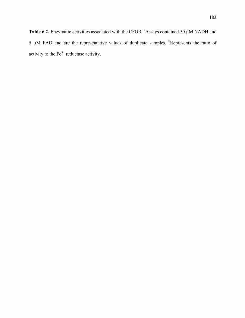
183
Table 6.2. Enzymatic activities associated with the CFOR. aAssays contained 50 µM NADH and
5 µM FAD and are the representative values of duplicate samples. bRepresents the ratio of
activity to the Fe3+ reductase activity.

184
NAD(P)H-dependent enzymatic activity (substrate is listed)
Avg sp acta (mU · mg -1)
Relative activityb
AQDS reduction 2100±50 3.8
1,2-naphthquinone reduction 870±80 1.6
Cr6+ reduction 650±30 1.2
Fe3+ reduction 560±55 1.0
2-hydroxy-1,4-naphthquinone reduction 300±32 0.54
duroquinone reduction 250±70 0.45
As5+ reduction 210±65 0.38
Fumarate reduction 210±25 0.38
Cu2+ reduction 180±20 0.32
1,4-naphthquinone reduction 180±14 0.32
Co3+ reduction 150±16 0.27
Mn4+ reduction 130±55 0.23
benzoquinone reduction 130±30 0.23
menadione reduction 110±28 0.20
Se6+ reduction none detected 0

185
Table 6.3. Kinetic parameters of the CFOR. a kcat’s (turnover numbers) were calculated by
determining the amount of CFOR, based upon the molecular weight of the monomer (45 kDa) and
assuming one catalytic site per CFOR monomer, present in the reaction mixture. The kcat’s were
then determined from the Vmaxapp values.

186
Km
app (µM)
Vmaxapp
(mU · mg -1 protein) kcat
a
(s-1)
Fe3+ reduction Fe3+ 0.014 480 0.36
FAD 1.2 230 0.18
NADH 62 310 0.25
Cr6+ reduction Cr6+ 0.6 10,000 7.5
FAD 2.0 3300 2.5
NADH 76 3300 2.5
AQDS reduction AQDS 0.025 2500 1.9
FAD 5.8 2500 1.9
NADH 26 3300 2.5

187
Fig. 6.1. Proposed model in which an electron shuttle serves to reduce insoluble Fe3+ oxides. “Q”
represents any electron shuttle, e.g., the quinone analog AQDS. “Q” is the oxidized form of the
shuttle while “QH2” is the reduced form. The shuttle passes into the cytoplasm of the
dissimilatory iron-reducer and is reduced. The reduced shuttle carries the electrons outside to the
insoluble Fe3+ source where the Fe3+ is reduced to Fe2+.

188
e- e- Q
QH2
Fe2+
Solid Fe3+ oxide
Fe3+
Electrons
Cytoplasmic (Soluble) Fe3+ reductase
Dissimilatory, Fe3+-reducing microorganism

189
Fig. 6.2. Time course showing the linearity of the NAD(P)H-dependent Fe3+ reduction activity of
the crude C. ferrireducens soluble (cytoplasmic) protein fraction. The assay was performed using
the ferrozine method and contained: 50 µg of protein, 5 µM FAD, 0.05 mM NADH or NADPH,
and 100 µM Fe3+ citrate.
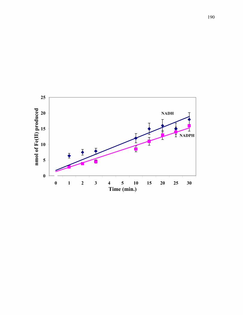
190
NADH
NADPH
0
5
10
15
20
25
0 1 2 3 4 5 10 15 20 25 30Time (min.)
nmol
of F
e(II
) pro
duce
d
191
Fig. 6.3. The effects of pH (A) and temperature (B) on the NAD(P)H-dependent Fe3+ reduction
activity of the CFOR. Assay mixtures contained: 2.5 µg of protein, 5 µM FAD, 0.05 mM NADH,
and 100 nM Fe3+ citrate.

192
A
0
10
20
30
40
50
60
70
5.4 5.9 6.4 6.9 7.4
pH
Fe3+
red
uctio
n ra
te (m
U/ m
g pr
otei
n

193
B
0
20
40
60
80
100
120
140
160
180
200
25 35 45 55 65 75 85 95
Temperature (ºC)
Fe3+
red
uctio
n ra
te (m
U •
mg-1
prot
ein)

194
Fig. 6.4. Initial velocity kinetics of the Fe3+ reduction activity of the CFOR. All data are
represented as the double- reciprocal plots. The effect of [Fe3+ citrate] on activity. Measurements
represent a combined replot for three, independent series with NADH as the constant substrate.
Assays followed the oxidation of NADH and contained: 2.5 µg of protein and 5 µM FAD.

195
10 µM NADH
50 µM NADH
100 µM NADH
00.010.020.030.040.050.060.070.080.09
-100 -50 0 50 1001/ [Fe3+ citrate (in µM)]
1/v
(mU
· m
g-1 p
rote
in)
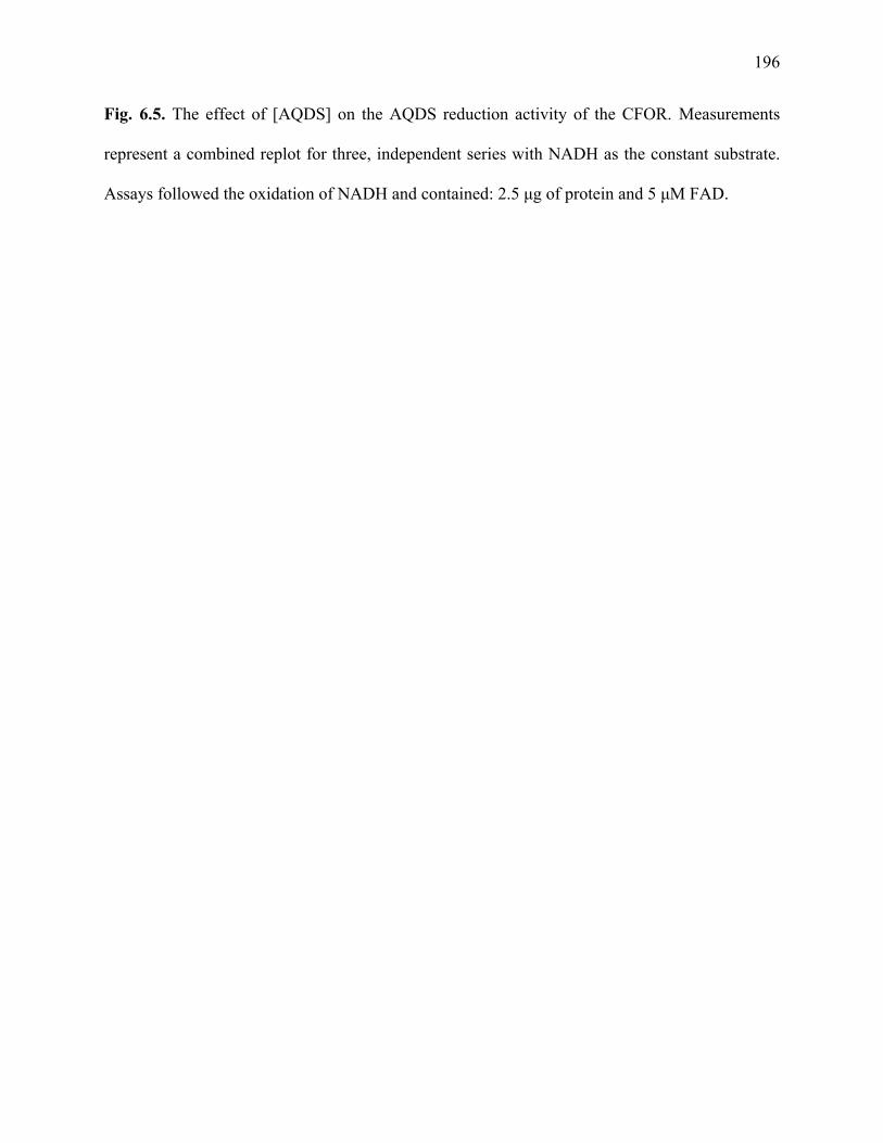
196
Fig. 6.5. The effect of [AQDS] on the AQDS reduction activity of the CFOR. Measurements
represent a combined replot for three, independent series with NADH as the constant substrate.
Assays followed the oxidation of NADH and contained: 2.5 µg of protein and 5 µM FAD.

197
50 µM NADH
100 µM NADH
10 µM NADH
-0.001
0
0.001
0.002
0.003
0.004
0.005
0.006
-40 -20 0 20 40 60 80 100
1/[AQDS (in µM)]
1/v
(mU
• m
g-1 p
rote
in)

198
Fig. 6.6. The effect of [Cr6+] on the Cr6+ reduction activity of the CFOR. Measurements
represent a combined replot for three, independent series with NADH as the constant substrate.
Assays followed the oxidation of NADH and contained: 2.5 µg of protein and 5 µM FAD.

199
0
0.005
0.01
0.015
0.02
0.025
0.03
0.035
0.04
-20 0 20 40 60 80 1001/[Na2Cr6+
2O7 (in µM)]
1/v
(mU
· m
g-1 p
rote
in)
10 µM NADH
50 µM NADH
100 µM NADH

200
Fig. 6.7. Product inhibition patterns for Fe3+ reduction by the CFOR. (A) NAD+ concentration
kept constant with 100 nM Fe3+ citrate, 5.0 µM FAD, and 0-50 µM NADH. (B) FADH2
concentration kept constant with 100 nM Fe3+ citrate, 50 µM NADH, and 0-50 µM FAD. (C)
Fe2+ sulfate heptahydrate concentration kept constant with 50 µM NADH, 5 µM FAD, and 0-10
µM Fe3+ citrate. Assays were performed by observing NADH oxidation.

201
A
0 µM NAD+
5 µM NAD+
10 µM NAD+
50 µM NAD+
-0.05
-0.03
-0.01
0.01
0.03
0.05
0.07
0.09
0.11
0.13
-1 -0.5 0 0.5 11/[NADH (in µM)]
1/v
(mU
• m
g-1 pr
otei
n)

202
B
0 µM FADH2
5 µM FADH2
10 µM FADH2
50 µM FADH2
-0.01
-0.005
0
0.005
0.01
0.015
0.02
0.025
-1 -0.5 0 0.5 11/[FAD (in µM)]
1/v
(mU
• m
g-1 p
rote
in)

203
C
0 µM Fe2+ sulfate
0.1 µM Fe2+ sulfate
1.0 µM Fe2+ sulfate
10 µM Fe2+ sulfate
-0.01
0.01
0.03
0.05
0.07
0.09
-100 -80 -60 -40 -20 0 20 40 60 80 100
1/[Fe3+ citrate (in µM)]
1/v
(mU
• m
g-1 pr
otei
n)

204
Fig. 6.8. Proposed mechanisms of substrate reduction by the CFOR. “X” refers to the substrate,
e.g., Fe3+, Cr6+, or AQDS, reduced (Xred) or oxidized (Xox). (A) 1. Substrate is reduced via the
oxidation of NADH with the enzyme bound FAD (yellow) serving as a cofactor, i.e., electron
mediator. Bound Fe (pink) is also present in the enzyme. 2. Exogenous FAD increases enzymatic
activity (indicated by thicker arrows) and possibly occupies a secondary site on the CFOR. (B)
Alternatively, exogenous FAD increases activity because the CFOR additionally functions as a
flavin reductase. The exogenous FAD is reduced by the CFOR. The reduced FAD in turn
reduces the substrate “X”.

205
A1
Xred
Xox
NAD+
NADH
CFOR
FADH2
FAD
Fe

206
A2
Xred
Xox
NAD+
NADH
FADH2
FAD
FAD
CFOR
Fe

207
B
Xred
Xox
NAD+
NADH
CFOR
FADH2
FAD
Fe
FADH2
FAD
FADH2Xox
Xred FAD
FAD
208
CHAPTER 7
IRON (III) REDUCTION: A NOVEL ACTIVITY OF THE HUMAN NAD(P)H
OXIDOREDUCTASE1
____________________________
1Onyenwoke, R. U., and J. Wiegel. To be submitted to FEBS Journal.

209
Abstract
NAD(P)H:quinone oxidoreductase (NQO1; EC 1.6.99.2) catalyzes a two electron transfer
involved in the protection of cells from reactive oxygen species. These reactive oxygen species
are often generated by the one electron reduction of quinones or quinone analogs. We report here
on the previously unreported Fe(III) reduction activity of human NQO1. Under steady state
conditions with Fe(III) citrate, the apparent Michaelis-Menten constant (Kmapp) was ~ 0.3 nM and
the apparent maximum velocity (Vmaxapp) was 16 U · mg-1. Substrate inhibition was observed
above 5 nM. NADH was the electron donor, Kmapp = 340 µM and Vmax
app = 46 U · mg-1. FAD
was also a cofactor with a Kmapp of 3.1 µM and Vmax
app of 89 U · mg-1. The turnover number for
NADH oxidation was 25 s-1. Possible physiological roles of the Fe(III) reduction by this enzyme
are discussed.
Introduction
The enzymatic reduction of quinones has been documented in the literature as early as 1954
(Martius 1954, 1960, 1961; Martius and Nitz –Litzow 1954; Maerkif and Martius 1960, 1961;
Ernster and Navazio 1958; Ernster et al. 1962; Hosoda et al. 1974; Ross 2004), with the majority
of this work focusing on NAD(P)H:quinone oxidoreductase 1 (NQO1), known historically as
DT-diaphorase for its unspecific reactivity with both DPNH and TPNH, i.e., NADH and
NADPH (Ernster 1958, 1967, 1987). NQO1 is described as a tightly associated physiological
dimer of identical subunits, each comprising 273 amino acids (Faig et al. 2000; Figure 7.1.).
NQO1 is a flavoprotein with each monomer containing one (1) catalytically essential, non-
covalently bound FAD prosthetic group (Smith et al. 1988, Faig et al. 2000, Cavelier et al. 2001).
It is also known that the addition of 0.2-0.5 µM FAD stimulates menadione reduction 1.5-1.7
210
fold by a mechanism which is presently not understood (Smith et al. 1988). NQO1 occurs widely
(Ernster et al. 1962; Prochaska and Talalay 1986; Smith et al. 1988; Prochaska 1988). Typically,
it catalyzes a two electron transfer (Iyanagi and Yamazaki 1969, 1970; Powis and Appel 1980).
This two electron transfer protects cells from reactive oxygen species formed when the products
of one electron transfers, e.g., semiquinones, are produced in the presence of oxygen (Ross
2004). NQO1 has a broad substrate specificity, various quinones and quinone analogs, azo dyes,
superoxide, and other electron acceptors are reduced (Ernster and Navazio 1958; Cui et al. 1995;
Siegel et al. 2004).
Based on work with rat liver mitochondrial fractions, the reduction of hexavalent
chromium [Cr(VI)] is one of the activities ascribed to NQO1 (Petrilli and de Flora 1988).
Presumably, the enzyme a) carries out multiple one electron reductions, or b) performs a
simultaneous reduction of two Cr(VI) to Cr(V), i.e., the typical two electron transfer model, to
alleviate Cr(VI) toxicity (Aiyar et al. 1992). Cr(VI) compounds are noted for 1) their mobility in
the environment (solubility), and 2) their DNA-damaging and cell-transforming effects (Levis
and Bianchi 1982; Petrilli and de Flora 1982; Viamajala et al. 2004). They are human
carcinogens (Yassi and Nieboer 1988; Hayes 1997). Reduced Cr is less mobile (less soluble)
and, thus, less toxic. Presumably it is unable to cross membranes (Levis and Bianchi 1982;
Langard 1982). Reduced Cr is even reported to be an essential nutrient (Cary 1982; Stearns et al.
1995).
NQO1 is one of the key enzymes in cellular chemoprotection. It is a highly promiscuous
enzyme that reduces a large number of substrates. The importance and versatility of NQO1
makes the study of its substrate specificity important. We have recently investigated the
involvement of NQO1 with the reduction of the key transition metal iron [Fe(III)]. To our

211
knowledge, Fe(III) has not been reported as a substrate for NQO1. Iron is one of the most
abundant elements on earth and is incorporated into numerous proteins that bind oxygen or are
involved in electron transfer reactions (Andrews et al. 1999). Iron reduction and oxidation is an
important mediator of oxidative damage via Fenton chemistry [reaction A] (Zigler et al. 1985;
Halliwell and Gutteridge 1984a, 1984b, 1992; Gutteridge and Halliwell 2000).
O2· - + Fe(III) → Fe(II) + O2
2O2· - + 2H+ → H2O2 + O2 (A)
H2O2 + Fe(II) → OH-
+ ·OH + Fe(III)
Here we report on some of the steady state kinetic properties of the human NQO1 and
demonstrate the enzymatic reduction of Fe(III). The exact implications of an iron reduction
activity by NQO1 are currently unclear.
Experimental procedures
Materials
β-NADH, NAD+, FAD, ferric citrate, ferrous sulfate heptahydrate, and ferrozine (3- (2-pyridyl)-
5,6-bis (4- phenyl-sulfonic acid)- 1,2,4-triazine) were obtained from Sigma Chemical Co., St.
Louis, MO, USA. Tris-HCl was purchased from J.T. Baker, Phillipsburg, NJ, USA. A 1 mM
stock of FADH2 was prepared by adding solid sodium dithionite to FAD and following the loss

212
of absorbance at 450 nm, i.e., the reduction of FAD to FADH2 (Louie et al. 2003). Recombinant
human (rh)NQO1, purified from Escherichia coli (Beall et al. 1994), was the generous gift of Dr.
David Siegel, University of Colorado Health Sciences Center, Denver, Colorado, USA. The
purified NQO1 was at a final protein concentration of 3.3 mg/ml in 25 mM Tris-HCL (pH 7.4)
containing 250 mM sucrose and 5 µM FAD.
Enzyme Activity Assays
Unless otherwise noted, all enzyme assays were performed in triplicate under anaerobic
conditions using N2 sparged Hungate tubes (Ljungdahl and Wiegel 1986) at 37°C for 15 min.
Data indicated ~15% of the substrate [Fe(III)] had been consumed in 15 min. Reduction of
Fe(III) to Fe(II) was monitored by the use of the Fe(II) capture reagent ferrozine at 562 nm
[molar extinction coefficient (ε562) is 27.9 mM-1cm-1 (Dailey and Lascelles 1977)] and by
measuring the rate of NADH oxidation at 340 nm [ε340 = 6.22 mM-1cm-1 (Ernster et al. 1962)]
both on a Beckman DU-64 spectrophotometer. However, due to its higher sensitivity, the
ferrozine assay was the only method employed when the concentrations of Fe(III) in the assay
were below 0.1 nM. The reaction mixture (final volume 1.0 ml (pH 7.4) contained: 0.1 M Tris-
HCl, 0.05-0.5 mM NADH, 0.5-10 µM FAD, 0.5 mM ferrozine (when required), 0.025 nM-100
µM Fe(III) citrate, and 0.5-2.5 µg of rhNQO1. Product inhibition studies were performed in the
reaction mixture using 1) 0-50 µM NAD+ with 0.5 nM Fe(III) citrate, 5.0 µM FAD, and 0-50 µM
NADH; 2) 0-50 µM FADH2 with 0.5 nM Fe(III) citrate, 50 µM NADH, and 0-50 µM FAD; or 3)
0-1 nM Fe(II) sulfate heptahydrate with 50 µM NADH and 5 µM FAD, and 0-1 nM Fe(III)
citrate. Reactions were initiated by the addition of rhNQO1.

213
Kinetic data analysis
Data were expressed as the double-reciprocal plots. Nonlinear regression analysis was performed
with the kinetic software package Aca-Stat 5.1.25.
Results
The enzyme NQO1 oxidizes NADH (34±3 U · mg-1 protein) and reduces Fe(III) to Fe(II) by
either a one electron reduction of one Fe(III) or by reducing 2 Fe(III) simultaneously, i.e., a two
electron reaction mechanism. As already stated, FAD has been previously shown to stimulate
menadione reduction (Smith et al. 1988). Therefore, it was not surprising that exogenous FAD
resulted in higher activities, i.e., the Fe(III) reduction rate was 2.0-2.5 fold lower than the above
reported velocity without 0.5 µM FAD (Fig. 7.2. and additional data not shown). As controls, the
Fe(III) reduction rate with chemically reduced FAD and no NQO1 was determined as well as the
rate of FAD reduction by NQO1 [no Fe(III) present]. The Fe(III) reduction rate with 0.5 µM of
reduced FAD, and no NQO1, in the assay was 0.28±0.03 mU total activity, as compared to 85±7
mU with no reduced FAD and 2.5 µg NQO1 (i.e., a typical assay as described above). The rate of
FAD (0.5 µM) reduction, i.e., no Fe(III) present, by NQO1 was 1.4±0.2 mU total activity with
2.5 µg NQO1. Thus, the kinetic analysis of Fe(III) reduction by NQO1 requires consideration of
three compounds: NADH, the complexed Fe(III), and FAD. Steady state kinetic data collected
included initial velocity studies with concentrations of Fe(III) citrate, FAD, and NADH. The
apparent Michaelis-Menten constant (Kmapp) and apparent maximum velocity (Vmax
app) were:
NADH (Kmapp = 340 µM and Vmax
app = 46 U · mg-1), FAD (Kmapp = 3.1 µM and Vmax
app = 89 U ·
mg-1), and Fe(III) citrate (Kmapp = 0.3 nM, Vmax
app = 16 U · mg-1). Furthermore, based on the

214
molecular weight of monomeric NQO1, a turnover number of 25 s-1 was calculated for NADH
oxidation.
The steady state kinetic data were then used to investigate the reaction mechanism for the
enzyme. The best fit plot (Fig. 7.3.) for Fe(III) citrate saturation in the presence of various
NADH concentrations yields an intersection at a point close to the x-axis. This data suggests that
Fe(III) citrate and NADH bind independently [random sequential in the nomenclature of Cleland
(1977)]. A similar result was obtained on examination of the effect of FAD with a series of
[Fe(III) citrate], i.e., based on interpretation of these kinetic data FAD and Fe(III) citrate also
bind independently of each other (data not shown). A sequential reaction mechanism is again
indicated by the effect of increasing [NADH] when FAD is treated as the variable substrate and
increasing [FAD] when NADH is the variable substrate (data not shown).
In order to obtain some further data on this proposed reaction mechanism, and to possibly
determine if substrate binding and product release are random or ordered, the product inhibition
patterns were analyzed. As expected for a sequential type reaction mechanism, NAD+ was a
competitive inhibitor versus NADH (Fig. 7.4.A.) when Fe(III) citrate was at a sub-saturating
level (0.5 nM). In contrast Fe(II) sulfate yielded mixed inhibition [properties of both competitive
and uncompetitive inhibition] (Fig. 7.4.C.) with an apparent KI'< KI (see Fig. 7.5.) when NADH
was present at a sub-saturating level (50 µM). These results indicate that the reaction mechanism
is specifically random sequential (Segel 1975), but due to the presently unknown role of
FAD/FADH2 (i.e., bound as cofactor vs. reversible binding as substrate forming an enzyme-
substrate complex) further data is necessary to determine the specific reaction mechanism, e.g.,
random bi bi, random uni bi, etc. Increasing [FADH2] stimulated rather than inhibited the
reaction (Fig. 7.4.B.). Indeed the affinity for FAD is not even significantly altered by differing
215
concentrations of FADH2 as the double-reciprocal plots converge at a point close to the x-axis
(Fig. 7.4.B.).
Discussion
The calculated Vmaxapp values listed above for NQO1 are comparable to values obtained for
soluble, bacterial metal reductases, e.g., the two Fe(III) reductases of Paracoccus denitrificans
[FerA; Fe(III)-NTA (Km = 17 µM) and NADH (Km = 5.5 µM) with a velocity, or Vmax, of 20 U ·
mg-1 and FerB; Fe(III)-NTA (Km = 800 µM) and NADH (Km = 2.6 µM) with a velocity of 5.6 U ·
mg-1 (Mazoch et al. 2004)], the Fe(III) reductase of Geobacter sulfurreducens [Fe(III)-NTA
(Kmapp
= 1 mM) and NADH (Kmapp
= 25 µM) with a velocity of 65 U · mg-1 (Kaufmann and
Lovley 2001)], and the Cr(VI) reductase of Escherichia coli [NADH; Km = 17.2 µM and Vmax=
130.7 U · mg-1 (Bae et al. 2005)]. However, the calculated values are significantly lower than the
reported Vmax’s of human NQO1 for menadione reduction [estimated Km and Vmax values for
NADH and menadione are: 200 µM and 762 U · mg-1, and 3.3 µM and 294 U · mg-1, respectively
(Smith et al. 1988)]. However, the human NQO1 exhibited a far higher affinity for Fe(III).
The random sequential reaction mechanism for NQO1 reported here is contrary to 1)
work that has been done with human, rat, and mouse NQO1 to study the reaction mechanism of
the enzyme through steady state and stopped flow kinetic methods (Hosoda et al. 1974), and 2)
the mechanistic model predicted by crystallographic studies (Faig et al. 2000; Cavelier and
Amzel 2001; Li et al. 1995; Figs. 7.6.-7.8.). These studies indicated a ping-pong type of reaction
mechanism for the binding of NADPH, quinone, and/or the common NQO1 inhibitors dicumarol
(Hosoda et al. 1974) or Cibacron Blue (Li et al. 1995; Fig. 7.6.). However, it should be noted that
these authors were observing quinone reduction and not Fe(III) reduction. Crystal structures have
216
indicated the quinone and nicotinamide [NAD(P)+] must occupy the same site (Faig et al. 2000;
Cavelier and Amzel 2001; Li et al. 1995; Figs. 7.6.-7.7.). Therefore, the rationale for the ping-
pong mechanism has been hydride transfer from NAD(P)H to the enzyme-bound FAD followed
by the release of NAD(P)+ and hydride transfer from reduced FAD to the quinone followed by
the release of the hydroquinone (Cavelier and Amzel 2001). Interestingly, Sparla et al. (1999)
have observed a similar disparity in mechanism for an enzymatic reaction when they reported
their work with a NADH-specific, FAD-containing, soluble reductase from maize seedlings
capable of Fe(III) citrate reduction. These authors report a sequential mechanism (whether
random or ordered was not commented upon by the authors) when Fe(III) citrate was the
electron acceptor but a ping-pong reaction mechanism when a quinone was the electron acceptor.
Another mechanistic issue is the activation of NQO1 by reduced FAD. This apparent
activation raises yet another possibility that the enzyme could function as an FAD reductase
(Fontecave et al. 1994; Fig. 7.9.B.). The FADH2 would then chemically reduce Fe(III), or any of
the other numerous substrates NQO1 has been reported to reduce, e.g., quinones and quinone
analogs, azo dyes, superoxide, and Cr(VI). It is conceivable FAD occasionally dissociates from
the enzyme, as it is non-covalently bound (Smith et al. 1988; Faig et al. 2000; Cavelier and
Amzel 2001), and then reduces Fe(III) adventitiously. Even though NQO1 has been well-
described as a flavoprotein, it could also have this dual activity as an FAD reductase, i.e., its
activity is a combination of flavoprotein oxidoreductase and FAD reductase. When supplied as
substrate, the FAD (0.5 µM) reduction rate was calculated as 560±50 mU · mg-1 protein. This
concentration of FAD and concentrations used in this work (0.5-8.0 µM) are physiologically
relevant as estimates of total cellular FAD in human tissue are 0.5-10 µM (Ortega et al. 1999).
217
Thus, NQO1 is an FAD reductase though this activity is significantly below the value of 34±3 U
· mg-1 protein calculated for Fe(III) reduction.
Another issue of physiological relevance is substrate inhibition by 5 nM Fe(III) citrate.
The fact that Fe(III) citrate becomes a potent inhibitor of Fe(III) reductase activity at elevated
concentrations can not presently be explained beyond simple substrate inhibition. However, this
inhibition is assumed to be physiologically irrelevant as the human body’s pool of free, unbound
iron is estimated to be well below 1 nM Fe(III) [complexed Fe3+ exists in equilibrium with free
Fe3+ at ~10-17-10-18 M free Fe3+ (Dale et al. 2004)], a reason for the use of a multitude of
relatively low Fe(III) citrate concentrations in the present study (Figs. 7.2., 7.3.; see Materials
and Methods).
Finally, to address the physiological role of this enzymatic reduction of Fe(III), a role of
this enzyme in the prevention of iron-mediated oxidative damage by superoxide (O2·)
(Gutteridge and Halliwell 2000) seem possible despite the subsequent formation of the hydroxyl
radical (·OH). Dicker and Cederbaum (1993) showed the hydroxyl radical (·OH) was formed
during the reoxidation of the menadione semi(hydro)quinone, which is produced by rat liver
cytosol (presumably the rat liver quinone oxidoreductase), only in the presence of a chelated
Fe(III) [e.g. Fe(III) EDTA, Fe(III) ATP, or Fe(III) citrate] chemical catalyst, i.e., Fenton
chemistry. However, no oxidation of NAD(P)H or reduction of Fe(III) in the absence of the
menadione, which is reduced by the rat liver cytosol to produce the menadione semi-
(hydro)quinone, was reported (Dicker and Cederbaum 1993).
The conclusion that Fe(III) is not an appropriate direct substrate for an
NAD(P)H:quinone oxidoreductase, but rather a chemical catalyst, is in contrast to what has been
seen in this present work; namely chelated Fe(III), and also FAD, can be directly enzymatically

218
reduced by the human NQO1 via the oxidiation of NADH (Fig. 7.9.). Differences in the assay
conditions might explain these results, i.e., an anaerobic assay and purified, human
oxidoreductase were used in this work while Dicker and Cederbaum (1993) used aerobic
conditions and a crude rat liver soluble fraction. Clearly, more work is required. What has been
shown here is that the human NAD(P)H:quinone oxidoreductase can function as a) an Fe(III)
reductase at a low substrate concentration, i.e., in the 10-11 M Fe(III) citrate range, via a predicted
sequential reaction mechanism, or b) an FAD reductase, a more likely role based on the
available, intracellular concentration of Fe3+ (~10-17-10-18 M). Thus, novel activities have been
demonstrated for this enzyme.
Acknowledgements
We thank Dr. David Siegel for the generous gift of the recombinant human NQO1, purified from
Escherichia coli, and for helpful suggestions in the preparation of this manuscript. We thank Drs.
Whitman and Adams for invaluable discussions and editorial help.

219
Fig. 7.1. Schematic representation (ribbon diagram) of the human NQO1 homodimer. The
monomers are represented in light green and dark green. Secondary structure numbering is
indicated. The bound FAD is shown in one of the two sites. Figure from Faig et al. 2000.

220

221
Fig. 7.2. Initial velocity kinetics of the iron reduction activity of human NQO1. All data are
represented as the double- reciprocal plots. The effect of [Fe(III) citrate] on the iron reduction
activity of human NQO1. Assays included 0.1 mM NADH. Ferrozine assays were employed.

222
0
0.5
1
1.5
2
2.5
3
3.5
4
4.5
-20000 -10000 0 10000 20000 30000 40000
1/[Fe(III) citrate (in µM)]
1/v
(U ·
mg-1
pro
tein
)
0.5 µM FAD
2.0 µM FAD
8.0 µM FAD

223
Fig. 7.3. The combined, double reciprocal replot of the effect of [Fe(III) citrate] on the iron
reduction activity of human NQO1. Measurements represent a plot for three, independent series
with NADH as the constant substrate. Ferrozine assays were employed.

224
-0.5
0
0.5
1
1.5
2
2.5
-7500 -2500 2500 7500 12500 17500 22500 27500 32500 37500
1/[Fe(III) citrate (µM)]
1/v
(U •
mg-1
pro
tein
)
0.05 mM NADH
0.1 mM NADH
0.4 mM NADH

225
Fig. 7.4. Product inhibition patterns for the reaction catalyzed by human NQO1. (A) NAD+
concentration kept constant with 0.5 nM Fe(III) citrate, 5.0 µM FAD, and 0-50 µM NADH. (B)
FADH2 concentration kept constant with 0.5 nM Fe(III) citrate, 50 µM NADH, and 0-50 µM
FAD. (C) Fe(II) sulfate heptahydrate concentration kept constant with 50 µM NADH, 5 µM
FAD, and 0-1 nM Fe(III) citrate. Assays followed NADH oxidation.

226
A
0 µM NAD+
5 µM NAD+
10 µM NAD+
50 µM NAD+
0
0.05
0.1
0.15
0.2
0.25
0.3
-1 -0.8 -0.6 -0.4 -0.2 0 0.2 0.4 0.6 0.8 1
1/[NADH (in µM)]
1/v
(U •
mg-1
pro
tein
)
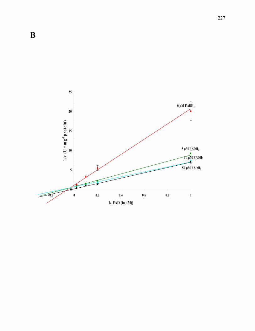
227
B
0 µM FADH2
5 µM FADH2
10 µM FADH2
50 µM FADH2
0
5
10
15
20
25
-0.2 0 0.2 0.4 0.6 0.8 1
1/[FAD (in µM)]
1/v
(U •
mg-1
prot
ein)

228
C
0 nM Fe2+ sulfate
0.05 nM Fe2+ sulfate
0.5 nM Fe2+ sulfate
1 nM Fe2+ sulfate
-0.3
-0.1
0.1
0.3
0.5
0.7
0.9
-100 -80 -60 -40 -20 0 20 40 60 80 1001/[Fe3+ citrate (in nM)]
1/v
(U •
mg-1
prot
ein)

229
Fig. 7.5. The kinetic scheme for a reversible enzyme inhibitor (Segel 1975). Modified to show
the possible binding scheme for substrate [Fe(III)] and product [Fe(II)] to NQO1.

230
NQO1 + Fe3+ NQO1-Fe3+ NQO1 + Fe2+ + + Fe2+ Fe2+ NQO1-Fe2+ + Fe3+ NQO1-Fe2+ - Fe3+
KI KI'

231
Fig. 7.6. The combined data from two (2) NQO1 complexes showing the superposition of
cofactor (FAD), inhibitor (Cibacron blue), and substrate (duroquinone). Complex I contained
NQO1 with bound FAD, Cibacron blue, and duroquinone. Complex II contained NQO1 with
bound FAD and NADP+. The information from complexes I and II was then combined. FAD is
bound in the same position in both complexes. NADP+ (carbon, gray; oxygen, red; nitrogen,
blue; phosphorous, yellow) is in the position found in complex II. Duroquinone (green) is in the
position found in complex I; it fully overlaps the nicotinamide ring of NADP+ in complex II.
Cibacron blue (blue) is in the position found in complex I. Three of its four rings overlap the
position of the ADP of NADP+ found in complex II. Figure from Li et al. 1995.

232

233
Fig. 7.7. The proposed mechanism of quinone reduction by NQO1. (A) The binding of NADP+
to NQO1. (B) The binding of substrate (duroquinone) to NQO1. Both NADP+ and duroquinone
cannot simultaneously occupy the site. The quinone is in an optimal position to receive a hydride
from the FADH2 (FAD represented in schematic). Figure from Li et al. 1995.

234
A

235
B
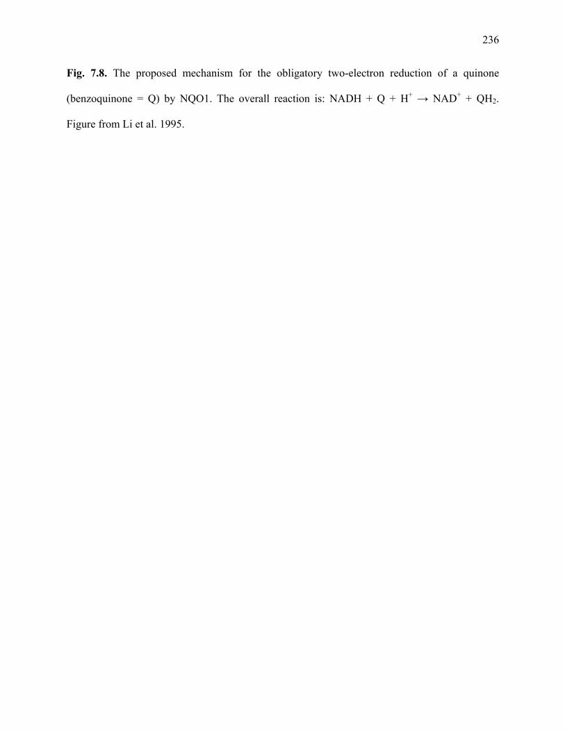
236
Fig. 7.8. The proposed mechanism for the obligatory two-electron reduction of a quinone
(benzoquinone = Q) by NQO1. The overall reaction is: NADH + Q + H+ → NAD+ + QH2.
Figure from Li et al. 1995.
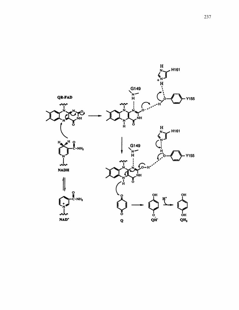
237

238
Fig. 7.9. Proposed mechanism of Fe3+ reduction by the NQO1. (A) 1. Fe3+ is reduced via the
oxidation of NADH with the enzyme bound FAD (yellow) serving as cofactor, i.e., electron
mediator. 2. Exogenous FAD increases enzymatic activity (indicated by thicker arrows) and
possibly occupies a secondary site on the NQO1. (B) Alternatively, exogenous FAD increases
activity because the NQO1 additionally functions as a flavin reductase. The exogenous FAD is
reduced by the NQO1. The reduced FAD in turn reduces the Fe3+.

239
A1
Fe2+
Fe3+
NAD+
NADH
NQO1
FADH2
FAD
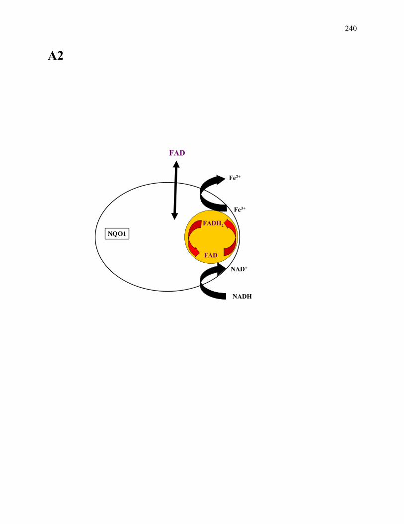
240
A2
Fe2+
Fe3+
NAD+
NADH
FADH2
FAD
FAD
NQO1

241
B
Fe2+
Fe3+
NAD+
NADH
NQO1
FADH2
FAD
FADH2
FAD
FADH2Fe3+
Fe2+ FAD
FAD

242
CONCLUSION
This dissertation has 1) provided a detailed summary of the ‘Firmicutes’ in terms of a broad
genomic overview of endosporulation in the lineage and a more focused exploration of the
phylogeny using a few specific taxa (i.e., iron-reducing species, the Thermoanaerobacterium, the
Thermoanaerobacter, and Caldicellulosiruptor acetigenus) as examples, and 2) described and
provided novel biochemical characterizations of enzymes that are possibly applicable throughout
the ‘Firmicutes’ and beyond.
Endosporulation is a unique characteristic of the ‘Firmicutes’ but is not restricted to, or a
shared trait of, particular lineages within the ‘Firmicutes’. The study described here even
indicates the genes for endosporulation may not be conserved among distinct phylogenetic
lineages. This fact is suggestive of convergent evolution for the development of the endospore-
forming phenotype, i.e., the ability to produce endospores arose independently multiple times.
However, the complexity of the process, i.e., over 150 genes and gene products are involved,
allows for other possible conclusions. It is likely the ability to identify all the genes responsible
for endosporulation from whole genome sequences correctly is currently lacking, or the current
system of phylogeny in use may not be making the correct assumptions about true lineage
determinations for the non-sporulating genera, also lacking B. subtilis-related sporulation genes.
Based on the work presented in this dissertation, it is apparent that many questions about the
phylogenetic status of the ‘Firmicutes’ have yet to be answered.
Apart from this global overview of the current status of the ‘Firmicutes’ lineage, work
has also been presented on the purification and characterization of a soluble oxidoreductase from

243
Carboxydothermus ferrireducens (the CFOR). The CFOR is likely involved in redox reactions;
possibly having a functional role in quinone reduction due to its high activities with quinones,
and due to the fact that the enzyme averts the production of the semiquinone radical by
performing a two electron reduction step to produce an unreactive hydroquinone species. This
activity is similar to the here reported novel iron reduction activity of the human NAD(P)H:
quinone oxidoreductase, a well-described and characterized enzyme known to be highly
promiscuous in terms of its substrate specificity.
Collectively, this dissertation serves as glimpse into the diversity of a lineage containing
members with numerous distinctive physiologies and properties. Presently it is unknown how
many, as of yet, undescribed taxa belong to the ‘Firmicutes’. With the notion that only about 1%
of the estimated microorganisms have been isolated and described, the elucidation of possible
novel lineages within the ‘Firmicutes’ may help to clarify the systematics of this group.

244
REFERENCES
Abhilashkumar, R., S. Mohan, K. Jayakumar, and R. K. Raj. 2001. Functional importance of the different ubiquinones in the filarial parasite Setaria digitati. Biochem. Biophys. Res. Comm. 283:938–942. Ackerley, D. F., C. F. Gonzalez, C. H. Park, R. Blake, M. Keyhan, and A. Matin. 2004. Chromate-reducing properties of soluble flavoproteins from Pseudomonas putida and Escherichia coli. Appl. Environ. Microbiol. 70:873-882. Adeney, W. E. 1894. On the reduction of manganese peroxide in sewage. Sci. Proc. R. Dublin Soc. 8:247-251. Aduseopoku, J., and W. J. Mitchell. 1988. Diauxic growth of Clostridium thermosaccharolyticum on glucose and xylose. FEMS Microbiol. Lett. 50:45-49. Afkar, E., and Y. Fukumori. 1999. Purification and characterization of triheme cytochrome c7 from the metal-reducing bacterium, Geobacter metallireducens. FEMS Microbiol. Lett. 175:205-210.
Ahring, B. K., K. Jensen, P. Nielsen, A. B. Bjerre, and A. S. Schmidt. 1996. Pretreatment of wheat straw and conversion of xylose and xylan to ethanol by thermophilic anaerobic bacteria. Bioresource Technol. 58:107-113.
Ahring, B. K., D. Licht, A. S. Schmidt, P. Sommer, and A. B. Thomsen. 1999. Production of ethanol from wet oxidised wheat straw by Thermoanaerobacter mathranii. Bioresource Technol. 68:3-9.
Aiyar, J., S. de Flora, and K. E. Wetterhahn. 1992. Reduction of chromium(VI) to chromium(V) by rat liver cytosolic and microsomal fractions: is DT-diaphorase involved? Carcinogenesis 13:1159–1166.
Aleshin, A. E., P. H. Feng, R. B. Honzatko, and P. J. Reilly. 2003. Crystal structure and evolution of a prokaryotic glucoamylase. J. Mol. Biol. 327:61-73.
Alister, A., E. Herlitz, R. Borquez, and M. Roeckel. 1990. Production of thermostable beta-galactosidase with Thermoanaerobacter ethanolicus. Annals New York Aca. Sci. 613:605-609.
Allison, L. E., and G. D. Scarseth. 1942. A biological reduction method for removing free iron oxides from soils and colloidal clays. J. Am. Soc. Agron. 34:616-623.

245
Allmaier, G., C. Schaffer, P. Messner, U. Rapp, and F. J. Mayer-Posner. 1995. Accurate determination of the molecular weight of the major surface layer protein isolated from Clostridium thermosaccharolyticum by time-of-flight mass spectrometry. J. Bacteriol. 177:1402-1404. Altaras, N. E., M. R. Etzel, and D. C. Cameron. 2001. Conversion of sugars to 1,2-propanediol by Thermoanaerobacterium thermosaccharolyticum HG-8. Biotechnol. Prog. 17:52-56. Altman, E., J. R. Brisson, P. Messner, and U. B. Sleytr. 1990. Chemical characterization of the regularly arranged surface layer glycoprotein of Clostridium thermosaccharolyticum D120-70. Eur. J. Biochem. 188:73-82. Altman, E., C. Schaffer, J. R. Brisson, and P. Messner. 1995. Characterization of the glycan structure of a major glycopeptide from the surface layer glycoprotein of Clostridium thermosaccharolyticum E207-71. Eur. J. Biochem. 229:308-315. Altman, E., C. Schaffer, J. R. Brisson, and P. Messner. 1996. Isolation and characterization of an amino sugar-rich glycopeptide from the surface layer glycoprotein of Thermoanaerobacterium thermosaccharolyticum E207-71. Carbohydr. Res. 295:245-253. Altschul, S. F., W. Gish, W. Miller, E. W. Myers, and D. J. Lipman. 1990. Basic local alignment search tool. J. Mol. Biol. 215:403–410. Amann, G., K. O. Stetter, E. Llobet-Brossa, R. Amann, and J. Anton. 2000. Direct proof for the presence and expression of two 5% different 16S rRNA genes in individual cells of Haloarcula marismortui. Extremophiles 4:373–376. Anderson, G. J., E. Lesuisse, A. Dancis, D. G. Roman, P. Labbe, and R. D. Klausner. 1992. Ferric iron reduction and iron assimilation in Saccharomyces cerevisiae. J. Inorg. Biochem. 47:249-255. Andrews, N. C., M. D. Fleming, and H. Gunshin. 1999. Iron transport across biological membranes. Nutr. Rev. 57:114-123.
Angelidaki, I., S. P. Petersen, and B. K. Ahring. 1990. Effects of lipids on thermophilic anaerobic digestion and reduction of lipid inhibition upon addition of bentonite. Appl. Microbiol. Biotechnol. 33:469–472.
Angert, E. R., A. E. Brooks, and N. R. Pace. 1996. Phylogenetic analysis of Metabacterium polyspora: clues to the evolutionary origin of daughter cell production in Epulopiscium species, the largest bacteria. J. Bacteriol. 178:1451–1456.
Antranikian, G. 1989. The formation of extracellular, thermoactive amylase and pullulanase in batch culture by Thermoanaerobacter finni. Appl. Biochem. Biotechnol. 20-21:267-279.
246
Antranikian, G. 1990. Physiology and enzymology of thermophilic anaerobic bacteria degrading starch. FEMS Microbiol. Rev. 75:201-208. Antranikian, G., C. Herzberg, F. Mayer, and G. Gottschalk. 1987a. Changes in the cell envelope structure of Clostridium sp. strain EM1 during massive production of α-amylase and pullulanase. FEMS Microbiol. Lett. 41:193-197.
Antranikian, G., P. Zablowski, and G. Gottschalk. 1987b. Conditions for the overproduction and excretion of thermostable α-amylase and pullulanase from Clostridium thermohydrosulfuricum DSM 567. Appl. Microbiol. Biotechnol. 27:75-81.
Archibald, F. 1986. Manganese: its acquisition by and function in the lactic acid bacteria. Crit. Rev. Microbiol. 13:63-109. Armand, S., C. Vieille, C. Gey, A. Heyraud, J. G. Zeikus, and B. Henrissat. 1996. Stereochemical course and reaction products of the action of beta-xylosidase from Thermoanaerobacterium saccharolyticum strain B6A-RI. Eur. J. Biochem. 236:706-713.
Arni, R. K., L. Watanabe, M. Fontes, D. S. Burdette, and J. G. Zeikus. 1996. Crystallization of the secondary alcohol dehydrogenase from Thermoanaerobacter ethanolicus 39E. Prot. Peptide Lett. 3:423-426.
Arnold, R. G., T. J. DiChristina, and M. R. Hoffman. 1988. Reductive dissolution of Fe(III) oxides by Pseudomonas sp. 200. Biotechnol. Bioeng. 32:1081-1096. Ash, C., J. A. E. Farrow, S. Wallbanks, and M. D. Collins. 1991. Phylogenetic heterogeneity of the genus Bacillus revealed by comparative analysis of small-subunit-ribosomal RNA sequences. Lett. Appl. Microbiol. 13:202-206. Askwith, C., D. Eide, A. Van Ho, P. S. Bernard, L. Li, S. Davis-Kaplan, D. M. Sipe, and J. Kaplan. 1994. The FET3 gene of S. cerevisiae encodes a multicopper oxidase required for ferrous iron uptake. Cell 76:403-410. Askwith, C. C., D. de Silva, and J. Kaplan. 1996. Molecular biology of iron acquisition in Saccharomyces cerevisiae. Mol. Microbiol. 20:27-34. Assfalg, M., I. Bertini, M. Bruschi, C. Michel, and P. Turano. 2002. The metal reductase activity of some multiheme cytochromes c: NMR structural characterization of the reduction of chromium(VI) to chromium(III) by cytochrome c(7). Proc. Natl. Acad. Sci. USA. 99:9750-9754. Aubert, C., E. Lojou, P. Bianco, M. Rousset, M. C. Durand, M. Bruschi, and A. Dolla. 1998. The Desulfuromonas acetoxidans triheme cytochrome c7 produced in Desulfovibrio desulfuricans retains its metal reductase activity. Appl. Environ. Microbiol. 64:1308-1312.

247
Bae, W. C., H. K. Lee, Y. C. Choe, D. J. Jahng, S. H. Lee, S. J. Kim, J. H. Lee, and B. C. Jeong. 2005. Purification and characterization of NADPH-dependent Cr(VI) reductase from Escherichia coli ATCC 33456. J. Microbiol. 43:21-27. Balashova, V. V., and G. A. Zavarzin. 1980. Anaerobic reduction of ferric iron by hydrogen bacteria. Microbiology 48:635-639. Balkwill, D. L., D. Maratea, and R. P. Blakemore. 1980. Ultrastructure of a magnetotactic spirillum. J. Bacteriol. 141:1399-1408. Balkwill, D. L., T. L. Kieft, T. Tsukuda, H. M. Kostandarithes, T. C. Onstott, S. Macnaughton, J. Bownas, and J. K. Fredrickson. 2004. Identification of iron-reducing Thermus strains as Thermus scotoductus. Extremophiles 8:37-44. Barton, L. L., R. M. Plunkett, and B. M. Thomson. 2003. Reduction of metals and nonessential elements by anaerobes. In: Ljungdahl, L., M. Adams, L. Barton, J. Ferry, and M. Johnson (eds.), Biochemistry and physiology of anaerobic bacteria, Springer-Verlag, New York, NY, pp 220-234. Baskaran, S., D. A. L. Hogsett, and L. R. Lynd. 1995a. Optimization of a chemically-defined, minimal medium for Clostridium thermosaccharolyticum. Appl. Biochem. Biotechnol. 51-52:399-411. Baskaran, S., H. J. Ahn, and L. R. Lynd. 1995b. Investigation of the ethanol tolerance of Clostridium thermosaccharolyticum in continuous culture. Biotechnol. Prog. 11:276-281. Beall, H. D., R. T. Mulcahy, D. Siegel, R. D. Traver, N. W. Gibson, and D. Ross. 1994. Metabolism of bioreductive antitumor compounds by purified rat and human DT-diaphorases. Cancer Res. 54:3196-3201. Belogurova, N. G., T. P. Mosolova, S. V. Kalyuzhnyy, and S. D. Varfolomeyev. 1991. Kinetics of growth and metabolism of Clostridium thermosaccharolyticum culture. Isolation and characteristics of its plasmids. Appl. Biochem. Biotechnol. 27:1-8. Belogurova, N. G., E. P. Del'ver, S. V. Kaliuzhnyi, S. D. Varfolomeev, O. V. Rodzevich, and A. A. Belogurov. 1992. Nucleotide sequence of the pNB2 plasmid from thermophilic bacteria Clostridium thermosaccharolyticum. Mol. Biol. (Mosk). 26:173-178. Belogurova, N. G., E. E. Davydova, M. V. Sobennikova, S. D. Varfolomeev, E. P. Del'ver, and A. A. Belogurov. 2002. Replication protein RepN encoded by the RC plasmid of thermophilic bacterium Thermoanaerobacterium saccharolyticum: mutational analysis and deletion mapping of domains responsible for its lethal effect. Mol. Biol. (Mosk). 36:106-113.
Ben-Bassat, A., and J. G. Zeikus. 1981. Thermobacteroides acetoethylicus gen. nov. sp. nov., a new chemoorganotrophic, anaerobic, thermophilic bacterium. Arch. Microbiol. 128:365-370.
248
Bentley, S. D., K. F. Chater, A. M. Cerdeno-Tarraga, G. L. Challis, N. R. Thomson, K. D. James, D. E. Harris, M. A. Quail, H. Kieser, D. Harper, A. Bateman, S. Brown, G. Chandra, C. W. Chen, M. Collins, A. Cronin, A. Fraser, A. Goble, J. Hidalgo, T. Hornsby, S. Howarth, C. H. Huang, T. Kieser, L. Larke, L. Murphy, K. Oliver, S. O’Neil, E. Rabbinowitsch, M. A. Rajandream, K. Rutherford, S. Rutter, K. Seeger, D. Saunders, S. Sharp, R. Squares, S. Squares, K. Taylor, T. Warren, A. Wietzorrek, J. Woodward, B. G. Barrell, J. Parkhill, and D. A. Hopwood. 2002. Complete genome sequence of the model actinomycete Streptomyces coelicolor A3(2). Nature 417:141-147.
Beyer, R. E., J. Segura-Aguilar, S. Di Bernardo, M. Cavazzoni, R. Fato, D. Fiorentini, M. C. Galli, M. Setti , L. Landi, and G. Lenaz. 1996. The role of DT-diaphorase in the maintenance of the reduced antioxidant form of coenzyme Q in membrane systems. Proc. Natl. Acad. Sci. USA 93:2528–2532. Bligh, E. G., and W. J. Dyer. 1959. A rapid method of total lipid extraction and purification. Can. J. Biochem. Physiol. 37:911–917. Bochner, B. R., and B. N. Ames. 1982. Complete analysis of cellular nucleotides by two-dimensional thin layer chromatography. J. Biol. Chem. 257:9759-9769. Bock, K., J. Schuster-Kolbe, E. Altman, G. Allmaier, B. Stahl, R. Christian, U. B. Sleytr, and P. Messner. 1994. Primary structure of the O-glycosidically linked glycan chain of the crystalline surface layer glycoprotein of Thermoanaerobacter thermohydrosulfuricus L111-69. Galactosyl tyrosine as a novel linkage unit. J. Biol. Chem. 269:7137-7144. Bogdahn, M., and D. Kleiner. 1986. N2 fixation and NH4+ assimilation in the thermophilic anaerobes Clostridium thermosaccharolyticum and Clostridium thermoautotrophicum. Arch. Microbiol. 144:102-104.
Bogin, O., M. Peretz, and Y. Burstein. 1997. Thermoanaerobacter brockii alcohol dehydrogenase: characterization of the active site metal and its ligand amino acids. Protein Sci. 6:450-458.
Bogin, O., M. Peretz, and Y. Burstein. 1998a. Probing structural elements of thermal stability in bacterial oligomeric alcohol dehydrogenases. I. Construction and characterization of chimeras consisting of secondary ADHs from Thermoanaerobacter brockii and Clostridium beijerinckii. Lett. Peptide Sci. 5:399-408.
Bogin, O., M. Peretz, Y. Hacham, Y. Korkhin, F. Frolow, A. J. Kalb, and Y. Burstein. 1998b. Enhanced thermal stability of Clostridium beijerinckii alcohol dehydrogenase after strategic substitution of amino acid residues with prolines from the homologous thermophilic Thermoanaerobacter brockii alcohol dehydrogenase. Prot. Sci. 7:1156-1163.
Bonch-Osmolovskaya, E. A., M. L. Miroshnichenko, N. A. Chernykh, N. A. Kostrikina, E. V. Pikuta, and F. A. Rainey. 1997. Reduction of elemental sulfur by moderately thermophilic

249
organotrophic bacteria and the description of Thermoanaerobacter sulfurophilus sp. nov. Mikrobiologiya 66:581-587 [(in Russian). English tranlation: Microbiology 66:483-489.]
Bonch-Osmolovskaya, E. A., M. L. Miroshnichenko, N. A. Chernykh, N. A. Kostrikina, E. V. Pikuta, and F. A. Rainey. 1998. Validation list no. 66. Int. J. Syst. Bacteriol. 48:631-632.
Boone, D. R., Y. Liu, Z. Zhao, D. L. Balkwill, G. R. Drake, T. O. Stevens, and H. C. Aldrich. 1995. Bacillus infernus sp. nov., an Fe(III)- and Mn(IV)-reducing anaerobe from the deep terrestrial subsurface. Int. J. Syst. Bacteriol. 45:441-448. Bork, P., C. Ouzounis, G. Casari, R. Schneider, C. Sander, M. Dolan, W. Gilbert, and P. M. Gillevet. 1995. Exploring the Mycoplasma capricolum genome: a minimal cell reveals its physiology. Mol. Microbiol. 16:955–967. Bowman, J. P., S. A. McCammon, D. S. Nichols, J. H. Skerratt, S. M. Rea, P. D. Nichols, and T. A. McMeekin. 1997. Shewanella gelidimarina sp. nov. and Shewanella frigidimarina sp. nov., novel Antarctic species with the ability to produce eicosapentaenoic acid (20:5 omega 3) and grow anaerobically by dissimilatory Fe(III) reduction. Int. J. Syst. Bacteriol. 47:1040-1047. Bradbeer, C. 1993. The proton motive force drives the outer membrane transport of cobalamin in Escherichia coli. J. Bacteriol. 175:3146-3150. Brechtel, E., and H. Bahl. 1999. In Thermoanaerobacterium thermosulfurigenes EM1 S-layer homology domains do not attach to peptidoglycan. J. Bacteriol. 181:5017-5023. Brechtel, E., M. Matuschek, A. Hellberg, E. M. Egelseer, R. Schmid, and H. Bahl. 1999. Cell wall of Thermoanaerobacterium thermosulfurigenes EM1: isolation of its components and attachment of the xylanase XynA. Arch. Microbiol. 171:159-165.
Bredholt, S., J. Sonne-Hansen, P. Nielsen, I. M. Mathrani, and B. K. Ahring. 1999. Caldicellulosiruptor kristjanssonii sp. nov., a cellulolytic, extremely thermophilic, anaerobic bacterium. Int. J. Syst. Bacteriol. 49:991-996.
Breves, R., K. Bronnenmeier, N. Wild, F. Lottspeich, W. L. Staudenbauer, and J. Hoffmeister. 1997. Genes encoding two different beta-glucosidases of Thermoanaerobacter brockii are clustered in a common operon. Appl. Environ. Microbiol. 63:3902-3910.
Brewer, P. G., and D. W. Spencer. 1971. Colorimetric determination of manganese in anoxic waters. Limnol. Oceanogr. 16:107-112. Brickman, T. J., and M. A. McIntosh. 1992. Overexpression and purification of ferric enterobactin esterase from Escherichia coli. Demonstration of enzymatic hydrolysis of enterobactin and its iron complex. J. Biol. Chem. 267:12350-12355.

250
Bridge, T. A. M., and D. B. Johnson. 1998. Reduction of soluble iron and reductive dissolution of ferric iron-containing minerals by moderately thermophilic iron-oxidizing bacteria. Appl. Environ. Microbiol. 64:2181-2186.
Brill, J. A., and J. Wiegel. 1997. Differentiation between spore-forming and asporogenic bacteria using a PCR and Southern hybridization based method. J. Microbiol. Methods 31:29-36.
Brock, T. D. 1967. Life at high temperatures. Science 158:1012-1019.
Brock, T. D., and H. Freeze. 1967. Thermus aquaticus gen. n. and sp. n., a non-sporulating extreme thermophile. J. Bacteriol. 98:289-297.
Brock, T. D. 1978. Thermophilic microorganisms and life at high temperatures. New York: Springer-Verlag.
Bronnenmeier, K., H. Meissner, S. Stocker, and W. L. Staudenbauer. 1995. α-D-Glucuronidases from the xylanolytic thermophiles Clostridium stercorarium and Thermoanaerobacterium saccharolyticum. Microbiology 141:2033-2040.
Bryant, F., and L. G. Ljungdahl. 1981. Characterization of an alcohol-dehydrogenase from Thermoanaerobacter ethanolicus active with ethanol and secondary alcohols. Biophy. Res. Comm. 100:793-799.
Bryant, F. O., J. Wiegel, and L. G. Ljungdahl. 1988. Purification and properties of primary and secondary alcohol-dehydrogenases from Thermoanaerobacter ethanolicus. Appl. Environ. Microbiol. 54:460-465.
Bryant, F. O., J. Wiegel, and L. G. Ljungdahl. 1992. Comparisons of alcohol dehydrogenases from wild-type and mutant strain, JW200 Fe4, of Thermoanaerobacter ethanolicus. Appl. Microbiol. Biotechnol. 37:490-495. Burchhardt, G., A. Wienecke, and H. Bahl. 1991. Isolation of the pullulanase gene from Clostridium thermosulfurogenes (DSM 3896) and its expression in Escherichia coli. Curr. Microbiol. 22:91-95.
Burdette, D., and J. G. Zeikus. 1994a. Purification of acetaldehyde dehydrogenase and alcohol dehydrogenases from Thermoanaerobacter ethanolicus 39E and characterization of the secondary-alcohol dehydrogenase (2 degrees Adh) as a bifunctional alcohol dehydrogenase--acetyl-CoA reductive thioesterase. Biochem. J. 302:163-170.
Burdette, D. S., and J. G. Zeikus. 1994b. Secondary alcohol-dehydrogenase from Thermoanaerobacter ethanolicus 39E-a model enzyme for the study of protein thermophilicity and catalytic specificity. Abstr. Papers Amer. Chem. Soc. 207:159-BIOT Part 1.
Burdette, D. S., V. M. Tchernajenko, and J. G. Zeikus. 1995. Cloning, mutagenesis, and physical biochemical-characterization of the secondary alcohol-dehydrogenases from

251
Thermoanaerobacter ethanolicus- a model enzyme for the study of protein thermophilicity. Abstr. Papers Amer. Chem. Soc 209:143-BIOT Part 1
Burdette, D. S., C. Vieille, and J. G. Zeikus. 1996. Cloning and expression of the gene encoding the Thermoanaerobacter ethanolicus 39E secondary-alcohol dehydrogenase and biochemical characterization of the enzyme. Biochem. J. 316:115-122.
Burdette, D. S., F. Secundo, R. S. Phillips, J. Dong, R. A. Scott, and J. G. Zeikus. 1997. Biophysical and mutagenic analysis of Thermoanaerobacter ethanolicus secondary-alcohol dehydrogenase activity and specificity. Biochem. J. 326:717-724.
Burdette, D. S., V. V. Tchernajencko, and J. G. Zeikus. 2000. Effect of thermal and chemical denaturants on Thermoanaerobacter ethanolicus secondary-alcohol dehydrogenase stability and activity. Enzyme Microb. Technol. 27:11-18.
Burdette, D. S., S. H. Jung, G. J. Shen, R. I. Hollingsworth, and J. G. Zeikus. 2002. Physiological function of alcohol dehydrogenases and long-chain (C-30) fatty acids in alcohol tolerance of Thermoanaerobacter ethanolicus. Appl. Environ. Microbiol. 68:1914-1918.
Butler, J. E., F. Kaufmann, M. V. Coppi, C. Nunez, and D. R. Lovley. 2004. MacA, a diheme c-type cytochrome involved in Fe(III) reduction by Geobacter sulfurreducens. J. Bacteriol. 186:4042-4045. Byrer, D. E., F. A. Rainey, and J. Wiegel. 2000. Novel strains of Moorella thermoacetica form unusually heat-resistant spores. Arch. Microbiol. 174:334-339. Caccavo, F., R. P. Blakemore, and D. R. Lovley. 1992. A hydrogen-oxidizing, Fe(III)-reducing microorganism from the Great Bay estuary, New Hampshire. Appl. Environ. Microbiol. 58:3211-3216. Caccavo, F., D. J. Lonergan, D. R. Lovley, M. Davis, J. F. Stolz, and M. J. McInerney. 1994. Geobacter sulfurreducens sp. nov., a hydrogen- and acetate-oxidizing dissimilatory metal-reducing microorganism. Appl. Environ. Microbiol. 60:3752-3759. Caccavo, F., B. Frǿlund, F. van O Kloeke, and P. H. Nielsen. 1996a. Deflocculation of activated sludge by the dissimilatory Fe(III)-reducing bacterium Shewanella alga strain BrY. Appl. Environ. Microbiol. 62:1487-1490. Caccavo, F, J. D. Coates, R. A. Rossello-Mora, W. Ludwig, K. H. Schleifer, D. R. Lovley, and M. J. McInerney. 1996b. Geovibrio ferrireducens, a phylogenetically distinct dissimilatory Fe(III)-reducing bacterium. Arch. Microbiol. 165:370-376. Caccavo, F., P. C. Schamberger, K. Keiding, and P. H. Nielsen. 1997. Role of hydrophobicity in adhesion of the dissimilatory Fe(III)-reducing bacterium Shewanella alga to amorphous Fe(III) oxide. Appl. Environ. Microbiol. 63:3837-3843.

252
Cameron, D. C., and C. L. Cooney. 1986. A novel fermentation – the production of R(-)-1,2- propanediol and acetol by by Clostrdium thermosaccharolyticum. Biotechnology 4:651-654. Cameron, D. C., N. E. Altaras, M. L. Hoffman, and A. J. Shaw. 1998. Metabolic engineering of propanediol pathways. Biotechnol. Prog. 14:116-125. Campbell, M. F., and Z. J. Ordal. 1968. Inhibition of sporulation of Clostridium thermosaccharolyticum. Appl. Microbiol. 16:1949-1951. Campbell, L. L., and B. Pace. 1968. Physiology of growth at high temperatures. J. Appl. Bacteriol. 31:24-35. Canganella, F, and J. Wiegel. 1993. The potential of thermophilic clostridia in biotechnology. In: Woods (ed.), The clostridia and biotechnology, Butterworths Publ., Stoneham, MA, pp 391-429. Cann, I. K. O., S. Kocherginskaya, M. R. King, B. A. White, and R. I. Mackie. 1999. Molecular cloning, sequencing, and expression of a novel multidomain mannanase gene from Thermoanaerobacterium polysaccharolyticum. J. Bacteriol. 181:1643-1651. Cann, I. K. O., P. G. Stroot, K. R. Mackie, B. A. White, and R. I. Mackie. 2001. Characterization of two novel saccharolytic, anaerobic thermophiles, Thermoanaerobacterium polysaccharolyticum sp. nov. and Thermoanaerobacterium zeae sp. nov., and emendation of the genus Thermoanaerobacterium. Int. J. Syst. Evol. Microbiol. 51:293-302.
Carreira, L. H., J. Wiegel, and L. G. Ljungdahl. 1983. Production of ethanol from biopolymers by anaerobic, thermophilic, and extreme thermophilic bacteria. I. Regulation of carbohydrate utilization in mutants of Thermoanaerobacter ethanolicus. Biotechnol. Bioeng. Symp. 13:183-191.
Cary, E. E. 1982. In: Biological and environmental aspects of chromium, Elsevier: Amsterdam, pp 49-64. Cavelier, G., and L. M. Amzel. 2001. Mechanism of NAD(P)H:quinone reductase: Ab initio studies of reduced flavin. Proteins 43:420-432.
Cayol J. L., B. Ollivier, B. K. C. Patel, G. Ravot, M. Magot, E. Ageron, P. A. D. Grimont, and J. L. Garcia. 1995. Description of Thermoanaerobacter brockii subsp. lactiethylicus subsp. nov., isolated from a deep subsurface French oil well, a proposal to reclassify Thermoanaerobacter finnii as Thermoanaerobacter brockii subsp. finnii comb. nov., and an emended description of Thermoanaerobacter brockii. Int. J. Syst. Bacteriol. 45:783-789.
Cejka, Z., and W. Baumeister. 1987. 3-Dimensional structure of the surface protein of Clostridium thermosaccharolyticum. FEMS Microbiol. Lett. 44:13-18.
253
Champine, J. E., B. Underhill, J. M. Johnston, W. W. Lilly, and S. Goodwin. 2000. Electron transfer in the dissimilatory iron-reducing bacterium Geobacter metallireducens. Anaerobe 6:187-196.
Childers, S. E., and D. R. Lovley. 2001. Differences in Fe(III) reduction in the hyperthermophilic archaeon, Pyrobaculum islandicum, versus mesophilic Fe(III)-reducing bacteria. FEMS Microbiol. Lett. 195:253-258. Childers, S. E., S. Ciufo, and D. R. Lovley. 2002. Geobacter metallireducens accesses insoluble Fe(III) oxide by chemotaxis. Nature 416:767-769. Chiu, H., E. Johnson, I. Schroder, and D. C. Rees. 2001. Crystal structures of a novel ferric reductase from the hyperthermophilic archaeon Archaeoglobus fulgidus and its complex with NADP+. Structure 9:311-319.
Christian, R., P. Messner, C. Weiner, U. B. Sleytr, and G. Schulz. 1988. Structure of a glycan from the surface-layer glycoprotein of Clostridium thermohydrosulfuricum strain L111-69. Carbohydr. Res. 176:160-163.
Clarke, F. M. 1949. Some nitrogen requirements of Clostridium thermosaccharolyticum. J. Bacteriol. 57:465-471. Cleland, W. W. 1977. Determining the chemical mechanisms of enzyme-catalyzed reactions by kinetic studies. Adv. Enzymol. Relat. Areas Mol. Biol. 45:273-387. Coates, J. D., E. J. Phillips, D. J. Lonergan, H. Jenter, and D. R. Lovley. 1996. Isolation of Geobacter species from diverse sedimentary environments. Appl. Environ. Microbiol. 62:1531-1536. Coates, J. D., D. J. Ellis, E. Roden, K. Gaw, E. L. Blunt-Harris, and D. R. Lovley. 1998. Recovery of humics-reducing bacteria from a diversity of sedimentary environments. Appl. Environ. Microbiol. 64:1504-1509. Coates, J. D., D. J. Ellis, C. V. Gaw, and D. R. Lovley. 1999. Geothrix fermentens gen. nov. sp. nov., an acetate-oxidizing Fe(III) reducer capable of growth via fermentation. Int. J. Syst. Bacteriol. 49:1615-1622. Coates, J. D., V. K. Bhupathiraju, L. A. Achenbach, M. J. McInerney, and D. R. Lovley. 2001. Geobacter hydrogenophilus, Geobacter chapellei and Geobacter grbiciae, three new, strictly anaerobic, dissimilatory Fe(III)-reducers. Int. J. Syst. Bacteriol. 51:581-588. Collins, M. D., P. A. Lawson, A. Willems, J. J. Cordoba, J. Fernandez-Garayzabal, P. Garcia, J. Cai, H. Hippe, and J. A. E. Farrow. 1994. The phylogeny of the genus Clostridium: proposal of five new genera and eleven new species combinations. Int. J. Syst. Bacteriol. 44:812-826.

254
Cook, G. M. 2000. The intracellular pH of the thermophilic bacterium Thermoanaerobacter wiegelii during growth and production of fermentation acids. Extremophiles 4:279-284. Cook, G. M., P. H. Janssen, and H. W. Morgan. 1991. Endospore formatiom by Thermoanaerobium brockii HTD4. Syst. Appl. Microbiol. 14:240-244.
Cook, G. M., P. H. Janssen, and H. W. Morgan. 1993. Uncoupler-resistant glucose-uptake by the thermophilic glycolytic anaerobe Thermoanaerobacter thermohydrosulfuricus (Clostridium thermohydrosulfuricm). Appl. Environ. Microbiol. 59:2984-2990.
Cook, G. M., P. H. Janssen, J. B. Russell, and H. W. Morgan. 1994. Dual mechanisms of xylose uptake in the thermophilic bacterium Thermoanaerobacter thermohydrosulfuricus. FEMS Microbiol. Lett. 116:257-262.
Cook, G. M., and H. W. Morgan. 1994. Hyperbolic growth of Thermoanaerobacter thermohydrosulfuricus (Clostridium thermohydrosulfuricm) increases ethanol-production in pH-controlled batch culture. Appl. Microbiol. Biotechnol. 41:84-89.
Cook, G. M., F. A Rainey, B. K. C. Patel, and H. W. Morgan. 1996. Characterization of a new obligately anaerobic thermophile, Thermoanaerobacter wiegelii sp. nov. Int. J. Syst. Bacteriol. 46:123-127. Cox, C. D. 1994. Deferration of laboratory media and assays for ferric and ferrous ions. Methods Enzym. 235:315-328. Cross, T. 1968. Thermophilic actinomycetes. J. Appl. Bacteriol. 31:36-53.
Crowther, R., and U. B. Sleytr. 1977. An analysis of the fine structure of the surface layer from two strains of clostridia, including correction of distorted images. J. Ultrastruct. Res. 58:41.
Cui, K., A. Y. Lu, and C. S. Yang. 1995. Subunit functional studies of NAD(P)H:quinone oxidoreductase with a heterodimer approach. Proc. Natl. Acad. Sci. USA 92:1043-1047. Dailey, H. A., and J. Lascelles. 1977. Reduction of iron and synthesis of protoheme by Spirillum itersonii and other organisms. J. Bacteriol. 129:815-820. Dale, S. E., A. Doherty-Kirby, G. Lajoie, and D. E. Heinrichs. 2004. Role of siderophore biosynthesis in virulence of Staphylococcus aureus: identification and characterization of genes involved in production of a siderophore. Infect. Immun. 72:29-37. Dancis, A., D. G. Roman, G. J. Anderson, A. G. Hinnebusch, and R. D. Klausner. 1992. Ferric reductase of Saccharomyces cerevisiae: molecular characterization, role in iron uptake, and transcriptional control by iron. Proc. Natl. Acad. Sci. 89:3869-3873. Dancis, A. 1998. Genetic analysis of iron uptake in the yeast Saccharomyces cerevisiae. J. Pediatr. 132:S24-S29.

255
Das, A., and F. Caccavo. 2000. Dissimilatory Fe(III) oxide reduction by Shewanella alga BrY requires adhesion. Curr. Microbiol. 40:344-347. Das, A., and F. Caccavo. 2001. Adhesion of the dissimilatory Fe(III)-reducing bacterium Shewanella alga BrY to crystalline Fe(III) oxides. Curr. Microbiol. 42:151-154.
De Ley, J., H. Cattoir, and A. Reynaerts. 1970. The quantitative measurement of DNA hybridization from renaturation rates. Eur. J. Biochem. 12:133-142.
Delver, E. P., N. G. Belogurova, E. E. Tupikova, S. D. Varfolomeyev, and A. A. Belogurov. 1996. Characterization, sequence and mode of replication of plasmid pNB2 from the thermophilic bacterium Clostridium thermosaccharolyticum. Mol. Gen. Genet. 253:166-172. Desai, S. G., M. L. Guerinot, and L. R. Lynd. 2004. Cloning of L-lactate dehydrogenase and elimination of lactic acid production via gene knockout in Thermoanaerobacterium saccharolyticum JW/SL-YS485. Appl. Microbiol. Biotechnol. 65:600-605. De Silva, D. M., C. C. Askwith, D. Eide, and J. Kaplan. 1995. The FET3 gene product required for high affinity iron transport in yeast is a cell surface ferroxidase. J. Biol. Chem. 270:1098-1101. DiChristina, T. J., C. M. Moore, and C. A. Haller. 2002. Dissimilatory Fe(III) and Mn(IV) reduction by Shewanella putrefaciens requires ferE, a homolog of the pulE (gspE) type II protein secretion gene. J. Bacteriol. 184:142-151. Dicker, E., and A. I. Cederbaum. 1993. Requirement for iron for the production of hydroxyl radicals by rat liver quinone reductase. J. Pharmacol. Exp. Ther. 266:1282-1290. DiDonato, L. N., E. S. Shelobolina, S. S. Sullivan, K. P. Nevin, T. L. Woodard, and D. R. Lovley. 2004. Characterization of a family of small periplasmic c-type cytochromes in Geobacter sulfurreducens and their role in Fe(III) respiration, abstr. K-135, Abstr. 104th Annu. Meet. Am. Soc. Microbiol. American Society for Microbiology, Washington, DC, pp 379. Dix, D. R., J. T. Bridgham, M. A. Broderius, C. A. Byersdorfer, and D. J. Eide. 1994. The FET4 gene encodes the low affinity Fe(II) transport protein of Saccharomyces cerevisiae. J. Biol. Chem. 269:26092-26099. Dobbin, P. S., J. N. Butt, A. K. Powell, G. A. Reid, and D. J. Richardson. 1999. Characterization of a flavocytochrome that is induced during the anaerobic respiration of Fe3+ by Shewanella frigidimarina NCIMB400. Biochem. J. 342:439-448.
Dotzauer, C., M. A. Ehrmann, and R. F. Vogel. 2002. Occurrence and detection of Thermoanaerobacterium and Thermoanaerobacter in canned food. Food Technol. Biotechnol. 40:21-26.
256
Egami, F. 1975. Origin and early evolution of transition element enzymes. J. Biochem. (Tokyo) 77:1165-1169. Ehrlich, H. L. 1963. Bacteriology of manganese nodules.I. Bacterial action on manganese in nodule enrichments. Appl. Microbiol. 11:15-19. Ehrlich, H. L. 1966. Reactions with manganese by bacteria from marine ferromanganese nodules. Dev. Ind. Microbiol. 7:279-286. Ehrlich, H. L. 1970. The microbiology of manganese nodules. U. S. Clearinghouse, Fed. Sci. Tech. Inform., A. D. 1970, no. 716,508. Ehrlich, H. L. 1971. Bacteriology of manganese nodules.V. Effect of hydrostatic pressure on bacterialoxidation of Mn(II) and reduction of MnO2. Appl. Microbiol. 21:306-310. Ehrlich, H. L. 1974. Response of some activities of ferromangenese bacteria to hydrostatic pressure. In: Colwell, R. R., and R. Y. Morita (eds.), Effect of the ocean environment on microbial activities. University Park Press, Baltimore, MD, pp 208-221. Eide, D., and M. L. Guerinot. 1997. Metal ion uptake in eukaryotes. ASM News 63:199-205. Emoto, Y. 1933. Die Mikroorganismem der Thermen. Botan. Mag. (Tokyo) 47:268-295. Engle, M., Y. Li, F. Rainey, S. DeBlois, V. Mai, A. Reichert, F. Mayer, P. Messner, and J. Wiegel. 1996. Thermobrachium celere gen. nov., sp. nov., a rapidly growing thermophilic, alkalitolerant, and proteolytic obligate anaerobe. Int. J. Syst. Bacteriol. 46:1025-1033.
Erbeznik, M., K. A. Dawson, and H. J. Strobel. 1998a. Cloning and characterization of transcription of the xylAB operon in Thermoanaerobacter ethanolicus. J. Bacteriol. 180:1103-1109.
Erbeznik, M., M. Ray, K. A. Dawson, and H. J. Strobel. 1998b. Xylose transport by the anaerobic thermophile Thermoanaerobacter ethanolicus and the characterization of a D-xylose-binding protein. Curr. Microbiol. 37:295-300.
Erbeznik, M., H. J. Strobel, K. A. Dawson, and C. R. Jones. 1998c. The D-xylose binding protein, XylF, from Thermoanaerobacter ethanolicus 39E: cloning, molecular analysis, and expression of the structural gene. J. Bacteriol. 180:3570-3577.
Erbeznik, M., H. J. Strobel, and K. A. Dawson. 2000. Organization and sequence of histidine biosynthesis genes hisH, -A, -F, and -IE in Thermoanaerobacter ethanolicus. Curr. Microbiol. 40:140-142.
Erbeznik, M., S. E. Hudson, A. B. Herrman, and H. J. Strobel. 2004. Molecular analysis of the xylFGH operon, coding for xylose ABC transport, in Thermoanaerobacter ethanolicus. Curr. Microbiol. 48:295-299.

257
Ernster, L. 1958. Diaphorase activities in liver cytoplasmic fractions. Fed. Proc.17:216.
Ernster, L. 1967. DT-diaphorase. Meth. Enzymol. 10:309-317.
Ernster, L. 1987. DT-diaphorase: a historical review. Chemica Scripta 27A:1-13. Ernster, L., and F. Navazio. 1958. Soluble diaphorase in animal tissues. Acta Chem. Scand. 12: 595-602. Ernster, L., L. Danielson, and M. Ljunggren. 1962. DT diaphorase. I. Purification from the soluble fraction of rat-liver cytoplasm, and properties. Biochim. Biophys. Acta 58:171-188. Errington, J. 1993. Bacillus subtilis sporulation: regulation of gene expression and control of morphogenesis. Microbiol. Rev. 57:1-33. Faig, M., M. A. Bianchet, P. Talalay, S. Chen, S. Winski, D. Ross, and L. M. Amzel. 2000. Structures of recombinant human and mouse NAD(P)H:quinone oxidoreductases: species comparison and structural changes with substrate binding and release. Proc. Natl. Acad. Sci. USA 97:3177-3182.
Fardeau, M. L., J. L. Cayol, M. Magot, and B. Ollivier. 1993. H2 oxidation in the presence of thiosulfate, by a Thermoanaerobacter strain isolated from an oil-producing well. FEMS Microbiol. Lett. 113:327-332.
Fardeau, M. L., J. L. Cayol, M. Magot, and B. Ollivier. 1994. Hydrogen oxidation abilities in the presence of thiosulfate as electron-acceptor within the genus Thermoanaerobacter. Curr. Microbiol. 29:269-272.
Fardeau, M. L., C. Faudon, J. L. Cayol, M. Magot, B. K. Patel, and B. Ollivier. 1996. Effect of thiosulphate as electron acceptor on glucose and xylose oxidation by Thermoanaerobacter finnii and a Thermoanaerobacter sp. isolated from oil field water. Res. Microbiol. 147:159-165.
Fardeau, M. L., B. K. C. Patel, M. Magot, and B. Ollivier. 1997. Utilization of serine, leucine, isoleucine, and valine by Thermoanaerobacter brockii in the presence of thiosulfate or Methanobacterium sp. as electron acceptors. Anaerobe 3:405-410.
Fardeau, M. L., M. Magot, B. K. C. Patel, P. Thomas, J. L. Garcia, and B. Ollivier. 2000. Thermoanaerobacter subterraneus sp. nov., a novel thermophile isolated from oilfield water. Int. J. Syst. Evol. Microbiol. 50:2141-2149.
Fardeau, M. L., M. B. Salinas, S. L'Haridon, C. Jeanthon, F. Verhe, J. L. Cayol, B. K. Patel, J. L. Garcia, and B. Ollivier. 2004. Isolation from oil reservoirs of novel thermophilic anaerobes phylogenetically related to Thermoanaerobacter subterraneus: reassignment of T. subterraneus, Thermoanaerobacter yonseiensis, Thermoanaerobacter tengcongensis and Carboxydibrachium pacificum to Caldanaerobacter subterraneus gen. nov., sp. nov., comb. nov. as four novel subspecies. Int. J. Syst. Evol. Microbiol. 54:467-474.
258
Farrow, J. A., C. Ash, S. Wallbanks, and M. D. Collins. 1992. Phylogenetic analysis of the genera Planococcus, Marinococcus and Sporosarcina and their relationships to members of the genus Bacillus. FEMS Microbiol. Lett. 72:167-172.
Farrow, J. A., P. A. Lawson, H. Hippe, U. Gauglitz, and M. D. Collins. 1995. Phylogenetic evidence that the gram-negative nonsporulating bacterium Tissierella (Bacteroides) praeacuta is a member of the Clostridium subphylum of the gram-positive bacteria and description of Tissierella creatinini sp. nov. Int. J. Syst. Bacteriol. 45:436-440.
Faudon, C., M. L. Fardeau, J. Heim, B. Patel, M. Magot, and B. Ollivier. 1995. Peptide and amino-acid oxidation in the presence of thiosulfate by members of the genus Thermoanaerobacter. Curr. Microbiol. 31:152-157.
Felsenstein, J. 1989. PHYLIP – Phylogeny inference package (version 3.2). Cladistics 5:164-166.
Fenchel, T., and T. H. Blackburn. 1979. Bacteria and mineral cycling. Academic Press, Inc. Ltd., London.
Feng, P. H., S. Berensmeier, K. Buchholz, and P. J. Reilly. 2002. Production, purification, and characterization of Thermoanaerobacterium thermosaccharolyticum glucoamylase. Starch-Starke 54:328-337. Fish, W. W. 1988. Rapid colorimetric micromethod for the quantitation of complexed iron in biological samples. Methods Enzymol. 158:357-364. Fokina, N. A., and G. A. Velikodvorskaia. 1997. Cloning and expression of the gene for thermostable beta-galactosidase from Thermoanaerobacter ethanolicus in Escherichia coli: purification and properties of the product. Mol.Gen. Mikrobiol. Virusol. 2:34-36. Fontecave, M., R. Eliasson, and P. Reichard. 1987. NAD(P)H:flavin oxidoreductase of Escherichia coli. A ferric iron reductase participating in the generation of the free radical of ribonucleotide reductase. J. Biol. Chem. 262:12325-12331. Fontecave, M., J. Coves, and J. L. Pierre. 1994. Ferric reductases or flavin reductases? Biometals 7:3-8. Forsmark-Andrée, P., G. Dallner, and L. Ernster. 1995. Endogenous ubiquinol prevents protein modification accompanying lipid peroxidation in beef heart submitochondrial particles. Free Radic. Biol. Med. 19:749–757. Fortin, D., R. Goulet, and M. Roy. 2000. Seasonal cycling of Fe and S in a constructed wetland: the role of sulfate-reducing bacteria. Geomicrobiol. J. 17:221-235. Francis, C. A., and B. M. Tebo. 2002. Enzymatic manganese(II) oxidation by metabolically dormant spores of diverse Bacillus species. Appl. Environ. Microbiol. 68:874-880.

259
Fredrickson, J. K., and Y. A. Gorby. 1996. Environmental processes mediated by iron-reducing bacteria. Curr. Opin. Biotechnol. 7:287-294. Fredrickson, J. K., J. M. Zachara, R. K. Kukkadapu, Y. A. Gorby, S. C. Smith, and C. F. Brown. 2001. Biotransformation of Ni-substituted hydrous ferric oxide by an Fe(III)-reducing bacterium. Environ. Sci. Technol. 35:703-712. Freier-Schroder, D., J. Wiegel, and G. Gottschalk. 1989. Butanol formation by Clostridium thermosaccharolyticum at neutral pH. Biotechnol. Lett. 11:831-836. Friedrich, A. B., and G. Antranikian. 1996. Keratin degradation by Fervidobacterium pennivorans, a novel thermophilic anaerobic species of the order Thermotogales. Appl. Environ. Microbiol. 62:2875-2882. Galushko, A. S., and B. Schink. 2000. Oxidation of acetate through reactions of the citric acid cycle by Geobacter sulfurreducens in pure culture and insyntrophic coculture. Arch. Microbiol. 174:314-321. Ganghofner, D., J. Kellermann, W. L. Staudenbauer, and K. Bronnenmeier. 1998. Purification and properties of an amylopullulanase, a glucoamylase, and an alpha-glucosidase in the amylolytic enzyme system of Thermoanaerobacterium thermosaccharolyticum. Biosci. Biotechnol. Biochem. 62:302-308.
Garrity, G. M., and J. G. Holt. 2001. Taxonomic outline of the Archaea and Bacteria. In: Boone, D. R., and R. W. Castenholz (eds.), Bergey’s manual of systematic bacteriology, vol 1, 2nd edn., Springer, Berlin Heidelberg New York, pp 119-166.
Garrity, G. M., K. L. Johnson, J. Bell, and D. B. Searles. 2002. Taxonomic outline of the Procaryotes. In: Bergey’s manual of systematic bacteriology, 2nd edn release 3.0. Springer Verlag, New York, NY, pp 1-366, DOI: 10.1007/bergeysoutline200210.
Garrity, G. M., J. A. Bell, and T. G. Lilburn. 2003. Taxonomic outline of the prokaryotes. In: Bergey’s manual of systematic bacteriology, 2nd edn release 4.0. DOI 10.1007/bergeysoutline200310.
Gaspard, S., F. Vazquez, and C. Holliger. 1998. Localization and solubilization of the iron(III) reductase of Geobacter sulfurreducens. Appl. Environ. Microbiol. 64:3188-3194. Gavrilov, S. N., E. A. Bonch-Osmolovskaia, and A. I. Slobodkin. 2003. Physiology of organotrophic and lithotrophic growth of the thermophilic iron-reducing bacteria Thermoterrabacterium ferrireducens and Thermoanaerobacter siderophilus. Mikrobiologiia 72:161-167. Gavrilov, S. N., A. I. Slobodkin, F. T. Robb, E. A. Bonch-Osmolovskaya, and S. de Vries. Characterization of membrane-bound Fe(III)-EDTA reductase activities from thermophilic

260
Gram-positive dissimilatory iron-reducing bacterium Thermoterrabacterium ferrireducens. Mikrobiologiia, in press. Gennis, R. B., and V. Stewart. 1996. Respiration, p. 217–261. In Neidhardt, F. C. (ed.), Escherichia coli and Salmonella typhimurium: Cellular and molecular biology, 2nd ed. ASM Press, Washington, DC.
Gerhardt, P., and R. E. Marquis. 1989. Spore thermoresistance mechanisms. In: Smith, I., R. A. Slepecky, and P. Setlow (eds.), Regulation of prokaryotic development, American Society for Microbiology, Washington, DC, pp 43-63.
Germain, P., F. Toukourou, and L. Donaduzzi. 1986. Ethanol production by anaerobic thermophilic bacteria: regulation of lactate dehydrogenase activity in Clostridium thermohydrosulfuricum. Appl. Microbiol. Biotechnol. 24:300-305. Geyer, R., A. D. Peacock, D. C. White, C. Lytle, and G. J. Van Berkel. 2004. Atmospheric pressure chemical ionization and atmospheric pressure photoionization for simultaneous mass spectrometric analysis of microbial respiratory ubiquinones and menaquinones. J. Mass. Spectrom. 39:922–929. Ghiorse, W. C., and H. L. Ehrlich. 1976. Electron transport components of the MnO2 reductase system and the location of the terminal reductase in a marine Bacillus. Appl. Environ. Microbiol. 31:977-985. Ghiorse, W. C. 1988. Microbial reduction of manganese and iron. In: Zehnder, A. J. B. (ed.), Biology of anaerobic microorganisms. John Wiley & Sons, Inc., New York, NY, pp 305-331.
Gibbons, N. E., and R. G. E. Murray. 1978. Proposals concerning the higher taxa of bacteria. Int. J. Syst. Bacteriol. 28:1-6.
Glasauer, S., S. Langley, and T. J. Beveridge. 2001. Sorption of Fe (hydr)oxides to the surface of Shewanella putrefaciens: cell-bound fine-grained minerals are not always formed de novo. Appl. Environ. Microbiol. 67:5544-5550. Glasauer S, S. Langley, and T. J. Beveridge. 2002. Intracellular iron minerals in a dissimilatory iron-reducing bacterium. Science 295:117-119. Gold, T. 1992. The deep, hot biosphere. Proc. Natl. Acad. Sci. USA 89:6045-6049. Gonzalez, C. F., D. F. Ackerley, S. V. Lynch, and A. Matin. 2005. ChrR, a soluble quinone reductase of Pseudomonas putida that defends against H2O2. J. Biol. Chem. 280:22590-22595. Gorby, Y. A., T. J. Beveridge, and R. P. Blakemore. 1988. Characterization of the bacterial magnetosome membrane. J. Bacteriol. 170:834-841.

261
Gorby, Y. A., and D. R. Lovley. 1991. Electron-transport in the dissimilatory iron reducer, GS-15. Appl. Environ. Microbiol. 57:867-870.
Gould, G. W. 1984. Mechanisms of resistance and dormancy. In: Hurst, A., and G. W. Gould (eds.), The bacterial spore, vol 2, Academic, London, pp 173-209.
Grassia, G. S., K. M. McLean, P. Glenat, J. Bauld, and A. J. Sheehy. 1996. A systematic survey for thermophilic fermentative bacteria and archaea in high temperature petroleum reservoirs. FEMS Microbiol. Ecol. 21:47-58.
Greene, A. C., B. K. C. Patel, and A. J. Sheehy. 1997. Deferribacter thermophilus gen. nov., sp. nov., a novel thermophilic manganese- and iron-reducing bacterium isolated from a petroleum reservoir. Int. J. Syst. Bacteriol. 47:505-509.
Grossman, A. D. 1995. Genetic networks controlling the initiation of sporulation and the development of genetic competence in Bacillus subtilis. Ann. Rev. Gen. 29:477-508.
Grossman, T. H., M. Tuckman, S. Ellestad, and M. S. Osburne. 1993. Isolation and characterization of Bacillus subtilis genes involved in siderophore biosynthesis: relationship between B. subtilis sfpO and Escherichia coli entD genes. J. Bacteriol. 175:6203-6211. Guckert, J. B., C. P. Antworth, P. D. Nichols, and D. C. White. 1985. Phospholipid, ester-linked fatty acid profiles as reproducible assays for changes in prokaryotic community structure of estuarine sediments. FEMS Microbiol. Ecol. 31:147–158. Guerinot, M. L. 1994. Microbial iron transport. Annu. Rev. Microbiol. 48:743-772. Gutteridge, J. M., and B. Halliwell. 2000. Free radicals and antioxidants in the year 2000. A historical look to the future. Ann. N Y Acad. Sci. 899:136-147. Haeckel, K., and H. Bahl. 1989. Cloning and expression of the thermostable α-amylase gene from Clostridium thermosulfurogenes (DSM 3896) in Escherichia coli. FEMS Microbiol. Lett. 60:333-338. Haldrup, A., S. G. Petersen, and F. T. Okkels. 1998. The xylose isomerase gene from Thermoanaerobacterium thermosulfurogenes allows effective selection of transgenic plant cells using D-xylose as the selection agent. Plant Mol. Biol. 37:287-296. Halle, F., and J.-M. Meyer. 1992. Ferrisiderophore reductases of Pseudomonas: purification, properties and cellular location of the Pseudomonas aeruginosa ferripyoverdine reductase. Eur. J. Biochem. 209:613-620. Halliwell, B., and J. M. Gutteridge. 1984a. Role of iron in oxygen radical reactions. Methods Enzymol. 105:47-56.
262
Halliwell, B., and J. M. Gutteridge. 1984b. Oxygen toxicity, oxygen radicals, transition metals and disease. Biochem. J. 219:1-14. Halliwell, B., and J. M. Gutteridge. 1992. Biologically relevant metal ion-dependent hydroxyl radical generation. An update. FEBS Lett. 307:108-112. Hamana, K., M. Niitsu, K. Samejima, and T. Itoh. 2001. Polyamines of the thermophilic eubacteria belonging to the genera Thermosipho, Thermaerobacter and Caldicellulosiruptor. Microbios. 104:177-185. Hammacott, J. E., P. H. Williams, and A. M. Cashmore. 2000. Candida albicans CFL1 encodes a functional ferric reductase activity that can rescue a Saccharomyces cerevisiae fre1 mutant. Microbiology 146:869-876. Hancock, R. E. W., and V. Braun. 1976. Nature of the energy requirement for the irreversible adsorption of bacteriophages T1 and f80 to Escherichia coli. J. Bacteriol. 125:409-415. Harder, E. C. 1919. Iron-depositing bacteria and their geological relations. U.S. Geological Survey professional paper 113, Department of the Interior, Government Printing Office, Washington, DC. Hausinger, R. P., J. F. Honer, and C. Walsh. 1986. Separation of flavins and flavin analogs by high-performance liquid chromatography. Methods Enzymol. 122:199–209. Hayes, R. B. 1997. The carcinogenicity of metals in humans. Cancer Causes Control 8:371-385. Hedrick, D. B., and D. C. White. 1986. Microbial respiratory quinones in the environment. I. A sensitive liquid chromatographic method. J. Microbiol. Methods 5:243–254.
Heiss, C., and R. S. Phillips. 2000. Asymmetric reduction of ethynyl ketones and ethynylketoesters by secondary alcohol dehydrogenase from Thermoanaerobacter ethanolicus. J. Chem. Soc.-Perkin Transactions 1:2821-2825.
Heiss, C., M. Laivenieks, J. G. Zeikus, and R. S. Phillips. 2001a. Mutation of cysteine-295 to alanine in secondary alcohol dehydrogenase from Thermoanaerobacter ethanolicus affects the enantioselectivity and substrate specificity of ketone reductions. Bioorg. Med. Chem. 9:1659-1666.
Heiss, C., M. Laivenieks, J. G. Zeikus, and R. S. Phillips. 2001b. The stereospecificity of secondary alcohol dehydrogenase from Thermoanaerobacter ethanolicus is partially determined by active site water. J. American Chem. Soc. 123:345-346.
Heitmann, T., E. Wenzig, and A. Mersmann. 1996. A kinetic model of growth and product formation of the anaerobic microorganism Thermoanaerobacter thermohydrosulfuricus. J. Biotechnol. 50:213-223.

263
Hernandez, M. E., and D. K. Newman. 2001. Extracellular electron transfer. Cell Mol. Life Sci. 58:1562-1571.
Hespell, R. B. 1992. Fermentation of xylans by Butyrivibrio fibrisolvens and Thermoanaerobacter strain B6A-utilization of uronic-acids and xylanolytic activities. Curr. Microbiol. 25:189-195.
Hibino, Y., Y. Nosoch, and J. J. Samejima. 1974. Structure stability relationship in proteins: new approaches to stabilising enzymes. Biochemistry 75:553-561.
Hild, H. M., D. C. Stuckey, and D. J. Leak. 2003. Effect of nutrient limitation on product formation during continuous fermentation of xylose with Thermoanaerobacter ethanolicus JW200 Fe(7). Appl. Microbiol. Biotechnol. 60:679-686. Hill, P. W., T. R. Klapatch, and L. R. Lynd. 1993. Bioenergetics and end-product regulation of Clostridium thermosaccharolyticum in response to nutrient limitation. Biotechnol. Bioeng. 42:873-883.
Hippe, H., J. R. Andreesen, and G. Gottschalk. 1992. The genus Clostridium nonmedical. In: Balows, A. (ed.), The prokaryotes, vol 2, Springer, Berlin Heidelberg New York, pp 1800-1866.
Hollaus, F., and H. Klaushofer. 1973. Identification of hyperthermophilic obligate anaerobic bacteria from extraction juices of beet sugar factories. Int. Sugar J. 75:237-241, 271-275. Hollaus, F., and U. Sletyr. 1972. On the taxonomy and fine structure of some hyperthermophilic saccharolytic clostridia. Arch. Microbiol. 86:129-146. Holt, P. J., R. E. Williams, K. N. Jordan, C. R. Lowe, and N. C. Bruce. 2000. Cloning, sequencing and expression in Escherichia coli of the primary alcohol dehydrogenase gene from Thermoanaerobacter ethanolicus JW200. FEMS Microbiol. Lett. 190:57-62. Hosoda, S., W. Nakamura, and K. Hayashi. 1974. Properties and reaction mechanism of DT diaphorase from rat liver. J. Biol. Chem. 249:6416-6423. Hoster, F., R. Daniel, and G. Gottschalk. 2001. Isolation of a new Thermoanaerobacterium thermosaccharolyticum strain (FH1) producing a thermostable dextranase. J. Gen. Appl. Microbiol. 47:187-192. Howard, D. H. 1999. Acquisition, transport, and storage of iron by pathogenic fungi. Clin. Microbiol. Rev. 12:394-404. Hsu, E. J., and Z. J. Ordal. 1969a. Sporulation of Clostridium thermosaccharolyticum under conditions of restricted growth. J. Bacteriol. 97:1511-1512. Hsu, E. J., and Z. J. Ordal. 1969b. Sporulation of Clostridium thermosaccharolyticum. Appl. Microbiol. 18:958-960.

264
Hsu, E. J., and Z. J. Ordal. 1970. Comparative metabolism of vegetative and sporulating cultures of Clostridium thermosaccharolyticum. J. Bacteriol. 102:369-376. Huber, R., T. A. Langworthy, H. Konig, M. Thomm, C. R. Woese, U. B. Sleytr, and K. O. Stetter. 1986. Thermotoga maritima sp. nov. represents a new genus of unique extremely thermophilic eubacteria growing up to 90°C. Arch. Microbiol. 144:324-333. Huber, R., J. K. Kristjansson, and K. O. Stetter. 1987. Pyrobaculum gen. nov., a new genus of neutrophilic, rod-shaped archaebacteria from continental solfataras growing optimally at 100°C. Arch. Microbiol. 149:95-101. Huber, R. E., N. J. Roth, and H. Bahl. 1996. Quaternary structure, Mg2+ interactions, and some kinetic properties of the beta-galactosidase from Thermoanaerobacterium thermosulfurigenes EM1. J. Protein Chem. 15:621-629.
Huß, V. A. R., H. Festl, and K. H. Schleifer. 1983. Studies on the spectrophotometric determination of DNA hybridization from renaturation rates. Syst. Appl. Microbiol. 4:184-192.
Hyun, H. H., and J. G. Zeikus. 1985a. Simultaneous and enhanced production of thermostable amylases and ethanol from starch cocultures of Clostridium thermosulfurogenes and Clostridium thermohydrosulfuricum. Appl. Environ. Microbiol. 49:1174-1181. Hyun, H. H., and J. G. Zeikus. 1985b. Regulation and genetic enhancement of β-amylase production in Clostridium thermosulfurogenes. J. Bacteriol. 164:1162-1170. Hyun, H. H., and J. G. Zeikus. 1985c. General biochemical characterization of thermostable pullulanase and glucoamylase from Clostridium thermohydrosulfuricum. Appl. Environ. Microbiol. 49:1168-1173. Hyun, H. H., and J. G. Zeikus. 1985d. Regulation and genetic enhancement of glucoamylase and pullulanase production in Clostridium thermohydrosulfuricum. J. Bacteriol. 164:1146-1152.
Ireton, K., and A. Grossman. 1994. DNA-related conditions controlling the initiation of sporulation in Bacillus subtilis. Cell Mol. Biol. Res. 40:193-198.
Iyanagi, T., and I. Yamazaki. 1969. One-electron-transfer reactions in biochemical systems. 3. One-electron reduction of quinones by microsomal flavin enzymes. Biochim. Biophys. Acta 172:370-381. Iyanagi, T., and I. Yamazaki. 1970. One-electron-transfer reactions in biochemical systems. V. Difference in the mechanism of quinone reduction by the NADH dehydrogenase and the NAD(P)H dehydrogenase (DT-diaphorase). Biochim. Biophys. Acta 216:282-294. Jahn-Schmid, B., M. Graninger, M. Glozik, S. Kupcu, C. Ebner, F. M. Unger, U. B. Sleytr, and P. Messner. 1996. Immunoreactivity of allergen (Bet v 1) conjugated to crystalline bacterial cell surface layers (S-layers). Immunotechnology 2:103-113.

265
Jaenicke, R. 2000. Stability and stabilization of globular proteins in solution. J. Biotechnol. 79:193-203. Jin, F., K. Yamasato, and K. Toda. 1988. Clostridium thermocopriae sp. nov., a celluloylic thermophile from animal feces, compost, soil, and a hot spring in Japan. Int. J. Syst. Bacteriol. 38:279-281. Johnson, D. L. 1971. Simultaneous determination of arsenate and phosphate in natural waters. Environ. Sci. Technol. 5:411–414. Johnson, D. B., and T. A. Bridge. 2002. Reduction of ferric iron by acidophilic heterotrophic bacteria: evidence for constitutive and inducible enzyme systems in Acidiphilium spp. J. Appl. Microbiol. 92:315-321. Johnson, A. H., and J. L. Stokes. 1966. Managanese oxidation by Sphaerotilus discophorus. J. Bacteriol. 91:1543-1547. Jones, H. E., P. A. Trudinger, L. A. Chambers, and N. A. Pyliotis. 1976. Metal accumulation by bacteria with particular reference to dissimilatory sulphate-reducing bacteria. Z. Allg. Mikrobiol. 16:425-435. Jones, R. W., and P. B. Garland. 1977. Sites and specificity of the reduction of bipyridylium compounds with anaerobic respiratory enzymes of Escherichia coli. Biochem. J. 164:199-211.
Jones, C. R., M. Ray, K. A. Dawson, and H. J. Strobel. 2000. High-affinity maltose binding and transport by the thermophilic anaerobe Thermoanaerobacter ethanolicus 39E. Appl. Environ. Microbiol. 66:995-1000.
Jones, C. R., M. Ray, and H. J. Strobel. 2002a. Transcriptional analysis of the xylose ABC transport operons in the thermophilic anaerobe Thermoanaerobacter ethanolicus. Curr. Microbiol. 45:54-62.
Jones, C. R., M. Ray, and H. J. Strobel. 2002b. Cloning and transcriptional analysis of the Thermoanaerobacter ethanolicus strain 39E maltose ABC transport system. Extremophiles 6:291-299.
Jorgensen, S. T., M. Tangney, R. L. Starnes, K. Amemiya, and P. L. Jorgensen. 1997. Cloning and nucleotide sequence of a thermostable cyclodextrin glycosyltransferase gene from Thermoanaerobacter sp. ATCC 53627 and its expression in Escherichia coli. Biotechnol. Lett. 19:1027-1031.
Jorgensen, F., O. C. Hansen, and P. Stougaard. 2004. Enzymatic conversion of d-galactose to d-tagatose: heterologous expression and characterisation of a thermostable l-arabinose isomerase from Thermoanaerobacter mathranii. Appl. Microbiol. Biotechnol. 64:816-822.

266
Joulian, C., N. B. Ramsing, and K. Ingvorsen. 2001. Congruent phylogenies of most common small-subunit rRNA and dissimilatory sulfite reductase gene sequences retrieved from estuarine sediments. Appl. Environ. Microbiol. 67:3314-3318. Jung, S., J. G. Zeikus, and R. I. Hollingsworth. 1994. A new family of very long chain alpha,omega-dicarboxylic acids is a major structural fatty acyl component of the membrane lipids of Thermoanaerobacter ethanolicus 39E. J. Lipid Res. 35:1057-1065.
Kannan, V., and R. Mutharasan. 1985. Ethanol fermentation characteristics of Thermoanaerobacter ethanolicus. Enzyme Microbial Technol. 7:87-89.
Kaplan, J., and T. V. O’Halloran. 1996. Iron metabolism in eukaryotes: mars and venus at it again. Science 271:1510-1512. Karnholz, A., K. Kusel, A. Gossner, A. Schramm, and H. L. Drake. 2002. Tolerance and metabolic response of acetogenic bacteria toward oxygen. Appl. Environ. Microbiol. 68:1005-1009. Kashefi, K., and D. R. Lovley. 2000. Reduction of Fe(III), Mn(IV), and toxic metals at 100 degrees C by Pyrobaculum islandicum. Appl .Environ. Microbiol. 66:1050-1056. Kashefi, K., J. M. Tor, D. E. Holmes, C. V. Gaw Van Praagh, A. L. Reysenbach, and D. R. Lovley. 2002a. Geoglobus ahangari gen. nov., sp. nov., a novel hyperthermophilic archaeon capable of oxidizing organic acids and growing autotrophically on hydrogen with Fe(III) serving as the sole electron acceptor. Int. J. Syst. Evol. Microbiol. 52:719-728. Kashefi, K., D. E. Holmes, A. L. Reysenbach, and D. R. Lovley. 2002b. Use of Fe(III) as an electron acceptor to recover previously uncultured hyperthermophiles: isolation and characterization of Geothermobacterium ferrireducens gen. nov., sp. nov. Appl. Environ. Microbiol. 68:1735-1742. Kashefi, K., D. E. Holmes, J. A. Baross, and D. R. Lovley. 2003. Thermophily in the Geobacteraceae: Geothermobacter ehrlichii gen. nov., sp. nov., a novel thermophilic member of the Geobacteraceae from the “Bag City” hydrothermal vent. Appl. Environ. Microbiol. 69:2985-2993. Kaufmann, F., and D. R. Lovley. 2001. Isolation and characterization of a soluble NADPH- dependent Fe(III) reductase from Geobacter sulfurreducens. J. Bacteriol. 183:4468-4476.
Kevbrina, M. V., and M. A. Pusheva. 1996. Excretion of acetate in homoacetogenic bacteria. Microbiology 65:10-14.
Kevbrina, M. V., A. M. Ryabokon, and M. A. Pusheva. 1996. Acetate formation from CO-containing gas mixtures by free and immobilized cells of the thermophilic homoacetogenic bacterium Thermoanaerobacter kivui. Microbiology 65:656-660.

267
Khijniak, T. V., A. I. Slobodkin, V. Coker, J. C. Renshaw, F. R. Livens, E. A. Bonch-Osmolovskaya, N. K. Birkeland, N. N. Medvedeva-Lyalikova, and J. R. Lloyd. 2005. Reduction of uranium(VI) phosphate during growth of the thermophilic bacterium Thermoterrabacterium ferrireducens. Appl. Environ. Microbiol. 71:6423-6426.
Kieft, T. L., J. K. Fredrickson, T. C. Onstott, Y. A. Gorby, H. M. Kostandarithes, T. J. Bailey, D. W. Kennedy, S. W. Li, A. E. Plymale, C. M. Spadoni, and M. S. Gray. 1999. Dissimilatory reduction of Fe(III) and other electron acceptors by a Thermus isolate. Appl. Environ. Microbiol. 65:1214-1221.
Kim, T. J., B. C. Kim, and H. S. Lee. 1997. Production of cyclodextrin using raw corn starch without a pretreatment. Enzyme Microbial Technol. 20:506-509.
Kim, B. C., R. Grote, D. W. Lee, G. Antranikian, and Y. R. Pyun. 2001a. Thermoanaerobacter yonseiensis sp. nov., a novel extremely thermophilic, xylose-utilizing bacterium that grows at up to 85 degrees C. Int. J. Syst. Evol. Microbiol. 51:1539-1548. Kim, Y. S., Y. J. Im, S. H. Rho, D. Sriprapundh, C. Vieille, S. W. Suh, J. G. Zeikus, and S. H. Eom. 2001b. Crystallization and preliminary X-ray studies of Trp138Phe/Val185Thr xylose isomerases from Thermotoga neapolitana and Thermoanaerobacterium thermosulfurigenes. Acta Crystallogr. D. Biol.Crystallogr. 57:1686-1688. King, W. E., and L. Davis. 1914. Potassium tellurite as an indicator of microbial life. Am. J. Public Health 4:917-932. King, M. R., B. A. White, H. P. Blaschek, B. M. Chassy, R. I. Mackie, and I. K. Cann. 2002. Purification of a thermostable alpha-galactosidase from Thermoanaerobacterium polysaccharolyticum. J. Agric. Food Chem. 50:5676-5682. Klapatch, T. R., D. A. L. Hogsett, S. Baskaran, S. Pal, and L. R. Lynd. 1994. Organism development and characterization for ethanol-production using thermophilic bacteria. Appl. Biochem. Biotechnol. 45-46:209-223. Klapatch, T. R., A. L. Demain, and L. R. Lynd. 1996a. Restriction endonuclease activity in Clostridium thermocellum and Clostridium thermosaccharolyticum. Appl. Microbiol. Biotechnol. 45:127-131. Klapatch, T. R., M. L. Guerinot, and L. R. Lynd. 1996b. Electrotransformation of Clostridium thermosaccharolyticum. J. Ind. Microbiol. 16:342-347. Klaushofer, H., and E. Parkkinen. 1965. Zur frage der bedeutung aerober und anaerober thermophiler sporgenbildner als infektionsurasache in rübenzucker-fabriken. I. Clostridium thermohydrosulfuricum eine neue arte eines saccharoseabbauenden, thermophilen, schwefelwasserstoffbilden Clostridiums. Z. Zukerindustr. (Boehmen) 90:445-449.

268
Kleifeld, O., A. Frenkel, O. Bogin, M. Eisenstein, V. Brumfeld, Y. Burstein, and I. Sagi. 2000. Spectroscopic studies of inhibited alcohol dehydrogenase from Thermoanaerobacter brockii: proposed structure for the catalytic intermediate state. Biochemistry 39:7702-7711.
Kleifeld, O., A. Frenkel, J. M. L. Martin, and I. Sagi. 2003a. Active site electronic structure and dynamics during metalloenzyme catalysis. Nature Struc. Biol. 10:98-103.
Kleifeld, O., S. P. Shi, R. Zarivach, M. Eisenstein, and I. Sagi. 2003b. The conserved Glu-60 residue in Thermoanaerobacter brockii alcohol dehydrogenase is not essential for catalysis. Prot. Sci. 12:468-479.
Kleifeld, O., L. Rulisek, O. Bogin, A. Frenkel, Z. Havlas, Y. Burstein, and I. Sagi. 2004. Higher metal-ligand coordination in the catalytic site of cobalt-substituted Thermoanaerobacter brockii alcohol dehydrogenase lowers the barrier for enzyme catalysis. Biochemistry 43:7151-7161. Klingeberg, M., H. Hippe and G. Antranikian. 1990. Production of novel pullulanases at high concentrations by two newly isolated thermophilic clostridia. FEMS Microbiol. Lett. 69:145-152. Klinke, H. B., A. B. Thomsen, and B. K. Ahring. 2001. Potential inhibitors from wet oxidation of wheat straw and their effect on growth and ethanol production by Thermoanaerobacter mathranii. Appl. Microbiol. Biotechnol. 57:631-638. Knegtel, R. M., R. D. Wind, H. J. Rozeboom, K. H. Kalk, R. M. Buitelaar, L. Dijkhuizen, and B. W. Dijkstra. 1996. Crystal structure at 2.3 A resolution and revised nucleotide sequence of the thermostable cyclodextrin glycosyltransferase from Thermonanaerobacterium thermosulfurigenes EM1. J. Mol. Biol. 256:611-622. Knight, V. V., F. Caccavo, S. Wudyka, and R. Blakemore. 1996. Synergistic iron reduction and citrate dissimilation by Shewanella alga and Aeromonas veronii. Arch. Microbiol. 166:269-274. Konings, W. N., S. V. Albers, S. Koning, and A. J. Driessen. 2002. The cell membrane plays a crucial role in survival of bacteria and archaea in extreme environments. Antonie Van Leeuwenhoek 81:61-72. Korkhin, Y., A. J. Kalb, M. Peretz, O. Bogin, Y. Burstein, and F. Frolow. 1998. NADP-dependent bacterial alcohol dehydrogenases: crystal structure, cofactor-binding and cofactor specificity of the ADHs of Clostridium beijerinckii and Thermoanaerobacter brockii. J. Mol. Biol. 278:967-981. Korkhin, Y., A. J. Kalb, M. Peretz, O. Bogin, Y. Burstein, and E. Frolow. 1999. Oligomeric integrity - the structural key to thermal stability in bacterial alcohol dehydrogenases. Prot. Sci. 8:1241-1249.
269
Kozianowski, G., F. Canganella, F. A. Rainey, H. Hippe, and G. Antranikian. 1997. Purification and characterization of thermostable pectate-lyases from a newly isolated thermophilic bacterium, Thermoanaerobacter italicus sp. nov. Extremophiles 1:171-182.
Kozianowski, G., F. Canganella, F. A. Rainey, H. Hippe, and G. Antranikian. 1998. Validation list no. 67. Int. J. Syst. Bacteriol. 48:1083-1084.
Kumar, S., K. Tamura, and M. Nei. 1994. MEGA: molecular evolutionary genetics analysis software for microcomputers. Comput. Appl. Biosci. 10:189-191.
Kupcu, S., C. Mader, and M. Sara. 1995. The crystalline cell-surface layer from Thermoanaerobacter thermohydrosulfuricus L111-69 as an immobilization matrix-influence of the morphological properties and the pore-size of the matrix on the loss of activity of covalently bound enzymes. Biotechnol. Appl. Biochem. 21:275-286.
Kupcu, S., U. B. Sleytr, and M. Sara. 1996. Two-dimensional paracrystalline glycoprotein S-layers as a novel matrix for the immobilization of human IgG and their use as microparticles in immunoassays. J. Immunol. Methods 196:73-84.
Kuriki, T., and T. Imanaka. 1999. The concept of the alpha-amylase family: structural similarity and common catalytic mechanism. J. Biosci. Bioengin. 87:557-565.
Lacis, L. S., and H. G. Lawford. 1985. Thermoanaerobacter ethanolicus in a comparison of the growth efficiencies of thermophilic and mesophilic anaerobes. J. Bacteriol. 163:1275-1278.
Lacis, L. S., and H. G. Lawford. 1988a. Ethanol-production from xylose by Thermoanaerobacter ethanolicus in batch culture and continuous culture. Arch. Microbiol. 150:48-55.
Lacis, L. S., and H. G. Lawford. 1988b. Effect of growth-rate on ethanol-production by Thermoanaerobacter ethanolicus in glucose-limited or xylose-limited continuous culture. Biotechnol. Lett. 10:603-608.
Lacis, L. S., and H. G. Lawford. 1989. Analysis of the variation in ethanol yield from glucose or xylose with continuously grown Thermoanaerobacter ethanolicus. Appl. Biochem. Biotechnol. 20-21:479-490.
Lacis, L. S., and H. G. Lawford. 1991. Thermoanaerobacter ethanolicus growth and product yield from elevated levels of xylose or glucose in continuous cultures. Appl. Environ. Microbiol. 57:579-585.
Lacis, L. S., and H. G. Lawford. 1992. Strain selection in carbon-limited chemostats affects reproducibility of Thermoanaerobacter ethanolicus fermentations. Appl. Environ. Microbiol. 58:761-764.
270
Laemmli, U. K. 1970. Cleavage of structural proteins during the assembly of the head of bacteriophage T4. Nature 277:680–685.
Lamed, R., and J. G. Zeikus. 1980. Ethanol production by thermophilic bacteria: relationship between fermentation product yields of and catabolic enzyme activities in Clostridium thermocellum and Thermoanaerobacter brockii. J. Bacteriol. 144:569-578.
Landuyt, S. M., E. J. Hsu, B. T. Wang, and S. S. Tsay. 1995. Conversion of paraffin oil to alcohols by Clostridium thermosaccharolyticum. Appl. Environ. Microbiol. 61:1153-1155.
Lane, D. J. 1991. 16S/23S rRNA sequencing. In: Stackebrandt, E., and M. Goodfellow (eds.) Nucleic Acid Techniques in Bacterial Systematics, Chichester: Wiley, pp 115-175.
Langard, S. 1982. Absorption, transport, and excretion of chromium in man and animals. In: Langard, S. (ed.), Biological and environmental effects of chromium, Elsevier Biomedical Press, New York, pp 149-159.
Larsen, L., P. Nielsen, and B. K. Ahring. 1997. Thermoanaerobacter mathranii sp. nov., an ethanol-producing, extremely thermophilic anaerobic bacterium from a hot spring in Iceland. Arch. Microbiol. 168:114-119.
Larsen, L., P. Nielsen, and B. K. Ahring. 1998. Validation List No. 64. Int. J. Syst. Bacteriol. 48:327-328. Larsen, R. A., T. E. Letain, and K. Postle. 2003. In vivo evidence of TonB shuttling between the cytoplasmic and outer membrane in Escherichia coli. Mol. Microbiol. 49:211-218. Leang, C., M. V. Coppi, and D. R. Lovley. 2003. OmcB, a c-type polyheme cytochrome, involved in Fe(III) reduction in Geobacter sulfurreducens. J. Bacteriol. 185:2096-2103. Lee, C. K., and Z. J. Ordal. 1967. Regulatory effect of pyruvate on the glucose metabolism of Clostridium thermosaccharolyticum. J. Bacteriol. 94:530-536.
Lee, C., B. C. Saha, and J. G. Zeikus. 1990. Characterization of Thermoanaerobacter glucose-isomerase in relation to saccharidase synthesis and development of single-step processes for sweetener production. Appl. Environ. Microbiol. 56:2895-2901.
Lee, C., and J. G. Zeikus. 1991. Purification and characterization of thermostable glucose-isomerase from Clostridium thermosulfurogenes and Thermoanaerobacter strain B6A. Biochem. J. 273:565-571.
Lee, Y. E., and J. G. Zeikus. 1993. Genetic organization, sequence and biochemical characterization of recombinant beta-xylosidase from Thermoanaerobacterium saccharolyticum strain B6A-RI. J. Gen. Microbiol. 139:1235-43.

271
Lee, Y. E., S. E. Lowe, and J. G. Zeikus. 1993a. Gene cloning, sequencing, and biochemical characterization of endoxylanase from Thermoanaerobacterium saccharolyticum B6A-RI. Appl. Environ. Microbiol. 59:3134-3137. Lee, Y. E., S. E. Lowe, and J. G. Zeikus. 1993b. Regulation and characterization of xylanolytic enzymes of Thermoanaerobacterium saccharolyticum B6A-RI. Appl. Environ. Microbiol. 59:763-771. Lee, Y. E., M. V. Ramesh, and J. G. Zeikus. 1993c. Cloning, sequencing and biochemical characterization of xylose isomerase from Thermoanaerobacterium saccharolyticum strain B6A-RI. J. Gen. Microbiol. 139:1227-1234. Lee, Y. E., M. K. Jain, C. Lee, S. E. Lowe, and J. G. Zeikus. 1993d. Taxonomic distinction of saccharolytic thermophilic anaerobes: description of Thermoanaerobacterium xylanolyticum gen. nov., sp. nov., and Thermoanaerobacterium saccharolyticum gen. nov., sp. nov.; reclassification of Thermoanaerobium brockii, Clostridium thermosulfurogenes, and Clostridium thermohydrosulfuricum E100-69 as Thermoanaerobacter brockii comb. nov., Thermoanaerobacterium thermosulfurigenes comb. nov., and Thermoanaerobacter thermohydrosulfuricus comb. nov., respectively; and transfer of Clostridium thermohydrosulfuricum 39E to Thermoanaerobacter ethanolicus. Int. J. Syst. Bacteriol. 43:41-51.
Lee, Y. J., I. D. Wagner, M. E. Brice, V. V. Kevbrin, G. L. Mills, C. S. Romanek, and J. Wiegel. 2005. Thermosediminibacter oceani gen. nov., sp. nov. and Thermosediminibacter litoriperuensis sp. nov., new anaerobic thermophilic bacteria isolated from Peru Margin. Extremophiles 9:375-383.
Leemhuis, H., B. W. Dijkstra, and L. Dijkhuizen. 2003. Thermoanaerobacterium thermosulfurigenes cyclodextrin glycosyltransferase. Eur. J. Biochem. 270:155-162.
Leigh, J. A., and R. S. Wolfe. 1983. Acetogenium kivui, a thermophilic acetogenic bacterium. Int. J. Syst. Bacteriol. 33:886.
Leigh, J. A., F. Mayer, and R. S. Wolfe. 1981. Acetogenium kivui, a new thermophilic hydrogen-oxidizing, acetogenic bacterium. Arch. Microbiol. 129:275-280.
Leu, J. Y., C. P. McGovern-Traa, A. J. R. Porter, W. J. Harris, and W. A. Hamilton. 1998. Identification and phylogenetic analysis of thermophilic sulfate-reducing bacteria in oil field samples by 16S rDNA gene cloning and sequencing. Anaerobe 4:165-174.
Lever, M. 1973. Colorimetric and fluorometric carbohydrate determination with p-hydroxybenzoic acid hydrazide. Biochem. Med. 7:274-281.
Levis, A. G., and V. Bianchi. 1982. Mutagenic and cytogenetic effects of chromium compounds. In: Langard, S. (ed.), Biological and environmental effects of chromium, Elsevier Biomedical Press, New York, NY, pp 171-208.

272
Li, R., M. A. Bianchet, P. Talalay, and L. M. Amzel. 1995. The three-dimensional structure of NAD(P)H:quinone reductase, a flavoprotein involved in cancer chemoprotection and chemotherapy: mechanism of the two-electron reduction. Proc. Natl. Acad. Sci. USA 92:8846-8850.
Li, C. M., J. Heatwole, S. Soelaiman, and M. Shoham. 1999. Crystal structure of a thermophilic alcohol dehydrogenase substrate complex suggests determinants of substrate specificity and thermostability. Prot. Struc. Func. Genet. 37:619-627.
Li, Y., H. P. Blaschek, and D. Tan. 2002. Molecular characterization and utilization of the CAK1 filamentous viruslike particle derived from Clostridium beijerinckii. J. Ind. Microbiol. Biotechnol. 28:118-126.
Lin, F. P., and K. L. Leu. 2002. Cloning, expression, and characterization of thermostable region of amylopullulanase gene from Thermoanaerobacter ethanolicus 39E. Appl. Biochem. Biotechnol. 97:33-44.
Lind, D. L., R. M. Daniel, D. A. Cowan, and H. W. Morgan. 1989. Beta-galactosidase from a strain of the anaerobic thermophile, Thermoanaerobacter. Enzyme Microbial Technol. 11:180-186.
Liu, S. Y., F. A. Rainey, H. W. Morgan, F. Mayer, and J. Wiegel. 1996a. Thermoanaerobacterium aotearoense sp. nov., a slightly acidophilic, anaerobic thermophile isolated from various hot springs in New Zealand, and emendation of the genus Thermoanaerobacterium. Int. J. Syst. Bacteriol. 46:388-396. Liu, S. Y., F. C. Gherardini, M. Matuschek, H. Bahl, and J. Wiegel. 1996b. Cloning, sequencing, and expression of the gene encoding a large S-layer-associated endoxylanase from Thermoanaerobacterium sp. strain JW/SL-YS 485 in Escherichia coli. J. Bacteriol. 178:1539-1547. Liu, S. Y., J. Wiegel, and F. C. Gherardini. 1996c. Purification and cloning of a thermostable xylose (glucose) isomerase with an acidic pH optimum form Thermoanaerobacterium sp. strain JW/SL-YS 489. J. Bacteriol. 178:5938-5945. Liu, S. V., J. Zhou, C. Zhang, D. R. Cole, M. Gajdarziska-Josifovska, and T. J. Phelps. 1997. Thermophilic Fe(III)-reducing bacteria from the deep subsurface: the evolutionary implications. Science 277:1106-1109. Ljungdahl, L. G., and J. Wiegel. 1986. Working with anaerobic bacteria. In: Demain, A. L., and N. A. Solomon. (eds.), Manual of industrial microbiology and biotechnology, American Society for Microbiology, Washington, DC, pp 84-96.
Ljungdahl, L. G., F. Bryant, L. Carreira, T. Saiki, and J. Wiegel. 1981. Some aspects of thermophilic and extreme thermophilic anaerobic microorganisms. Basic Life Sci. 18:397-419.

273
Lloyd, L. F., O. S. Gallay, J. Akins, and J. G. Zeikus. 1994. Crystallization and preliminary x-ray diffraction studies of xylose isomerase from Thermoanaerobacterium thermosulfurigenes strain 4B. J. Mol. Biol. 240:504-506. Lloyd, J. R., E. L. Blunt-Harris, and D. R. Lovley. 1999. The periplasmic 9.6- kilodalton c-type cytochrome of Geobacter sulfurreducens is not an electron shuttle to Fe(III). J. Bacteriol. 181:7647-7649. Lloyd, J. R., C. Leang, A. L. Hodges Myerson, M. V. Coppi, S. Cuifo, B. Methe, S. J. Sandler, and D. R. Lovley. 2003. Biochemical and genetic characterization of PpcA, a periplasmic c-type cytochrome in Geobacter sulfurreducens. Biochem. J. 369:153-161.
Londesborough, J. 1986. Phosphorylation of proteins in Clostridium thermohydrosulfuricum. J. Bacteriol. 165:595-601.
Lonergan, D. J., H. L. Jenter, J. D. Coates, E. J. P. Phillips, T. M. Schmidt, and D. R. Lovley. 1996. Phylogenetic analysis of dissimilatory Fe(III)-reducing bacteria. J. Bacteriol. 178:2402-2408. Lojou, E., P. Bianco, and M. Bruschi. 1998a. Kinetic studies on the electron transfer between various c-type cytochromes and iron (III) using a voltammetric approach. Electrochim. Acta 43:2005-2013. Lojou, E., P. Bianco, and M. Bruschi. 1998b. Kinetic studies on the electron transfer between bacterial c-type cytochromes and metal oxides. J. Electroanal. Chem. 452:167-177. Lorenz, W. W., and J. Wiegel. 1997. Isolation, analysis, and expression of two genes from Thermoanaerobacterium sp. strain JW/SL-YS485: a beta-xylosidase and a novel acetyl xylan esterase with cephalosporin C deacetylase activity. J. Bacteriol. 179:5436-5441. Louie, T. M., X. S. Xie, and L. Xun. 2003. Coordinated production and utilization of FADH2 by NAD(P)H-flavin oxidoreductase and 4-hydroxyphenylacetate 3-monooxygenase. Biochemistry 42:7509-7517.
Lovitt, R. W., R. Longin, and J. G. Zeikus. 1984. Ethanol production by thermophilic bacteria: physiological comparison of solvent effects on parent and alcohol-tolerant strains of Clostridium thermohydrosulfuricum. Appl. Environ. Microbiol. 48:171-177.
Lovley, D. R. 1991. Dissimilatory Fe(III) and Mn(IV) reduction. Microbiol. Rev. 55:259-287. Lovley, D. R. 1995a. Bioremediation of organic and metal contaminants with dissimilatory metal reduction. J. Ind. Microbiol. 14:85-93. Lovley, D. R. 1995b. Microbial reduction of iron, manganese, and other metals. In: Sparks, D. L. (ed.), Advances in Agronomy, vol. 54, Academic Press, San Diego, CA, pp 175-231.
274
Lovley, D. R. 2000. Fe(III) and Mn(IV) reduction. In: Lovley, D. R. (ed.), Environmental microbe-metal ineractions, Washington, DC, pp 3-30. Lovley, D. R., and E. L. Blunt-Harris. 1999. Role of humic-bound iron as an electron transfer agent in dissimilatroy Fe(III) reduction. Appl. Environ. Microbiol. 65:4252-4254.
Lovley, D. R., and J. D. Coates. 1997. Bioremediation of metal contamination. Curr. Opin. Biotechnol. 8:285-289. Lovley, D. R., and J. D. Coates. 2000. Novel forms of anaerobic respiration of environmental relevance. Curr. Opin. Microbiol. 3:252-256. Lovley, D. R., and D. J. Lonergan. 1990. Anaerobic oxidation of toluene, phenol, and p-cresol by the dissimilatory iron-reducing organisms, GS-15. Appl. Environ. Microbiol. 56:1858-1864. Lovley, D. R., and E. J. P. Phillips. 1988a. Manganese inhibition of microbial iron reduction in anaerobic sediments. Geomicrobiol. J. 6:145-155. Lovley, D. R., and E. J. P. Phillips. 1988b. Novel mode of microbial energy metabolism: organic carbon oxidation coupled to dissimilatory reduction of iron and manganese. Appl. Environ. Microbiol. 54:1472-1480. Lovley, D. R., and E. J. P. Phillips. 1994. Reduction of chromate by Desulfovibrio vulgaris and its c3 cytochrome. Appl. Environ. Microbio. 60:726-728. Lovley, D. R., M. J. Baedecker, D. J. Longergan, I. M. Cozzarelli, E. J. P. Phillips, and D. I. Siegel. 1989. Oxidation of aromatic contaminents coupled to microbial iron reduction. Nature 339:297-300. Lovley, D. R., S. J. Giovannoni, D. C. White, J. E. Champine, E. J. Phillips, Y. A. Gorby, and S. Goodwin. 1993. Geobacter metallireducens gen. nov. sp. nov., a microorganism capable of coupling the complete oxidation of organic compounds to the reduction of iron and other metals. Arch. Microbiol. 159:336-344. Lovley, D. R., E. J. Phillips, D. J. Lonergan, and P. K. Widman. 1995. Fe(III) and S0 reduction by Pelobacter carbinolicus. Appl. Environ. Microbiol. 61:2132-2138. Lovley, D. R., J. D. Coates, E. L. Blunt-Harris, E. J. P. Phillips, and J. C. Woodward. 1996. Humic substances as electron acceptors for microbial respiration. Nature 382:445-448. Lovley, D. R., J. L. Fraga, E. L. Blunt-Harris, L. A. Hayes, E. J. P. Phillips, and J. D. Coates. 1998. Humic substances as a mediator for microbially catalyzed metal reduction. Acta Hydrochim. Hydrobiol. 26:152-157. Lovley, D. R., K. Kashefi, M. Vargas, J. M. Tor, and E. L. Blunt-Harris. 2000. Reduction of humic substances and Fe(III) by hyperthermophilic microorganisms. Chem.Geol. 169:289-298.

275
Lowe, S. E., M. K. Jain, and J. G. Zeikus. 1993. Biology, ecology, and biotechnological applications of anaerobic-bacteria adapted to environmental stresses in temperature, pH, salinity, or substrates. Microbiol. Rev. 57:451-509.
Lowry, O. H., N. J. Rosebrough, A. L. Farr, and R. J. Randall. 1951. Protein measurement with the Folin phenol reagent. J. Biol. Chem. 193:265-275. Ludwig, W., O. Strunk, R. Westram, L. Richter, H. Meier, Yadhukumar, A. Buchner, T. Lai, S. Steppi, G. Jobb, W. Förster, I. Brettske, S. Gerber, A. W. Ginhart, O. Gross, S. Grumann, S. Hermann, R. Jost, A. König, T. Liss, R. Lüßmann, M. May, B. Nonhoff, B. Reichel, R. Strehlow, A. Stamatakis, N. Stuckmann, A. Vilbig, M. Lenke, T. Ludwig, A. Bode, and K. H. Schleifer. 2004. ARB: a software environment for sequence data. Nucl. Acids Res. 32:1363-1371.
Lupas, A., H. Engelhardt, J. Peters, U. Santarius, S. Volker, and W. Baumeister. 1994. Domain structure of the Acetogenium kivui surface layer revealed by electron crystallography and sequence analysis. J. Bacteriol. 176:1224-1233.
Luther, G. W., P. A. Shellenbarger, and P. J. Brendel. 1996. Dissolved organic Fe(III) and Fe(II) complexes in salt marsh porewaters. Geochim.Cosmochim. Acta 60:951–960. Luu, Y. S., and J. A. Ramsay. 2003. Microbial mechanisms of accessing insoluble Fe(III) as an energy source. J. Microbiol. Biotechnol. 19:215-225. Luu, Y., B. A. Ramsay, and J. A. Ramsay. 2003. Nitrilotriacetate stimulation of anaerobic Fe(III) respiration by mobilization of humic materials in soil. Appl. Environ. Microbiol. 69:5255-5262. Lynd, L. R., S. Baskaran, and S. Casten. 2001. Salt accumulation resulting from base added for pH control, and not ethanol, limits growth of Thermoanaerobacterium thermosaccharolyticum HG-8 at elevated feed xylose concentrations in continuous culture. Biotechnol. Prog. 17:118-125. Madi, E., G. Antranikian, K. Ohmiya, and G. Gottshalk. 1987. Thermostable amylolytic enzymes from a new Clostridium isolate. Appl. Environ. Microbiol. 53:1661-1667. Maerkif, F., and C. Martius. 1960. Vitamin K reductase, preparation and properties. Biochem. Z. 333:111-135. Maerkif, F. and C. Martius. 1961. Vitamin K reductase from cattle and rat liver. Biochem. Z. 334:293-303. Magnuson, T. S., A. L. Hodges-Myerson, and D. R. Lovley. 2000. Characterization of a membrane-bound NADH-dependent Fe3+ reductase from the dissimilatory Fe3+-reducing bacterium Geobacter sulfurreducens. FEMS Microbiol. Lett. 185:205-211.
276
Magnuson, T. S., N. Isoyama, A. L. Hodges-Myerson, G. Davidson, M. J. Maroney, G. G. Geesey, and D. R. Lovley. 2001. Isolation, characterization and gene sequence analysis of a membrane-associated 89 kDa Fe(III) reducing cytochrome c from Geobacter sulfurreducens. Biochem. J. 359:147-152. Mai, V., and J. Wiegel. 1999. Recombinant DNA applications in thermophiles. In: Demain, Davis, and Hershberger (eds.), ASM manual of industrial microbiology and biotechnology, 2nd edition, ASM Press, Washington, DC, pp 511-519. Mai, V., and J. Wiegel. 2001. Advances in development of a genetic system for Thermoanaerobacterium spp.: expression of genes encoding hydrolytic enzymes, development of a second shuttle vector, and integration of genes into the chromosome. Appl. Environ. Microbiol. 66:4817-4821. Mai, V., W. W. Lorenz, and J. Wiegel. 1997. Transformation of Thermoanaerobacterium sp. strain JW/SL-YS485 with plasmid pIKM1 conferring kanamycin resistance. FEMS Microbiol. Lett. 148:163-167.
Mai, V., J. Wiegel, and W. W. Lorenz. 2000. Cloning, sequencing, and characterization of the bifunctional xylosidase-arabinosidase from the anaerobic thermophile Thermoanaerobacter ethanolicus. Gene 247:137-143.
Martin, M. T., M. Alcalde, F. J. Plou, L. Dijkhuizen, and A. Ballesteros. 2001. Synthesis of malto-oligosaccharides via the acceptor reaction catalyzed by cyclodextrin glycosyltransferases. Biocatalysis Biotransform. 191:21-35.
Martin, M. T., F. J. Plou, M. Alcalde, and A. Ballesteros. 2003. Immobilization on Eupergit C of cyclodextrin glucosyltransferase (CGTase) and properties of the immobilized biocatalyst. J. Mol. Catalysis B-Enzymatic 21:299-308.
Martin, M. T., M. A. Cruces, M. Alcalde, F. J. Plou, M. Bernabe, and A. Ballesteros. 2004. Synthesis of maltooligosyl fructofuranosides catalyzed by immobilized cyclodextrin glucosyltransferase using starch as donor. Tetrahedron 60:529-534.
Martius, C. 1954. The place of vitamin K1 in respiratory chain; preliminary report. Biochem. Z. 326:26-27. Martius, C. 1960. In: Boyer, P. D., and H. A. M. K. Lardy. (eds.), The enzymes, vol. 7, Academic Press, New York, NY, pp 517-532. Martius, C. 1961. The metabolic relationships between the different K vitamins and the synthesis of the ubiquinones. Am. J. Clin. Nutr. 9:97-103. Martius, C., and D. Nitz –Litzow. 1954. Demonstration of an in vitro effect of vitamin K1 on oxidative phosphorylation. Biochim. Biophys. Acta 13:289-290.
277
Maruta, K., K. Mukai, H. Yamashita, M. Kubota, H. Chaen, S. Fukuda, and M. Kurimoto. 2002. Gene encoding a trehalose phosphorylase from Thermoanaerobacter brockii ATCC 35047. Biosci. Biotechnol. Biochem. 66:1976-1980.
Mathupala, S. P., and J. G. Zeikus. 1993. Improved purification and biochemical-characterization of extracellular amylopullulanase from Thermoanaerobacter ethanolicus 39E. Appl. Microbiol. Biotechnol. 39:487-493.
Mathupala, S. P., S. E. Lowe, S. M. Podkovyrov, and J. G. Zeikus. 1993. Sequencing of the amylopullulanase (apu) gene of Thermoanaerobacter ethanolicus 39E, and identification of the active site by site-directed mutagenesis. J. Biol. Chem. 268:16332-16344.
Mathupala, S. P., J. H. Park, and J. G. Zeikus. 1994. Evidence for alpha -1,6 and alpha -1,4- glucosidic bond-cleavage in highly branched glycogen by amylopullulanase from Thermoanaerobacter ethanolicus. Biotechnol. Lett. 16:1311-1316.
Matteuzzi, D., F. Hollaus, and B. Biavati. 1978. Proposal of neotype for Clostridium thermosaccharolyticum and merging of Clostridium tartarivorium with Clostridium thermosaccharolyticum. Int. J. Syst. Bacteriol. 28:528-531. Matuschek, M., G. Burchhardt, K. Sahm, and H. Bahl. 1994. Pullulanase of Thermoanaerobacterium thermosulfurigenes EM1 (Clostridium thermosulfurogenes): molecular analysis of the gene, composite structure of the enzyme, and a common model for its attachment to the cell surface. J. Bacteriol. 176:3295-3302. Matuschek, M., K. Sahm, A. Zibat, and H. Bahl. 1996. Characterization of genes from Thermoanaerobacterium thermosulfurigenes EM1 that encode two glycosyl hydrolases with conserved S-layer-like domains. Mol. Gen. Genet. 252:493-496. Matuschek, M., K. Sahm, and H. Bahl. 1997. Molecular characterization of genes encoding a novel ABC transporter in Thermoanaerobacterium thermosulfurigenes EM1. Curr Microbiol. 35:237-239. Mayer, F., and G. Gottschalk. 2004. The bacterial cytoskeleton and its putative role in membrane vesicle formation observed in a Gram-positive bacterium producing starch-degrading enzymes. J. Mol. Microbiol. Biotechnol. 6:127-132.
Mayer, M. A. G., K. Bronnenmeier, W. H. Schwarz, C. Schertler, and W. L. Staudenbauer. 1995. Isolation and properties of acetate kinase-negative and phosphotransacetylase-negative mutants of Thermoanaerobacter thermohydrosulfuricus. Microbiology-UK 141:2891-2896.
Mayer, F., B. Vogt, and C. Poc. 1998. Immunoelectron microscopic studies indicate the existence of a cell shape preserving cytoskeleton in prokaryotes. Naturwissenschaften 85:278-282.

278
Mazoch, J., R. Tesarik, V. Sedlacek, I. Kucera, and J. Turanek. 2004. Isolation and biochemical characterization of two soluble iron (III) reductases from Paracoccus denitrificans. Eur. J. Biochem. 271:553-562. Mazoy, R., and M. L. Lemos. 1996. Ferric-reductase activities in whole cells and cell fractions of Vibrio (Listonella) anguillarum. Microbiol. 142:3187–3193. Mazoy, R., E. M. Lopez, B. Fouz, C. Amaro, and M. L. Lemos. 1999. Ferric-reductase activities in Vibrio vulnificus biotypes 1 and 2. FEMS Microbiol. Lett. 172:205-211. McClung, L. S. 1935. Studies on anaerobic bacteria; IV. Taxonomy of cultures of a thermophilic species causing ‘swells’ of canned foods. J. Bacteriol. 29:189-203. McGeary, D, and C. Plummer. 1997. Physical geology: Earth revealed. WCW McGraw- Hill, New York, NY. McKinlay, J. B., and J. G. Zeikus. 2004. Extracellular iron reduction is mediated in part by neutral red and hydrogenase in Escherichia coli. Appl. Environ. Microbiol. 70:3467-3474.
McMahon, M., and P. Mulcahy. 2002. Bioaffinity purification of NADP(+)-dependent dehydrogenases: studies with alcohol dehydrogenase from Thermoanaerobacter brockii. Biotechnol. Bioengin. 77:517-527.
Melasniemi, H. 1987. Characterization of α-amylase and pullulanase activities of Clostridium thermohydrosulfuricum. Biochem. J. 246:193-197.
Melasniemi, H., and M. Paloheimo. 1989. Cloning and expression of the Clostridium thermohydrosulfuricum α-amylase-pullulanase gene in Escherichia coli. J. Gen. Microbiol. 135:1755-1762.
Meng, M., M. Bagdasarian, and J. G. Zeikus. 1993a. The role of active-site aromatic and polar residues in catalysis and substrate discrimination by xylose isomerase. Proc. Natl. Acad. Sci. USA. 90:8459-8463. Meng, M., M. Bagdasarian, and J. G. Zeikus. 1993b. Thermal stabilization of xylose isomerase from Thermoanaerobacterium thermosulfurigenes. Biotechnology 11:1157-1161. Mercer, W. A., and R. H. Vaughn. 1951. The characteristics of some thermophilic, tartrate fermenting anaerobes. J. Bacteriol. 62:27-37. Mesbah, M., U. Premachandran, and W. Whitman. 1989. Precise measurement of the G+C content of deoxyribonucleic acid by high-performance liquid chromatography. Int. J. Syst. Bacteriol. 39:159-167.

279
Messner, P., R. Christian, C. Neuninger, and G. Schulz. 1995. Similarity of "core" structures in two different glycans of tyrosine-linked eubacterial S-layer glycoproteins. J. Bacteriol. 177:2188-2193.
Meyer, J., J. Gagnon, L. C. Sieker, A. Vandorsselaer, and J. M. Moulis. 1990. Rubredoxin from Clostridium thermosaccharolyticum, amino acid sequence, mass spectrophotometric and preliminary crystallographic data. Biochem. J. 271:839-841. Miehe, H. 1907. Die Selbsterhitzung des Heus. Gustav Fischer Verlag, Jena.
Miroliaei, M., and M. Nemat-Gorgani. 2002. Effect of organic solvents on stability and activity of two related alcohol dehydrogenases: a comparative study. Int. J. Biochem. Cell Biol. 34:169-175.
Mistry, F. R., and C. L. Cooney. 1989. Production of ethanol by Clostridium thermosaccharolyticum.1. Effect of cell recycle and environmental parameters. Biotechnol. Bioeng. 34:1295-1304. Mitchell, P. 1961. Coupling of phosphorylation to electron and hydrogen transfer by a chemi-osmotic type of mechanism. Nature 191:144-148.
Mitchell, R. W., B. Hahnhagerdal, J. D. Ferchak, and E. K. Pye. 1982. Characterization of beta-1,4- glucosidase activity in Thermoanaerobacter ethanolicus. Biotechnol. Bioengin. 12:461-467.
Mladenovska, Z., I. M. Mathrani, and B. K. Ahring. 1995. Isolation and characterization of Caldicellulosiruptor lactoaceticus sp. nov., an extremely thermophilic, cellulolytic, anaerobic bacterium. Arch. Microbiol. 163:223-230.
Moody, M. D., and H. A. Dailey. 1985. Ferric iron reductase of Rhodopseudomonas sphaeroides. J. Bacteriol. 163:1120-1125.
Morse, R., K. O’Hanlon, and M. D. Collins. 2002. Phylogenetic, amino acid content and indel analyses of the beta subunit of DNA-dependent RNA polymerase of Gram-positive and Gram-negative bacteria. Int. J. Syst. Evol. Microbiol. 52:1477-1488.
Mosolova, T. P., S. V. Kalyuzhnyi, N. G. Belogurova, and S. D. Varfolomeev. 1991. Effects of antibiotics, temperature, and pH on the growth and metabolism of Clostridium thermosaccharolyticum. Microbiology 60:340-345. Mori, Y. 1990. Characterization of a symbiotic co-culture of Clostridium thermo-hydrosulfuricum YM3 and Clostridium thermocellum YM4. Appl. Environ. Microbiol. 56:37-42.
Mori, Y. 1995. Nutritional interdependence between Thermoanaerobacter thermohydro-sulfuricus and Clostridium thermocellum. Arch. Microbiol. 164:152-154.

280
Morrison, L. E., and F. W. Tanner. 1922. Studies on thermophilic bacteria. I. Aerobic thermophilic bacteria from water. J. Bacteriol. 7:343-366.
Munch, J. C., and J. C. G. Ottow. 1977. Modelluntersuchungen zum Mechanismus der bakteriellen Eisenreduktion in Hydromorphen Boden. Z. Pflanzenernaehr. Bodenkd. 140:549-562. Munch, J. C., and J. C. G. Ottow. 1980. Preferential reduction of amorphous to crystalline iron oxides by bacterial activity. Soil Sci. 129:15-21. Munch, J. C., and J. C. G. Ottow. 1983. Reductive transformation mechanism of ferric oxides in hydromorphic coils. Environ. Biogeochem. Ecol. Bull. (Stockholm) 35:383-394. Myers, C. R., and J. M. Myers. 1992. Localization of cytochromes to the outer membrane of anaerobically grown Shewanella putrefaciens MR-1. J. Bacteriol. 174:3429-3438. Myers, C. R., and J. M. Myers. 1993a. Ferric reductase is associated with the outer membrane of anaerobically grown Shewanella putrifaciens MR-1. FEMS Microbiol. Lett. 108:15-22. Myers, C. R., and J. M. Myers. 1993b. Role of menaquinone in the reduction of fumarate, nitrate, iron(III) and manganese(IV) by Shewanella putrefaciens MR-1. FEMS Microbiol. Lett. 114:215–222. Myers, C. R., and J. M. Myers. 1994. Role of menaquinone in the reduction of fumarate, nitrate, iron (III) and manganese (IV) by Shewanella putrefaciens MR-1. FEMS Microbiol. Lett. 114:215-222. Myers, C. R., and J. M. Myers. 1997. Outer membrane cytochromes of Shewanella putrifaciens MR-1: spectral analysis, and purification of the 83-kDa c-type cytochrome. Biochim. Biophys. Acta 1326:307-318. Myers, C. R., and J. M. Myers. 2000. Role of the tetraheme cytochrome CymA in anaerobic electron transport in cells of Shewanella putrefaciens MR-1 with normal levels of menaquinone. J. Bacteriol. 182:67-75 Myers, C. R., and K. H. Nealson. 1988a. Bacterial manganese reduction and growth with manganese oxide as the sole electron acceptor. Science 240:1319-1321. Myers, C. R., and K. H. Nealson. 1988b. Microbial reduction of manganese oxides: interactions with iron and sulfur. Geochim. Cosmochim. Acta 52:2727-2732. Myers, C. R., and K. H. Nealson. 1990. Respiration-linked proton translocation coupled to anaerobic reduction of manganese (IV) and iron (III) in Shewanella putrefaciens MR-1. J. Bacteriol. 172:6232-6238.

281
Myers, C. R., and K. H. Nealson. 1991. Iron mineralization by bacteria: metabolic coupling of iron reduction to cell metabolism in Alteromonas putrefaciens strain MR-1. In: Franklin, R.B., and R.P. Blakmore (eds.), Iron biominerals. Plenum Press, New York, NY, pp 131-149. Myers, J. M., W. E. Antholine, and C. R. Myers. 2004. Vanadium(V) reduction by Shewanella oneidensis MR-1 requires menaquinone and cytochromes from the cytoplasmic and outer membranes. Appl. Environ. Microbiol. 70:1405–1412. Nakajima, Y., T. Fujiwara, and Y. Fukumori. 1998. Purification and characterization of a [3Fe-4S][4Fe-4S] type ferredoxin from the hyperthermophilic archaeon, Pyrobaculum islandicum. J. Biochem. 123:521-527.
Nakamura, K., E. Hashizume, T. Shibata, Y. Nakamura, S. Mala, and K. Yamane. 1995. Small cytoplasmic RNA (scRNA) gene from Clostridium perfringens can replace the gene for the Bacillus subtilis scRNA in both growth and sporulation. Microbiology 141:2965-2975.
Nakano, H., T. Kiso, K. Okamoto, T. Tomita, M. B. A. Manan, and S. Kitahata. 2003. Synthesis of glycosyl glycerol by cyclodextrin glucanotransferases. J. Biosci. Bioengin. 95:583-588.
Narberhaus, F., A. Pich, and H. Bahl. 1994. Synthesis of heat shock proteins in Thermoanaerobacterium thermosulfurigenes EM1 (Clostridium thermosulfurogenes EM1). Curr. Microbiol. 29:13-18. Nazina, T. N., T. P. Tourova, A. B. Poltaraus, E. V. Novikova, A. A. Grigoryan, A. E. Ivanova, A. M. Lysenko, V. V. Petrunyaka, G. A. Osipov, S. S. Belyaev, and M. V. Ivanov. 2001. Taxonomic study of aerobic thermophilic bacilli: descriptions of Geobacillus subterraneus gen. nov., sp. nov. and Geobacillus uzenensis sp. nov. from petroleum reservoirs and transfer of Bacillus stearothermophilus, Bacillus thermocatenulatus, Bacillus thermoleovorans, Bacillus kaustophilus, Bacillus thermoglucosidasius and Bacillus thermodenitrificans to Geobacillus as the new combinations G. stearothermophilus, G. thermocatenulatus, G. thermoleovorans, G. kaustophilus, G. thermoglucosidasius and G. thermodenitrificans Int. J. Syst. Evol. Microbiol. 51:433-446. Nealson, K. H., and B. L. Cox. 2002. Microbial metal-ion reduction and Mars: extraterrestrial expectations? Curr. Opin. Microbiol. 5:296-300. Nealson, K. H., and C. R. Myers. 1992. Microbial reduction of manganese and iron: new approaches to carbon cycling. Appl. Environ. Microbiol. 58:439-443. Nealson, K. H., and D. Saffarini. 1994. Iron and manganese in anaerobic respiration: environmental significance, physiology, and regulation. Annu. Rev. Microbiol. 48:311-343. Nevin, K. P., and D. R. Lovley. 2000. Lack of production of electron-shuttling compounds or solubilization of Fe(III) during reduction of insoluble Fe(III) oxide by Geobacter metallireducens. Appl. Environ. Microbiol. 66:2248-2251.

282
Nevin, K. P., and D. R. Lovley. 2002. Mechanisms for accessing insoluble Fe(III) oxide during dissimilatory Fe(III) reduction by Geothrix fermentans. Appl. Environ. Microbiol. 68:2294-2299. Newman, D. K., and R. Kolter. 2000. A role for excreted quinones in extracellular electron transfer. Nature 405:94-97.
Ng, T. K., A. Ben-Bassat, and J. G. Zeikus. 1981. Ethanol production by thermophilic bacteria: fermentation of cellulosic substrates by co-cultures of Clostridium thermocellum and Clostridium thermohydrosulfuricum. Appl. Environ. Microbiol. 41:1337-1343.
Ng, T. K., and J. G. Zeikus. 1982. Differential metabolism of cellobiose and glucose by Clostridium thermocellum and Clostridium thermohydrosulfuricum. J. Bacteriol. 150:1391-1399.
Nicholson, W. L., N. Munakata, G. Horneck, H. J. Melosh, and P. Setlow. 2000. Resistance of Bacillus endospores to extreme terrestrial and extraterrestrial environments. Microbiol. Mol. Biol. Rev. 64:548-572.
Nielsen, P., I. M. Mathrani, and B. K. Ahring. 1993. Thermoanaerobium acetigenum spec. nov., a new anaerobic, extremely thermophilic, xylanolytic non-spore forming bacterium isolated from an Icelandic hot spring. Arch. Microbiol. 159:460-464.
Nielsen, P., I. M. Mathrani, and B. K. Ahring. 1994. Thermoanaerobium acetigenum sp. nov. In: Validation of the publication of new names and new combinations previously effectively published outside the IJSB, List no. 49. Int. J. Syst. Bacteriol. 44:370-371.
Nikitina, O. A., S. S. Zatsepin, S. V. Kalyuzhnyi, E. I. Rainina, S. D. Varfolomeev, A. L. Zubov, and V. I. Lozinskii. 1993. Hydrogen-production by thermophilic, anaerobic bacteria Clostridium thermosaccharolyticum, immobilized in polyvinyl-alcohol cryogel. Microbiology 62:296-301. Noguchi, Y., T. Fujiwara, K. Yoshimatsu, and Y. Fukumori. 1999. Iron reductase for magnetite synthesis in the magnetotactic bacterium Magnetospirillum magnetotacticum. J. Bacteriol. 181:2142-2147. Obuekwe, C., W. Westlake, and F. Cook. 1981. Effect of nitrate on reduction of ferric iron by a bacterium isolated from crude oil. Can. J. Microbiol. 27:692-697. Ohnishi, K., Y. Niimura, K. Yokoyama, M. Hidaka, H. Masaki, T. Uchimura, H. Suzuki, T. Uozumi, M. Kozaki, K. Komagata, and T. Nishino. 1994. Purification and analysis of a flavoprotein functional as NADH oxidase from Amphibacillus xylanus overexpressed in Escherichia coli. J. Biol. Chem. 269:31418-31423.
Okada, H., E. Fukushi, S. Onodera, T. Nishimoto, J. Kawabata, M. Kikuchi, and N. Shiomi. 2003. Synthesis and structural analysis of five novel oligosaccharides prepared by

283
glucosyltransfer from beta-D-glucose 1-phosphate to isokestose and nystose using Thermoanaerobacter brockii kojibiose phosphorylase. Carbo. Res. 338:879-885.
Oliveira, P. L., and M. F. Oliveira. 2002. Vampires, Pasteur and reactive oxygen species. Is the switch from aerobic to anaerobic metabolism a preventive antioxidant defence in blood-feeding parasites? FEBS Lett. 525:3-6. Olsen, G., H. Matsuda, R. Hagstrom, and R. Overbeek. 1994. FastDNAml: a tool for construction of phylogenetic trees of DNA sequences using maximum likelihood. CABIOS 10:41-48. Olsson, J. M., L. Xia, L. C. Eriksson, and M. Bjornstedt. 1999. Ubiquinone is reduced by lipoamide dehydrogenase and this reaction is potently stimulated by zinc. FEBS Lett. 448:190–192. Onyenwoke, R. U., J. A. Brill, K. Farahi, and J. Wiegel. 2004. Sporulation genes in members of the low G+C Gram-type positive phylogenetic branch (‘Firmicutes’). Arch. Microbiol. 182:182-192. Onyenwoke, R. U., Y.-J. Lee, S. Dabrowski, B. Ahring, and J. Wiegel. 2006. Reclassification of Thermoanaerobium acetigenum X6BT as Caldicellulosiruptor acetigenus X6BT comb. nov. and emendation of the genus description. Int. J. Syst. Evol. Microbiol. 56:1391-1395. Onyenwoke, R. U., and J. Wiegel. Genus I. Thermoanaerobacterium In: Garrity, G. M. (ed.), Bergey’s Manual of Systematic Bacteriology. Springer, New York, NY, in press. Onyenwoke, R. U., and J. Wiegel. Genus VIII. Thermoanaerobacter In: Garrity, G. M. (ed.), Bergey’s Manual of Systematic Bacteriology. Springer, New York, NY, in press. Onyenwoke, R. U., and J. Wiegel. Iron (III) reduction: a novel activity of the human NAD(P)H: oxidoreductase. submitted. Onyenwoke, R. U., R. Geyer, and J. Wiegel. Characterization of a soluble oxidoreductase with an Fe3+ reduction activity from Thermoterrabacterium ferrireducens. in preparation. Onyenwoke, R. U., J. Hanel, R. C. Davis, A.-L. Reysenbach, and J. Wiegel. Fe(III) reduction by novel chemolithotrophic strains of glycolytic thermophiles. in preparation.
Orphan, V. J., L. T. Taylor, D. Hafenbradl, and E. F. Delong. 2000. Culture-dependent and culture-independent characterization of microbial assemblages associated with high-temperature petroleum reservoirs. Appl. Environ. Microbiol. 66:700-711.
Ortega, M. R., M. E. Quintas, R. M. Martínez, P. Andrés, A. M. López-Sobaler, and A. M. Requejo. 1999. Riboflavin levels in maternal milk: the influence of Vitamin B2 status during the third trimester of pregnancy. J. Am. Coll. Nutr. 18:324-329.

284
Paidhungat, M., K. Ragkousi, and P. Setlow. 2001. Genetic requirements for induction of germination of spores of Bacillus subtilis by Ca(2+)-dipicolinate. J. Bacteriol. 183:4886-4893.
Park, C. H., M. Keyhan, B. Wielinga, S. Fendorf, and A. Matin. 2000. Purification to homogeneity and characterization of a novel Pseudomonas putida chromate reductase. Appl. Environ. Microbiol. 66:1788-1795.
Parkkinen, E. 1986. Conversion of starch into ethanol by Clostridium thermohydrosulfuricum. Appl. Microbiol. Biotechnol. 25:213-219.
Patel, B. K. C., J. A. Hudson, H. W. Morgan, and R. M. Daniel. 1988. Non-diauxic fermentation of acid hydrolysed pine by a strain of Clostridium thermohydrosulfuricum isolated from a New Zealand hot spring. FEMS Microbiol. Lett. 56:285-288.
Payne, S. M. 1993. Iron and virulence in the family enterobacteriaceae. CRC Microbiol. 16:81-111.
Peretz, M., and Y. Burstein. 1989. Amino acid sequence of alcohol dehydrogenase from the thermophilic bacterium Thermoanaerobium brockii. Biochemistry 28:6549-6555.
Peretz, M., L. M. Weiner, and Y. Burstein. 1997a. Cysteine reactivity in Thermoanaerobacter brockii alcohol dehydrogenase. Prot. Sci. 6:1074-1083.
Peretz, M., O. Bogin, S. Telor, A. Cohen, G. S. Li, J. S. Chen, and Y. Burstein. 1997b. Molecular cloning, nucleotide sequencing, and expression of genes encoding alcohol dehydrogenases from the thermophile Thermoanaerobacter brockii and the mesophile Clostridium beijerinckii. Anaerobe 3:259-270.
Peteranderl, R., F. Canganella, A. Holzenburg, and J. Wiegel. 1993. Induction and regeneration of autoplasts from Clostridium thermohydrosulfuricum JW102 and Thermoanaerobacter ethanolicus JW200. Appl. Environ. Microbiol. 59:3498-3501. Petrilli, F. L., and S. de Flora. 1982. Interpretations on chromium mutagenicity and carcinogenicity. In: Sorsa, M., and H. Vainio (eds.), Mutagens in our environment, Alan R. Liss, Inc., New York, pp 453-464. Petrilli, F. L., and S. de Flora. 1988. Metabolic reduction of chromium as a threshold mechanism limiting its in vivo activity. Sci. Total Environ. 71:357-364.
Pham, V. T., and R. S. Phillips. 1990. Effects of substrate structure and temperature on the stereospecificity of secondary alcohol-dehydrogenase from Thermoanaerobacter ethanolicus. J. American Chem. Soc. 112:3629-3632.
Pham, V. T., R. S. Phillips, and L. G. Ljungdahl. 1989. Temperature-dependent enantiospecificity of secondary alcohol-dehydrogenase from Thermoanaerobacter ethanolicus. J. American Chem. Soc. 111:1935-1936.
285
Pheil, C. G., and Z. J. Ordal. 1967. Sporulation of the “thermophilic anaerobes”. Appl. Microbiol. 15:893-898.
Phillips, R. S. 1992. Temperature effects on stereochemistry of enzymatic-reactions. Enzyme Microbial Technol. 14:417-419.
Phillips, R. S. 1996. Temperature modulation of the stereochemistry of enzymatic catalysis: prospects for exploitation. Trends Biotechnol. 14:13-16.
Phillips, R. S. 2002a. Tailoring the substrate specificity of secondary alcohol dehydrogenase. Can. J. Chem.-Revue Can. Chimie 80:680-685.
Phillips, R. S. 2002b. How does active site water affect enzymatic stereorecognition? J. Mol. Catalysis B-Enzymatic 19:103-107.
Phillips, E. J., D. R. Lovley, and E. E. Roden. 1993. Composition of non-microbially reducible Fe(III) in aquatic sediments. Appl. Environ. Microbiol. 59:2727-2729.
Phillips, R. S., C. Zheng, V. T. Pham, F. A. C. Andrade, and M. A. C. Andrade. 1994. Effects of temperature on the stereochemistry of enzymatic-reactions. Biocatalysis 10:77-86.
Pierre, J. L., M. Fontecave, and R. R. Crichton. 2002. Chemistry for an essential biological process: the reduction of ferric iron. Biometals 15:341-346. Pitts, K. E., P. S. Dobbin, F. Reyes-Ramirez, A. J. Thomson, D. J. Richardson, and H. E. Seward. 2003. Characterization of the Shewanella oneidensis MR-1 decaheme cytochrome MtrA: expression in Escherichia coli confers the ability to reduce soluble Fe(III) chelates. J. Biol. Chem. 278:27758-27765.
Podkovyrov, S. M., D. Burdette, and J. G. Zeikus. 1993. Analysis of the catalytic center of cyclomaltodextrinase from Thermoanaerobacter ethanolicus 39E. FEBS Lett. 317:259-262.
Polglase, W. J., W. T. Pun, and J. Withaar. 1966. Lipoquinones of Escherichia coli. Biochim. Biophys. Acta 118:425–426. Ponnamperuma, F. N. 1972. The chemistry of submerged soils. Adv. Agron. 24:29-96. Postle, K. 1993. TonB protein and energy transduction between membranes. J. Bioenerg. Biomembr. 25:591-601. Powis, G. and P. L. Appel. 1980. Relationship of the single-electron reduction potential of quinones to their reduction by flavoproteins. Biochem. Pharmacol. 29:2567-2572. Prévot, A. R. 1938. Études de systématique bactérienne. IV. Critique de la conception actuelle du genre Clostridium. Ann. Inst. Pasteur (Paris) 61:72-91.

286
Pringsheim, E. G. 1949. The filamentous bacteria Sphaerotilus, Leptothrix, Cladothrix, and their relation to iron and manganese. Phil. Trans. R. Soc. London B. Biol. Sci. 233:453-482. Prochaska, H. J. 1988. Purification and crystallization of rat liver NAD(P)H:(quinone-acceptor) oxidoreductase by cibacron blue affinity chromatography: identification of a new and potent inhibitor. Arch. Biochem. Biophys. 267:529-538. Prochaska, H. J., and P. Talalay. 1986. Purification and characterization of two isofunctional forms of NAD(P)H: quinone reductase from mouse liver. J. Biol. Chem. 261:1372-1378.
Rabinkov, A., T. Miron, D. Mirelman, M. Wilchek, S. Glozman, E. Yavin, and L. Weiner. 2000. S-allylmercaptoglutathione: the reaction product of allicin with glutathione possesses SH-modifying and antioxidant properties. Biochim. Biophys. Acta-Mol. Cell Res. 1499:144-153.
Radianingtyas, H., and P. C. Wright. 2003. Alcohol dehydrogenases from thermophilic and hyperthermophilic archaea and bacteria. FEMS Microbiol. Rev. 27:593-616.
Rainey, F. A., and E. Stackebrandt. 1993. Transfer of the type species of the genus Thermobacteroides to the genus Thermoanaerobacter as Thermoanaerobacter acetoethylicus (Ben-Bassat and Zeikus 1981) comb. nov., description of Coprothermobacter gen. nov., and reclassification of Thermobacteroides proteolyticus as Coprothermobacter proteolyticus (Ollivier et al. 1985) comb. nov. Int. J. Syst. Bacteriol. 43:857-859.
Rainey, F. A., N. L. Ward, H. W. Morgan, R. Toalster, and E. Stackebrandt. 1993. Phylogenetic analysis of anaerobic thermophilic bacteria- aid for their reclassification. J. Bacteriol. 175:4772-4779.
Rainey, F. A., A. M. Donnison, P. H. Janssen, D. Saul, A. Rodrigo, P. L. Bergquist, R. M. Daniel, E. Stackebrandt, and H. W. Morgan. 1994. Description of Caldicellulosiruptor saccharolyticus gen. nov., sp. nov.: an obligately anaerobic, extremely thermophilic, cellulolytic bacterium. FEMS Microbiol. Lett. 120:263–266.
Rainey, F. A., A. M. Donnison, P. H. Janssen, D. Saul, A. Rodrigo, P. L. Bergquist, R. M. Daniel, E. Stackebrandt, and H. W. Morgan. 1995. Caldicellulosiruptor gen. nov. In validation of the publication of new names and new combinations previously effectively published outside the IJSB, List no. 52. Int. J. Syst. Bacteriol. 45:197-198.
Rainey, F. A., N. L. Ward-Rainey, P. H. Janssen, H. Hippe, and E. Stackebrandt. 1996. Clostridium paradoxum DSM 7308T contains multiple 16S rRNA genes with heterogeneous intervening sequences. Microbiology 142:2087-2095.
Ramesh, M. V., S. M. Podkovyrov, S. E. Lowe, and J. G. Zeikus. 1994. Cloning and sequencing of the Thermoanaerobacterium saccharolyticum B6A-RI apu gene and purification and characterization of the amylopullulanase from Escherichia coli. Appl. Environ. Microbiol. 60:94-101.
287
Rao, P. V., C. M. Krishna, and J. S. Zigler. 1992. Identification and characterization of the enzymatic activity of zeta-crystallin from guinea pig lens. A novel NADPH:quinone oxidoreductase. J. Biol. Chem. 267:96-102. Ratering, S., and S. Schnell. 2000. Localization of iron-reducing activity in paddy soil by profile studies. Biogeochemistry 48:341-365.
Riessen, S., and G. Antranikian. 2001. Isolation of Thermoanaerobacter keratinophilus sp. nov., a novel thermophilic, anaerobic bacterium with keratinolytic activity. Extremophiles 5:399-408.
Roden, E. E., and D. R. Lovley. 1993. Dissimilatory Fe(III) reduction by the marine microorganism Desulfuromonas acetoxidans. Appl. Environ. Microbiol. 59:734-742. Roh, Y., S. V. Liu, G. Li, H. Huang, T. J. Phelps, and J. Zhou. 2002. Isolation and characterization of metal-reducing Thermoanaerobacter strains from deep subsurface environments of the Piceance Basin, Colorado. Appl. Environ. Microbiol. 68:6013-6020. Ross, D. 2004. Quinone reductases multitasking in the metabolic world. Drug Metab. Rev. 36:639-654.
Ryabokon, A. M., M. A. Pusheva, E. N. Detkova, and E. I. Rainina. 1995. Reduction of CO2 to acetate by immobilized cells of the thermophilic homoacetogenic bacterium Thermoanaerobacter kivui. Microbiology 64:657-661.
Saddler, J. N., and M. K. H. Chan. 1984. Conversion of pretreated lignocellulosic substrates to ethanol by Clostridium thermocellum in mono-culture and co-culture with Clostridium thermosaccharolyticum and Clostridium thermohydrosulphuricum. Can. J. Microbiol. 30:212-220.
Saha, B. C., R. Lamed, C. Y. Lee, S. P. Mathupala, and J. G. Zeikus. 1990. Characterization of an endo-acting amylopullulanase from Thermoanaerobacter strain B6A. Appl. Environ. Microbiol. 56:881-886.
Sahm, K., M. Matuschek, H. Muller, W. J. Mitchell, and H. Bahl. 1996. Molecular analysis of the amy gene locus of Thermoanaerobacterium thermosulfurigenes EM1 encoding starch-degrading enzymes and a binding protein-dependent maltose transport system. J. Bacteriol. 178:1039-1046.
Saitou, N., and M. Nei. 1987. The neighbor-joining method: a new method for reconstructing phylogenetic trees. Mol. Biol. Evol. 4: 406-425.
Sanchezriera, F., D. C. Cameron, and C. L. Cooney. 1987. Influence of environmental –factors in the production of R(-)-1,2-propanediol by Clostridium thermosaccharolyticum. Biotechnol. Lett. 9:449-454.
288
Sára, M., and U. B. Sletyr. 1989. Use of regularly structured bacterial cell envelope layers as matrix for the immobilization of macromolecules. Appl. Microbiol. Biotechnol. 30:184-189.
Sára, M., and U. B. Sleytr. 1996a. Biotechnology and biomimetic with crystalline bacterial cell surface layers (S-layers). Micron 27:141-156.
Sára, M., and U. B. Sleytr. 1996b. Crystalline bacterial cell surface layers (S-layers): from cell structure to biomimetics. Prog. Biophys. Mol. Biol. 65:83-111.
Sára, M., I. Kalsner, and U. B. Sleytr. 1988. Surface properties from the S-layer of Clostridium thermosaccharolyticum D120-70 and Clostridium thermohydrosulfuricum L111-69. Arch. Microbiol. 149:527-533.
Sára, M., S. Küpcü, and U. B. Sletyr. 1989. Localization of the carbohydrate residue of the S-layer of the glycoprotein from Clostridium thermohydrosulfuricum L111-69. Arch. Microbiol. 151:416-420.
Sauer, U., A. Treuner, M. Buchholz, J. D. Santangelo, and P. Durre. 1994. Sporulation and primary sigma factor homologous genes in Clostridium acetobutylicum. J. Bacteriol. 176:6572-6582.
Sauer, U., J. D. Santangelo, A. Treuner, M. Buchholz, and P. Durre. 1995. Sigma-factor and sporulation genes in Clostridium. FEMS Microbiol. Rev. 17:331-340.
Schaffer, C., K. Dietrich, B. Unger, A. Scheberl, F. A. Rainey, H. Kahlig, and P. Messner. 2000. A novel type of carbohydrate-protein linkage region in the tyrosine-bound S-layer glycan of Thermoanaerobacterium thermosaccharolyticum D120-70. Eur. J. Biochem. 267:5482-5492.
Schink, B., and J. G. Zeikus. 1980. Microbial methanol formation: a major end product of pectin metabolism. Curr. Microbiol. 4:387-389.
Schink, B., and J. G. Zeikus. 1983a. Clostridium thermohydrosulfurigenes sp. nov., a new thermophile that produces elemental sulfur from thiosulfate. J. Gen. Microbiol. 129:1149-1158. Schink, B., and J. G. Zeikus. 1983b. In validation list no. 12. Int. J. Syst. Bacteriol. 33:896-897.
Schmid, U., H. Giesel, S. M. Schoberth, and H. Sahm. 1986. Thermoanaerobacter finnii spec. nov., a new ethanologenic sporogenous bacterium. Syst. Appl. Microbiol. 8:80-85.
Schmid, U., K. L. Schimz, and H. Sahm. 1989. Determination of intracellular pyridine nucleotide levels by bioluminescence using anaerobic bacteria as a model. Anal. Biochem. 180:17-23.
Schroder, I., E. Johnson, and S. de Vries. 2003. Microbial ferric iron reductases. FEMS Microbiol. Rev. 27:427-447.

289
Schwalb, C., S. K. Chapman, and G. A. Reid. 2003. The tetraheme cytochrome CymA is required for anaerobic respiration with dimethyl sulfoxide and nitrite in Shewanella oneidensis. Biochemistry 42:9491–9497.
Scopes, R. K., and K. Truscott. 1998. Chaperonins from Thermoanaerobacter species. Mol. Chaperones Meth. Enzymology 290:161-169.
Secundo, F., and R. S. Phillips. 1996. Effects of pH on enantiospecificity of alcohol dehydrogenases from Thermoanaerobacter ethanolicus and horse liver. Enzyme Microbial Technol. 19:487-492.
Seebach, D., M. F. Züger, F. Giovannini, B. Sonnleitner, and A. Fiechter. 1984. Preparative microbial reduction of β-oxoesters with Thermoanaerobacter brockii. Angew. Chem. Int. Ed. Engl. 23:151-152.
Seeliger, S., R. Cord-Ruwisch, and B. Schink. 1998. A periplasmic and extracellular c-type cytochrome of Geobacter sulfurreducens acts as a ferric iron reductase and as an electron carrier to other acceptors or to partner bacteria. J. Bacteriol. 180:3686-3691. Segel, I. H. 1975. Enzyme kinetics: behavior and analysis of rapid equilibrium and steady state enzyme systems. John Wiley & Sons, Inc. Toronto, Canada. Semple, K., and D. Westlake. 1987. Characterization of iron-reducing Alteromonas putrefaciens strains from oil field fluids. Can. J. Microbiol. 33:366-371.
Setlow, P. 1995. Mechanisms for the prevention of damage to DNA in spores of Bacillus species. Annu. Rev. Microbiol. 49:29-54.
Setlow, P. 2001. Resistance of spores of Bacillus species to ultraviolet light. Environ. Mol. Mutagen. 38:97–104.
Severance, S., S. Chakraborty, and D. J. Kosman. 2004. The Ftr1p iron permease in the yeast plasma membrane: orientation, topology and structure-function relationships. Biochem. J. 380:487-496.
Sha, D., M. Davis, and T. Mamone. 1997. Purification and characterization of a thermostable DNA polymerase from Thermoanaerobacter thermohydrosulfuricus. FASEB J. 11:A1134-A1134.
Shao, W., and J. Wiegel. 1992. Purification and characterization of a thermostable beta-xylosidase from Thermoanaerobacter ethanolicus. J. Bacteriol. 174:5848-5853.
Shao, W., and J. Wiegel. 1995. Purification and characterization of two thermostable acetyl xylan esterases from Thermoanaerobacterium sp. strain JW/SL-YS485. Appl. Environ. Microbiol. 61:729-733.

290
Shao, W., S. DeBlois, and J. Wiegel. 1995a. A high-molecular weight, cell-associated xylanase isolated from exponentially growing Thermoanaerobacterium sp. strain JW/SL-YS485. Appl. Environ. Microbiol. 61:937-940. Shao, W., S. K. C. Obi, J. Puls, and J. Wiegel. 1995b. Purification and characterization of the alpha-glucuronidase from Thermoanaerobacterium sp. strain JW/SL-YS485, an important enzyme for the utilization of substituted xylans. Appl. Environ. Microbiol. 61:1077-1081. Siegel, D., D. L. Gustafson, D. L. Dehn, J. Y. Han, P. Boonchoong, L. J. Berliner, and D. Ross. 2004. NAD(P)H:quinone oxidoreductase 1: role as a superoxide scavenger. Mol. Pharmacol. 65:1238-1247. Simpson, H. D., and D. A. Cowan. 1997. Controlling the enantioselectivity of sec-alcohol dehydrogenase from Thermoanaerobacterium sp. Ket4B1. Prot. Pep. Lett. 4:25-32.
Sissons, C. H., K. R. Sharrock, R. M. Daniel, and H. W. Morgan. 1987. Isolation of cellulolytic anaerobic extreme thermophiles from New Zealand thermal sites. Appl. Environ. Microbiol. 53: 832-838.
Siunov, A. V., D. V. Nikitin, N. E. Suzina, V. V. Dmitriev, N. P. Kuzmin, and V. I. Duda. 1999. Phylogenetic status of Anaerobacter polyendosporus, an anaerobic, polysporogenic bacterium. Int. J. Syst. Bacteriol. 49:1119-1124.
Skerman, V. B. D., V. McGowan, and P. H. A. Sneath. 1980. Approved lists of bacterial names. Int. J. Syst. Bacteriol. 30:225-420.
Slepecky, R. A., and H. E. Hemphill. 1992. The genus Bacillus – nonmedical. In: Balows, A. (ed.), The prokaryotes, vol. 2, Springer, Berlin Heidelberg New York, pp 1663-1698.
Sleytr, U. B., and A.M. Glauert. 1973. Evidence for an empty core in a bacterial flagellum. Nature 241:542-543.
Sleytr, U. B., P. Messner, D. Pum, and M. Sára. 1999. Crystalline bacterial cell surface layers (S layers): from supramolecular cell structure to biomimetics and nanotechnology. Angewandte Chemie-Int. Ed. 38:1035-1054.
Slobodkin, A. I., and J. Wiegel. 1997. Fe(III) as an electron acceptor for hydrogen oxidation in thermophilic anaerobic enrichment cultures from geothermal areas. Extremophiles 1:106-109. Slobodkin, A., A. L. Reysenbach, N. Strutz, M. Dreier, and J. Wiegel. 1997. Thermoterrabacterium ferrireducens gen. nov., sp. nov., a thermophilic anaerobic dissimilatory Fe(III)-reducing bacterium from a continental hot spring. Int. J. Syst. Bacteriol. 47:541-547. Slobodkin, A. I., C. Jeanthon, S. L’Haridon, T. Nazina, M. Miroshnichenko, and E. Bonch-Osmolovskaya. 1999a. Dissimilatory reduction of Fe(III) by thermophilic bacteria and archaea in deep subsurface petroleum reservoirs of western Siberia. Curr. Microbiol. 39:99-102.

291
Slobodkin, A. I., T. P. Tourova, B. B. Kuznetsov, N. A. Kostrikina, N. A. Chernyh, and E. A. Bonch-Osmolovskaya. 1999b. Thermoanaerobacter siderophilus sp. nov., a novel dissimilatory Fe(III)-reducing, anaerobic, thermophilic bacterium. Int. J. Syst. Bacteriol. 49:1471-1478. Slobodkin, A., B. Campbell, S. C. Cary, E. Bonch-Osmolovskaya, and C. Jeanthon. 2001. Evidence for the presence of thermophilic Fe(III)-reducing microorganisms in deep-sea hydrothermal vents at 13°N (east Pacific rise). FEMS Microbiol. Ecol. 36:235-243. Slobodkin, A. I., T. G. Sokolova, A. M. Lysenko, and J. Wiegel. 2006. Reclassification of Thermoterrabacterium ferrireducens as Carboxydothermus ferrireducens comb. nov., and emended description of the genus Carboxydothermus. Int. J. Syst. Evol. Microbiol. 56:2349-2351. Smith, P. K., R. I. Krohn, G. T. Hermanson, A. K. Mallia, F. H. Gartner, M. D. Provenzano, E. K. Fujimoto, N. M. Goeke, B. J. Olson, and D. C. Klenk. 1985. Measurement of protein using bicinchoninic acid. Anal. Biochem. 150:76-85. Smith, D., L. F. Martin, and R. Wallin. 1988. Human DT-diaphorase, a potential cancer protecting enzyme. Its purification from abdominal adipose tissue. Cancer Lett. 42:103-112.
Sneath, P. H. A. 1984. Endospore-forming Gram-positive rods and cocci. In: Sneath, P. H. A. (ed.), Bergey’s manual of systematic bacteriology, vol 2, Williams & Wilkins, Baltimore, MD, pp 1104-1207.
Soballe, B., and R. K. Poole. 1999. Microbial ubiquinones: multiple roles in respiration, gene regulation and oxidative stress management. Microbiology 145:1817-1830. Sohn, J. H., K. K. Kwon, J. H. Kang, H. B. Jung, and S. J. Kim. 2004. Novosphingobium pentaromativorans sp. nov., a high-molecular-mass polycyclic aromatic hydrocarbon-degrading bacterium isolated from estuarine sediment. Int. J. Syst. Evol. Microbiol. 54:1483-1487. Sokolova, T., J. Hanel, R. U. Onyenwoke, A. -L. Reysenbach, A. Banta, R. Geyer, J. M. Gonzalez, W. B. Whitman, and J. Wiegel. 2006. Novel chemolithotrophic, thermophilic, anaerobic bacteria Thermolithobacter ferrireducens gen. nov., sp. nov. and Thermolithobacter carboxydivorans sp. nov. Extremophiles, in press.
Sommer, P., T. Georgieva, and B. K. Ahring. 2004. Potential for using thermophilic anaerobic bacteria for bioethanol production from hemicellulose. Biochem. Soc. Transactions 32:283-289.
Sonne-Hansen, J., I. M. Mathrani, and B. K. Ahring. 1993. Xylanolytic anerobic thermophiles from Icelandic hot-springs. Appl. Microbiol. Technol. 38:537-541.
Sparla, F., V. Preger, P. Pupillo, and P. Trost. 1999. Characterization of a novel NADH-specific, FAD-containing, soluble reductase with ferric citrate reductase activity from maize seedlings. Arch. Biochem. Biophys. 363:301-308.

292
Specka, U., F. Mayer, and G. Antranikian. 1991. Purification and properties of a thermoactive glucoamylase from Clostridium thermosaccharolyticum. Appl. Environ. Microbiol. 57:2317-2323.
Sriprapundh, D., C. Vieille, and J. G. Zeikus. 2000. Molecular determinants of xylose isomerase thermal stability and activity: analysis of thermozymes by site-directed mutagenesis. Protein Eng. 13:259-265. Stackebrandt, E., and C. R. Woese. 1984. The phylogeny of prokaryotes. Microbiol. Sci. 1:117-22. Stearman, R., D. S. Yuan, Y. Yamaguchi-Iwai, R. D. Klausner, and A. Dancis. 1996. A permease-oxidase complex involved in high-affinity iron uptake in yeast. Science 271:1552-1557. Stearns, D. M., J. J. Belbruno, and K. E. Wetterhahn. 1995. A prediction of chromium (III) accumulation in humans from chromium dietary supplements. FASEB J. 9:1650-1657. Stetter, K. O. 1986. Diversity of extremely thermophilic archaebacteria. In: Brock, T. D. (ed.), Thermophiles: general, molecular, and applied microbiology, John Wiley & Sons, New York, N.Y., pp 39.
Stragier, P. 2002. A gene odyssey: exploring the genomes of endospore-forming bacteria. In: Sonenshein, A. L. (ed.), Bacillus subtilis and its closest relatives: from genes to cells, ASM Press, Washington DC, pp 519-525.
Stragier, P., and R. Losick. 1996. Molecular genetics of sporulation in Bacillus subtilis. Annu. Rev. Genet. 30:297-341.
Straub, K. L., M. Benz, B. Schink, and F. Widdel. 1996. Anaerobic, nitrate-dependent microbial oxidation of ferrous iron. Appl. Environ. Microbiol. 62:1458-1460. Straub, K. L., M. Benz, and B. Schink. 2001. Iron metabolism in anoxic environments at near neutral pH. FEMS Microbiol. Ecol. 34:181-186.
Subbotina, I. V., N. A. Chernykh, T. G. Sokolova, I. V. Kublanov, E. A. Bonch-Osmolovskaia, and A. V. Lebedinskii. 2003a. Oligonucleotide probes for the detection of Thermoanaerobacter. Mikrobiologiia 72:374-382.
Subbotina, I. V., N. A. Chernyh, T. G. Sokolova, I. V. Kublanov, E. A. Bonch-Osmolovskaya, and A. V. Lebedinsky. 2003b. Oligonucleotide probes for the detection of representatives of the genus Thermoanaerobacter. Microbiology 72:331-339.
Svensson, B. 1994. Protein engineering in the alpha-amylase family-catalytic mechanism, substrate-specificity, and stability. Plant Mol. Biol. 25:141-157.

293
Szewzyk, U., R. Szewzky, and T. A. Stenstrom. 1994. Thermophilic, anaerobic-bacteria isolated from a deep borehole in granite in Sweden. PNAS USA 91:1810-1813.
Tanaka, M., M. Haniu, K. T. Yasunobu, R. H. Himes, and J. M. Akagi. 1973. The primary structure of the Clostridium thermosaccharolyticum ferredoxin, a heat-stable ferredoxin. J. Biol. Chem. 248:5215-5217. Terai, T., Y. Kamahora, and Y. Yamamura. 1958. Tellurite reductase from Mycobacterium avium. J. Bacteriol. 75:535-539. Thauer, R. K., K. Jungermann, and K. Decker. 1977. Energy conservation in chemotrophic anaerobic bacteria. Bacteriol. Rev. 41:100-180.
Thomas, T. M., and R. K. Scopes. 1998. The effects of temperature on the kinetics and stability of mesophilic and thermophilic 3-phosphoglycerate kinases. Biochem. J. 330:1087-1095.
Thompson, J. D., T. J. Gibson, F. Plewniak, F. Jeanmougin, and D. G. Higgins. 1997. The CLUSTAL_X windows interface: flexible strategies for multiple sequence alignment aided by quality analysis tools. Nucleic Acids Res. 25:4876-4882.
Todd, M. J., S. Walke, G. Lorimer, K. Truscott, and R. K. Scopes. 1995. The single-ring Thermoanaerobacter brockii chaperonin 60 (Tbr-EL7) dimerizes to Tbr-EL14.Tbr-ES7 under protein folding conditions. Biochemistry 34:14932-14941.
Torry-Smith, M., P. Sommer, and B. K. Ahring. 2003. Purification of bioethanol effluent in an UASB reactor system with simultaneous biogas formation. Biotechnol. Bioeng. 84:7-12.
Tripp, A. E., D. S. Burdette, J. G. Zeikus, and R. S. Phillips. 1998. Mutation of serine-39 to threonine in thermostable secondary alcohol dehydrogenase from Thermoanaerobacter ethanolicus changes enantiospecificity. J. Amer. Chem. Soc. 120:5137-5141.
Truscott, K. N., and R. K. Scopes. 1998. Sequence analysis and heterologous expression of the groE genes from Thermoanaerobacter sp. Rt8.G4. Gene 217:15-23.
Truscott, K. N., P. B. Hoj, and R. K. Scopes. 1994. Purification and characterization of chaperonin 60 and chaperonin 10 from the anaerobic thermophile Thermoanaerobacter brockii. Eur. J. Biochem. 222:277-284.
Tsiroulnikov, K., H. Rezai, E. Bonch-Osmolovskaya, P. Nedkov, A. Gousterova, V. Cueff, A. Godfroy, G. Barbier, F. Metro, J. M. Chobert, P. Clayette, D. Dormont, J. Grosclaude, and T. Haertle. 2004. Hydrolysis of the amyloid prion protein and nonpathogenic meat and bone meal by anaerobic thermophilic prokaryotes and streptomyces subspecies. J. Agric. Food Chem. 52:6353-6360.
Tucker, F. L., J. F. Walper, M. D. Appleman, and J. Donohue. 1962. Complete reduction of tellurite to pure tellurium metal by microorganisms. 83:1313-1314.

294
Turunen, M., E. Parkkinen, J. Londesborough, and M. Korhola. 1987. Distinct forms of lactate dehydrogenase purified from ethanol- and lactate producing cells of Clostridium thermohydrosulfuricum. J. Gen. Microbiol. 133:2865-2873.
Urone, P. F. 1955. Stability of colorimetric reagent for chromium: S-diphenylcarbazides in various solvents. Anal. Chem. 27:1354–1355. Vadas, A., H. G. Monbouquette, E. Johnson, and I. Schroder. 1999. Identification and characterization of a novel ferric reductase from the hyperthermophilic archaeon Archaeoglobus fulgidus. J. Biol. Chem. 274:36715-36721. Vancanneyt, M., P. Devos, and J. Deley. 1987. Ethanol-production from glucose by Clostridium thermosaccharolyticum strains- effect of pH and temperature. Biotechnol. Lett. 9:567-572. van Rijssel, M., and T. A. Hansen. 1989. Fermentation of pectin by a newly isolated Clostridium thermosaccharolyticum strain. FEMS Microbiol. Lett. 61:41-46. van Rijssel, M., I. Vanderveen, and T. A. Hansen. 1992. A lithotrophic Clostridium strain with extremely thermoresistant spores isolated from a pectin-limited continuous culture of Clostridium thermosaccharolyticum strain Haren. FEMS Microbiol. Lett. 91:171-175. van Rijssel, M., M. P. Smidt, G. Vankouwen, and T. A. Hansen. 1993. Involvement of an intracellular oligogalacturonate hydrolase in metabolism of pectin by Clostridium thermosaccharolyticum. Appl. Environ. Microbiol. 59:837-842. van Waasbergen, L. G., J. A. Hoch, and B. M. Tebo. 1993. Genetic analysis of the marine manganese-oxidizing Bacillus sp. strain SG-1: protoplast transformation, Tn917 mutagenesis, and identification of chromosomal loci involved in manganese oxidation. J. Bacteriol. 175:7594-7603. van Waasbergen, L. G., M. Hildebrand, and B. M. Tebo. 1996. Identification and characterization of a gene cluster involved in manganese oxidation by spores of the marine Bacillus sp. strain SG-1. J. Bacteriol. 12:3517-3530. Vargas, M., K. Kashefi, E. L. Blunt-Harris, and D. R. Lovley. 1998. Microbiological evidence for Fe(III) reduction on early Earth. Nature 395:65-67. Vetriani, C., H. W. Jannasch, B. J. MacGregor, D. A. Stahl, and A.-L. Reysenbach. 1999. Population structure and phylogenetic characterization of marine benthic Archaea in deep-sea sediments. Appl. Environ. Microbiol. 65:4375-4384. Viamajala, S., B. M. Peyton, R. K. Sani, W. A. Apel, and J. N. Petersen. 2004. Toxic effects of chromium(VI) on anaerobic and aerobic growth of Shewanella oneidensis MR-1. Biotechnol. Prog. 20:87-95.

295
Vieille, C., and G. J. Zeikus. 2001. Hyperthermophilic enzymes: sources, uses, and molecular mechanisms for thermostability. Microbiol. Mol. Biol. Rev. 65:1-43.
Vocadlo, D. J., J. Wicki, K. Rupitz, and S. G. Withers. 2002a. A case for reverse protonation: identification of Glu160 as an acid/base catalyst in Thermoanaerobacterium saccharolyticum beta-xylosidase and detailed kinetic analysis of a site-directed mutant. Biochemistry 41:9736-9746. Vocadlo, D. J., J. Wicki, K. Rupitz, and S. G. Withers. 2002b. Mechanism of Thermoanaerobacterium saccharolyticum beta-xylosidase: kinetic studies. Biochemistry 41:9727-9735. Wachstein, M. 1949. Reduction of potassium tellurite by living tissues. Proc. Soc. Exptl. Biol. Med. 72:175-178.
Walker, J. C. G. 1987. Was the Archaean biosphere upside down? Nature 329:710-712. Wang, P. C., T. Mori, K. Komori, M. Sasatsu, K. Toda, and H. Ohtake. 1989. Isolation and characterization of an Enterobacter cloacae strain that reduces hexavalent chromium under anaerobic conditions. Appl. Environ. Microbiol. 55:1665-1669. Wang, G., and R. J. Maier. 2004. An NADPH quinone reductase of Helicobacter pylori plays an important role in oxidative stress resistance and host colonization. Infect. Immun. 72:1391- 1396.
Wayne, L. G., D. J. Brenner, R. R. Colwell, P. A. D. Grimont, O. Kandler, M. I. Krichevsky, L. H. Moore, W. E. C. Moore, R. G. E. Murray, E. Stackebrandt, M. P. Starr, and H. G. Trüper. 1987. Report of the ad hoc committee on reconciliation of approaches to bacterial systematics. Int. J. Syst. Bacteriol. 37:463-464.
Weber, V., S. Weigert, M. Sara, U. B. Sleytr, and D. Falkenhagen. 2001. Development of affinity microparticles for extracorporeal blood purification based on crystalline bacterial cell surface proteins. Ther. Apheresis 5:433-438. Wedler, F. C, and F. M. Hoffman. 1974. Enzymes of industrial interest: a traditional product. Biochemistry 13:3207-3234.
Weimer, P. J. 1985. Thermophilic anaerobic fermentation of hemicellulose and hemicellulose-derived aldose sugars by Thermoanaerobacter strain B6A. Arch. Microbiol. 143:130-136.
Wessling-Resnick, M. 2000. Iron transport. Annu. Rev. Nutr. 20:129-151. White, D. C., R. Geyer, A. D. Peacock, D. B. Hedrick, S. S. Koenigsberg, Y. Sung, J. He, and F. E. Loffler. 2005. Phospholipid furan fatty acids and ubiquinone-8: lipid biomarkers that may protect Dehalococcoides strains from free radicals. Appl. Environ. Microbiol. 71:8426–8433.
296
Wiegel, J. 1981. Distinction between the gram reaction and the gram type of bacteria. Int. J. Syst. Bacteriol. 31:88. Wiegel, J. 1990. Temperature spans for growth: a hypothesis and discussion. FEMS Microbiol. Rev. 75:155-170. Wiegel, J. 1992. The obligately anaerobic thermophilic bacteria. In: Kristjansson, J. K. (ed.), Thermophilic bacteria, CRC-Press, Boca Raton, FL, pp 105-184. Wiegel, J. 1998a. Lateral gene exchange, an evolutionary mechanism for extending the upper or lower temperature limits for growth of a microorganism? A hypothesis. In: Wiegel, J., and M. Adams (eds.), Thermophiles, the molecular key to the evolution and origin of life?, Taylor and Francis, London, pp 175-185. Wiegel, J. 1998b. Anaerobic alkalithermophiles, a novel group of extremophiles. Extremophiles 2:257-267.
Wiegel, J., and M. W. W. Adams. 1998. Thermophiles: the keys to molecular evolution and the origin of life? Taylor and Francis, Philadelphia, PA, pp 346.
Wiegel, J., and L. G. Ljungdahl. 1981. Thermoanaerobacter ethanolicus gen. nov., spec. nov., a new, extreme thermophilic, anaerobic bacterium. Arch. Microbiol. 128:343-348.
Wiegel, J., and L. G. Ljungdahl. 1982. In Validation of the publication of new names and new combinations previously effectively published outside the IJSB. Int. J. Syst. Bacteriol. 32:384-385.
Wiegel, J., and L. Quandt. 1982. Determination of the Gram-type using the reaction between polymyxin B and lipopolysaccharides of the outer cell wall of whole bacteria. J. Gen. Microbiol. 128:2261-2270.
Wiegel, J., L. G. Ljungdahl, and J. R. Lawson. 1979. Isolation from soil and properties of the extreme thermophile Clostridium thermohydrosulfuricum. J. Bacteriol. 139:800-810.
Wiegel, J., L. H. Carreira, C. P. Mothershed, and J. Puls. 1983. Production of ethanol from bio-polymers by anaerobic, thermophilic, and extreme thermophilic bacteria. 2. Thermoanaerobacter ethanolicus JW200 and its mutants in batch cultures and resting cell experiments. Biotechnol. Bioengin. 13:193-205. Wiegel, J., S.-U. Kuk, and G. W. Kohring. 1989. Clostridium thermobutyricum sp. nov., a moderate thermophile isolated from a cellulolytic culture, that produces butyrate as the major product. Int. J. Syst. Bacteriol. 39:199-204.
Wiegel, J., R. Tanner, and F. A. Rainey. 2004. An introduction to the family Clostridiaceae. In: Dworkin, M. (ed.), The prokaryotes: an evolving electronic resource for the microbiological community, 3rd edn., Springer, New York, http://www.springeronline.com.

297
Wilder, M., R. C. Valentine, and J. M. Akagi. 1963. Ferrodoxin of Clostridium thermosaccharolyticum. J. Bacteriol. 86:861-865. Wildung, R. E., Y. A. Gorby, K. M. Krupka, N. J. Hess, S. W. Li, A. E. Plymale, J. P. McKinley, and J. K. Fredrickson. 2000. Effect of electron donor and solution chemistry on products of dissimilatory reduction of technetium by Shewanella putrefaciens. Appl. Environ. Microbiol. 66:2451-2460.
Wimmer, B., F. Lottspeich, J. Ritter, and K. Bronnenmeier. 1997. A novel type of thermostable alpha-D-glucosidase from Thermoanaerobacter thermohydrosulfuricus exhibiting maltodextrinohydrolase activity. Biochem. J. 328:581-586.
Wind, R. D., W. Liebl, R. M. Buitelaar, D. Penninga, A. Spreinat, L. Dijkhuizen, and H. Bahl. 1995. Cyclodextrin formation by the thermostable alpha-amylase of Thermoanaerobacterium thermosulfurigenes EM1 and reclassification of the enzyme as a cyclodextrin glycosyltransferase. Appl. Environ. Microbiol. 61:1257-1265. Wind, R. D., J. C. Uitdehaag, R. M. Buitelaar, B. W. Dijkstra, and L. Dijkhuizen. 1998. Engineering of cyclodextrin product specificity and pH optima of the thermostable cyclodextrin glycosyltransferase from Thermoanaerobacterium thermosulfurigenes EM1. J. Biol. Chem. 273:5771-5779. Wisotzkey, J. D., P. Jurtshuk, G. E. Fox, G. Deinhard, and K. Poralla. 1992. Comparative sequence analyses on the 16S rRNA (rDNA) of Bacillus acidocaldarius, Bacillus acidoterrestris, and Bacillus cycloheptanicus and proposal for creation of a new genus, Alicyclobacillus gen. nov. Int. J. Syst. Bacteriol. 42:263-269. Woese, C. R. 1987. Bacterial evolution. Microbiol. Rev. 51:221-271. Woese, C. R., G. E. Fox, L. Zablen, T. Uchida, L. Bonen, K. Pechman, B. J. Lewis, and D. Stahl. 1975. Conservation of primary structure in 16S ribosomal RNA. Nature 254:83-86. Woese, C. R., O. Kandler, and M. L. Wheelis. 1990. Towards a natural system of organisms: proposal for the domains Archaea, Bacteria, and Eucarya. Proc. Nati. Acad. Sci. USA 87:4576-4579. Wolf, M., T. Muller, T. Dandekar, and J. D. Pollack. 2004. Phylogeny of Firmicutes with special reference to Mycoplasma (Mollicutes) as inferred from phosphoglycerate kinase amino acid sequence data. Int. J. Syst. Evol. Microbiol. 54:871-875. Wolin, E. A., M. J. Wolin, and R. S. Wolfe. 1963. Formation of methane by bacterial extracts. J. Biol. Chem. 238:2882-2886. Wu, M., Q. Ren, A. S. Durkin, S. C. Daugherty, L. M. Brinkac, R. J. Dodson, R. Madupu, S. A. Sullivan, J. F. Kolonay, W. C. Nelson, L. J. Tallon, K. M. Jones, L. E. Ulrich, J. M. Gonzalez, I. B. Zhulin, F. T. Robb, and J. A. Eisen. 2005. Life in hot carbon monoxide: the
298
complete genome sequence of Carboxydothermus hydrogenoformans Z-2901. PLoS Genet. 1:563–574. Wynter, C., B. K. C. Patel, P. Bain, J. DeJersey, S. Hamilton, and P. A. Inkerman. 1996. A novel thermostable dextranase from a Thermoanaerobacter species cultured from the geothermal waters of the Great Artesian Basin of Australia. FEMS Microbiol. Lett. 140:271-276.
Wynter, C. V. A., M. Chang, J. DeJersey, B. Patel, P. A. Inkerman, and S. Hamilton. 1997. Isolation and characterization of a thermostable dextranase. Enzyme Microbial Technol. 20:242-247.
Xue, Y., Y. Xu, Y. Liu, Y. Ma, and P. Zhou. 2001. Thermoanaerobacter tengcongensis sp. nov., a novel anaerobic, saccharolytic, thermophilic bacterium isolated from a hot spring in Tengcong, China. Int. J. Syst. Evol. Microbiol. 51:1335-1341.
Yagi, T. 1986. Purification and characterization of NADH dehydrogenase complex from Paracoccus denitrificans. Arch. Biochem. Biophys. 250:302-311.
Yamamoto, K., R. Murakami, and Y. Takamura. 1998. Isoprenoid quinone, cellular fatty acid composition and diaminopimelic acid isomers of newly classified thermophilic anaerobic Gram-positive bacteria. FEMS Microbiol. Lett. 161:351-358.
Yang, H., A. Jonsson, E. Wehtje, P. Adlercreutz, and B. Mattiasson. 1997. The enantiomeric purity of alcohols formed by enzymatic reduction ketones can be improved by optimisation of the temperature and by using a high co-substrate concentration. Biochim. Biophys. Acta Gen. Subj. 1336:51-58.
Yang, J. K., H. J. Yoon, H. J Ahn, B. I. Lee, J. D. Pedelacq, E. C. Liong, J. Berendzen, M. Laivenieks, C. Vieille, G. J. Zeikus, D. J. Vocadlo, S. G. Withers, and S. W. Suh. 2004. Crystal structure of beta-D-xylosidase from Thermoanaerobacterium saccharolyticum, a family 39 glycoside hydrolase. J. Mol. Biol. 335:155-165. Yassi, A., and E. Nieboer. 1988. Carcinogenicity of chromium compounds. In: Nriagu, J. O., and E. Nieboer (eds.), Chromium in the natural and human environments, John Wiley and Sons, New York, NY, pp 443-496.
Zamost, B. L., H. K. Nielsen, and R. L. Starnes. 1991. Thermostable enzymes for industrial applications. J. Indus. Microbiol. 8:71-81.
Zehnder, A. J. B., and W. Stumm. 1988. Geochemistry and biogeochemistry of anaerobic habitats. In: Zehnder, A. J. B. (ed.), Biology of anaerobic microorganisms. John Wiley & Sons, Inc., New York, NY, pp 1-38. Zeikus, J. G., P. W. Hegge, and M. A. Anderson. 1979. Thermoanaerobacter brockii gen. nov. and sp. nov., a new chemoorganotrophic, caldoactive, anaerobic bacterium. Arch. Microbiol. 122:41-48.

299
Zeikus, J. G., A. Ben-Bassat, and P. Hegge. 1980. Microbiology of methanogenesis in thermal, volcanic environments. J. Bacteriol. 143:432-440.
Zeikus, J. G., A. Ben-Bassat, T. K. Ng, and R. J. Lamed. 1981. Thermophilic ethanol fermentations. Basic Life Sci. 18:441-461.
Zeikus, J. G., P. W. Hegge, and M. A. Anderson. 1983. Validation of the publication of new names and new combinations previously effectively published outside the IJSB. List No. 11. Int. J. Syst. Bacteriol. 33:672-674.
Zeikus, J. G., C. Lee, Y. E. Lee, and B. C. Saha. 1991. Thermostable saccharidases-new sources, uses, and biodesigns. ACS Symp. Series 460:36-51.
Zheng, C. S., and R. S. Phillips. 1992. Effect of coenzyme analogs on enantioselectivity of alcohol-dehydrogenase. J. Chem. Soc. -Perkin Transactions 1:1083-1084.
Zheng, C., V. T. Pham, and R. S. Phillips. 1992. Asymmetric reduction of ketoesters with alcohol-dehydrogenase from Thermoanaerobacter ethanolicus. Bioorg. Med. Chem. Lett. 2:619-622.
Zheng, C. S., V. T. Pham, and R. S. Phillips. 1994. Asymmetric reduction of aliphatic and cyclic-ketones with secondary alcohol-dehydrogenase from Thermoanaerobacter ethanolicus – effects of substrate structure and temperature. Catalysis Today 22:607-620.
Zhou, J., S. Liu, B. Xia, C. Zhang, A. V. Palumbo, and T. J. Phelps. 2001. Molecular characterization and diversity of thermophilic iron-reducing enrichment cultures from deep subsurface environments. J. Appl. Microbiol. 90:96-105.
Zigler, J. S., H. M. Jernigan, D. Garland, and V. N. Reddy. 1985. The effects of "oxygen radicals" generated in the medium on lenses in organ culture: inhibition of damage by chelated iron. Arch. Biochem. Biophys. 241:163-172.
Zolotarev, Y. A., A. K. Dadayan, Y. A. Borisov, E. M. Dorokhova, V. S. Kozik, N. N. Vtyurin, E. V. Bocharov, R. N. Ziganshin, N. A. Lunina, S. V. Kostrov, T. V. Ovchinnikova, and N. F. Myasoedov. 2003. The effect of three-dimensional structure on the solid state isotope exchange of hydrogen in polypeptides with spillover hydrogen. Bioorg. Chem. 31:453-463.

300
APPENDIX A
NOVEL CHEMOLITHOTROPHIC, THERMOPHILIC, ANAEROBIC BACTERIA
THERMOLITHOBACTER FERRIREDUCENS GEN. NOV., SP. NOV. AND
THERMOLITHOBACTER CARBOXYDIVORANS SP. NOV.1
____________________________
1Sokolova, T., J. Hanel, R. U. Onyenwoke, A.-L. Reysenbach, A. Banta, R. Geyer, J. M.
Gonzalez, W.B. Whitman, and J. Wiegel. Accepted by Extremophiles.
Reprinted here with kind permission of Springer Science and Business Media (publisher).

301
Abstract
Three thermophilic strains of chemolithoautotrophic Fe(III)-reducers were isolated from mixed
sediment and water samples (JW/KA-1 and JW/KA-2T: Calcite Spring, Yellowstone N.P., WY,
USA; JW/JH-Fiji-2: Savusavu, Vanu Levu, Fiji). All were Gram stain positive rods (~0.5 x 1.8
µm). Cells occurred singly or in V-shaped pairs, and they formed long chains in complex media.
All utilized H2 to reduce amorphous iron (III) oxide/hydroxide to magnetite at temperatures from
50 to 75oC (opt. ~73oC). Growth occurred within the pH60C range of 6.5 to 8.5 (opt. pH60C 7.1-
7.3). Magnetite production by resting cells occurred at pH60C 5.5 to 10.3 (opt. 7.3). The iron (III)
reduction rate was 1.3 µmol Fe(II) produced × h-1 × ml-1 in a culture with 3 × 10⁷ cells, one of
the highest rates reported. In the presence or absence of H2, JW/KA-2T did not utilize CO. The
G+C content of the genomic DNA of the type strain is 52.7±0.3 mol %. Strains JW/KA-1 and
JW/KA-2T each contain two different 16S rRNA gene sequences. The 16S rRNA gene sequences
from JW/KA-1, JW/KA-2T, or JW/JH-Fiji-2 possessed >99% similarity to each other but also
99% similarity to the 16S rRNA gene sequence from the anaerobic, thermophilic,
hydrogenogenic CO-oxidizing bacterium ‘Carboxydothermus restrictus’ R1. DNA-DNA
hybridization between strain JW/KA-2T and strain R1T yielded 35% similarity. Physiological
characteristics and the 16S rRNA gene sequence analysis indicated that the strains represent two
novel species and are placed into the novel genus Thermolithobacter within the phylum
‘Firmicutes’. In addition, the levels of 16S rRNA gene sequence similarity between the lineage
containing the Thermolithobacter and well-established members of the three existing classes of
the ‘Firmicutes’ is less than 85%. Therefore, Thermolithobacter is proposed to constitute the first
genus within a novel class of the ‘Firmicutes’, Thermolithobacteria. The Fe(III)-reducing
Thermolithobacter ferrireducens gen. nov., sp. nov. is designated as the type species with strain

302
JW/KA-2T (ATCC 700985 T, DSM 13639 T) as its type strain. Strain R1T is the type strain for the
hydrogenogenic, CO-oxidizing Thermolithobacter carboxydivorans sp. nov. (DSM 7242 T, VKM
2359 T).
Introduction
Anaerobic, microbially-mediated processes, such as carbon monoxide oxidation and autotrophic
iron (III) reduction, may have been important on primitive Earth, which contained a reduced
atmosphere and very low levels of oxygen (Lovley 1991; Gold 1992). Increasingly biotic
dissimilatory iron reduction has become a topic of interest due to its possible role in
biogeochemical cycling and potential importance in the evolution of microbial life (Lovley
1991).
Iron is the third most abundant element in the Earth’s crust (McGeary and Plummer
1997), and it is generally believed that iron-reducing bacteria are more abundant in nature than
their representation in culture collections. Of particular interest are thermophilic, iron-reducing,
chemolithoautotrophs, which could have been involved in the precipitation of ancient Banded
Iron Formations. Bacteria from a variety of phylogenetic groups are capable of iron reduction
(Boone et al. 1995; Lonergan et al. 1996; Slobodkin et al. 1997, 1999). Three out of eight
published thermophilic, iron- reducing bacteria (Table A.1.) belong to the Gram-type positive
‘Firmicutes’ (Gibbons and Murray 1978; Garrity et al. 2002). Others belong to the Gram-type
negative Flexistipes, Deferribacter thermophilus (Greene et al. 1997); Proteobacteria,
‘Geothermobacterium ferrireducens’ (Kashefi et al. 2002b) and Geothermobacter ehrlichii
(Kashefi et al. 2003); Thermatogales, Thermotoga maritima (Huber et al. 1986; Vargas et al.
1998); and the Deinococcus-Thermus clade, Thermus scotoductus (Balkwill et al. 2004). In

303
addition, two archaeal, thermophilic iron-reducers are known: Geoglobus ahangari (Kashefi et
al. 2002a), and Pyrobaculum islandicum (Kashefi and Lovley 2000).
Autotrophic, dissimilatory iron reduction was first demonstrated for
Thermoterrabacterium ferrireducens and Thermoanaerobacter siderophilus (Slobodkin et al.
1997, 1999). Moreover, Slobodkin and Wiegel (1997) showed that several different Fe(III)-
reducing microorganisms must exist growing at temperatures up to 90oC. Since that time, the
autotrophic, dissimilatory iron-reducers Geoglobus ahangari (Kashefi et al. 2002a) and
‘Geothermobacterium ferrireducens’ (Kashefi et al. 2002b) have been isolated, and
Pyrobaculum islandicum has been shown to grow autotrophically (Kashefi and Lovley 2000).
Another type of chemolithotrophic growth that often presumably exists in tandem with
dissimilatory, chemolithotrophic iron-reduction is hydrogenogenic, CO-oxidation. Members of
both groups have been found to inhabit unique microbial communities and both often rely on the
energy of reduced inorganic compounds (Slobodkin and Wiegel 1997; Slobodkin et al. 1999).
Hydrogenogenic, CO-oxidizing anaerobic prokaryotes base their metabolism on the anaerobic
oxidation of carbon monoxide to equimolar hydrogen gas and carbon dioxide production,
according to the reaction CO + H2O → CO2 + H2 (∆G0 = -20 kJ). The name “hydrogenogens”
has been proposed for this physiological group because hydrogen is the only reduced product of
their metabolism, and physiological groups of anaerobic prokaryotes are frequently named after
the major reduced product of their metabolism. CO-oxidizing hydrogenogenic prokaryotes
belong to the bacterial genera Carboxydothermus (Svetlichnyi et al. 1991, 1994),
Caldanaerobacter (Sokolova et al. 2001), Carboxydocella (Sokolova et al. 2002), Thermosinus
(Sokolova et al. 2004a) and Thermincola (Sokolova et al. 2005) and to the hyperthermophilic
archael genus Thermococcus (Sokolova et al. 2004b). Hydrogenogenic CO-oxidizing

304
prokaryotes have been shown to grow obligately dependent on CO (Sokolova et al. 2002),
fermentatively (Sokolova et al. 2001; Sokolova et al. 2004a), and employing the reduction of
Fe(III) and various other electron acceptors (Sokolova et al. 2004a, 2004b; Henstra and Stams
2004).
Three strains of thermophilic iron-reducers were isolated from hot springs in Yellowstone
National Park and Fiji and have been partly described (Hanel, J., 2000, thesis, The University of
Georgia; Wiegel et al. 2003). Based on their physiological features and 16S rRNA gene sequence
analysis they were proposed as ‘Ferribacter thermautotrophicus’ gen. nov., sp. nov. (Wiegel et
al. 2003). However, their 16S rRNA gene sequences were similar to the sequence of the
hydrogenogenic CO-oxidizing strain R1T, which was described previously as
‘Carboxydothermus restrictus’ sp. nov. (Svetlichnyi et al. 1994). Currently neither name has
been validated. The similarity of the above-mentioned strains 16S rRNA gene sequences, and
their lack of similarity with other described ‘Firmicutes’ indicate that they should be 1) assigned
as two novel species within a novel genus and 2) proposed as a novel lineage (class) within the
‘Firmicutes’. Because the previously suggested names would exclude the specific properties of
the other species, we propose to assign them to the novel genus Thermolithobacter, a name
which includes the key properties of both species.
Materials and methods
Environmental samples
A combined sample of water, organic filamentous material, and sediment containing 10-15 ppm
Fe from a runoff of a hot spring close to the Yellowstone river at the Calcite Spring area from
305
Yellowstone National Park (44°54.291’ N, 110°24.242’ W) contained white and black bacterial
filaments. The thermal spring sampling point had a temperature gradient from 60-85°C with a
pH of 7.6. The sample from Fiji contained water and sediment [Na/K ratio was 25.7, 1158 ppm
Na, 1606 ppm Ca, 5250 ppm Cl, 228 ppm SO4, and 0.06 ppm Fe] (Cox 1981; Patel, B.K.C.,
1985, thesis, Waikato University, Hamilton, New Zealand) from Nakama springs (main spring;
used by local residents for cooking) south of the soccer field in Savusavu on Vanu Levu. The
estimated temperature for the spring is around 170°C (boiling; partly superheated) with a pH
~7.5. Samples were collected in sterile, N2-flushed 100 ml Pyrex jars, capped with butyl rubber
stoppers and brought to the laboratory in Athens, GA, where they were stored at 4-7oC for
several weeks before the enrichments were started.
Strain R1T was isolated from a terrestrial hot spring at the Raoul Island, Archipelago
Kermadeck (New Zealand).
Media and cultivation
Mineral medium was prepared anaerobically by the Hungate technique (Ljungdahl and Wiegel
1986) under an atmosphere of hydrogen and carbon dioxide gases (80:20 v/v), or under 100%
CO. It contained (per liter of de-ionized water): 0.33 g KH2PO4, 0.33 g NH4Cl, 0.33 g KCl, 0.33
g MgCl2·2H2O, 0.33 g CaCl2·2H2O, 2.0 g NaHCO3, 1 ml vitamin solution (Wolin et al. 1963),
and 1.2 ml trace element solution (Slobodkin et al. 1997). The pH25C was adjusted to 7.0 with
10% (w/v) NaOH under an atmosphere of either hydrogen and carbon dioxide gases (80:20 v/v)
or 100% CO. To this mineral medium, 90 mM amorphous Fe(III) oxide/hydroxide [mineral Fe
medium; prepared as described by Slobodkin et al. (1997)] or 20 mM 9,10-anthraquinone 2,6-
disulfonic acid (AQDS) were typically added as electron acceptors. Medium was routinely
306
sterilized by autoclaving at 121oC for 1 hr due to the possible presence of heat stable spores
(Byrer et al. 2000).
JW/KA-1, JW/KA-2T, and JW/JH-Fiji-2 were isolated from mineral Fe-medium
enrichments positive for Fe reduction. Briefly, positive enrichments were used to inoculate (2%
v/v) mineral AQDS medium. Agar shake roll tubes using AQDS medium supplemented with
2.5% Bacto agar were inoculated and incubated at 60oC. After 24 h, isolated colonies were
picked in an anaerobic chamber (Coy Products) under a N2/H2 atmosphere (95:5 v/v) and used to
inoculate AQDS medium. Cultures were incubated for 48 h at 60oC. Those tubes displaying
AQDS reduction and cell growth were used to inoculate (2% v/v) mineral Fe medium. After 48
h, tubes displaying black magnetic precipitation and cell growth were repeatedly used for
subsequent (nine times for JW/KA-2T) picking of colonies from agar shake- roll-tube-dilution
rows to ensure obtaining axenic cultures. The picked colonies were re-suspended in 0.5 ml of
pre-reduced media and used to inoculate the subsequent round of agar shake-roll-tube-dilution
rows. The cell suspensions were examined microscopically to ensure that they contained mainly
single cells and no clumps of cells. Stock cultures were stored in 50% v/v glycerol in AQDS and
mineral Fe media at -75oC. For routine use, cultures were maintained in both iron oxyhydroxide
and AQDS media.
The enrichments and pure cultures of JW/KA-1, JW/KA-2T, and JW/JH-Fiji-2 were
typically grown in Hungate or Balch tubes at 60oC, and all transfers were carried out with
disposable syringes and hypodermic needles. No reducing agents were added because the
reducing agents dithionite, cysteine, Ti(III) citrate (Gaspard et al. 1998) and HCl-cysteine reduce
Fe(III). Strain R1T was grown at 70-72oC in 50 ml serum flasks containing 10 ml of the liquid

307
mineral medium described above, except that AQDS and Fe(III) oxide/hydroxide were omitted
(Svetlichnyi et al. 1994). The medium was reduced with 0.5 g/l Na2S 9 H2O under 100% of CO.
Determination of growth
Growth was determined by direct cell counts and visually by determinations of reduced iron
(black magnetic precipitation). Besides cell counting and measuring the change in OD600nm, the
growth of the CO-utilizing strain was additionally measured by following CO utilization and H2
production employing gas chromatography (Sokolova et al. 2004a).
Microscopy
Light microscopic observations were performed using slides coated by 2% (w/v) ultra pure agar
and an Olympus VANOX equipped with phase-contrast objectives. Electron microscopy of
negatively stained cells was performed with a JEOL CX-100 transmission electron microscope
(Valentine et al. 1968). Light and electron microscopy of strain R1T were performed as described
by Svetlichnyi et al. (1994).
Quantification of Fe(II)
Fe(II) was determined employing the 2,2-dipyridyl iron assay of Balashova and Zavarzin (1980).
In this assay, all species of iron are dissolved in 0.5 ml of 0.6 M HCl, but only Fe(II) reacts with
the added 2,2-dipyridyl. Iron was quantified using an Fe(II) (FeCl2) standard curve.

308
Temperature and pH ranges for growth
The temperature range for the iron-reducers was determined in both mineral Fe- and AQDS-
media at pH25C 7.3 using a temperature gradient incubator (Scientific Industries, Inc., Bohemia,
N.Y.) with shaking (~20 strokes/min.). To compensate for the effect of temperature on pH
measurements, pH60C was determined with a pH meter calibrated with standards and the
electrode equilibrated at 60oC (Wiegel 1998). Sterile anaerobic 1 M NaOH and 3 M HCl were
used to adjust the pH of the medium prior to inoculation. The temperature and pH25C ranges for
the growth of strain R1T were determined as described by Svetlichnyi et al. (1994) without
shaking.
Carbon sources (substrate utilization)
Growth on potential carbon sources was assessed using Hungate tubes containing mineral
medium and Fe(III) in which the autotrophic carbon source, CO2, was omitted and various other
substrates were supplied. In the case of strain R1T potential carbon sources which could be
utilized during growth on H2 were assessed using 50 ml serum flasks containing 10 ml of the
same mineral medium, except 20 mM AQDS was supplied instead of Fe(III). Cell numbers and
iron or AQDS reduction were used to determine growth. Negative controls contained identical
media and carbon supplements but were inoculated with autoclaved suspensions of cells. This
control tested the possibility of abiotic Fe(III) or AQDS reduction, which is common with
carbohydrates at high temperatures (Slobodkin et al. 1997). To avoid false positive results,
cultures were transferred into the same media using 1% (v/v) inocula before being scored as
positive.

309
Electron acceptors
The ability to utilize various electron acceptors was tested in mineral medium without the
addition of Fe(III). Utilization was assumed when cell counts increased from 1.0×10⁶ to 2.0×10⁷
in the second and third subsequent subcultures, and when the reduction of ferric citrate and
AQDS was accompanied by changes in color from black to clear (reduced form) and yellow to
brown or black (reduced form), respectively. When nitrate, thiosulfate, sulfate or elemental
sulfur was added as the potential electron acceptor, the medium was reduced with sodium
sulfide. The inoculum was 1% (v/v). A culture without addition of an electron acceptor was used
as a control.
Lipid extraction and fractionation
Duplicate samples of strain JW/JH-Fiji-2 (0.5 g/ml) were extracted using a modified Bligh and
Dyer (1959) extraction method (White et al. 2005). Lipids were separated on a silicic acid
column with chloroform, acetone, and methanol into neutral lipid, glycolipid, and polar lipid
fractions (Guckert et al. 1985). Solvents were removed under a gentle stream of nitrogen, and the
fractions were stored at -20°C. The neutral lipid fraction was resolved in methanol and analyzed
for respiratory quinones by HPLC-tandem mass spectrometry (Geyer et al. 2004). The
phospholipid fatty acids (PLFAs) in the polar lipid fraction were derivatized with
trimethylchlorosilane in methanol to produce fatty acid methylesters (FAMEs) and analyzed on a
GC-MS system after the addition of FAME 19:0 as an internal standard (Geyer et al. 2005).

310
G+C content
The G+C mol% was determined by the HPLC method of Mesbah et al. (1989). DNA for the
analysis was extracted using the mini-BeadBeater DNA extraction system (Biospec Products).
DNA from strain R1T was isolated as described by Marmur (1961).
DNA extraction, sequencing, and 16S rRNA gene sequence phylogenetic analysis
Genomic DNA was extracted from cell pellets obtained from cultures of JW/KA-2T, JW/KA-1
and JW/JH-Fiji-2 (after multiple rounds of isolating single cell colonies) using the DNeasy
Tissue kit (Qiagen). Purifed DNA was used to amplify the small-subunit rRNA gene by PCR as
previously described (Götz et al. 2002). PCR products (approximately 1500 base pairs) were
purified using the PCRpure Spin Kit (Intermountain Scientific, Kaysville, Utah) and cloned into
pCR2.1 vector using the TOPO-TA cloning kit (Invitrogen). Plasmid DNA was prepared using
an alkaline-lysis method (Sambrook et al. 1989). Sequencing reactions were performed using the
ABI PRISM BigDyeTerminator Cycle Sequencing Kit and an ABI 310 Genetic Analyzer (PE
Biosystems) according to the manufacturer’s protocol. DNA was extracted, amplified, cloned,
and sequenced three different times to note any discrepancies that might arise during these
procedures. Three clones were sequenced of each strain to ensure the cultures were pure, and to
again note any discrepancies that might exist within the sequences. Additionally, PCR products
were sequenced directly to determine whether or not a single product was present. Both strands
of clones JW/KA-1, JW/KA-2T, and JW/JH-Fiji- 2 were completely sequenced using a suite of
16S rRNA-specific primers (Vetriani et al. 1999) to generate an overlapping set of sequences
which were assembled into one contiguous sequence. Sequence alignments were performed
using the software suite ARB (Ludwig et al. 2004). Phylogenetic analysis was performed using

311
1307 homologous nucleotides. Using a subset of sequences, and based on manual sequence
alignments and secondary structure comparisons, all nucleotides were used to construct distance
matrices by pairwise analysis with the Jukes and Cantor correction (Jukes and Cantor 1969).
Maximum-likelihood, maximum-parsimony and neighbor-joining analyses were done as
previously described (Götz et al. 2002). The FastDNAml (Olsen et al. 1994) tree is presented,
and the T value was optimized to T=1.2. Bootstrap values were based on 1,000 trials.
DNA-DNA hybridization
DNA-DNA hybridization was performed by the German culture collection (DSMZ). DNA was
isolated using a French pressure cell (Thermo Spectronic) and purified by chromatography on
hydroxyapatite as described by Cashion et al. (1977). DNA–DNA hybridization was carried out
as described by De Ley et al. (1970), with the modifications described by Huss et al. (1983),
using a model Cary 100 Bio UV/VIS spectrophotometer equipped with a Peltier-thermostatted
6 x 6 multicell changer and a temperature controller with in-situ temperature probe (Varian).
Nucleotide sequence accession numbers of novel isolates
The 16S rRNA gene sequences of strain JW/KA-2T sequences A and B and JW/JH-Fiji-2 were
deposited into GenBank under the accession numbers AF282253, AF282254, and AF282252,
respectively. The sequence of strain R1T was deposited as DQ095862.

312
Results
Morphology
As examined by phase contrast microscopy, the iron-reducing strains JW/KA-1, JW/JH-Fiji-2
and JW/KA-2T cells were slightly curved rods, approximately 1.8 by 0.5 µm. They were usually
found in V-shaped pairs, although chains were not uncommon. In mineral Fe-media, tumbling
motility was observed during light microscopic imaging of cultures incubated under optimal
growth conditions. Electron microscopy revealed the presence of 2-3 peritrichously located
flagella (Fig. A.1.). Spores were never observed by phase contrast or electron microscopy.
Cultures were unable to withstand 5 min. of heat treatment at 100oC, thus, heat resistant spores
appeared to be absent. Cells stained Gram positive. Colonies formed on the agar surface in roll
tubes (mineral medium with AQDS as electron acceptor) and were typically small (1-2 mm in
diameter), flat, round, and white in color after 24 hours.
The CO-oxidizing strain R1T cells were short, straight rods, motile with peritrichious
flagella. They divided by binary fusion, and ultra-thin sections revealed a Gram-type positive
cell wall (Wiegel 1981; Svetlichnyi et al. 1994).
Chemolithoautotrophic growth
The iron-reducing strain JW/KA-2T and the CO-oxidizing strain R1T were obligately anaerobic
as O2 concentrations of 0.2% in the gas phase were bactericidal. Although the relationship was
not strictly proportional, an increase in cell number of strain JW/KA-2T was observed with a
concomitant increase in magnetite formation (Fig. A.2.A.) during growth on CO2/H2 with Fe(III)
as the electron acceptor. The amount of Fe(II) produced per cell in a growing culture of strain

313
JW/KA-2T was initially high, but steadily decreased as the cells entered the later phases of
growth. Both resting cells and growing cells in the culture may have been responsible for Fe(III)
reduction. Strain JW/KA-2T utilized CO2, yeast extract, casamino acids, fumarate, and crotonate
in the presence of H2. In the absence of H2, JW/KA-2T used formate as an electron donor and
Fe(III) oxide/hydroxide, 9,10-anthraquinone 2,6-disulfonic acid, fumarate, and thiosulfate as
terminal electron acceptors. None of the iron-reducing strains were able to grow on CO or a
CO/H2 mixture. In contrast, strain R1T grew chemolithoautotrophically on CO (Fig. A.2.B.) and
produced equimolar quantities of H2 and CO2. The minimal generation time was 1.3 h. However,
strain R1T did not grow with either Fe(III) citrate or Fe(III) oxide/hydroxide in mineral media
supplied with HCO3-, acetate, lactate, yeast extract, formate, or citrate under 100% H2 or 100%
CO atmosphere. Strain R1T did not grow in media supplemented with AQDS, HCO3-, acetate,
lactate, yeast extract, formate, or citrate under 100% H2, and did not reduce AQDS during
growth on CO. Strain R1T did not grow on yeast extract, formate, citrate, lactate or a H2:CO2
mixture in media supplemented with SO42-, S2O3
2-, or elemental sulfur.
The temperature range for growth of the iron-reducing strain JW/KA-2T at pH60C 7.0 was
50-75oC (opt. 73-74oC) [Fig. A.3.A.]; no growth occurred below 47oC or above 77oC. The pH60C
range for growth of JW/KA-2T was between 6.5 and 8.5 (opt. ~7.1-7.3) [Fig. A.3.B.]; no growth
occurred below pH60C 5.5 or above 9.0. Using a temperature of 72oC and a pH60C of 7.3, the
shortest doubling time observed was 46 minutes.
The temperature and pH ranges for magnetite production by resting cells of JW/KA-2T
were broader than those for growth. The temperature range (Fig. A.3.C.) was from 50 to 90oC
(opt. 74oC) and pH60C range from 5.5 to 10.3 (opt. 7.3) [Fig. A.3.D.].

314
Experiments to determine an optimal headspace to liquid medium ratio revealed a
requirement for ~1.5 volumes of headspace for each volume of liquid. Large headspace volumes
(100:1 and 200:1) resulted in no growth or iron reduction (data not shown). Experiments with
varying levels of atmospheric H2 indicated that 100% H2 was optimal for both cell yields and
Fe(III) reduction. However, growth was observed at H2 concentrations as low as 0.1% (vol/vol)
in the headspace.
The observed stoichiometry of iron reduction to H2 uptake was 1.3 mol of Fe(II)
produced per mol of H2 utilized. The theoretical ratio is 2 mol of Fe(II) produced per 1 mol of
hydrogen utilized, but some of the hydrogen was used in reduction of CO2 to biomass (0.35 mol
CO2 reduced/mol Fe(III) reduced).
By refeeding the JW/KA-2T cultures at 24 h intervals with H2/CO2 gas mixtures, a
maximal cell yield of 108 cells · ml-1 was obtained. The bacteria exhibited a high rate of Fe(III)
reduction to Fe(II). For example, Thermoterrabacterium ferrireducens (Slobodkin et al. 1997),
reduces Fe(III) to Fe(II) at a maximal rate of 0.6 µmol × h-1 × ml-1 in cultures containing 1.2 ×
109 cells. Under optimal growth conditions, strain JW/KA-2T reduced approximately 1.3 µmol of
Fe(III) to Fe(II) × h-1 × ml-1 in cultures containing 3.0 × 10⁷ cells.
Substrate utilization
In the presence of Fe(III) as an electron acceptor and H2 as an electron donor, the iron-reducing
strain JW/KA-2T utilized CO2, fumarate (20 mM), yeast extract (1%), casamino acids (1%), and
crotonate (20 mM), and (without the addition of H2) formate (20 mM) as carbon sources, but no
growth (with or without Fe(III)) on tryptone (1.0%), glucose (20 mM), galactose (20 mM),
xylose (20 mM), sucrose (20 mM), propionate (20 mM), starch (5 g/l), sodium acetate (30 mM),
315
succinate (20 mM), lactic acid (20 mM), glycerol (130 mM), cellobiose (20 mM), ethanol (20
mM), 1-butanol (20 mM), 2-propanol (20 mM), acetone (20 mM), phenol (10 mM), ethylene
glycol (20 mM), 1,3 propanediol (20 mM), catechol (20 mM) or olive oil (10 ml/l) as carbon and
energy sources. In the controls, glucose, sucrose, xylose, cellobiose, galactose, and starch
reduced Fe(III) to Fe(II) abiotically at rates similar to those of a growing culture. However, no
increase in cell numbers was observed with these substrates; i.e. growth was not observed with
these particular substrates.
Electron acceptors
The oxidation of hydrogen gas by the iron-reducing strain JW/KA-2T was coupled to the
reduction of Fe(III) to magnetite and siderite, as determined by x-ray diffraction analysis (data
not shown). Strain JW/KA-2T also used AQDS (20 mM), thiosulfate (20 mM) and fumarate (20
mM) as electron acceptors but not ferric citrate, nitrate, sulfate (all at 20 mM), elemental sulfur
(sublimated, 0.1% w/v), elemental sulfur (precipitated, 0.1% w/v), crystalline iron (III) oxide, 15
mM manganese (IV) added as MnO2 (Kostka and Nealson 1998), 100 µM selenium (VI) added
as selenium acetate, 10 µM uranium (VI) added as uranyl acetate, or 100 µM arsenic (V) added
as potassium arsenate.
The CO-oxidizing strain R1T did not use either Fe(III) or AQDS as electron acceptors
under 100% H2, and it did not reduce AQDS during growth on CO. Strain R1T did not grow on
CO in the presence of NO3-, Fe(III) oxide/hydroxide, Fe(III) citrate, or SO3
2-. Strain R1T grew on
CO in the presence of SO42-, S2O3
2-, or fumarate, though the presence of the previously
mentioned electron acceptors did not stimulate the growth and no indication of the reduction of
either SO42-, S2O3
2-, or elemental sulfur in media supplemented with yeast extract, formate,

316
acetate, pyruvate, citrate, succinate, lactate or H2:CO2, respectively, was observed. Strain R1T
also did not use either NO3-, SO3
2-, or fumarate as electron acceptor while growing on H2:CO2
(66:34).
Antibiotic susceptibility
During growth at 60oC in mineral iron medium, strain JW/KA-2T was resistant to tetracycline
and ampicillin at 100 µg · ml-1 (higher concentrations not tested). It was resistant to 10 µg · ml-1
rifampicin, erythromycin, streptomycin and chloramphenicol but not at 100 µg · ml-1. The latter
four were bactericidal at 100 µg · ml-1. It should be noted that tetracycline and ampicillin lose
their effectiveness rapidly at this incubation temperature (Peteranderl et al. 1990), so the exact
concentration of these antibiotics JW/KA-2T is resistant to is unclear. However, due to their
bactericidal action they could still be used to grant some selective advantage to strain JW/KA-2T
in enrichment cultures. Strain R1T grows with antibiotics as previously described (Svetlichnyi et
al. 1994).
Lipid extraction and fractionation
The phospholipid fatty acid (PLFA) profile of strain JW/JH-Fiji-2, which showed 99% 16S
rRNA gene sequence similarity to JW/KA-1 and JW/KA-2T, contained the following fatty acids
(mol%): i15:0 (32.2 ± 6.4), a15:0 (4.1 ± 0.9), i16:0 (0.8 ± 0.1), 16:0 (15.9 ± 1.0), i17:0 (35.0 ±
4.5), a17:0 (11.2 ± 1.5), and 18:0 (0.8 ± 0.2). The neutral lipid fraction of strain JW/JH-Fiji-2
contained the following respiratory quinones (mol%): demethylmenaquinone-9 (82.7) as the
major compound, menaquinone (MK)-4 (2.2), MK-5 (0.7), MK-6 (2.2) and ubiquinone (UQ)-6
(9.0), UQ-7 (0.6), UQ-9 (1.4), UQ-10 (1.2) as minor compounds. The minor quinones, except

317
UQ-9, were also found in the control (culture media) and could derive from passive
accumulation in the membrane (Geyer, R., unpublished results).
G+C content
The mean (± standard deviation from three measurements) guanosine (G) plus cytosine (C)
content of the genomic DNA of strain JW/KA-2T was 52.7 ± 0.3 mol% G+C as determined by
HPLC (Mesbah et al. 1989). Strain R1T DNA G+C content was 52 ± 1 mol% and was
determined by melting-point analysis (Marmur and Doty 1962), using Escherichia coli K12 as a
reference.
Phylogeny
DNA was extracted, amplified, cloned, and sequenced three different times from strains JW/KA-
1, JW/KA-2T, and JW/JH-Fiji- 2. Three clones were sequenced for each of the above three listed
strains to ensure that the cultures were pure, and to note any discrepancies that may exist within
the sequences. Additionally, PCR products were sequenced directly to determine whether or not
a single product was present. Sequencing of the PCR products indicated that there were two 16S
rRNA gene sequences, designated A and B, present in both JW/KA-1 and JW/KA-2T, i.e. each
strain has two 16S rRNA gene sequences. The two strains differed slightly in the cell and colony
morphology at the time of isolation. When comparing the two 16S rRNA gene sequences from
one strain, a similarity of 98.9% was observed with the changes distributed in the variable
regions throughout the 16S rRNA gene sequence. The sequences were checked from three
different colony isolations and three different DNA isolations of each strain after the strains were
isolated nine times as a single colony in subsequent agar roll tube cultures. It should be noted
318
here that comparable situations have been reported before, i.e., single-cell-derived pure cultures
contained two different 16S rRNA gene sequences with up to 5% inferred differences in
substitutions and bracketing different species (Amann et al. 2000; Onyenwoke et al. 2006). For
strain JW/JH-Fiji-2, only one 16S rRNA gene sequence was observed. Further isolates need to
be obtained and analyzed to test whether the occurrence of two 16S rRNA gene sequences is
common among strains of this novel species.
The 16S rRNA gene sequences of strain R1T and strains JW/KA-2T, JW/KA-1 and
JW/JH-Fiji-2 possessed 99% sequence similarity. The 16S rRNA gene sequences of JW/KA-2T,
JW/KA-1 and JW/JH-Fiji-2 possessed only low similarity to any other previously described
thermophilic or mesophilic iron-reducers. The closest neighbors on the 16S rRNA gene
sequence-based phylogenetic tree is Thermanaeromonas toyohensis (Fig. A.4.), as well as the
Desulfotomaculum species, several of which are also capable of chemolithoautotrophic growth,
and the acetogenic Moorella species, of which two are also able to grow
chemolithoautotrophically with CO (Menon and Ragsdale 1996; Wiegel unpublished results).
The chemolithoautotrophic, iron-reducing strains do not produce acetate although they share
with Moorella species a high (52 to 56) mol% G+C content.
DNA-DNA hybridization
DNA-DNA hybridization between strains R1T and JW/KA-2T was 35%.
319
Discussion
Iron-reducing strains
Physiologically, the three iron-reducing strains JW/KA-1, JW/KA-2T, and JW/JH-Fiji-2 were
different from other iron-reducing, thermophilic bacteria. They required H2 or formate as their
electron donor. They were not able to grow chemoorganotrophically. One of the distinguishing
features of Thermolithobacter ferrireducens was that it only grew to low cell densities. However,
the specific rates of Fe(III) reduction per biomass unit were approximately 10 times higher than
those which have been reported for other iron-reducers (Table A.2.). The 16S rRNA gene
sequence analysis has distinguished these strains from other thermophilic iron-reducers, such as
Bacillus infernus (which is only capable of iron reduction under heterotrophic conditions)
(Boone et al. 1995), Thermoterrabacterium ferrireducens (which utilizes glycerol, and grows to
higher densities in similar media) (Slobodkin et al. 1997), and Thermoanaerobacter siderophilus
(which also utilizes sulfite and elemental sulfur as an electron acceptor) (Slobodkin et al. 1999).
In agreement with the 16S rRNA gene sequence analyses, we propose that the strains JW/KA-1,
JW/JH-Fiji-2 and JW/KA-2T represent a novel genus and species, Thermolithobacter
ferrireducens.
These microorganisms may also be interesting models for early life on Earth, and
possibly extra-terrestrial life forms. Of special note in this regard are their ability to grow
autotrophically in mineral medium, the use of Fe(III) as a terminal electron acceptor, and a high
Fe(III) reduction rate. Presuming that a reservoir of Fe(III) could be provided, JW/KA-2T would
require little more than H2, CO2, trace minerals, and elevated temperatures from geothermal
activity to thrive; i.e., as might be present in deep subsurface environments. Properties of

320
Thermolithobacter ferrireducens, i.e., reducing Fe(III) in the range of pH60C 5.5 to 10.3 and
temperature ranges of 50 to 90oC, indicate that this or closely related organisms could have
contributed to geologically important iron reduction, such as forming the Precambrian Banded
Iron Formations, especially since the resting cells showed an extended pH and temperature range
and high Fe(III) reduction rates. Studies have indicated that, on Earth, ultra-fine grain magnetite
is frequently the product of microbial Fe(III)-reduction (Gold 1992). Similar ultra-fine grains of
magnetite have been observed in the Martian meteorite ALH84001 (McKay et al. 1996). It is of
note to mention Thermoterrabacterium ferrireducens (Slobodkin et al. 1997) was isolated from a
hot spring runoff at Calcite Spring a few meters away from the sample site from which strain
JW/KA-2T was isolated. However, these sampling sites are far apart, which suggests that this
bacterium could also be widespread in various geothermal environments (Slobodkin and Wiegel
1997).
CO-oxidizing strain
The CO-oxidizing strain R1T was distinguished by 16S rRNA gene sequence analysis from any
other known hydrogenogenic, CO-trophic bacteria. Strain R1T differed from Carboxydothermus
hydrogenoformans, the type species of the genus that strain R1T was originally assigned, by the
inability of strain R1T to grow employing AQDS reduction (Henstra and Stams 2004) and in the
ability of strain R1T to grow fermentatively on several carbohydrates while C. hydrogenoformans
is unable to grow fermentatively.

321
Novel lineage Thermolithobacteria
In spite of their differences, strain R1T and strains JW/KA-1, JW/KA-2T, and JW-Fiji-2
possessed sufficient 16S rRNA gene sequence similarity to assign them to the same genus.
Taking into account that strains JW/KA-1, JW/KA-2T, and JW-Fiji-2 only grow
chemolithotrophically and strain R1T prefers chemolithoautotrophic growth (cell yield during the
growth on CO is 10 times higher than during the growth on glucose, data not shown), we
propose Thermolithobacter as a novel genus within the phylum ‘Firmicutes’.
The phylum ‘Firmicutes’ is divided into three classes: the ‘Clostridia’, Mollicutes, and
‘Bacilli’ (Garrity et al. 2002). Levels of 16S rRNA gene sequence similarity between the lineage
containing the Thermolithobacter and well-established members of the three classes of the
‘Firmicutes’, i.e., represented by the type species of the type genus of the ‘Bacilli’ and
‘Clostridia’, Bacillus subtilis and Clostridium butyricum, respectively, are less than 85%, and the
Thermolithobacter appear to form their own distinct lineage apart from the members of the three
existing classes (Fig. A.5.). Even though Heliobacterium chlorum (a member of class 1) and
Alicyclobacillus acidocaldarius (class 3) are the closest neighbors to the Thermolithobacter on
the phylogenetic tree (Fig. A.5.), it should be noted that the classifications for these taxa are still
tentative (Garrity et al. 2002). The 16S rRNA gene sequences of representative members of
closely related (Schloss and Handelsman 2004) phylogenetic taxa outside the ‘Firmicutes’ were
included in the tree (Fig. A.5.). The tree also shows the phylogenetic position of
Thermolithobacter is indicative of a distinct lineage within the ‘Firmicutes’. It is therefore
proposed that the Thermolithobacter represent a new class within phylum BXIII ‘Firmicutes’,
Thermolithobacteria (Gibbons and Murray 1978; Garrity et al. 2002).

322
Description of Thermolithobacteria classis nov.
Thermolithobacteria (Ther.mo.li.tho.bac.te’ri.a. Gr. adj. thermos, hot; Gr. masc. n. lithos, stone;
N. L. bacter masc. equivalent of Gr. neut. n. baktron, rod staff ; suff. –ia, ending proposed by
Gibbons and Murray (1978) to denote a class; N. L. neut. pl. n. Thermolithobacteria,
thermophilic, lithotrophically growing rods).
Class of the ‘Firmicutes’. Segregation of these organisms is warranted by: 1) their distinct
phylogenetic lineage within the ‘Firmicutes’, and 2) their obligate, or preferred,
chemolithotrophic growth. Description is the same as that of the genus. Type order:
Thermolithobacterales.
Description of Thermolithobacterales ord. nov.
Thermolithobacterales (Ther.mo.li.tho.bac.ter’ales. N.L. masc. n. Thermolithobacter, the type
genus of the order; suff. –ales, ending to denote order; N.L. fem. pl. n. Thermolithobacterales,
the order of the genus Thermolithobacter).
Description is the same as that for the genus. Type family: Thermolithobacteraceae.
Description of Thermolithobacteraceae fam. nov.
Thermolithobacteraceae (Ther.mo.li.tho.bac.te.ra’ce.ae. N.L. masc. n. Thermolithobacter, the
type genus of the family; N.L. suff. –aceae, ending denoting a family; N.L. fem. pl. n.
Thermolithobacteraceae, the family of Thermolithobacter).
Description is the same as that for the genus. Type genus: Thermolithobacter.

323
Description of Thermolithobacter gen. nov.
Thermolithobacter (Ther.mo.li. tho.bac’ter. Gr. adj. thermos, hot; Gr. masc. n. lithos, stone; N. L.
masc. n. bacter equivalent of Gr. neut. n. baktron, rod staff; N. L. masc. n. Thermolithobacter,
thermophilic lithotrophically growing rods). Cells are short rods. Gram-type positive cell wall.
Bacterium. Obligate anaerobe. Thermophile. Neutrophile. Chemolithotroph. DNA G+C content
is 52-53 +1 mol%. Type species: Thermolithobacter ferrireducens.
Description of Thermolithobacter ferrireducens sp. nov.
Thermolithobacter ferrireducens [fer.ri.re.du’cens. L.n. ferrum, iron; N.L. part. adj. reducens,
leading back, bringing back, and in the chemistry converting to a different state; N. L. part. adj.
ferrireducens, reducing iron (III)]. Has the characteristics of the genus. Rod-shaped cells,
approximately 1.8 by 0.5 µm, occurring in singles, V-shaped pairs or chains. Exhibit tumbling
motility via 2-3 peritrichously located flagella; spores not observed. Grows under anaerobic
conditions in the presence of H2/CO2 in a temperature range of 50-75oC (opt. ~73oC). The pH60C
range for growth is 6.5-8.5 (opt. 7.1-7.3). Uses as electron acceptor, Fe(III), 9,10-anthraquinone-
2,6-disulfonic acid, thiosulfate, and fumarate. No growth occurs with sulfur (precipitated or
sublimated), nitrate, sulfate, ferric citrate, crystalline ferric hydroxide, Mn(IV), U(VI), Se(VI) or
As(V). Besides growing with H2/CO2, the species is able to grow in the presence of H2 with
casamino acids, yeast extract, fumarate, and crotonate, and also without the addition of H2 with
formate. Resistant to tetracycline and ampicillin at 100 µg/ml (higher concentrations not tested),
and resistant to 10 µg/ml rifampicin, erythromycin, streptomycin and chloramphenicol but not at
100 µg/ml (all tests performed at 60ºC in mineral iron medium). The latter four were bactericidal
at 100 g/ml. The phospholipid derived fatty acids consist mainly (78%) of terminal branched

324
species i15:0, i17:0, and a17:0 which is consistent with the affiliation to the Gram-type positive
bacteria. The most abundant respiratory quinone species is demethylmenaquinone-9 (84%) if
grown with yeast and H2. The G+C content of the DNA of the type strain is 52.7 ± 0.3 mol %.
Type strain is JW/KA-2T (=ATCC 700985T =DSM 13639T), isolated from a mixed sample of
geothermally heated sediment and water collected from a hot spring outflow at Calcite Spring in
the Yellowstone National Park (Wyoming, USA).
Description of Thermolithobacter carboxidivorans sp. nov.
Thermolithobacter carboxidivorans [car.bo.xy.di.vo’rans. N.L. neut. n. carboxydum carbon
monoxide; L. part. adj. vorans devouring, digesting; N.L. part. adj. carboxidivorans, digesting
carbon monoxide]. Has the characteristics of the genus. Cells are short, straight rods, about 0.5
by 1-2 µm, arranged singly or in short chains. Cells are motile due to peritrichous flagella. On
solid medium produces round, white, semi- translucent colonies, one (1) mm in diameter. Grows
within the temperature range from 40 to 78oC (opt. 70oC). The pH for growth ranges from 6.6 to
7.6 (opt. 6.8-7.0). Grows on CO autotrophically, producing hydrogen as the sole reduced
product. Growth and CO consumption are inhibited by penicillin, erythromycin, and
chloramphenicol but not streptomycin, rifampicin, or tetracycline (all 100 µg/ml). Does not
reduce SO42-, S2O3
2-, or fumarate during growth with CO. Does not reduce SO42-, S2O3
2-, or
elemental sulfur with yeast extract, formate, acetate, pyruvate, citrate, succinate, lactate or
H2:CO2. Does not grow on a H2:CO2 mixture in the presence or absence of either NO3-, SO3
2-, or
fumarate. Does not grow on CO in the presence of Fe(III) citrate, Fe(III) oxide/hydroxide, or
NO3-. The G+C content of the DNA of the type strain is 52 ± 1 mol%.The type strain is strain

325
R1T (=DSM 7242T, = VKM 2359T), isolated from a terrestrial hot spring at Raoul Island,
Archipelago Kermadeck.

326
Acknowledgements
We would like to thank Kaya Aygen for his assistance during the purification of strain JW/KA-
2T and Dr. Dorothy Byrer for the electron micrographs. We are grateful to Dr. Christopher
Romanek and Robert Thomas for performing the x-ray diffraction analyses. We thank J.P.
Euzeby for valuable assistance in using proper nomenclature. JMG acknowledges support from a
Ramon y Cajal program and project REN2002-00041 both from the Spanish Ministry of
Education and Science. This work was partly supported by Programms of Russian Academy of
Sciences “Molecular and cell biology,” “Biosphere and evolution” and in its later stage by an
NSF-MCB 0238407 grant.

327
References Amann, G., K. O. Stetter, E. Llobet-Brossa, R. Amann, and J. Anton. 2000. Direct proof for the presence and expression of two 5% different 16S rRNA genes in individual cells of Haloarcula marismortui. Extremophiles 4:373-376. Balashova, V. V., and G. A. Zavarzin. 1980. Anaerobic reduction of ferric iron by hydrogen bacteria. Microbiology 48:635-639. Balkwill, D. L., T. L. Kieft, T. Tsukuda, H. M. Kostandarithes, T. C. Onstott, S. Macnaughton, J. Bownas, and J. K. Fredrickson. 2004. Identification of iron-reducing Thermus strains as Thermus scotoductus. Extremophiles 8:37-44. Bligh, E. G., and W. J. Dyer. 1959. A rapid method of total lipid extraction and purification. Can. J. Biochem. Physiol. 37:911-917. Boone, D. R., Y. Liu, Z. Zhao, D. Balkwill, G. Drake, T. O. Stevens, and H. C. Aldrich. 1995. Bacillus infernus sp. nov., an Fe(III)- and Mn(IV)-reducing anaerobe from the deep terrestrial subsurface. Int. J. Syst. Bacteriol. 45:441-448. Byrer, D. E., F. A. Rainey, and J. Wiegel. 2000. Novel strains of Moorella thermoacetica form unusually heat-resistant spores. Arch. Microbiol. 174:334-339. Cashion, P., M. A. Hodler-Franklin, J. McCully, and M. Franklin. 1977. A rapid method for base ratio determination of bacterial DNA. Anal. Biochem. 81:461–466. Cox, M. 1981. Preliminary geothermal investigation in the Lambasa area,Vanna Levu, Ministry of Lands and Mineral Resources Department, Geothermal Report Number 2, Supplementary sheet number 1, Fiji Government Printing Department, Fiji. De Ley, J., H. Cattoir, and A. Reynaerts. 1970. The quantitative measurement of DNA hybridization from renaturation rates. Eur. J. Biochem. 12:133-142. Garrity, G. M., K. L. Johnson, J. Bell, and D. B. Searles. 2002. Taxonomic outline of the Procaryotes. In: Bergey’s manual of systematic bacteriology, 2nd edn, release 3.0, Springer-Verlag, New York, pp 1-366 DOI: 10.1007/bergeysoutline200210. Gaspard, S., F. Vazquez, and C. Holliger. 1998. Localization and solubilization of the iron (III) reductase of Geobacter sulfurreducens. Appl. Environ. Microbiol. 64:3188-3194. Geyer, R., A. D. Peacock, D. C. White, C. Lytle, and G. J. Van Berkel. 2004. Atmospheric pressure chemical ionization and atmospheric pressure photoionization for simultaneous mass spectrometric analysis of microbial respiratory ubiquinones and menaquinones. J. Mass Spectrom. 39:922-929.

328
Geyer, R., A. D. Peacock, A. Miltner, H. H. Richnow, D. C. White, and M. Kästner. 2005. In situ assessment of microbial activity using microcosms loaded with 13C-labelled benzene or toluene. Environ. Sci. Technol. 39:4983-4989. Gibbons, N. E., and R. G. E. Murray. 1978. Proposals concerning the higher taxa of the bacteria. Int. J. Syst. Bacteriol. 28:1-6. Götz, D., A. Banta, T. J. Beveridge, A. I. Rushdi, B. R. T. Simoneit, and A.-L. Reysenbach. 2002. Persephonella marina gen. nov., sp. nov. and Persephonella guaymasensis sp. nov., two novel thermophilic hydrogen-oxidizing microaerophiles from deep-sea hydrothermal vents. Int. J. Syst. Evol. Microbiol. 52:1349-1359. Greene, A. C., B. K. C. Patel, and A. Sheehy. 1997. Deferribacter thermophilus gen. nov., sp. nov., a novel thermophilic manganese- and iron-reducing bacterium isolated from a petroleum reservoir. Int. J. Syst. Bacteriol. 47:505-509. Gold, T. 1992. The deep, hot biosphere. Proc. Natl. Acad. Sci. USA. 89:6045-6049. Guckert, J. B., C. P. Antworth, P. D. Nichols, and D. C. White. 1985. Phospholipid, ester-linked fatty acid profiles as reproducible assays for changes in prokaryotic community structure of estuarine sediments. FEMS Microbiol. Ecol. 31:147-158. Henstra, A. M., and A. J. M. Stams. 2004. Novel physiological features of Carboxydothermus hydrogenoformans and Thermoterrabacterium ferrireducens. Appl. Environ. Microbiol. 70:7236-7240. Huber, R., T. A. Langworthy, H. König, M. Thomm, C. R. Woese, U. B. Sleytr, and K. O. Stetter. 1986. Thermotoga maritima sp. nov. represents a new genus of unique extremely thermophilic eubacteria growing up to 90°C. Arch. Microbiol. 144:324-333. Huss, V. A. R., H. Festl, and K. H. Schleifer. 1983. Studies on the spectrometric determination of DNA hybridization from renaturation rate. Syst. Appl. Microbiol. 4:184-192. Jukes, T. H., and C. R. Cantor. 1969. Evolution of protein molecules. In: Munro, H. N. (ed.) Mammalian protein metabolism, Academic Press, New York, pp 21–132. Kashefi, K., and D. Lovley. 2000. Reduction of Fe(III), Mn(IV), and toxic metals at 100oC by Pyrobaculum islandicum. Appl. Environ. Microbiol. 66:1050-1056. Kashefi, K., J. M. Tor, D. E. Holmes, C. V. Gaw Van Praagh, A. L. Reysenbach, and D. R. Lovley. 2002a. Geoglobus ahangari gen. nov., sp. nov., a novel hyperthermophilic archaeon capable of oxidizing organic acids and growing autotrophically on hydrogen with Fe(III) serving as the sole electron acceptor. Int. J. Syst. Evol. Microbiol. 52:719-728. Kashefi, K., D. E. Holmes, A. L. Reysenbach, and D. R. Lovley. 2002b. Use of Fe(III) as an electron acceptor to recover previously uncultured hyperthermophiles: isolation and
329
characterization of Geothermobacterium ferrireducens gen. nov., sp. nov. Appl. Environ. Microbiol. 68:1735-1742. Kashefi, K., D. E. Holmes, J. A. Baross, and D. R. Lovley. 2003. Thermophily in the Geobacteraceae: Geothermobacter ehrlichii gen. nov., sp. nov., a novel thermophilic member of the Geobacteraceae from the “Bag City” hydrothermal vent. Appl. Environ. Microbiol. 69:2985-2993. Kostka, J., and K. Nealson. 1998. Isolation, cultivation and characterization of iron- and manganese-reducing bacteria. In: Burlage, R. S., R. Atlas, D. Stahl, G. Geesey, and G. Sayler (eds.), Techniques in microbial ecology, pp 58-78. Lonergan, D. J., H. L. Jentre, J. D. Coates, E. J. P. Phillips, T. M. Schmidt, and D. R. Lovley. 1996. Phylogenetic analysis of dissimilatory Fe(III)-reducing bacteria. J. Bacteriol. 178:2402-2408. Lovley, D. R. 1991. Dissimilatory Fe(III) and Mn(IV) reduction. Microbiol. Rev. 55:259-287. Ljungdahl, L. G., and J. Wiegel. 1986. Working with anaerobic bacteria. In: Demain, A. L., and N. A. Solomon (eds.) Manual of industrial microbiology and biotechnology, American Society for Microbiology, Washington, DC, pp 84-94. Ludwig, W., O. Strunk, R. Westram, L. Richter, H. Meier, Yadhukumar, A. Buchner, T. Lai, S. Steppi, G. Jobb, W. Förster, I. Brettske, S. Gerber, A. W. Ginhart, O. Gross, S. Grumann, S. Hermann, R. Jost, A. König, T. Liss, R. Lüßmann, M. May, B. Nonhoff, B. Reichel, R. Strehlow, A. Stamatakis, N. Stuckmann, A. Vilbig, M. Lenke, T. Ludwig, A. Bode, and K. H. Schleifer. 2004. ARB: a software environment for sequence data. Nucl. Acids Res. 32:1363-1371. Marmur, J. 1961. A procedure for the isolation of deoxyribonucleic acid from microorganisms. J. Mol. Biol. 3:208-218. Marmur, J., and P. Doty. 1962. Determination of the base composition of deoxyribonucleic acid from its thermal denaturation temperature. J. Mol. Biol. 5:109-118. McGeary, D., and C. Plummer. 1997. (eds.) Physical geology: Earth revealed. WCW McGraw-Hill, New York. McKay, D. S., E. K. Gibson, K. L. Thomas-Keprta, H. Vali, C. S. Romanek, S. J. Clemett, X. D. Chillier, C. R. Maechling, and R. N. Zare. 1996. Search for past life on Mars: possible relic biogenic activity in martian meteorite ALH84001. Science 273:924-930. Menon, S., and S. W. Ragsdale. 1996. Unleashing hydrogenase activity in carbon monoxide dehydrogenase/acetyl-CoA synthase and pyruvate: ferredoxin oxidoreductase. Biochemistry 35:15814-15821.

330
Mesbah, M., U. Premachandran, and W. Whitman. 1989. Precise measurement of the G+C content of deoxyribonucleic acid by high-performance liquid chromatography. Int. J. Syst. Bacteriol. 39:159-167. Olsen, G., H. Matsuda, R. Hagstrom, and R. Overbeek. 1994. FastDNAml: a tool for construction of phylogenetic trees of DNA sequences using maximum likelihood. CABIOS 10:41-48. Onyenwoke, R. U., Y.-J. Lee, S. Dabrowski, B. Ahring, and J. Wiegel. 2006. Reclassification of Thermoanaerobium acetigenum X6BT as Caldicellulosiruptor acetigenus X6BT comb. nov. and emendation of the genus description. Int. J. Syst. Evol. Microbiol. 56:1391-1395. Peteranderl, R., E. B. Shotts, and J. Wiegel. 1990. Stability of antibiotics under growth conditions for thermophilic anaerobes. Appl. Environ. Microbiol. 56:1981-1983. Sambrook, J., E. F. Fritsch, and T. Maniatis. 1989. Molecular cloning: A laboratory manual, 2nd ed. Cold Spring Harbor, Laboratory Press. Schloss, P. D., and J. Handelsman. 2004. Status of the microbial census. Microbiol. Mol. Biol. Rev. 68:686-691. Slobodkin, A. I., and J. Wiegel. 1997. Fe(III) as an electron acceptor for hydrogen oxidation in thermophilic anaerobic enrichment cultures from geothermal areas. Extremophiles 1:106-109. Slobodkin, A., A. L. Reysenbach, N. Strutz, M. Dreier, and J. Wiegel. 1997. Thermoterrabacterium ferrireducens gen. nov., sp. nov., a thermophilic anaerobic dissimilatory Fe(II)-reducing bacterium from a continental hot spring. Int. J. Syst. Bacteriol. 47:541-547. Slobodkin, A. I., T. P. Tourova, B. B. Kuznetsov, N. A. Kostrikina, N. A. Chernyth, and E. A. Bonch-Osmolovskaya. 1999. Thermoanaerobacter siderophilus sp. nov., a novel dissimilatory Fe(III)-reducing, anaerobic, thermophilic bacterium. Int. J. Syst. Bacteriol. 49:1471-1478. Sokolova, T. G., J. M. Gonzalez, N. A. Kostrikina, N. A. Chernyh, T. P. Tourova, C. Kato, E. A. Bonch-Osmolovskaya, and F. T. Robb. 2001. Carboxydobrachium pacificum gen. nov., sp. nov., a new anaerobic, thermophilic, CO-utilizing marine bacterium from Okinawa Trough. Int. J. Syst. Evol. Microbiol. 51:141-149. Sokolova, T. G., N. A. Kostrikina, N. A. Chernyh, T. P. Tourova, T. V. Kolganova, and E. A. Bonch-Osmolovskaya. 2002. Carboxydocella thermautotrophica gen. nov., sp. nov., a novel anaerobic CO-utilizing thermophile from Kamchatkan hot spring. Int. J. Syst. Evol. Microbiol. 52:1961-1967. Sokolova, T. G., J. M. Gonzalez, N. A. Kostrikina, N. A. Chernyh, T. V. Slepova, E. A. Bonch-Osmolovskaya, and F. T. Robb. 2004a. Thermosinus carboxydivorans gen. nov., sp.

331
nov., a new anaerobic thermophilic, carbon-monoxide-oxidizing, hydrogenogenic bacterium from a hot pool of Yellowstone National Park. Int. J. Syst. Evol. Microbiol. 54:2353-2359. Sokolova, T. G., C. Jeanthon, N. A. Kostrikina, N. A. Chernyh, A. V. Lebedinsky, E. Stackebrandt, and E. A. Bonch-Osmolovskaya. 2004b. The first evidence of anaerobic CO oxidation coupled with H2 production by a hyperthermophilic archaeon isolated from a deep-sea hydrothermal vent. Extremophiles 8:317-323. Sokolova, T. G., N. A. Kostrikina, N. A. Chernyh, T. V. Kolganova, T. P. Tourova, and E. A. Bonch-Osmolovskaya. 2005. Thermincola carboxydiphila gen. nov., sp. nov., a new anaerobic carboxydotrophic hydrogenogenic bacterium from a hot spring of Lake Baikal area. Int. J. Syst. Evol. Microbiol. 55:2069-2073. Svetlichnyi, V. A., T. G. Sokolova, M. Gerhardt, M. Ringpfeil, N. A. Kostrikina, and G. A. Zavarzin. 1991. Carboxydothermus hydrogenoformans gen. nov., sp. nov., a CO-utilizing thermophilic anaerobic bacterium from hydrothermal environments of Kunashir Island. Syst. Appl. Microbiol. 14:254-260. Svetlichnyi, V. A., T. G. Sokolova, N. A. Kostrikina, and A. M. Lysenko. 1994. A new thermophilic anaerobic carboxydotrophic bacterium Carboxydothermus restrictus sp. nov. Microbiology (English translation of Mikrobiologiya) 63:294-297. Valentine, R. C., B. M. Shapiro, and E. R. Stadman. 1968. Regulation of glutamine synthetase XII electron microscopy of the enzyme from E. coli. Biochemistry 7:2143-2152. Vargas, M., K. Kashefi, E. L. Blunt-Harris, and D. R. Lovley. 1998. Microbiological evidence for Fe(III) reduction on early Earth. Nature 395:65-67. Vetriani, C., H. W. Jannasch, B. J. MacGregor, D. A. Stahl, and A.-L. Reysenbach. 1999. Population structure and phylogenetic characterization of marine benthic Archaea in deep-sea sediments. Appl. Environ. Microbiol. 65:4375-4384. White, D. C., R. Geyer, A. D. Peacock, D. B. Hedrick, S. S. Koenigsberg, Y. Sung, J. He, and F. E. Loffler. 2005. Phospholipid furan fatty acids and ubiquinone-8: lipid biomarkers that may protect Dehalococcoides strains from free radicals. Appl. Environ. Microbiol. 71:8426-8433. Wiegel, J. 1981. Distinction between the gram reaction and the gram type of bacteria. Int. J. Syst. Bacteriol. 31:88. Wiegel, J. 1998. Anaerobic alkalithermphiles, a novel group of extremophiles. Extremophiles 2:257-267. Wiegel, J., J. Hanel, and K. Aygen. 2003. Chemolithoautotrophic thermophilic iron (III)-reducer. In: Ljungdahl, L. G., M. W. Adams, L. L. Barton, J. G. Ferry, and M. K. Johnson (eds.), Biochemistry and physiology of anaerobic bacteria, Springer-Verlag, New York, NY.

332
Wolin, E. A., M. J. Wolin, and R. S. Wolfe. 1963. Formation of methane by bacterial extracts. J. Biol. Chem. 238:2882-2886.

333
Table A.1. Differentiation of JW/KA-2T from other Fe(III)-reducing thermophilic micro-
organisms.

334
Properties Strain JW/KA-2T Bacillus infernus
Boone et al. 1995 Thermoterrabacterium ferrireducens Slobodkin et al. 1997
Deferribacter thermophilus Greene et al. 1997
Chemolithoautotrophic + - + -
Organic electron donors none sugars sugars sugars
Metabolism obligate anaerobe obligate anaerobe obligate anaerobe microaerophilic
Spore formation not observed yes not observed not observed
Flagella 2-3, peritrichous none none monotrichous (polar)
Motility tumbling none tumbling none
Cell size (avg) 0.5 x 1.8 µm 0.7 x 6.0 µm 0.3 x 2.0 µm 0.4 x 3.0 µm
Temp range (°C) 50-75, opt. 72 39-65, opt. 61 50-74, opt. 65 50-65, opt. 60
pH range 6.5-8.5, opt. 7.3 7.3-7.8, opt. 7.3 5.5-7.6, opt. 6.1 5.0-8.0, opt. 6.5
Gram stain + + + -
Reduction of Mn(IV) - + - +
G + C content (mol%) 53 ND 41 34

335
Thermoanaerobacter siderophilus Slobodkin et al. 1999
Thermotoga maritima Huber et al. 1986
‘Geothermobacterium ferrireducens’ Kashefi et al. 2002b
Geothermobacter ehrlichii Kashefi et al. 2003
Thermus scotoductus Balkwill et al. 2004
+ - + - -
sugars sugars none sugars, amino acids, peptides sugars and amino acids
obligate anaerobe obligate anaerobe obligate anaerobe obligate anaerobe facultative anaerobe
not observed not observed not observed not observed not observed
2-3, peritrichous monotrichous (subpolar) monotrichous monotrichous
(subpolar) none
tumbling + + + none
0.5 x 6.5 µm 0.6 x 5.0 µm 0.5 x 1.0 µm 0.5 x 1.2 µm 0.5 x 1.5 µm
39-78, opt. 70 55 -90, opt. 80 65 -100, opt. 85 -90 35 -65, opt. 55 42-73, opt. 65
4.8-8.2, opt. 6.4 5.5-9.0, opt. 6.5 ND (grew 6.8 -7.0) 5.0-7.75, opt. 6.0 6-8, opt. 7.5
+ - - - -
+ ND - - ND
32 46 ND 62.6 64.5

336
Table A.2. A comparison of the rates of Fe(III) reduction by Thermolithobacter ferrireducens
strain JW/KA-2T to other iron-reducing bacteria.
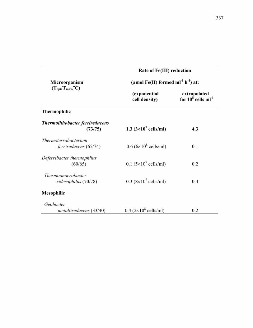
337
Rate of Fe(III) reduction Microorganism (µmol Fe(II) formed ml-1 h-1) at: (Topt/Tmax,oC)
(exponential extrapolated cell density) for 108 cells ml-1
Thermophilic Thermolithobacter ferrireducens
(73/75) 1.3 (3×10⁷ cells/ml) 4.3 Thermoterrabacterium
ferrireducens (65/74) 0.6 (6×10⁸ cells/ml) 0.1 Deferribacter thermophilus
(60/65) 0.1 (5×10⁷ cells/ml) 0.2 Thermoanaerobacter
siderophilus (70/78) 0.3 (8×10⁷ cells/ml) 0.4 Mesophilic Geobacter
metallireducens (33/40) 0.4 (2×10⁸ cells/ml) 0.2

338
Fig. A.1. Electron micrograph of JW/KA-2T. Negative staining preparation using uranyl acetate.
( ) indicates flagella, ( ) indicates Fe precipitation.

339
0.5 µm

340
Fig. A.2. Growth and (A) Fe(II) formation by JW/KA-2T and (B) CO utilization/ H2 production
by strain R1T. A. Cell growth of strain JW/KA-2T (-♦-) paralleled Fe(II) accumulation (-•-)
when cells were grown in a mineral Fe medium at pH25C 7.2 and 60oC as compared to a sterile
control (-∈-). Gas atmosphere was 80:20 H2-CO2 (vol/vol). B. Cell growth of strain R1T (-▲-)
was accompanied by CO utilization (-♦-) and H2 production (-■-) when cells were grown in a
mineral medium at pH25C 7.2 and 70-72oC. Gas atmosphere was 100% CO.
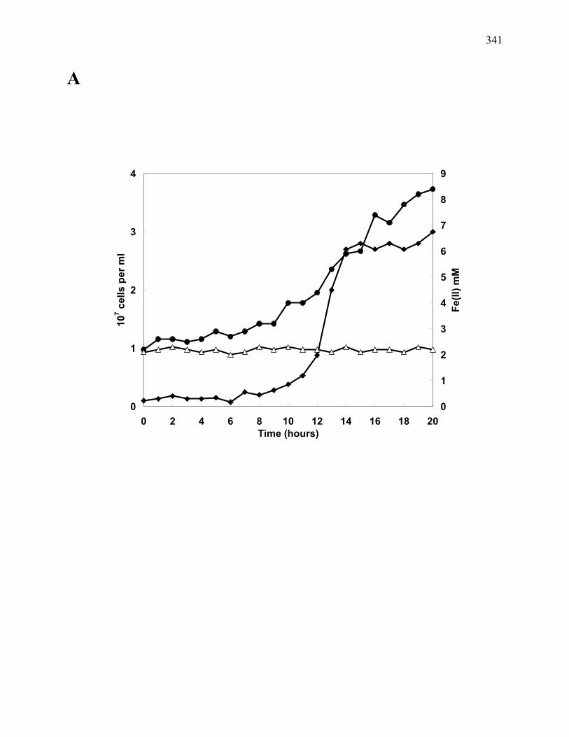
341
A
0
1
2
3
4
0 2 4 6 8 10 12 14 16 18 20Time (hours)
107 c
ells
per
ml
0
1
2
3
4
5
6
7
8
9
Fe(II
) mM

342
0
0.05
0.1
0.15
0.2
0.25
0 20 40 60 80Time (hours)
CO
, H2 m
mol
ml-1
6
6.5
7
7.5
8
8.5
cells
, log
B

343
Fig. A.3. Influence of incubation (A) temperature and (B) pH on growth of JW/KA-2T, and the
influence of incubation (C) temperature and (D) pH on Fe(III) reduction by resting cells of
JW/KA-2T. A. Fe-medium with a pH60C 7.3 was inoculated with 1 × 10⁶ cells/ml. After 24 hour
incubation with shaking (20 rpm), cells were counted using a Petroff-Hausser counting chamber.
B. Fe medium was inoculated with 1 × 10⁶ cells/ml and incubated at 72oC. After 24 hour
incubation with shaking (20 rpm), cells were counted using a Petroff-Hausser counting chamber.
C. Resting cells were inoculated to a concentration of 5 × 10⁷ cells/ml, and incubated for 24
hours in mineral media with a pH60C of 7.2 under an 80:20 H2-CO2 (vol/vol) atmosphere. Fe(II)
accumulation was monitored (- -) as compared to a sterile control (-▲-). A pH meter calibrated
at 60oC was used to determine pH of the media. D. Resting cells (- -) were inoculated to a
concentration of 5×10⁷ cells/ml, incubated at 60oC with shaking (20 rpm). Fe(II) accumulation
was monitored (- -) as compared to a sterile control (-▲-).

344
A
0
1
2
3
4
45 50 55 60 65 70 75 80
Temperature (oC)
107 c
ells
ml-1
b.
0
1
2
3
4
4 6 8 10
pH60C
107 c
ells
ml-1
B

345
00.20.40.60.8
11.21.4
5 6 7 8 9 10 11pH60C
mic
rom
oles
Fe(
II) m
l-1 h
our-1
C
D
0
0.2
0.4
0.6
0.8
1
1.2
35 40 45 50 55 60 65 70 75 80 85 90 95
Temperature (oC)
Fe(II
) mic
rom
oles
ml -1
h-1

346
Fig. A.4. Phylogenetic tree. Unrooted phylogenetic tree showing the position of Thermolitho-
bacter ferrireducens strains JW/KA-2T, with sequences A and B, and JW/JH-Fiji-2, and T.
carboxydivorans strain R1T. Two 16S rRNA gene sequences were also observed for strain
JW/KA-1. The tree was generated by maximum-likelihood. Reliability values at each internal
branch are percentages obtained from 1000 trials. The scale bar represents the expected number
of changes per sequence position. Thermus aquaticus was used as outgroup. The superscript “T”
denotes that the strain is the type strain of the species.

347

348
Fig. A.5. Phylogenetic tree of higher taxa. Phylogenetic tree constructed from the 16S rRNA
gene with maximum likelihood correction for synonymous changes using the neighbor-joining
algorithm. GenBank accession numbers are indicated in parentheses. The three classes of the
phylum ‘Firmicutes’ are indicated. For the ‘Clostridia’ (class 1), the order 1 Clostridiales are
indicated by ( ) while the order 2 ‘Thermoanaerobacteriales’ and order 3 Halanaerobiales are
indicated by ( ) and ( ), respectively. The class ‘Bacilli’ is divided into the order 1 Bacillales
( ) and order 2 ‘Lactobacillales’ ( ) while the members of the class Mollicutes are indicated
by ( ) with the order number indicated, i.e., 1order 1 Mycoplasmatales, 2order 2
Entomoplasmatales, 3order 3 Acholeplasmatales, and 4order 4 Anaeroplasmatales (Garrity et al.
2002). To have a meaningful tree, each order is only represented by the type species of the type
genus for the class and for the first family for each of the different orders. Numbers at nodes
indicate bootstrap support values for 100 replicates with only values above 75 displayed on the
tree. Scale bar denotes number of nucleotide substitutions per site. Escherichia coli was used as
the outgroup.

349

350
APPENDIX B
FE(III) REDUCTION BY NOVEL CHEMOLITHOTROPHIC STRAINS OF GLYCOLYTIC
THERMOPHILES1
____________________________
1Onyenwoke, R. U., J. Hanel, R. C. Davis, and J. Wiegel. To be submitted to the International
Journal of Systematic and Evolutionary Microbiology.
351
Abstract
Based on 16S rRNA analysis, two novel strains, JW/JH-1 from Yellowstone National Park, WY,
USA and JW/JH-Fiji-1 from Fiji: Vanua Levu Island, were isolated as thermophilic,
chemolithoautotrophic iron-reducers. They belong to the clade of the glycolytic ‘Caloramator
celere’ (basonym Thermobrachium celere) and Clostridium thermobutyricum, respectively.
However, the glycolytic type strains of both species were unable to couple growth to the
reduction of amorphous Fe(III) oxides, or Fe(III) citrate in the absence of H2/CO2.
Introduction
In recent years, evidence has indicated that a great deal more of the world’s Fe(II) might be the
product of microbial activity than was previously estimated. Despite observations dating back
nearly 80 years that bacteria were capable of biotic iron reduction (Harder 1919; Pringsheim
1949), it had simply been assumed that the reduction of Fe(III) to Fe(II) was an abiotic reaction.
Mesophilic iron-reducing bacteria have been well-documented and studied in recent years
(Lovley and Longergan 1990; Longergan et al. 1996), however, thermophilic iron-reducers have
been rarely described (Boone et al. 1995; Slobodkin et al. 1997, 1999; Greene et al. 1997;
Kashefi et al. 2003; Sokolova et al., in press). A thermophilic iron-reducer was not even
described until Bacillus infernus (Boone et al. 1995). Of the first described iron reducers, all
required organic carbon sources, i.e., were heterotrophs. Slobodkin et al. (1997) demonstrated
chemolithoautotrophic growth for Thermoterrabacterium ferrireducens (i.e. able to grow with
CO2, H2, and amorphous Fe(III) oxides alone) and later also for Thermoanaerobacter
siderophilus (Slobodkin et al. 1999). Thermophilic, autotrophic, iron reduction has gained
interest because it: 1) is under-represented by current culture collections, and 2) may exist in

352
biosphere pockets deep within the Earth (and possibly other planets) (Gold 1992). This growing
interest has been demonstrated by the more recent isolations of the autotrophic iron reducers
Geoglobus ahangari (Kashefi et al. 2002a), ‘Geothermobacterium ferrireducens’ (Kashefi et al.
2002b), and Thermolithobacter ferrireducens (Sokolova et al., in press; this dissertation
Appendix A) and by the observation that Pyrobaculum islandicum is capable of autotrophic
growth on iron (Kashefi and Lovley 2000).
Iron reduction can impact environmental systems in a number of ways: organic matter
oxidation, aromatic degradation, banded iron formation, and inhibition/stimulation of other
microbial populations (Lovley 1995). As mentioned earlier, biotic Fe(III) reduction has been an
underestimated phenomenon and is, therefore, not a routine characterization step for novel
microorganisms. We report here on Fe(III) reduction by anaerobic glycolytic and
chemolithoautotrophic thermophiles carrying out Fe(III) reduction.
Materials and methods
Environmental samples
A combined water, organic filamentous material, and sediment sample from a runoff of a hot
spring close to the river at the Calcite Spring area from Yellowstone National Park contained
white and black bacterial filaments and about 10-15 ppm iron in the sediment. The sample from
Fiji contained water and sediment from a hot spring runoff channel at the soccer field on Vanua
Levu Island. Samples were collected in sterile, N2-flushed 100 ml Pyrex jars, capped with butyl
rubber stoppers, and brought to the laboratory in Athens, GA, where they were stored at 4-7oC
for several weeks before the enrichments were started.

353
Enrichment and isolation of organisms
‘Caloramator celere’ strain JW/JH-1 and Clostridium thermobutyricum JW/JH-Fiji-1 were
isolated from the Yellowstone and Fiji samples, respectively, using an anaerobic media prepared
using the Hungate technique (Ljungdahl and Wiegel 1986; Hanel, J., M.S. thesis, The University
of Georgia) under a headspace of H2/CO2 (80:20 v/v) containing (per liter deionized water): 0.33
g of KH2PO4, 0.33 g of NH4Cl, 0.33 g of KCl, 0.33 g of MgCl2·2H2O, 0.33 g of CaCl2·2H2O, 2.0
g of NaHCO3, 1 ml of vitamin solution (Wolin et al. 1963) and 1.2 ml of trace element solution
(Slobodkin et al. 1997) with a pH25C adjusted to 7.0 with 10% (w/v) NaOH. To this mineral
medium, 90 mM amorphous Fe(III) oxide/hydroxide (mineral Fe medium; prepared as described
by Slobodkin et al. 1997) or 20 mM 9,10-anthraquinone 2,6-disulfonic acid (AQDS medium)
were typically added as electron acceptors. Medium was routinely sterilized by autoclaving at
121oC for 1 hour due to the possible presence of heat stable spores with possible D10 times of
nearly 2 hr (Byrer et al. 2000). The enrichment and pure cultures of JW/JH-1 and JW/JH-Fiji-1
were typically grown in Hungate or Balch tubes at 60oC, and all transfers were carried out with
disposable syringes and hypodermic needles. Since the reducing agents dithionite, cysteine,
Ti(III) citrate (Gaspard et al. 1998) and HCl-cysteine (unpublished results) reduce Fe(III), no
reducing agents were added.
For isolations, approximately 1 g of sediment and 2 ml of liquid from the mixed sample
were transferred to 50 ml serum bottles containing 10 ml of the anaerobic mineral medium
supplemented with 90 mM Fe(III) oxide/hydroxide and 20 mM formate, and the enrichments
were incubated at 60oC without shaking. After 72 hours, the presence of iron reducers was
indicated by the occurrence of magnetic black precipitate (magnetite). Positive enrichments were
transferred three times to the same medium to ensure viability. After the final transfer, the

354
enrichment displaying the most magnetic precipitate was selected and used to inoculate (2% v/v)
several Hungate tubes containing mineral medium supplemented with AQDS as the soluble
electron acceptor. All tubes were reduced (yellow to dark brown) in 24 h and displayed growth.
The cultures were then inoculated into agar roll tubes containing AQDS media supplemented
with 2.5% Bacto agar and incubated at 60oC. After 24 h, isolated colonies were picked in an
anaerobic chamber (Coy Products) under N2:H2 (95:5 v/v) and inoculated into AQDS medium,
which was then incubated for 48 h at 60oC. Tubes displaying positive AQDS reduction and cell
growth were used to inoculate (2% v/v) mineral Fe media and were incubated for 48 h. Tubes
displaying formation of black magnetic precipitation production and cell growth were then
repeatedly transferred into agar roll tubes until pure cultures were obtained. Stock cultures were
stored in glycerol (50% v/v) in both AQDS and mineral Fe media at -80oC. For routine use,
cultures were maintained in both media.
Microscopy
Light microscopic observations were performed using an Olympus VANOX equipped with
phase-contrast objectives and a Petroff-Hausser counting chamber.
Temperature and pH ranges for growth
The temperature range for the iron reducers was determined in both mineral Fe-medium and
AQDS-media at pH25C 7.3 (Wiegel 1998) using a temperature gradient incubator (Scientific
Industries, Inc., Bohemia, N.Y.) with shaking (~20 strokes/min.). The pH60C [pH determined
with a pH meter calibrated with standards and electrode equilibrated at 60oC (Wiegel 1998)] was

355
determined in both media at 60oC under shaking at ~20 rpm. Sterile anaerobic 1 M NaOH and 3
M HCl were used to adjust the pH of the medium prior to inoculation.
Carbon sources (substrate utilization)
Growth on potential carbon sources was assessed using Hungate tubes containing mineral
medium and Fe(III) in which the autotrophic carbon source, CO2, was omitted and substrates
were supplied. Cell numbers and iron or AQDS reduction were used to determine growth.
Negative controls included the use of identical media and carbon supplements, inoculated with
autoclaved, sterile suspensions in order to test the possibility of abiotic iron or AQDS reduction
such as is common with carbohydrates (Slobodkin et al. 1997). All potential carbon sources were
sterilized by autoclavation with the exception of pyruvate, which was filter-sterilized. The
inocula were 1% (v/v).
Electron acceptors
The ability to utilize various compounds as electron acceptors was tested for growth in mineral
medium without the addition of Fe(III). Utilization was assumed when optical densities (600 nm)
increased by 0.2, and the reduction of ferric citrate and AQDS was accompanied by changes in
color from black to clear (reduced form) and yellow to brown or black (reduced form),
respectively. When nitrate, thiosulfate, sulfate or elemental sulfur was added as potential electron
acceptors, the medium was reduced with sodium sulfide. The inoculum was 1% (v/v). A culture
without addition of an electron acceptor was used as a control.

356
Iron reduction and Fe(II) analysis
Fe(II) was determined employing the 2,2-dipyridyl iron assay in which all species of iron are
dissolved in 0.5 ml of 0.6 N HCl. Only Fe(II) will react with the added 2,2-dipyridyl in glacial
acetic acid buffer (Balashova and Zavarzin 1980). Iron was quantified using an Fe(II) standard
curve of FeCl2 ranging from 0.5 mM to 50 mM.
Antibiotic susceptibility
Antibiotic susceptibility testing was carried out in mineral media supplemented with tryptone
(1%) and yeast extract (0.4%) at 60oC without shaking. The antibiotics were added to the media
from filter sterilized stock solutions to the appropriate, final concentration (10 or 100 µg/ml). An
inoculated control was made for each strain by the omission of an antibiotic to the media.
Growth was observed at 12, 24, and 48 hrs.
G+C content
The G+C mol% was determined by the HPLC method of Mesbah et al. (1989). DNA for the
analysis was extracted using the mini-BeadBeater DNA extraction system (Biospec Products).
DNA extraction, sequencing, and 16S rRNA phylogenetic analysis
Genomic DNA was extracted from cell pellets obtained from cultures of JW/JH-1 and JW/JH-
Fiji-1 using the DNeasy Tissue kit (Qiagen). The 16S rRNA genes were amplified by PCR from
genomic DNA in reactions containing 50 mM KCl, 30 mM Tris-HCl (pH 8.3), 2 mM MgCl2,
200 µM each of dATP, dCTP, dGTP, dTTP, 0.05% Igepal (Sigma), 1 U Taq Polymerase, and 20
pmol each primer (8F, AGAGTTTGATCCTGGCTCAG and 1492R, GGTTACCTTGTTAC-

357
GACTT) with thermal cycling conditions as follows: 94oC for 2 min; 94oC for 30 s, 50oC for 30
s, 72oC for 1.5 min for 30 amplification cycles; and an extension cycle at 72oC for 10 min. PCR
products (approximately 1500 base pairs) were purified using the PCRpure Spin Kit
(Intermountain Scientific, Kaysville, Utah) and cloned into a pCR2.1 vector using the TOPO-TA
cloning kit (Invitrogen). Plasmid DNA was prepared using an alkaline-lysis method (Sambrook
et al. 1989). Sequencing reactions were performed using the ABI PRISM BigDyeTerminator
Cycle Sequencing Kit and an ABI 310 Genetic Analyzer (PE Biosystems) according to the
manufacturer’s protocol. DNA was extracted, cloned, amplified and sequenced three different
times. Three clones were sequenced of each strain to ensure that the cultures were pure, and to
note any discrepancies that may exist within the sequences. Additionally, PCR products were
sequenced directly to determine whether a single product was present. Both strands of JW/JH-1
and JW/JH-Fiji-1 were completely sequenced using a suite of 16S rRNA-specific primers
(Vetriani et al. 1999) to generate an overlapping set of sequences which were assembled into one
contiguous sequence. Sequence alignments were performed using the software suite ARB
(Ludwig et al. 2004). The alignment was manually edited considering the expected sequence
secondary structure. An unrooted phylogenetic tree was constructed by maximum-likelihood
using the program FastDNAml (Olsen et al. 1994) embeded in ARB. Bootstrap values were
based on 100 trials. The reliability value (bootstraps) of each internal branch indicates, as a
percentage, how often the corresponding cluster was found. The organisms used in the
phylogenetic analysis and the GenBank accession numbers for their 16S rRNA gene sequences
were: Clostridium butyricum (X77834; outgroup), Thermoanaerobacter ethanolicus (L09162),
Moorella thermoautotrophica (X77849), Moorella thermoacetica (M59121), Moorella glycerini
(U82327), Desulfotomaculum nigrificans (AY742958), Desulfotomaculum thermobenzoicum

358
(AJ294429), Desulfotomaculum thermocisternum (U33455), Desulfotomaculum geothermicum
(AJ294428).
DNA-DNA hybridization
DNA-DNA hybridizations were performed using…..
Nucleotide sequence accession numbers of novel isolates
The 16S rRNA sequences of strain JW/JH-1 and JW/JH-Fiji-1 were deposited into GenBank
under the accession numbers AF286863 and AF286862, respectively.
Results and Discussion
Morphology
JW/JH-1 cells were rods ~0.5 by 10-12 µm, usually appearing as single cells, with some pairs,
during early growth (24 hr) while older cultures (72 hr) contained long chains of cells, up to 10,
in addition to paired and single cells (Fig. B.1.B.). Retarded motility was observed. Young
cultures (6 hr) stained Gram positive while older cultures tended to stain Gram negative.
Colonies formed on the agar (2.2%) surface in roll tubes (mineral medium supplemented with
yeast extract (0.4%) and tryptone (1.0%)) and are ~1 mm in diameter, cream-colored, with
filamentous edges.
As examined by phase contrast microscopy, cells of strain JW/JH-Fiji-1 were rods ~0.5
by 6-8 µm, usually arranged in pairs or appearing as single cells during early growth (24 hr)
while older cultures (72 hr) often contain long chains, ≥12 cells (Fig. B.1.A.). Retarded motility

359
was observed. Young (6 hr) and older (72 hr) cultures always stained Gram negative. Colonies
formed on the agar (2.2%) surface in roll tubes (mineral medium supplemented with yeast
extract (0.4%) and tryptone (1.0%)) and are ~0.5 mm in diameter, cream-colored, with smooth
edges.
Spores were never observed by phase contrast microscopy for either strain. Both strains
were unable to withstand 5 min of heat treatment at 100oC. Thus, heat resistant spores appeared
to be absent in these strains under the employed conditions.
Temperature and pH ranges for growth
The temperature range for growth of strain JW/JH-1 was 45-68oC (pH25C 7.3) with an opt. 63oC
(Fig. B.2.A.); no growth occurred below 40oC or above 70oC. The pH60C range for growth of
JW/JH-1 was 5.5-9.5, with an opt. ~8.0 (Fig. B.2.B.); no growth occurred below pH60C 5.0 or
above 10.
The temperature range for growth of strain JW/JH-Fiji-1 was 47-72oC (pH25C 7.3), opt.
65oC (Fig. B.3.A.); no growth occurred below 40oC or above 75oC. The pH60C range for growth
of JW/JH-Fiji-1 was 5.5-8.5, opt. ~7.2 (Fig. B.3.B.); no growth occurred below pH60C 5.0 or
above 9.0.
Substrate utilization
Substrates utilized by strain JW/JH-1 include: yeast extract (0.3% and as low as 0.05%), sucrose
(0.2%), raffinose (0.2%), inositol (0.2%), crotonate (0.1%), and benzoate (0.1%) but not fructose
(0.2%), glucose (0.2%), or ribose (0.2%) (with either Fe(III) oxyhydroxide (90 mM) or AQDS
(20 mM) as electron acceptor) (Table B.1.). In the presence of atmospheric H2, but not in its
360
absence, as an electron donor, strain JW/JH-1 utilized: CO2, formate (10 mM), glycerol (3 ml/l),
and acetate (20 mM) but not casamino acids (1%), glucose (20 mM), cellobiose (20 mM), or
pyruvate (20 mM).
Strain JW/JH-Fiji-1 utilized: yeast extract 0.3% (and as low as 0.05%), sucrose (0.2%),
corn starch (0.1%), ribose (0.2%), fructose (0.2%), glucose (0.2%), succinate (10 mM), toluene
(1 mM), acetone (40 mM), phenol (5 mM), benzoate (10 mM), formate (10 mM) but not
galactose (0.2%), xylose (0.2%), or maltose (0.2%) (with either Fe(III) oxyhydroxide (90 mM)
or AQDS (20 mM) as electron acceptor) (Table B.1.). JW/JH-Fiji-1 utilized CO2 and formate (10
mM) in the presence of a H2 headspace. Formate was also utilized in the absence of H2. Neither
casamino acids (1%), cellobiose (20 mM), glycerol (3 ml/l), acetate (20 mM) nor pyruvate (20
mM) was utilized. In the controls, glucose, sucrose, xylose, cellobiose, galactose, and starch
reduced Fe(III) to Fe(II) abiotically at rates similar to that of a growing culture, however, no
increase in cell numbers was observed.
Electron acceptors
The oxidation of hydrogen gas was coupled to the reduction of Fe(III) to magnetite and siderite
(determined by x-ray diffraction analysis). Besides amorphous Fe(III) oxyhydroxide (90 mM),
JW/JH-1 and JW/JH-Fiji-1 utilized AQDS (20 mM) and thiosulafate (20 mM) as electron
acceptors with atmospheric H2/CO2 (80:20 vol/vol) but not Fe(III) citrate (25 mM), nitrate,
sulfate, elemental sulfur (sublimated, 0.1%), elemental sulfur (precipitated, 0.1%), crystalline
Fe(III) oxide, manganese (IV) (15 mM) added as MnO2 (Kostka and Nealson 1998), or selenium
(VI) (100 µM) added as selenium acetate. However, JW/JH-1 and JW/JH-Fiji-1 were unable to
361
utilize amorphous Fe(III) oxyhydroxide (90 mM), AQDS (20 mM) or thiosulafate (20 mM) as
electron acceptors when the H2/CO2 headspace (80:20vol/vol) was replaced with CO (100%).
Both strains grew to higher cell densities (2.0 x 10⁸ cells/ml) on an Fe(III) mineral
medium supplemented with 1% yeast extract and 1% tryptone than in the absence of yeast
extract and tryptone (3.0 x 10⁷ cells/ml).
Iron reduction and Fe(II) analysis
In the process of dissimilatory Fe(III) reduction, 2 mol of Fe(II) were formed for every 1 mol of
H2 oxidized. Strains JW/JH-1 and JW/JH-Fiji-1 produced Fe(II) and oxidized H2 at ratios of: 0.9-
1.1 ± 0.2 and 0.8-0.9 ± 0.1, respectively. Thus, per mole of H2 consumed for the reduction of
Fe(III), 1 mole of H2 was used for anabolic reactions.
Antibiotic susceptibility
Growing at 60oC on rich media, strain JW/JH-Fiji-1 was susceptible to 10 µg/ml: rifampicin,
erythromycin, streptomycin, chloramphenicol, tetracycline, and ampicillin. Strain JW/JH-1
showed an identical susceptibility profile to JW/JH-Fiji-1, except JW/JH-1 was resistant to
streptomycin at 10 µg/ml (but not at 100 µg/ml).
G+C content
The mean (± standard deviation from three measurements) guanosine (G) plus cytosine (C)
content of the genomic DNA of strains JW/JH-1 and JW/JH-1 Fiji-1 were ?? ± ?? and 62.5 ±
0.05 mol% G+C, respectively, as determined by HPLC (Mesbah et al. 1989).

362
Description of ‘Caloramator celere’ strain JW/JH-1
Cells of strain JW/JH-1 are rods ~0.5 by 10-12 µm, usually appearing as single cells, with some
pairs, during early growth (24 hr) while older cultures (72 hr) contain long chains of cells, up to
10, in addition to paired and single cells. Retarded flagellation and sluggish motility is observed.
Young cultures (6 hr) stain Gram positive while older cultures tend to stain Gram negative.
Colonies formed on the agar (2.2%) surface in roll tubes (mineral medium supplemented with
yeast extract (0.4%) and tryptone (1.0%)) are ~1 mm in diameter, cream-colored, with
filamentous edges.
The temperature range for growth of strain JW/JH-1 is 45-68oC [pH25C 7.3] (opt. 63oC);
with no growth at or below 40oC, or at or above 70oC. The pH60C range for growth of JW/JH-1 is
5.5-9.5 (opt. ~8.0); no growth at or below pH60C 5.0, or at or above 10. Strain JW/JH-1 utilizes
yeast extract, sucrose, raffinose, inositol, crotonate, and benzoate. In the presence of atmospheric
H2 as an electron donor, strain JW/JH-1 utilizes: CO2, formate, glycerol, and acetate. Amorphous
Fe(III) oxyhydroxide, AQDS, and thiosulafate are utilized as electron acceptors with H2/CO2
(gas phase 80:20) but not Fe(III) citrate, nitrate, sulfate, elemental sulfur (sublimated), elemental
sulfur (precipitated), crystalline Fe(III) oxide, manganese (IV) added as MnO2, or selenium (VI).
Strain JW/JH-1 was isolated from a combined water, organic filamentous material, and
sediment sample from a runoff of a hot spring close to the river at the Calcite Spring area from
Yellowstone National Park containing white and black bacterial filaments and about 10-15 ppm
iron in the sediment.
Strain JW/JH-1 was deposited in the American Type Culture Collection ATCC 700984
and the German Collection of Microorganisms DSM 13655.

363
Description of Clostridium thermobutyricum strain JW/JH-Fiji-1
Cells of strain JW/JH-Fiji-1 are rods ~0.5 by 6-8 µm, usually arranged in pairs or appearing as
single cells during early growth (24 hr) while older cultures (72 hr) often contain long chains,
≥12 cells. Retarded flagellation and sluggish motility is observed. Young (6 hr) and older (72 hr)
cultures stain Gram negative. Colonies formed on the agar (2.2%) surface in roll tubes (mineral
medium supplemented with yeast extract (0.4%) and tryptone (1.0%) and are ~0.5 mm in
diameter, cream-colored, with smooth edges. The temperature range for growth of strain JW/JH-
Fiji-1 is 47-72oC [pH25C 7.3] (opt. 65oC); no growth at or below 40oC, or at or above 75oC. The
pH60C range for growth of JW/JH-Fiji-1 is 5.5-8.5 (opt. ~7.2); no growth at or below pH60C 5.0,
or at or above 9.0. Strain JW/JH-Fiji-1 utilizes: yeast extract, sucrose, corn starch, ribose,
fructose, glucose, succinate, toluene, acetone, phenol, benzoate, and formate. JW/JH-Fiji-1
utilizes CO2 and formate (10 mM) in the presence of H2 as headspace gas. Amorphous Fe(III)
oxyhydroxide, AQDS, and thiosulfate can serve as electron acceptors with H2/CO2 as headspace
gas but not Fe(III) citrate, nitrate, sulfate, elemental sulfur (sublimated), elemental sulfur
(precipitated), crystalline Fe(III) oxide, manganese (IV) added as MnO2, or selenium (VI).
Strain JW/JH-Fiji-1 was isolated from a sample from Fiji containing water and sediment
from a hot spring runoff channel at the soccer field in Savusavu on Vanua Levu Island.
The G+C content of the DNA of the strain is 62.5 ± 0.05 mol %. Strain JW/JH-Fiji-1 was
deposited in the American Type Culture Collection ATCC 700983 and the German Collection of
Microorganisms DSM 13654.
364
References Boone, D. R., Y. Liu, Z. Zhao, D. Balkwill, G. Drake, T. O. Stevens, and H. C. Aldrich. 1995. Bacillus infernus sp. nov., an Fe(III)- and Mn(IV)-reducing anaerobe from the deep terrestrial subsurface. Int. J. Syst. Bacteriol. 45:441-448. Byrer, D. E., F. A. Rainey, and J. Wiegel. 2000. Novel strains of Moorella thermoacetica form unusually heat-resistant spores. Arch. Microbiol. 174:334-339. De Ley, J., H. Cattoir, and A. Reynaerts. 1970. The quantitative measurement of DNA hybridization from renaturation rates. Eur. J. Biochem. 12:133-142. Gaspard, S., F. Vazquez, and C. Holliger. 1998. Localization and solubilization of the iron (III) reductase of Geobacter sulfurreducans. Appl. Environ. Microbiol. 64:3188-3194. Gold, T. 1992. The deep, hot biosphere. Proc. Natl. Acad. Sci. USA 89:6045–6049. Greene, A. C., B. K. C. Patel, and A. Sheehy. 1997. Deferribacter thermophilus gen. nov., sp. nov., a novel thermophilic manganese- and iron-reducing bacterium isolated from a petroleum reservoir. Int. J. Syst. Bacteriol. 47:505-509. Harder, E. C. 1919. Iron-depositing bacteria and their geologic relations. U.S. Geological Survey professional paper 113 Department of the Interior, Government Printing Office, Washington, DC. Huss, V. A. R., H. Festl, and K. H. Schleifer. 1983. Studies on the spectrometric determination of DNA hybridization from renaturation rate. Syst. Appl. Microbiol. 4:184-192. Kashefi, K., and D. Lovley. 2000. Reduction of Fe(III), Mn(IV), and toxic metals at 100oC by Pyrobaculum islandicum. Appl. Environ. Microbiol. 66:1050-1056. Kashefi, K., J. M. Tor, D. E. Holmes, C. V. Gaw Van Praagh, A. L. Reysenbach, and D. R. Lovley. 2002a. Geoglobus ahangari gen. nov., sp. nov., a novel hyperthermophilic archaeon capable of oxidizing organic acids and growing autotrophically on hydrogen with Fe(III) serving as the sole electron acceptor. Int. J. Syst. Evol. Microbiol. 52:719-728. Kashefi, K., D. E. Holmes, A. L. Reysenbach, and D. R. Lovley. 2002b. Use of Fe(III) as an electron acceptor to recover previously uncultured hyperthermophiles: isolation and characterization of Geothermobacterium ferrireducens gen. nov., sp. nov. Appl. Environ. Microbiol. 68:1735-1742. Kashefi, K., D. E. Holmes, J. A. Baross, and D. R. Lovley. 2003. Thermophily in the Geobacteraceae: Geothermobacter ehrlichii gen. nov., sp. nov., a novel thermophilic member of the Geobacteraceae from the “Bag City” hydrothermal vent. Appl. Environ. Microbiol. 69:2985-2993.

365
Ljungdahl, L. G., and J. Wiegel. 1986. Working with anaerobic bacteria. In: Demain, A. L., and N. A. Solomon (eds.) Manual of industrial microbiology and biotechnology, American Society for Microbiology, Washington, D.C., pp 84-94. Lonergan, D. J., H. L. Jentre, J. D. Coates, E. J. P. Phillips, T. M. Schmidt, and D. R. Lovley. 1996. Phylogenetic analysis of dissimilatory Fe(III)-reducing bacteria. J. Bacteriol. 178:2402-2408. Lovley, D. R., and D. J. Lonergan. 1990. Anaerobic oxidation of toluene, phenol, and p-cresol by the dissimilatory iron-reducing organisms, GS-15. Appl. Environ. Microbiol. 56:1858-1864. Lovley, D. R. 1995. Microbial reduction of iron, manganese, and other metals. Advances in Agronomy 54:175-231. Ludwig, W., O. Strunk, R. Westram, L. Richter, H. Meier, Yadhukumar, A. Buchner, T. Lai, S. Steppi, G. Jobb, W. Förster, I. Brettske, S. Gerber, A. W. Ginhart, O. Gross, S. Grumann, S. Hermann, R. Jost, A. König, T. Liss, R. Lüßmann, M. May, B. Nonhoff, B. Reichel, R. Strehlow, A. Stamatakis, N. Stuckmann, A. Vilbig, M. Lenke, T. Ludwig, A. Bode, and K. H. Schleifer. 2004. ARB: a software environment for sequence data. Nucl. Acids Res. 32:1363-1371. Mesbah, M., U. Premachandran, and W. Whitman. 1989. Precise measurement of the G+C content of deoxyribonucleic acid by high-performance liquid chromatography. Int. J. Syst. Bacteriol. 39:159-167. Olsen, G., H. Matsuda, R. Hagstrom, and R. Overbeek. 1994. FastDNAml: a tool for construction of phylogenetic trees of DNA sequences using maximum likelihood. CABIOS 10:41-48. Pringsheim, E. G. 1949. The filamentous bacteria Sphaerotilus, Leptothrix, Cladothrix, and their relation to iron and manganese. Phil. Trans. R. Soc. London B. Biol. Sci. 233:453-482. Slobodkin, A., A. L. Reysenbach, N. Strutz, M. Dreier, and J. Wiegel. 1997. Thermoterrabacterium ferrireducens gen. nov., sp. nov., a thermophilic anaerobic dissimilatory Fe(III)-reducing bacterium from a continental hot spring. Int. J. Syst. Bacteriol. 47:541-547. Slobodkin, A. I., T. P. Tourova, B. B. Kuznetsov, N. A. Kostrikina, N. A. Chernyth, and E. A. Bonch-Osmolovskaya. 1999. Thermoanaerobacter siderophilus sp. nov., a novel dissimilatory Fe(III)-reducing, anaerobic, thermophilic bacterium. Int. J. Syst. Bacteriol. 49:1471-1478. Sokolova, T., J. Hanel, R. U. Onyenwoke, A. -L. Reysenbach, A. Banta, R. Geyer, J. M. Gonzalez, W. B. Whitman, and J. Wiegel. 2006. Novel chemolithotrophic, thermophilic, anaerobic bacteria Thermolithobacter ferrireducens gen. nov., sp. nov. and Thermolithobacter carboxydivorans sp. nov. Extremophiles, in press.

366
Vetriani, C., H. W. Jannasch, B. J. MacGregor, D. A. Stahl, and A.-L. Reysenbach. 1999. Population structure and phylogenetic characterization of marine benthic Archaea in deep-sea sediments. Appl. Environ. Microbiol. 65:4375-4384. Wiegel, J. 1998. Anaerobic alkalithermphiles, a novel group of extremophiles. Extremophiles 2:257-267. Wolin, E. A., M. J. Wolin, and R. S. Wolfe. 1963. Formation of methane by bacterial extracts. J. Biol. Chem. 238:2882-2886.
367
Table B.1. Substrates utilized by strains JW/JH-Fiji-1, Clostrium thermobutyricum JW171KT
(Wiegel et al. 1989), JW/JH-1, and Thermobrachium celere JW/YL-NZ35T (Engle et al. 1996).
The designation of: (+) indicates growth (a visibly turbid culture), (-) indicates no growth, and
(±) indicates weak growth (not a visibly turbid culture) at OD 600 nm. OD reading for (±)
designation was ≥0.03 as compared to both: an uninoculated control (media containing the
substrate but uninoculated) and an inoculated control, i.e., this control was inoculated with the
bacterium but lacks the substrate. aReduction of Fe(III) oxide, formation of black precipitates
from amorphous brown Fe(III) oxide, indicated growth. bReduction of AQDS, yellow to black,
indicated growth. NT=not tested. Neither strain JW/JH-Fiji-1 nor strain JW/JH-1 utilized:
rhamnose or propionate (both at 0.2%), trehalose (0.1%), ethanol (0.3%), lactic acid (20 mM), 2-
propanol (40 mM), CO + Fe(III) oxide (90 mM), or CO + AQDS (20 mM). A superscript “T”
indicates the type strain of a species.

368
Substrate JW/JH-Fiji-1 JW171KT JW/JH-1 JW/YL-NZ35T
Yeast extract (0.3%) + + + + Sucrose (0.2%) + - + + Cellobiose (0.2%) - + - - Corn starch (0.1%) + - - NT Raffinose (0.2%) - - + NT Inositol (0.2%) - NT + NT Crotonate (0.1%) - NT + NT Xylose (0.2%) - + - - Ribose (0.2%) + + - - Fructose (0.2%) + + - + Glucose (0.2%) + + - + Galactose (0.2%) - - - + Maltose (0.2%) - + - + Succinate (10 mM) + - - NT Fumarate (10 mM) - - - NT Ethanol (0.3%) - - - NT Toluene (1 mM) + NT - NT Acetone (40 mM) + NT - NT Phenol (5 mM) + NT - NT Benzoate (10 mM) + NT + NT Formic acid (10 mM) + - - NT H2/CO2 + Fe(III) oxide (90 mM)
a+ - + NT
H2/CO2 + AQDS (20 mM) b+ - + NT

369
Fig. B.1. Phase-contrast images of: (A) JW/JH-Fiji-1 and (B) JW/JH-1. Bars are 10 µm in length.

370
A
B

371
Fig. B.2. Influence of incubation (A) temperature and (B) pH on growth of JW/JH-1. A. Media
with a pH60C 7.3 was inoculated with 1 × 10⁶ cells/ml. After 24 hours of incubation with shaking
(20 rpm), cells were counted using a Petroff-Hausser counting chamber. B. Mineral media was
inoculated with 1 × 10⁶ cells/ml and incubated for 24 hours at 60oC with shaking (20 rpm).

372
A
T. celere strain JW/JH-1
0.0E+00
1.0E+07
2.0E+07
3.0E+07
40 50 60 70Temp (oC)

373
B
T. celere strain JW/JH-1
0.0E+00
1.0E+07
2.0E+07
3.0E+07
4.0E+07
4 6 8 10 12
pH60C
Cel
ls/m
l

374
Fig. B.3. Influence of incubation (A) temperature and (B) pH on growth of JW/JH-Fiji-1. A.
Media with a pH60C 7.3 was inoculated with 1 × 10⁶ cells/ml. After 24 hours of incubation with
shaking (20 rpm), cells were counted using a Petroff-Hausser counting chamber. B. Mineral
media was inoculated with 1 × 10⁶ cells/ml and incubated for 24 hours at 60oC with shaking (20
rpm).

375
A
C. thermobutryricum strain JW/JH-Fiji-1
0.0E+00
1.0E+07
2.0E+07
3.0E+07
4.0E+07
40 45 50 55 60 65 70 75
Temp (oC)
Cel
ls/m
l

376
B
C. thermobutyricum strain JW/JW-Fiji-1
0.0E+00
1.0E+07
2.0E+07
3.0E+07
4.0E+07
4 6 8 10 12
pH60C
Cel
ls/m
l
377
Fig. B.4. Fitch tree showing the estimated phylogenetic relationships of strains JW/JH-1 and
JW/JH-Fiji-1 based on 16S rRNA gene sequence data with Jukes-Cantor correction for
synonymous changes. The 16S rRNA gene data used represent Escherichia coli DSM30083T
nucleotide positions 105–1450. Numbers at nodes indicate bootstrap support percentages for 100
replicates. Bar, 0.02 nucleotide substitutions per site. GenBank accession numbers are indicated
after the strain identifier. The superscript “T” denotes the strain is the type strain for the species.
The thermophilic, iron-reducing bacteria are indicated in bold.

378