fine structure of division in ciliate protozoa i
TRANSCRIPT
FINE STRUCTURE OF DIVISION
IN CILIATE PROTOZOA
I. Micronuclear Mitosis in Blepharisma
R. A. JENKINS
From the Department of Biochemistry and Biophysics, Iowa State University, Ames, Iowa 50010.The author's present address is the Department of Zoology and Physiology, University of Wy-oming, Laramie, Wyoming 82070
ABSTRACT
The mitotic, micronuclear division of the heterotrichous genus Blepharisma has been studiedby electron microscopy. Dividing ciliates were selected from clone-derived mass cultures andfixed for electron microscopy by exposure to the vapor of 2 % osmium tetroxide; individualBlepharisma were encapsulated and sectioned. Distinctive features of the mitosis are the pres-ence of an intact nuclear envelope during the entire process and the absence of centrioles atthe polar ends of the micronuclear figures. Spindle microtubules (SMT) first appear in ad-vance of chromosome alignment, become more numerous and precisely aligned by meta-phase, lengthen greatly in anaphase, and persist through telophase. Distinct chromosomaland continuous SMT are present. At telophase, daughter nuclei are separated by a spindleelongation of more than 40 u, and a new nuclear envelope is formed in close apposition tothe chromatin mass of each daughter nucleus and excludes the great amount of spindlematerial formed during division. The original nuclear envelope which has remained struc-turally intact then becomes discontinuous and releases the newly formed nucleus into thecytoplasm. The micronuclear envelope seems to lack the conspicuous pores that are typicalof nuclear envelopes. The morphology, size, formation, and function of SMT and the natureof micronuclear division are discussed.
INTRODUCTION
To date electron microscopy of protozoan nucleihas resulted in the description of numerous anddiverse structures which are not always reconcil-able in terms of a unifying story of nuclearmorphology and division. Information on themicronucleus in division is particularly lackingand may be due, as Nanney and Rudzinska(26) suggest, to its small size although other fac-tors, such as problems in fixation and identifica-tion of appropriate stages, have made an electronmicroscope study more difficult. A lack of ultra-structural information is emphasized by the factthat Pitelka's generally complete work (27)
includes almost no description of micronucleiand by the recent suggestion that a true mitosisdoes not occur in ciliate micronuclei (9).
The ciliate micronucleus may offer certain ad-vantages in attempts to analyze the dynamicprocesses involved in nuclear division. Among cur-rent needs are a chemical dissection of essentiallyclean mitotic apparatus microtubules after theapproach utilized by Gibbons for ciliary shafts(10) and a more complete knowledge of the un-structured, internal environment which immedi-ately surrounds the kinetic elements (49). Sincemicronuclear mitosis is completed within the
463
on December 25, 2018jcb.rupress.org Downloaded from http://doi.org/10.1083/jcb.34.2.463Published Online: 1 August, 1967 | Supp Info:
confines of an intact nuclear envelope, the un-structured environment of the achromatic appa-ratus is more likely to be peculiar to the divisionprocess than in the case of nuclear envelope break-down and the formation of the so-called mixo-plasm (34) as occurs in most mitoses. Also, theisolation of the mitotic apparatus free of cyto-plasmic contamination may be accomplished bythe in vitro disruption of isolated, intact, dividingmicronuclei. Small numbers of dividing micro-nuclei are now routinely isolated by the author and
are found, by phase microscopy, to be free of cyto-plasmic contamination. Assuming that the SMT
of these nuclei can be stabilized through the isola-tion procedure (electron microscopy has not yetbeen done), it would seem a simpler task to isolatemicronuclei, then disrupt the envelope, and col-lect clean spindle material in a controlled en-vironment than to isolate the fibrous mitotic
apparatus from the cytoplasmic material in whichit is bathed, as is usually done, and then attemptto isolate SMT free of contaminating material.Synchronous cultures of ciliates could easily pro-vide ample numbers of dividing micronuclei formost types of experimentation.
Microtubules have been described previouslyfor dividing ciliate nuclei (4, 30, 36, 37), but allmicronuclear stages have been studied only insituations where the elements of the achromatic
apparatus were not preserved because of the fixa-tion procedures employed (6-8). Spindle micro-tubules (SMT) are well preserved by the fixationtechnique utilized in this work, and all mitoticstages are reported. The major aim of this paper isto give a more complete description than had beenpresented of the fine structure of micronucleardivision and to provide further information on thegeneral involvement of microtubules in mitosis.T'he work has served the author as an introduction
to further investigations such as the isolation ofdividing micronuclei and the microtubules con
tained therein. The observations reported are
restricted to the genus Blepharisma and then only
to the micronucleus; fine structure studies of themicronuclear mitosis and macronuclear division
in Didinium and the macronuclear division of
Blepharisma will be subjects of subsequent reports(in preparation).
MATERIALS AND METHODS
The majority of the observations reported here weremade on either Blepharisma undulans americanum Suzuki
or an organism (obtained from General BiologicalSupply House, Chicago, Ill.) which is similar in sizebut distinct from B. undulans americanum; this organismwill be referred to as Blepharisma sp. Turtox strain.The other species (subspecies, strains; see Hirshfieldet al., reference 14 and Suzuki, reference 45) utilizedwere Blepharisma intermedium, obtained from Dr. HenryHirshfield (New York University, New York) andBlepharisma undulansjaponicus Suzuki which, along withB. undulans americanum, was obtained from Dr. A. C.
Giese (Stanford University, Palo Alto, Calif.). Masscultures were derived from clones initiated from theoriginal stock and were maintained under conditionsof reduced light at 23 °C in a 0.1 , Cerophyl-lettuceinfusion buffered with 5 mM phosphate at pH 6.8 andinoculated with Bacillus subtilis at least 24 hr beforethe introduction of ciliates.
Dividing Blepharisma were easily selected from con-centrated cultures which were transferred in mass toa fresh culture medium I day before their intendeduse. Organisms with the typical division morphology(46) were selected individually by imicropipet until20-30 were collected. A small drop of culture fluidcontaining the ciliates was placed at the center of amicroscope slide coverslip which was then invertedand placed over the mouth of a container of aqueous2%, osmium tetroxide. A fixation time of 4 min at23°C was used routinely. Dehydration was accom-plished by rapid changes of acetone or ethanol,usually two 5-min changes of 50%C;, one 10-min changeof 75%, one 5-min change of 95, two 5-min changesof absolute, and two 5-min changes of propyleneoxide for epoxy embedding. Organisms were em-bedded either in methacrylate (a Na2 SO 4-driedmixture consisting of 57 ml n-butyl methacrylate, 43ml ethyl methacrylate, 1.5 ml divinylbenzene (DVB)and I g benzoyl peroxide) or in an Epon-Aralditemixture (2) containing 5% D. E. R. 732; micrographsmade from epoxy-embedded organisms will be identi-fied in the figure legends. In all cases, organisms wereembedded singly in capsules or, with epoxy, in a flatmatrix so that specific orientations could be achievedmore easily. As a final step in the preparation ofblocks for sectioning, each organism was stagedaccording to the general scheme used by Suzuki (46);then drawings were made and used as aids in fixingorientation of structures in electron micrographs.
Thin sections were cut with an LKB or Reichertultramicrotome, were mounted on grids with thinParlodion or Formvar supporting films and werestained with calcium perinanganate or doubly stainedwith uranyl acetate and lead citrate (31). Sectionswere examined with an RCA EMU 3F electronmicroscope operated at 100 kv; a 150 u condenseraperture and a 35 A objective aperture were em-ployed.
464 THE JOURNAL OF CELL BIOLOGY - VOLUME 34, 1967
The work reported here results from the study ofsome 40 organisms in known stages of division.
OBSERVATIONS
The ultrastructure of Blepharisma undulans has beenreported recently by Kennedy (18) and byDembitzer and Hirshfield (5). Since the presentstudy has revealed interphase features generallyconsistent with those described by these workers,a detailed description of the general fine structureof the nondividing organism will not be presentedhere.
Also, since observations of micronuclear be-havior for each of the four strains utilized in thisstudy are essentially the same and since mostmitotic stages have been studied in at least twostrains, no distinction as to strain will be made inthe text; however, figure legends will includeidentification of specific organisms.
Interphase
The interphase micronucleus is a nearly spher-ical body 1.5-2 in diameter (Fig. 1, Mi) and islimited by a two-membrane envelope in whichpores are not visible (Fig. 2, NE). That the en-velopes of the macronucleus and micronucleusare very different with respect to the presenceof pores is clearly shown in Fig. 2 where theenvelope of each nucleus is cut tangentially. Poreswith dense annuli are obvious in the macro-nuclear envelope (NP) and absent in the micro-nuclear envelope (Fig. 2, NP, Ma, and NE). Themicronucleoplasm has a cortex consisting of afine matrix surrounding an electron-opaque,reticular, central region which is a densely packedfibrillar structure (Fig. 1, CR). The structure ofthe interphase nucleus is essentially identical withthat described by Seshachar (42) and similar tothat described for other ciliates (7, 8, 17, 43).Occasionally, micronuclei similar to those de-scribed by Kennedy (18) are observed whichseemingly lack the less dense cortex but arethought not to represent the interphase condition.The several micronuclei (from 6 to 20) are foundthroughout the cytoplasm, but most are locatednear the macronucleus.
Several cytoplasmic features of Blepharisma are
shown to good advantage in Fig. 1. Mitochrondria
(M), vesicles (V), and paraglycogen granules
(P) are distributed throughout the cytoplasm.Pigment granules (PG) are confined primarily
to the region just beneath the pellicle, but some
are found deeper in the cell. Golgi bodies (G),often paired, are numerous and are present duringthe entire division cycle.
Prophase
At least two of the micronuclei (PMi) near themacronucleus in Fig. 1 are taken to be in prophasewhile one other is quite obviously in interphase(Mi). Although the micronuclear division is cor-related with macronuclear changes, e.g. meta-phase and the condensed stage, all micronuclei donot divide in synchrony, and it appears that somemay forego division completely in both cell divi-sion and in regeneration following merotomy.
In most descriptions of mitosis, the beginningof nuclear division (prophase) is marked bycytological changes observable at the lightmicroscope level, usually a visible condensationof the chromosomes. In the earliest known divisionstages studied here, organisms with very early oralprimordia but no noticeable macronuclear change,micronuclei are usually enlarged to a diameter of3 4 p and lack dense central chromatin; insteadseveral small, dense chromosomes are distributedthroughout the nucleoplasm (Fig. , CH). Otherauthors (8, 29, 41, 44, 46) report similar prophasevolume changes for Tetrahymena, Nassulopsis,Paramecium, Frontonia, and Blepharisma, respec-tively.
At a slightly later stage, possibly equivalent toprometaphase as described in other cells, spindlemicrotubules (SMT) are present in small numbersbut without precise array, and chromosomes areclearly visible but not yet aligned at a metaphaseplate (Fig. 3, SMT and CH). There is no indica-tion of well-defined association of these early SMTwith a specific center of formation; that is, neithercentrioles nor kinetochores are identifiable. Asis the case for all stages, SMT appear to terminateat or very near the inner membrane of the nuclearenvelope (Fig. 3, arrow). Structural changes(porosity) in the nuclear envelope have not beendetected either during earlier swelling or at thisstage.
Metaphase
In metaphase, when the chromosomes arealigned at the equatorial plate, well-defined SMTare present in great numbers (Figs. 4 and 5,SMT); SMT diameters are apparently subject todifferences in embedment, since they measureapproximately 16 my in methacrylate and 22 mg
R. A. JENKINS Micronuclear Mitosis 465
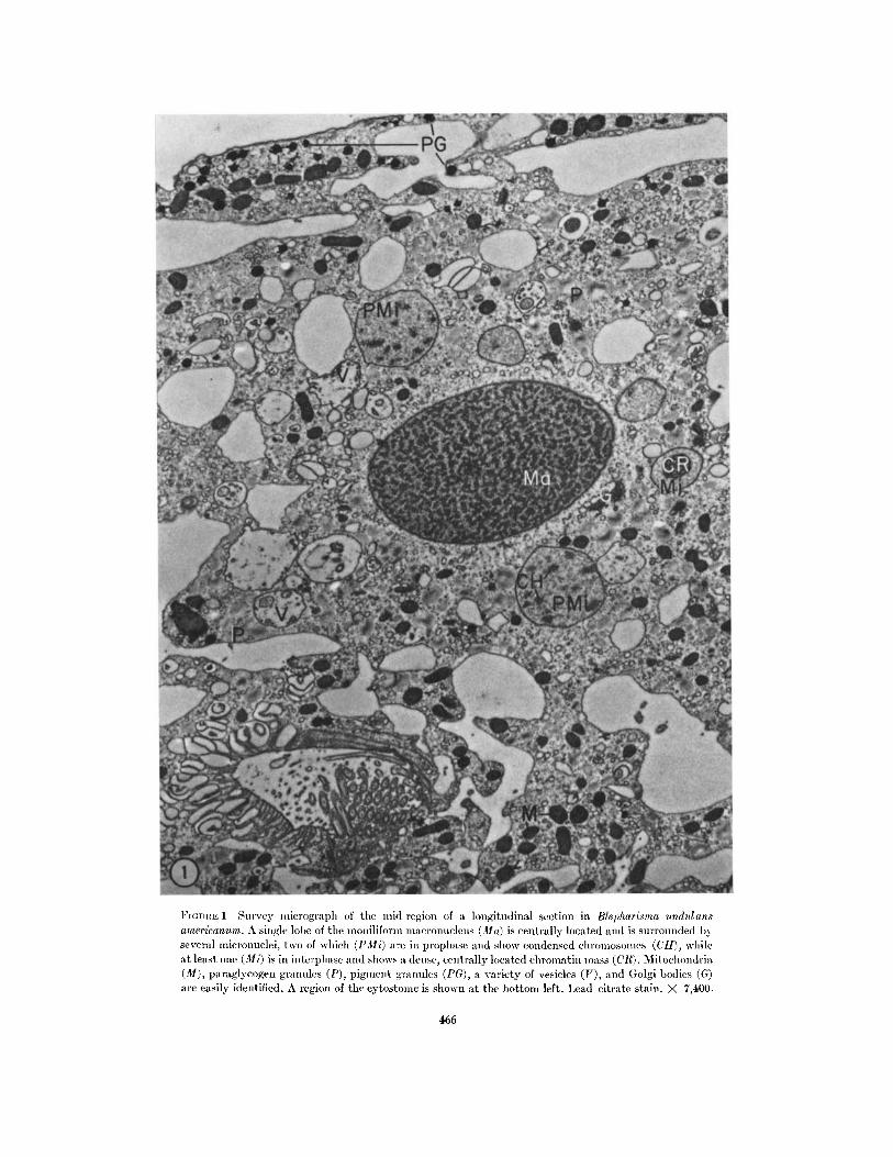
FrGITHtE 1 Survey micrograpll of the mid-region of a longitudinal section in Blepharisma undulansamericanurn. A single lobe of the moniliform macronucleus (la) is centrally located and is surrounded byseveral mlicronuclei, two of which (I'Mi) are in prophase and show condensed cllromlosonles (CII), whileat least one (Mi) is in interphase and shows a dense, centrally located chromatin mass (ClR). Mitochondria(M), paraglycogen granules (P), pigmlent granules (PG), a variety of vesicles (V), and (;olgi bodies (G)are easily identified. A region of the cytostomle is shown at the bottom left. Lead citrate stain. X 7,400.
466
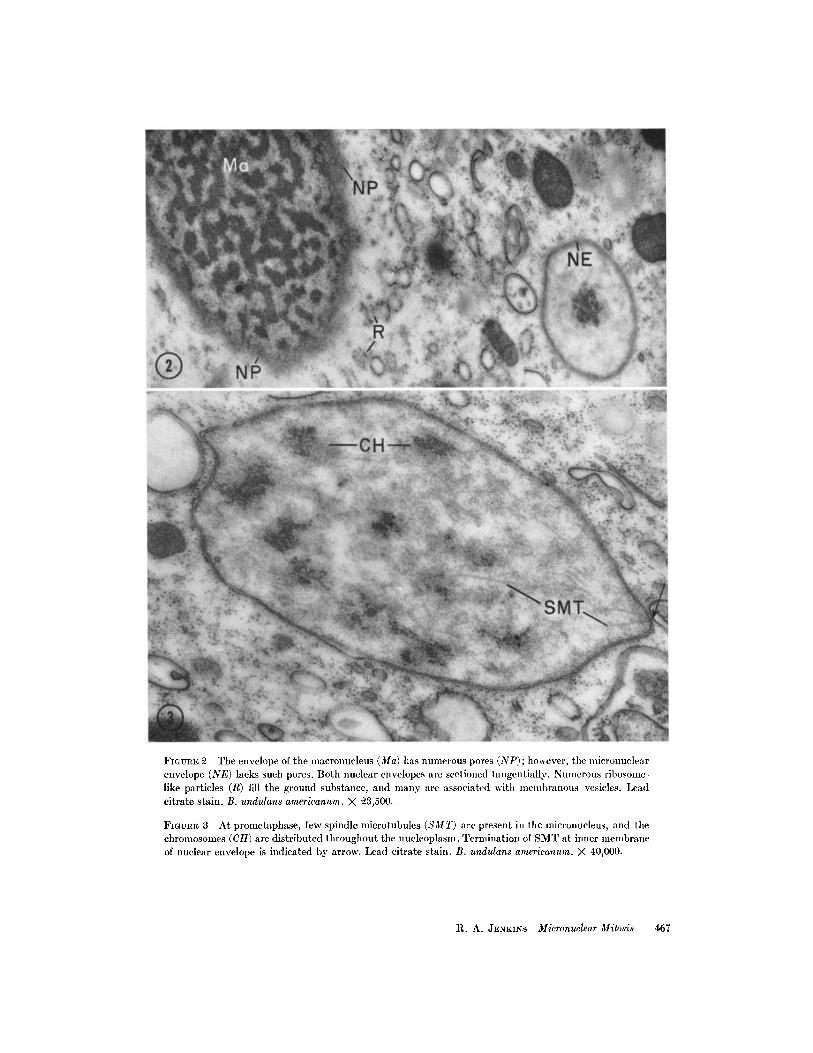
FIGURE 2 The envelope of the macronucleus (Ma) has numerous pores (NP); however, the nlicronuclearenvelope (NE) lacks such pores. Both nuclear envelopes are sectioned tangentially. Numerous ribosome-like particles (R) fill the ground substance, and many are associated with membranous vesicles. Leadcitrate stain. B. undulans americanum. X 23,500.
FIGuRE 3 At prometaphase, few spindle microtubules (SMT) are present in the micronucleus, and thechromosomes (CH) are distributed throughout the nucleoplasm. Termination of SMT at inner membraneof nuclear envelope is indicated by arrow. Lead citrate stain. B. undulans americanum. X 40,000.
R. A. JENKINS Micronuclear Mitosis 467
in the epoxy mixture. Some SMT terminate at
chromosomes while others pass between them andextend from pole to pole (Fig. 4, SMT and Fig. 5,SMT and C). Early metaphase nuclei are barrel-shaped with slightly depressed polar regions.Later, the mitotic figure is more elongate, typicallyfusiform, and in Fig. 4 shows some evidence of avery early separation of chromosomes (CH).
When sectioned across their long dimension,late metaphase micronuclei reveal numerousSMT evenly distributed throughout the nucleus,except within the body of the chromosomes, andcompletely contained by the nuclear envelope(Fig. 7, SMT and CH); SMT cut in exact cross-section present a circular profile with a dense
cortex and a less dense center. Spindle micro-tubules are surrounded by a fine, fibrous materialwhich is possibly an integral part of the tubule(Figs. 4, 5, and 6, FM); differential concentra-tions of this material are not observed at the
chromosome plate or at the polar region. Nor isthere a concentration layered just inside thenuclear envelope as was found in Diplodinium(37). Metaphase chromosomes are 250-300mu masses with poorly defined substructure except
for a generally fibrous appearance (Figs. 4-7, and9, CH). Longitudinal sections (Fig. 5, CH) andcross-sections (Fig. 7, CH) reveal the same mor-phology and indicate that the chromosome hasthe form of a tightly coiled, spherical mass. Well-defined structures (kinetochores) marking chromo-some connections with SMT have not been ob-served (Fig. 6, arrows).
The macronucleus shows a more homogeneousnucleoplasm at micronuclear metaphase, and,
as its elongation begins, microtubules are ob-served for the first time at an extranuclear location,situated adjacent to the nuclear envelope anddirected parallel with the axis of the elongation(Figs. 4, and cross-section Fig. 8, E). These micro-tubules have the same morphology as the SMT
of the micronucleus and are similar in location
to those described by others (36, 37). A complete
description of this "extranuclear division appa-ratus" will be the subject of a subsequent report.
Anaphase
Early anaphase micronuclei provide the clear-
est demonstration that both continuous and
chromosomal fibers (composed of SMT) are pres-
ent in the micronuclear mitotic apparatus (Fig. 6,
SMT'). The interzonal space previously occupied
by the now separated chromosomes contains finefibrous material; this indicates that at least a por-tion of this material is apparently an unstructuredenvironment which surrounds the SMT (Fig. 6,FM). In mid-anaphase, when a chromosomeseparation of 4 5 /p has been effected, SMT arepresent at the poleward side in greatest numbers,but they are also present in the interzone (Fig.9, I, SMT). There is some evidence for a slighttorsion of the entire nuclear apparatus at thisstage; unfortunately this is shown to better ad-vantage in sections adjacent to that shown inFig. 9. Suzuki (47) shows drawings which havesuch a feature; Schwartz (41) cites twisting ofthe anaphase figure as a typical feature in isolatedmicronuclei of P. bursaria; a similar phenomenonhas been described in dividing amebae nuclei(21). The nuclear envelope remains continuous,but on occasion a less regular separation betweencomponent membranes occurs (Fig. 9, arrows).
Measurement of several metaphase nucleireveals a plate-to-pole distance of approximately2.25 Ap. There appears to be a slight shorteningof this distance as the interzonal distance increasesduring anaphase. Based on limited measurementsof anaphase figures, a plate-to-pole distance ofabout 1.75 /z is found, while distal ends of thesame nuclei are separated by several times thisdistance. Chromosome separation is primarilyby elongation of continuous SMT.
In very late anaphase micronuclei, the chromo-somes of the daughter nuclei are fused as singlemasses which resemble the interphase form andare located at each end of the mitotic figure(Figs. 10 and 11, CR). Chromosome-to-pole SMTare no longer detectable, and an abundance offine fibrous material surrounds the SMT at thistime (Fig. 11, FM). A short time later, there isindication that the chromatin mass has "rolledto the side" and continuous SMT extend beyondit and distend the nuclear envelope (Fig. 14,SMT).
Telophase
During anaphase the spatial separation ofdaughter nuclei is completed, and the chromatinmass fully regains its spherical form and the densereticulum typical of interphase (compare CR,Figs. 1 and 10). The two presumptive daughternuclei are enclosed, during the early part oftelophase, within a common milieu as describedby the intact nuclear envelope. When an early
468 TuE JOURNAL OF CELL BIOLOGY VOLUME 4, 1967
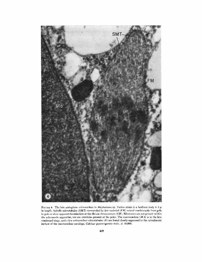
FiGunE 4 The late metaphase micronucleus in Blepharisma sp. Turtox strain is a fusiform body 4-5 gin length. Spindle microtubules (SMT) surrounded by fine material (FM) extend continuously from poleto pole or show apparent termination at the fibrous chromosomes (CH). Ribosomes are not present withinthe achromatic apparatus, nor are centrioles present at the poles. The macronucleus (MA) is in the latecondensed stage, and a few extranuclear microtubules (E) are found closely appressed to the cytoplasmicsurface of the macronuclear envelope. Calcium permanganate stain. X 40,000.
469
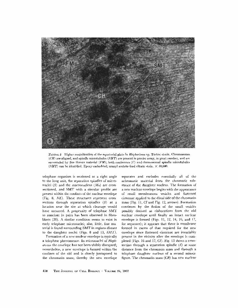
FIGURE 5 Higher magnification of the equatorial plate in Blepharisma sp. Turtox strain. Chromosomes(CH) are aligned, and spindle microtubules (SMT) are present in precise array, in great number, and aresurrounded by fine fibrous material (FM); both continuous (C) and chromosomal spindle microtubules(SMT) can be identified. Epoxy-embedded; uranyl acetate-lead citrate stain. X 50,000.
telophase organism is sectioned at a right angleto the long axis, the separation spindles of micro-
nuclei (S) and the macronucleus (Ma) are cross-sectioned, and SMT with a circular profile arepresent within the confines of the nuclear envelope(Fig. 8, NE). These structures represent cross-
sections through separation spindles (S) at alocation near the site at which cleavage wouldhave occurred. A propensity of telophase SMTto associate in pairs has been observed in fibro-blasts (20). A similar condition seems to exist inearly telophase micronuclei; also, little, fine ma-terial is found surrounding SMT in regions distantto the daughter nuclei (Figs. 8 and 13, SMT).
Formation of a new nuclear envelope is typicallya telophase phenomenon. In micronuclei of Bleph-
arisma the envelope has not been visibly disrupted;nevertheless, a new envelope is formed within theconfines of the old and is closely juxtaposed tothe chromatin mass; thereby the new envelope
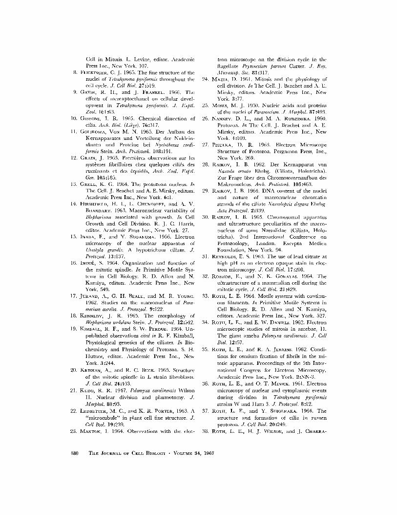
separates and excludes essentially all of theachromatic material from the chromatic sub-stance of the daughter nucleus. The formation ofa new nuclear envelope begins with the appearanceof small membranous vesicles and flattenedcisternae applied to the distal side of the chromatinmass (Fig. 11, CI and Fig. 12, arrows). Formationcontinues by the fusion of the small vesiclespossibly derived as elaborations from the oldnuclear envelope until finally an intact nuclearenvelope is formed (Figs. 11, 12, 14, 16, and 17,for sequence); it appears that there is membraneformed in excess of that required for the newenvelope since flattened cisternae are invariablypresent in the vicinity after the envelope is com-pleted (Figs. 16 and 17, CI). Fig. 13 shows a cross-section through a separation spindle (S) at somedistance from the chromatin mass and through atelophase daughter nucleus of a second mitoticfigure. The chromatin mass (CR) has new nuclear
470 THE JOURNAL OF CELL BIOLOGY VOLUME 34, 1967
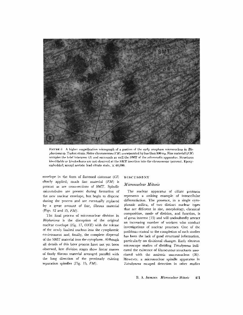
FIGURE 6 A higher magnification micrograph of a portion of the early anaphase micronucleus in Ble-pharisma sp. Turtox strain. Sister chromosomes (CH) areseparated byless than 300 mn. Fine material (FM)occupies the brief interzone (I) and surrounds as well the SMT of the achromatic apparatus. Structuresidentifiable as kinetochores are not observed at the SMT insertion into the chromosome (arrows). Epoxy-embedded; uranyl acetate-lead citrate stain. X 60,000.
envelope in the form of flattened cisternae (CI)closely applied; much fine material (FM) ispresent as are cross-sections of SMT. Spindlemicrotubules are present during formation ofthe new nuclear envelope, but begin to disperseduring the process and are eventually replacedby a great amount of fine, fibrous material(Figs. 12 and 15, FM).
The final process of micronuclear division inBlepharisma is the disruption of the originalnuclear envelope (Fig. 17, ONE) with the releaseof the newly limited nucleus into the cytoplasmicenvironment and, finally, the complete dispersalof the SMT material into the cytoplasm. Althoughall details of this later process have not yet beenobserved, late division stages show linear massesof finely fibrous material arranged parallel withthe long direction of the previously existingseparation spindles (Fig. 15, FM).
DISCUSSION
MIicronuclear Mitosis
The nuclear apparatus of ciliate protozoarepresents a striking example of intracellulardifferentiation. The presence, in a single cyto-plasmic milieu, of two distinct nuclear typesthat are different in size, morphology, chemicalcomposition, mode of division, and function, isof great interest (13) and will undoubtedly attractan increasing number of workers who conductinvestigations of nuclear processes. One of theproblems central to the completion of such studies
has been the lack of good structural information,
particularly on divisional changes. Early electron
microscope studies of dividing Tetrahymena indi-cated the existence of filamentous structures asso-
ciated with the amitotic macronucleus (36).However, a micronuclear spindle apparatus inTetrahymena escaped detection in other studies
R. A. JENKINS Micronuclear Mitosis 471
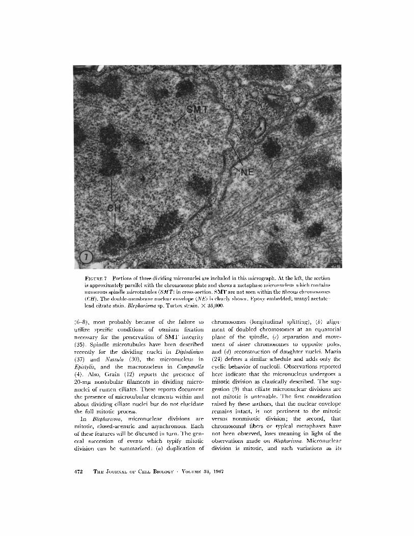
FIGURE 7 Portions of three dividing micronuclei are included in this mIicrograph. At the left, the sectionis approximately parallel with the chromosome plate and shows a inetaphase micronucleus which containsnumerous spindle Ilicrotubules (SM T) in cross-section. SMT are not seen within the fibrous chlromosomlles(CH). Trhe double-membrane nuclear envelope (NE) is clearly shown. Epoxy-embedded; uranyl acetate-lead citrate stain. Blepharisma sp. Tultox strain. X 35,000.
(6 8), most probably because of the failure to
utilize specific conditions of smium fixationnecessary for the preservation of SMT integrity
(35). Spindle microtubules have been describedrecently for the dividing nuclei in Diplodinium
(37) and Nassula (30), the micronucleus inEpistylis, and the macronucleus in Campanella
(4). Also, Grain (12) reports the presence of
20-mgu nontubular filaments in dividing micro-nuclei of rumen ciliates. These reports document
the presence of microtubular elements within and
about dividing ciliate nuclei but do not elucidate
the full mitotic process.In Blepharisma, micronuclear divisions are
mitotic, closed-acentric and asynchronous. Each
of these features will be discussed in turn. The gen-
eral succession of events which typify mitoticdivision can be summarized: (a) duplication of
chromosomes (longitudinal splitting), (b) align-ment of doubled chromosomes at an equatorialplane of the spindle, (c) separation and move-ment of sister chromosomes to opposite poles,and (d) reconstruction of daughter nuclei. Mazia(24) defines a similar schedule and adds only thecyclic behavior of nucleoli. Observations reportedhere indicate that the micronucleus undergoes amitotic division as classically described. The sug-gestion (9) that ciliate micronuclear divisions arenot mitotic is untenable. The first considerationraised by these authors, that the nuclear enveloperemains intact, is not pertinent to the mitoticversus nonmitotic division; the second, thatchromosomal fibers or typical metaphases havenot been observed, loses meaning in light of theobservations made on Blepharisma. Micronucleardivision is mitotic, and such variations as its
472 TIIE JOURNAL OF CELL BIOLOGY VOLUME 34, 1967
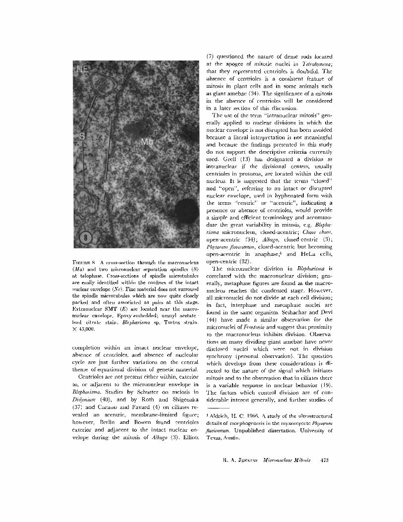
FIuaRE 8 A cross-section through the macronucleus(Ma) and two icronuclear separation spindles (S)at telophase. Cross-sections of spindle icrotubulesare easily identified within the confines of the intactnuclear envelope (Ne). Fine material does not surroundthe spindle microtubules which are now quite closelypacked and often associated as pairs at this stage.Extranuclear SMT (E) are located near the macro-nuclear envelope. Epoxy-embedded; uranyl acetate-lead citrate stain. Blepharisma sp. Turtox strain.X 45,000.
completion within an intact nuclear envelope,absence of centrioles, and absence of nucleolarcycle are just further variations on the centraltheme of equational division of genetic material.
Centrioles are not present either within, exterior
to, or adjacent to the micronuclear envelope in
Blepharisma. Studies by Schuster on meiosis in
Didymium (40), and by Roth and Shigenaka
(37) and Carasso and Favard (4) on ciliates re-
vealed an acentric, membrane-limited figure;
however, Berlin and Bowen found centrioles
exterior and adjacent to the intact nuclear en-
velope during the mitosis of Albugo (3). Elliott
(7) questioned the nature of dense rods located
at the apogee of mitotic nuclei in Tetrahymena;
that they represented centrioles is doubtful. The
absence of centrioles is a consistent feature of
mitosis in plant cells and in some animals such
as giant amebae (34). The significance of a mitosisin the absence of centrioles will be consideredin a later section of this discussion.
The use of the term "intranuclear mitosis" gen-erally applied to nuclear divisions in which thenuclear envelope is not disrupted has been avoidedbecause a literal interpretation is not meaningful
and because the findings presented in this studydo not support the descriptive criteria currentlyused. Grell (13) has designated a division as
intranuclear if the divisional centers, usually
centrioles in protozoa, are located within the cellnucleus. It is suggested that the terms "closed"
and "open", referring to an intact or disrupted
nuclear envelope, used in hyphenated form withthe terms "centric" or "acentric", indicating a
presence or absence of centrioles, would provide
a simple and efficient terminology and accommo-date the great variability in mitosis, e.g. Blepha-
risma micronucleus, closed-acentric; Chaos chaos,
open-acentric (34); Albugo, closed-centric (3);Physarum fiavicomum, closed-acentric but becoming
open-acentric in anaphase;' and HeLa cells,
open-centric (32).The micronuclear division in Blepharisma is
correlated with the macronuclear division; gen-
erally, metaphase figures are found as the macro-
nucleus reaches the condensed stage. However,
all micronuclei do not divide at each cell division;
in fact, interphase and metaphase nuclei are
found in the same organism. Seshachar and Devi
(44) have made a similar observation for the
micronuclei of Frontonia and suggest that proximityto the macronucleus inhibits division. Observa-
tions on many dividing giant amebae have neverdisclosed nuclei which were not in division
synchrony (personal observation). The question
which develops from these considerations is di-rected to the nature of the signal which initiatesmitosis and to the observation that in ciliates there
is a variable response in nuclear behavior (19).The factors which control division are of con-
siderable interest generally, and further studies of
' Aldrich, H. C. 1966. A study of the ultrastructuraldetails of morphogenesis in the myxomycete Physarum
fiavicomum. Unpublished dissertation. University ofTexas, Austin.
R. A. JENKINS Micronuclear Mitosis 473
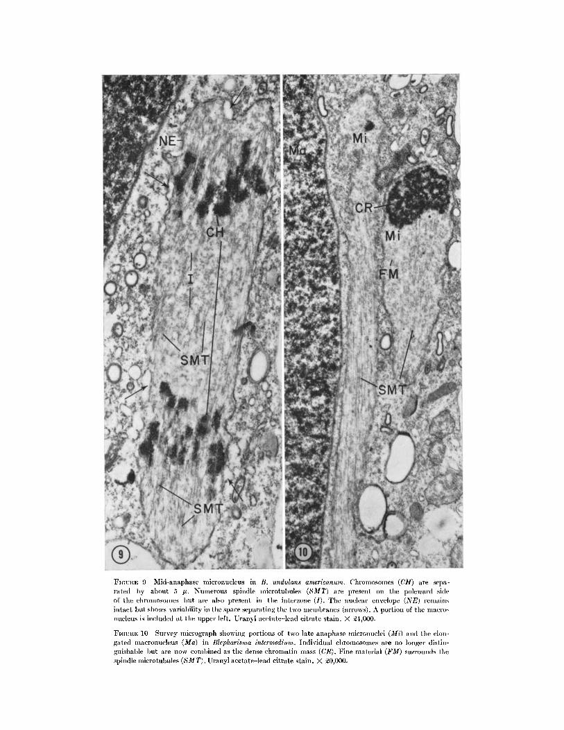
FIGIUTE 9 Mid-anaphase micronucleus in B. undulans americanum. Chromosomes (CH) are sepa-rated by about 5 u. Numerous spindle microtubules (SMT) are present on the poleward sideof the chromosomes but are also present in the interzone (I). The nuclear envelope (NE) remainsintact but shows variability in the space separating the two membranes (arrows). A portion of the macro-nucleus is included at the upper left. Uranyl acetate-lead citrate stain. X 24,000.
FIGURE 10 Survey micrograph showing portions of two late anaphase micronuclei (Mi) and the elon-gated macronucleus (Ma) in Blepharisma intermedium. Individual chromosomes are no longer distin-guishable ut are now combined as the dense chromatin mass (CR). Fine material (FM) surrounds thespindle microtubules (SMT). UJranyl acetate-lead citrate stain. X 20,000.
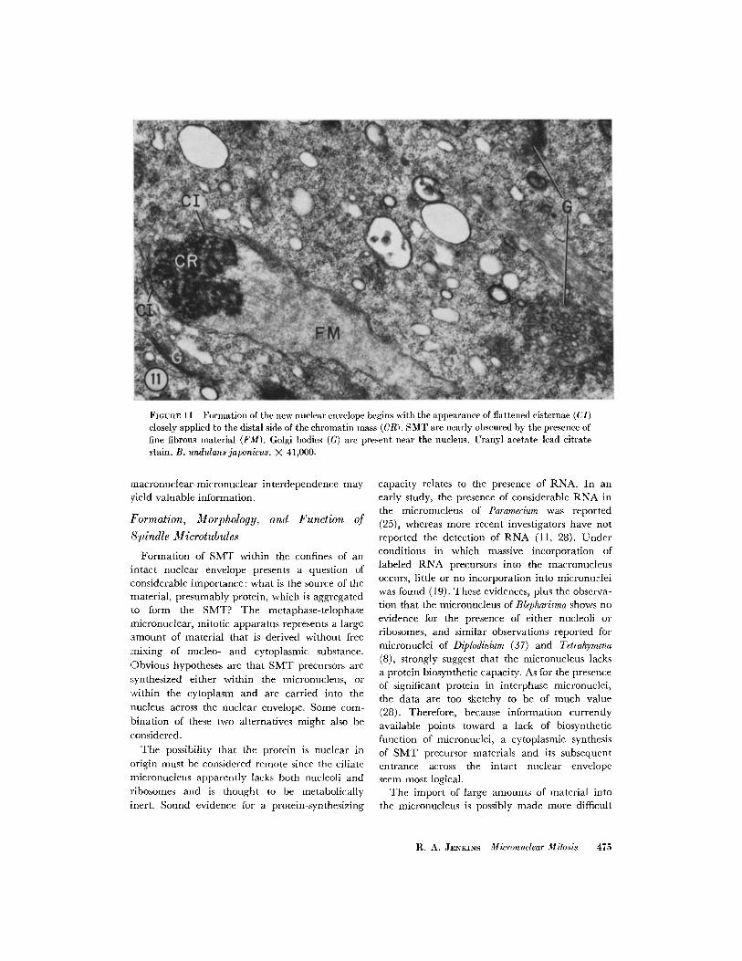
FIGURE 11 Formation of the new nuclear envelope begins with the appearance of flattened cisternae (CI)closely applied to the distal side of the chromatin mass (CR). SMT are nearly obscured by the presence offine fibrous material (FM). Golgi bodies (G) are present near the nucleus. Uranyl acetate-lead citratestain. B. undulans japonicus. X 41,000.
macronuclear-micronuclear interdependence mayyield valuable information.
Formation, Morphology, and Function of
Spindle Mllicrotubules
Formation of SMT within the confines of anintact nuclear envelope presents a question ofconsiderable importance: what is the source of thematerial, presumably protein, which is aggregatedto form the SMT? The metaphase-telophase:micronuclear, mitotic apparatus represents a largeamount of material that is derived without freemixing of nucleo- and cytoplasmic substance.Obvious hypotheses are that SMT precursors aresynthesized either within the micronucleus, orwithin the cytoplasm and are carried into thenucleus across the nuclear envelope. Some com-bination of these two alternatives might also beconsidered.
The possibility that the protein is nuclear inorigin must be considered remote since the ciliatemicronucleus apparently lacks both nucleoli andribosomes and is thought to be metabolicallyinert. Sound evidence for a protein-synthesizing
capacity relates to the presence of RNA. In anearly study, the presence of considerable RNA inthe micronucleus of Paramecium was reported(25), whereas more recent investigators have notreported the detection of RNA (11, 28). Underconditions in which massive incorporation oflabeled RNA precursors into the macronucleusoccurs, little or no incorporation into micronucleiwas found (19). These evidences, plus the observa-tion that the micronucleus of Blepharisma shows noevidence for the presence of either nucleoli orribosomes, and similar observations reported formicronuclei of Diplodinium (37) and Tetrahymena(8), strongly suggest that the micronucleus lacksa protein biosynthetic capacity. As for the presenceof significant protein in interphase micronuclei,the data are too sketchy to be of much value(28). Therefore, because information currentlyavailable points toward a lack of biosyntheticfunction of micronuclei, a cytoplasmic synthesisof SMT precursor materials and its subsequententrance across the intact nuclear envelopeseem most logical.
The import of large amounts of material intothe micronucleus is possibly made more difficult
R. A. JENKINS Micronuclear Mitosis 475
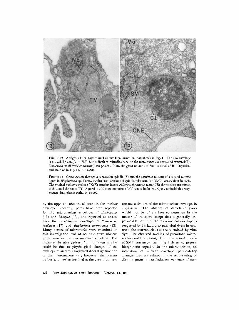
FIGunE 12 A slightly later stage of nuclear envelope formation than shown in Fig. 11. The new envelopeis essentially complete (NE) but difficult to visualize because the membranes are sectioned tangentially.Numerous small vesicles (arrows) are present. Note the great amount of fine material (FM). Organismand stain as in Fig. 11. X 43,000.
FIGURE 13 Cross-section through a separation spindle (S) and the daughter nucleus of a second mitoticfigure in Blepharisma sp. Turtox strain; cross-sections of spindle microtubules (SMT) are evident in each.The original nuclear envelope (ONE) remains intact while the chromatin mass (CR) shows close appositionof flattened cisternae (CI). A portion of the macronucleus (Ma) is also included. Epoxy-embedded; uranylacetate-lead citrate stain. X 4,000.
by the apparent absence of pores in the nuclearenvelope. Recently, pores have been reportedfor the micronuclear envelopes of Blepharisma(18) and Urostyla (15), and reported as absentfrom the micronuclear envelopes of Parameciumcaudatum (17) and Blepharisma intermedium (42).Many dozens of micronuclei were examined inthis investigation and at no time were obviouspores seen in the micronuclear envelope. Thedisparity in observations from different studiescould be due to physiological changes of theenvelope related to a suggested short stage functionof the micronucleus (8); however, the presentauthor is somewhat inclined to the view that pores
are not a feature of the micronuclear envelope inBlepharisma. The absence of detectable pores
would not be of absolute consequence in thematter of transport except that a generally im-
permeable nature of the micronuclear envelope issuggested by its failure to pass vital dyes; in con-trast, the macronucleus is easily stained by vitaldyes. The observed swelling of premitotic micro-nuclei could represent, if not the actual uptakeof SMT precursor (assuming little or no proteinbiosynthetic capacity for the micronucleus), anindication of nuclear envelope permeabilitychanges that are related to the sequestering ofdivision protein; morphological evidence of such
476 TIIE JOURNAL OF CELL BIOLOGY VOLUME 34, 1967
changes was not obtained in this study. In micro-
nuclei of Nassulopsis the increase in dimension
occurs only during the G2 phase, after DNA
synthesis is completed (29); therefore, in this
organism at least, the swelling cannot be attributed
to DNA synthesis directly and is more probably a
result of some predivision activity of the micro-nucleus. Morphological features that wouldsupport either micronuclear biosynthesis or
facile membrane transport of spindle precursorare not demonstrated in this study. Thus an ex-
planation for the immediate source of SMT ma-terial cannot be advanced at this time.
Let us turn our attention to the initiation ofSMT formation. Inou6 has continually empha-sized that fibers of the mitotic apparatus areorganized and oriented by "centers" such askinetochores and centrioles (16). Neither centriolesnor kinetochores have been observed in micro-
nuclear mitosis in Blepharisma; the only likely
"center of aggregation" in a closed-acentric
mitosis is a region of the chromosome, here not
distinguished by defined structure. However, the
role of centers of formation might be questioned
on three major counts. First, the nearly ubiquitous
occurrence of microtubules in cells and particu-
larly in areas of cells where direct association witha center cannot be demonstrated (22, 47, 48)indicates a more independent scheme. Taylor
(47) has suggested that, if microtubules dooriginate from organizing or initiating molecular
configurations, these centers may reside on mem-
brane surfaces. Roth et al. (38) emphasize thesignificance of the association of microtubules withmembrane surfaces. The present study as well
as that of Tilney and Porter (48) on axopodiademonstrate the termination of great numbers ofmicrotubules near the nuclear envelope, albeit
to opposite sides; there is, however, no indication
of any elaboration at this site. Manton (23) re-ports the apparent termination of SMT on piecesof nuclear envelope in P. parvum. The significanceof the association of microtubules with membranesurfaces is not yet clear but seems worthy of
further consideration. Second, the electron micro-
scopic observation of well-defined kinetochoreswith any indication of tubular morphology isnot consistent, nor is a firm connection of SMTto centrioles ever clearly shown. It may be, how-
ever, that the structure of each of these junctions
(centers) is highly labile and that fixation suffi-
cient to preserve the formed or forming SMT is
not sufficient to preserve the fine detail of a struc-
ture (region) representing a site of aggregation.Third, within the acentric mitotic apparatus
SMT are present which are continuous from pole
to pole and lack connections to either chromo-
somes or centrioles and, in the case of micro-
nuclei, lack direct contact with the cytoplasm. It
is probable that these continuous SMT are respon-sible for the greater separation of daughter nuclei
in many cells (for survey see Roth et al. 38).
Since these SMT are essentially free from direct
association with known centers, it is appropriateto implement a hypothesis which considers their
formation as resulting from changes in the micro-environment that are known to control micro-
tubule assembly in vitro such as pH, ionic strength
(1), or the presence of a specific linker substance(RNA; 39, 49, 50); such a concept might entail
growth of the microtubule by insertion of material
along its length or addition at the ends (33) as
well as the presence of initial seeds.Spindle microtubules of the dividing micro-
nucleus are morphologically identical with micro-
tubules described for the great number of cells
previously referred to. Attention is called, how-
ever, to the fact that reported differences in the
diameter of SMT are apparently due to the useof different preparative techniques, as earlier
suggested by Ledbetter and Porter (22). Organisms
handled in an identical manner up to the em-
bedding step showed SMT approximately 22 mg
in diameter when in epoxy and 16 mg in diam-eter when in methacrylate-DVB; considerable
caution should be used in attaching significanceto differences in diameter. As is the case in other
cells studied with the electron microscope, no
changes in SMT diameter or morphology can
be determined for different stages of division.
That the fine material found adhering to the
surface of SMT is an integral part of the micro-
tubular morphology has been suggested (34).There is a great amount of fine, fibrous materialassociated with the SMT of the micronucleus in
Blepharisma, but it is not clear whether this ma-
terial is wholly a lateral component of the micro-
tubule or represents, at least in part, an unstruc-
tured phase serving possibly as precursor material.The material closely resembles that found sur-rounding the microtubules of the ciliary shaft.
Single SMT of early telophase separation spin-
dles are found to be continuous for several microns;
the logical conclusion is that individual continu-
ous SMT undergo a great increase in length
during the course of mitosis. In this regard, SMT
R. A. JENKINS Micronuclear Mitosis 477
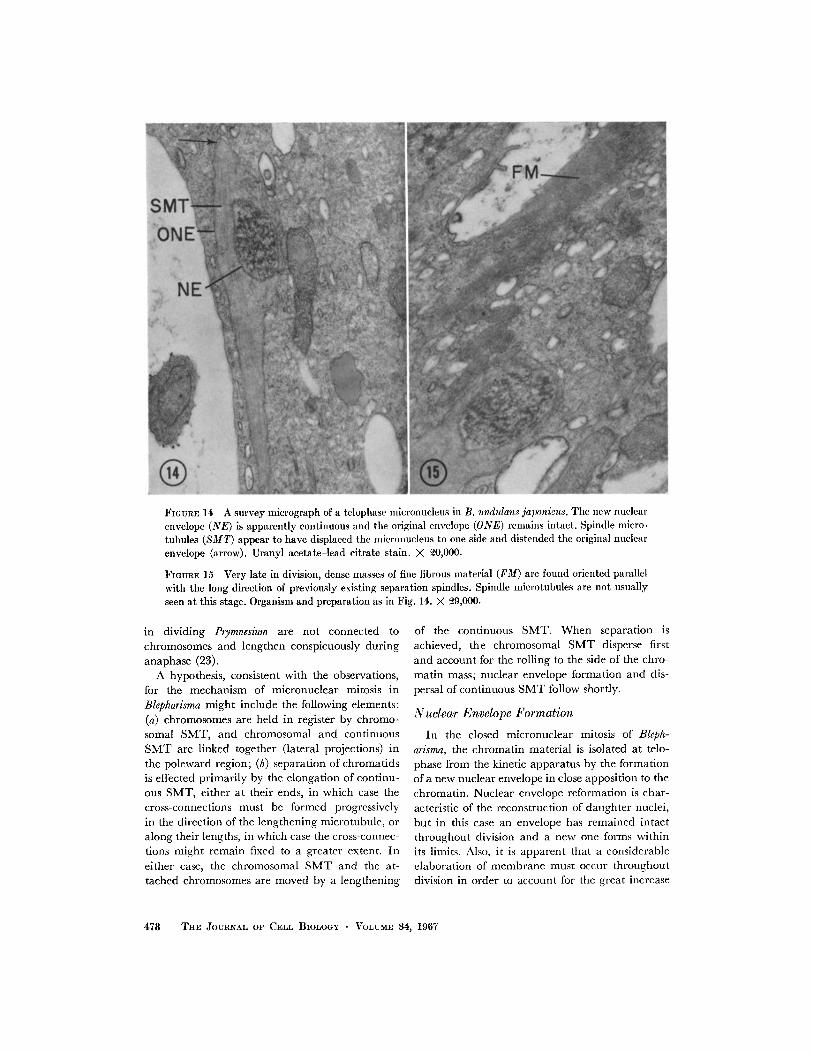
FIGURE 14 A survey micrograph of a telophase micronucleus in B. undulans japonicus. The new nuclear
envelope (NE) is apparently continuous and the original envelope (ONE) remains intact. Spindle micro-tubules (SMT) appear to have displaced the micronucleus to one side and distended the original nuclear
envelope (arrow). Uranyl acetate-lead citrate stain. X 20,000.
FIGURE 15 Very late in division, dense masses of fine fibrous material (FM) are found oriented parallel
with the long direction of previously existing separation spindles. Spindle microtubules are not usually
seen at this stage. Organism and preparation as in Fig. 14. X 29,000.
in dividing Prymnesium are not connected tochromosomes and lengthen conspicuously duringanaphase (23).
A hypothesis, consistent with the observations,for the mechanism of micronuclear mitosis inBlepharisma might include the following elements:(a) chromosomes are held in register by chromo-somal SMT, and chromosomal and continuousSMT are linked together (lateral projections) inthe poleward region; (b) separation of chromatidsis effected primarily by the elongation of continu-ous SMT, either at their ends, in which case thecross-connections must be formed progressivelyin the direction of the lengthening microtubule, oralong their lengths, in which case the cross-connec-tions might remain fixed to a greater extent. Ineither case, the chromosomal SMT and the at-tached chromosomes are moved by a lengthening
of the continuous SMT. When separation is
achieved, the chromosomal SMT disperse first
and account for the rolling to the side of the chro-
matin mass; nuclear envelope formation and dis-
persal of continuous SMT follow shortly.
Nuclear Envelope Formation
In the closed micronuclear mitosis of Bleph-
arisma, the chromatin material is isolated at telo-
phase from the kinetic apparatus by the formation
of a new nuclear envelope in close apposition to the
chromatin. Nuclear envelope reformation is char-
acteristic of the reconstruction of daughter nuclei,
but in this case an envelope has remained intact
throughout division and a new one forms within
its limits. Also, it is apparent that a considerable
elaboration of membrane must occur throughout
division in order to account for the great increase
478 THE JOURNAL OF CELL BIOLOGY VOLUME 34, 1967
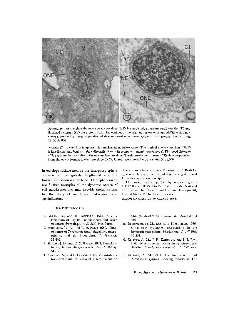
FIGURE 16 At the time the new nuclear envelope (NE) is completed, numerous small vesicles (V) andflattened cisternae (CI) are present within the confines of the original nuclear envelope (ONE) which nowshows a greater than usual separation of its component membranes. Organism and preparation as in Fig.14. X 39,000.
FIGUsE 17 A very late telophase micronucleus in B. intermedium. The original nuclear envelope (ONE)is less distinct and begins to show discontinuities in itscomponent membranes (arrows). Flattened cisternae(CI) are found in proximity to the new nuclear envelope. The dense chromatin mass (CR) shows separationfrom the newly formed nuclear envelope (NE). Uranyl acetate-lead citrate stain. X 40,000.
in envelope surface area as the interphase sphere
converts to the greatly lengthened structure
formed as division is completed. These phenomena
are further examples of the dynamic nature of
cell membranes and may provide useful systems
for the study of membrane elaboration and
specialization.
The author wishes to thank Professor L. E. Roth forguidance during the course of this investigation andfor review of the manuscript.
The work was supported by research grantsHD1260 and HD2585 to Dr. Roth from the NationalInstitute of Child Health and Human Development,United States Public Health Service.
Received for publicaiton 10 December 1966.
REFERENCES
1. ABRAM, D., and H. KOFFLER. 1964. In vitro
formation of flagella-like filaments and otherstructures from flagellin. J. Mol. Biol. 9:168.
2. ANDERSON, W. A., and R. A. ELLIS. 1965. Ultra-
structure of Typanosoma lewisi: flagellum, micro-tubules, and the kinetoplast. J. Protozool.12:483.
3. BERLIN, J. D., and C. C. BOWEN. 1964. Centriolesin the fungus Albzugo candida. Am. J. Botany.51:650.
4. CARASSO, N., and P. FAVARD. 1965. Microtubules
fusoriaux dans les micro et macronucleus de
cilies peritriches en division. J. Microscop. 4:395.
5. DEMBITZER, H. M., and H. I. HIRSHFIELD. 1966.
Some new cytological observations in theheterotrichous ciliate, Blepharisma. J. Cell Biol.30:201.
6. ELLIOTT, A. M., J. R. KENNEDY, and I. J. BAK.1962. Macronuclear events in synchronouslydividing Telrahymena pyriformis. J. Cell Biol.12:515.
7. ELLIOTT, A. M. 1963. The fine structure ofTetrahymena pyriformis during mitosis. In The
R. A. JENKINS Micronuclear Mitosis 479
Cell in Mitosis. L. Levine, editor. AcademicPress Inc., New York. 107.
8. FLICKINGER, C. J. 1965. The fine structure of the
nuclei of Tetrahymena pyriformis throughout thecell cycle. J. Cell Biol. 27:519.
9. GAVIN, R. H., and . FRANKEL. 1966. The
effects of mercaptoethanol on cellular devel-opment in Tetrahymena pyriformis. J. Exptl.Zool. 161:63.
10. GIBBONS, I. R. 1965. Chemical dissection ofcilia. Arch. Biol. (Liege). 76:317.
11. GOLIKOWA, VON M. N. 1965. Der Aufbau des
Kernapparates und Verteilung der Nuklein-siiuren und Proteine bei Nyctotherus cordi-formis Stein. Arch. Protistenk. 108:191.
12. GRAIN, J. 1965. Premieres observations sur lessystlmes fibrillaires chez quelques cilihs desruminants et des quidhs, Arch. Zool. Exptl.Gen. 105:185.
13. GRELL, K. G. 1964. The protozoan nucleus. InThe Cell. J. Brachet and A. E. Mirsky, editors.Academic Press Inc., New York. 6:1.
14. HIRSHFIELD, H. I., L. CHUNOSOFF, and A. V.BHANDARY. 1963. Macronuclear variability of
Blepharisma associated with growth. In CellGrowth and Cell Division. R. J. C. Harris,editor. Academic Press Inc., New York. 27.
15. INABA, F., and Y. SUGANUMA. 1966. Electronmicroscopy of the nuclear apparatus ofUrostyla grandis. A hypotrichous ciliate. J.Protozool. 13:137.
16. INouE, S. 1964. Organization and function ofthe mitotic spindle. In Primitive Motile Sys-tems in Cell Biology. R. D. Allen and N.Kamiya, editors. Academic Press Inc., NewYork. 549.
17. JURAND, A., G. H. BEALE, and M. R. YOUNG.
1962. Studies on the macronucleus of Para-mecium aurelia. J. Protozool. 9:122.
18. KENNEDY, J. R. 1965. The morphology ofBlepharisma undulans Stein. J. Protozool. 12:542.
19. KIMBALL, R. F., and S. W. PERDUE. 1964. Un-published observations cited in R. F. Kimball,Physiological genetics of the ciliates. In Bio-chemistry and Physiology of Protozoa. S. H.Hutner, editor. Academic Press Inc., NewYork. 3:244.
20. KRISHAN, A., and R. C. BUCK. 1965. Structure
of the mitotic spindle in L strain fibroblasts.J. Cell Biol. 24:433.
21. KUDO, R. R. 1947. Pelomyxa carolinensis WilsonII. Nuclear division and plasmotomy. J.Morphol. 80:93.
22. LEDBETTER, M. C., and K. R. PORTER, 1963. A
"microtubule" in plant cell fine structure. J.Cell Biol. 19:239.
23. MANTON, I. 1964. Observations with the elec-
tron microscope on the division cycle in theflagellate Prymnesium parrum Carter. J. Roy.Microscope. Soc. 83:317.
24. MAZIA, D. 1961. Mitosis and the physiology ofcell division. In The Cell. J. Brachet and A. E.Mirsky, editors. Academic Press Inc., NewYork. 3:77.
25. MosEs, M. J. 1950. Nucleic acids and proteinsof the nuclei of Paramecium. J. Morphol. 87:493.
26. NANNEY, D. L., and M. A. RUDZINSKA. 1960.
Protozoa. In The Cell. J. Brachet and A. E.
Mirsky, editors. Academic Press Inc., NewYork. 4:109.
27. PITELKA, D. R. 1963. Electron Microscope
Structure of Protozoa. Pergamon Press, Inc.,
New York. 269.
28. RAKOV, I. B. 1962. Der Kernapparat vonNassula ornata Ehrbg. (Ciliata, Holotricha).
Zur Frage fiber den Chromosomenaufbau desMakronucleus. Arch. Protistenk. 105:463.
29. RAIKOV, I. B. 1964. DNA content of the nucleiand nature of macronuclear chromatin
strands of the ciliate Nassulopsis elegans EhrbgActa Protozool. 2:339.
30. RAIKOV, I. B. 1965. Chromosomal apparatus
and ultrastructure peculiarities of the macro-nucleus of some Nassulidae (Ciliata, Holo-tricha). 2nd International Conference onProtozoology, London. Exerpta MedicaFoundation, New York. 94.
31. REYNOLDS, E. S. 1963. The use of lead citrate athigh pH as an electron opaque stain in elec-tron microscopy. J. Cell Biol. 17:208.
32. ROBBINs, E., and N. K. GONATAS. 1964. Theultrastructure of a mammalian cell during themitotic cycle. J. Cell Biol. 21:429.
33. ROTH, L. E. 1964. Motile systems with continu-ous filaments. In Primitive Motile Systems inCell Biology. R. D. Allen and N. Kamiya,editors. Academic Press Inc., New York. 527.
34. ROTH, L. E., and E. W. DANIELS. 1962. Electronmicroscopic studies of mitosis in amebae, IT.
The giant ameba Pelomyxa carolinensis. J. Cell
Biol. 12:57.
35. ROTH, L. E., and R. A. JENKINS. 1962. Condi-tions for osmium fixation of fibrils in the mi-totic apparatus. Proceedings of the 5th Inter-
national Congress for Electron Microscopy.Academic Press Inc., New York. 2:NN-3.
36. ROTH, L. E., and 0. T. MINICK. 1961. Electronmicroscopy of nuclear and cytoplasmic eventsduring division in Tetrahymena pytiformis
strains W and Ham 3. J. Protozool. 8:12.37. ROTH, L. E., and Y. SHIGENAKA. 1964. The
structure and formation of cilia in rumenprotozoa. J. Cell Biol. 20:249.
38. ROTH, L. E., H. J. WILSON, and J. CHAKRA-
480 THE JOURNAL OF CELL BIOLOGY VOLUME 34, 1967
BORTY. 1966. Anaphase structure in mitoticcells typified by spindle elongation. J. Ultra-struct. Res. 14:460.
39. SAKAI, H. 1966. Studies on sulfhydryl groupsduring cell division of sea-urchin eggs, VIII.Some properties of mitotic apparatus proteins.Biochim. Biophys. Acta, 112:132.
40. SCHUSTER, F. 1964. Electron microscope obser-
vations on spore formation in the true slimemold Didymium nigripes. J. Protozool. 11:207.
41. SCHWARTZ, V. 1965. Die Teilungsspindel des
Mikronucleus von Paramecium bursaria, Zoolo-gischer Anzieger. 28 (Suppl.):123.
42. SESHACHAR, B. R. 1964. Observations on the
fine structure of the nuclear apparatus ofBlepharisma intermedium Bhandary (Ciliata:Spirotricha). J. Protozool. 11:402.
43. SESHACHAR, B. R. 1965. The fine structure of
the nuclear apparatus and the chromosomesof Spirostomum ambiguum Ehrbg. Acta Protozool.3:337.
44. SESHACHAR, B. R., and R. V. DEVI. 1964. Cy-
tology of Frontonia elliptica (Beardsley). ActaProtozool. 2:247.
45. SzuKI, S. 1954. Taxonomic studies on Blepha-
risma undulans Stein with special reference to
the macronuclear variation. J. Sci. Hiroshima
Univ. Ser. B. 15:205.
46. SUZUKi, S. 1959. Morphogenesis in the regenera-
tion of Blepharisma undulans japonicus Suzuki.Yamagato-Daigaku Kiyo, Shizen Kagaku. 4:85.
47. TAYLOR, A. C. 1966. Microtubules in the micro-spikes and cortical cytoplasm of isolated cells.
J. Cell Biol. 28:155.48. TILNEY, L. G., and K. R. PORTER. 1965. Studies
on microtubules in Heliozoa I. The fine
structure of Actinosphaerium nucleofilium (Bar-rett), with particular reference to the axial rod
structure. Protoplasma. 60:317.
49. WENT, H. A. 1966. The behavior of centrioles
and the structure and formation of the achro-
matic figure. Protoplasmatalogia. 6:1.50. ZIMMERMAN, A. M. 1963. Chemical aspects of
the isolated mitotic apparatus. In The Cell in
Mitosis. L. Levine, editor. Academic PressInc., New York. 159.
R. A. JENKINS Micronuclear Mitosis 481