film photography's limitations in imaging skin of color underlies racial imaging disparities;...
TRANSCRIPT
LETTERS
NOTES & COMMENTS
Film photography’s limitations in imagingskin of color underlies racial imagingdisparities; new digital photography featuresfacilitate the imaging of skin of color
To the Editor: I read with interest Ebede’s October2006 contribution to the Journal entitled ‘‘Disparitiesin dermatology educational resources.’’1 While cul-ture biases of photographers likely sustained thesedisparities, the smaller range in tones that markseruptions in dark skin and film camera technologicallimitations also underlies the situation that Ebedeobserved. PubMed reports do not described num-mular dermatitis in persons of dark skin color (ie,African Americans), in whom purple on a brownbackground is difficult to image. In patients withdark skin color—in whom erythema migrans on abrown background can be hard to see and difficult toimage—only one report with no images noted anincidence of Lyme disease.2
Photographing skin of color and maintaining thecutaneous detail was a challenge for film photogra-phy. To shoot film of dermatoses in dark skin, wherethe range of tones between normal and pathologicskin is narrow, the film had to be overexposed (extralight added when the film was exposed to an object)and underdeveloped (less time given to a film printto reach its final state while being created; alsoknown as pull processing) to hold detail in theshadows without blowing out pathologic highlights.A flash provides additional light to capture and tostandardize image detail; however, a flash washesout color and, in particular when photographing oilyblack skin, leaves flash artifacts.
New technological developments in digital pho-tography might eliminate the disparity of ease ofimaging black and white skin. The most importantdevelopment in this regard is image stabilization,which keeps the lens focused on an object evenwhile the camera moves because of hand tremors,without a flash, and without washing out color orproducing flash artifacts.
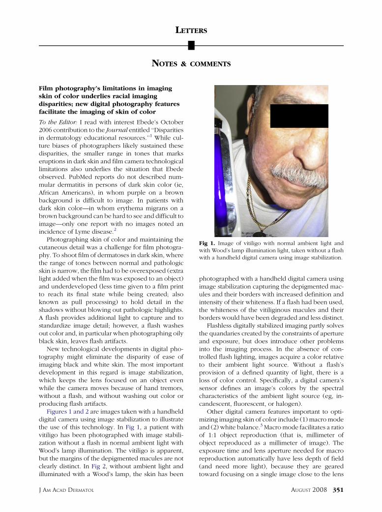
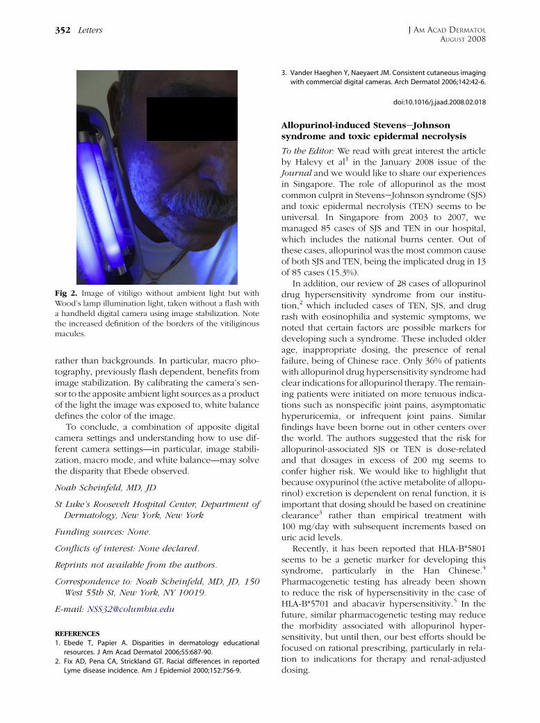
Figures 1 and 2 are images taken with a handhelddigital camera using image stabilization to illustratethe use of this technology. In Fig 1, a patient withvitiligo has been photographed with image stabili-zation without a flash in normal ambient light withWood’s lamp illumination. The vitiligo is apparent,but the margins of the depigmented macules are notclearly distinct. In Fig 2, without ambient light andilluminated with a Wood’s lamp, the skin has been
J AM ACAD DERMATOL
photographed with a handheld digital camera usingimage stabilization capturing the depigmented mac-ules and their borders with increased definition andintensity of their whiteness. If a flash had been used,the whiteness of the vitiliginous macules and theirborders would have been degraded and less distinct.
Flashless digitally stabilized imaging partly solvesthe quandaries created by the constraints of apertureand exposure, but does introduce other problemsinto the imaging process. In the absence of con-trolled flash lighting, images acquire a color relativeto their ambient light source. Without a flash’sprovision of a defined quantity of light, there is aloss of color control. Specifically, a digital camera’ssensor defines an image’s colors by the spectralcharacteristics of the ambient light source (eg, in-candescent, fluorescent, or halogen).
Other digital camera features important to opti-mizing imaging skin of color include (1) macro modeand (2) white balance.3 Macro mode facilitates a ratioof 1:1 object reproduction (that is, millimeter ofobject reproduced as a millimeter of image). Theexposure time and lens aperture needed for macroreproduction automatically have less depth of field(and need more light), because they are gearedtoward focusing on a single image close to the lens
Fig 1. Image of vitiligo with normal ambient light andwith Wood’s lamp illumination light, taken without a flashwith a handheld digital camera using image stabilization.
AUGUST 2008 351
Allopurinol-induced StevenseJohnsonsyndrome and toxic epidermal necrolysis
To the Editor: We read with great interest the articleby Halevy et al1 in the January 2008 issue of theJournal and we would like to share our experiencesin Singapore. The role of allopurinol as the mostcommon culprit in StevenseJohnson syndrome (SJS)and toxic epidermal necrolysis (TEN) seems to beuniversal. In Singapore from 2003 to 2007, wemanaged 85 cases of SJS and TEN in our hospital,which includes the national burns center. Out ofthese cases, allopurinol was the most common causeof both SJS and TEN, being the implicated drug in 13of 85 cases (15.3%).
In addition, our review of 28 cases of allopurinoldrug hypersensitivity syndrome from our institu-tion,2 which included cases of TEN, SJS, and drugrash with eosinophilia and systemic symptoms, wenoted that certain factors are possible markers fordeveloping such a syndrome. These included olderage, inappropriate dosing, the presence of renalfailure, being of Chinese race. Only 36% of patientswith allopurinol drug hypersensitivity syndrome hadclear indications for allopurinol therapy. The remain-ing patients were initiated on more tenuous indica-tions such as nonspecific joint pains, asymptomatichyperuricemia, or infrequent joint pains. Similarfindings have been borne out in other centers overthe world. The authors suggested that the risk forallopurinol-associated SJS or TEN is dose-relatedand that dosages in excess of 200 mg seems toconfer higher risk. We would like to highlight thatbecause oxypurinol (the active metabolite of allopu-rinol) excretion is dependent on renal function, it isimportant that dosing should be based on creatinineclearance3 rather than empirical treatment with100 mg/day with subsequent increments based onuric acid levels.
Recently, it has been reported that HLA-B*5801seems to be a genetic marker for developing thissyndrome, particularly in the Han Chinese.4
Pharmacogenetic testing has already been shownto reduce the risk of hypersensitivity in the case ofHLA-B*5701 and abacavir hypersensitivity.5 In thefuture, similar pharmacogenetic testing may reducethe morbidity associated with allopurinol hyper-sensitivity, but until then, our best efforts should befocused on rational prescribing, particularly in rela-tion to indications for therapy and renal-adjusteddosing.
J AM ACAD DERMATOL
AUGUST 2008
352 Letters
rather than backgrounds. In particular, macro pho-tography, previously flash dependent, benefits fromimage stabilization. By calibrating the camera’s sen-sor to the apposite ambient light sources as a productof the light the image was exposed to, white balancedefines the color of the image.
To conclude, a combination of apposite digitalcamera settings and understanding how to use dif-ferent camera settings—in particular, image stabili-zation, macro mode, and white balance—may solvethe disparity that Ebede observed.
Noah Scheinfeld, MD, JD
St Luke’s Roosevelt Hospital Center, Department ofDermatology, New York, New York
Funding sources: None.
Conflicts of interest: None declared.
Reprints not available from the authors.
Correspondence to: Noah Scheinfeld, MD, JD, 150West 55th St, New York, NY 10019.
E-mail: [email protected]
REFERENCES
1. Ebede T, Papier A. Disparities in dermatology educational
resources. J Am Acad Dermatol 2006;55:687-90.
2. Fix AD, Pena CA, Strickland GT. Racial differences in reported
Lyme disease incidence. Am J Epidemiol 2000;152:756-9.
Fig 2. Image of vitiligo without ambient light but withWood’s lamp illumination light, taken without a flash witha handheld digital camera using image stabilization. Notethe increased definition of the borders of the vitiliginousmacules.
3. Vander Haeghen Y, Naeyaert JM. Consistent cutaneous imaging
with commercial digital cameras. Arch Dermatol 2006;142:42-6.
doi:10.1016/j.jaad.2008.02.018