figure 43.0 specialized lymphocytes attacking a cancer cell
TRANSCRIPT
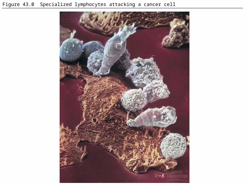
Figure 43.0 Specialized lymphocytes attacking a cancer cell
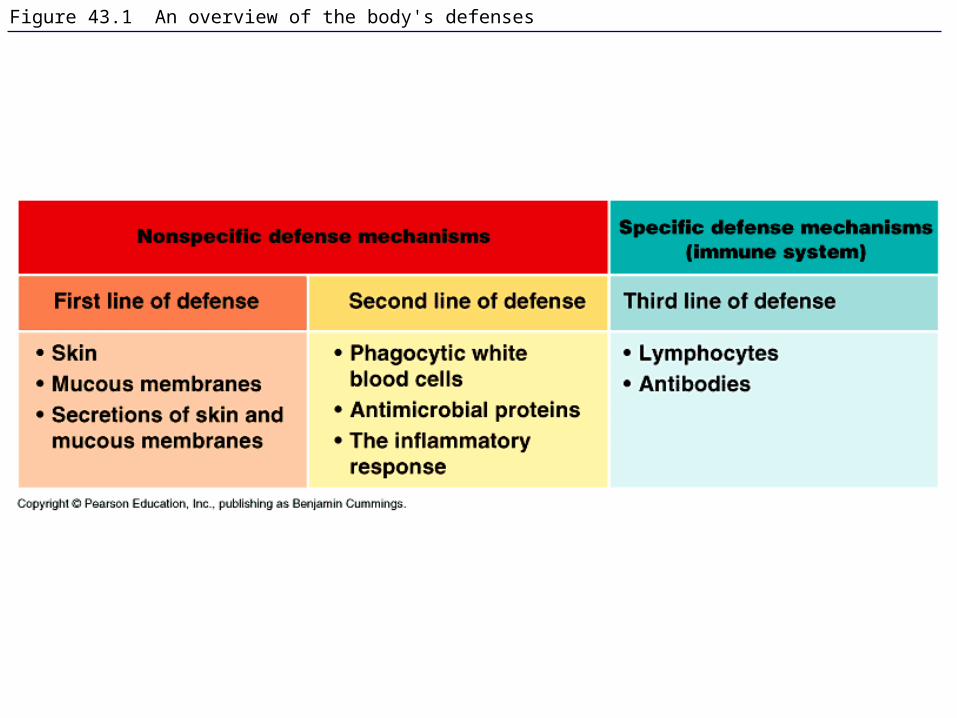
Figure 43.1 An overview of the body's defenses
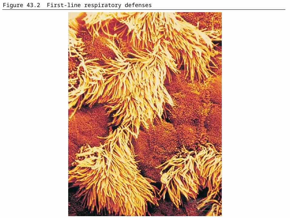
Figure 43.2 First-line respiratory defenses

Figure 43.3 Phagocytosis by a macrophage

Figure 43.3x Macrophage
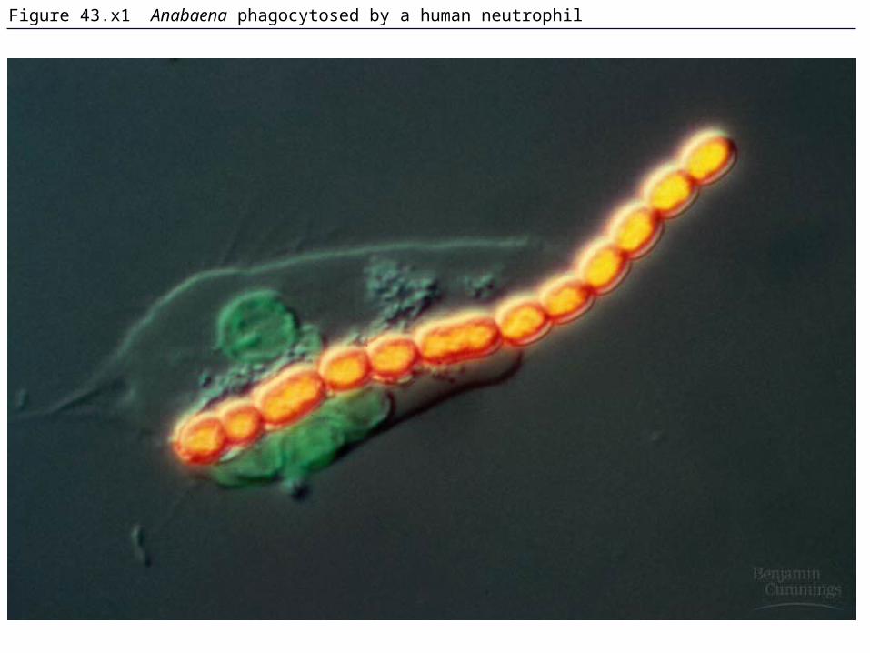
Figure 43.x1 Anabaena phagocytosed by a human neutrophil

Figure 43.4 The human lymphatic system

Figure 43.5 A simplified view of the inflammatory response

Figure 43.6 Clonal selection

Figure 43.7 Immunological memory

Figure 43.8 The development of lymphocytes

Figure 43.8x B lymphocyte

Figure 43.9 The interaction of T cells with MHC molecules
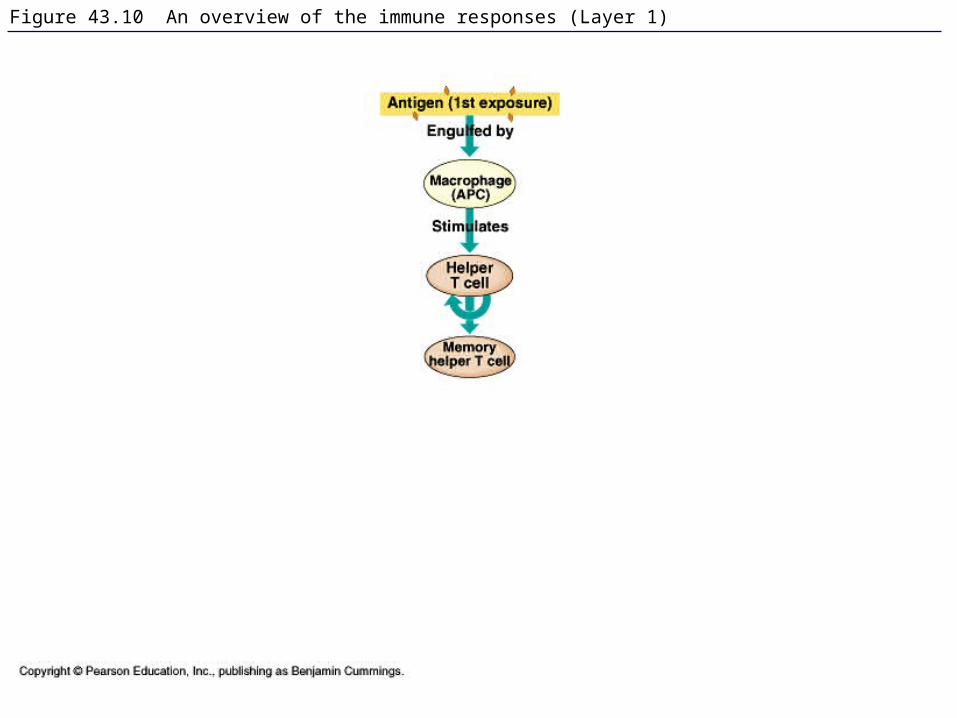
Figure 43.10 An overview of the immune responses (Layer 1)

Figure 43.10 An overview of the immune responses (Layer 2)

Figure 43.10 An overview of the immune responses (Layer 3)
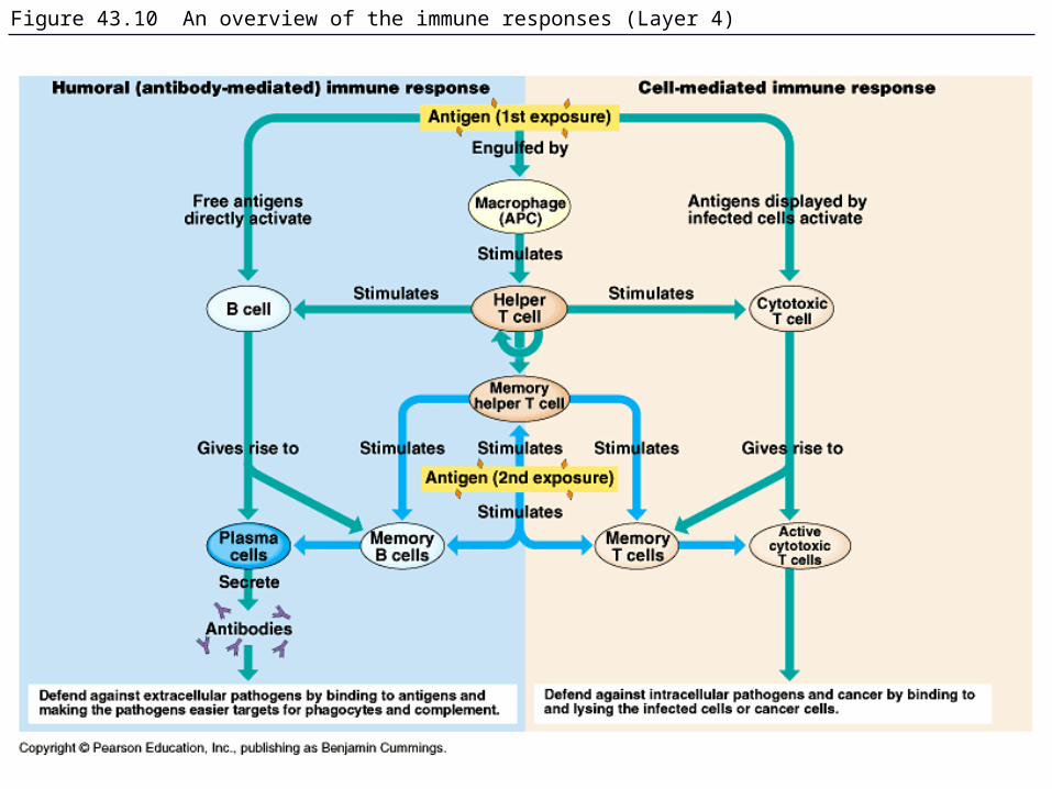
Figure 43.10 An overview of the immune responses (Layer 4)
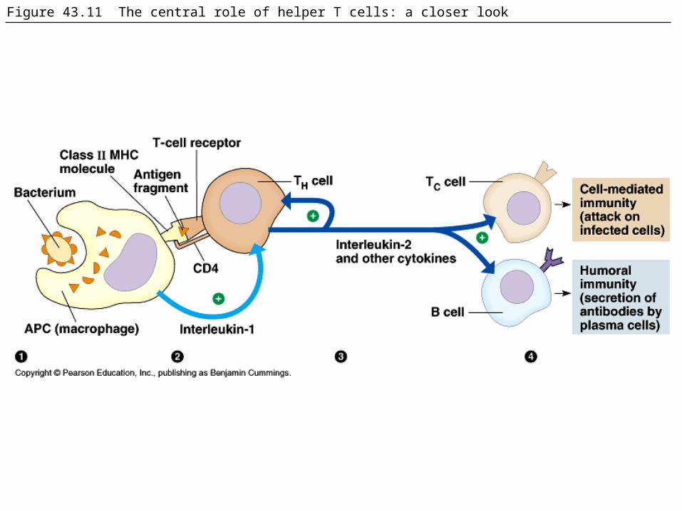
Figure 43.11 The central role of helper T cells: a closer look

Figure 43.12a The functioning of cytotoxic T cells

Figure 43.12b A cytotoxic T cell has lysed a cancer cell
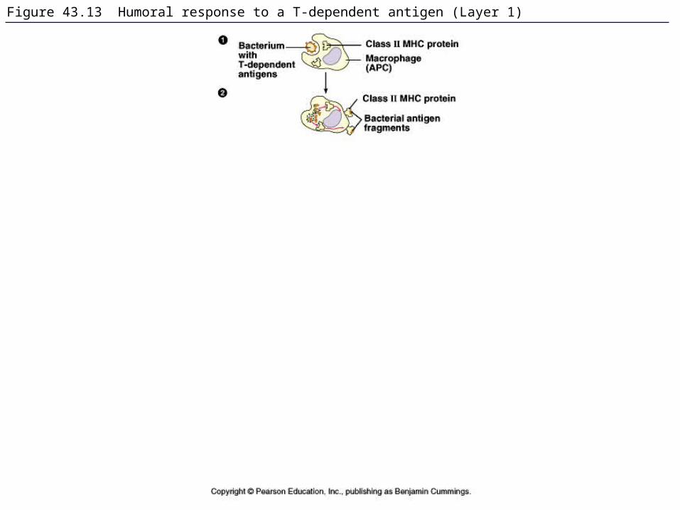
Figure 43.13 Humoral response to a T-dependent antigen (Layer 1)

Figure 43.13 Humoral response to a T-dependent antigen (Layer 2)

Figure 43.13 Humoral response to a T-dependent antigen (Layer 3)

Figure 43.14 Epitopes (antigenic determinants)

Figure 43.15c Antibody molecule

Figure 43.15a,b The structure of a typical antibody molecule

Figure 19.6 DNA rearrangement in the maturation of an immunoglobulin (antibody) gene

Table 43.1 The Five Classes of Immunoglobulins

Figure 43.16 Effector mechanisms of humoral immunity
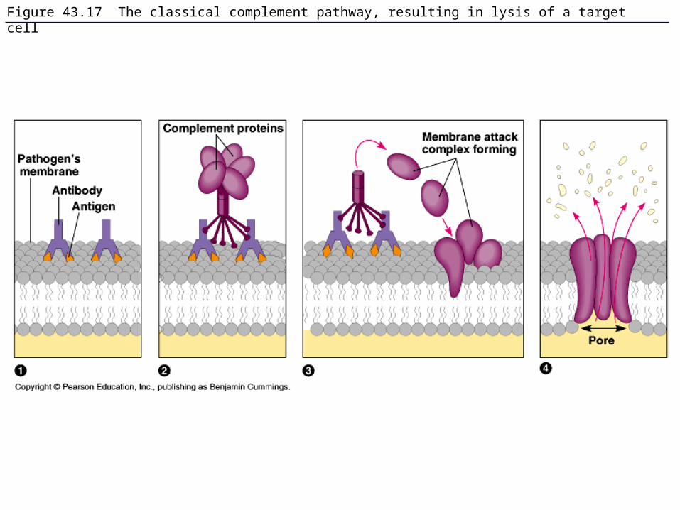
Figure 43.17 The classical complement pathway, resulting in lysis of a target cell

Figure 43.x2 Vaccination
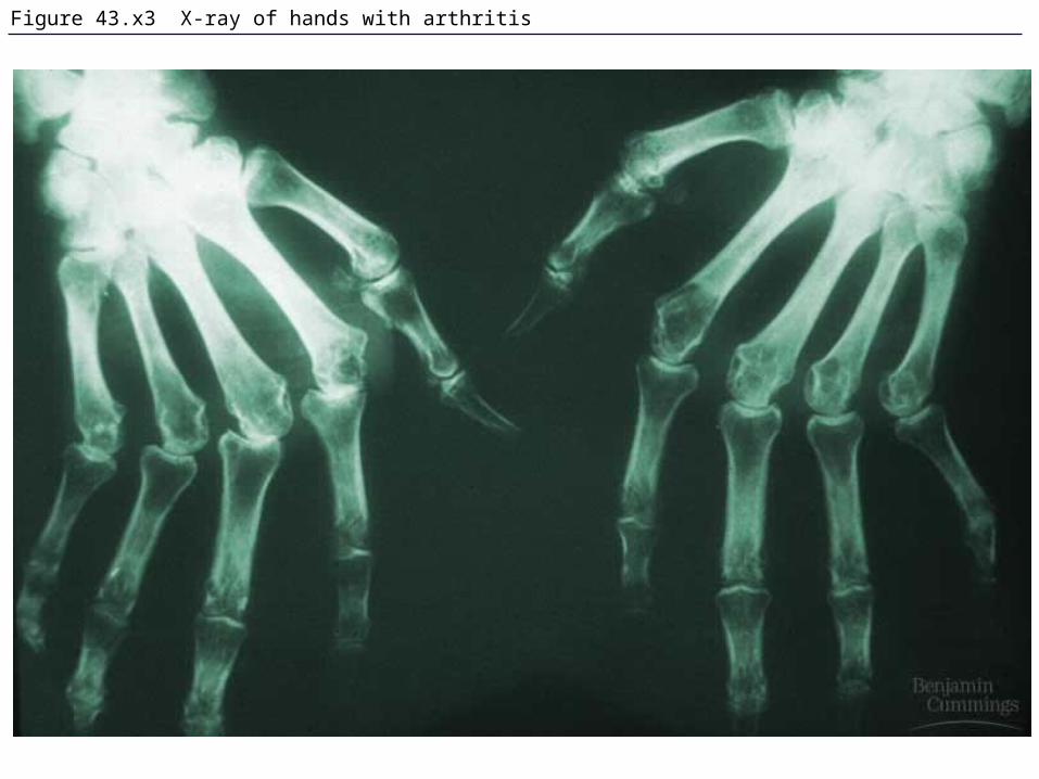
Figure 43.x3 X-ray of hands with arthritis

Figure 43.18 Mast cells, IgE, and the allergic response

Figure 43.x4 Alternaria spores, a cause of allergies in humans

Figure 43.19 A T cell infected with HIV

Figure 43.19x1 HIV on a lymphocyte, detail

Figure 43.19x2 HIV budding

Figure 43.19x2a T cell infected with HIV
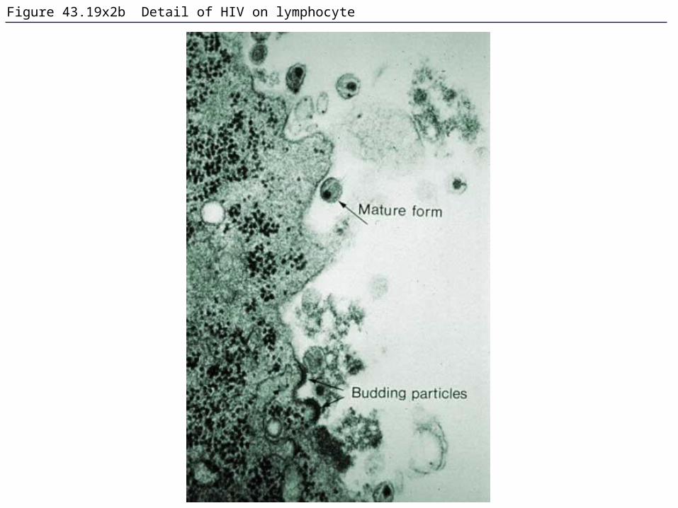
Figure 43.19x2b Detail of HIV on lymphocyte

Figure 43.20 The stages of HIV infection

Figure 43.x5 AIDS posters