fetal growth restriction - vanderbilt university medical … · · 2014-12-03fetal growth...
TRANSCRIPT

Fetal Growth Restriction: Diagnosis and Management
40th Annual Perinatal Conference
Vanderbilt University
December 5, 2014
Kimberly B Fortner, MD

At the end of this presentation, participants will be able:
• To classify IUGR fetus
• To understand the cardiovascular changes that occur in IUGR fetuses
• To understand management issues of IUGR fetuses

PUBMED: FGR…IUGR
18, 920 papers

Growth Restriction Defined
• WHO classification
• 1969: newborn less than 2500 gm (low-birth weight)
• Classification of low-birth weight
• Preterm neonate/AGA
• Preterm neonate/SGA
• Term neonate/SGA

Fetal Growth Restriction
• Definitions:
• EFW < 10th percentile1
• EFW < 5th percentile2
• EFW < 3rd percentile
• EFW > 2 SD below mean for gestational age
• AC <10th percentile or more than 2 week lag
The definition remains controversial because it does not make distinction among fetuses who are constitutionally small, growth restricted and small, and growth restricted but not small
1Battaglia et al J Pediat, 1967; 2Zhang et al, Am J Epidemiol 2011
Standard for Fetal Growth
• Between 10th – 90th percentiles for fixed gestational age (2.5th – 97.5th percentile)
• Denver standards (1960-1970s)
• Regardless, the goal of prenatal detection of small fetus is to REDUCE morbidity and mortality by employing some intervention.

Rate of Normal Fetal Growth
• First phase • <16 weeks gestation • “Cellular hyperplasia”
• Second phase • 16-32 weeks • Simultaneous hyperplasia and hypertrophy
• Third phase • >32 weeks • “Cellular hypertrophy” (rapid increase)
Hyperplasia Hypertrophy

Normal Fetal Growth
• 14-15 weeks gestation • 5 gm/day
• 20 weeks • 10 gm/day
• 32-34 weeks • 30-35 gm/day
Mean peaks 230-285 gm/week (32-34 wk)
Decreases at 41-42 weeks
Slightly less growth per week for multiples

Types of IUGR
• Asymmetrical (70-80%)
• Normal length; weight/ abdomen is below normal
• Symmetrical (20-30%)
• Length and weight are below normal
• Global impairment of early fetal cellular hyperplasia
• Ponderal index
• Birth weight (gm)/crown-heel length (cm)3 x100
• Potential error with the length

Epidemiology
• Incidence 10% ( any population)
• Developed countries: 4-8%
• Developing countries: 6-30%

Risk Factors
• Maternal medical conditions
• Smoking/substance abuse
• Severe malnutrition
• Placental disease
• Multiples
• Genetic disorders
• Exposure to teratogens

Perinatal mortality/morbidity
Fetal growth is important because
Inverse relationship between fetal/neonatal weight percentile and adverse perinatal outcome
Modified from Manning F. Fetal Medicine 1995

Perinatal mortality/morbidity
• Early studies (1980s)
• Infant 1500-2500 gm near term (5-30X mortality rate)
• Infant less than 1500gm near term (50-100X)
• Overall mortality rate
• Generally 50% higher
• Increases with weight below 6th percentile
• 2 times mortality at 10-15th percentile
• Higher for preterm infants

EFW < 10th percentile
Normal
• 70% ??
• Female sex
• Maternal ethnicity
• Parity
• Maternal BMI
Pathologic
• 30%
Manning FA, Fetal Medicine, 1995

Etiologies
• Genetic
• Congenital anomalies
• Infection
• Multiple gestation
• Placenta
• Maternal nutrition/environmental
• Maternal vascular disease

Genetic /Congenital Anomalies
• Among malformed fetus FGR frequency = 20-60%1
• Conversely, among FGR Anomaly frequency = 10%2
• Aneuploidy
• Trisomy 13, 18, 21
• Abnormalities X/Y
• Congenital anomalies
• Any anomaly; 22% IUGR
• Single umbilical artery
1Khoury MJ et al, Pediatrics, 1998. 2Mendez H. Am Jmed Genet. 1985

Infection
• Rubella
• Due to capillary endothelial damage during organogenesis
• Cytomegalovirus
• Cytolysis and localized necrosis leading to decreased cell size
• Protozoan infections
• Toxoplasmosis
• Plasmodium (malaria)

Multiple Gestation
• Fetal growth rate
• Weight LINEAR < 34-38 wk
• Decline occurs when fetal mass near 3000-3500 gm
• Peak weekly weight gain = 28-32 wk (160-170 gm/wk)
• IUGR, mild
• Decreased cell size
• Vascular anastomoses (mono/di)

Etiologies
Genetic
Congenital anomalies
Infection
Multiple gestation
• Placenta
• Maternal nutrition/environmental
• Maternal vascular disease

Placental Factors
“Umbrella that covers our ignorance in terms of etiology and pathogenesis of the
utero-placental chronic dysfunction”
Assali, Eur J Obstet Gynecol 1975

Placental Insufficiency
It is not the cause of IUGR but is rather the consequence of a disease process that often we do not understand

Placental Factors
• Placenta increase in size throughout gestation
• IUGR, placental growth plateaus at 36 weeks
• Abnormalities: cord, placenta hemangiomas
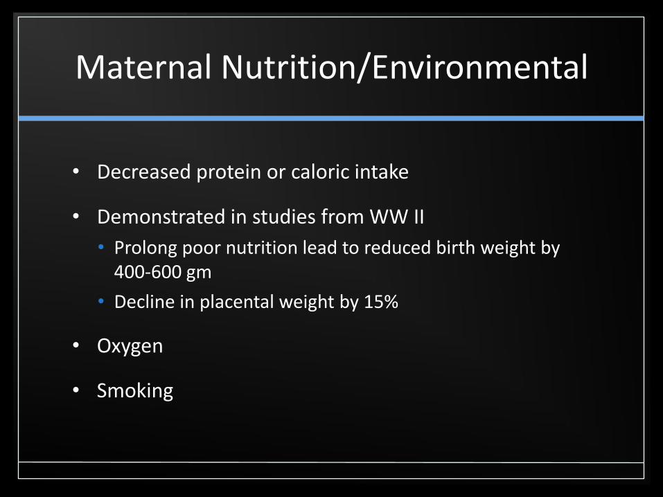
Maternal Nutrition/Environmental
• Decreased protein or caloric intake
• Demonstrated in studies from WW II
• Prolong poor nutrition lead to reduced birth weight by 400-600 gm
• Decline in placental weight by 15%
• Oxygen
• Smoking

Maternal Disease
• Accounts for 25-30% of IUGR infants
• Hypertensive disease
• Obstructive arterionecrosis (preeclampsia)
• Thrombophilia
• Uterine anomalies

Evaluation
• History and Physical
• Fundal height (sensitivity ranges 13-86%)
• Abdominal palpation (sensitivity 30-50%)
• Ultrasound
• Importance of dates • Abdominal circumference (sensitivity 60%)
• Composite EFW (sensitivity 90%)
• Umbilical artery Doppler

Management
The optimal approach to monitoring the fetus with suspected growth restriction has not been established; there is essentially no evidence from randomized trials
Grivell RM et al, Cochrane Database Syst Rev 2012

Cornerstone of Management
Serial Evaluation of:
• Growth
• Fetal behavior (BPP)
• Impedance to blood flow in fetal arterial and venous vessels
With Purpose of:
• Identify fetus at risk for inutero demise & neonatal morbidity
• May benefit from preterm delivery
Grivell RM et al, Cochrane Database Syst Rev 2012

Fetal Growth
• Follow estimated weight percentiles
• Follow growth velocity
• Serial sonograms q 2-4 week intervals
• Persistent growth deficiency strengthens likelihood

Umbilical Artery Doppler
Fitzgerald was the first to
obtain Doppler signal in
pregnancy in 1977

Doppler in AGA and IUGR Fetuses

Umbilical Artery: High
Placental Vascular
Resistance

Umbilical Artery Doppler and Placental Vascular Histology
Giles WB et al, BJOG 1985

Umbilical Artery Doppler and Outcome
• Reduce perinatal death and unnecessary induction of labor in the preterm growth restricted fetus
• Meta-analysis use of Doppler ultrasonography reduced the odds of perinatal death by 38% (95% CI 15-55)
Alfirevic Z et al, Am J Obstet Gynecol 1995

Doppler vs. No Doppler, Outcome = Any perinatal death
(after randomization)
Zarko A et al, Cochrane Database of Systematic Reviews 2013

Doppler vs. No Doppler, Outcome = Stillbirth
Zarko A et al, Cochrane Database of Systematic Reviews 2013

Brain Circulation
MCA Waveforms at 24 weeks
B= “Brain sparing effect”
A= Normal


Ductus
Venosus
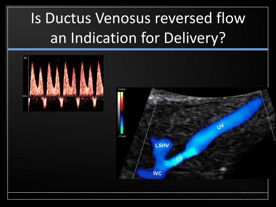
Is Ductus Venosus reversed flow an Indication for Delivery?

Temporal Sequence of Fetal Deterioration
DECREASED Umbilical
Venous volume
Redistribution of fetal blood flow
INCREASED Umbilical Artery
Doppler
DECREASED MCA Doppler
Godfrey et al. PLOS one, 2012

Biophysical Profile
• Evaluation of multiple acute and chronic fetal physiologic parameters
• Easy to perform
• Reliable test of fetal well-being
≤ 5cm
• Both FGR and Oligo = INCREASED risk of perinatal mortality
> 5cm
• NOT highly associated with either FGR or fetal demise
Dayal AK et al, Am J Obstet Gynecol, 1999

FGR Delivery?
• GRIT1 24-36 wks
• Delaying delivery of very preterm FGR fetus results in stillbirths, but immediate delivery = almost equal number of neonatal deaths; Neither approach results in better long-term neurodevelopmental outcome
• DIGITAT2,3 36 wks (IOL vs. Expectant)
• No difference in morbidity score; development @ 2yrs same
• TRUFFLE4 26-32 wks (cohort)
• Fetal outcome better. Death and severe morbidity significantly related to EGA (@study entry & @ delivery) and with presence of maternal hypertensive morbidity
1Eur J Obstet Gynecol Reprod Biol 1996; 2Boers KE et al, BMJ 2010; 3Van Wyk L et al, Am J
Obstet Gynecol 2012; 4Lees C et al, Ultrasound Obstet Gynecol 2013

Timing of Intervention
Immediate Delivery
Abnormal Ductus Venosus
≥ 32 wks with Reversed EDF
≥34 wks with AEDF
≥37 wks with Abnormal S/D, poor interval growth, …
≥38-39 wks with FGR, normal Doppler studies
Inpatient / Betamethasone
Women needing daily maternal or fetal assessment
<32 wks with Reversed EDF

Long-term Sequelae
• Potential intrapartum complications
• Neonatal asphyxia
• Meconium
• Adult disease
• IQ / Neurodevelopment
• Obstructive lung disease
• Barker Hypothesis (Hypertension, Diabetes)

THANK YOU!
• Enjoy the rest of your day…
• Questions?