female thermal sensitivity to hot and cold during rest and ... · b oxylane research, decathlon...
TRANSCRIPT

Physiology & Behavior 152 (2015) 11–19
Contents lists available at ScienceDirect
Physiology & Behavior
j ourna l homepage: www.e lsev ie r .com/ locate /phb
Female thermal sensitivity to hot and cold during rest and exercise
Nicola Gerrett a,⁎, Yacine Ouzzahra a,1, Bernard Redortier b, Thomas Voelcker b, George Havenith a
a Environmental Ergonomics Research Centre, Design School, Loughborough University, Loughborough, Leicestershire LE113TU, UKb Oxylane Research, Decathlon Campus, Villeneuve d'Ascq, Lille, France
H I G H L I G H T S
• Females are more sensitive to innocuous cold compared to innocuous heat stimulation.• Regional differences to cold stimulation occur across the body in females.• Regional differences are more homogenous to hot stimulation in females.• Exercise reduces thermal magnitude sensation in both hot and cold stimuli.
⁎ Corresponding author at: Institute of Sport and EWorcester, Henwick Road, Worcester WR26AJ, UK.
E-mail addresses: [email protected] (N. Gerrett), y(Y. Ouzzahra), [email protected] (B. [email protected] (T. Voelcker), G.Havenith@
1 Institute forHealth andBehaviour, University of Luxemb
http://dx.doi.org/10.1016/j.physbeh.2015.08.0320031-9384/Crown Copyright © 2015 Published by Elsevie
a b s t r a c t
a r t i c l e i n f oArticle history:Received 22 June 2015Received in revised form 18 August 2015Accepted 25 August 2015Available online 5 September 2015
Keywords:FemalesBody mappingExerciseRegionalThermal sensitivity
Regional differences in thermal sensation to a hot or cold stimulus are often limited to male participants, in arested state and cover minimal locations. Therefore, magnitude sensation to both a hot and cold stimulus wereinvestigated during rest and exercise in 8 females (age: 20.4 ± 1.4 years, mass: 61.7 ± 4.0 kg, height: 166.9 ±5.4 cm, VO2max: 36.8 ± 4.5 ml·kg−1·min−1). Using a repeated measures cross over design, participants restedin a stable environment (22.3 ± 0.9 °C, 37.7 ± 5.5% RH) whilst a thermal probe (25 cm2), set at either 40 °C or20 °C, was applied in a balanced order to 29 locations across the body. Participants reported their thermal sensa-tion after 10 s of application. Following this, participants cycled at 50% VO2max for 20 min and then 30% VO2max
whilst the sensitivity test was repeated. Females experienced significantly stronger magnitude sensations tothe cold than the hot stimulus (5.5 ± 1.7 and 4.3 ± 1.3, p b 0.05, respectively). A significant effect of locationwas found during the cold stimulation (p b 0.05). Thermal sensation was greatest at the head then the torsoand declined towards the extremities. No significant effect of location was found in response to the hot stimula-tion and the pattern across the body was more homogenous. In comparison to rest, exercise caused a significantoverall reduction in thermal sensation (5.2±1.5 and 4.6±1.7, respectively, p b 0.05). Bodymapswere producedfor both stimuli during rest and exercise, which highlight sensitive areas across the body.Crown Copyright © 2015 Published by Elsevier Inc. This is an open access article under the CC BY-NC-ND license
(http://creativecommons.org/licenses/by-nc-nd/4.0/).
1. Introduction
Central and peripheral thermoreceptors are distributed all over thebody and are responsible for both sensory and thermoregulatoryresponses to maintain thermal equilibrium. Behavioural thermoregula-tion is the first line of defence against thermal disturbances and this isprimarily controlled by peripheral thermoreceptors which provideimmediate feedback about the thermal state of the body and initiate a
xercise Science, University of
[email protected]),lboro.ac.uk (G. Havenith).ourg,Walferdange, Luxembourg.
r Inc. This is an open access article un
set of desired actions to correct the thermal imbalance [1]. Both Burkeand Mekjavić [2] and Nakamura et al. [3] speculated that the centralnervous system assigns weighing factors for each body segment andthat this iswhat determines the regional differences in sensitivity ratherthan receptor density. Regional differences in thermal sensitivity havepreviously beenmeasured on a limited number of small locations acrossthe body (b5) which then have been interpreted as fully representingthat particular area (e.g. legs, arms, front/back torso and face). However,intra-segment variations to a cold or hot stimulus have recently beenidentified [4,5]. More research is required to fully understand thermalsensitivity across wider areas of the body.
Despite there being a large body of literature exploring thermalsensitivity much of the research is limited to male participants, despitethe fact that females have been shown to be more sensitive to a varietyof stimuli [6,7,8]. One recent study compared sex difference in responseto a hot stimulus (40 °C) across 31 locations on the body and found that
der the CC BY-NC-ND license (http://creativecommons.org/licenses/by-nc-nd/4.0/).

12 N. Gerrett et al. / Physiology & Behavior 152 (2015) 11–19
overall females were significantly more sensitive to heat thanmales [4].Regional differences in perceptual responses were more prominent infemales and thermal hot sensation was greatest at the head, than thetorso and declined towards the extremities. Using a similar technique,Ouzzahra et al. [5] assessed regional distribution of sensitivity on thetorso to a cold thermal stimulus (20 °C) inmale participants. Comparingthe findings of these two studies indicates that male regional sensitivityacross the torso is not the same when stimulated with cold or hot stim-uli. There are more regional differences in response to a cold stimuluswhereas the response is more homogenous to a hot stimulus. Howeverin both studies the anterior torso was more sensitive than the posteriortorso. To the authors knowledge there is limited research comparingthermal sensitivity to a cold and hot stimulus in female participantsacross multiple (N5) locations on the body.
Exercise has been shown to alter ones sensory perceptions to a vari-ety of different stimuli [9,10,11,12,13,14]. A large body of literature isavailable demonstrating the reduction in pain during or after exercisein males and females (see Koltyn [15] for a review). This also includesnoxious thermal stimulation and the reduction is associated with exer-cise induced analgesia (EIA). EIA is associated with the activation of theendogenous opioid system during exercise in which various peptidesare released that have a similar effect to that of morphine (i.e. theycause a reduction in pain sensitivity) [16]. Large amounts of researchexist regarding EIA and noxious stimulation using magnitude estima-tion but only two studies have investigated innocuous thermal sensitiv-ity with this technique [4,5].
Ouzzahra et al. [5] found that during exercise thermal sensitivity to acold stimulus was attenuated in males compared to rest and suggestedthat the reductionwas likely a result of exercise induced analgesia (EIA).Gerrett et al. [4] also found that sensitivity to a hot stimulus (40 °C) wasreduced during exercise in both males and females. The response wasmore prominent inmales despite only the females having a significantlyelevated Tc temperature during exercise. This was the first study to con-firm that exercise caused a reduction in innocuous heat sensitivity. It isbelieved that during exercise the release of corticotrophin concomitant-ly with β-endorphin reduces the pain perception thresholds [17,18].Males have been reported to have significantly higher β-endorphinthan females at rest and in response to endurance exercise [19]. Thismay explain why males had a significantly reduced thermal sensitivitycompared to females during exercise. However, whether female sensi-tivity reduces during exercise to a cold innocuous stimulus has not yetbeen investigated. It would be of interest to determinewhether femalesexperience the same decline in sensitivity to a cold stimulus as to thatexperienced to a hot stimulus.
The aim of the study is to investigate the regional differences in ther-mal sensitivity to a hot and cold stimulus in female participants duringrest and exercise. It is hypothesised that, similar to males, significantregional variations will exist between hot and cold perceptions infemales. It is further hypothesised that exercise will cause a significantreduction in thermal sensitivity to both innocuous cold and hot stimuli.
2. Methods
2.1. Participants
Eight Caucasian females (age: 20.4 ± 1.4 years, mass: 61.7 ± 4.0 kg,height: 166.9 ± 5.4 cm, VO2max: 36.8 ± 4.5 ml·kg−1·min−1) wererecruited from the staff and student population of LoughboroughUniversity. The selection criteria included only Caucasian females,aged between 18 and 45 years to reduce any systemic errors due toethnic or age-related differences in thermoregulatory responses. Six ofthe eight participants were taking oral contraceptives. Menstrualcycle phase was not controlled for during the experimental session.However the stage of menstrual cycle in each participant was notedand a range of stages was tested during the experiment, thus providinga representative sample of menses state in the results.
2.2. Experimental design
The aim of the investigation was to compare regional sensitivity to ahot and a cold stimulus in females during rest and exercise. To achievethese aims a randomised cross over design was opted for, with allparticipants taking part in both conditions (hot and cold sensitivity)separated by at least 2 days. During both conditions, participants restedand exercised on a cycle ergometer whilst regional thermal sensitivitiesto a thermal probe were investigated. A total of 29 regional bodysegments were chosen to ensure that each area of the body was fullyinvestigated (see Gerrett et al. [4]). These included the front and backtorso, the arms and legs (upper, lower, front and back), head, face andneck and the extremities. The testing sequence of the segments wasrandomised to prevent any order effects. However, the order of restand exercise in the tests was not randomised as rest had to precedeexercise due to the elevation of Tc caused by the latter. This increasecould have a lasting effect in any following rest exposures. To counteractany order effect, participants were thoroughly familiarised with theprocedure before the start of the actual test.
2.3. Experimental protocol
Each participant completed a pre-test session for anthropometricmeasurements; stature and bodymass. They then completed a submax-imal fitness test based on the Åstrand Rhyming methods [20]. The testconsisted of four progressive exercise stages on an electromagneticallybraked cycle ergometer (Lode Excalibur, Groningen, Netherlands)each lasting 5 min. Heart rate (Polar Electro Oy, Kemple, Finland) wasrecorded during the last minute of each stage. Estimation of VO2max
was then calculated from the ACSMmetabolic equation for cycling [20].During the test, participants were familiarised with the thermal
probe and sensation scales across a number of locations. Participantswere then invited back to the laboratory on two different occasions toconduct the main trial with at least 2 days separating trials. For themain trial, pre- and post-test nude weight was recorded. Participantsself-inserted a rectal probe 10 cm beyond the anal sphincter. Four skinthermistors (Grant Instrument Ltd, Cambridge, UK) were attached atthe chest, upper arm, thigh and calf using 3M™ Transpore™ surgicaltape (3M United Kingdom PLC). Mean skin temperature (T�sk) was esti-mated using the following calculations as proposed by Ramanathan[21]:
T�sk ¼ 0:3 � Tricepð Þ þ 0:3 � Chestð Þ þ 0:2 � Quadricepsð Þ þ 0:2 � Calfð Þ:
Body temperature (Tb) was estimated using the following calculationof Tc and T�sk in an 8:2 ratio [22]:
Tb ¼ 0:8 � Tc þ 0:2 � T�sk:
Markings were made on the body using a washable pen to indicateeach measurement site for the application of the thermal probe. Thelocations of each stimulus application, all taken on the left hand sideof the body, are shown in Fig. 1. Dressed in shorts, sports bra, socksand trainers, participants sat in a controlled environment (22.3 ±0.9 °C, 37.7 ± 5.5% RH) for 15 min to allow physiological responsesto stabilise. During the stabilisation period participants were onceagain familiarisedwith the sensation scales and allowed to practise ratingtheir sensations to a range of hot and cold stimuli across different regionson the body.
After the stabilisation period, whilst still at rest, thermal sensitivityof each body site along the left hand side of the body to the thermalstimulus was investigated in a balanced order. Each stimulus site wassubjected to the following: the measurement of Tsk using an infraredthermometer (FLUKE 566 IR THERMOMETER, Fluke Corporation,Eindhoven, Netherlands), immediately followed by probe applicationfor 10 s. The temperature controlled thermal probe was similar to that
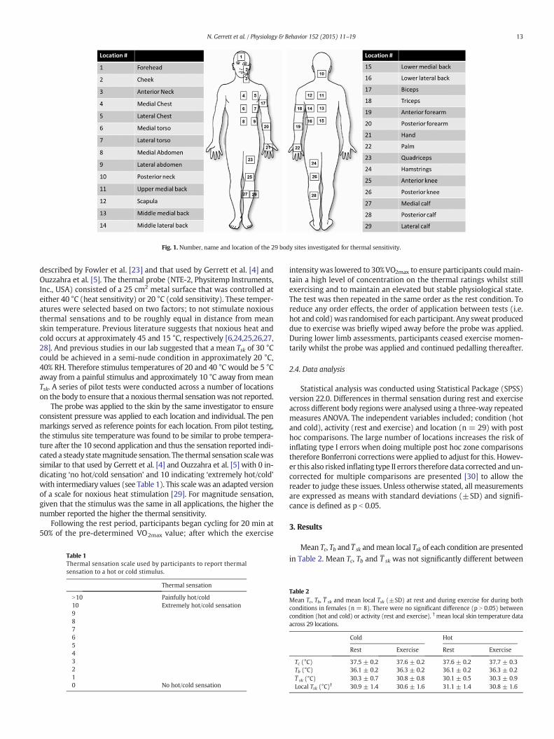
Fig. 1. Number, name and location of the 29 body sites investigated for thermal sensitivity.
13N. Gerrett et al. / Physiology & Behavior 152 (2015) 11–19
described by Fowler et al. [23] and that used by Gerrett et al. [4] andOuzzahra et al. [5]. The thermal probe (NTE-2, Physitemp Instruments,Inc., USA) consisted of a 25 cm2 metal surface that was controlled ateither 40 °C (heat sensitivity) or 20 °C (cold sensitivity). These temper-atures were selected based on two factors; to not stimulate noxiousthermal sensations and to be roughly equal in distance from meanskin temperature. Previous literature suggests that noxious heat andcold occurs at approximately 45 and 15 °C, respectively [6,24,25,26,27,28]. And previous studies in our lab suggested that a mean Tsk of 30 °Ccould be achieved in a semi-nude condition in approximately 20 °C,40% RH. Therefore stimulus temperatures of 20 and 40 °C would be 5 °Caway from a painful stimulus and approximately 10 °C away frommeanTsk. A series of pilot tests were conducted across a number of locationson the body to ensure that a noxious thermal sensationwas not reported.
The probe was applied to the skin by the same investigator to ensureconsistent pressure was applied to each location and individual. The penmarkings served as reference points for each location. From pilot testing,the stimulus site temperature was found to be similar to probe tempera-ture after the 10 second application and thus the sensation reported indi-cated a steady statemagnitude sensation. The thermal sensation scalewassimilar to that used by Gerrett et al. [4] and Ouzzahra et al. [5] with 0 in-dicating ‘no hot/cold sensation’ and 10 indicating ‘extremely hot/cold’with intermediary values (see Table 1). This scale was an adapted versionof a scale for noxious heat stimulation [29]. For magnitude sensation,given that the stimulus was the same in all applications, the higher thenumber reported the higher the thermal sensitivity.
Following the rest period, participants began cycling for 20 min at50% of the pre-determined VO2max value; after which the exercise
Table 1Thermal sensation scale used by participants to report thermalsensation to a hot or cold stimulus.
Thermal sensation
N10 Painfully hot/cold10 Extremely hot/cold sensation9876543210 No hot/cold sensation
intensitywas lowered to 30%VO2max to ensure participants couldmain-tain a high level of concentration on the thermal ratings whilst stillexercising and to maintain an elevated but stable physiological state.The test was then repeated in the same order as the rest condition. Toreduce any order effects, the order of application between tests (i.e.hot and cold)was randomised for each participant. Any sweat produceddue to exercise was briefly wiped away before the probe was applied.During lower limb assessments, participants ceased exercise momen-tarily whilst the probe was applied and continued pedalling thereafter.
2.4. Data analysis
Statistical analysis was conducted using Statistical Package (SPSS)version 22.0. Differences in thermal sensation during rest and exerciseacross different body regionswere analysed using a three-way repeatedmeasures ANOVA. The independent variables included; condition (hotand cold), activity (rest and exercise) and location (n = 29) with posthoc comparisons. The large number of locations increases the risk ofinflating type I errors when doing multiple post hoc zone comparisonstherefore Bonferroni correctionswere applied to adjust for this. Howev-er this also risked inflating type II errors therefore data corrected andun-corrected for multiple comparisons are presented [30] to allow thereader to judge these issues. Unless otherwise stated, all measurementsare expressed as means with standard deviations (±SD) and signifi-cance is defined as p b 0.05.
3. Results
Mean Tc, Tb and T�sk andmean local Tsk of each condition are presented
in Table 2. Mean Tc, Tb and T�sk was not significantly different between
Table 2Mean Tc, Tb, T
�sk and mean local Tsk (±SD) at rest and during exercise for during both
conditions in females (n = 8). There were no significant difference (p N 0.05) betweencondition (hot and cold) or activity (rest and exercise). † mean local skin temperature dataacross 29 locations.
Cold Hot
Rest Exercise Rest Exercise
Tc (°C) 37.5 ± 0.2 37.6 ± 0.2 37.6 ± 0.2 37.7 ± 0.3Tb (°C) 36.1 ± 0.2 36.3 ± 0.2 36.1 ± 0.2 36.3 ± 0.2T�sk (°C) 30.3 ± 0.7 30.8 ± 0.8 30.1 ± 0.5 30.3 ± 0.9Local Tsk (°C)† 30.9 ± 1.4 30.6 ± 1.6 31.1 ± 1.4 30.8 ± 1.6

14 N. Gerrett et al. / Physiology & Behavior 152 (2015) 11–19
conditions (p N 0.05).Mean Tc, Tb andT�skwashigher during exercise com-
pared to rest but not significantly different (p N 0.05). The local Tsk datacollected from each of the 29 locations prior to stimulation wasaveraged and the results were not significantly different between condi-tions or activity type (p N 0.05).
3.1. Regional sensitivity to hot and cold stimuli
Female sensitivity to a hot and cold stimulus during rest and exerciseare illustrated in Figs. 2 and 3, respectively. A significant overall effect ofcondition (hot versus cold) was observed, as females reported a signif-icantly higher magnitude sensation for the cold stimulus in comparisonto the hot stimulus (5.5 ± 1.7 and 4.3 ± 1.3, p b 0.05, respectively). Asignificant overall effect of activity was also found as thermal sensation
Fig. 2. Female (n = 8) regional magnitude sensation to a cold stimulus (20 °C) during rest asymmetry [51,52]. Areas in grey were not investigated.
was higher during rest than exercise (5.2 ± 1.5 and 4.6 ± 1.7, respec-tively, p b 0.05). No significant overall effect of location was found(p N 0.05). However, when the overall effect of location was analysedfor cold and hot stimuli separately, the results indicated no significantoverall effect of location for the hot stimulus but a significant overalleffect for the cold stimulus (inclusive of rest and exercise). Pairwisecomparisons with Bonferroni corrections revealed no significant differ-ences (p N 0.05), howeverwithout corrections formultiple comparisonssignificant differences were observed and these are listed in Table 3.
During the cold stimulus, the head regionwas very sensitive and thecheek was significantly more sensitive than many locations across thebody (p b 0.05). During rest, the anterior torso was generally moresensitive than the posterior torso but this pattern reversed during exer-cise. The lateral lower back was significantly less sensitive than mostlocations across the torso and head region. During both rest and
nd exercise. All measurements were taken from the left hand side of the body assuming
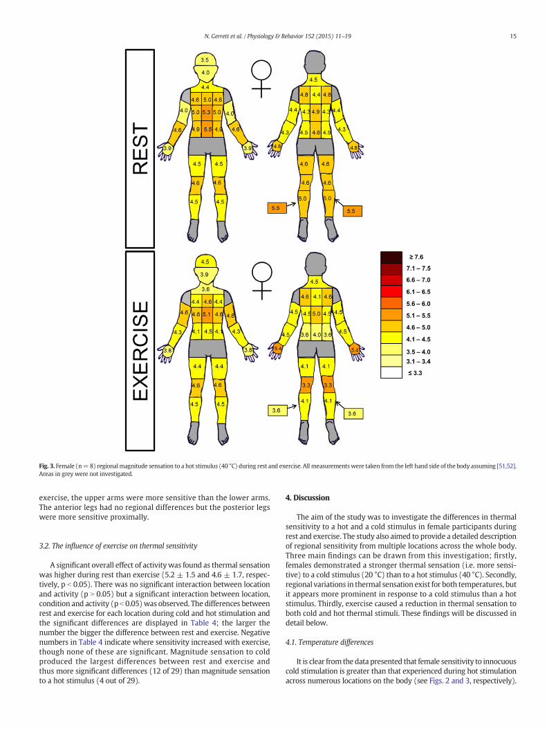
Fig. 3. Female (n=8) regionalmagnitude sensation to a hot stimulus (40 °C) during rest and exercise. All measurementswere taken from the left hand side of the body assuming [51,52].Areas in grey were not investigated.
15N. Gerrett et al. / Physiology & Behavior 152 (2015) 11–19
exercise, the upper arms were more sensitive than the lower arms.The anterior legs had no regional differences but the posterior legswere more sensitive proximally.
3.2. The influence of exercise on thermal sensitivity
A significant overall effect of activitywas found as thermal sensationwas higher during rest than exercise (5.2 ± 1.5 and 4.6 ± 1.7, respec-tively, p b 0.05). There was no significant interaction between locationand activity (p N 0.05) but a significant interaction between location,condition and activity (p b 0.05)was observed. The differences betweenrest and exercise for each location during cold and hot stimulation andthe significant differences are displayed in Table 4; the larger thenumber the bigger the difference between rest and exercise. Negativenumbers in Table 4 indicate where sensitivity increased with exercise,though none of these are significant. Magnitude sensation to coldproduced the largest differences between rest and exercise andthus more significant differences (12 of 29) than magnitude sensationto a hot stimulus (4 out of 29).
4. Discussion
The aim of the study was to investigate the differences in thermalsensitivity to a hot and a cold stimulus in female participants duringrest and exercise. The study also aimed to provide a detailed descriptionof regional sensitivity from multiple locations across the whole body.Three main findings can be drawn from this investigation; firstly,females demonstrated a stronger thermal sensation (i.e. more sensi-tive) to a cold stimulus (20 °C) than to a hot stimulus (40 °C). Secondly,regional variations in thermal sensation exist for both temperatures, butit appears more prominent in response to a cold stimulus than a hotstimulus. Thirdly, exercise caused a reduction in thermal sensation toboth cold and hot thermal stimuli. These findings will be discussed indetail below.
4.1. Temperature differences
It is clear from the data presented that female sensitivity to innocuouscold stimulation is greater than that experienced during hot stimulationacross numerous locations on the body (see Figs. 2 and 3, respectively).

Table 3Significance of pairwise comparison between locations for female magnitude sensation to a cold stimulus (data is inclusive of rest and exercise).
1 2 3 4 5 6 7 8 9 10 11 12 13 14 15 16 17 18 19 20 21 22 23 24 25 26 27 28 29
23 *
4 * *
5 *
6 *
78 *
910 * *
11 **
1213 *
14 * *
15 * * * * * * *
16 * * *
17 * * *
1819 * *
20 *
21 * *
22 * *
23 *
24 * *
25 *
2627 * *
28 * * * *
29 * * * *
⁎p b 0.05 without Bonferroni correction.⁎⁎p b 0.001 without Bonferroni correction.
Table 4The differences inmagnitude sensation between rest and exercise for each location duringcold or hot stimulation. No significant differences with Bonferroni correction.
LocationΔ magnitude sensation (rest–exercise)
Cold Hot
Forehead 0.5 ± 0.7 −1.0 ± 0.5Cheek 0.1 ± 0.4 0.1 ± 0.1Anterior neck 0.1 ± 0.0 0.8 ± 0.5Posterior neck −0.4 ± 0.4 0.0 ± 0.1Medial chest 0.9 ± 0.1 0.3 ± 0.3Lateral chest 1.9 ± 0.4⁎ 0.4 ± 0.3Medial torso 1.4 ± 0.1⁎ 0.4 ± 0.6Lateral torso 1.0 ± 0.0⁎ 0.1 ± 0.1Medial abdomen 1.4 ± 0.7⁎ 0.8 ± 0.2Lat abdomen 1.1 ± 0.3⁎ 1.0 ± 0.1⁎
Upper medial back 1.5 ± 0.2⁎ 0.1 ± 0.2Scapula 0.8 ± 0.9 0.3 ± 0.3Middle medial back 0.6 ± 0.1⁎ −0.3 ± 0.3Middle lateral back 0.6 ± 0.3 −0.1 ± 0.1Lower medial back 0.6 ± 0.2 0.9 ± 0.8Lower lateral back 1.3 ± 1.0 0.6 ± 0.0Biceps 1.6 ± 0.3 −0.6 ± 0.1Triceps 0.5 ± 0.1 −0.1 ± 0.3Anterior forearm 1.4 ± 0.5⁎ −0.3 ± 0.0Posterior forearm 1.3 ± 0.5 0.4 ± 0.4Palm −0.3 ± 0.4 0.1 ± 0.2Back of hand 0.1 ± 0.1 −0.6 ± 0.2⁎
Quadriceps 0.8 ± 1.5 0.1 ± 0.6Front knee 1.0 ± 0.4⁎ 0.4 ± 0.4⁎
Lateral gastrocnemius 0.9 ± 0.4 0.0 ± 0.5Hamstring 1.1 ± 0.3⁎ 0.4 ± 0.4Posterior knee 1.1 ± 0.4⁎ 1.4 ± 0.0Post. Gastrocnemius 0.8 ± 0.6⁎ 0.9 ± 0.7Medial gastrocnemius 0.2 ± 0.7 1.9 ± 0.2⁎
Mean 0.9 ± 0.6 0.3 ± 0.6
⁎ p b 0.05 (without Bonferroni corrections).
16 N. Gerrett et al. / Physiology & Behavior 152 (2015) 11–19
Overall females reported a significantly higher magnitude sensation forthe cold stimulus in comparison to the hot stimulus (5.5 ± 1.7 and4.3 ± 1.3, p b 0.05, respectively). Whilst this is the first paper to deter-mine such differences in females using magnitude estimation, similarfindings have been reported in in both males and females using themethod of limits/threshold detection [31,32]. Using this technique,Golja et al. [32] also reported that females were more sensitive thanmales to cold and warmth and the differences in sensitivity to warmand cold temperatures was more pronounced in females than males.Whilst no gender comparison can be made based on the present study,the data suggests that thermal sensitivity to cold is greater than warmthusing magnitude estimation.
Factors that influence thermal sensitivity that may account fordifferences observed in this study reside in the stimulation of peripheralthermoreceptors. According to Bullock, Boyle and Wang [33], thethermal sensation experienced is dependent upon the direction andmagnitude of the change in temperature. In the present study, meanTsk during both conditions was 30.3 ± 0.6 °C and the average local Tskof each site prior to stimulation was 30.8 ± 1.6 °C. As a result the differ-ence in Tsk and the temperature of the thermal probe was similarbetween the two conditions (approximately 10 °C). In addition, thethermal probe was held in place for 10 s before a thermal sensationscore was reported in order to achieve a steady state score. This effec-tively removed the influence of initial Tsk, whichwas found to be similarto probe temperature after the 10 second application in pilot tests.Therefore the differences in thermal sensitivity to a hot and cold stimu-lus cannot be associated with the magnitude of change in Tsk. A morelikely explanation is one associated with properties of the receptorsbeing stimulated. Hensel, Andres, and Düring [34] revealed that warmreceptors tend to be located deeper in the epidermis thanmore superfi-cial cold receptors, warm receptors are also outnumbered by cold recep-tors and their nerves conduct signals at much slower speeds [26]. In

17N. Gerrett et al. / Physiology & Behavior 152 (2015) 11–19
recent years the molecular characteristics of these sensory neuronshave markedly advanced, of which the transient receptor potential(TRP) family has been identified as thermosensitive fibres [35]. Of thefour proposed heat activated sensors and two cold activated sensors,the specific TRP stimulated in this study during hot stimulation wasTRPV3 (activated at temperature N 33 °C) and TRPM8 (activated attemperatures b23 °C) [36,35]. Hensel [26] associated cold activatedfibres with the thinly myelinated Aδ fibres whose conduction velocitiesare faster than heat activated fibres along unmyelinated C fibres. Recentresearch by Caterina& Julius [36] & Patapoutian et al. [35] has associatedTRPM8 with both Aδ and C fibres found in animals [37,38] and to someextent in humans [39]. Much of the research associated with the TRPfamily is still in its infancy and more research is required to enhanceour understanding of the TRP family and its relationship with tempera-ture sensation.
As thermoreceptors serve to protect the body for thermal imbalance,the greater number of cold compared with warm thermoreceptors andtheir faster conducting velocities may suggest we are in more dangerfrom cold exposure than heat. Hypothermia imposes physiological andbehavioural responses that are metabolically costly (e.g. shivering,piloerection and movement) and it may be argued that the body is bet-ter adapted to heat than cold due to the large number of sweat glands toenhance evaporative heat loss during hyperthermia. As our peripheralthermoreceptors drive our thermoregulatory behaviours, if we are toprotect the body against hypothermia or peripheral cold injuries, suchas frost bite, then a heightened sensitivity to cold is advantageous.This may explain why females were more sensitive to cold stimulationthan heat.
It should also be noted that the stimulation temperature selectedmay have also impacted the findings. The stimulation temperaturewere chosen so not to stimulate noxious thermal sensations by beingapproximately 5 °C away from average noxious thresholds [6,24,25,26,27,28] and 10 °C away from mean Tsk. Noxious thermal threshold toheat is consistently reported to occur at approximately 45 °C across nu-merous locations on the body [6,24]. Cold noxious thermal thresholdhowever can vary across different locations and ranging from 12 to23 °C [40]. The larger variation to noxious cold threshold may haveresulted in some areas being closer to the noxious threshold comparedto the hot stimulus and thus not representing and comparable stimulus.Future research in this area could determine individual's pain thresholdfor different locations and then stimulate each individual location relativeto its sensitivity. This may provide more conclusive findings for regionaldifferences to innocuous cold and warm sensitivity.
4.2. Regional differences
The visual presentation of bodymaps to observe regional differenceshas become popular in recent thermoregulatory research [41,42,43,5,4]as it allows an easy and clear understanding of regional differences.From Figs. 2 and 3 it is clear to conceptualise the regional thermalsensitivities to a hot but even more so to a cold stimulation. Althoughno significant overall effect of location was found (p N 0.05), whenanalysed individually the results indicated no significant overall effectof location for the hot stimulus but a significant overall effect for thecold stimulus. Comparing the findings of Ouzzahra et al. [5] and Gerrettet al. [4] on male sensitivity across the torso it is evident that males toodo not experience similar regional sensitivity to a cold and to a hotstimulus. Similar to the present observation in females, males alsoshow a more apparent topographical variation in response to a coldstimulus and a more homogenous to a hot one. Interestingly, the moreregional differences observed to cold compared to warm innocuousthermal sensitivity in the present study seems comparable to regionalnoxious thermal sensitivity reported elsewhere [40,6,24]. Previously,in our paper on gender differences in responses to a hot stimulus asignificant effect of location was found in females [4] which was absentin the present study. This may be due to the lower sample size of the
present study inflating the risk of type II errors. Therefore interestedreaders may wish to refer to Gerrett et al. [4] for hot thermal sensitivitydata based on stronger statistical power.
The currentfindings support the general consensus of regional sensi-tivity distribution [44,45,3] which suggests that sensitivity is greatest atthe head, followed by the torso and declines towards the extremities.This tends to be the case regardless of methods used (e.g. method oflimits [31]; stimulus modality [46] and temperatures explored [3]).The head region was very sensitive and the cheek was significantlymore sensitive than many locations across the body (p b 0.05). Thehead has consistently been defined as a sensitive area due to the largenumber of thermoreceptors and the importance of keeping the brainwithin a thermo-prescriptive zone [47,45,44,48]. Early research in thisarea has associated regional differences in thermal sensitivity withthermoreceptor distribution [47]. Whilst the overall pattern observedin the present study supports this theory there are some exceptions tothe rule. The hands are thought to be densely packed with varioustypes of receptors, yet the palms of the hands were one of the leastsensitive areas during rest to a cold and hot stimulus [49,47]. This wasalso observed by Gerrett et al. [4]. Perhaps the hands are more sensitiveto changes in temperature and would be deemed sensitive using themethod of limits/threshold detection. Both Burke and Mekjavić [2] andNakamura et al. [3] speculated that the central nervous system assignsweighing factors for each body segment and that this iswhat determinesthe regional differences in sensitivity rather than receptor density. Asthe head and torso contain vital organs it is important to maintainthermal homeostasis within these regions, which may explain theregional differences observed.
The anterior torso was generally more sensitive than the posteriortorso which has also been observed in males using the same techniqueand the same temperature (20 °C) [5]. In the present study, the laterallower back was significantly less sensitive than most locations acrossthe torso and head region, whilst the lateral lower abdomen was oneof the most sensitive area on the body. The lateral abdomen areashave consistently been defined as highly sensitive areas in the presentstudy and our previous studies [4,5]. Although this has been linked toenhanced thermal sensitivity there is a possibility that in these areasthe sensation magnitude is affected differently by the mechanical com-ponent of the stimulus. As the lateral abdomen is often a ‘ticklish’ areafor many individuals, indicating a higher mechanical sensitivity, theinfluence of mechanoreceptors may be higher there, possibly influenc-ing the sensation. However, against this argument is the fact that sensa-tionmagnitudewas not recorded until a steady state valuewas reached,10 s after application, and keeping the probe still during stimulation.
4.3. Rest and exercise
Exercise induced analgesia (EIA) is a phenomenonwhereby exercisecauses a reduction in the transmission of sensory information alongafferentfibres. The result is a reduction in sensitivity to a variety of stim-uli. The majority of research associated with EIA has been associatedwith noxious stimulation, in particular to thermal noxious stimulation[14,13,12,11,50]. However recent research has demonstrated that EIAis not limited to noxious sensitivity but also affects innocuous thermalsensitivity [4,5]. This has been confirmed in males and females forinnocuous heat sensitivity [4] and for innocuous cold sensitivity inmales only [5]. In the present study, a significant effect of activity wasfound as thermal sensation was higher during rest than exercise(5.2 ± 1.5 and 4.6 ± 1.7, respectively, p b 0.05). As Tc was not signifi-cantly different between rest and exercise in either condition confirmsthat the thermal state of the body did not influence the thermal sensa-tion reported and the reduction in thermal sensation could be a resultof EIA. Although the exercise protocol was designed to elevate Tc andpromote a steady state response, Tc was measured using rectal ther-mometer which may be subjected to time delays. As such, changes tothe thermal state of the body may have occurred during exercise and

18 N. Gerrett et al. / Physiology & Behavior 152 (2015) 11–19
thus influenced the results. Further research in this area may wish toadopt more sensitive measures of Tc, such as oesophageal temperature,which is not subjected to time delays as sometimes observed usingrectal thermometer.
Table 4 shows the locations across the body that had significantchanges in sensitivity from rest to exercise; the larger the number thebigger the difference between rest and exercise. Magnitude sensationto cold produced the largest differences between rest and exercise andthus more significant differences (12 of 29) than magnitude sensationto a hot stimulus (4 out of 29). Thefindings of the present study indicatethat EIA occurs in females and appears more prominent in cold(mean ± SD at rest = 6.2 ± 1.8 and exercise = 5.3 ± 2.0) than hot(mean ± SD at rest = 4.6 ± 1.4 and exercise = 4.3 ± 1.5) innocuoussensitivity. Even with a larger sample size (n = 12) these differencesbetween hot and cold are still evident as Gerrett et al. [4] reported hotthermal sensitivity during rest (4.9 ± 18) and exercise (4.6 ± 1.8).Gerrett et al. [4] found that areas which displayed no significant differ-ences between rest and exercise generally have a low sensitivity in com-parison to other sites, suggesting that EIA is site specific or a given levelof responsemagnitude or sensitivity is required for EIA to have an effect.As cold thermal sensitivity was significantly greater than hot sensitivity(5.5 ± 1.7 and 4.3 ± 1.3, p b 0.05, respectively) and more areas weresignificantly different between rest and exercise during cold stimulationalso supports this theory. In the cold the areaswhichwere influenced byexercisewere areas of the anterior torsowhich generally were themostsensitive areas on the body. Therefore it seems that the greater theresponsemagnitude the stronger influence EIA has upon thermal innoc-uous sensitivity. The authors are unaware ofwhether this also occurs fornoxious thermal sensitivity.
4.4. Application of research
The application of these findings is important for the design ofclothing, in particular sports clothing and protective clothing. The datacan enhance the evaluation of such clothing using thermal manikins,modelling of human thermophysiological responses, and climate controlin cars or buildings in an attempt to avoid Tsk fluctuations in areas sensi-tive to cold and heat. Previous research has shown that data collectedon males regarding thermal sensitivity cannot be directly applied toboth genders and this paper now adds to the small body of literature onfemale thermal sensitivity.
5. Conclusions
The main finding of this study is that females are more sensitive(i.e. reported stronger thermal sensation) to innocuous cold (20 °Cstimulus) compared to innocuous heat (40 °C stimulus) stimulation. Inaddition, females display more regional differences in thermal sensitiv-ity to the cold, with the head being the most sensitive, followed by thetorso and then the extremities. These finding are consistent with previ-ous literature in the area on other groups. In addition, exercise causes areduction in magnitude of the thermal sensation in both hot and coldstimuli.
Author notes
The present research was done in the context of an industry-co-funded PhD by Oxylane Research (Decathlon R&D Department) andthe Loughborough Design School (Environmental Ergonomics ResearchCentre). Bernard Redortier and Thomas Voelcker, members of the spon-soring industry (Oxylane Research), contributed to the conception anddesign of the experiment and contributed to the paper write-up. NicolaGerrett, Yacine Ouzzahra and George Havenith were fully responsiblefor the conduct of the trial and the data analysis. The authors declarethat they have no conflict of interest.
References
[1] A.A. Romanovsky, Thermoregulation: some concepts have changed. Functional archi-tecture of the thermoregulatory system, Am. J. Physiol. Regul. Integr. Comp. Physiol.292 (2007) R37–R46, http://dx.doi.org/10.1152/ajpregu.00668.2006.
[2] W.E. Burke, I.B.Mekjavić, Estimationof regional cutaneous cold sensitivity by analysis ofthe gasping response, J. Appl. Physiol. 71 (1991) 1933–1940.
[3] M. Nakamura, T. Yoda, L.I. Crawshaw, S. Yasuhara, Y. Saito, M. Kasuga, et al., Regionaldifferences in temperature sensation and thermal comfort in humans, J. Appl. Physiol.105 (2008) 1897–1906, http://dx.doi.org/10.1152/japplphysiol.90466.2008.
[4] N. Gerrett, Y. Ouzzahra, S. Coleby, S. Hobbs, B. Redortier, T. Voelcker, G. Havenith,Thermal sensitivity to warmth during rest and exercise. A sex comparison, Eur. J.Appl. Physiol. 114 (2014) 1451–1462.
[5] Y. Ouzzahra, G. Havenith, B. Redortier, Regional distribution of thermal sensitivity tocold at rest and during mild exercise in males, J. Therm. Biol. 37 (2014) 517–523,http://dx.doi.org/10.1016/j.jtherbio.2012.06.003.
[6] R.B. Fillingim,W.Maixner, S. Kincaid, S. Silva, Sex differences in temporal summationbut not sensory-discriminative processing of thermal pain, Pain 75 (1998) 121–127,http://dx.doi.org/10.1016/S0304-3959(97)00214-5.
[7] M.W. Otto, M.J. Dougher, Sex differences and personality factors in responsivity topain, Percept. Mot. Skills 61 (1985) 383–390.
[8] W. Velle, Sex differences in sensory functions, Perspect. Biol. Med. 30 (1987) 490–522.[9] M.N. Janal, E.W.D. Colt, W.C. Clark, M. Glusman, Pain sensitivity, mood and plasma
endocrine levels in man following long-distance running: effects of naloxone, Pain19 (1984) 13–25, http://dx.doi.org/10.1016/0304-3959(84)90061-7.
[10] M.D. Hoffman, M.A. Shepanski, S.B. Ruble, Z. Valic, J.B. Buckwalter, P.S. Clifford,Intensity and duration threshold for aerobic induced analgesia to pressure pain,Arch. Phys. Med. Rehabil. 85 (2004) 1183–1187.
[11] A. Pertovaara, T. Huopaniemi, A. Virtanen, G. Johansson, The influence of exercise ondental pain thresholds and the release of stress hormones, Physiol. Behav. 33 (1984)923–926, http://dx.doi.org/10.1016/0031-9384(84)90230-0.
[12] P. Kemppainen, A. Pertovaara, T. Huopaniemi, G. Johansson, Elevation of dental painthreshold induced in man by physical exercise is not reversed by cyproheptadine-mediated suppression of growth hormone release, Neurosci. Lett. 70 (1986)388–392, http://dx.doi.org/10.1016/0304-3940(86)90585-9.
[13] P. Kemppainen, A. Pertovaara, T. Huopaniemi, G. Johansson, S.L. Karonen, Modificationof dental pain and cutaneous thermal sensitivity by physical exercise inman, Brain Res.360 (1985) 33–40, http://dx.doi.org/10.1016/0006-8993(85)91217-X.
[14] R. Guieu,O. Blin, J. Pouget, G. Serratrice, Nociceptive threshold andphysical activity, Can.J. Neurol. Sci. 19 (1992) 69–71, http://dx.doi.org/10.1016/S0306-4565(03)00010-X.
[15] K.F. Koltyn, Analgesia following exercise: a review, Sports Med. 29 (2000) 85–98,http://dx.doi.org/10.2165/00007256-200029020-00002.
[16] A. Beaumont, J. Hughes, Biology of opioid peptides, Annu. Rev. Pharmacol. 19 (1979)245–267.
[17] P. Kemppainen, P. Paalasmaa, A. Pertovaara, A. Alila, G. Johansson, Dexamethasoneattenuates exercise-induced dental analgesia in man, Brain Res. 519 (1990)329–332.
[18] R. Guillemin, T. Vargo, J. Rossier, S. Minick, N. Ling, C. Rivier, et al., Beta-endorphinand adrenocorticotropinare secreted concomitantly by the pituitary gland, Science197 (1997) 1367–1369.
[19] W.J. Kraemer, S.J. Fleck, R. Callister, M. Shealy, G.A. Dudley, C. Maresh, et al., Trainingresponses of plasma β-endorphin, adrenocorticotropin, and cortisol, Med. Sci. SportsExerc. 21 (1989) 146–153.
[20] B.A. Franklin, M.H. Whaley, E.T. Howley, G.T. Balady, ACSM's Guidelines for ExerciseTesting and Prescription, Lippincott Williams & Wilkins, Philadelphia, USA, 2000.
[21] N.L. Ramanathan, A new weighting system for mean surface temperature of thehuman body, J. Appl. Physiol. 19 (1964) 531.
[22] J.D. Hardy, E.F. Dubois, Basal metabolism, radiation, convection and evaporation attemperatures from 22 °C to 35 °C, J. Nutr. 15 (1938) 477–492.
[23] C.J. Fowler, M. Carroll, D. Burns, N. Howe, K. Robinson, A portable system formeasur-ing cutaneous thresholds for warming and cooling, J. Neurol. Neurosurg. Psychiatry50 (1987) 1211–1215, http://dx.doi.org/10.1136/jnnp.50.9.1211.
[24] S. Lautenbacher, F. Strian, Sex differences in pain and thermal sensitivity: the role ofbody size, Atten. Percept. Psychophys. 50 (1991) 179–183, http://dx.doi.org/10.3758/BF03212218.
[25] K.D. Davis, G.E. Pope, Noxious cold evokes multiple sensations with distinct timecourses, Pain 98 (2002) 179–185.
[26] H. Hensel, Thermoreception and temperature regulation, Monogr. Physiol. Soc. 38(1981) 1–31.
[27] D.C. Spray, Cutaneous temperature receptors, Annu. Rev. Physiol. 48 (1986)625–638.
[28] A. Dhaka, V. Viswanath, A. Patapoutian, TRP ion channels and temperature sensation,Annu. Rev. Neurosci. 29 (2006) 135–161.
[29] K.L. Casey, T.J. Morrow, Arousal-related changes in the response of VP thalamicneurons to somatic and spinothalamic stimulation in the awake monkey, Pain 18(1984) 313.
[30] G. Havenith, A. Fogarty, R. Bartlett, C. Smith, V. Ventenat, Male and female upperbody sweat distribution during running measured with technical absorbents, Eur.J. Appl. Physiol. 104 (2008) 245–255, http://dx.doi.org/10.1007/s00421-007-0636-z.
[31] J.Y. Lee, M. Saat, C. Chou, N. Hashiguchi, T.Wijayanto, H.Wakabayashi, et al., Cutaneouswarm and cool sensation thresholds and the inter-threshold zone in Malaysian andJapanese males, J. Therm. Biol. 35 (2010) 70–76.
[32] P. Golja,M.J. Tipton, I.B.Mekjavic, Cutaneous thermal thresholds—the reproducibilityof their measurements and the effect of gender, J. Therm. Biol. 28 (2003) 341–346.
[33] J. Bullock, J. Boyle, M.B.Wang, Physiology, 4th edition Lippincott Williams &WilkinsPhiladelphia, USA, 2001.
19N. Gerrett et al. / Physiology & Behavior 152 (2015) 11–19
[34] H. Hensel, K.H. Andres, M. Düring, Structure and function of cold receptors, PfügersArchiv. 352 (1984) 1–10.
[35] A. Patapoutian, A.P. Perier, G.M. Story, V. Viswanath, ThermoTRPs and beyond:mechanisms of temperature sensation, Nat. Rev. Neurosci. 5 (2003) 1169–1176.
[36] M.J. Caterina, D. Julius, The vanilloid receptor: amolecular gateway to thepainpathway,Annu. Rev. Neurosci. 24 (2001) 487–517.
[37] I. Darien-Smith, K.O. Johnson, R. Dykes, Cold fiber population innervating palmarand digital skin of the monkey: responses to cooling pulses, J. Neurophysiol. 36(1973) 325–346.
[38] H. Hensel, A. Iggo, I. Witt, A quantitative study of sensitive cutaneous thermoreceptorswith C afferent fibres, J. Physiol. 153 (1960) 113–126.
[39] M. Campero, J. Serra, H. Bostock, J.L. Ochoa, Slowly conducting afferents activated byinnocuous low temperature in human skin, J. Physiol. 535 (2001) 855–865.
[40] G. Havenith, E.J.G. van de Linde, R. Heus, Pain, thermal sensation and cooling rates ofhands while touching cold materials, Eur. J. Appl. Physiol. 65 (1992) 43–51.
[41] D. Fournet, L. Ross, T. Voelcker, B. Redortier, G. Havenith, Body mapping of thermoreg-ulatory and perceptual responses of males and females running in the cold, J. Therm.Biol. 38 (2013) 339–344, http://dx.doi.org/10.1016/j.jtherbio.2013.04.005.
[42] C.J. Smith, G. Havenith, Body mapping of sweating patterns in male athletes in mildexercise-induced hyperthermia, Eur. J. Appl. Physiol. 111 (2011) 1391–1404, http://dx.doi.org/10.1007/s00421-010-1744-8.
[43] C.J. Smith, G. Havenith, Body mapping of sweating patterns in athletes: a sex compari-son, Med. Sci. Sports Exerc. 44 (2012) 2350–2361, http://dx.doi.org/10.1249/MSS.0b013e318267b0c4.
[44] M. Cabanac, Selective brain-cooling in humans: “fancy” or fact? FASEB J. 7 (1993)1143–1146.
[45] E.R. Nadel, J.W. Mitchell, J.A.J. Stolwijk, Differential thermal sensitivity in the humanskin, Pflugers Arch. 340 (1973) 71–76.
[46] H. Zhang, Human Thermal Sensation and Comfort in Transient and Non-uniformThermal Environments(PhD. Thesis) CEDR,University of California, Berkeley, California,USA, 2003.
[47] H. Strughold, R. Porz, Die dichte der kaltpunkte auf der haut des mens- chlichenkorpers, Z. Biol. 91 (1931) 563–571.
[48] T. Nagasaka, H. Brinnel, J. Hales, T. Ogawa, Selective brain cooling in hyperthermia:the mechanisms and medical implications, Med. Hypotheses 50 (1998) 203–211,http://dx.doi.org/10.1016/S0306-9877(98)90019-6.
[49] H. Jasper, W. Penfield, Epilepsy and the Functional Anatomy of the Human Brain,2nd edition Little Brown and Co., Boston, USA, 1954.
[50] P. Paalasmaa, P. Kemppainen, A. Pertovaara, Modulation of skin sensitivity by dynamicand isometric exercise inman, Eur. J. Appl. Physiol. Occup. Physiol. 62 (1991) 279–285,http://dx.doi.org/10.1007/BF00571553.
[51] D. Claus, M.J. Hilz, I. Hummer, B. Neundörfer, Methods of measurement of thermalthresholds, Acta Neurol. Scand. 76 (1987) 288–296.
[52] D. Meh, M. Deništič, Quantitative assessment of thermal and pain sensitivity,J.Neurol. Sci. 127 (1994) 164–169.