faunistic studies on marine ciliates from the antarctic … · faunistic studies on marine ciliates...
TRANSCRIPT

Acta Protozool. (2002) 41: 23 - 61
Faunistic Studies on Marine Ciliates from the Antarctic Benthic Area,Including Descriptions of One Epizoic Form, 6 New Species and, 2 NewGenera (Protozoa: Ciliophora)
Weibo SONG1 and Norbert WILBERT2
1Laboratory of Protozoology, Ocean University of Qingdao, Qingdao, China; 2Institut für Zoophysiologie, Universität Bonn, Bonn,Germany
Summary. The morphology and taxonomy of 22 marine benthic ciliates including 2 new genera and 6 new species from the Antarctic areaare described using live observations and silver impregnation techniques: Metacystis sp., Litonotus antarcticus sp. n., Dysteria calkinsiKahl, 1931, Chlamydonella sp., Orthodonella shenae sp. n., Aegyriana paroliva gen. n., sp. n. Pleuronema coronatum Kent, 1881, Cyclidiumvaribonneti Song, 2000, Metanophrys antarctica sp. n., Scyphidia sp., Zoothamnium sp., Condylostoma remanei Spiegel in Kahl, 1932,Heterostentor coeruleus gen. n., sp. n., Holosticha diademata (Rees, 1884), Metaurostylopsis rubra sp. n., Uronychia binucleata Young,1922, Diophrys oligothrix Borror, 1965, Diophrys scutum Dujardin, 1842, Euplotes balteatus (Dujardin, 1841), Aspidisca polypoda(Dujardin, 1841), Aspidisca crenata Fabre-Domerque, 1885 and Aspidisca quadrilineata Kahl, 1932. The diagnosis for the new genusHeterostentor: slightly to strongly contractile, free-living Heterotrichina with elongated body shape; no conspicuous buccal cavity orcytopharynx; oral apparatus Stentor-like, restricted apically around small truncated frontal plate; without peristomial kineties, paroralmembrane highly degenerated or absent; somatic ciliature with suture on ventral side. According to the rule of the ICZN (1985),reestablishment for the �non-existing�genus Aegyriana gen. n. has been made, its diagnosis: dorsoventrally flattened Dysteriidae with tail-shaped podite, oral structure reduced to 3 fragment-like kineties, postoral kineties making no gap in median; one bald space presentsubcaudally, which is completely surrounded by kinetosomes and where the podite is positioned; cytopharyngeal rods dominant. Basedon the data obtained, the following species are considered as junior synonyms: Euplotes alatus Kahl, 1932; E. quinquecarinatus Gelei, 1951;E. magnicirratus Carter, 1972 and E. plicatum Valbonesi et al., 1997 [possibly conspecific with Eupotes balteatus (Dujardin, 1841) Kahl1932]; Diophrys magnus Raikov & Kovaleva, 1968 and D. kasymovi Agamaliev, 1971 (junior synonyms of Diophrys scutum Dujardin, 1842).One new combination is suggested: Scyphidia marioni (Van As et al., 1998) comb. n. (formerly Mantoscyphidia marioni Van As et al., 1998).
Key words: Antarctic benthic ciliates, morphology, new genus, new species, taxonomy.
INTRODUCTION
In recent decades, the ciliated protozoa in the Ant-arctic area have become of great interest to protozoolo-gists working both in the taxonomic and ecologicalfields, simply because of the special faunistic structure
and high degree of morphological as well as biologicaldiversity (Dragesco 1963; Fenchel and Lee 1972;Thompson 1972; Thompson and Croom 1978; Corlissand Snyder 1986; Agatha et al. 1990, 1993; Valbonesiand Luporini 1990a, b, c; Wilbert et al. 1993; Petz 1994,1995; Petz et al. 1995; Foissner 1996; Song and Wilbert1999, 2000a).
However, these investigations concerned basicallyonly the pelagic forms or those found in the sea ice;ciliates living in the periphyton communty remain largely
Address for correspondence: Norbert Wilbert, Institut fürZoophysiologie, Poppelsdorfer Schloss, 53115 Bonn, Germany;E-mail: [email protected]
24 W. Song and N. Wilbert
unknown though it is commonly accepted that in thisbiotope lives a different community, usually with greaterspecies richness, as revealed in studies performed inother geographical habitats (Kahl 1931, 1935; Dietz1964; Agamaliev 1968; Hartwig 1973; Deroux 1974,1978; Song and Wilbert 1989; Foissner et al. 1991;Wilbert 1995).
In January-February, 2000, a comprehensivetaxonomic survey of the periphytic ciliates wasthus carried out, supported by a DFG (DeutscheForschungsgemeinschaft) research project, in order tosupply some basic data about these organisms.
MATERIALS AND METHODS
Organisms and preparation. Specimens were collected(January-February, 2000) from a rock pool and in the littoral ofPotter Cove, King George Island (62o 14�S; 58o 40�W)
Specimens were either observed immediately with a pre-cooledmicroscope and photographed by a video camera or maintained fordays in the laboratory. Wheat grains were added to the medium toprovide bacterial food. Nitrate silver and protargol impregnationsaccording to Song and Wilbert (1995) and Wilbert (1975) were ap-plied to reveal the silverline system and the infraciliature.
Counts and measurements on stained specimens were performedat a magnification of x1250. Drawings were made with the help of acamera lucida. Terminology and systematics basically follow Kahl(1932), Lom (1961) and Corliss (1979).
Ecological features. The water temperature in the period ofsampling was about 1oC, salinity about 33 � (Wiencke et al. 1998).
Deposition of slides. Holotypes as permanent slides (usingeither protargol or silver nitrate impregnations) are deposited in theOberösterreichisches Landesmuseum, A-4040 Linz, Austria, whileparatypes and slides for little-known species are in the collection ofthe senior author in the Laboratory of Protozoology, College ofFisheries, Ocean University of Qingdao, China.
RESULTS
Order Prostomatida Schewiakoff, 1896Genus Metacystis Cohn, 1866Metacystis sp. (Figs 2A-C, 18-19)We observed this organism on a protargol-impreg-
nated slide with only a few individuals, and the lorica wasnot found. Hence identification is difficult and we cangive here only a description of the infraciliature.
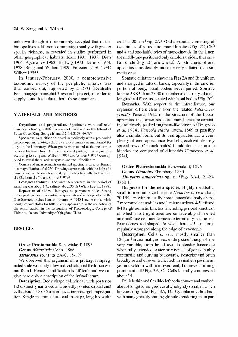
Description. Body shape cylindrical with posterior1/3 distinctly narrowed and broadly pointed caudal end;cells about 160 x 35 µm in size after protargol impregna-tion. Single macronucleus oval in shape, length x width
ca 15 x 20 µm (Fig. 2A). Oral apparatus consisting oftwo circles of paired circumoral kineties (Fig. 2C, CK)and 4 and one-half circles of monokinetids. In the latter,the middle one positioned only on �dorsal side�, thus onlyhalf circle (Fig. 2C, arrowhead). All structures of oralapparatus considerably more densely ciliated than so-matic ones.
Somatic ciliature as shown in Figs 2A and B: uniformand arranged in tufts or bands, especially in the anteriorportion of body, basal bodies never paired. Somatickineties (SK) about 25-30 in number and loosely ciliated,longitudinal fibres associated with basal bodies (Fig. 2C).
Remarks. With respect to the infraciliature, ourorganism differs clearly from the related Pelatractusgrandis Penard, 1922 in the structure of the buccalapparatus: the former has a circumoral structure consist-ing of closely packed fragment-like kineties (Dragescoet al. 1974). Vasicola ciliata Tatem, 1869 is possiblyalso a similar form, but its oral apparatus has a com-pletely different appearance: two belts, each with denselyspaced rows of monokinetids; in addition, its somatickineties are composed of dikinetids (Dragesco et al.1974).
Order Pleurostomatida Schewiakoff, 1896Genus Litonotus Ehrenberg, 1830Litonotus antarcticus sp. n. (Figs 3A-I, 21-23;
Table 1)Diagnosis for the new species. Highly metabolic,
small to medium-sized marine Litonotus in vivo about70-150 µm with basically broad lanceolate body shape,2 macronuclear nodules and 1 micronucleus; 4-5 left and8-10 right somatic kineties (including perioral kineties),of which most right ones are considerably shortenedanteriad; one contractile vacuole terminally positioned.Extrusomes rod-shaped, in vivo about 4-5 µm long,regularly arranged along the edge of cytostome.
Description. Cells in vivo mostly smaller than120 µm (in �normal�, non-extending state) though shapevery variable, from broad oval to slender lanceolatewhen fully extended. Anteriorly typical of genus, highlycontractile and curving backwards. Posterior end oftenbroadly round or even truncated; in smaller specimens,yet not seldom with narrowed end, but never formingprominent tail (Figs 3A, C). Cells laterally compressedabout 3:1.
Pellicle thin and flexible; left body convex and vaulted,about 4 longitudinal grooves often slightly spiral, in whichkineties originate (Figs 3A, D). Cytoplasm colourless,with many greasily shining globules rendering main part

Antarctic benthic marine ciliates 25
of body rather opaque (especially at low magnification).Cells basically hyaline anteriorly and in area of mouth;food vacuoles containing brown or green inclusion(algae) or pinnate diatoms. 2 macronuclear nodules, inmid-body, ellipsoid, with several small spherical or singlelarge nucleolus. Micronucleus between macronuclearnodules. Contractile vacuole small, terminally positioned.Extrusomes rod-shaped, straight and conspicuousin vivo, about 4-6 µm long; densely distributed in oralregion and scattered in other parts of body (Fig. 3I).
Cilia ca 6-8 µm long, movement genus-typical, slowlygliding on substrate.
Oral slit about half of body length. Oral structuretypical of genus (Foissner 1984): 3 perioral kineties, i.e.2 on right and 1 on left of cytostome. Anterior portion ofboth row 1 and 2 (about length of oral slit) composed ofdensely positioned dikinetids, while basal body pairs inrow 3 are widely spaced (Figs 3G, H). Characteristically,perioral kinety 2 terminating about half way along body(Fig. 3F, double-arrowheads). Nematodesmata long andhighly developed, originating from perioral kinetids about90% of cell length (Fig. 3I).
Somatic ciliature as shown in Figs 3F and G. Withexception of number 2 (numbered form dorsal to ventralin Fig. 3F, the leftmost-but-one), all other somatic kinetieson right side anteriorly shortened considerably (arrow-heads in Fig. 3F). On left side, almost consistently with4 kineties (including perioral kinety 3), of which only twokineties extending along whole length of cell, while therightmost and the one between the P3 and dorsal brosseshortened anteriad (double-arrowheads in Fig. 3G). Dorsalbrosse (DK) composed of about 20 basal body pairs,each with short bristle-like cilium, continuous posteriorlywith row of densely spaced monokinetids (Fig. 3G).
Comparison. Up to now, over 15 small Litonotus-species from various habitats have been investigatedusing modern methods, of which at least 5 forms -basedon general ciliature, body shape and size, position ofcontractile vacuole and the habitat- should be comparedwith the new species described here: L. lamella,L. yeanae, L. obtus, L. lamella uninucleata andL. emmerichi (Borror 1963, Fryd-Versavel et al. 1975,Wilbert and Kahan 1981, Dragesco and Dragesco-Kernéis 1986, Song 1991). Among them, L. lamellauninucleata is a very small species with only onemacronucleus (Wilbert and Kahan 1981). The new
Fig. 1. Sampling site (asterisk)
Table 1. Morphometric characterization of Litonotus antarcticus sp. n. All data are based on protargol-impregnated specimens. Measurementsin µm. CV - coefficient of variation in %; Max - maximum; Mean - arithmetic mean; Min - minimum; SD - standard deviation; SE - standarderror of mean
Character Min Max Mean SD SE CV n
Body length 60 114 88.9 22.63 6.82 25.5 11Body width 25 46 37.5 5.72 1.72 15.3 11Number of somatic kineties on left side* 4 5 4.1 0.33 0.11 8.1 9Number of somatic kineties on right side** 8 10 8.8 0.87 0.29 9.9 9Number of macronuclei 1 2 1.87 0.35 0.09 18.8 13Number of dikinetids in perioral kinety 3 13 15 - - - - -Number of dikinetids in dorsal brosse 18 20 - - - - -
* perioral kinety 2 not counted; **including perioral kinety 1

26 W. Song and N. Wilbert
species can be separated from L. yeanae, L. obtus andL. emmerichi in combined characters of size, number ofsomatic kineties on right side and the distinctly differentarrangement of ciliature of right side: in all other knownforms the kineties never shortened anteriorly.
Compared with the form described by Fryd-Versavelet al. (1975), under the name of L. lamella, the presentspecies can be recognized in body shape (never cau-dally-pointed in posterior end vs. having a narrowed tail)and a definitely different ciliature pattern on the rightside, i.e. all kineties in L. lamella on right side extend thewhole length of the cell (vs. being shortened anteriad).
Order Cyrtophorida Fauré-Fremiet in Corliss (1956)Genus Orthodonella Bhatia, 1936Orthodonella shenae sp. n. (Figs 4A-G, 20)
Diagnosis for the new species. Large marineOrthodonella species in vivo about 120-180 x 40-70 µm in size with �moveable� and conspicuous snout-like projection, with which the cell glides on the sub-strate; ca 45 somatic kineties on ventral and 25 on dorsalside; dominant synhymenium consisting of about80 paired kinetosomes, which is arranged in a typicalpattern of the genus; pharyngeal basket composed of ca13 rods; 2 contractile vacuoles on left cell side; one ovalmacronucleus.
Dedication. We dedicate this species to the protozo-ologist, Prof. Shen Yunfen, the Institute of Hydrobiology,Chinese Academy of Sciences, to express our respectfor her contributions to ciliate studies.
Description. Cells generally acontractile; outline elon-gate elliptical, right margin convex, left straight, con-
Figs 2A-C. Metacystis sp. from protargol impregnation. A - general view; B -anterior portion, arrowheads marking the double-rowed circumoralkinety; C - detail of anterior portion, to show the oral structure; arrowhead indicating the perioral kinety which surrounds only one side of thecell. CK - circumoral kineties, SK -�normal� somatic kineties. Scale bar - 50 µm

Antarctic benthic marine ciliates 27
Figs 3A-I. Litonotus antarcticus sp. n. from life (A-E) and protargol impregnation (F-I). A - left view; B - lateral view; C - to show differentbody shapes; D - left view of anterior portion, arrows marking the extrusomes; E - extrusomes; F - right view of infraciliature, arrowheadsindicating the shortened anterior end of some kineties, double-arrowheads referring the posterior end of perioral kinety 2; note that the leftmostkinety on the right side terminates sub-apically (arrow); G - left view of infraciliature; arrowhead indicates the posterior end of perioral kinety2, while the double-arrowheads mark the anterior end of the rightmost kinety on the left side; H - left view, to show the detail of the anteriorportion and the buccal apparatus; I - left view, to demonstrate the nematodesmata (cytopharyngeal fibres) and the distribution of theextrusomes. CV - contractile vacuole, DK - dorsal brosse, Ex - extrusomes, F - cytopharyngeal fibres, Ma - macronucleus, Mi - micronucleus,P
1-3 - perioral kinety 1-3. Scale bars: A, F - 40 µm, E - 15 µm

28 W. Song and N. Wilbert
Figs 4A-G. Orthodonella shenae sp. n. from life (A-D) and after protargol impregnation (E-G). A - ventral view; B - lateral view; C - ventralview, arrowheads mark the movable snout-like projection; D - ventrolateral view, to show the cell gliding on the substrate, arrow indicates thedirection of movement; E - right ventrolateral view; F, G - ventral and dorsal view of the same specimen, to show the general infraciliature;arrowheads indicate the �broken� kineties, double-arrowheads the snout. Nu - nucleoli, Sy - synhymenium. Scale bars: A - 80 µm, F - 50 µm

Antarctic benthic marine ciliates 29
spicuous snout-like projection extending far to left whenviewed ventrally, both anterior and posterior end broadlypointed or tapering (Fig. 4A). Dorsoventrally about3:1 flattened. Cytoplasm colourless, often with manylarge, shiny granules (3-5 µm across). Food vacuolesusually with pinnate diatoms and flagellates. Two con-tractile vacuoles positioned along left body margin, onein anterior 1/3 and another in posterior 1/3 of bodylength. Nematodesmata (pharyngeal rods) conspicuous,forming funnel-shaped basket, extending posterior-left(Figs 4A, C). Single macronucleus slightly behind mid-body, ellipsoidal , containing many small spherical chro-matin bodies and one large centrally positioned nucleolus(Fig. 4G, Nu). Micronucleus not discernible.
Movement very distinctive: when gliding, characteris-tically ventral side up, often with its snout swinging up-and-down and �attaching� to substrate, giving an impres-sion that the organism is searching for food with thesnout (Fig. 4D).
Synhymenium positioned only on ventral side, ob-liquely from right margin to apical end of snout (Fig. 4F,double-arrowheads), in which about 80 pairs of kineto-somes are closely packed.
Somatic cilia about 10 µm long. All ventral kinetiesterminating at synhymenium: preorally about 8 kinetiescurving around cytostome with 4-5 rows �broken� orseparated by cytostome (Fig. 4F, arrowheads); postorallymeridional, considerably more densely ciliated than dor-
Figs 5A-E. Aegyriana paroliva gen. n., sp. n. from life (A, C) and after protargol impregnation (B, D, E). A - ventral view of a typical individual;B - dorsal view of anterior portion, to show the right somatic kineties, which shift to dorsal side; C - ventral views, to show different bodyshapes; D - detail of buccal �basket�, to show the toothed cytopharyngeal rods; E - ventral view of infraciliature; arrows indicate the shortenedleft somatic kineties, double-arrowheads mark the perioral kineties, which are close to the curved right somatic kineties, while the arrowheadsshow the fragment-like kineties, which are posterior to the podite. Cs - cytostome, SK - right somatic kineties, Tf - terminal fragment of kinety.Scale bars - 30 µm

30 W. Song and N. Wilbert
sal side. In snout portion, kineties appear to be continu-ous posteriorly with those on dorsal side (Figs 4F, G,double-arrowheads). On dorsal side, all kineties con-spicuously loosely ciliated, extending whole dorsal sidewith anterior ends curved to left and converging in snoutregion (Fig. 4G).
Comparison. Considering the habitat, size and bodyshape, only one species in the genus Orthodonella canbe compared with our new form, O. hamata (Gruber,1884). However, the Antarctic species differs clearlyfrom it in the number and position of contractile vacuoles(2 and positioned laterally on cell margin vs. 1, caudally
Figs 6A-G. Chlamydonella sp. from life (A, D-F) and after protargol impregnation (B, C, G). A - ventral view; B, C - ventral view, to showdifferent body shapes and macronucleus; D - lateral view; E - dorsal view, to demonstrate the dorsal hump; F - dorsal view, showing a cellpacked with large diatoms; G - ventral view of the infraciliature; arrowheads indicate the perioral kineties, while the double-arrowheads markthe external right equatorial kinety. Nd - nematodesmata, SK - left somatic kineties, Tf - terminal fragment of kinety. Scale bars - 20 µm

Antarctic benthic marine ciliates 31
positioned) (Ozaki and Yagiu 1941, Jankowski 1968,Wilbert 1986).
Genus Aegyriana gen. n.This genus was originally erected by Deroux (1975)
but no type species was fixed, hence it is a nomen nudumaccording to Art 13(b) of the ICZN (1985). To maintainstability, we re-establish it here.
Diagnosis for the new genus. Dorsoventrally flat-tened Dysteriidae with tail-shaped podite, oral structurereduced to 3 fragment-like kineties; preoral kinetiesarched to left; postoral kineties making no gap in median;one bald space present subcaudally, which is completelysurrounded by kinetosomes and in which the podite ispositioned; cytopharyngeal rods dominant.
Type species Aegyriana oliva (Claparède &Lachmann, 1858)
Aegyriana paroliva sp. n. (Figs 5A-E)Diagnosis for the new species. Marine metabolic
Aegyriana in vivo 60-80 x 30-50 µm with 33-40 somatickineties, of which the leftmost 10-15 evenly shortened
posteriad; buccal basket prominent, consisting of ca 26-30 cytopharyngeal rods; single macronucleus oval inshape.
Description. Body shape variable, but generallyelliptical or D-shaped, right margin convex, left slightlysigmoid or straight with posterior end broadly rounded orirregularly truncated, inconspicuous snout-like projectionon anterior left (Figs 5A, C). Dorsoventrally rather thick,about 2:1 flattened, ventral flat, dorsal side stronglyvaulted. Cytoplasm colourless or greyish, containingnumerous small greasily shining globules and food vacu-oles commonly with long pinnate diatoms, which renderscells highly opaque or even dark grey (Fig. 5A). Nocontractile vacuoles observed. Buccal basket genus-typical and considerably wide, nematodesmata(cytopharyngeal rods) conspicuous and toothed, directedleftwards. Macronucleus ellipsoid, about 20 x 15 µm insize, located in mid-body (Fig. 5C).
Ciliation basically on ventral side with densely spacedbasal bodies. Preorally about 7 kineties arched to left
Figs 7A-D. Dysteria calkinsi Kahl, 1931 from life (A, B) and after protargol impregnation (C, D). A - right lateral view; B - left ventrolateralview, to show an ovoid-shaped cell; C - left lateral view, showing general infraciliature and nuclear apparatus; D - right lateral view ofinfraciliature, arrow indicates left equatorial field of kineties. CoK - circumoral kineties, Cph - cytopharynx, Fe - fragment of external rightkinety, Ma - macronucleus, P - podite. Scale bars: A - 30 µm, D - 40 µm

32 W. Song and N. Wilbert
margin of cell with some anterior-most kineties consis-tently shifted dorsally (Fig. 5B, SK); terminal fragment(Tf) composed of about 10 basal bodies, positioned onleft frontal end of body. Perioral kineties likely in3 fragments and separated from each other (very diffi-cult to spot in our form since they are always on themargin of anterior end of cell, see Fig. 5E, double-arrowheads). On ventral side, kineties arranged as shownin Fig. 5E: anteriorly most kineties terminating beneathbuccal region and making dominant bald area, some10-15 of which are shortened posteriorly (Fig. 5E,arrows). About 8 kineties at posterior end �interrupted�by podite and hence forming one big bald area, right ofwhich there are about 10 kineties, and ca 4 to the left.
Comparison. Only 2 species have been described inthis genus: Aegyriana oliva (Claparède & Lachmann,1858) and A. minima Deroux, 1975. The latter is smalland has far fewer somatic kineties, and hence can beclearly separated from this new species (Deroux 1975).The big form, Aegyriana oliva is, according to rede-scriptions by Kahl (1930) and Deroux (1975), similar toour new form, it can be distinguished, however, in bodyshape (oval in outline and strongly vaulted or even folded
like a Dysteria in vivo) and probably less-developedbuccal basket (Deroux 1975).
Genus Chlamydonella Deroux in Petz et al. (1995)According to the newly-defined diagnosis (Song and
Wilbert 2000a), the genus Chlamydonella is character-ized by: Lynchellids without plasmatic protrusions onventral side; somatic kineties making no noticeable na-ked gap between left and right areas; perioral kinetiescontinuous or slightly fragmented with leftmost rowsparallel to each other, which are arched transversely;cytopharyngeal rods (nematodesmata) toothed. Macro-nucleus basically dimorphic. The form described herecorresponds to this definition perfectly.
Chlamydonella sp. (Figs 6A-G)We have many well-impregnated specimens but have
failed to observe some critical features such as thesituation of the contractile vacuoles. Thus it has to bedescribed as an uncertain species though we definitelybelieve that it represents a new member in this genus.
Description. Body size and shape slightly variable(30-60 x 20-30 µm in size, but usually smaller than 40 µmlong); mostly oval with conspicuous snout-shaped pro-jection on anterior left when viewed ventrally; both ends
Table 2. Morphological comparison of some closely-related Litonotus-species, which inhabit marine or brackish water. Measurements inµm. Ma - macronuclei, SK - somatic kinety
Species Size in vivo No, SK No, SK Body shape No, Ma Referenceson left on rightside side
L. yinae 30-60 4-5 6-7 lanceolate, with 2 Song 1991bnarrowed posterior end,non-metabolic
L. lamella uninucleolata* 30-50 - 6 as above 1 Wilbert andKahan 1981
L. obtusus* ca 60 - 5 as above 2 Borror 1963
L. lamella* 40-200 5 10-16 lanceolate, with 2 Dragesco andnarrowed posterior end Dragesco-
Kernéis1986
L. antarcticus sp. n. 70-150 4-5 8-10 with broadly rounded 2 present paperposterior end,highly metabolic
L. emmerichi 60-90 4-5 5 with narrowed 2 Petz et al. 1995posterior end,non-metabolic
*Systematic identities not confirmed in the current table

Antarctic benthic marine ciliates 33
rounded (Figs 6A, E, F). Dorsoventrally about 1:2 flat-tened, ventral side flat, dorsal vaulted (Fig. 6D). Cyto-plasm colourless to greyish, often containing numeroustiny granules and several to many different-sized dia-toms (Figs 6A, F). Cytostome prominent, sub-apicallylocated; buccal basket consisting of about 12cytopharyngeal rods or nematodesmata (Nd in Fig. 6G),extending leftwards and posteriorly (Figs 6B, G). Con-tractile vacuoles not observed. One large ellipsoid ma-cronucleus positioned in mid-body containing severallarge nucleoli (Fig. 6B). Micronucleus not recognizable.
Infraciliature as shown in Fig. 6G, 3 rightmost kinetiesextending preorally, with anterior portion curved to leftmargin; one terminal fragment (Tf) positioned on distalmargin of cell. All other kineties (ca 11-14 in number)terminating at about cytostome level, making no sutureposteriad. In addition to these �normal� kineties, oneexternal right kinety which is fragment-like with about
6 basal bodies always recognizable in mid-body (Fig. 6G,double-arrowheads).
Oral structure consisting of two long rows of dikinetids,which are transversely arched preorally (Fig. 6G, arrow-heads).
Remarks. The form described here is similar to itscongener, C. pseudochilodon (Deroux, 1970), whichwas also isolated from the Antarctic area (Petz et al.1995), but differs from the latter in smaller size (vs.75 µm on average), differently humped dorsal side andthe body shape (Deroux 1970, 1976; Petz et al. 1995).
Genus Dysteria Huxley, 1857Much misinterpretation was/is involved in descrip-
tions of the structure or position of organelles of theseorganisms. This genus is characterized by the highlybilaterally (vs. dorsoventrally flattened in most othergroups) flattened body shape, hence all somatic ciliationis �squeezed� into a narrow region ventrally, while the
Table 4. Morphometric characterization of Scyphidia sp. Data are either from life (in vivo) or based on protargol-impregnated specimens(*) Measurements in µm
Character Min Max Mean SD M n
Body length in vivo 74 115 95.5 12.0 96 33Body width in vivo 31 43 36.9 3.7 36 33Peristomial collar diameter in vivo 26 46 35.0 6.9 35 15Scopula diameter in vivo 20 32 24.2 2.8 26 33Length of macronucleus* 21 30 24.2 3.0 24 27Width of macronucleus* 12 15 13.7 1.2 14 27Length of micronucleus* 6 8 6.8 0.8 7 27Width of micronucleus* 3 4 3.7 0.48 4 27Distance between aboral ciliary wreath and scopula* 15 25 20.3 3.3 18 27
Table 3. Morphometric characterization of Metanophrys antarctica sp. n. All data are based on protargol-impregnated specimensMeasurements in µm
Character Min Max Mean SD SE CV n
Body length 28 41 34.9 4.73 1.26 13.5 14Body width 20 31 26.1 3.26 0.94 12.5 12Length of buccal field 17 21 18.3 1.49 0.41 8.2 13Length of macronucleus 15 19 16.7 1.60 0.61 9.6 7Width of macronucleus 12 15 13.1 1.22 0.46 9.2 7Number of macronuclei 1 1 1 0 0 0 >30Number of somatic kineties 14 14 14 0 0 0 12Number of kinetosomes in somatic kinety 1* 17 19 - - - - 3Number of kinetosomes in somatic kinety n* 18 19 - - - - 3
* Paired kinetosomes counted as single.
34 W. Song and N. Wilbert
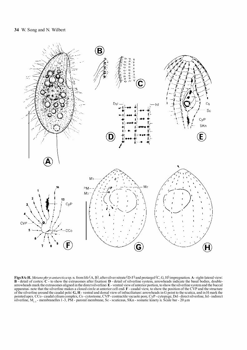
Figs 8A-H. Metanophrys antarctica sp. n. from life (A, B), after silver nitrate (D-F) and protargol (C, G, H) impregnation. A - right-lateral view;B - detail of cortex; C - to show the extrusomes after fixation; D - detail of silverline system, arrowheads indicate the basal bodies, double-arrowheads mark the extrusomes aligned in the direct silverline; E - ventral view of anterior portion, to show the silverline system and the buccalapparatus; note that the silverline makes a closed circle at anterior cell end; F - caudal view, to show the position of the CVP and the structureof the silverline around the caudal pole; G, H - ventral and dorsal view of infraciliature; arrowheads in G point to the scutica, and in H mark thepointed apex. CCo - caudal cilium complex, Cs - cytostome, CVP - contractile vacuole pore, CyP - cytopyge, Dsl - direct silverline, Isl - indirectsilverline, M
1-3 - membranelles 1-3, PM - paroral membrane, Sc - scuticaus, SKn - somatic kinety n. Scale bar - 20 µm

Antarctic benthic marine ciliates 35
left and right field of somatic kineties are positionedtogether (the latter are reduced to left equatorial fieldwith densely packed, fragment-like kineties).
Dysteria calkinsi Kahl, 1931, (Figs 7A-D, 24)Description. In vivo about 30-50 x 20-25 µm. Body
bilaterally about 1:2-3 flattened. From side view, cellsslender and more or less quadrilateral with 2 moderatelyconspicuous longitudinal furrows; anteriorly with someirregularly positioned ridges (Fig. 7A). When viewedventrally, right side flat while left arched (Fig. 7B).Podite (attachment organelle, P) about 15 µm long,distally pointed, posteriorly on narrow ventral side. Cy-
toplasm colourless or slightly greyish, containing severallarge food vacuoles (likely with flagellates). Cytostomeventrally at anterior end, diagonally oriented, with incon-spicuous nematodesmata (cytopharyngeal rods) extend-ing dorsad (Fig. 7D, Cph). Contractile vacuoles notobserved. Macronucleus (Ma) about in mid-body, ca25 x 15 µm in size, characteristically dimorphic: anteriorhalf with numerous fine nucleoli, while many largegranules of nucleoli visiable in posterior half (Figs 7C,D).
Movement genus-typical, very quiet, slowly crawlingand slightly thigmotactic. Infraciliature as shown in
Figs 9A-F. Scyphidia sp. from life (A-C), protargol (D, E) and silver nitrate (F) impregnation. A-C - to show zooids with different body sizesand shapes; arrow in C indicates the peak of the disc; arrowheads mark the finely striped pellicle; D - general view, to demonstrate theinfraciliature and nuclear apparatus; arrowheads indicate the argentophilic fibres; E - oral apparatus; arrowheads mark the separated row of thepolykinety 2; F - detail of silverline system, arrowheads point to the aboral ciliary wreath. ACW - aboral ciliary wreath, AD - attaching disc,Cph - cytopharynx, CV - contractile vacuole, G - germinal row, H - haplokinety, P - polykinety, Ma - macronucleus, Mi - micronucleus,P
1-3 - peniculi 1-3. Scale bars - 40 µm

36 W. Song and N. Wilbert
Fig. 7D: when viewed from right side, �right� filed ofciliature composed of (consistently !) 5 densely spacedkineties of variable length, formed by groups of ca4 basal bodies, extending from anterior left to posterior
right (Fig. 7D). To left of distal end of these kineties, onefragment of kineties (Fe) always present. In mid-body,about 6 short rows of densely packed basal bodiesforming left equatorial field (arrow in Fig. 7D). Circu-
Figs 10A-J. Heterostentor coeruleus gen. n., sp. n. from life (A-E, G) and after protargol impregnation (F, H-J). A - a typical individual; B,C - to show extending and contractile forms; D - detail of pellicle, arrowhead indicates the pigments; E - anterior portion, note the pigmentscondense beneath the buccal apparatus (double-arrowheads); F - portion of cortex, arrowhead marks the granules beneath the pellicle; G - todemonstrate a cell gliding on substrate; H - to show the somatic kineties, arrowheads mark the shortened anterior end of some kineties whichmake the �suture�. I - anterior part of cell, to show the adoral zone of membranelles, note no definite cytopharynx present; arrowheads markthe shortened kineties which form the suture, while the double-arrowheads indicate the somatic kinety No. 1; J - caudal view, to demonstratethe close-set caudal cilia. CC - caudal cilia, Ma - macronucleus. Scale bars - 120 µm

Antarctic benthic marine ciliates 37
moral kineties (CoK) genus-typical, dorsally aroundcytostome (Fig. 7D).
Remarks. This species has never been reinvestigatedusing modern methods since it was described by Kahl(1931). We identify our population according to the size,body shape and the habitat (it was originally found in theAtlantic). The only point we cannot confirm: Kahl (1931)described it having 2 diagonally positioned contractilevacuoles, which were not detected in this Antarctic form(further observations needed).
It differs form D. marina in larger size (30-50 vs.25 µm), slender body shape (vs. oval) and furrowed sideview (vs. smooth) (Kahl 1931, Borror 1963).
Order Scuticociliatida Small, 1967Genus Cyclidium O. F. Müller, 1786Cyclidium varibonneti Song, 2000, (Fig. 36)This Antarctic population was found in great numbers
and resembles very closely that in the original report(Song 2000), hence the identification is quite certain anda further description is unnecessary.
Genus Metanophrys De Puytorac et al., 1974According to the new definition (Song and Wilbert
2000b), the genus Metanophrys is distinguished by:(a) possessing a Parauronema-like buccal apparatus(2- or more-rowed membranelle 1, the paroral mem-brane extending anteriorly to about the middle of the
Figs 11A-F. Condylostoma remanei Spiegel in Kahl, 1932 from life (C) and after protargol impregnation (A, B, D-F). A, B - ventral and dorsalview of infraciliature, with arrowheads indicating the shortened somatic kineties; C - schematic diagram, to show �typical� body shape;D - detail of buccal apparatus; E - tail portion; F - to show the somatic kineties. AZM - adoral zone of membranelles, BC - buccal cavity,Ci - membranelles-like cirri, PM - paroral membrane. Scale bar - 100 µm

38 W. Song and N. Wilbert

Antarctic benthic marine ciliates 39
membranelles 2), and (b) the pointed apex (i.e. with noconspicuous frontal plate). So the form we present hereshould be clearly assigned into this taxon.
Metanophrys antarctica sp. n. (Figs 8A-H, 32-35;Table 3)
Diagnosis for the new species. Medium-sizedmarine Metanophrys in vivo 30-45 x 20-30 µm; bodygenerally ovoid with slightly pointed apical end; onemacro- and one micronucleus; oral field about 2/5 ofcell length; buccal apparatus genustypical withdistinctly small membranelles; 14 somatic kineties with
ca 18 mono- and dikinetids each; single contractilevacuole terminally located; one prolonged caudal cilium.
Description. Cells in vivo mostly 30-40 x 20-25 µm,body shape oval to slender bag-shaped, circular in cross-section; apical end slightly pointed while posterior gener-ally rounded. Ventral surface gently indented aroundbuccal area, dorsally convex (Fig. 8A). Buccal field withshallow depression, about 40% of cell length. Pelliclerigid and slightly notched, below it densely spaced spindle-like extrusomes (ca 2 µm in length) (Fig. 8B), whichafter fixation usually appear thicker (Fig. 8C). Cilia
Figs 12A-J. Metaurostylopsis rubra sp. n., from life (A-F) and after protargol impregnation (G-J). A-D - to show different body shapes andarrangement of cortical granules (B); arrowheads in C indicate the contractile vacuole, arrow in D marks the densely arranged pigments atanterior end of cell; E - to show pigments beneath the pellicle; F - lateral view; G - detail of structure in buccal field, with arrowhead markingthe position where the midventral rows and the ventral row are conjoined; H, J - ventral and dorsal view of infraciliature, with arrowheadsindicating the fragment-like kineties anterior to the right marginal rows, while the double-arrowheads mark the �isolated� dikinetids;I - micronuclei. AZM - adoral zone of membranelles; BC - buccal cirrus; DK
2, 3 - dorsal kinety 2,3; Cph - cytopharynx; EM - endoral membrane;
FC - frontal cirri; FTC - frontoterminal cirri; LMR - left marginal rows; MVR - midventral rows; PM - paroral membrane;RMR - right marginal rows; TC - transverse cirri; VR -ventral row. Scale bars: A - 80 µm, H - 50 µm
Figs 13A-B. Holosticha diademata (Rees, 1884) after protargol impregnation. A, B - ventral and dorsal view of the same specimen, to show theinfraciliature and nuclear apparatus; arrowheads indicate the �gap� between the anterior and posterior part of adoral zone of membranelles,while the arrow marks the �hook-like� arrangement of cirri in left marginal row. BC - buccal cirrus, FTC - frontoterminal cirri, LMR - leftmarginal rows, MVR - midventral rows, RMR - right marginal rows, TC - transverse cirri. Scale bar - 30 µm
40 W. Song and N. Wilbert
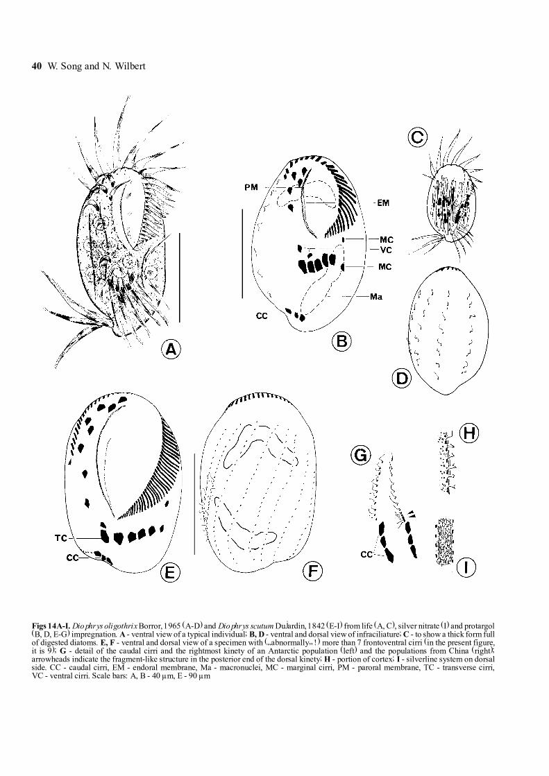
Figs 14A-I. Diophrys oligothrix Borror, 1965 (A-D) and Diophrys scutum Dujardin, 1842 (E-I) from life (A, C), silver nitrate (I) and protargol(B, D, E-G) impregnation. A - ventral view of a typical individual; B, D - ventral and dorsal view of infraciliature; C - to show a thick form fullof digested diatoms. E, F - ventral and dorsal view of a specimen with (�abnormally� !) more than 7 frontoventral cirri (in the present figure,it is 9); G - detail of the caudal cirri and the rightmost kinety of an Antarctic population (left) and the populations from China (right);arrowheads indicate the fragment-like structure in the posterior end of the dorsal kinety; H - portion of cortex; I - silverline system on dorsalside. CC - caudal cirri, EM - endoral membrane, Ma - macronuclei, MC - marginal cirri, PM - paroral membrane, TC - transverse cirri,VC - ventral cirri. Scale bars: A, B - 40 µm, E - 90 µm

Antarctic benthic marine ciliates 41
about 8 to 10 µm long, single caudal cilium ca 15 µm inlength (Fig. 8A).
Cytoplasm colourless, often with several to manylarge shining granules (3-5 µm in diameter, inactive foodvacuoles) and diatoms, which often render specimensrather dark. No food vacuoles observed. Contractilevacuole small, terminally located at posterior end of cell
(Fig. 8A). One large oval macronucleus centrally lo-cated; micronucleus uncertain, possibly adjacent to ma-cronucleus.
Infraciliature as shown in Figs 8G, H. Somatic kinetieslongitudinally arranged, extending over entire length ofbody, which are mainly composed of dikinetids through-out except the posterior 1/4 of each kinety. Each row
Figs 15A-F. Euplotes balteatus (Dujardin, 1841) Kahl, 1932 from life (A, B), protargol (C, D) and silver nitrate (E, F) impregnation. A - ventralview of a typical individual; B - dorsal view, to show the ridges; C, D - ventral and dorsal view of infraciliature; arrow in C indicates the enlargedtransverse cirrus; E, F - ventral and dorsal view of the silverline system. CC - caudal cirri, CVP - pore of contractile vacuole, MC - marginal cirri,PM - paroral membrane. Scale bars - 30 µm
42 W. Song and N. Wilbert
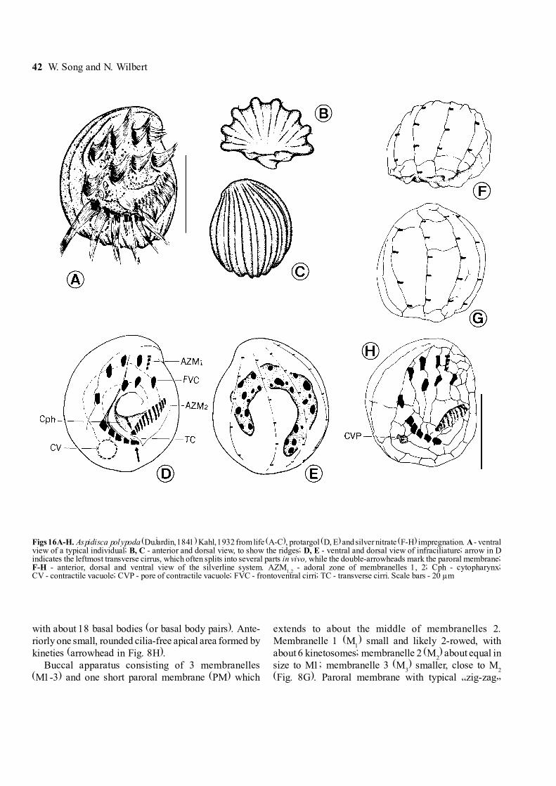
with about 18 basal bodies (or basal body pairs). Ante-riorly one small, rounded cilia-free apical area formed bykineties (arrowhead in Fig. 8H).
Buccal apparatus consisting of 3 membranelles(M1-3) and one short paroral membrane (PM) which
extends to about the middle of membranelles 2.Membranelle 1 (M
1) small and likely 2-rowed, with
about 6 kinetosomes; membranelle 2 (M2) about equal in
size to M1; membranelle 3 (M3) smaller, close to M
2
(Fig. 8G). Paroral membrane with typical �zig-zag�
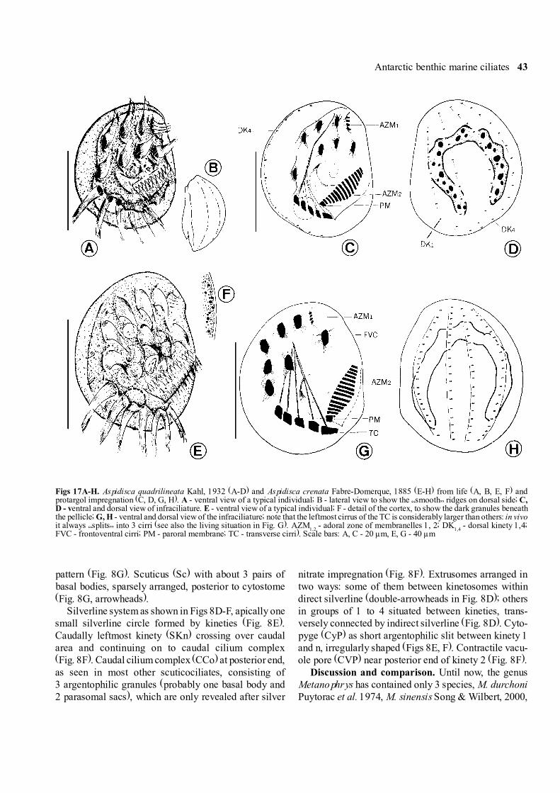
Figs 16A-H. Aspidisca polypoda (Dujardin, 1841) Kahl, 1932 from life (A-C), protargol (D, E) and silver nitrate (F-H) impregnation. A - ventralview of a typical individual; B, C - anterior and dorsal view, to show the ridges; D, E - ventral and dorsal view of infraciliature; arrow in Dindicates the leftmost transverse cirrus, which often splits into several parts in vivo, while the double-arrowheads mark the paroral membrane;F-H - anterior, dorsal and ventral view of the silverline system. AZM
1,2 - adoral zone of membranelles 1, 2; Cph - cytopharynx;
CV - contractile vacuole; CVP - pore of contractile vacuole; FVC - frontoventral cirri; TC - transverse cirri. Scale bars - 20 µm
Antarctic benthic marine ciliates 43
pattern (Fig. 8G). Scuticus (Sc) with about 3 pairs ofbasal bodies, sparsely arranged, posterior to cytostome(Fig. 8G, arrowheads).
Silverline system as shown in Figs 8D-F, apically onesmall silverline circle formed by kineties (Fig. 8E).Caudally leftmost kinety (SKn) crossing over caudalarea and continuing on to caudal cilium complex(Fig. 8F). Caudal cilium complex (CCo) at posterior end,as seen in most other scuticociliates, consisting of3 argentophilic granules (probably one basal body and2 parasomal sacs), which are only revealed after silver
nitrate impregnation (Fig. 8F). Extrusomes arranged intwo ways: some of them between kinetosomes withindirect silverline (double-arrowheads in Fig. 8D); othersin groups of 1 to 4 situated between kineties, trans-versely connected by indirect silverline (Fig. 8D). Cyto-pyge (CyP) as short argentophilic slit between kinety 1and n, irregularly shaped (Figs 8E, F). Contractile vacu-ole pore (CVP) near posterior end of kinety 2 (Fig. 8F).
Discussion and comparison. Until now, the genusMetanophrys has contained only 3 species, M. durchoniPuytorac et al. 1974, M. sinensis Song & Wilbert, 2000,
Figs 17A-H. Aspidisca quadrilineata Kahl, 1932 (A-D) and Aspidisca crenata Fabre-Domerque, 1885 (E-H) from life (A, B, E, F) andprotargol impregnation (C, D, G, H). A - ventral view of a typical individual; B - lateral view to show the �smooth� ridges on dorsal side; C,D - ventral and dorsal view of infraciliature. E - ventral view of a typical individual; F - detail of the cortex, to show the dark granules beneaththe pellicle; G, H - ventral and dorsal view of the infraciliature; note that the leftmost cirrus of the TC is considerably larger than others: in vivoit always �splits� into 3 cirri (see also the living situation in Fig. G). AZM
1,2 - adoral zone of membranelles 1, 2; DK
1,4 - dorsal kinety 1,4;
FVC - frontoventral cirri; PM - paroral membrane; TC - transverse cirri). Scale bars: A, C - 20 µm, E, G - 40 µm
44 W. Song and N. Wilbert

Antarctic benthic marine ciliates 45
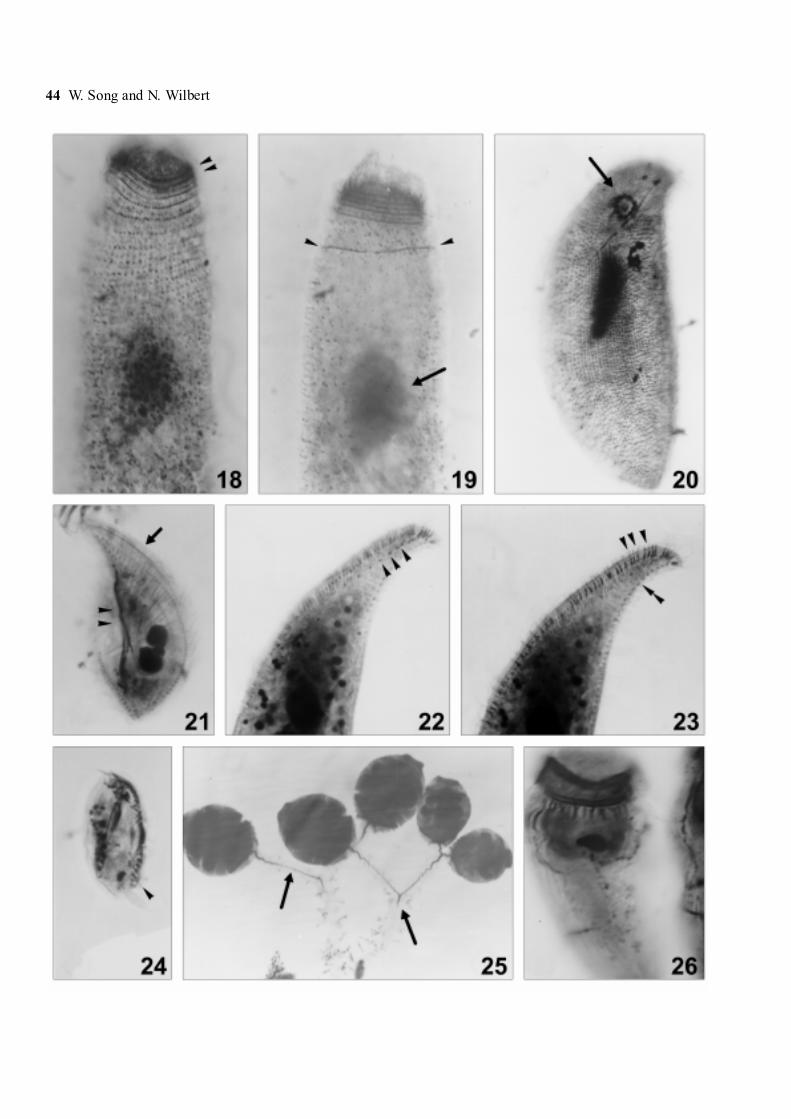
Figs 18-26. Photomicrographs of Metacystis sp. (18, 19), Orthodonella shenae sp. n. (20), Litonotus antarcticus sp. n.(21-23), Dysteriacalkinsi (24) and Zoothamnium sp. (25, 26) after protargol impregnation. 18, 19 - infraciliature of anterior portion; arrowheads in Fig. 18indicate the double-rowed circumoral kineties, and in Fig. 19 mark the posterior-most perioral kinety; arrow in Fig. 19 ponts to macronucleus(arrow). 20 - ventral view of infraciliature; arrow indicates the cytostome. 21 - right view of infraciliature; arrowheads mark the fibres, whilethe arrow indicates the first perioral kinety; 22 - left view; arrowheads mark the 3rd perioral kinety; 23 - left view of the same specimen as inFig. 22, to show extrusomes (arrowheads) and the dorsal brosse (double-arrowheads). 24 - to show the ventral kineties. 25 - four zooids, arrowsmark the myoneme; 26 - general appearance of a zooid, note the argentophilic fibres extending all over the cell
Figs 27-31. Photomicrographs of Scyphidia sp. 27 - ventral view of the host, Nacella concinna, with arrowheads marking the gills. 28 - zooidsof Scyphidia sp. on the outer edge of a gill plate (arrowheads); 29 - a typical zooid in vivo. Arrow points to the peak on the perioral disc;30 - protargol impregnated specimens, showing general infraciliature and nuclear apparatus; double-arrowheads indicate the peniculi, while thearrowhead marks the aboral ciliary wreath, 31 - silverline system after silver nitrate impregnation, note the pellicular pores between the close-set silverlines (arrowheads). Scale bars: 27- 5 mm, 28 - 200 µm, 29-31 - 50 µm

46 W. Song and N. Wilbert
and M. elongata (Biggar & Wenrich, 1932) Grolière etal. (1978). Our new species differs from M. elongatain body size (30-45 vs. 100-120 µm), number of somatickineties (14 vs. 15-20) and structure of oral apparatus:the latter has highly developed, extremely longmembranelles 1 and 2 (Grolière et al. 1978). M. durchonihas 2 contractile vacuole pores situated subcaudallywithin 2nd and 3rd somatic kinety, and a single-rowedmembranelle 1 (Puytorac et al. 1974). Additionally, thesilverline surrounding the caudal pole forms a small andin particular closed circle (misinterpretation ?) and is,therefore, distinctly different from those of the Antarcticorganism. The species recently reported by the presentauthors, M. sinensis, is similar to this new species in sizebut can be identified definitely by lower number ofsomatic kineties (9-10 vs. 14), differently arrangedextrusomes and the structure of membranelle 1: in theformer membranelle 1 is much longer with ca 8 pairs ofkinetosomes (Song and Wilbert 2000b).
Genus Pleuronema Dujardin, 1836Pleuronema coronatum Kent, 1881, (Fig. 37)This species was identified from protargol impreg-
nated specimens. All morphometrical data correspondwith the previous descriptions perfectly. For details seeDragesco (1968) and Song and Wilbert (1997).
Order Peritrichida Stein, 1859Genus Scyphidia Dujardin, 1841The family Epistylididae Kahl, 1933, to which
Scyphidia belongs, comprises sessile peritrichs thateither attach directly by way of the scopula or are fixedto the substrate by a non-contractile stalk. This familyhas been subdivided by Guhl (1979) into two subfamilies:(1) Epistylinae, with the diagnosis: oral region as a rulewith distinctly set-off marginal ridge (= peristome withlip), the disk is not stalked, being continuous with the lip;brief description �epistyliform�. (2) Opercularinae, whichis characterized by: peristome without lip, disc can beabsent and when present is usually stalked; brief de-scription �operculariform�.
The traditional genus Scyphidia Dujardin, 1841 in-cludes both epistyliform and operculariform species.Hence it was divided into two genera by Guhl (1979):Scyphidia and Scyphidiella (for the operculariform).In 1985, Jankowski included Scyphidiella in thegenus Scyphidia but placed the epistyliform speciesoriginally assigned to Scyphidia by Guhl in a total of4 genera according to the habitats and living styles:Mantoscyphidia Jankowski, 1980: reserved for speciesliving on freshwater and marine mollusks; Myoscyphidia
Jankowski, 1985 for species on aquatic plants and in theperiphyton of ponds; Speleoscyphidia Jankowski, 1980:with one limnetic free-living species and RiboscyphidiaJankowski, 1980 for species living on marine and fresh-water fishes (Jankowski 1980).
The classification of Scyphidia according to its hostsis definitely questionable, chiefly in view of the fact thatmany symphorionts do not exhibit the degree of hostspecificity required for such a system. For this reasonwe consider it unsuitable and maintain the systematicarrangement by Guhl (1979). Based on this new under-standing, Mantoscyphidia marioni Van As et al., 1998should be transferred into Scyphidia as a new combina-tion, Scyphidia marioni (Van As et al. 1998) comb. n.
The Antarctic form described here was found in largenumbers on the gills of the Antarctic limpet Nacellaconcinna collected in the lower littoral at low tide andall its morphology was investigated in detail (i.e. livingmorphology, infraciliature and silverline system). Sincesome taxonomically reliable characters (i.e. theinfraciliature and silverline system) are unavailable inmost known congeners, which have been basically de-scribed using �classical� methods, while many otherfeatures (e.g. the body shape, size, position of thecontractile vacuole or the appearance of the nuclearapparatus) overlap extensively among them, it is ex-tremely difficult to recognize or to separate them (Cuénot1891, Hirshfield 1949, Lom and Corliss 1968, Fish andGoodwin 1976, Van As et al. 1998). As a result, it iscompletely impossible for us to identify our species oreven to compare it with its �related� forms.
In our opinion, this genus urgently needs a detailedrevision and all �known� as well as uncertain speciesneed re-confirmation or careful description/comparisonbefore a definitive assignment is made. Hence, we treatour form as an �unrecognized� species here, whosetaxonomic identification awaits further comparison andinvestigations.
Scyphidia sp. (Figs 9A-E, 27-31, 61; Table 4)Description. Zooids in extended state usually cylin-
drical or conical with prominent attaching �foot� onsurface of gills, body about 70- 120 µm in length, whencontracted, oval to broadly spindle-shaped (Figs 9B, C).Peristome with widely projecting collar. Peristomial discslightly convex with a central peak (Fig. 29, arrowhead).Contractile vacuole large, below peristomial collar anddorsally positioned. Buccal cavity conspicuous;cytopharynx short, limited to the upper third of body.Pellicle delicately ringed and discernible only at highmagnification. Endoplasm greyish, often with several

Antarctic benthic marine ciliates 47
large shining globules (inactive food vacuoles). Macro-nucleus variable in shape, slender to broad oviform,always positioned in posterior end of body.
This organism not very sensitive to stimulation, onisolated gills no formation of swarmers being induced(even after more than 2 weeks).
Infraciliature as shown in Figs 9D, E, haplokinety (H)und polykinety (P) spiral through about one anda half turns before entering vestibule (buccal cavity). Asin other peritrichs, polykinety in buccal cavity differenti-ating three distinct peniculi (P
1-3), which finally spiral to
reach the cytopharynx. Germinal kinety (G) begins far
Table 5. Morphometric comparison of Metaurostylopsis rubra sp. n. (upper line), M. marina (Kahl, 1932) Song et al. (2001) (middle line)and Holosticha diademata (Rees, 1884) (lower line). All data are based on protargol-impregnated specimens. Measurements in µm
Character Min Max Mean SD SE CV n
Body length 132 226 181.1 32.54 10.29 18.0 1086 120 107.1 9.20 2.06 9.3 2052 77 66.7 9.17 3.46 13.7 12
Body width 72 101 83.3 11.64 4.75 13.8 738 60 47.0 5.80 1.85 12.2 2021 54 42.0 6.49 1.88 15.5 12
Length of buccal field 54 72 60.3 5.59 2.11 9.3 735 46 40.6 2.90 0.67 7.1 1920 38 28.6 6.39 1.84 22.4 12
Number of adoral membranelles 35 46 38.9 3.76 1.19 9.7 1027 30 28.4 1.10 0.26 4.3 1921 33 28.1 3.81 1.21 13.6 10
Number of buccal cirri 1 1 1 0 0 0 >201 1 1 0 0 0 251 1 1 0 0 0 12
Number of frontal cirri 4 4 4 0 0 0 144 4 4 0 0 0 254 4 4 0 0 0 12
Number of frontoterminal cirri 5 8 6.2 0.98 0.30 15.9 113 6 4.5 0.96 0.24 21.7 162 2 2 0 0 0 13
Number of cirral pairs in midventral rows 8 11 9.5 1.04 0.31 11.0 117 11 9.3 1.14 0.29 12.2 167 10 8.7 1.03 0.31 12.4 12
Number of cirri in the ventral row 8 13 9.8 1.69 0.53 17.2 104 7 - - - - 5- - - - - - -
Number of left marginal rows 6 9 7.8 0.87 0.25 11.2 123 5 4.1 0.57 0.14 14.1 161 1 1 0 0 0 >30
Number of right marginal rows 6 7 6.3 0.49 0.14 7.8 123 5 4.1 0.44 0.11 10.9 161 1 1 0 0 0 >30
Number of transverse cirri 4 6 4.6 0.77 0.21 16.6 135 9 6.8 1.00 0.24 15.2 177 11 9.75 1.22 0.35 12.5 12
Number of cirri in left marginal row - - - - - - -- - - - - - -
12 16 14.3 1.49 0.43 10.4 12Number of cirri in right marginal row - - - - - - -
- - - - - - -14 21 17.7 1.95 0.60 11.0 11
Number of dorsal kineties* 3 3 3 0 0 0 143 3 3 0 0 0 164 5 4.25 0.45 0.13 10.6 12
* Only the complete rows counted

48 W. Song and N. Wilbert
Figs 32-40. Photomicrographs of Metanophrys antarctica sp. n. (32-35), Cyclidium varibonneti (36), Pleuronema coronatum (37) andCondylostoma remanei (38-40) after silver nitrate (32-35) and protargol (36-40) impregnation. 32 - ventral view of silverline system;arrowhead marks the cytostome, while double-arrowheads indicate the membranelle 1; 33 - to show the arrangement of the extrusomes (double-arrowheads); arrow marks the cytostome; 34 - caudal-lateral view; arrow indicates the pore of contractile vacuole, while the double-arrowheadspoint to the cytopyge; 35 - caudal view, arrow marks the caudal-cilium-complex. 36 - general view of some individuals, arrowheads indicate thedensely ciliated somatic kinety n-1; 37 - ventral view of infraciliature. 38 - posterior portion; arrow indicates the portion where the shortenedkineties converge, double-arrowheads mark the long tail; 39 - anterior portion, arrow indicates the paroral membrane; 40 - general view fromdorsal side
Antarctic benthic marine ciliates 49
up in vestibule and parallel to haplokinety. In vestibulewhere the peniculi differentiate, G and H separate fromthe polykinety and run towards the opposite wall. Bothpeniculi 1 and 2 considerably longer than P3 (Fig. 9E).Aboral ciliary wreath (ACW) band-like, formed byshort, diagonal kineties, each comprising ca 3 kineto-somes (Fig. 9C).
Silverline system striated type, very closely spacedless than 1µm between the individual lines, with numer-ous pellicular pores between silverlines (Fig. 9F). Itresembles the silverlinesystem of Scyphidia physarumLachmann, 1856, but the latter has a more widely spacedstriation ( Foissner and Schiffmann 1979)
Remarks. Our Scyphidia is possibly close toMantoscyphidia marioni Van As et al., 1998, which isalso isolated as an ectocommensal organism from Nacellaoff South Africa, but has symbiotic flagellates (Van Aset al. 1998). However, the two forms can be camparedonly on the basis of characters from life, for the latterwas described without giving data or the infraciliatureand silverline system. Mantoscyphidia marioni is smallerthan the Antarctic form in vivo and differs from thelatter in having symbiotic flagellates.
Other related symphorionts on marine mollusks areS. hydrobiae Kahl, 1933, 70 µm long, found in the KielBay on tentacles and snout of Hydrobia species;S. patellae Cuénot, 1891, 30-50 µm in length, on the gillsof Patella vulgata, Atlantic; S. ubiquita Hirshfield,1949, 102 x 43 µm in size, on the gills and in the mantlecavity of the genera Acmea, Lottia, Tegula, Fissurella,Littorina and Gibbula (Hirshfield 1949, Lom and Corliss1968, Fish and Goodwin 1976); S. acanthophora Fish& Goodwin, 1976, 108 x 37 µm, in the mantle cavity ofGibbula umbilicalis and Monodonta lineata, IrishSea. For completeness, mention should also be made ofthe Atlantic species S. scorpaenae Fabre-Domergue,1888, which is epizoic on the gills of Scorpaena, 53 µmlong. Our Antarctic form differs from all these men-tioned above in the combined characters of body shapeand size. The peaked structure projecting above the disccan be found in either freshwater or marine species.
Genus Zoothamnium Bory de St Vincent, 1826Zoothamnium sp. (Figs 25, 26)All colonies observed are rather senile and somewhat
�abnormal� in stalk and myoneme structure, whichrender species identification difficult. Hence it is consid-ered as an incertae sedis.
Zooids elongate, in vivo about 100 µm long withdominant double-layered peristomial border; pelliclesmooth (at least at low magnification), endoplasm trans-
parent. Contractile vacuole large, apically located. Ma-cronucleus C-shaped, thick and transversely positioned.Colony large, stalk branching regularly dichotomous.There seems no differentiation of macro- and microzooids.
This species is fairly similar to the well-knownZoothamnium duplicatum Kahl, 1933 but larger in sizeand relatively slender (more elongated) in the shape ofzooids (Song 1991a). Further investigations are definitelyneeded.
Order Heterotrichida Stein 1859Genus Heterostentor gen. n.Diagnosis. Slightly to strongly contractile, free-living
Heterotrichina with elongated body shape; no conspicu-ous buccal cavity or cytopharynx; oral apparatus Sten-tor-like, restricted apically around small truncated fron-tal plate; without peristomial kineties, paroral membranehighly degenerated or absent; somatic ciliature withsuture on ventral side.
Type species. Heterostentor coeruleus sp. n.Etymology. The denomination �Heterostentor�
(Hetero- Latin, different) indicates that this new genusis similar to the well-known taxon Stentor. Masculinegender.
Remarks. This new genus is characterized by thecombination of the following traits: (1) without conspicu-ous buccal cavity; (2) structure of adoral zone ofmembranelles like that in Stentor, but has no peristomialkineties; (3) paroral membrane not present or highlydegenerated when present and (4) free-living with elon-gated body shape.
Heterostentor coeruleus sp. n. (Figs 10A-J, 41-44)Diagnosis for the new species. Slightly asymmet-
ric and contractile Heterotrichina with bright blue colourand elongate body shape; in vivo about 200-300 µm longwith ca 50-70 membranelles in AZM which form a non-closed �6� pattern; no paroral membrane present; 100-120 somatic kineties in mid-body; one oval to ellipsoidmacronucleus 60-80 x 30-40 µm in size; marine habitat.
Description. Body shape basically as shown inFig. 10A, mostly elongated cylindrical to slightly fusi-form, dorsoventrally somewhat flattened, length to widthabout 1:3-5; posteriorly narrowed to rounded tapering;anterior end narrowed and more or less head-like (simi-lar to some prostomatids), oral bulge vaulted or slightlytruncated (Figs 10A, G). Cells highly contractile but notsensitive to disturbance, hence in vivo observations,body shape almost constant, not metabolic or flexible(the contractility can be seen only after fixation). Pellicleas in some Stentor, rough with densely packed, dark-

50 W. Song and N. Wilbert
Figs 41-50. Photomicrographs of Heterostentor coeruleus gen. n., sp. n. (41-44), Euplotes balteatus (45-48) and Metaurostylopsis rubraisp. n. (49, 50) after silver nitrate (45-48) and protargol (41-44, 49, 50) impregnation. 41 - anterior portion, arrowheads mark the darkargentophilic granules; 42 - anterior portion, to show the buccal apparatus (adoral zone of membranelles, arrow); note the suture on ventral side(arrowhead); 43 - general view of somatic ciliature, arrowheads mark the macronucleus; 44 - posterior portion. 45-47 - dorsal view, to show theirregular (45) and regular (46, 47) lattice structure; 48 - ventral view, arrow indicates the 2 close-set marginal cirri. 49 - posterior portion, toshow the transverse cirri (arrow). 50 - general infraciliature on ventral side, arrowheads indicate the frontoventral cirri
Antarctic benthic marine ciliates 51
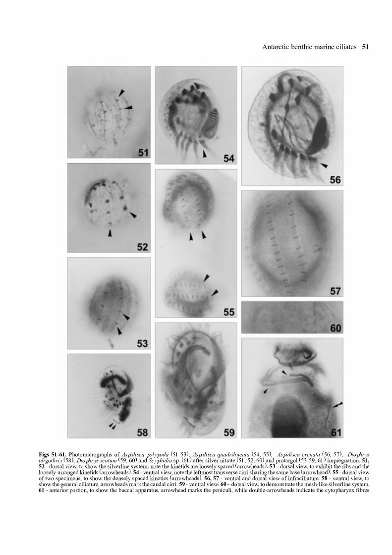
Figs 51-61. Photomicrographs of Aspidisca polypoda (51-53), Aspidisca quadrilineata (54, 55), Aspidisca crenata (56, 57), Diophrysoligothrix (58), Diophrys scutum (59, 60) and Scyphidia sp. (61) after silver nitrate (51, 52, 60) and protargol (53-59, 61) impregnation. 51,52 - dorsal view, to show the silverline system; note the kinetids are loosely spaced (arrowheads); 53 - dorsal view, to exhibit the ribs and theloosely-arranged kinetids (arrowheads). 54 - ventral view, note the leftmost transverse cirri sharing the same base (arrowhead); 55 - dorsal viewof two specimens, to show the densely spaced kineties (arrowheads). 56, 57 - ventral and dorsal view of infraciliature. 58 - ventral view, toshow the general ciliature, arrowheads mark the caudal cirri. 59 - ventral view; 60 - dorsal view, to demonstrate the mesh-like silverline system.61 - anterior portion, to show the buccal apparatus, arrowhead marks the peniculi, while double-arrowheads indicate the cytopharynx fibres

52 W. Song and N. Wilbert
blue pigments between ciliary rows (arrowhead inFig. 10D), which render the cell bright-blue (especially insome regions, e.g. anteriorly around/beneath adoral zoneof membrane, where the pigments seem distinctivelydense, double-arrowheads in Fig. 10E). Cytoplasm, dueto thickness and numerous inclusions, completely opaqueor even dark, often full of large lipid droplets; afterprotargol staining, always numerous black-coloured gran-ules close to cortex (3-5 µm across) (Fig. 10F, arrow-head), which seem difficult to bleach during protargolimpregnation (Fig. 10G). Macronucleus large, ellipsoid toelongate in shape, located in mid-body, appearing as onelarge transparent region in vivo (Fig. 10G); no micro-nucleus detected.
Cilia of membranelles about 20 µm long, in othersomatic area ca 10 µm in length. Movement slow, glidingon substrate (Fig. 10G).
Oral apparatus as shown in Fig. 10I, base ofmembranelles ca 10-15 µm long with possibly 2 rows ofkinetids. Adoral zone of membranelles (AZM) spiralingin a 6 shape, with small �gap� between the two ends, inthe centre of which no kineties are present; hence alarge, bald field is formed (BF in Fig. 10I). Buccal cavitypossibly not present or very inconspicuous. Very unusu-ally, no definite paroral membrane can be discerned(possibly severely degenerated ?) in our specimens.
Somatic kineties composed of densely packeddikinetids, generally both ciliated. Almost all kinetiesanteriorly terminating close to adoral zone of membranelles(Fig. 10I), while anteriorly on ventral side, some short-ened kineties forming suture like that in Stentor (Fig. 10I,arrowheads). But sometimes in middle portion of cell(ventral side ? It is difficult to determine the �ventral� or�dorsal� side in this form), a few kineties shortenedanteriorly terminating near right one and giving theappearance of one inconspicuous suture (arrowheads inFig. 10H). Caudally a group of close-set basal bodiesdensely arranged (CC in Fig. 10J).
Comparison. Considering the body shape, cell colour,nuclear apparatus and especially the unique ciliature, nosimilar species can be compared with this new organism.However, since the buccal apparatus is really veryunusual in structure, e.g. no paroral membrane has beenrecognized (non-present or degenerated ?), further stud-ies will presumably be necessary.
Genus Condylostoma Bory, 1824Condylostoma remanei Spiegel in Kahl, 1928,
(Figs 11A-E, 38-40)This species was in vivo merely insufficiently ob-
served by us, therefore only a brief mention of its
infraciliature can be given here (for further information,see Kahl 1932; Spiegel 1926; Villeneuve-Brachon 1940).
Description. The Antarctic population correspondsto the original very well (Spiegel 1926, Kahl 1928): cellscolourless and rather transparent, about 500-800 µmlong; body shape usually constant, cylindrical in bodyportion with one conspicuous, abruptly-narrowed longtail which is about 2/3 of body length. Buccal cavitydominant and genus-typical, about 30% of body length(Fig. 11C). Macronuclei with ca 12 ellipsoid nodules,beaded and slightly on right of body (Fig. 11G).
Somatic kineties composed of paired basal bodies,generally both ciliated with relatively short cilia(Figs 11A, F). About 22 somatic kineties, some of whichterminate at the base of the tail (Fig. 11B, arrowheads).Kineties on ventral side more loosely arranged thandorsally.
Adoral zone of membranelles (AZM) conspicuous,proximal portion invaginated, consisting of more than200 membranelles (Fig. 11D). At distal end of AZM,2 to 3 membrane-like cirri apically on margin of buccallip (Fig. 11D, Ci). Paroral membrane (PM) highly devel-oped, on right of dominant buccal cavity, terminatingposteriorly near cytopharynx (Fig. 11D).
Remarks. Differences from Condylostomalongicaudata Dragesco 1996: this species is in vivoconsiderably shorter (about 500-800 vs. 800-1600 µm inlength), has relatively lower number of somatic kineties(22 vs. 27 on average) and in proportion longer buccalfield compared with the body length (ca 1/4 vs. 1/6 ofbody length) (Dragesco 1996).
Order Hypotrichida Stein, 1859Genus Metaurostylopsis Song, Petz & Warren,
2001This newly-established genus Metaurostylopsis
differs from other similar genera withinthe family Urostylidae (e.g. Pseudokeronopsis,Thigmokeronopsis, Birojimia, Kahliella, Keronella,Australothrix) by having several marginal rows on eachside, which are generated completely from marginalcirral anlagen during morphogenesis, and by possessingboth frontoterminal and clearly differentiated frontal cirri(Song et al. 2001).
Metaurostylopsis rubra sp. n. (Figs 12A-J, 49, 50;Table 5)
Diagnosis for the new species. Large marineMetaurostylopsis, in vivo 150-300 x 50-90 µm withelongated body shape and brick-reddish cell colour;conspicuous cortical granules in rows on dorsal side;
Antarctic benthic marine ciliates 53
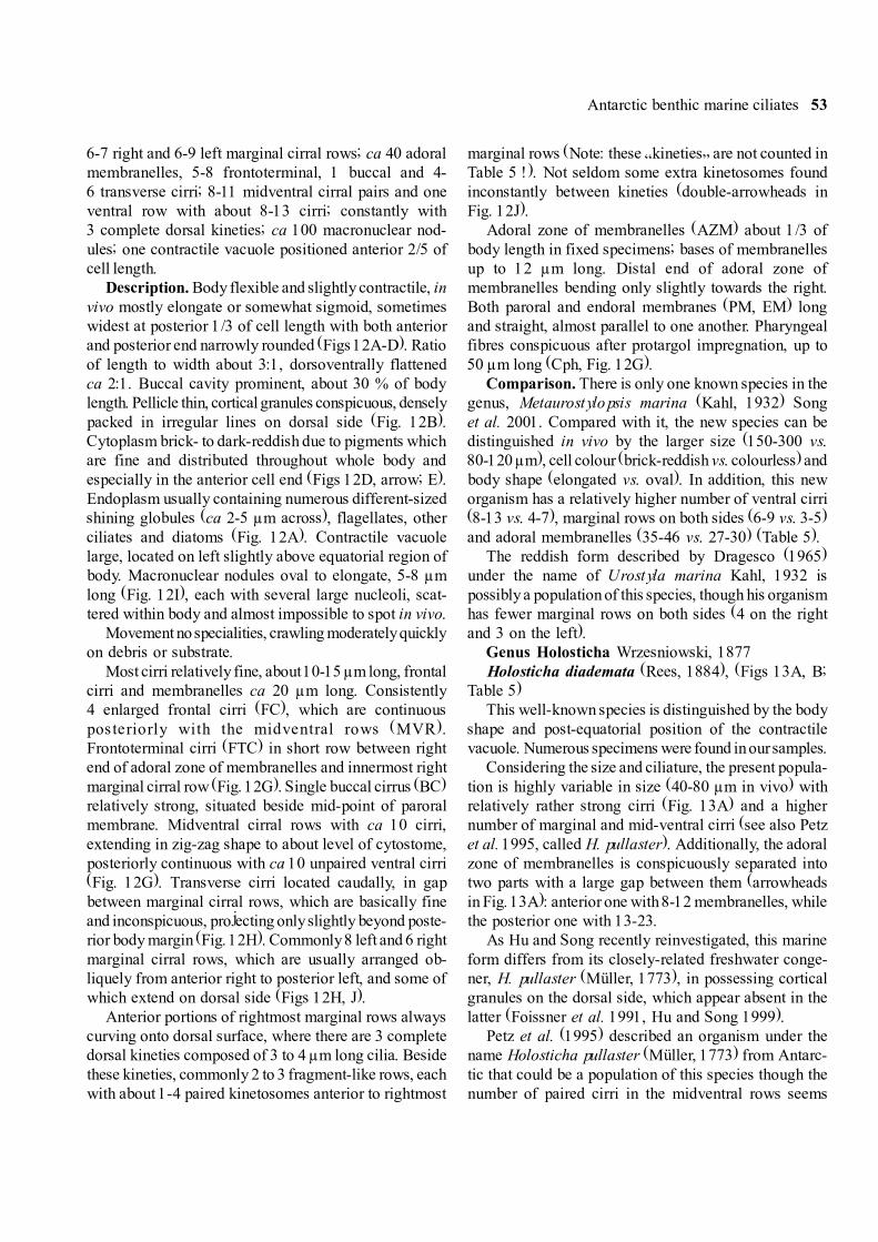
6-7 right and 6-9 left marginal cirral rows; ca 40 adoralmembranelles, 5-8 frontoterminal, 1 buccal and 4-6 transverse cirri; 8-11 midventral cirral pairs and oneventral row with about 8-13 cirri; constantly with3 complete dorsal kineties; ca 100 macronuclear nod-ules; one contractile vacuole positioned anterior 2/5 ofcell length.
Description. Body flexible and slightly contractile, invivo mostly elongate or somewhat sigmoid, sometimeswidest at posterior 1/3 of cell length with both anteriorand posterior end narrowly rounded (Figs 12A-D). Ratioof length to width about 3:1, dorsoventrally flattenedca 2:1. Buccal cavity prominent, about 30 % of bodylength. Pellicle thin, cortical granules conspicuous, denselypacked in irregular lines on dorsal side (Fig. 12B).Cytoplasm brick- to dark-reddish due to pigments whichare fine and distributed throughout whole body andespecially in the anterior cell end (Figs 12D, arrow; E).Endoplasm usually containing numerous different-sizedshining globules (ca 2-5 µm across), flagellates, otherciliates and diatoms (Fig. 12A). Contractile vacuolelarge, located on left slightly above equatorial region ofbody. Macronuclear nodules oval to elongate, 5-8 µmlong (Fig. 12I), each with several large nucleoli, scat-tered within body and almost impossible to spot in vivo.
Movement no specialities, crawling moderately quicklyon debris or substrate.
Most cirri relatively fine, about 10-15 µm long, frontalcirri and membranelles ca 20 µm long. Consistently4 enlarged frontal cirri (FC), which are continuousposteriorly with the midventral rows (MVR).Frontoterminal cirri (FTC) in short row between rightend of adoral zone of membranelles and innermost rightmarginal cirral row (Fig. 12G). Single buccal cirrus (BC)relatively strong, situated beside mid-point of paroralmembrane. Midventral cirral rows with ca 10 cirri,extending in zig-zag shape to about level of cytostome,posteriorly continuous with ca 10 unpaired ventral cirri(Fig. 12G). Transverse cirri located caudally, in gapbetween marginal cirral rows, which are basically fineand inconspicuous, projecting only slightly beyond poste-rior body margin (Fig. 12H). Commonly 8 left and 6 rightmarginal cirral rows, which are usually arranged ob-liquely from anterior right to posterior left, and some ofwhich extend on dorsal side (Figs 12H, J).
Anterior portions of rightmost marginal rows alwayscurving onto dorsal surface, where there are 3 completedorsal kineties composed of 3 to 4 µm long cilia. Besidethese kineties, commonly 2 to 3 fragment-like rows, eachwith about 1-4 paired kinetosomes anterior to rightmost
marginal rows (Note: these �kineties� are not counted inTable 5 !). Not seldom some extra kinetosomes foundinconstantly between kineties (double-arrowheads inFig. 12J).
Adoral zone of membranelles (AZM) about 1/3 ofbody length in fixed specimens; bases of membranellesup to 12 µm long. Distal end of adoral zone ofmembranelles bending only slightly towards the right.Both paroral and endoral membranes (PM, EM) longand straight, almost parallel to one another. Pharyngealfibres conspicuous after protargol impregnation, up to50 µm long (Cph, Fig. 12G).
Comparison. There is only one known species in thegenus, Metaurostylopsis marina (Kahl, 1932) Songet al. 2001. Compared with it, the new species can bedistinguished in vivo by the larger size (150-300 vs.80-120 µm), cell colour (brick-reddish vs. colourless) andbody shape (elongated vs. oval). In addition, this neworganism has a relatively higher number of ventral cirri(8-13 vs. 4-7), marginal rows on both sides (6-9 vs. 3-5)and adoral membranelles (35-46 vs. 27-30) (Table 5).
The reddish form described by Dragesco (1965)under the name of Urostyla marina Kahl, 1932 ispossibly a population of this species, though his organismhas fewer marginal rows on both sides (4 on the rightand 3 on the left).
Genus Holosticha Wrzesniowski, 1877Holosticha diademata (Rees, 1884), (Figs 13A, B;
Table 5)This well-known species is distinguished by the body
shape and post-equatorial position of the contractilevacuole. Numerous specimens were found in our samples.
Considering the size and ciliature, the present popula-tion is highly variable in size (40-80 µm in vivo) withrelatively rather strong cirri (Fig. 13A) and a highernumber of marginal and mid-ventral cirri (see also Petzet al. 1995, called H. pullaster). Additionally, the adoralzone of membranelles is conspicuously separated intotwo parts with a large gap between them (arrowheadsin Fig. 13A): anterior one with 8-12 membranelles, whilethe posterior one with 13-23.
As Hu and Song recently reinvestigated, this marineform differs from its closely-related freshwater conge-ner, H. pullaster (Müller, 1773), in possessing corticalgranules on the dorsal side, which appear absent in thelatter (Foissner et al. 1991, Hu and Song 1999).
Petz et al. (1995) described an organism under thename Holosticha pullaster (Müller, 1773) from Antarc-tic that could be a population of this species though thenumber of paired cirri in the midventral rows seems

54 W. Song and N. Wilbert
somewhat lower and all ciliature appears relativelyweaker. We consider these as population-dependentvariation and suggest that the name, H. pullaster begiven to the freshwater form without cortical granules.
Genus Uronychia Stein, 1859Uronychia binucleata Young, 1922A few specimens of this species were found in
protargol-impregnated slides. Both the morphology andinfraciliature agrees perfectly with the redescriptions byprevious authors (Curds and Wu 1983, Petz et al. 1995,Song and Wilbert 1997), so no further descriptions areneeded.
Genus Diophrys Dujardin, 1841Diophrys oligothrix Borror, 1965, (Figs 14A-D, 58;
Table 6)We agree with Borror (1965) and Czapik (1981) that
Diophrys oligothrix Borror, 1965 is a valid species,which differs from the well-known D. appendiculata(Ehrenberg, 1838) Levander, 1894 in having the non-broken dorsal kineties, which extend continuously overthe dorsal side instead of having a large gap in theequatorial region. Other features for D. oligothrix are:(a) paroral and endoral membrane parallel and closelylocated (vs. conspicuously separated); (b) basically4 dorsal kineties (vs. mostly 5) and (c) usually slender
(vs. more oval in shape) but larger (Song and Packroff1997).
The Antarctic population agrees basically well withthe original and subsequent descriptions (Borror 1965,Czapik 1981, Petz et al. 1995, Song and Packroff 1997).We thus supply only some additional data here.
Description. Cells in vivo about 60-80 µm long,usually more plump than the description given by Songand Packroff (1997), i.e. mostly oval to broadly oval withasymmetric posterior end: on right one prominent de-pression, where the caudal cirri transversely positioned(Fig. 14A).
Ciliary organelles in this population are relatively long,especially caudal cirri and some anterior membranelleswhich are conspicuously longer than others and spreadstiffly (Fig. 14A). Buccal field about 1/2 of body length.2 ridges on ventral side, one on right margin, another toleft, relatively short and less dominant, posterior tocytostome (Fig. 14A). Cytoplasm grayish or colourless,often filled with several to many food vacuoles whichrender the cell completely opaque. This organism seemsto feed preferably on flagellates and diatoms (Figs 14A,D). Consistently 2 elongated macronuclear nodules.
Behaviour and movement like a Euplotes, glidingwithout pause on substrate.
Table 6. Morphometric characterization of Euplotes balteatus (upper line) and Diophrys oligothrix (lower line). All data are based on protargol-impregnated specimens. Measurements in µm
Character Min Max Mean SD SE CV n
Body length 40 66 48.8 4.65 1.34 9.5 1252 95 70.3 16.96 5.36 24.1 10
Body width 30 44 34.2 2.44 0.71 7.2 1230 67 46.5 14.80 4.68 31.8 10
Length of buccal field 29 42 32.9 2.54 0.73 7.4 1234 54 41.8 7.38 2.33 17.7 10
Number of adoral membranelles 27 33 30.2 2.15 0.68 7.1 1031 37 34.3 2.06 0.78 6.0 7
Number of frontoventral cirri 10 10 10 0 0 0 137 7 7 0 0 0 11
Number of transverse cirri 5 5 5 0 0 0 145 5 5 0 0 0 11
Number of left marginal cirri 1 2 1.9 0.34 0.09 18.2 162 2 2 0 0 0 11
Number of caudal cirri 2 2 2 0 0 0 143 3 3 0 0 0 11
Number of dorsal kineties 8 10 9.2 0.62 0.15 6.9 164 5 4.4 0.50 0.10 11.4 23
umber of dikinetids in mid-dorsal rows 9 14 10.9 1.41 0.34 12.9 17- - - - - - -
Number of macronuclei 1 1 1 0 0 0 >302 2 2 0 0 0 >20

Antarctic benthic marine ciliates 55
Both somatic and oral ciliature correspond perfectlyto previous descriptions (Petz et al. 1995, Song andPackroff 1997). Compared with the China population,transverse cirri in the Antarctic form more anteriorlypositioned. Mostly 4 (seldom 5) dorsal kineties withrelatively longer cilia (about 5-7 µm) (Figs 14 B, D).
Diophrys scutum Dujardin, 1842 (Figs 14E-I, 59, 60)Syn. Diophrys magnus Raikov & Kovaleva, 1968Diophrys kasymovi Agamaliev, 1971This species was also newly reinvestigated by Song
and Packroff (1997) and hence no further completedescriptions are necessary. Here only some criticalpoints will be mentioned.
Description. This species often exhibits some vari-ability in ciliary pattern as observed by many previousauthors. The number of the frontoventral cirri is �nor-mally� 7, but not seldom 8 even 9 (Fig. 14E). The samesituation has been also observed in the transverse andcaudal cirri: in the former mostly 5, sometimes 6 (thesurplus cirri might derive from the FVT-anlagen duringthe binary division rather than from the retained parentalstructure), while in the latter usually 3, less frequently 4.
The dorsal ciliature of the Antarctic population seemsslightly different form the populations reported in previ-ous work. The posterior end of the rightmost dorsalkinety is unlike that described for the China population,i.e. not fragment-like, but as �normal� dikinetids anteriorto the caudal cirri (Fig. 14G).
Remarks. Raikov and Kovaleva (1968) described anew species form the Japan Sea, Diophrys magnus,which is characterized by slender body shape and two
macronuclei with more or less moniliform shape andseveral micronuclei. We consider it conspecific withD. scutum because the number of micronuclei couldhave been misinterpreted (their organisms were impreg-nated by Chatton-Lwoff methods), and it is quite com-mon in this species that the shape of body or macronucleivaries slightly.
According to Agamaliev (1971), the Caspian Seaspecies, Diophrys kasymovi Agamaliev, 1971 differsform D. scutum in having more frontoventral cirri (theauthor indicates that there are 6 transverse and8 frontoventral cirri in the former). In our opinion,however, this is a very weak criterion because thenumber of transverse as well as frontoventral cirrichanges frequently in D. scutum even within a cloneculture according to the authors� knowledge. Hence weconsider it as a junior synonym of the latter.
Genus Euplotes Ehrenberg, 1830Eupotes balteatus (Dujardin, 1841) Kahl, 1932,
(Figs 15A-F, 45-48; Table 6)Syn. Euplotes alatus Kahl, 1932 (?)E. quinquecarinatus Gelei, 1951 (?)E. magnicirralus Carter, 1972 (?)E. plicatum Valbonesi et al., 1997 (?)Since this species is highly polymorphic with ex-
tremely variations in size and other morphological fea-tures, we generalize the data obtained and give animproved definition here.
Improved diagnosis. Small to medium-sized marineEuplotes in vivo mostly 40-70 x 30-50 µm, 25-33 membranelles, 10 frontoventral, 5 transverse, 2 cau-
Table 7. Comparison of some small, morphologically-related marine Euplotes-species with silverline pattern of �double-eurystomus� on dorsalside and 10 frontoventral as well as 2 marginal cirri. Measurements in µm. AZM - adoral zone of membranelles, BB - basal bodies,DK - dorsal kineties, MC - marginal cirri, Mem - membranelles
Species* Body size No. No., BB in No., mem Feature ReferencesDK in vivo mid-rows in AZM of MC
E. balteatus 40-70 8-10 9-14 27-33 close-set present paperE. balteatus 30-150 8 11 25-30 widely separated Tuffrau 1964aE. balteatus 50 8 13-14 28-33 close-set Agamaliev 1968E. alatus ca 40 8 ca 10 ? ca 30 ? widely separated Borror 1968E. quinquecarinatus ca 60 9 ca 13 ? ca 30 ? widely separated Borror 1968E. magnicirratus 54** 8 13-17 ca 50 widely separated Carter 1972E. plicatum 42-55 10 14 22-25 widely separated Valbonesi et al. 1997E. rariseta 40-67 6 8-14 25-33 close-set Petz et al. 1995
* The taxonomic identifications not confirmed in current table! ** Data probably from impregnated specimens? Data not given, he re countedfrom illustrations of the original authors.
56 W. Song and N. Wilbert
dal and two close-set marginal cirri; 8 to 10 dorsalkineties with ca 11 dikinetids in mid-rows; silverlinesystem double-eurystomus type.
Description. Body shape generally oval, left convex,right straight with obliquely truncated posterior end(Fig. 15A). Dorsoventrally flattened about 2:1. At ante-rior region, dorsal surface more or less deck-like andstretching forwards, coving ventral part and distal end ofadoral zone membranelles, which is clearly discernibleafter silver impregnation (Fig. 15E). 3-4 conspicuousventral ridges between buccal field and right margin;
ridges among transverse cirri short but prominent(Fig. 15A). On dorsal side about 6-7 longitudinal dorsalridges extending over entire length of body, conspicuousin vivo (Fig. 15B). Cilia of cirri and membranellesca 10-15 µm long. Cytoplasm colourless, containingmany shining globules (ca 2-3 µm across) and foodvacuoles with possibly flagellates or bacteria. Macro-nucleus inverted C-shaped. Contractile vacuole at levelof transverse cirri (Fig. 15A).
Movement genus-typical, crawling on substrate, notfast and slightly jerking.
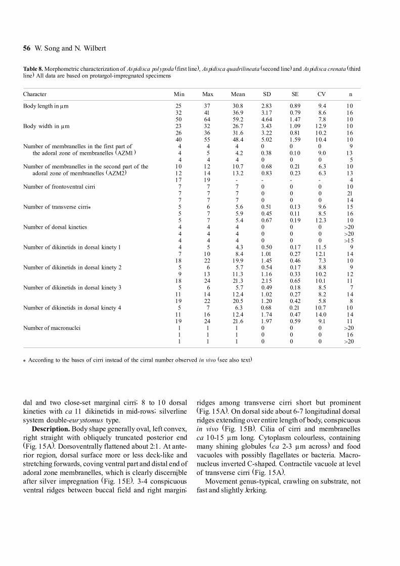
Table 8. Morphometric characterization of Aspidisca polypoda (first line), Aspidisca quadrilineata (second line) and Aspidisca crenata (thirdline) All data are based on protargol-impregnated specimens
Character Min Max Mean SD SE CV n
Body length in µm 25 37 30.8 2.83 0.89 9.4 1032 41 36.9 3.17 0.79 8.6 1650 64 59.2 4.64 1.47 7.8 10
Body width in µm 23 32 26.7 3.43 1.09 12.9 1026 36 31.6 3.22 0.81 10.2 1640 55 48.4 5.02 1.59 10.4 10
Number of membranelles in the first part of 4 4 4 0 0 0 9the adoral zone of membranelles (AZM1) 4 5 4.2 0.38 0.10 9.0 13
4 4 4 0 0 0 5Number of membranelles in the second part of the 10 12 10.7 0.68 0.21 6.3 10
adoral zone of membranelles (AZM2) 12 14 13.2 0.83 0.23 6.3 1317 19 - - - - 4
Number of frontoventral cirri 7 7 7 0 0 0 107 7 7 0 0 0 217 7 7 0 0 0 14
Number of transverse cirri* 5 6 5.6 0.51 0.13 9.6 155 7 5.9 0.45 0.11 8.5 165 7 5.4 0.67 0.19 12.3 10
Number of dorsal kineties 4 4 4 0 0 0 >204 4 4 0 0 0 >204 4 4 0 0 0 >15
Number of dikinetids in dorsal kinety 1 4 5 4.3 0.50 0.17 11.5 97 10 8.4 1.01 0.27 12.1 14
18 22 19.9 1.45 0.46 7.3 10Number of dikinetids in dorsal kinety 2 5 6 5.7 0.54 0.17 8.8 9
9 13 11.3 1.16 0.33 10.2 1218 24 21.3 2.15 0.65 10.1 11
Number of dikinetids in dorsal kinety 3 5 6 5.7 0.49 0.18 8.5 711 14 12.4 1.02 0.27 8.2 1419 22 20.5 1.20 0.42 5.8 8
Number of dikinetids in dorsal kinety 4 5 7 6.3 0.68 0.21 10.7 1011 16 12.4 1.74 0.47 14.0 1419 24 21.6 1.97 0.59 9.1 11
Number of macronuclei 1 1 1 0 0 0 >201 1 1 0 0 0 161 1 1 0 0 0 >20
* According to the bases of cirri instead of the cirral number observed in vivo (see also text)
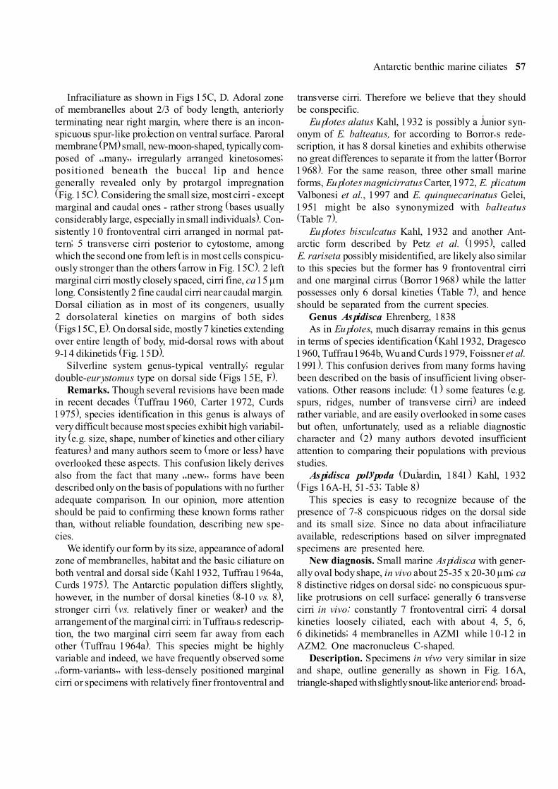
Antarctic benthic marine ciliates 57
Infraciliature as shown in Figs 15C, D. Adoral zoneof membranelles about 2/3 of body length, anteriorlyterminating near right margin, where there is an incon-spicuous spur-like projection on ventral surface. Paroralmembrane (PM) small, new-moon-shaped, typically com-posed of �many� irregularly arranged kinetosomes;positioned beneath the buccal lip and hencegenerally revealed only by protargol impregnation(Fig. 15C). Considering the small size, most cirri - exceptmarginal and caudal ones - rather strong (bases usuallyconsiderably large, especially in small individuals). Con-sistently 10 frontoventral cirri arranged in normal pat-tern; 5 transverse cirri posterior to cytostome, amongwhich the second one from left is in most cells conspicu-ously stronger than the others (arrow in Fig. 15C). 2 leftmarginal cirri mostly closely spaced, cirri fine, ca 15 µmlong. Consistently 2 fine caudal cirri near caudal margin.Dorsal ciliation as in most of its congeners, usually2 dorsolateral kineties on margins of both sides(Figs 15C, E). On dorsal side, mostly 7 kineties extendingover entire length of body, mid-dorsal rows with about9-14 dikinetids (Fig. 15D).
Silverline system genus-typical ventrally; regulardouble-eurystomus type on dorsal side (Figs 15E, F).
Remarks. Though several revisions have been madein recent decades (Tuffrau 1960, Carter 1972, Curds1975), species identification in this genus is always ofvery difficult because most species exhibit high variabil-ity (e.g. size, shape, number of kineties and other ciliaryfeatures) and many authors seem to (more or less) haveoverlooked these aspects. This confusion likely derivesalso from the fact that many �new� forms have beendescribed only on the basis of populations with no furtheradequate comparison. In our opinion, more attentionshould be paid to confirming these known forms ratherthan, without reliable foundation, describing new spe-cies.
We identify our form by its size, appearance of adoralzone of membranelles, habitat and the basic ciliature onboth ventral and dorsal side (Kahl 1932, Tuffrau 1964a,Curds 1975). The Antarctic population differs slightly,however, in the number of dorsal kineties (8-10 vs. 8),stronger cirri (vs. relatively finer or weaker) and thearrangement of the marginal cirri: in Tuffrau�s redescrip-tion, the two marginal cirri seem far away from eachother (Tuffrau 1964a). This species might be highlyvariable and indeed, we have frequently observed some�form-variants� with less-densely positioned marginalcirri or specimens with relatively finer frontoventral and
transverse cirri. Therefore we believe that they shouldbe conspecific.
Euplotes alatus Kahl, 1932 is possibly a junior syn-onym of E. balteatus, for according to Borror�s rede-scription, it has 8 dorsal kineties and exhibits otherwiseno great differences to separate it from the latter (Borror1968). For the same reason, three other small marineforms, Euplotes magnicirratus Carter, 1972, E. plicatumValbonesi et al., 1997 and E. quinquecarinatus Gelei,1951 might be also synonymized with balteatus(Table 7).
Euplotes bisculcatus Kahl, 1932 and another Ant-arctic form described by Petz et al. (1995), calledE. rariseta possibly misidentified, are likely also similarto this species but the former has 9 frontoventral cirriand one marginal cirrus (Borror 1968) while the latterpossesses only 6 dorsal kineties (Table 7), and henceshould be separated from the current species.
Genus Aspidisca Ehrenberg, 1838As in Euplotes, much disarray remains in this genus
in terms of species identification (Kahl 1932, Dragesco1960, Tuffrau 1964b, Wu and Curds 1979, Foissner et al.1991). This confusion derives from many forms havingbeen described on the basis of insufficient living obser-vations. Other reasons include: (1) some features (e.g.spurs, ridges, number of transverse cirri) are indeedrather variable, and are easily overlooked in some casesbut often, unfortunately, used as a reliable diagnosticcharacter and (2) many authors devoted insufficientattention to comparing their populations with previousstudies.
Aspidisca polypoda (Dujardin, 1841) Kahl, 1932(Figs 16A-H, 51-53; Table 8)
This species is easy to recognize because of thepresence of 7-8 conspicuous ridges on the dorsal sideand its small size. Since no data about infraciliatureavailable, redescriptions based on silver impregnatedspecimens are presented here.
New diagnosis. Small marine Aspidisca with gener-ally oval body shape, in vivo about 25-35 x 20-30 µm; ca8 distinctive ridges on dorsal side; no conspicuous spur-like protrusions on cell surface; generally 6 transversecirri in vivo; constantly 7 frontoventral cirri; 4 dorsalkineties loosely ciliated, each with about 4, 5, 6,6 dikinetids; 4 membranelles in AZM1 while 10-12 inAZM2. One macronucleus C-shaped.
Description. Specimens in vivo very similar in sizeand shape, outline generally as shown in Fig. 16A,triangle-shaped with slightly snout-like anterior end; broad-

58 W. Song and N. Wilbert
est always behind mid-body, both margins evenly con-vex; anteriorly broadly narrowed, while posteriorly moreor less notched because of the presence of dorsal ribs(Fig. 16A). Dorsoventrally only slightly flattened (about3:2) with 8 dominant ridges, which are clearly visibleeven at low magnification (Figs 16B, C) Pellicle rigid,appearance of ventral side basically similar to its conge-ners. Cytoplasm colourless or greyish, often with severallarge food vacuoles (feeding likely on flagellates). Con-tractile vacuole small, right of median, posterior totransverse cirri. Macronucleus C-shaped, with largeconspicuous nucleoli (Fig. 16E). Micronucleus not ob-served.
Movement moderately fast when compared with itsbody size, crawling on substrate without periods ofresting.
Ciliary structure rather stable (Figs 16D, E; Table 7).Cilia of frontoventral cirri about 10 µm long, in typicalpattern of genus. Transverse cirri relatively weak,ca 15 µm in length, subcaudally arranged in oblique row;right-most cirrus always splitting into two closely spacedones (Fig. 16A), though united at the base (Fig. 16D,arrow). Anterior part of adoral zone of membranelles(AZM1) with consistently 4 membranelles, located indeep concavity, while posterior part (AZM2) with onaverage 11 organelles, beneath lid-like spur. Paroralmembrane close to proximal end of AZM2 (double-arrowheads in Fig. 16D). Pharyngeal fibres curvedanteriorly. Dorsal kineties with quite sparsely-arrangedbasal body pairs (4-6 pairs each, Table 8), each alongdorsal ridge and generally extending from end to end ofcell (Fig. 16E).
Silverline system as shown in Figs 16F-H, basicallytypical of genus, contractile vacuole pore prominent(Fig. 16H, CVP).
Remarks With the small size, marine habitat, generaloutline and the distinctive 8 dorsal ribs (ridges), thisorganism is easy to recognize.
Aspidisca quadrilineata Kahl, 1932, (Figs 17A-D,54-55; Table 8)
To the authors� knowledge, no reinvestigations havebeen made since it was erected (Kahl 1932). Hence wepresent here a detailed redescription and an improveddiagnosis based on our observations.
New diagnosis. Small marine Aspidisca with gener-ally oval body shape, in vivo about 30-40 x 25-35 µm;with neither conspicuous dorsal ribs nor pronouncedspur-like protrusions throughout; 6 transverse in vivoand 7 frontoventral cirri; 4 dorsal kineties densely cili-
ated, each with about 8, 11, 12, 12 dikinetids; 4-5membranelles in AZM1 and 12-14 in AZM2. One ma-cronucleus C-shaped.
Description. This species generally rather similar topolypoda except for the dorsal ridges. In vivo slightlylarger, about 35 x 30 µm in size; outline tending to bemore oval with anterior end only very slightly snout-like;broadest in posterior 1/3 of body, with both cell marginsevenly convex; posterior end more broadly rounded thananterior (Fig. 17A). Dorsoventrally flattened about1.5-2:1. Pellicle armed as last form, 4 inconspicuousdorsal ridges (Fig. 17B). Cytoplasm colourless and opaque,often full of tiny granules. Single contractile vacuole onright near transverse cirri. Macronucleus slightly notchedanteriorly with conspicuous nucleoli (Fig. 17D). In ourprotargol impregnated specimens, no micronuclei re-vealed.
Movement as in last form, slow to moderately fast,gliding on substrate.
Ciliature as in last species (Figs 17C, D). Mostly6 transverse cirri in vivo (we observed individuals withsometimes even 7 cirri, in which case the leftmost twooften joined together); yet only 5 after impregnation(Fig. 17C). Fibres dominant, associated with transversecirri, extending to about anterior margin of cell. Mostly4 (seldom 5) membranelles in AZM1 and 12-14 inAZM2. Paroral membrane (PM) close to proximal endof AZM2 (Fig. 17C). Dorsal kineties with densely-arranged dikinetids (about 8-12 pairs each, Table 8)(Fig. 17D).
Remarks. In Wu and Curds� revision (1979), thisspecies was treated, without giving any explanation, asa synonym of Aspidisca polypoda, but in view of thecompletely different living appearance (i.e. with ca8 dominant dorsal ridges vs. inconspicuous 4 ribs; andthe infraciliature on dorsal side: the number of dikinetidsin each kineties, see Table 8), this form is doubtless avalid species.
This species might be similar to Aspidisca aculeata(Ehrenberg, 1838), which can be distinguished, however,by a conspicuous dorsal thorn and loosely ciliated dorsalkineties: each with about 4-8 (vs. 8-12) dikinetids ac-cording to the redescriptions by Kahl (1932), Borror(1965) and Agamaliev (1974).
Aspidisca crenata Fabre-Domergue, 1885, (Figs17E-H, 56-57; Table 8)
Our Antarctic population corresponds to the previousdescriptions by Kahl (1932) very well (having �7�transversale Cirren, Hinterrand ohne deutliche Zähne,

Antarctic benthic marine ciliates 59
nur schwach gekerbt oder gewellt�). Though Kahl in-sisted that this organism has 3-4 inconspicuous furrows(instead of �ribs� as in our own observations), webelieve that our identification is correct while the pres-ence of ribs or furrows is entirely an observer-dependentexpression.
New diagnosis. Medium-sized marine Aspidiscawith generally oval body shape and smooth cell margin,in vivo about 50-65 x 40-55 µm in size, dorsally with4 very inconspicuous ribs; 7 strong frontoventral cirri;basically 7 transverse cirri from life; 4 dorsal kinetiesdensely ciliated, each with about 20, 21, 21, 22 dikinetids;4 membranelles in AZM1 while 17-19 in AZM2; macro-nucleus C-shaped.
Description. Size of this Antarctic population in vivoabout 50-65 x 40-55 µm, body elliptical with inconspicu-ous snout-shaped anterior end; broadest in about mid-body, left and right margin evenly convex; both anteriorand posterior end broadly rounded; dorsoventrally highlyflattened (so in low magnification often appeared hya-line!). Ventral surface generally flattened with buccal lidcovering adoral zone of membranelles. On dorsal side,always 4 dominant ridges (Fig. 13C). Buccal area with1 distinct thorn-like projection (prostomial spur) on leftsubcaudally, which covers the AZM2. Pellicle rigid,beneath it always some tiny dark-gray granules (about1µm across) sparsely distributed, which can be observedonly at high magnifications from life (Fig. 17F). Cyto-plasm hyaline and colourless with large food vacuoles.Contractile vacuole about in median, at level of trans-verse cirri. Macronucleus C-shaped (Fig. 17H).
Movement crawling on substrate, typical of the ge-nus.
Ciliary pattern as shown in Figs 17G, H. Frontoventralcirri rather stiff and strong, cilia about 12-15 µm long;consistently 7 transverse cirri tightly arranged in obliquerow, among which the base of the right-most always 3cirri in vivo as in some other congeners (Fig. 17G).Fibres from transverse cirri strong and prominent, ex-tending anteriorly. AZM1 in deep concavity, containingonly 4 membranelles, while AZM2 on left margin, withabout 18 membranelles. Paroral membrane (PM) small,often difficult to recognize (Fig. 17G). Pharyngeal fibrescurved anteriorly. Kineties on dorsal side densely ciliated(Fig. 17H).
Remarks. Without knowing the infraciliature, theauthors inappropriately synonymized Aspidisca crenatawith A. leptaspis (Song and Wilbert 1997). According tothe data obtained recently and our new observations,however, the former should be considered as a valid but
separate species. It differs from the latter in havingclearly fewer membranelles in the AZM1 (4-5 vs. 7-8)and the frontoventral cirri (consistently 7 vs. 7 plus onesmall cirrus) (Song and Wilbert 1997).
It seems quite common that many �large� species(over 50 µm in length) possess 7 transverse cirri and ca20 membranelles in the posterior part of AZM, and, asnoticed by many previous authors, the body shape,especially the spur - outline might be highly variableamong them, hence species identification for these largeforms remains a huge difficulty. This problem might besolved only when the infraciliature of all uncertain formshas been revealed.
Acknowledgements. These studies were supported by personneland equipment funds from the Alfred Wegener Institute for Polarand Marine Research, Bremenhaven and by the DeutscheForschunsgemeinschaft as part of the program �Antarctic Researchwith Comparative Studies in Arctic Regions� awarded to NW, and avisiting grant awarded to WS by �The Natural Science Foundation ofChina� (project No. 39970098). Our thanks are also to Dr. AlanWarren, The Natural History Museum, London for kindly supplyingsome literature.
REFERENCES
Agamaliev F. G. (1968) Materials on morphology of somepsammophilic ciliates of the Caspian Sea. Acta Protozool. 6: 225-244 (in Russian, English summary)
Agamaliev F. G. (1971) Complements to the fauna of psammophilicciliates of the Western coast of the Caspian Sea. Acta Protozool.8: 379-407 (in Russian, English summary)
Agamaliev F. G. (1974) Ciliates of the solid surface overgrowth ofthe Caspian Sea. Acta Protozool. 13: 53-82 (in Russian, Englishsummary)
Agatha S., Spindler M., Wilbert N. (1993) Ciliated protozoa (Cilio-phora) from Arctic sea ice. Acta Protozool. 32: 261-268
Agatha S., Wilbert N., Spindler M., Elbrächter M. (1990) Euplotideciliates in sea ice of the Weddell Sea (Antarctica). Acta Protozool.29: 221-228
Borror A. C. (1963) Morphology and ecology of the benthic ciliatedprotozoa of Alligator Habor, Florida. Arch. Protistenkd. 106:465-534
Borror A. C. (1965) New and little known tidal marsh ciliates. Trans.Amer. Micros. Soc. 84: 550-565
Borror A. C. (1968) Systematic of Euplotes (Ciliophora,Hypotrichida); toward union of the old and the new. J. Protozool.15: 802-808
Carter H. P. (1972) Infraciliature of eleven species of the genusEuplotes. Trans. Amer. Micros. Soc. 91: 466-492
Corliss J. O. (1979) The Ciliated Protozoa: Characterization, Clas-sification and Guide to the Literature. 2nd ed. Pergamon Press,New York
Corliss J. O., Snyder R. A. (1986) A preliminary description ofseveral new ciliates from the Antarctica, including Cohnilembusgrassei n. sp. Protistologica 22: 39-46
Cuénot L (1891) Infusoires commensaux des Ligies, Patelles etArenicoles. Rev. Biol. Nord France 4: 81-89
Curds C. R. (1975) A guide to the species of the genus Euplotes(Hypotrichida Ciliatea). Bull. Br. Mus. Nat. Hist. (Zool.) 28: 1-61
Curds C. R., Wu I. C. H. (1983) A review of the Euplotidae(Hypotrichida, Ciliophora). Bull. Br. Mus. Nat. Hist. (Zool.) 44:191-247

60 W. Song and N. Wilbert
Czapik A. (1981) La morphogenèse chez le cilié� Diophrys oligothrixBorror. Acta Protozool. 20: 367-372
Deroux G. (1970) La série «Chlamydonellienne» chez lesChlamydodontidae (Holotriches, Cyrtophrdina Fauré�-Fremiet).Protistologica 6: 155-182
Deroux G. (1974) Les dispositifs adhésives ciliaires chez lesCyrtophorida et la famile des Hypocomidae. Protistologica 10:379-396
Deroux G. (1976) Plan cortical des Cyrtophorida III. Les structuresdifférenciatrices chez les Dysterina. Protistologica 12: 505-538
Deroux G. (1978) The hypostome ciliate order Synhymeniida: fromChilodontopsis of Blochmann to Nassulopsis of Fauré-Fremiet.Trans. Amer. Micros. Soc. 97: 458-469
Dietz G. (1964) Beitrag zur Kenntnis der Ciliatenfauna einigerBrackwassertümpel (étangs) der französischen Mittelmeerküste.Vie Milieu 15: 47-93
Dragesco J. (1960) Ciliés mésopsammiques littoraux. Systématique,morphologie, écologie. Trav. Stn. Biol. Roscoff 122: 1-356
Dragesco J. (1963) Compléments à la connaissance des ciliésmésopsammiques de Roscoff. II. Hypotriches. Cah. Biol. Mar. 4:251-275
Dragesco J. (1965) Ciliés mésopsammiques d�Afrique noire. Cah.Biol. Mar. 6: 357-399
Dragesco J. (1968) Les genres Pleuronema Dujardin, Schizocalyptranov. gen. et Histiobalantium Stokes (Ciliés holotricheshymostomes). Protistologica 4: 85-106
Dragesco J. (1996) Infraciliature et morphométrie de cinq espèces deciliés mésopsammiques méditerranéens. Cah. Biol. Mar. 37: 261-293
Dragesco J., Dragesco-Kernéis A. (1986) Ciliés libres de l�Afriqueintertropicale. Faune Tropicale 26: 1-559
Dragesco J., Iftode F., Fryd-Versavel G. (1974) Contribution à laconnaissance de quelques ciliés holotriches rhabdophores; I.Prostomiens. Protistologica 10: 59-75
Fenchel T., Lee C. C. (1972) Studies on ciliates associated with seaice from Antarctica 1. The nature of the fauna. Arch. Protistenkd.114: 231-236
Fish J. D., Goodwin B. J. (1976) Observations on the peritrichousciliate Scyphidia ubiquita from the west coast of Wales and adescription of an new species. J. Zool. (London) 179: 361-371
Foissner W. (1984) Taxonomie und Ökologie einiger Ciliaten (Proto-zoa, Ciliophora) des Saprobiensystems. I: Genera Litonotus,Amphileptus, Opisthodon. Hydrobiologia 119: 193-208
Foissner W. Schiffmann H. (1979) Morphologie und Silberliniensystemvon Pseudovorticella sauwaldensis nov. spec. und Scyphidiaphysarum Lachmann, 1856 (Ciliophora, Peritrichida) Ber. Nat.Med. Ver. Salzburg 3/4: 83-94
Foissner W. (1996) Faunistics, taxonomy and ecology of moss andsoil ciliates (Protozoa, Ciliophora) from Antarctica, with descrip-tion of new species, including Pleuroplitoides smithi gen. n.,sp. n. Acta Protozool. 35: 95-123
Foissner W., Blatterer H., Berger H., Kohmann F. (1991) Taxonomischeund ökologische Revision der Ciliaten des Saprobiensystems -Band I: Cyrtophorida, Oligotrichida, Hypotricha, Colpodea.Informat Bayer Land. Wasserwirt 1/91: 1-478
Fryd-Versavel G., Iftode F., Dragesco J. (1975) Contribution à laconnaissance de quepques ciliés Gymnostomes. II. Prostomiens,pleurostomiens: Morphologie, stomatogenèse. Protistologica 6:509-530
Grolière C. A., Puytorac de P., Grain J. (1978) Ciliés endocommensauxde l�Oursin Lytechinus variegatus des cotes de Ciudad del Carmen(Mexique) (Abstract). J. Protozool. 25: 554
Guhl W. (1979) Beitrag zur Systematik, Biologie und Morphologieder Epistylididae (Ciliata, Peritricha). Arch. Protistenkd. 121:417-483
Hartwig E (1973) Die Ciliaten des Gezeiten-Sandstrandes derNordseeinsel Sylt. I. Systematik. Abh math-naturw Kl Akad WissMainz Mikrofauna Meer 18: 1-69
Hirshfield H. (1949) The morphology of Urceolaria karyolobia,sp. nov., Trichodina tegula, sp. nov., and Scyphidia ubiquita,sp. nov., three new ciliates from southern California limpets andturbans. J. Morphol. 85: 1-28
Hu X., Song W.. (1999) On morphology of the marine Hypotrichousciliate, Holosticha diademata (Ciliophora, Hypotrichida), withcomparison of its related species. J. Ocean. Univ. Qingdao 29:469-473 (in Chinese, English summary)
ICZN (The International Commission on Zoological Nomenclature)(1985) International Code of Zoological Nomenclature. 3rd ed.Univ. California Press, Berkeley
Jankowski A. W. (1968) Taxonomy of the suborder Nassulina Jank.,1967 (Ciliophora, Ambihymenida). Zool. Zh. 47: 900-1001(in Russian, English summary))
Jankowski A. W. (1980) Une revue de la nouvelle systématique duphylum Ciliophora. Trud. Zool. Inst. Akad. Nauk USSR 94: 103-121 (in Russian)
Jankowski A. W. (1985) Life cycle and taxonomy of genera ofthe groups Scyphidia, Heteroplaria, Zoothamnium and Cothurnia(Class: Peritricha). Proc. Zool. Inst. USSR Akad. Sci. 129: 74-100
Kahl A. (1928) Die Infusorien (Ciliata) der Oldesloer Salzwasserstellen.Arch. Hydrobiol. 19: 50-123
Kahl A. (1930) Urtiere oder Protozoa I: Wimpertiere oder Ciliata(Infusoria). 1. Allgemeiner Teil und Prostomata. Tierwelt. Dtl. 18:1-180
Kahl A. (1931) Urtiere oder Protozoa I: Wimpertiere oder Ciliata(Infusoria). 2. Holotricha. Tierwelt. Dtl. 25: 181-398
Kahl A. (1932) Urtiere oder Protozoa. I: Wimpertiere oder Ciliata(Infusoria), 3. Spirotricha. Tierwelt. Dtl. 25: 399-650
Kahl A. (1933) Ciliata libera et ectocommensalia. In: Die Tierwelt derNord- und Ostsee (Eds. G. Grimpe and E. Wagler) 23: 29-146
Kahl A. (1935) Urtiere oder Protozoa I: Wimpertiere oder Ciliata(Infusoria). 4. Peritricha und Chonotricha. Tierwelt. Dtl. 25: 651-886
Lom J. (1961) On the buccal apparatus of peritrichous ciliates. Progr.Protozool. 1st Intern. Conf. Protozool. Prague 91-95
Lom J., Corliss J. O. (1968) Observations on the fine structure oftwo species of the peritrich ciliate genus Scyphidia and theirmode of attachment to the host. Trans. Amer. Micros. Soc. 87:493-509
Ozaki Y., Yagiu R. (1941) Studies on the marine ciliates of Japan,mainly from the Setonaikai (the inland sea of Japan) II. J. Sci.Hiroshima Univ. (Ser B) 9: 159-180
Petz W.. (1994) Morphology and morphogenesis of Strombidiumkryalis nov. spec. (Ciliophora, Strombidiida) from Antarctic seaice. Arch. Protistenkd. 144: 185-195
Petz W. (1995) Morphology and morphogenesis of Thigmokeronopsisantarctica nov. spec. and T. crystalis nov. spec. (Ciliophora,Hypotrichida) from Antarctic sea ice. Europ. J. Protistol. 31: 137-147
Petz W., Song W., Wilbert N. (1995) Taxonomy and ecology of theciliate fauna (Protozoa, Ciliophora) in the endopagial and pelagialof the Weddell Sea, Antarctica. Stapfia 40: 1-223
Puytorac P. de, Grolière C. A., Roque M., Detcheva R. (1974) Apropos d�un cilié Philasterina trouvé dans la cavité générale dupolychète Nereis diversicolor O. F. Müller. Protistologica 10:101-111
Raikov I. B., Kovaleva V. G. (1968) Complements to the fauna ofpsammobiotic ciliates of the Japan Sea (Posjet gulf). Acta Protozool.6: 309-333
Song W. (1991a) Contribution to the commensal ciliates on Penarusorientalis II. (Ciliophora, Peritrichida). J. Ocean. Univ. Qingdao21:45-55 (in Chinese, English summary)
Song W. (1991b) Litonotus yinae n. sp. (Protozoa, Ciliophora), aperiphytic and commensal species from Qingdao. Ophelia 34:181-189
Song W. (2000) Morphological and taxonomical studies on somemarine scuticociliates from China Sea, with description of twonew species, Philasterides armatalis sp. n. and Cyclidiumvaribonneti sp. n. (Protozoa: Ciliophora: Scuticociliatida). ActaProtozool. 39: 295-322
Song W., Packroff G. (1997) Taxonomische Untersuchungen anmarinen Ciliaten aus China mit Beschreibungen von 2 neuenArten, Strombidium globosaneum nov. spec. und Strombidiumplatum nov. spec. (Protozoa, Ciliophora). Arch. Protistenkd. 147:331-360
Antarctic benthic marine ciliates 61
Song W., Wilbert N. (1989) Taxonomische Untersuchungen anAufwuchsciliaten (Protozoa, Ciliophora) im PoppelsdorferWeiher, Bonn. Lauterbornia 3: 2-221
Song W., Wilbert N. (1995) Benthische Ciliaten des Süsswassers.In: Praktikum der Protozoologie (Ed. Röttger) Fischer Verlag,Stuttgart
Song W., Wilbert N. (1997) Morphological investigation on some freeliving ciliates (Protozoa, Ciliophora) from China Sea with de-scription of a new hypotrichous genus, Hemigastrostyla nov.gen. Arch. Protistenkd. 148: 413-444
Song W., Wilbert N. (2000a) Ciliates form Antarctic sea ice. PolarBiol. 23: 212-222
Song W., Wilbert N. (2000b) Redefinition and redescription of somemarine scuticociliates from China, with report of a new species,Metanophrys sinensis nov. spec. (Ciliophora, Scuticociliatida).Zool. Anz. 239: 45-74
Song W., Wilbert N., Warren A. (1999) Three new entocommensalciliates from digestive tract of sea urchins of the Weddell Sea,Antarctica (Protozoa, Ciliophora). Polar Biol. 22: 232-240
Song W., Petz W., Warren A. (2001) Morphology and morphogenesisof the poorly-known marine urostylid ciliate, Metaurostylopsismarina (Kahl, 1932) nov. gen., nov. comb., with a description ofthe new genus Metaurostylopsis (Protozoa, Ciliophora,Hypotrichida). Europ. J. Protistol. (in press)
Spiegel A. (1926) Einige neue marine Ciliaten. Arch. Protistenkd. 55:184-190
Thompson J. C. (1972) Ciliated protozoa of the Antarctic Peninsula.In: Antarctic Terrestrial Biology (Ed. G. A. Llano). Antarct. Res.Ser. 20: 261-288
Thompson J. C., Croom J. M. (1978) Systematics and ecology ofciliated protozoa from King George Island, South Shetland Is-lands. In: Biology of the Antarctic Seas VII (Ed. D. L .Pawson).Antarct. Res. Ser. 27: 41-67
Tuffrau M. (1960) Revision du genre Euplotes, fondée sur lacomparaison des structures superficielles. Hydrobiologia 15: 1-77
Tuffrau M. (1964a) Le maintien des caractères spécifiques à traversle polymorphisme d�Euplotes balteatus Dujardin, 1841. Arch.Zool. Exp. Gén. 104: 143-151
Tuffrau M. (1964b) La morphogenèse de bipartition et les structuresneuromotrices dans le genre Aspidisca (Ciliés hypotriches). Re-vue de quelques espèces. Cah. Biol. Mar. 5: 173-199
Valbonesi A., Luporini P. (1990a) Description of two new species ofEuplotes and Euplotes rariseta from Antarctica. Polar Biol. 11:47-53
Valbonesi A., Luporini P. (1990b) A new marine species of Euplotes(Ciliophora, Hypotrichida) from Antarctica. Bull. Br. Mus. Nat.Hist. (Zool.) 56: 57-61
Valbonesi A., Luporini P. (1990c) A new marine species of Uronychia(Ciliophora, Hypotrichida) from Antarctica: Uronychia antarctica.Boll. Zool. 57: 365-367
Valbonesi A., Apone F., Luporini P. (1997) Morphology and biologyof a new species of Euplotes, Euplotes plicatum sp. n. (Ciliophora:Euplotidae). Acta Protozool. 36: 287-294
Van As L. G., Basson L., Van As J. G. (1998) Two new species ofMantoscyphidia Jankowski, 1980 (Ciliophora: Peritrichia), gillsymbionts of limpets, from South Africa and the sub-Antarcticisland, Marion. Acta Protozool. 37: 101-111
Villeneuve-Brachon S. (1940) Recherches sur les ciliés hétérotriches:cinétome, argyrome, myonèmes. Formes nouvelles ou peuconnues. Arch. Zool. Exp. Gén. 82: 1-180
Wiencke C., Ferreyra G., Arntz W., Rinaldi C. (1998) The PotterCove coastal ecosystem, Antarctica. Ber. Polarforsch. 299: 1-326
Wilbert N. (1975) Eine verbesserte Technik der Protargolimprägnationfür Ciliaten. Mikrokosmos 64: 171-179
Wilbert N. (1986) Ciliates from saline lakes in Saskatchewan. Symp.Biol. Hung. 33: 249-256
Wilbert N. (1995) Benthic ciliates of salt lakes. Acta Protozool. 34:271-288
Wilbert N., Kahan D. (1981) Ciliates of Solar Lake on the Red Seashore. Arch. Protistenkd. 124: 70-95
Wilbert N., Petz W., Song W. (1993) Observations on the ciliatecommunity of the Antarctic sea ice and plankton (Ciliophora,Protozoa). Ber. Polarforsch. 121: 89-90
Wu I. C. H., Curds C. R. (1979) A guide to the species of the genusAspidisca. Bull. Br. Mus. Nat. Hist. (Zool.) 36: 1-34
Received on 19th March, 2001; accepted on 5th January, 2002