fatty acid activation in cyanobacteria mediated by acyl-acyl
TRANSCRIPT

Fatty Acid Activation in Cyanobacteria Mediated byAcyl-Acyl Carrier Protein Synthetase Enables FattyAcid Recycling1[W]
Danuta Kaczmarzyk and Martin Fulda*
Department of Plant Biochemistry, Albrecht-von-Haller-Institute, Georg-August-University Goettingen,D–37077 Goettingen, Germany
In cyanobacteria fatty acids destined for lipid synthesis can be synthesized de novo, but also exogenous free fatty acids fromthe culture medium can be directly incorporated into lipids. Activation of exogenous fatty acids is likely required prior to theirutilization. To identify the enzymatic activity responsible for activation we cloned candidate genes from Synechocystis sp. PCC6803 and Synechococcus elongatus PCC 7942 and identified the encoded proteins as acyl-acyl carrier protein synthetases (Aas).The enzymes catalyze the ATP-dependent esterification of fatty acids to the thiol of acyl carrier protein. The two proteinsequences are only distantly related to known prokaryotic Aas proteins but they display strong similarity to sequences that canbe found in almost all organisms that perform oxygenic photosynthesis. To investigate the biological role of Aas activity incyanobacteria, aas knockout mutants were generated in the background of Synechocystis sp. PCC 6803 and S. elongatus PCC7942. The mutant strains showed two phenotypes characterized by the inability to utilize exogenous fatty acids and by thesecretion of endogenous fatty acids into the culture medium. The analyses of extracellular and intracellular fatty acid profilesof aas mutant strains as well as labeling experiments indicated that the detected free fatty acids are released from membranelipids. The data suggest a considerable turnover of lipid molecules and a role for Aas activity in recycling the released fattyacids. In this model, lipid degradation represents a third supply of fatty acids for lipid synthesis in cyanobacteria.
Cyanobacteria present a diverse group of Gram-negative bacteria capable of oxygenic photosynthesis(Margulis, 1975). Their two photosystems, as well asother genetic and morphological similarities, identi-fied them as putative predecessors of chloroplasts ofeukaryotic plants (Wallace, 1982; Pakrasi, 1995). Thestructural similarities of cyanobacteria and chloro-plasts are reflected in part by equivalence of biochem-ical pathways and their components. For instance,cyanobacterial fatty acid and glycerolipid composi-tions closely resemble those of the inner envelope andthylakoid membranes of chloroplasts (Roughan et al.,1980; Heinz and Roughan, 1983). In cyanobacteria, aswell as in chloroplasts, fatty acids are synthesized by atype II fatty acid synthase (FAS) complex utilizing afreely dissociable acyl carrier protein (ACP; Froehlichet al., 1990). The products of FAS are released as acylACPs and may serve directly as substrates for acyl-transferases, incorporating the fatty acids into mem-brane lipids (Frentzen et al., 1983). The substratespecificity of the acyltransferases establishes in cyano-
bacteria as well as in plastids the typical prokaryoticfatty acid pattern characterized by C16 fatty acidsesterified to the sn-2 position. The correspondence ofmetabolic pathways between cyanobacteria and chlo-roplasts is reflected by the shared presence of closelyrelated enzymes that catalyze key reactions. Besidesthe many similarities, however, there are also cleardiscrepancies that in part account for the fact thatcyanobacteria are unicellular organisms, whereaschloroplasts are embedded in the metabolism of aeukaryotic cell. In terms of lipid metabolism, suchdifferences become obvious if one considers the factthat the plastidial FAS also supplies the extraplastidiccompartment with fatty acids (Browse et al., 1986).Fatty acid export from the chloroplast necessitates therelease of synthesized acyl chains from ACP to allowtransport across both envelope membranes. The re-lease is achieved by the action of acyl-ACP thioester-ases that hydrolyze the acyl-ACP thioester to liberatethe fatty acid (Voelker et al., 1997). In cyanobacteriasuch export would obviously result in an unfavorableloss of fatty acids, and consequently homologousproteins to acyl-ACP thioesterases cannot be foundhere. Whereas cyanobacteria seem to be unable torelease fatty acids enzymatically from their activatedstate, all cyanobacterial genomes available to dateencode an activity most likely responsible for theactivation of free fatty acids. The respective sequencesare annotated as acyl-CoA synthetases. Conservedmotifs in the amino acid sequence identify theseproteins as members of the well-established super-
1 This work was supported by the Deutsche Forschungsgemein-schaft (German Research Foundation; grant no. FU 430/3–1).
* Corresponding author; e-mail [email protected] author responsible for distribution of materials integral to the
findings presented in this article in accordance with the policydescribed in the Instructions for Authors (www.plantphysiol.org) is:Martin Fulda ([email protected]).
[W] The online version of this article contains Web-only data.www.plantphysiol.org/cgi/doi/10.1104/pp.109.148007
1598 Plant Physiology�, March 2010, Vol. 152, pp. 1598–1610, www.plantphysiol.org � 2010 American Society of Plant Biologists www.plantphysiol.orgon January 5, 2019 - Published by Downloaded from
Copyright © 2010 American Society of Plant Biologists. All rights reserved.

family of AMP-binding proteins. This protein familycomprises several hundred amino acid sequencesspreading across all organisms analyzed so far. Thefamily members are annotated in the PROSITE data-base under entry number PS00455. Although thesepredicted fatty acid-activating enzymes of cyanobac-teria are annotated as acyl-CoA synthetases due totheir sequence similarity to proteins with such enzy-matic activity, there is a much higher degree of sim-ilarity to certain AMP-binding proteins of plant originwith less-well-established function. These plant pro-teins are predicted to reside in chloroplasts and onemember of this subgroup from Arabidopsis (Arabidop-sis thaliana) designated as AAE15 was recently de-scribed as acyl-ACP synthetase. The conclusions werebased on the comparison of enzymatic activity be-tween plant extracts of wild-type and knockout mu-tant lines (Koo et al., 2005). Whereas the biological roleof this activity remained largely elusive, it was shownthat the capacity of plant extracts to elongate suppliedmedium fatty acids depended on AAE15 activity.Since the elongation of medium chain fatty acids inthe plastid depends on the FAS requiring acyl ACPs, itwas concluded that the fatty acids must have beenactivated by ACP. The elongated fatty acids ultimatelyappeared in membrane lipids. Together these findingssuggested that AAE15 is an acyl-ACP synthetase.Besides encoding a protein homologous to AAE15
fromArabidopsis, cyanobacteria are also able to utilizeexogenous fatty acids like it was shown for isolatedchloroplasts. It is well established that feeding differ-ent cyanobacteria with free fatty acids results in theincorporation of these fatty acids into membranelipids. For this process the activation of the fatty acidsis believed to be essential. This causal relationship wasclearly shown at least for other unicellular organismslike Escherichia coli and yeast (Saccharomyces cerevisiae)where the deletion of acyl-CoA synthetase activityresulted in the inability to utilize exogenous fatty acids(Overath et al., 1969; Knoll et al., 1995). It is not easy toassess how regularly cyanobacterial cells are exposedto exogenous free fatty acids in nature but at least formarine strains this is most likely a rather artificialsituation. Therefore, it can be speculated that thecapacity to activate free fatty acids might be of differ-ent relevance in the lipid metabolism of cyanobacteriain vivo.In this article, we investigated the fatty acid metab-
olism of cyanobacteria. We isolated candidate genespotentially encoding enzymes involved in fatty acidactivation from the strains Synechocystis sp. PCC 6803(hereafter Synechocystis) and Synechococcus elongatusPCC 7942 (hereafter Synechococcus) and performedheterologous expression in E. coli. The recombinantproteins were shown to possess acyl-ACP synthetaseactivity with broad substrate specificity. Knockoutmutant strains deficient in acyl-ACP synthetase activ-ity were characterized by secretion of endogenous freefatty acids into the culture medium. Combined withlabeling experiments, the results suggest an essential
role for acyl-ACP synthetase in fatty acid recycling incyanobacteria.
RESULTS
Fatty Acid-Activating Enzymes from CyanobacteriaDisplay Acyl-ACP Synthetase Activity
To investigate the fatty acid metabolism in cyano-bacteria, we sought to characterize the fatty acidactivation process as the entry point for acyl chainsinto lipid metabolism. In a first step the enzymaticactivity was studied in vitro. According to Cyanobase(www.kazusa.or.jp), Synechocystis encodes only a sin-gle candidate gene for fatty acid activation, annotatedas long-chain fatty acid CoA ligase and designated asopen reading frame slr1609. To obtain reliable resultsconcerning fatty acid activation in cyanobacteria ingeneral, we selected Synechococcus as a second modelorganism with considerable phylogenetic distance toSynechocystis. Based on sequence comparisons, Syne-chococcus also contained only a single candidate genefor fatty acid activation. The protein sequence is de-posited in GenBank as YP_399935 and the codingsequence is located within the genome from base-pairpositions 924079 to 926028. The Synechococcus proteinis annotated as long-chain fatty acid CoA ligase andshares 50% identity and 64% similarity with slr1609 ofSynechocystis.
To characterize the enzymatic function of the proteinencoded by slr1609 from Synechocystis and its homologfrom Synechococcus, both genes were cloned in framewith a C-terminal polyhistidine tag present in pETexpression vectors, and the enzymes were expressedin the E. coli strain Rosetta(DE3)pLysS. Purification ofthe enzymes was achieved by ultracentrifugation ofthe cell lysate to yield a membrane fraction, which wassolubilized in 2% Triton X-100 prior to metal affinitychromatography. Aliquots of the protein fractionscollected after respective preparation steps were ana-lyzed by SDS-PAGE (Supplemental Fig. S1). An ex-pressed recombinant protein of approximately 66 kDwas visible in the fractions collected from cells ex-pressing acyl-acyl carrier protein synthetases (Aas) ofeither Synechocystis (AasPCC 6803) or Synechococcus(AasPCC 7942). Under conditions employed, the het-erologous expression of AasPCC 7942 turned out to besignificantly more robust compared to the AasPCC6803. Consequently, it was easier to obtain sufficientamounts of purified AasPCC 7942 and, therefore, mostenzymatic studies were conducted with this enzyme.
Purified AasPCC 7942 and AasPCC 6803 weresubjected to enzyme assays to evaluate their capacityto activate free fatty acids. Since the proteins areannotated as acyl-CoA synthetases but share alsosequence similarity with the putative acyl-ACP syn-thetase AAE15 from Arabidopsis (Koo et al., 2005),CoA and ACP were both considered as possible ac-ceptors of the acyl group. The results unequivocally
Fatty Acid Activation in Cyanobacteria
Plant Physiol. Vol. 152, 2010 1599 www.plantphysiol.orgon January 5, 2019 - Published by Downloaded from
Copyright © 2010 American Society of Plant Biologists. All rights reserved.
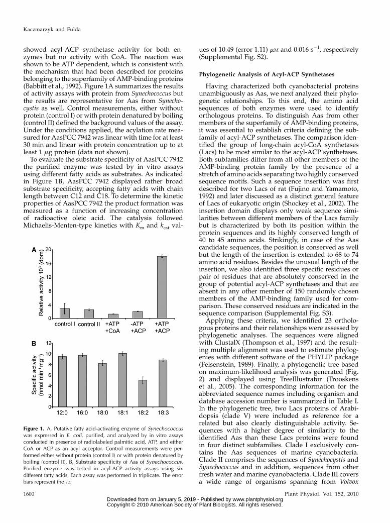
showed acyl-ACP synthetase activity for both en-zymes but no activity with CoA. The reaction wasshown to be ATP dependent, which is consistent withthe mechanism that had been described for proteinsbelonging to the superfamily of AMP-binding proteins(Babbitt et al., 1992). Figure 1A summarizes the resultsof activity assays with protein from Synechococcus butthe results are representative for Aas from Synecho-cystis as well. Control measurements, either withoutprotein (control I) or with protein denatured by boiling(control II) defined the background values of the assay.Under the conditions applied, the acylation rate mea-sured for AasPCC 7942 was linear with time for at least30 min and linear with protein concentration up to atleast 1 mg protein (data not shown).
To evaluate the substrate specificity of AasPCC 7942the purified enzyme was tested by in vitro assaysusing different fatty acids as substrates. As indicatedin Figure 1B, AasPCC 7942 displayed rather broadsubstrate specificity, accepting fatty acids with chainlength between C12 and C18. To determine the kineticproperties of AasPCC 7942 the product formation wasmeasured as a function of increasing concentrationof radioactive oleic acid. The catalysis followedMichaelis-Menten-type kinetics with Km and kcat val-
ues of 10.49 (error 1.11) mM and 0.016 s21, respectively(Supplemental Fig. S2).
Phylogenetic Analysis of Acyl-ACP Synthetases
Having characterized both cyanobacterial proteinsunambiguously as Aas, we next analyzed their phylo-genetic relationships. To this end, the amino acidsequences of both enzymes were used to identifyorthologous proteins. To distinguish Aas from othermembers of the superfamily of AMP-binding proteins,it was essential to establish criteria defining the sub-family of acyl-ACP synthetases. The comparison iden-tified the group of long-chain acyl-CoA synthetases(Lacs) to be most similar to the acyl-ACP synthetases.Both subfamilies differ from all other members of theAMP-binding protein family by the presence of astretch of amino acids separating two highly conservedsequence motifs. Such a sequence insertion was firstdescribed for two Lacs of rat (Fujino and Yamamoto,1992) and later discussed as a distinct general featureof Lacs of eukaryotic origin (Shockey et al., 2002). Theinsertion domain displays only weak sequence simi-larities between different members of the Lacs familybut is characterized by both its position within theprotein sequences and its highly conserved length of40 to 45 amino acids. Strikingly, in case of the Aascandidate sequences, the position is conserved as wellbut the length of the insertion is extended to 68 to 74amino acid residues. Besides the unusual length of theinsertion, we also identified three specific residues orpair of residues that are absolutely conserved in thegroup of potential acyl-ACP synthetases and that areabsent in any other member of 150 randomly chosenmembers of the AMP-binding family used for com-parison. These conserved residues are indicated in thesequence comparison (Supplemental Fig. S3).
Applying these criteria, we identified 23 ortholo-gous proteins and their relationships were assessed byphylogenetic analyses. The sequences were alignedwith ClustalX (Thompson et al., 1997) and the result-ing multiple alignment was used to estimate phylog-enies with different software of the PHYLIP package(Felsenstein, 1989). Finally, a phylogenetic tree basedon maximum-likelihood analysis was generated (Fig.2) and displayed using TreeIllustrator (Trooskenset al., 2005). The corresponding information for theabbreviated sequence names including organism anddatabase accession number is summarized in Table I.In the phylogenetic tree, two Lacs proteins of Arabi-dopsis (clade V) were included as reference for arelated but also clearly distinguishable activity. Se-quences with a higher degree of similarity to theidentified Aas than these Lacs proteins were foundin four distinct subfamilies. Clade I exclusively con-tains the Aas sequences of marine cyanobacteria.Clade II comprises the sequences of Synechocystis andSynechococcus and in addition, sequences from otherfresh water and marine cyanobacteria. Clade III coversa wide range of organisms spanning from Volvox
Figure 1. A, Putative fatty acid-activating enzyme of Synechococcuswas expressed in E. coli, purified, and analyzed by in vitro assaysconducted in presence of radiolabeled palmitic acid, ATP, and eitherCoA or ACP as an acyl acceptor. Control measurements were per-formed either without protein (control I) or with protein denatured byboiling (control II). B, Substrate specificity of Aas of Synechococcus.Purified enzyme was tested in acyl-ACP activity assays using sixdifferent fatty acids. Each assay was performed in triplicate. The errorbars represent the SD.
Kaczmarzyk and Fulda
1600 Plant Physiol. Vol. 152, 2010 www.plantphysiol.orgon January 5, 2019 - Published by Downloaded from
Copyright © 2010 American Society of Plant Biologists. All rights reserved.
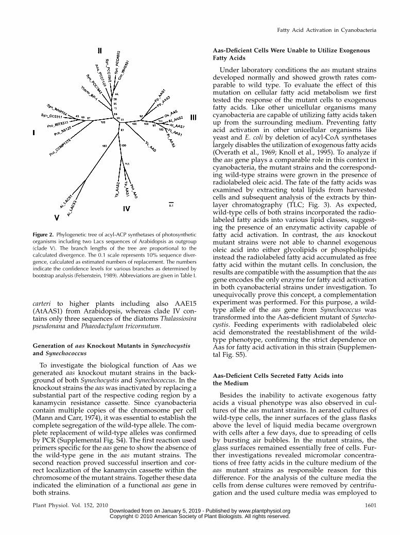
carteri to higher plants including also AAE15(AtAAS1) from Arabidopsis, whereas clade IV con-tains only three sequences of the diatoms Thalassiosirapseudonana and Phaeodactylum tricornutum.
Generation of aas Knockout Mutants in Synechocystisand Synechococcus
To investigate the biological function of Aas wegenerated aas knockout mutant strains in the back-ground of both Synechocystis and Synechococcus. In theknockout strains the aaswas inactivated by replacing asubstantial part of the respective coding region by akanamycin resistance cassette. Since cyanobacteriacontain multiple copies of the chromosome per cell(Mann and Carr, 1974), it was essential to establish thecomplete segregation of the wild-type allele. The com-plete replacement of wild-type alleles was confirmedby PCR (Supplemental Fig. S4). The first reaction usedprimers specific for the aas gene to show the absence ofthe wild-type gene in the aas mutant strains. Thesecond reaction proved successful insertion and cor-rect localization of the kanamycin cassette within thechromosome of the mutant strains. Together these dataindicated the elimination of a functional aas gene inboth strains.
Aas-Deficient Cells Were Unable to Utilize Exogenous
Fatty Acids
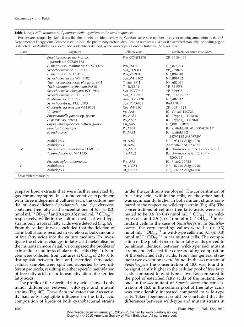
Under laboratory conditions the aas mutant strainsdeveloped normally and showed growth rates com-parable to wild type. To evaluate the effect of thismutation on cellular fatty acid metabolism we firsttested the response of the mutant cells to exogenousfatty acids. Like other unicellular organisms manycyanobacteria are capable of utilizing fatty acids takenup from the surrounding medium. Preventing fattyacid activation in other unicellular organisms likeyeast and E. coli by deletion of acyl-CoA synthetaseslargely disables the utilization of exogenous fatty acids(Overath et al., 1969; Knoll et al., 1995). To analyze ifthe aas gene plays a comparable role in this context incyanobacteria, the mutant strains and the correspond-ing wild-type strains were grown in the presence ofradiolabeled oleic acid. The fate of the fatty acids wasexamined by extracting total lipids from harvestedcells and subsequent analysis of the extracts by thin-layer chromatography (TLC; Fig. 3). As expected,wild-type cells of both strains incorporated the radio-labeled fatty acids into various lipid classes, suggest-ing the presence of an enzymatic activity capable offatty acid activation. In contrast, the aas knockoutmutant strains were not able to channel exogenousoleic acid into either glycolipids or phospholipids;instead the radiolabeled fatty acid accumulated as freefatty acid within the mutant cells. In conclusion, theresults are compatible with the assumption that the aasgene encodes the only enzyme for fatty acid activationin both cyanobacterial strains under investigation. Tounequivocally prove this concept, a complementationexperiment was performed. For this purpose, a wild-type allele of the aas gene from Synechococcus wastransformed into the Aas-deficient mutant of Synecho-cystis. Feeding experiments with radiolabeled oleicacid demonstrated the reestablishment of the wild-type phenotype, confirming the strict dependence onAas for fatty acid activation in this strain (Supplemen-tal Fig. S5).
Aas-Deficient Cells Secreted Fatty Acids into
the Medium
Besides the inability to activate exogenous fattyacids a visual phenotype was also observed in cul-tures of the aas mutant strains. In aerated cultures ofwild-type cells, the inner surfaces of the glass flasksabove the level of liquid media became overgrownwith cells after a few days, due to spreading of cellsby bursting air bubbles. In the mutant strains, theglass surfaces remained essentially free of cells. Fur-ther investigations revealed micromolar concentra-tions of free fatty acids in the culture medium of theaas mutant strains as responsible reason for thisdifference. For the analysis of the culture media thecells from dense cultures were removed by centrifu-gation and the used culture media was employed to
Figure 2. Phylogenetic tree of acyl-ACP synthetases of photosyntheticorganisms including two Lacs sequences of Arabidopsis as outgroup(clade V). The branch lengths of the tree are proportional to thecalculated divergence. The 0.1 scale represents 10% sequence diver-gence, calculated as estimated numbers of replacement. The numbersindicate the confidence levels for various branches as determined bybootstrap analysis (Felsenstein, 1989). Abbreviations are given in Table I.
Fatty Acid Activation in Cyanobacteria
Plant Physiol. Vol. 152, 2010 1601 www.plantphysiol.orgon January 5, 2019 - Published by Downloaded from
Copyright © 2010 American Society of Plant Biologists. All rights reserved.
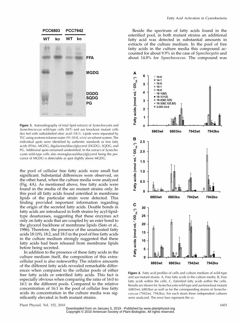
prepare lipid extracts that were further analyzed bygas chromatography. In a representative experimentwith three independent cultures each, the culture me-dia of Aas-deficient Synechocystis and Synechococcuscontained free fatty acid concentrations of 6.4 (SD 0.3)nmolmL21 OD750
21 and 8.4 (SD 0.5) nmolmL21 OD75021,
respectively, while in the culture media of wild-typestrains only traces of fatty acidswere detected (Fig. 4A).From these data it was concluded that the deletion ofaas in both strains resulted in secretion of bulk amountsof free fatty acids into the culture medium. To inves-tigate the obvious changes in fatty acid metabolism ofthe mutants in more detail, we compared the profiles ofextracellular and intracellular fatty acids (Fig. 4). Sam-ples were collected from cultures at OD750 of 2 to 3. Todistinguish between free and esterified fatty acidscellular samples were split and subjected to two dif-ferent protocols, resulting in either specific methylationof free fatty acids or in transmethylation of esterifiedfatty acids.
The profile of the esterified fatty acids showed onlyminor differences between wild-type and mutantstrains (Fig. 4C). These data indicated that Aas activ-ity had only negligible influence on the fatty acidcomposition of lipids of both cyanobacterial strains
under the conditions employed. The concentration offree fatty acids within the cells, on the other hand,was significantly higher in both mutant strains com-pared to the respective wild-type strain (Fig. 4B). Theconcentrations of cellular free fatty acids were esti-mated to be 0.6 (SD 0.4) nmol mL21 OD750
21 in wild-type cells and 2.5 (SD 0.4) nmol mL21 OD750
21 in aasmutant cells in the case of Synechocystis. In Synecho-coccus, the corresponding values were 1.4 (SD 0.3)nmol mL21 OD750
21 in wild-type cells and 3.1 (SD 0.8)nmol mL21 OD750
21 in aas mutant cells. The compo-sition of the pool of free cellular fatty acids proved tobe almost identical between wild-type and mutantstrains and reflected the corresponding compositionof the esterified fatty acids. From this general state-ment two exceptions were found. In the aasmutant ofSynechocystis the concentration of 18:0 was found tobe significantly higher in the cellular pool of free fattyacids compared to wild type as well as compared tothe pool of esterified fatty acids of the mutant. Sec-ond, in the aas mutant of Synechococcus the concen-tration of 16:0 in the cellular pool of free fatty acidswas considerably increased compared to wild-typecells. Taken together, it could be concluded that thedifferences between wild-type and mutant strains in
Table I. Acyl-ACP synthetases of photosynthetic organisms and related sequences
Proteins are grouped by clade. If possible the proteins are identified by the GenBank accession number. In case of ongoing annotation by the U.S.Department of Energy Joint Genome Institute (JGI), the preliminary protein identification number is given or if assembled manually the coding regionis denoted. For Arabidopsis also the locus identifiers defined by the Arabidopsis Genome Initiative (AGI) are given.
Clade Organism Abbreviation GenBank Accession No./JGI/AGI
I Prochlorococcus marinus sp.pastoris str. CCMP1378
Pro_CCMP1378 ZP_00104400
P. marinus sp. marinus str. CCMP1375 Pro_SS120 NP_874793Synechococcus sp. CC9311 Syn_CC9311 YP_729826P. marinus str. MIT 9313 Pro_MIT9313 NP_894048Synechococcus sp. WH 8102 Syn_WH8102 NP_896762
II Thermosynechococcus elongatus BP-1 Therm_BP-1 NP_682091Trichodesmium erythraeum IMS101 Tri_IMS101 YP_721558Synechococcus elongatus PCC 7942 Syn_PCC7942 YP_399935Synechococcus sp. PCC 7002 Syn_PCC7002 YP_001735222Anabaena sp. PCC 7120 Ana_PCC7120 NP_487642Synechocystis sp. PCC 6803 Syn_PCC6803 BAA17024Crocosphaera watsonii WH 8501 Cro_WH8501 ZP_00514521
III V. carteri Vc_AAS JGI Volca1 120723Physcomitrella patens ssp. patens Pp_AAS1 JGI Phypa1_1 169848P. patens ssp. patens Pp_AAS2 JGI Phypa1_1 140960Oryza sativa (japonica cultivar group) Os_AAS NP_001051878Populus trichocarpa Pt_AAS1 JGI scaffold_88: 413600-428931a
P. trichocarpa Pt_AAS2 JGI scaffold LG_I:24797125-24808759a
Arabidopsis At_AAS1 NP_193143 At4g14070Arabidopsis At_AAS2 AAM28629 At3g23790
IV Thalassiosira pseudonana CCMP 1335 Tp_AAS1 JGI chromosome 7: 511777-514965a
T. pseudonana CCMP 1335 Tp_AAS2 JGI chromosome 6: 1257611-1260333a
Phaeodactylum tricornutum Pht_AAS JGI Phatr2 35735V Arabidopsis At_LACS1 NP_182246 At2g47240
Arabidopsis At_LACS3 NP_176622 At1g64400
aAssembled manually.
Kaczmarzyk and Fulda
1602 Plant Physiol. Vol. 152, 2010 www.plantphysiol.orgon January 5, 2019 - Published by Downloaded from
Copyright © 2010 American Society of Plant Biologists. All rights reserved.
the pool of cellular free fatty acids were small butsignificant. Substantial differences were observed, onthe other hand, when the culture media were analyzed(Fig. 4A). As mentioned above, free fatty acids werefound in the media of the aas mutant strains only. Inthis pool all fatty acids found esterified in membranelipids of the particular strain were detected. Thisfinding provided important information regardingthe origin of the secreted fatty acids. Double bonds infatty acids are introduced in both strains by acyl-lipid-type desaturases, suggesting that these enzymes actonly on fatty acids that are coupled by an ester bond tothe glycerol backbone of membrane lipids (Sato et al.,1986). Therefore, the presence of the unsaturated fattyacids 18:1(9), 18:2, and 18:3 in the pool of free fatty acidsin the culture medium strongly suggested that thesefatty acids had been released from membrane lipidsbefore being secreted.In addition to the presence of these fatty acids in the
culture medium itself, the composition of this extra-cellular pool is also noteworthy. The relative amountsof the different fatty acids revealed remarkable differ-ences when compared to the cellular pools of eitherfree fatty acids or esterified fatty acids. This fact isespecially obvious when comparing the ratio of 16:0 to16:1 in the different pools. Compared to the relativeconcentration of 16:1 in the pool of cellular free fattyacids its concentration in the culture media was sig-nificantly elevated in both mutant strains.
Beside the spectrum of fatty acids found in theesterified pool, in both mutant strains an additionalfatty acid was detected in substantial amounts inextracts of the culture medium. In the pool of freefatty acids in the culture media this compound ac-counted for about 9.9% in the case of Synechocystis andabout 14.8% for Synechococcus. The compound was
Figure 3. Autoradiography of total lipid extracts of Synechocystis andSynechococcus wild-type cells (WT) and aas knockout mutant cells(ko) fed with radiolabeled oleic acid (18:1). Lipids were separated byTLC using acetone:toluene:water (91:30:8, v/v/v) as solvent system. Theindividual spots were identified by authentic standards as free fattyacids (FFAs), MGDG, digalactosyldiacylglycerol (DGDG), SQDG, andPG. Additional spots remained unidentified. In the extract of Synecho-cystis wild-type cells also monoglucosyldiacylglycerol being the pre-cursor of MGDG is detectable as spot slightly above MGDG.
Figure 4. Fatty acid profiles of cells and culture medium of wild-typeand aas mutant strains. A, Free fatty acids in the culture media. B, Freefatty acids within the cells. C, Esterified fatty acids within the cells.Results are shown for Synechocystiswild type and aas knockout mutant(6803wt, 6803ko) as well as for the corresponding strains of Synecho-coccus (7942wt, 7942ko). For each strain three independent cultureswere analyzed. The error bars represent the SD.
Fatty Acid Activation in Cyanobacteria
Plant Physiol. Vol. 152, 2010 1603 www.plantphysiol.orgon January 5, 2019 - Published by Downloaded from
Copyright © 2010 American Society of Plant Biologists. All rights reserved.
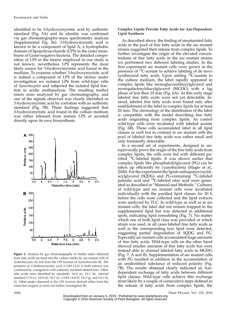
identified to be 3-hydroxymyristic acid by authenticstandard (Fig. 5A) and its identity was confirmedvia gas chromatography-mass spectrometry analysis(Supplemental Fig. S6). 3-Hydroxymyristic acid isknown to be a component of lipid A, a hydrophobicdomain of lipopolysaccharide (LPS) in the outer mem-brane of Gram-negative bacteria. The detailed compo-sition of LPS in the strains employed in our study isnot known; nevertheless LPS represents the mostlikely source for 3-hydroxymyristic acid found in themedium. To examine whether 3-hydroxymyristic acidis indeed a compound of LPS of the strains underinvestigation we isolated LPS from wild-type cellsof Synechocystis and subjected the isolated lipid frac-tion to acidic methanolysis. The resulting methylesters were analyzed by gas chromatography, andone of the signals observed was clearly identified as3-hydroxymyristic acid by coelution with an authenticstandard (Fig. 5B). These findings suggested that3-hydroxymyristic acid found in the culture mediumwas either released from mature LPS or secreteddirectly upon its own biosynthesis.
Complex Lipids Provide Fatty Acids for Aas-Dependent
Lipid Synthesis
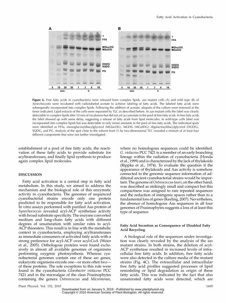
As described above, the finding of unsaturated fattyacids in the pool of free fatty acids in the aas mutantstrains suggested their release from complex lipids. Tofurther investigate the origin of the elevated concen-trations of free fatty acids in the aas mutant strainswe performed two different labeling studies. In thefirst experiment aas mutant cells were grown in thepresence of 14C-acetate to achieve labeling of de novosynthesized fatty acids. Upon adding 14C-acetate tothe culture medium, the label rapidly appeared incomplex lipids like monoglucosyldiacylglycerol andmonogalactosyldiacylglycerol (MGDG) with a lagphase of less than 10 min (Fig. 6A). At this early stagelabeled free fatty acids were not yet detectable. In-stead, labeled free fatty acids were found only afterestablishment of the label in complex lipids for at least20 min. The chronology of the distribution of the labelis compatible with the model describing free fattyacids originating from complex lipids. As controlwild-type cells were incubated with labeled acetate(Fig. 6B). These cells accumulated label in all lipidclasses as well but in contrast to aas mutant cells thepool of labeled free fatty acids was rather small andonly transiently detectable.
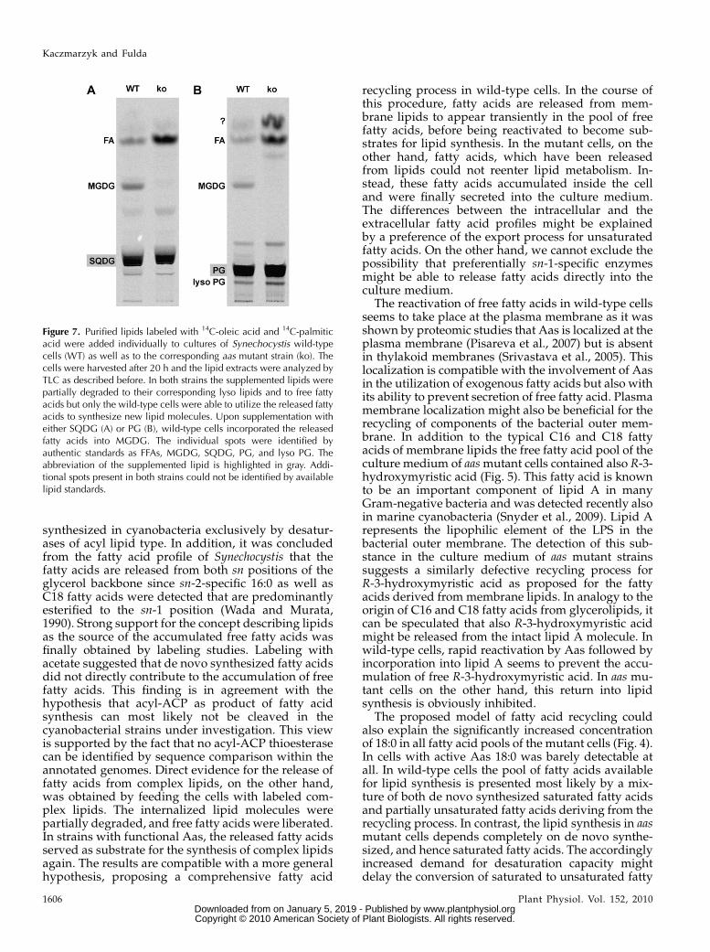
In a second set of experiments, designed to un-equivocally prove the origin of the free fatty acids fromcomplex lipids, the cells were fed with different pu-rified 14C-labeled lipids. It was shown earlier thatcomplex lipids like phosphatidylglycerol (PG) can betaken up efficiently by cyanobacteria (Hagio et al.,2000). For the experiment the lipids sulfoquinovosyldi-acylglycerol (SQDG) and PG-containing 14C-labeledpalmitic acid and 14C-labeled oleic acid were gener-ated as described in “Material andMethods.” Culturesof wild-type and aas mutant cells were incubatedindividually with the purified lipid classes for 20 hbefore the cells were collected and the lipid extractswere analyzed by TLC. In wild-type as well as in aasmutant cells, the label did not remain trapped in thesupplemented lipid but was detected in additionalspots, indicating lipid remodeling (Fig. 7). No matterwhich one of both lipid class was provided or whichstrain was used, in all cases labeled free fatty acids aswell as the corresponding lyso lipid were detected,suggesting partial degradation of SQDG and PG.Especially aasmutant cells accumulated huge amountsof free fatty acids. Wild-type cells on the other handshowed smaller amounts of free fatty acids but wereinstead able to channel labeled fatty acids to MGDG(Fig. 7, A and B). Supplementation of aas mutant cellswith PG resulted in addition in the accumulation ofan unidentified substance of reduced polarity (Fig.7B). The results obtained clearly indicated an Aas-dependent exchange of fatty acids between differentlipid classes. Wild-type cells achieve this exchangemost likely by a couple of consecutive steps defined asthe release of fatty acids from complex lipids, the
Figure 5. Analysis by gas chromatography of methyl esters obtainedfrom fatty acids secreted into the culture media by aas mutant cells ofSynechocystis (A) and from the LPS fraction of Synechocystis (B). Thepresence of 3-hydroxymyristic acid (3-OH-14:0) in both extracts wasconfirmed by comigration with authentic standard (dotted line). Otherfatty acids were identified by standards: 16:0 (a), 16:1 (b), internalstandard 17:0 (c), 18:0 (d), 18:1 (e), 3-OH-14:0 (f), 18:2 (g), and 18:3 (h;A). Other peaks observed in the LPS fraction derived either from theextraction reagent or were not further investigated (B).
Kaczmarzyk and Fulda
1604 Plant Physiol. Vol. 152, 2010 www.plantphysiol.orgon January 5, 2019 - Published by Downloaded from
Copyright © 2010 American Society of Plant Biologists. All rights reserved.
establishment of a pool of free fatty acids, the reacti-vation of these fatty acids to provide substrate foracyltransferases, and finally lipid synthesis to produceagain complex lipid molecules.
DISCUSSION
Fatty acid activation is a central step in fatty acidmetabolism. In this study, we aimed to address themechanism and the biological role of this enzymaticactivity in cyanobacteria. The genomes of sequencedcyanobacterial strains encode only one proteinpredicted to be responsible for fatty acid activation.In vitro assays performed with purified Aas protein ofSynechococcus revealed acyl-ACP synthetase activitywith broad substrate specificity. The enzyme convertedmedium and long-chain fatty acids with differentdegrees of unsaturation with similar rates to acyl-ACP thioesters. This result is in line with the metaboliccontext in cyanobacteria, employing acyltransferasesas immediate consumers of activated fatty acids with astrong preference for acyl-ACP over acyl-CoA (Weieret al., 2005). Orthologous proteins were found exclu-sively in almost all available genomes of organismsperforming oxygenic photosynthesis. Whereas cya-nobacterial genomes contain one of these aas genes,eukaryotic organisms encode one—ormore often two—of these proteins. The sole exceptions to this rule werefound in the cyanobacteria Gloeobacter violaceus PCC7421 and in the microalgae of the class Prasinophytescontaining the genera Ostreococcus and Micromonas,
where no homologous sequences could be identified.G. violaceus PCC 7421 is a member of an early branchinglineage within the radiation of cyanobacteria (Hondaet al., 1999) and is characterized by the lack of thylakoids(Rippka et al., 1974). To evaluate the question if theappearance of thylakoids and Aas activity is somehowconnected to the genomic sequence information of ad-ditional ancient cyanobacterial strains would be impor-tant. ThegenomeofOstreococcus tauri, on the otherhand,was described as strikingly small and compact but thiscompactness was assigned to rare repeated sequencesand the reduction of intergenic spaces rather than to afundamental loss of genes (Keeling, 2007).Nevertheless,the absence of homologous Aas sequences in all fourgenomes of Prasinophytes suggests a loss of at least thistype of sequence.
Fatty Acid Secretion as Consequence of Disabled Fatty
Acid Recycling
A biological role of the sequences under investiga-tion was clearly revealed by the analysis of the aasmutant strains. In both strains, the deletion of acyl-ACP synthetase resulted in increased levels of intra-cellular free fatty acids. In addition, free fatty acidswere also detected in the culture media of the mutantstrains (Fig. 4C). The extracellular and intracellularfree fatty acid profiles suggested processes of lipidremodeling or lipid degradation as origin of thesefatty acids. This was indicated by the fact that alsounsaturated fatty acids were detected, which are
Figure 6. Free fatty acids in cyanobacteria were released from complex lipids. aas mutant cells (A) and wild type (B) ofSynechocystis were incubated with radiolabeled acetate to achieve labeling of fatty acids. The labeled fatty acids weresubsequently incorporated into complex lipids. Following the addition of acetate, aliquots of the culture were removed at thetimes indicated. Lipid extracts of the cells were separated by TLC as described before. In aas mutant cells the label was clearlydetectable in complex lipids after 10 min of incubation but did not yet accumulate in the pool of free fatty acids. In free fatty acidsthe label showed up with some delay, suggesting a release of fatty acids from lipid molecules. In wild-type cells label wasincorporated into complex lipids but was detectable in only minor amounts in the pool of free fatty acids. The individual spotswere identified as FFAs, monoglucosyldiacylglycerol (MGlucDG), MGDG (MGalDG), digalactosyldiacylglycerol (DGDG),SQDG, and PG. Analysis of the spot close to the solvent front (?) by two-dimensional TLC revealed a mixture of at least fourdifferent components that were not further investigated.
Fatty Acid Activation in Cyanobacteria
Plant Physiol. Vol. 152, 2010 1605 www.plantphysiol.orgon January 5, 2019 - Published by Downloaded from
Copyright © 2010 American Society of Plant Biologists. All rights reserved.
synthesized in cyanobacteria exclusively by desatur-ases of acyl lipid type. In addition, it was concludedfrom the fatty acid profile of Synechocystis that thefatty acids are released from both sn positions of theglycerol backbone since sn-2-specific 16:0 as well asC18 fatty acids were detected that are predominantlyesterified to the sn-1 position (Wada and Murata,1990). Strong support for the concept describing lipidsas the source of the accumulated free fatty acids wasfinally obtained by labeling studies. Labeling withacetate suggested that de novo synthesized fatty acidsdid not directly contribute to the accumulation of freefatty acids. This finding is in agreement with thehypothesis that acyl-ACP as product of fatty acidsynthesis can most likely not be cleaved in thecyanobacterial strains under investigation. This viewis supported by the fact that no acyl-ACP thioesterasecan be identified by sequence comparison within theannotated genomes. Direct evidence for the release offatty acids from complex lipids, on the other hand,was obtained by feeding the cells with labeled com-plex lipids. The internalized lipid molecules werepartially degraded, and free fatty acids were liberated.In strains with functional Aas, the released fatty acidsserved as substrate for the synthesis of complex lipidsagain. The results are compatible with a more generalhypothesis, proposing a comprehensive fatty acid
recycling process in wild-type cells. In the course ofthis procedure, fatty acids are released from mem-brane lipids to appear transiently in the pool of freefatty acids, before being reactivated to become sub-strates for lipid synthesis. In the mutant cells, on theother hand, fatty acids, which have been releasedfrom lipids could not reenter lipid metabolism. In-stead, these fatty acids accumulated inside the celland were finally secreted into the culture medium.The differences between the intracellular and theextracellular fatty acid profiles might be explainedby a preference of the export process for unsaturatedfatty acids. On the other hand, we cannot exclude thepossibility that preferentially sn-1-specific enzymesmight be able to release fatty acids directly into theculture medium.
The reactivation of free fatty acids in wild-type cellsseems to take place at the plasma membrane as it wasshown by proteomic studies that Aas is localized at theplasma membrane (Pisareva et al., 2007) but is absentin thylakoid membranes (Srivastava et al., 2005). Thislocalization is compatible with the involvement of Aasin the utilization of exogenous fatty acids but also withits ability to prevent secretion of free fatty acid. Plasmamembrane localization might also be beneficial for therecycling of components of the bacterial outer mem-brane. In addition to the typical C16 and C18 fattyacids of membrane lipids the free fatty acid pool of theculture medium of aasmutant cells contained also R-3-hydroxymyristic acid (Fig. 5). This fatty acid is knownto be an important component of lipid A in manyGram-negative bacteria and was detected recently alsoin marine cyanobacteria (Snyder et al., 2009). Lipid Arepresents the lipophilic element of the LPS in thebacterial outer membrane. The detection of this sub-stance in the culture medium of aas mutant strainssuggests a similarly defective recycling process forR-3-hydroxymyristic acid as proposed for the fattyacids derived from membrane lipids. In analogy to theorigin of C16 and C18 fatty acids from glycerolipids, itcan be speculated that also R-3-hydroxymyristic acidmight be released from the intact lipid A molecule. Inwild-type cells, rapid reactivation by Aas followed byincorporation into lipid A seems to prevent the accu-mulation of free R-3-hydroxymyristic acid. In aas mu-tant cells on the other hand, this return into lipidsynthesis is obviously inhibited.
The proposed model of fatty acid recycling couldalso explain the significantly increased concentrationof 18:0 in all fatty acid pools of the mutant cells (Fig. 4).In cells with active Aas 18:0 was barely detectable atall. In wild-type cells the pool of fatty acids availablefor lipid synthesis is presented most likely by a mix-ture of both de novo synthesized saturated fatty acidsand partially unsaturated fatty acids deriving from therecycling process. In contrast, the lipid synthesis in aasmutant cells depends completely on de novo synthe-sized, and hence saturated fatty acids. The accordinglyincreased demand for desaturation capacity mightdelay the conversion of saturated to unsaturated fatty
Figure 7. Purified lipids labeled with 14C-oleic acid and 14C-palmiticacid were added individually to cultures of Synechocystis wild-typecells (WT) as well as to the corresponding aas mutant strain (ko). Thecells were harvested after 20 h and the lipid extracts were analyzed byTLC as described before. In both strains the supplemented lipids werepartially degraded to their corresponding lyso lipids and to free fattyacids but only the wild-type cells were able to utilize the released fattyacids to synthesize new lipid molecules. Upon supplementation witheither SQDG (A) or PG (B), wild-type cells incorporated the releasedfatty acids into MGDG. The individual spots were identified byauthentic standards as FFAs, MGDG, SQDG, PG, and lyso PG. Theabbreviation of the supplemented lipid is highlighted in gray. Addi-tional spots present in both strains could not be identified by availablelipid standards.
Kaczmarzyk and Fulda
1606 Plant Physiol. Vol. 152, 2010 www.plantphysiol.orgon January 5, 2019 - Published by Downloaded from
Copyright © 2010 American Society of Plant Biologists. All rights reserved.
acids, resulting eventually in higher levels of 18:0bound to lipids. Under such conditions, the activitiesresponsible for the release of fatty acids from lipidmolecules appear to access their substrate sometimeseven faster than the D9-desaturase, resulting in thedetection of substantial amounts of 18:0 also in thepools of free fatty acids.The data reported here for cyanobacterial strains
with disabled fatty acid activation are surprisinglysimilar to those found for mutants of yeast with acomparable defect (Scharnewski et al., 2008). In yeast,the inactivation of two out of five existing acyl-CoAsynthetases resulted in very similar phenotypes asdescribed here for the deletion of aas in cyanobacteria.In both cases, exogenous fatty acids were no longerused as substrates for lipid synthesis, and in additionthe mutant cells were characterized by secretion of freefatty acids into the culture medium. In cyanobacterialcells as well as in yeast cells, membrane lipids wereidentified as source of the released fatty acids, indi-cating permanent and significant lipid turnover. Inboth cases, wild-type cells were able to reintroducereleased free fatty acids back into the lipid metabo-lism. We conclude that the recycling of endogenousfatty acids is probably one of the most importanttasks of fatty acid activation in general. In addition,the striking similarities between distantly relatedorganisms might indicate a well-conserved strategyof lipid turnover behind a deceiving front of apparentlipid stability. Currently, we can only speculate aboutthe reasons for the establishment of such a ratherenergy-costly procedure. One possible explanationcould be a permanent adaptation of the lipid compo-sition or the molecular lipid species to changes of theenvironmental conditions. This might be economi-cally unviable under artificially constant parameterspresent in the laboratory but might be beneficial tocope with challenging conditions in the natural en-vironment.In conclusion, we have shown that the activation
of free fatty acids by Aas in cyanobacteria is essentialfor the incorporation of exogenously supplied freefatty acids into cellular lipid metabolism. The moreimportant role of Aas seems to be, nevertheless, theactivation of endogenous free fatty acids perma-nently released from membrane lipids. The resultingrecycling of fatty acids can be observed in distantlyrelated species like yeast as well, suggesting a well-conserved mechanism in cellular lipid metabolism ingeneral.
MATERIALS AND METHODS
Strains and Growth Conditions
Liquid cultures of the Glc-tolerant Synechocystis sp. PCC 6803 and
Synechococcus elongatus PCC 7942 were grown photoautotrophically in BG 11
media (Rippka et al., 1979) at 30�C. The cultures were grown under constant
illumination at a photosynthetic photon flux density of approximately 38
mmol photons m22 s21 and with aeration by sterile air. The growth was
monitored by measurement of OD750. For growth on solid media BG 11 was
supplemented with 20 mM HEPES-NaOH, pH 7.5, 0.3% (w/v) sodium
thiosulfate pentahydrate, and 1.5% (w/v) agar. Mutants of Synechocystis and
Synechococcus were grown in the presence of antibiotic for selection (15 mg
mL21 kanamycin or 10 mg mL21 chloramphenicol, respectively).
DNA Isolation and Generation of aas Knockout Mutantsin Cyanobacteria
Genomic DNA from Synechocystis and Synechococcus was isolated accord-
ing to a protocol described before (Porter, 1988). The aas mutants in the
background of Synechocystis and Synechococcus were created by replacing part
of the coding region by a cassette encoding a kanamycin resistance gene via
homologous recombination. The constructs were prepared as follows: The
open reading frame of slr1609 was amplified from genomic DNA of Synecho-
cystis with the specific primer pair GGAATTCATATGGACAGTGGC-
CATGGCGCT, 6803_KOfor and AGAATTCTCGAGAAACATTTCGTCAAT-
TAAATGTTG, 6803_KOrev and was subsequently cloned into the SmaI site of
the vector pUC19. A 230-bp fragment was excised from the gene by digestion
with NaeI and SmaI and replaced by a kanamycin resistance cassette released
from the vector pKRP11 (Reece and Phillips, 1995) by SmaI. An analogous
strategy was applied to prepare the construct for disruption of the homologous
gene from Synechococcus. Genomic DNAwas used as a template to amplify the
open reading frame including fragments of 957 bp upstream and of 645 bp
downstream the coding region using primers ACACGCATGCTTAAAT-
GACTTCTTGTGGAAAG, 7942_KOfor and AGAGATCTAGAGACGG-
CACCTCAACTCCTAGGT, 7942_KOrev. The obtained PCR product was
cloned into pGEM-T (Promega), a fragment of 1,704 bp was removed by
digestion with EcoRV and SmaI, and the kanamycin resistance cassette was
inserted into the vector.
For the construction of a plasmid required to achieve genetic complemen-
tation of the aas mutant strain of Synechocystis a variant of the knockout
construct described above was used as a starting point. This construct
contained a chloramphenicol resistance cassette instead of the described
kanamycin cassette. The chloramphenicol cassette was released from the
vector pKRP10 (Reece and Phillips, 1995). Fragments of Synechocystis DNA,
flanking the chloramphenicol cassette, served as borders for homologous
recombination. In one of the flanking fragments 25 bps distant from the
chloramphenicol cassette 3 bps, were changed (QuikChange II site-directed
mutagenesis kit, Stratagene) to introduce an EagI restriction site (GAT-
GAACTACACCAGCGGCCGCCATTTCAAGGG, 6803_QCfor and CCCT-
TGAAATGGCGGCCGCTGGTGTAGTTCATC, 6803_QCrev). A 3,920 bps
fragment, including the open reading frame of aas of Synechococcus plus
promoter and terminator, was amplified by PCR with forward and reverse
primers (ACAGCGGCCGCGATCGCGTCTCGAATCG, 7942_for, and ACAG-
CGGCCGCGCAGCACGATTTCAACTTGC, 7942_rev), adding NotI restric-
tion sites to both ends. The PCR fragment was cloned first into pGEM-T vector
and then transferred into the newly created EagI restriction site of the comple-
mentation construct. The resulting vector was used to transform Synechocystis
aas knockout cells. Cyanobacterial cells were transformed as described before
(Porter, 1988). For initial selection of transformants the DNA/cells mixture was
spread on solid BG 11 medium. After 24 h the appropriate antibiotic (0.45 mg
kanamycin or 0.2 mg chloramphenicol) was added into three evenly distributed
wells punched into the agar. Homozygousmutants were obtained by successive
streaking on BG 11 plates containing antibiotic.
Both the correct integration of the knockout cassette and the complete
segregation of the wild-type alleles were confirmed by PCR. For Synechocystis
the complete segregation was evaluated by 6803_con1 (CTCTACATCCTA-
GAAGACAGC) combined with 6803_con2 (GAATCCAATTCCCGTACTT-
GGTGC) and the correct insertion of the kanamycin resistance cassette was
confirmed by Kan_con3 (GATTCAGTCGTCACTCATGGTG) combined with
6803_con4 (CACAGCCGGGGCACACCGACAATG). For Synechococcus the
complete segregation was evaluated by the combination of 7942_con1
(CCGTAATCAGCGTGTAGATGATGG) and 7942_con2 (GATCGAACCGC-
TGTCCTCTAAGACG). The correct insertion of the kanamycin resistance cas-
sette was confirmed by Kan_con3 as given above combined with 7942_con4
(GTGTTCCGCGACAACGTTGCGACG). The successful integration of the con-
struct used for complementing the aasmutant of Synechocystiswas confirmed by
testing for the presence of aas of Synechococcus using the primer combination
7942_con5 (CCGTAATCAGCGTGTAGATGATGG) and 7942_con6 (GATC-
GAACCGCTGTCCTCTAAGACG).
Fatty Acid Activation in Cyanobacteria
Plant Physiol. Vol. 152, 2010 1607 www.plantphysiol.orgon January 5, 2019 - Published by Downloaded from
Copyright © 2010 American Society of Plant Biologists. All rights reserved.
Cloning of Cyanobacterial aas Genes for Expression in
Escherichia coli
The open reading frames of the candidate genes were amplified from
genomic DNA of Synechocystis and Synechococcus using specific primer pairs:
GGAATTCATATGGACAGTGGCCATGGCGCT, 6803_ORFfor, combined with
AGAATTCTCGAGAAACATTTCGTCAATTAAATGTTG, 6803_ORFrev, and
ACACCATGGCTGGAACCGCCCTCGCGCAAC, 7942_ORFfor, combined
with ACAGCGGCCGCACTCGCCGATTCAAACATCCCGT, 7942_ORFrev,
respectively. Primers were designed to convert the existing GTG start codons
into ATG and to remove the stop codons of both genes. The fragments obtained
were cloned into either pET24c or pET24d vectors, resulting in pETaas6803
and pETaas7942, respectively. Both genes were cloned in frame with the
polyhistidine-tag sequence of the vector. The primers were constructed
according to data provided by CyanoBase (http://www.kazusa.or.jp/cyano/
cyano.html) and the U.S. Department of Energy Joint Genome Institute
(http://www.jgi.doe.gov). The integrity of all cloned fragmentswas confirmed
by sequencing.
Enzyme Overproduction and Purification
Two hundred milliliter cultures of Rosetta (DE3)pLysS cells (Novagen)
carrying a pETaas construct were grown at 37�C with shaking to an OD600 0.8.
The protein expression was induced after 2 h incubation at 16�C by the
addition of 1 mM isopropylthio-b-galactoside and the cultures were further
incubated for 66 h at 16�C. The cultures were chilled on ice and the cells were
harvested by centrifugation at 3,200g. For protein purification some modifi-
cations were introduced to the method described before (Shanklin, 2000). The
cellular pellet was resuspended in 5 mL of extraction buffer (50 mM Tris-HCl,
pH 8.0) and cells were disrupted by sonification. The cellular extract was
clarified by centrifugation at 16,000g for 25 min at 4�C. The supernatant was
collected and MgCl2 was added to a final concentration of 20 mM. The
membrane fraction was collected by ultracentrifugation at 100,000g for 1 h
at 4�C. The supernatant was discarded and the membrane pellet was
resuspended in 300 mL of extraction buffer. An equal volume of the buffer
containing detergent (50 mM Tris-HCl, 20 mM MgCl2, 4% [v/v] Triton X-100,
pH 8.0) was added to solubilize the membranes. To improve the removal of
membrane-bound proteins the fraction was incubated on ice with agitation for
30 min before clarification by centrifugation at 100,000g for 30 min at 4�C.During centrifugation BD TALON resin (BD Biosciences) was equilibrated
with the column buffer (50 mM Tris-HCl, 20 mM MgCl2, 2% [v/v] Triton X-100,
pH 8.0). The clarified and solubilized membrane fraction was applied to the
resin and gently agitated at 4�C for 1 h on a platform shaker to allow the
polyhistidine-tagged protein to bind the resin. The resin was transferred to a 5
mL gravity-flow column andwashed sequentially with three column volumes
of the column buffer followed by three column volumes of the column buffer
containing 20 mM imidazole to remove nonspecifically bound proteins. The
target protein was eluted with the column buffer containing 100 mM EDTA. A
total of 130 to 150 mL fractions containing the polyhistidine-tagged protein
were collected and dialyzed overnight against 400 mL of the column buffer at
4�C. Protein fractions were analyzed by SDS-PAGE carried out as described
by Laemmli (1970) and gels were stained with Coomassie Brilliant Blue
(Serva).
Acyl-ACP Synthetase and Acyl-CoA Synthetase inVitro Assays
The acyl-ACP synthetase activity was measured according to a modified
method described previously (Rock and Cronan, 1981). The assay was
conducted in 1.5 mL microcentrifuge tubes in a volume of 40 mL. The assay
mixture contained 2.5 mM Tris-HCl, pH 8.0, 2 mM dithiothreitol, 10 mM MgCl2,
5 mM ATP, 10 mM LiCl, 2% (v/v) Triton X-100, 15 mM ACP, 30 mM14C-fatty acid
(specific activity 53.7–60 mCi mmol21), and defined amount (0.1–1 mg) of
protein sample.
The acyl-CoA synthetase activity assay was performed in the same way as
the acyl-ACP synthetase activity assay. The composition of the assay mixture
followed a slightly modified protocol described previously (Joyard and
Stumpf, 1981). In detail, the assay mixture was composed of 100 mM Tris-
HCl, pH 8.0, 10 mM MgCl2, 5 mM ATP, 2.5 mM dithiothreitol, 0.5 mM CoA, 2%
(v/v) Triton X-100, 30 mM14C-fatty acid (specific activity 53.7–60 mCi mmol21),
and protein sample in a final volume of 40 mL.
Phylogenetic Analysis
The amino acid sequences of Aas candidate proteins were aligned together
with Lacs sequences serving as outgroup using ClustalX (Thompson et al.,
1997). The resulting alignments were used to investigate phylogenetic rela-
tionships by employing the following software of the PHYLIP program
package (Felsenstein, 1989): PROTDIST in combination with FITCH as a
neighbor-joining method, and PROML as maximum-likelihood method.
Parameters used were: the Jones-Taylor-Thornton model, global rearrange-
ments, and randomized input order of sequences with three jumbles. The
bootstrap values were calculated with SEQBOOT and CONDENSE 100
replicates. Finally, the tree was displayed using TreeIllustrator (Trooskens
et al., 2005).
Lipid Analytical Methods
For analysis of fatty acid profiles in cells and media 8 mL aliquots of
cultures at OD750 of about 2 to 3 were collected. Cells were harvested by
centrifugation and washed twice with 1 mL 0.1 M NaHCO3. Intracellular and
extracellular lipid extractions were performed according the protocol
established previously (Bligh and Dyer, 1959). To recover free fatty acids
quantitatively the extraction mixture was acidified by addition of 10 mL
0.1 M hydrochloric acid in case of the cellular pellet and 100 mL 0.1 M
hydrochloric acid in case of the culture medium. Prior to extraction,
heptadecanoic acid (17:0) as internal standard for free fatty acids (15 mg)
and triheptadecanoylglycerol as internal standard for esterified fatty acids
(20 mg) were added.
Free fatty acids from intracellular and extracellular lipid extracts were
methylated according to a modified protocol described earlier (Stumpe et al.,
2001). In short, the lipid extract (40 mL) was transferred to a new glass tube and
dried under a stream of nitrogen. Methanol (400 mL) was added together with
10 mL of 1-ethyl-3-3-dimethylaminopropylcarbodiimide (0.1 mg mL21 in
methanol) and incubated for 2 h at 22�C. The reaction was stopped by adding
200 mL of 0.1 M Tris-HCl, pH 7.5. The methyl esters of free fatty acids were
extracted with 1 mL of hexane followed by centrifugation at 1,000g for 2 min.
The upper hexane phase was transferred to a 1.5 mL microcentrifuge tube,
dried, and resuspended in acetonitrile (12 mL) and analyzed by gas chroma-
tography.
Esterified fatty acids from intracellular lipid extracts were transmethylated
according to a modified protocol described earlier (Christie, 1982). Lipid
extract (40 mL) was transferred to a 2-mL microcentrifuge tube and dried
under a stream of nitrogen. A total of 333 mL methanol:toluene (1:1, v/v) and
167 mL 0.5 M sodium methoxide (CH3NaO) were added and the mixture was
incubated at 22�C. After 20 min, the reaction was stopped by adding 500 mL of
1 M NaCl and 50 mL of 32% hydrochloric acid. The methyl esters were
extracted with 1 mL of hexane followed by centrifugation at 2,300g for 2 min.
The upper hexane phase was transferred to a new 1.5 mL microcentrifuge
tube, dried, and resuspended in acetonitrile (15 mL). The fatty acid methyl
esters were analyzed by gas chromatography using an Agilent 6890 series gas
chromatograph equipped with a capillary DB-23 column (Agilent).
Extraction and Analysis of LPS
LPS was extracted from the cells of Synechocystiswith the LPS extraction kit
(Intron Biotechnology, Molecular Solutions Europe) according to the manu-
facturer’s protocol and subjected to acidic methanolysis that was performed as
described previously (Miquel and Browse, 1992). The resultant methyl esters
were analyzed by gas chromatography. The analysis of the hydroxy fatty acids
was carried out using Agilent 5973 network mass selective detector connected
to Agilent 6890 gas chromatograph. Electron energy of 70 eV, an ion source
temperature of 230�C, and a temperature of 260�C for the transfer line was
used.
Fatty Acid Uptake Assay
Cyanobacterial cells were collected from 10 mL cultures at OD750 of about
3.6 by centrifugation, resuspended in 2 mL of fresh BG 11 medium, and
transferred to 2 mL microcentrifuge tubes. Radiolabeled fatty acid [1-14C]18:1
(Amersham Biosciences; specific activity 56 mCi mmol21) was individually
added in amounts corresponding to 0.25 mCi and the tubes were placed on a
platform shaker under light. After 1 h incubation 0.5 mL of each culture were
transferred to a new microcentrifuge tube and the cells were collected by
Kaczmarzyk and Fulda
1608 Plant Physiol. Vol. 152, 2010 www.plantphysiol.orgon January 5, 2019 - Published by Downloaded from
Copyright © 2010 American Society of Plant Biologists. All rights reserved.
centrifugation at 3,000g. The remaining 1.5 mL were further incubated for
additional 15 h before another aliquot of 0.5 mL were recovered from each
culture and treated as described above. A total of 0.4 mL of each supernatant
was used to determine the radioactivity present in the culture medium by
liquid scintillation counting. The residual supernatant was discarded. Cell
pellets were washed twice with 0.1 M NaHCO3 before being subjected to total
lipid extraction as described above. Different lipid classes were separated by
TLC using acetone/toluene/water (91/30/8, v/v/v) as solvent and were
visualized by fluorography.
Synthesis of 14C-Labeled Glycerolipids
Radiolabeled lipids were generated by incubating 20 mL Synechocystis
wild-type cells at OD750 5 with 3 mCi of [1-14C]palmitic acid and 3 mCi [1-14C]
oleic acid (Amersham Biosciences). After 20 h incubation on the platform
shaker under light at 30�C, cells were harvested, washed with 0.1 M NaHCO3,
and subjected to lipid extraction. Lipid extract was fractionated into neutral
lipids, glycolipids, and phospholipids using Strata SI-1 silica column (Phe-
nomenex). The individual lipid classes were purified twice by TLC and finally
dissolved in 2.5% (v/v) Triton X-100.
Labeling Experiments
A total of 1.5 mCi of radiolabeled [1-14C]acetate (Amersham Biosciences)
was added to 8 mL culture of Synechocystis aas knockout strain at OD750 13.
Culture was grown for 24 h under light, at 30�C, aerated by shaking on a
platform shaker. Aliquots of 0.5 mL were collected at defined time points.
Cells were harvested by centrifugation, washed with 0.1 M NaHCO3, and
subjected to total lipid extraction followed by TLC.
Radiolabeled lipids generated as described above were added in volumes
of 20 to 40 mL corresponding to 0.1 mCi to 20 mL culture of wild type and aas
knockout strain of Synechocystis at OD750 0.9. Cultures were grown for 16 h
under light, at 30�C, aerated by shaking on a platform shaker. Cells were
harvested by centrifugation and lipids were extracted and analyzed as
described above.
Sequence data from this article can be found in the GenBank/EMBL data
libraries under accession numbers YP_399935 (amino acid sequence of Aas of
S. elongatus PCC 7942) and BAA17024 (amino acid sequence of Aas of
Synechocystis sp. PCC 6803).
Supplemental Data
The following materials are available in the online version of this article.
Supplemental Figure S1. Heterologous expression of cyanobacterial aas
genes in E. coli.
Supplemental Figure S2. Michaelis-Menten plot for Aas of Synechococcus
assayed with oleic acid.
Supplemental Figure S3. Alignment of the amino acid sequences of
AasPCC 7942 and AasPCC 6803.
Supplemental Figure S4. Confirmation of the successful disruption of aas
genes in Synechocystis and Synechococcus by PCR.
Supplemental Figure S5. The aas mutant strain of Synechocystis can be
complemented by the aas gene from Synechococcus.
Supplemental Figure S6. Mass spectra of 3-hydroxymyristic acid.
ACKNOWLEDGMENTS
We are grateful to Kay Marin for providing the strain Synechocystis sp.
PCC6803. We thank Jay Shockey, Ingo Heilmann, and Ivo Feussner for
critical comments on the manuscript and for valuable discussions. We also
thank Conny Goebel for her help with the identification of 3-hydroxymyr-
istic acid.
Received September 23, 2009; accepted December 30, 2009; published January
8, 2010.
LITERATURE CITED
Babbitt PC, Kenyon GL, Martin BM, Charest H, Slyvestre M, Scholten
JD, Chang KH, Liang PH, Dunaway-Mariano D (1992) Ancestry of the
4-chlorobenzoate dehalogenase: analysis of amino acid sequence iden-
tities among families of acyl:adenyl ligases, enoyl-CoA hydratases/
isomerases, and acyl-CoA thioesterases. Biochemistry 31: 5594–5604
Bligh EG, Dyer WJ (1959) A rapid method of total lipid extraction and
purification. Can J Biochem Physiol 37: 911–917
Browse J, Warwick N, Somerville CR, Slack CR (1986) Fluxes through the
prokaryotic and eukaryotic pathways of lipid synthesis in the ‘16:3’
plant Arabidopsis thaliana. Biochem J 235: 25–31
Christie WW (1982) A simple procedure for rapid transmethylation of
glycerolipids and cholesteryl esters. J Lipid Res 23: 1072–1075
Felsenstein J (1989) PHYLIP—Phylogeny Interference Package (Version
3.2). Cladistics 5: 164–166
Frentzen M, Heinz E, McKeon TA, Stumpf PK (1983) Specificities and se-
lectivities of glycerol-3-phosphate acyltransferase and monoacylglycerol-
3-phosphate acyltransferase from pea and spinach chloroplasts. Eur J
Biochem 129: 629–636
Froehlich JE, Poorman R, Reardon E, Barnum SR, Jaworski JG (1990)
Purification and characterization of acyl carrier protein from two
cyanobacteria species. Eur J Biochem 193: 817–825
Fujino T, Yamamoto T (1992) Cloning and functional expression of a novel
long-chain acyl-CoA synthetase expressed in brain. J Biochem 111:
197–203
Hagio M, Gombos Z, Varkonyi Z, Masamoto K, Sato N, Tsuzuki M, Wada
H (2000) Direct evidence for requirement of phosphatidylglycerol in
photosystem II of photosynthesis. Plant Physiol 124: 795–804
Heinz E, Roughan PG (1983) Similarities and differences in lipid metab-
olism of chloroplasts isolated from 18:3 and 16:3 plants. Plant Physiol 72:
273–279
Honda D, Yokota A, Sugiyama J (1999) Detection of seven major evolu-
tionary lineages in cyanobacteria based on the 16S rRNA gene sequence
analysis with new sequences of five marine Synechococcus strains. J Mol
Evol 48: 723–739
Joyard J, Stumpf PK (1981) Synthesis of long-chain acyl-CoA in chloroplast
envelope membranes. Plant Physiol 67: 250–256
Keeling PJ (2007) Ostreococcus tauri: seeing through the genes to the
genome. Trends Genet 23: 151–154
Knoll LJ, Johnson DR, Gordon JI (1995) Complementation of Saccharo-
myces cerevisiae strains containing fatty acid activation gene (FAA)
deletions with a mammalian acyl-CoA synthetase. J Biol Chem 270:
10861–10867
Koo AJ, Fulda M, Browse J, Ohlrogge JB (2005) Identification of a plastid
acyl-acyl carrier protein synthetase in Arabidopsis and its role in the
activation and elongation of exogenous fatty acids. Plant J 44: 620–632
Laemmli UK (1970) Cleavage of structural proteins during the assembly of
the head of bacteriophage T4. Nature 227: 680–684
Mann N, Carr NG (1974) Control of macromolecular composition and cell
division in the blue-green algae Anacystis nidulans. J Gen Microbiol 83:
399–405
Margulis L (1975) Symbiotic theory of the origin of eukaryotic organelles;
criteria for proof. Symp Soc Exp Biol 1975: 21–38
Miquel M, Browse J (1992) Arabidopsis mutants deficient in polyunsatu-
rated fatty acid synthesis: biochemical and genetic characterization
of a plant oleoyl-phosphatidylcholine desaturase. J Biol Chem 267:
1502–1509
Overath P, Pauli G, Schairer HU (1969) Fatty acid degradation in Esche-
richia coli: an inducible acyl-CoA synthetase, the mapping of old mu-
tants, and the isolation of regulatory mutants. Eur J Biochem 7: 559–574
Pakrasi HB (1995) Genetic analysis of the form and function of photosys-
tem I and photosystem II. Annu Rev Genet 29: 755–776
Pisareva T, Shumskaya M, Maddalo G, Ilag L, Norling B (2007) Proteo-
mics of Synechocystis sp. PCC 6803: identification of novel integral
plasma membrane proteins. FEBS J 274: 791–804
Porter RD (1988) DNA transformation. Methods Enzymol 167: 703–712
Reece KS, Phillips GJ (1995) New plasmids carrying antibiotic-resistance
cassettes. Gene 165: 141–142
Rippka R, Deruelles J, Waterbury JB, Herdman M, Stanier RY (1979)
Generic assignments, strain histories and properties of pure cultures of
cyanobacteria. J Gen Microbiol 111: 1–61
Fatty Acid Activation in Cyanobacteria
Plant Physiol. Vol. 152, 2010 1609 www.plantphysiol.orgon January 5, 2019 - Published by Downloaded from
Copyright © 2010 American Society of Plant Biologists. All rights reserved.
Rippka R, Waterbury JB, Cohen-Bazire G (1974) A cyanobacterium which
lacks thylakoids. Arch Microbiol 100: 419–436
Rock CO, Cronan JE Jr (1981) Acyl-acyl carrier protein synthetase from
Escherichia coli. Methods Enzymol 71: 163–168
Roughan PG, Holland R, Slack CR (1980) The role of chloroplasts and
microsomal fractions in polar-lipid synthesis from [1-14C]acetate by
cell-free preparations from spinach (Spinacia oleracea) leaves. Biochem J
188: 17–24
Sato N, Seyama Y, Murata N (1986) Lipid-linked desaturation of palmitic
acid in monogalactosyl diacylglycerol in the blue-green alga (cyano-
bacterium) Anabaena variablis studies in vivo. Plant Cell Physiol 27:
819–835
Scharnewski M, Pongdontri P, Mora G, Hoppert M, Fulda M (2008)
Mutants of Saccharomyces cerevisiae deficient in acyl-CoA synthetases
secrete fatty acids due to interrupted fatty acid recycling. FEBS J 275:
2765–2778
Shanklin J (2000) Overexpression and purification of the Escherichia coli
inner membrane enzyme acyl-acyl carrier protein synthase in an active
form. Protein Expr Purif 18: 355–360
Shockey JM, Fulda MS, Browse JA (2002) Arabidopsis contains nine long-
chain acyl-coenzyme A synthetase genes that participate in fatty acid
and glycerolipid metabolism. Plant Physiol 129: 1710–1722
Snyder DS, Brahamsha B, Azadi P, Palenik B (2009) Structure of com-
positionally simple lipopolysaccharide from marine synechococcus.
J Bacteriol 191: 5499–5509
Srivastava R, Pisareva T, Norling B (2005) Proteomic studies of the
thylakoid membrane of Synechocystis sp. PCC 6803. Proteomics 5:
4905–4916
Stumpe M, Kandzia R, Gobel C, Rosahl S, Feussner I (2001) A pathogen-
inducible divinyl ether synthase (CYP74D) from elicitor-treated potato
suspension cells. FEBS Lett 507: 371–376
Thompson JD, Gibson TJ, Plewniak F, Jeanmougin F, Higgins DG (1997)
The CLUSTAL_X windows interface: flexible strategies for multiple
sequence alignment aided by quality analysis tools. Nucleic Acids Res
25: 4876–4882
Trooskens G, De Beule D, Decouttere F, Van Criekinge W (2005) Phylo-
genetic trees: visualizing, customizing and detecting incongruence.
Bioinformatics 21: 3801–3802
Voelker TA, Jones A, Cranmer AM, Davies HM, Knutzon DS (1997)
Broad-range and binary-range acyl-acyl-carrier protein thioesterases
suggest an alternative mechanism for medium-chain production in
seeds. Plant Physiol 114: 669–677
Wada H, Murata N (1990) Temperature-induced changes in the fatty acid
composition of the cyanobacterium, Synechocystis PCC6803. Plant
Physiol 92: 1062–1069
Wallace DC (1982) Structure and evolution of organelle genomes. Micro-
biol Rev 46: 208–240
Weier D, Muller C, Gaspers C, Frentzen M (2005) Characterisation of
acyltransferases from Synechocystis sp. PCC6803. Biochem Biophys Res
Commun 334: 1127–1134
Kaczmarzyk and Fulda
1610 Plant Physiol. Vol. 152, 2010 www.plantphysiol.orgon January 5, 2019 - Published by Downloaded from
Copyright © 2010 American Society of Plant Biologists. All rights reserved.