farnesoid x receptor (fxr) gene deficiency impairs urine ... · farnesoid x receptor (fxr) gene...
TRANSCRIPT

Farnesoid X receptor (FXR) gene deficiency impairsurine concentration in miceXiaoyan Zhanga,b,1, Shizheng Huanga,1, Min Gaoa, Jia Liua, Xiao Jiaa, Qifei Hana, Senfeng Zhenga, Yifei Miaoc, Shuo Lia,Haoyu Wenga, Xuan Xiaa, Shengnan Dua, Wanfu Wuc, Jan-Åke Gustafssonc,d,2, and Youfei Guana,b,2
aDepartment of Physiology and Pathophysiology, Peking University Health Science Center, Key Laboratory of Cardiovascular Science of the Ministry of Education,Beijing 100191, China; bDepartment of Physiology, Shenzhen University Health Science Center, Shenzhen 518060, China; cCenter for Nuclear Receptors and CellSignaling, University of Houston, Houston, TX 77204; and dDepartment of Biosciences and Nutrition, Karolinska Institutet, S-141 86 Novum, Sweden
Contributed by Jan-Åke Gustafsson, December 23, 2013 (sent for review November 15, 2013)
The farnesoid X receptor (FXR) is a ligand-activated transcriptionfactor belonging to the nuclear receptor superfamily. FXR is mainlyexpressed in liver and small intestine, where it plays an importantrole in bile acid, lipid, and glucose metabolism. The kidney alsohas a high FXR expression level, with its physiological functionunknown. Here we demonstrate that FXR is ubiquitously distrib-uted in renal tubules. FXR agonist treatment significantly loweredurine volume and increased urine osmolality, whereas FXR knock-out mice exhibited an impaired urine concentrating ability, whichled to a polyuria phenotype. We further found that treatment ofC57BL/6 mice with chenodeoxycholic acid, an FXR endogenousligand, significantly up-regulated renal aquaporin 2 (AQP2) expres-sion, whereas FXR gene deficiency markedly reduced AQP2 ex-pression levels in the kidney. In vitro studies showed that theAQP2 gene promoter contained a putative FXR response elementsite, which can be bound and activated by FXR, resulting in a sig-nificant increase of AQP2 transcription in cultured primary innermedullary collecting duct cells. In conclusion, the present studydemonstrates that FXR plays a critical role in the regulation ofurine volume, and its activation increases urinary concentratingcapacity mainly via up-regulating its target gene AQP2 expressionin the collecting ducts.
water homeostasis | bile acid receptor
The farnesoid X receptor (FXR) is a member of the nuclearreceptor superfamily, with the typical functional domains
including the DNA binding domain and the ligand binding do-main. Upon binding to its ligands, FXR forms a heterodimerwith another nuclear receptor, retinoid X receptor, whereuponthe receptor dimer binds to the FXR response element (FXRE)located in the promoter regions of FXR target genes, therebyregulating the transcription of these genes (1–3). The single FXRgene gives rise to two isoforms, designated as FXRα and FXRβ,as a result of alternative use of the promoters (4). In addition,each FXR isoform has two variants (FXRα1/FXRβ1 and FXRα2/β2), depending on the presence or absence of an insert of fouramino acids (MYTG) immediately adjacent to the DNA bindingdomain in the hinge domain (5). Because FXRβ constitutes apseudogene in humans and primates, all recent studies focuson FXRα (6).FXR is highly expressed in the liver and intestine, where it
functions as an intracellular sensor of bile acids and is requiredfor the negative feedback regulation of bile acid biosynthesis andits enterohepatic cycle (7, 8). Most recently FXR has been foundto be involved in the regulation of adrenal and vascular function,as well as hepatic glucose and lipid metabolism, which suggestsan important role for FXR in many tissues beyond the hep-atointestinal system (9–12). Among most tissues tested, thekidney exhibits the highest expression levels (13). However, therole of FXR in the kidney remains largely unknown. It has beenpreviously reported that FXR may negatively regulate sterolregulatory element-binding proteins-1c activity and attenuatealbuminuria levels in both type 1 and type 2 diabetes models (14–16), suggesting that FXR might represent a therapeutic target for
the treatment of diabetic nephropathy. It is currently unclearwhether FXR plays an important role in the regulation ofrenal physiology.The kidney is a key organ in the maintenance of water and
sodium homeostasis. It regulates urine output by a counter-current mechanism. Although almost all renal tubules are in-volved in urine volume regulation, renal collecting ducts areessential for urine concentration. Urine volume depends on thefunction of many aquaporin water channels (AQPs) located inthe apical and basolateral membrane of epithelial cells alongrenal tubules. To date, at least seven AQPs are expressed in thekidney, four of which (AQP1–4) play a major role in renal waterreabsorption (17). AQP1 mediates constitutive water reabsorp-tion in the proximal tubules and thin descending limbs of Henle.In the principal cells of the collecting ducts, water enters the cellthrough apical aquaporin 2 (AQP2) and exits via basolateralAQP3 and AQP4. Unlike most other AQPs, AQP2 is under tightcontrol by its major transcriptional activator, arginine vasopres-sin (AVP) or antidiuretic hormone. Increase of circulating AVPconcentrations induces expression, phosphorylation, and trans-location of AQP2, leading to urine concentration. In contrast,decrease of circulating AVP levels or dysfunction of AVP re-ceptor V2 reduces AQP2 expression and its apical localiza-tion, resulting in polyuria or urine dilution. In addition to AVP,other factors, such as hypertonicity, inflammation, insulin, aldo-sterone, prostaglandins, and extracellular calcium levels are alsoinvolved in AQP2 transcription, translation, and posttranslationalmodifications (18).Hepatorenal syndrome (HRS) is a life-threatening medical
condition that consists of rapid deterioration in kidney functionin individuals with cirrhosis or, less commonly, with fulminant
Significance
Farnesoid X (FXR) receptor is a ligand-activated transcriptionfactor that regulates bile acid, lipid and glucose metabolism. Itcan be activated by endogenous bile acids, including cheno-deoxycholic acid. This study uncovers a unique role for FXR toregulate urine volume independently of antidiuretic hormoneand identifies aquaporin 2 as a direct target gene of FXR inrenal collecting ducts. These findings provide a unique mech-anism involved in the regulation of urinary concentratingprocess. The present study may also help understand thepathogenesis of hepatorenal syndrome, a state with increasedcirculating bile acid levels and impaired renal function.
Author contributions: X.Z., S.H., J.-Å.G., and Y.G. designed research; X.Z., S.H., M.G., J.L.,X.J., Q.H., S.Z., Y.M., S.L., H.W., X.X., S.D., and W.W. performed research; J.-Å.G. and Y.G.contributed new reagents/analytic tools; X.Z. and S.H. analyzed data; and X.Z., S.H., J.-Å.G.,and Y.G. wrote the paper.
The authors declare no conflict of interest.1X.Z. and S.H. contributed equally to this work.2To whom correspondence may be addressed. E-mail: [email protected] [email protected].
This article contains supporting information online at www.pnas.org/lookup/suppl/doi:10.1073/pnas.1323977111/-/DCSupplemental.
www.pnas.org/cgi/doi/10.1073/pnas.1323977111 PNAS | February 11, 2014 | vol. 111 | no. 6 | 2277–2282
MED
ICALSC
IENCE
S

liver failure. Patients with HRS or cholestasis usually exhibitascites and peripheral edema with high plasma bile acid levels,impaired renal water and sodium excretion, but histologicallynormal kidney tissue (19, 20). It is estimated that 18% of indi-viduals with cirrhosis and ascites will develop HRS within 1 y oftheir diagnosis with cirrhosis, and 39% of these individuals willdevelop HRS within 5 y of diagnosis (21). Although multiplefactors, including nitric oxide, prostaglandin, and renin–angio-tensin systems, have been found to be involved in the patho-genesis of deteriorated renal function, the precise mechanismsremain incompletely characterized. Because FXR is the receptorof bile acids and is highly abundant in the kidney, we anticipatethat FXR may participate in the regulation of renal water andsodium homeostasis and is involved in the development of fluidretention in liver cirrhosis or cholestasis (22).In the present study we show that FXR plays a critical role in
the regulation of renal water reabsorption, mainly by controllingthe transcription of AQP2 gene. We also provide evidence thatAQP2 gene is a direct target gene of FXR. These findings un-cover a previously unidentified role of FXR in whole-bodywater homeostasis.
ResultsExpression of FXR in the Kidney. As previous studies reported, thekidney is the organ with the highest expression levels of FXR(Fig. 1A). In the kidney, FXR represents the nuclear receptorwith the highest expression levels among all members of thenuclear receptor superfamily (13). In the present study we de-termined the intrarenal localization of FXR in mouse. FXRmRNA expression was highest in the cortex, followed by theouter medulla and inner medulla (Fig. 1B). Western blot analysisusing the cell nuclear fraction of mouse kidney showed that FXRprotein expression level in the cortex was much higher than thatin the outer medulla and inner medulla (Fig. 1C). Immunofluo-rescence study further demonstrated that FXR expression wasmainly localized in the nuclei of renal cells, with much lower ex-pression in the cytoplasm (Fig. 1D). FXR exhibited a ubiquitousexpression pattern in the kidney, with an exception in renal glo-meruli, where very little FXR expression was evident (Fig. 1 D andE). Immunohistochemistry indicated that FXR was expressed inalmost all of the segments of renal tubules, with relatively higherexpression in the proximal tubules and thick ascending limbs,followed by the distal tubules, thin descending limbs, and collect-ing ducts (Fig. S1). There were also some FXR signals in in-terstitial fibroblasts, located between the collecting ducts inrenal medulla (Fig. S1).
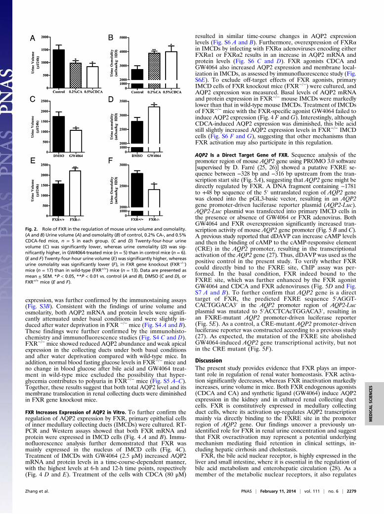
Effect of FXR on Urine Concentration. The kidney is a key organregulating water and salt metabolism. To test whether FXR isinvolved in regulation of urine concentration, cholic acid (CA)and chenodeoxycholic acid (CDCA), two endogenous agonistsfor FXR (2, 23), were used to treat C57BL/6 mice (0.2% CA and0.5% CDCA mixed in diet) for 5 d. Mice were housed in met-abolic cages for 24 h with free access to water. Treatment of micewith CA and CDCA resulted in a significant decrease in urinevolume and increase in urine osmolality (Fig. 2 A and B). Se-lective FXR agonist GW4064 treatment also resulted in a similarphenotype (Fig. 2 C and D). In contrast, FXR gene knockoutmice exhibited much more daily urine output and much lowerurine osmolality than wild-type mice. (Fig. 2 E and F). Thesefindings suggest that FXR may play an important role in regu-lation of urine concentration. The polyuria phenotype observedin FXR−/− mice seems to be the result of the defect in the kid-ney. Urine concentrating response to water restriction and des-mopressin (dDAVP) treatment was similar between FXR+/+ andFXR−/− mice (Fig. S2 A–C).
Effect of FXR on Renal AQP2 Expression. To explore the mechanismthrough which FXR influences the urine concentration, geneexpression profiles were examined by using the Affymetrix chip.After treatment of wild-type mice with CDCA for 5 d, expression
levels of a total of 242 genes were significantly changed (foldchange ≥1.5, P ≤ 0.05). Among them, 173 genes were up-regu-lated and 69 genes were down-regulated, with some of thesegenes involved in regulation of water homeostasis (Table S1). Asshown in Fig. 3A, CDCA treatment markedly up-regulatedAQP2 gene expression in the kidney. Real-time PCR analysisdemonstrated that renal AQP2 mRNA levels were significantlyhigher in CA- and CDCA-fed mice compared with the controlmice (Fig. 3B). As previously reported, glycosylation is importantfor cell surface expression of AQP2 (24). Both CA and CDCAtreatment resulted in a marked increase in total AQP2 proteinlevels, as well as the glycosylated (multiple bands with highmolecular weights) and nonglycosylated (a sharp band with lowmolecular weight) forms of AQP2 protein (Fig. 3C). Immuno-histochemical assay showed that AQP2 protein expression wasmainly increased in medullary collecting ducts, with little changein cortical collecting ducts of CDCA-fed mice (Fig. S3A).Treatment of mice with GW4064 also significantly increasedAQP2 expression levels both at mRNA and protein levels (Fig. 3D and E). In sharp contrast, FXR knockout mice exhibited sig-nificantly reduced AQP2 mRNA and protein abundance (Fig. 3F and G). Reduced AQP2 protein expression, especially apical
Fig. 1. Intrarenal localization of FXR in normal mice. (A) Mouse tissue dis-tribution of FXRα mRNA determined by real-time PCR. The results wereexpressed as relative expression levels after standardization by 18S. TheFXRα mRNA levels were normalized as the average of jejunum mRNA levels = 1.WAT, white adipose tissue; BAT, brown adipose tissue. n = 6. Data arepresented as mean ± SEM. (B) Real-time PCR analysis showing FXR mRNAlevels in renal cortex, outer medullar (OM), and inner medulla (IM). Data arepresented as mean ± SEM. *P < 0.05, **P < 0.01 vs. cortex. n = 9. (C) Westernblot assay demonstrating FXR protein levels in renal cortex, OM, and IM. (Dand E) Intrarenal localization of FXR protein in mouse cortex (D) and me-dulla (E) as assessed by immunofluorescence. Diffuse FXR immunoreactivitywas observed in renal cortex and medulla. FXR signals (arrowheads) weremainly located in the nuclei (red) of epithelial cells of the proximal tubules(PT), thick ascending limbs (TAL), distal convoluted tubules (DT), and col-lecting ducts (CD). Faint FXR signals were evident in the glomeruli (G). (D andE, Insets) Negative immunostaining with the IgG (without the primary an-tibody against FXR) in FXR wild-type mice and with the FXR antibody in FXRgene knockout mice, respectively. No specific signal indicates high specificityof the FXR antibody used in the present study.
2278 | www.pnas.org/cgi/doi/10.1073/pnas.1323977111 Zhang et al.
expression, was further confirmed by the immunostaining assays(Fig. S3B). Consistent with the findings of urine volume andosmolarity, both AQP2 mRNA and protein levels were signifi-cantly attenuated under basal conditions and were slightly in-duced after water deprivation in FXR −/− mice (Fig. S4 A and B).These findings were further confirmed by the immunohisto-chemistry and immunofluorescence studies (Fig. S4 C and D).FXR−/− mice showed reduced AQP2 abundance and weak apicalexpression in the collecting ducts under both basal conditionsand after water deprivation compared with wild-type mice. Inaddition, normal blood fasting glucose levels in FXR−/− mice andno change in blood glucose after bile acid and GW4064 treat-ment in wild-type mice excluded the possibility that hyper-glycemia contributes to polyuria in FXR−/− mice (Fig. S5 A–C).Together, these results suggest that both total AQP2 level and itsmembrane translocation in renal collecting ducts were diminishedin FXR gene knockout mice.
FXR Increases Expression of AQP2 in Vitro. To further confirm theregulation of AQP2 expression by FXR, primary epithelial cellsof inner medullary collecting ducts (IMCDs) were cultured. RT-PCR and Western assays showed that both FXR mRNA andprotein were expressed in IMCD cells (Fig. 4 A and B). Immu-nofluorescence analysis further demonstrated that FXR wasmainly expressed in the nucleus of IMCD cells (Fig. 4C).Treatment of IMCDs with GW4064 (2.5 μM) increased AQP2mRNA and protein levels in a time-course-dependent manner,with the highest levels at 6-h and 12-h time points, respectively(Fig. 4 D and E). Treatment of the cells with CDCA (80 μM)
resulted in similar time-course changes in AQP2 expressionlevels (Fig. S6 A and B). Furthermore, overexpression of FXRαin IMCDs by infecting with FXRα adenoviruses encoding eitherFXRα1 or FXRα2 results in an increase in AQP2 mRNA andprotein levels (Fig. S6 C and D). FXR agonists CDCA andGW4064 also increased AQP2 expression and membrane local-ization in IMCDs, as assessed by immunofluorescence study (Fig.S6E). To exclude off-target effects of FXR agonists, primaryIMCD cells of FXR knockout mice (FXR−/−) were cultured, andAQP2 expression was measured. Basal levels of AQP2 mRNAand protein expression in FXR−/− mouse IMCDs were markedlylower than that in wild-type mouse IMCDs. Treatment of IMCDsof FXR−/− mice with the FXR-specific agonist GW4064 failed toinduce AQP2 expression (Fig. 4 F andG). Interestingly, althoughCDCA-induced AQP2 expression was diminished, this bile acidstill slightly increased AQP2 expression levels in FXR−/− IMCDcells (Fig. S6 F and G), suggesting that other mechanisms thanFXR activation may also participate in this regulation.
AQP2 Is a Direct Target Gene of FXR. Sequence analysis of thepromoter region of mouse AQP2 gene using PROMO 3.0 software[supervised by D. Farré (25, 26)] showed a putative FXRE se-quence between −328 bp and −316 bp upstream from the tran-scription start site (Fig. 5A), suggesting that AQP2 gene might bedirectly regulated by FXR. A DNA fragment containing −1781to +48 bp sequence of the 5′ untranslated region of AQP2 genewas cloned into the pGL3-basic vector, resulting in an AQP2gene promoter-driven luciferase reporter plasmid (AQP2-Luc).AQP2-Luc plasmid was transfected into primary IMCD cells inthe presence or absence of GW4064 or FXR adenovirus. BothGW4064 and FXR overexpression significantly increased tran-scription activity of mouse AQP2 gene promoter (Fig. 5 B and C).A previous study reported that dDAVP can increase cAMP levelsand then the binding of cAMP to the cAMP-responsive element(CRE) in the AQP2 promoter, resulting in the transcriptionalactivation of the AQP2 gene (27). Thus, dDAVP was used as thepositive control in the present study. To verify whether FXRcould directly bind to the FXRE site, ChIP assay was per-formed. In the basal condition, FXR indeed bound to theFXRE site, which was further enhanced by the FXR agonistGW4064 and CDCA and FXR adenoviruses (Fig. 5D and Fig.S7 A and B). To further confirm that AQP2 gene is a directtarget of FXR, the predicted FXRE sequence 5′AGGT-CACTGGACA3′ in the AQP2 promoter region of AQP2-Lucplasmid was mutated to 5′ACCTCAcTGGACA3′, resulting inan FXRE-mutant AQP2 promoter-driven luciferase reporter(Fig. 5E). As a control, a CRE-mutant AQP2 promoter-drivenluciferase reporter was constructed according to a previous study(27). As expected, the mutation of the FXRE site abolishedGW4064-induced AQP2 gene transcriptional activity, but notin the CRE mutant (Fig. 5F).
DiscussionThe present study provides evidence that FXR plays an impor-tant role in regulation of renal water homeostasis. FXR activa-tion significantly decreases, whereas FXR inactivation markedlyincreases, urine volume in mice. Both FXR endogenous agonists(CDCA and CA) and synthetic ligand (GW4064) induce AQP2expression in the kidney and in cultured renal collecting ductcells. FXR is constitutively expressed in medullary collectingduct cells, where its activation up-regulates AQP2 transcriptionmainly via directly binding to the FXRE site in the promoterregion of AQP2 gene. Our findings uncover a previously un-identified role for FXR in renal urine concentration and suggestthat FXR overactivation may represent a potential underlyingmechanism mediating fluid retention in clinical settings, in-cluding hepatic cirrhosis and cholestasis.FXR, the bile acid nuclear receptor, is highly expressed in the
liver and small intestine, where it is essential in the regulation ofbile acid metabolism and enterohepatic circulation (28). As amember of the metabolic nuclear receptors, it also regulates
Fig. 2. Role of FXR in the regulation of mouse urine volume and osmolality.(A and B) Urine volume (A) and osmolality (B) of control, 0.2% CA-, and 0.5%CDCA-fed mice, n = 5 in each group. (C and D) Twenty-four-hour urinevolume (C) was significantly lower, whereas urine osmolality (D) was sig-nificantly higher, in GW4064-treated mice (n = 5) than in control mice (n = 6).(E and F) Twenty-four-hour urine volume (E) was significantly higher, whereasurine osmolality was significantly lower (F), in FXR gene knockout (FXR−/−)mice (n = 17) than in wild-type (FXR+/+) mice (n = 13). Data are presented asmean ± SEM. *P < 0.05, **P < 0.01 vs. control (A and B), DMSO (C and D), orFXR+/+ mice (E and F).
Zhang et al. PNAS | February 11, 2014 | vol. 111 | no. 6 | 2279
MED
ICALSC
IENCE
S

expression of many target genes relevant for lipid and glucosemetabolism at the transcription level (29–31). Increasing evi-dence suggests that FXR may play an important role in liverdetoxification, regeneration, and carcinogenesis (6, 32, 33). FXRhas also been found to be expressed in many “nonclassical” bilesalt target tissues, including the vasculature and macrophages,where FXR influences vascular tension and regulates the un-loading of cholesterol from foam cells, respectively (34). It hasbeen previously reported that FXR is expressed in the adrenalglands and that its activation by GW4064 modulates adrenalcholesterol metabolism and glucocorticoid synthesis (35). Thepresent study demonstrates that the kidney has constitutivelyhigh expression of FXR, with a ubiquitous expression patternalong renal tubules, suggesting that FXR may play an importantrole in renal physiology and pathophysiology. In support, recentstudies by Levi Moshe’s group have demonstrated that FXRmodulates renal lipid metabolism and is involved in the path-ogenesis of diabetic nephropathy and represents an attractivetherapeutic target for this disease (16, 36).The present study provides evidence that FXR plays an im-
portant role in urine concentration. Treatment with endogenousFXR agonists (CA and CDCA) and synthetic FXR agonistGW4064 significantly decreased urine volume and increasedurine osmolality in mice, indicative of enhanced urine concen-tration. In contrast, FXR gene deficiency mice exhibited increasedurine output with reduced osmolality, suggesting an attenuated
urine concentrating ability. Microarray analysis found that a groupof genes involved in controlling water reabsorption was up-regulated, especially the aquaporin 2 water channel. Regulationof AQP2 expression by FXR was further confirmed by decreasedAQP2 expression in FXR gene-deleted mice and increasedAQP2 expression in FXR agonist-treated mice, as assessed byreal-time PCR, Western blot, and immunohistochemistry assays.Collectively these findings indicate that FXR plays an importantrole in renal water homeostasis, possibly through regulating theexpression of AQP2 in renal collecting ducts.It was previously reported that the nuclear receptor liver X
receptors (LXRs), which have a “yin-yang” relationship with FXRin regulation of bile acid metabolism, also participate in water ho-meostasis regulation. LXRβ deficiency results in central diabetesinsipidus and impaired renal proximal tubule aquaporin-1 expres-sion in mice (37). This finding suggests that LXR and FXR may actin concert in maintaining renal water homeostasis. In addition, FXRhas been shown to play an important role in glucose metabolism.Although FXR−/− mice exhibit insulin resistance, there was nodifference in fasting blood glucose levels between wild-type andFXR−/− mice (16). Therefore, it seems unlikely that hyperglycemiais responsible for polyuria in FXR−/− mice.The present study also identifies AQP2 as a direct target gene
of FXR. The kidney is the key organ in maintenance of waterhomeostasis. Normally, 99% of water in urine filtered by glo-meruli is reabsorbed by renal tubules and collecting ducts.Among them, 70% is reabsorbed by the proximal tubules, 15%by the loops of Henle, and the rest by the distal tubules andcollecting ducts (38). The collecting ducts play a determiningrole in final urine volume, depending on AQP2 expression andlocalization. Collecting duct AQP2 abundance is largely regu-lated by the AVP/V2 receptor system and to a less extent byprostaglandins and the transcription factors nuclear factor kappaB and tonicity-responsive enhancer binding protein (39). Thepresent study has uncovered a unique mechanism by whichAQP2 is regulated by FXR, independent of AVP and otherknown factors. Mouse AQP2 promoter contains a typical FXREsequence and can be bound to FXR and activated by FXRagonists, resulting in a significant increase in AQP2 gene tran-scription. The identification of AQP2 as a direct target gene ofFXR may not only help to understand the molecular mecha-nisms involved in maintaining normal urine concentrating ca-pacity but also provide a possible explanation for increased fluidretention in clinical settings with elevated circulating bile acidlevels, such as the hepatorenal syndrome and cholestasis. On thebasis of our findings, we anticipate that in the state of severe liverdamage elevated plasma bile acids can activate FXR in renalcollecting ducts, resulting in impaired urine excretion through anFXR/AQP2-dependent mechanism.It is interesting to note that, in cultured primary medullary
collecting duct cells of FXR gene knockout mice, endogenousFXR agonist CDCA still slightly increased the transcription ofAQP2 gene, indicating that other mechanisms may also be in-volved. It has been previously reported that in addition to the nu-clear receptor FXR, bile acids can also activate a G protein-coupledreceptor TGR5 and exert their biological effects through a fewnongenomic signaling cascades (40). It is also known that othernuclear receptors, like pregnane X receptor, vitamin D receptor,and constitutive androstane receptor, may respond to bile acids,albeit to a more restricted set of bile acid species (41).Taken together, our study demonstrates that FXR is consti-
tutively expressed in the epithelial cells along renal tubules, espe-cially renal collecting ducts. FXR activation enhances, whereasFXR gene deficiency lowers, urine concentrating capacity. FXRcan directly bind to the FXRE site in the promoter region of theAQP2 gene, thereby increasing its gene expression in the col-lecting duct cells. These findings not only uncover a uniquemechanism in the maintenance of renal water homeostasis butmay also help to understand the pathogenesis of fluid retentionfrequently occurring in diseases like the hepatorenal syndromeand cholestasis.
Fig. 3. Effect of FXR on AQP2 expression in mouse kidney. (A) Heat mapvisualization of changes of four major aquaporins in the kidneys of CDCA-fed mice. AQP2 was the one with the most significantly induction. Foldchange of each gene symbol was visualized by red–green color scale: greenfor down-regulation, black for insignificant change, and red for up-regula-tion. (B and C) Both mRNA (B) and protein (C) levels of AQP2 were increasedin the kidneys of 0.2% CA- and 0.5% CDCA-fed mice. (D and E) Treatment ofmice with a specific FXR agonist, GW4064, via i.p. injection for 3 d up-regulatedAQP2 expression at both mRNA (D) and protein (E ) levels in the kidneys.(F and G) FXR gene-deficient mice exhibited diminished AQP2 expressionat both mRNA (F) and protein (G) levels. Data are presented as mean ± SEM.*P < 0.05, **P < 0.01 vs. control (A and B), DMSO (C and D) or FXR +/+ mice(E and F). β-Actin was used as an internal control.
2280 | www.pnas.org/cgi/doi/10.1073/pnas.1323977111 Zhang et al.

Materials and MethodsAnimals. All experiments were reviewed and approved by the Animal Careand Use Review Committee of Peking University Health Science Center. FXRknockout mice, purchased from the Jackson Laboratory, were maintained onstandardmouse chow and housed on a 12-h light/black cycle under controlledtemperature (22–24 °C) and humidity (50–65%) in the animal facility ofPeking University Health Science Center. Experiments were performed withmale wild-type and FXR knockout mice, aged 4–6 mo.
Bile Acid and GW4064 Treatment. Fifteen male mice (12 wk old) were dividedinto three groups randomly: one was placed on a control diet (n = 5), theother two on the same diet supplemented with 0.2% CA (n = 5) or 0.5%CDCA (n = 5), respectively. Five days later, 24-h urine samples were col-lected, and then the mice were killed. For GW4064 treatment, 11 malemice (12 wk old) were divided into two groups randomly. The mice inboth groups received i.p. injection once daily for 3 d. One group of micewas injected with DMSO as vehicle (n = 6). The other group was injectedwith 30 mg/kg GW4064 dissolved in DMSO (n = 5). Three days later,24-h urine samples and the kidneys of the mice were collected forfurther analysis.
Urine Collection and Osmolality Analysis. Mice were housed in individualmetabolic cages (Tecniplast) with free access to water and food for collectionof 24-h urine. Body weight, urine excretion, and water consumption weremeasured. Mice were placed in individual metabolic cages for 24 h beforemeasurement. Urine samples were centrifuged at 3,000 × g at 4 °C for 5 min,
and the supernatants were saved for osmolality analysis using a freezingpoint depression osmometer (Micro-Osmometer 3300).
Construction of Mouse AQP2-Luc. The DNA sequence of mouse AQP2 promoterregion of 2,000 bp upstream of the transcription start site was analyzed usingpromoter 3.0 (http://alggen.lsi.upc.es/cgi-bin/promo_v3/promo/promoinit.cgi?dirDB=TF_8.3). Mouse AQP2 promoter fragment between −1781 and+48 was amplified by PCR using mouse kidney genomic DNA as templateand cloned in pGL3-luciferase reporter vector (Promega). The forwardprimer 5′-GCC CAA TCT TGA TAC-3′ and the reverse primer 5′-TCT TCCTCC CTC CCT CTC TCT-3′ were used for the PCR. Site-directed mutagen-esis of the FXRE-like site at −327 and −326 and CREB site at −223 and −222 wasaccomplished using the Fast Mutagenesis System (FM111; TransGen Biotech)by using FXRE mutagenic primers (forward primer: 5′-CAG CCT TTT AGT CAAAGAGAA CCT CAC TGGACA-3′; FXREmutagenic reverse primer: 5′- GGT TCT CTTTGA CTA AAA GGC TGG CCA AGG AAG-3′) and CRE mutagenic primers (forwardprimer: 5′- AAC GAG GAA AAC AGA GTG GTC AAT CCT TAT-3′; CRE mutagenicreverse primer: 5′- CAC TCT GTT TTC CTC GTT TTT TCC TCA GTT-3′). The clonedmouse AQP2 promoter fragments were confirmed by DNA sequencing.
ChIP Assay. ChIP assays were performed using a ChIP assay kit (CA92590;Millipore) according to the manufacturer’s instructions. Soluble chromatin
Fig. 4. FXR activation with GW4064 induced AQP2 expression in culturedprimary IMCD cells. (A) RT-PCR analysis of FXRα and FXRβ mRNA expressionin three preparations of cultured primary IMCD cells. (B) Western blot assayshowing FXR protein expression in the nuclei of primary IMCD cells. (C )Nuclear localization of FXR (red) and AQP2 (green) in a primary IMCD cell.The nucleus was visualized by staining the cell with DAPI (blue). (D and E)Primary IMCD cells were treated with 2.5 μM GW4064 for the indicated timepoints. Both AQP2 mRNA (D) and protein (E) levels were induced by theGW4064 treatment. (F and G) GW4064 treatment for 6 h failed to induceAQP2 expression at both mRNA (F) and protein (G) levels in primary IMCDcells isolated from FXR− /− mice. Data are presented as mean ± SEM. *P <0.05, **P < 0.01 vs. DMSO (D, n = 3) and FXR+ /+ mice (F, n = 4).
Fig. 5. FXR activation increased AQP2 transcription via the transactivationmechanism. (A) DNA sequence analysis revealed a potential FXRE-like sitewithin the proximal promoter region of mouse AQP2 gene located between−328 bp and −316 bp upstream from the transcription start site. (B) GW4064(n = 8) treatment for 6 h significantly increased the luciferase activity of themouse AQP2-Luc reporter in primary IMCD cells. dDAVP (1 nM, n = 8) 0.**P < 0.01, ***P < 0.001 vs. DMSO (n = 6). (C) FXR overexpression signifi-cantly increased the AQP2-luc reporter activity. Cells were infected with Ad-FXRα1 or Ad-FXRα2 for 36 h. Adenovirus carrying cDNA encoding control VP16alone (Ad-VP16) was used as the control for AdFXRα1 (n = 11) or AdFXRα2 (n =11). ***P < 0.001 vs. Ad-VP16 (n = 9). (D) ChIP assay revealed that FXR can bindto the FXRE-like site in mouse AQP2 promoter. GW4064 could enhance thebinding of FXR to the FXRE-like site. Input, positive control; FXR, anti-FXRantibody precipitated DNA; IgG, IgG precipitated DNA as negative control.(E) Schematic structure of the AQP2-Luc reporter. Site-directed mutagenesisof the putative FXRE from AGGTCACTGGACA to ACCTCACTGGACA, andCRE from GACGTCA to GTGGTCA was shown. (F ) Mutation of the puta-tive FXRE, but not the CRE, completely abolished the stimulatory effectof GW4064 on the luciferase activity of the AQP2-Luc reporter in IMCDs.**P < 0.01, ***P < 0.001 vs. DMSO, n = 8. Data are presented as mean ± SEM.
Zhang et al. PNAS | February 11, 2014 | vol. 111 | no. 6 | 2281
MED
ICALSC
IENCE
S

was prepared from primary IMCD cells treated with 80 μmol/L CDCA or 2.5μmol/L GW4064 for 6 h. Chromatin was immunoprecipitated with antibodies(2 μg) directed against FXR (sc13063; Santa Cruz Biotechnology). Final DNAextractions were sequenced at −395 to −137 in the AQP2 promoter, and theprimers used were as follows: forward 5′-GCC TAT CAC CCC ATC TTA GCT-3′;antisense 5′-CCC ACA TTT CCT CAC AGT T-3′.
Chemicals and reagents, methods of real-time PCR, Western blot, immu-nostaining, microarray, dDAVP treatment, blood glucose measurement,primary culture of mouse IMCD cells, and luciferase assay are described in SIMaterials and Methods.
Statistical Analysis. The data were analyzed using the Prism software package(GraphPad Software). Data are presented as mean ± SEM. A two-sidedStudent t test was used to analyze individual differences. A value of P < 0.05was considered to be statistically significant.
ACKNOWLEDGMENTS. This work was supported by Ministry of Science andTechnology Grant 2012CB517504/2010C8912500/2011ZX09102-011-12 (toY.G.) and Natural Science Foundation Grants 81030003/81270275 (to Y.G.)and 81200511 (to X.Z.). J.-Å.G. is supported by the Swedish Research Counciland by Robert A. Welch Foundation Grant E-0004.
1. Laffitte BA, et al. (2000) Identification of the DNA binding specificity and potentialtarget genes for the farnesoid X-activated receptor. J Biol Chem 275(14):10638–10647.
2. Makishima M, et al. (1999) Identification of a nuclear receptor for bile acids. Science284(5418):1362–1365.
3. Gustafsson JA (1999) Seeking ligands for lonely orphan receptors. Science 284(5418):1285–1286.
4. Zhang Y, Kast-Woelbern HR, Edwards PA (2003) Natural structural variants of thenuclear receptor farnesoid X receptor affect transcriptional activation. J Biol Chem278(1):104–110.
5. Huber RM, et al. (2002) Generation of multiple farnesoid-X-receptor isoforms throughthe use of alternative promoters. Gene 290(1-2):35–43.
6. Wang YD, Chen WD, Moore DD, Huang W (2008) FXR: A metabolic regulator and cellprotector. Cell Res 18(11):1087–1095.
7. Ananthanarayanan M, Balasubramanian N, Makishima M, Mangelsdorf DJ, Suchy FJ(2001) Human bile salt export pump promoter is transactivated by the farnesoid Xreceptor/bile acid receptor. J Biol Chem 276(31):28857–28865.
8. Chiang JY, Kimmel R, Weinberger C, Stroup D (2000) Farnesoid X receptor responds tobile acids and represses cholesterol 7alpha-hydroxylase gene (CYP7A1) transcription.J Biol Chem 275(15):10918–10924.
9. Bishop-Bailey D, Walsh DT, Warner TD (2004) Expression and activation of the far-nesoid X receptor in the vasculature. Proc Natl Acad Sci USA 101(10):3668–3673.
10. Lee H, et al. (2006) FXR regulates organic solute transporters alpha and beta in theadrenal gland, kidney, and intestine. J Lipid Res 47(1):201–214.
11. Sinal CJ, et al. (2000) Targeted disruption of the nuclear receptor FXR/BAR impairs bileacid and lipid homeostasis. Cell 102(6):731–744.
12. Ma K, Saha PK, Chan L, Moore DD (2006) Farnesoid X receptor is essential for normalglucose homeostasis. J Clin Invest 116(4):1102–1109.
13. Lanz RB, et al. (2006) Nuclear Receptor Signaling Atlas (www.nursa.org): Hyperlinkingthe nuclear receptor signaling community. Nucleic Acids Res 34(Database issue):D221–D226.
14. Jiang T, et al. (2007) Farnesoid X receptor modulates renal lipid metabolism, fibrosis,and diabetic nephropathy. Diabetes 56(10):2485–2493.
15. Proctor G, et al. (2006) Regulation of renal fatty acid and cholesterol metabolism,inflammation, and fibrosis in Akita and OVE26 mice with type 1 diabetes. Diabetes55(9):2502–2509.
16. Wang XX, et al. (2010) Diabetic nephropathy is accelerated by farnesoid X receptordeficiency and inhibited by farnesoid X receptor activation in a type 1 diabetesmodel. Diabetes 59(11):2916–2927.
17. Noda Y, Sohara E, Ohta E, Sasaki S (2010) Aquaporins in kidney pathophysiology. NatRev Nephrol 6(3):168–178.
18. Hasler U, Leroy V, Martin PY, Féraille E (2009) Aquaporin-2 abundance in the renalcollecting duct: New insights from cultured cell models. Am J Physiol Renal Physiol297(1):F10–F18.
19. Setchell KD, Matsui A (1983) Serum bile acid analysis. Clin Chim Acta 127(1):1–17.20. Jonassen TE, Nielsen S, Christensen S, Petersen JS (1998) Decreased vasopressin-
mediated renal water reabsorption in rats with compensated liver cirrhosis. Am JPhysiol 275(2 Pt 2):F216–F225.
21. Ginès A, et al. (1993) Incidence, predictive factors, and prognosis of the hepatorenal
syndrome in cirrhosis with ascites. Gastroenterology 105(1):229–236.22. Tain Y-L, Hsieh C-S, Chen C-C, Huang L-T (2008) Toward nitric oxide deficiency in
hepatorenal syndrome: Is farnesoid X receptor the link? Biosci Hypotheses 1(3):
145–146.23. Wang H, Chen J, Hollister K, Sowers LC, Forman BM (1999) Endogenous bile acids are
ligands for the nuclear receptor FXR/BAR. Mol Cell 3(5):543–553.24. Baumgarten R, Van De Pol MH, Wetzels JF, Van Os CH, Deen PM (1998) Glycosylation
is not essential for vasopressin-dependent routing of aquaporin-2 in transfected
Madin-Darby canine kidney cells. J Am Soc Nephrol 9(9):1553–1559.25. Farré D, et al. (2003) Identification of patterns in biological sequences at the ALGGEN
server: PROMO and MALGEN. Nucleic Acids Res 31(13):3651–3653.26. Messeguer X, et al. (2002) PROMO: Detection of known transcription regulatory el-
ements using species-tailored searches. Bioinformatics 18(2):333–334.27. Matsumura Y, Uchida S, Rai T, Sasaki S, Marumo F (1997) Transcriptional regulation of
aquaporin-2 water channel gene by cAMP. J Am Soc Nephrol 8(6):861–867.28. Inagaki T, et al. (2005) Fibroblast growth factor 15 functions as an enterohepatic
signal to regulate bile acid homeostasis. Cell Metab 2(4):217–225.29. Han SI, et al. (2004) Bile acids enhance the activity of the insulin receptor and gly-
cogen synthase in primary rodent hepatocytes. Hepatology 39(2):456–463.30. Thomas C, Pellicciari R, Pruzanski M, Auwerx J, Schoonjans K (2008) Targeting bile-
acid signalling for metabolic diseases. Nat Rev Drug Discov 7(8):678–693.31. Watanabe M, et al. (2004) Bile acids lower triglyceride levels via a pathway involving
FXR, SHP, and SREBP-1c. J Clin Invest 113(10):1408–1418.32. Huang W, et al. (2006) Nuclear receptor-dependent bile acid signaling is required for
normal liver regeneration. Science 312(5771):233–236.33. Wang C, et al. (2013) PARP1 promotes oxidative stress-induced liver cell death via
suppressing FXR. Mol Cell Biol 33(22):4492–503.34. Hageman J, Herrema H, Groen AK, Kuipers F (2010) A role of the bile salt receptor
FXR in atherosclerosis. Arterioscler Thromb Vasc Biol 30(8):1519–1528.35. Hoekstra M, et al. (2012) FXR agonist GW4064 increases plasma glucocorticoid levels
in C57BL/6 mice. Mol Cell Endocrinol 362(1-2):69–75.36. Wang XX, et al. (2009) The farnesoid X receptor modulates renal lipid metabolism
and diet-induced renal inflammation, fibrosis, and proteinuria. Am J Physiol Renal
Physiol 297(6):F1587–F1596.37. Gabbi C, et al. (2012) Central diabetes insipidus associated with impaired renal
aquaporin-1 expression in mice lacking liver X receptor β. Proc Natl Acad Sci USA
109(8):3030–3034.38. O’Callaghan CA (2009) The Renal System at a Glance (Wiley-Blackwell, Chichester,
UK), 3rd Ed.39. Hasler U, et al. (2008) NF-kappaB modulates aquaporin-2 transcription in renal col-
lecting duct principal cells. J Biol Chem 283(42):28095–28105.40. Watanabe M, et al. (2006) Bile acids induce energy expenditure by promoting in-
tracellular thyroid hormone activation. Nature 439(7075):484–489.41. Schaap FG, Trauner M, Jansen PL (2013) Bile acid receptors as targets for drug de-
velopment. Nat Rev Gastroenterol Hepatol, 10.1038/nrgastro.2013.151.
2282 | www.pnas.org/cgi/doi/10.1073/pnas.1323977111 Zhang et al.