f h ulq d ' ls wh ud / d x [ d q llg d h · 5 h y lv lr q r i wk h $ iur wur s lf d o 6 s h f...
TRANSCRIPT

Revision of the Afrotropical Species of Parapachycerina(Diptera: Lauxaniidae)
Authors: Davies, Gregory B. P., and Miller, Raymond M.
Source: African Invertebrates, 49(2) : 131-158
Published By: KwaZulu-Natal Museum
URL: https://doi.org/10.5733/afin.049.0208
BioOne Complete (complete.BioOne.org) is a full-text database of 200 subscribed and open-access titlesin the biological, ecological, and environmental sciences published by nonprofit societies, associations,museums, institutions, and presses.
Your use of this PDF, the BioOne Complete website, and all posted and associated content indicates youracceptance of BioOne’s Terms of Use, available at www.bioone.org/terms-of-use.
Usage of BioOne Complete content is strictly limited to personal, educational, and non - commercial use.Commercial inquiries or rights and permissions requests should be directed to the individual publisher ascopyright holder.
BioOne sees sustainable scholarly publishing as an inherently collaborative enterprise connecting authors, nonprofitpublishers, academic institutions, research libraries, and research funders in the common goal of maximizing access tocritical research.
Downloaded From: https://bioone.org/journals/African-Invertebrates on 04 Feb 2020Terms of Use: https://bioone.org/terms-of-use

African Invertebrates Vol. 49 (2) Pages 131–158 Pietermaritzburg December, 2008
http://www.africaninvertebrates.org.za
Revision of the Afrotropical species of Parapachycerina(Diptera: Lauxaniidae)
Gregory B. P. Davies1 and Raymond M. Miller2
1Natal Museum, P. Bag 9070, Pietermaritzburg, 3200 South Africa; [email protected] of Biological & Conservation Sciences, University of KwaZulu-Natal, P. Bag X01, Scottsville,
Pietermaritzburg, 3209 South Africa; [email protected]
ABSTRACT
Parapachycerina Stuckenberg, a genus of small, usually yellow-orange sapromyziform lauxaniids (Diptera:Lauxaniidae) restricted to the Old World, is redescribed. The African and Malagasy representatives arerevised, with four new species described: P. bispina sp. n., P. infuscata sp. n., P. lalitra sp. n. and P. talea sp.n. There are now five Afrotropical Parapachycerina species, including the type species, P. munroiStuckenberg, 1971. Further, the paper provides an identification key to the Afrotropical species, a summaryof the available biological information, and preliminary remarks on the phylogenetic relationships of thegenus.
KEY WORDS: Diptera, Lauxaniidae, Parapachycerina, Neogeomyza, Neopachycerina, Tanyura, taxonomy,identification key.
INTRODUCTION
The Afrotropical lauxaniid fauna comprises approximately 28 genera and at least 90described species (Stuckenberg 1971; Miller 1980). Lauxaniids are generally smallacalyptrate flies, which can be identified in the vast Afrotropical acalyptrate assemblageby, inter alia, their (a) lack of vibrissae, (b) two reclinate fronto-orbital bristles, (c) de-cussate (cruciate) postvertical bristles, (d) complete subcosta, (e) lack of costal breaksand (f) dorsal pre-apical tibial bristles.
The lauxaniid fauna in sub-Saharan Africa, as elsewhere in the world, is foundprincipally in forest (e.g. Homoneura van der Wulp, Cainohomoneura Stuckenberg,Prosamyza Stuckenberg), moist savannah and grassland (e.g. Chaetolauxania Kertész,Diplochasma Knab), and shows low species richness and diversity in arid regions (e.g.<2% of lauxaniid specimen lots in the Natal Museum Diptera Collection originate fromthe drier, western provinces of South Africa, viz. Northern Cape, North-West and FreeState).
Despite the widespread occurrence and diversity of lauxaniids in sub-Saharan Africa,little research has been published on the Afrotropical representatives in recent years. Inthis paper we revise Parapachycerina. The genus was erected by Stuckenberg (1971:539) and, to date, only three species are recognized in the published literature (Shewell1977; Miller 1980; Evenhuis & Okadome 1989): the type species, P. munroi Stuckenberg,1971 and two Oriental species (both originally described as Lauxania species), P. hirsuti-seta (de Meijere, 1910) and P. cuneifera (Kertész, 1913).
In his major review of Old World lauxaniids, Stuckenberg (1971: 588) indicated that‘there are a number of species of Parapachycerina in tropical Africa, all undescribed,and these will be dealt with fully in a forthcoming monograph’. Later events precludedthe production of this monograph, and it is the task of this paper to describe these newAfrican and Malagasy Parapachycerina species.
The four goals of this paper are: (1) the description of new Afrotropical (includingMalagasy) species; (2) the provision of an updated dichotomous key to the Afrotropical
Downloaded From: https://bioone.org/journals/African-Invertebrates on 04 Feb 2020Terms of Use: https://bioone.org/terms-of-use

132 AFRICAN INVERTEBRATES, VOL. 49 (2), 2008
species; (3) the description and illustration of the salient morphological features of thegenus (except female postabdomen); and (4) a preliminary comparison with putativelyrelated genera.
MATERIAL AND METHODS
Terminology generally follows Stuckenberg (1971) and Kim (1994), including theirpostabdominal terminology. Departures from their terminology are discussed hereunder.Antennal terminology is according to Stuckenberg (1999). A rather bewildering arrayof terms has been applied to cuticular processes (chaetation) in dipterology. In thispaper, chaetotaxy largely follows Miller (1977a: 153), although the term ‘setulae’ isused more broadly here also to refer to very short, fine processes with alveoli. Althoughlauxaniidologists have used the term ‘costal spinules’ (e.g. Shewell 1987; Kim 1994),these processes are socketed (alveolus-derived) and are thus setulae rather than spinules,which are immovable, socket-less cuticular processes (‘microtrichia’ in D.K. McAlpine’s(1973) terminology). Oddities also emerge when some standard chaetae change theirlength and shape; for example, in Parapachycerina, the anterior fronto-orbital bristle isa tiny, fine process and, structurally speaking, is not a bristle, which is defined in Miller(1977a) as a distinctly long, larger, more robust seta. Pro tempore, we continue to usesuch terms despite the inherent contradictions. Proboscidal terminology followsBroadhead (1984); specifically she identified remarkable serrated lamella-like modifi-cations of the pseudotracheal canals as ‘scoops’ and ‘prongs’, and a hamate ‘beak’ at‘the anterior tip of the labial gutter’ (Broadhead 1984: 646). Contra most dipterologicalpapers, the ‘face’ is here termed the ‘prefrons’, a well-known synonym (e.g. D.K.McAlpine 1973: 17; J.F. McAlpine 1981: 10). The term face, although traditionally andwidely-used in dipterology, is too general and open to misunderstanding by non-dipterists; its use in the discipline should be discontinued. The area usually called the‘frons’ in dipterology (i.e. the area above the antennal sockets and below the vertex) ishere termed the ‘postfrons’. Following the arguments of D.K. McAlpine (2007: 32),the bristles usually termed ‘postocellar bristles’ (e.g. by most lauxaniid researchers) arehere called ‘postvertical bristles’. Following Stuckenberg (1971: figs 1, 2) the term‘sapromyziform costa’ is used to indicate that the stubby costal setulae (as opposed tothe fine costal setae) diminish in size and terminate before reaching R
4+5 (vein 3), in
contrast to the ‘homoneuriform costa’ (where the costal setulae reach R4+5
). Measure-ments are in millimetres (mm) and averages are cited with ranges in parentheses (exceptwhere there is no variability in measurements). Standard abbreviations for abdominalsclerites are used (e.g. T3 for tergite 3, S3 for sternite 3). Scanning electron microscope/microscopy is abbreviated as SEM. Male terminalia were removed from relaxedspecimens, and treated for approximately 12–24 hours in KOH, before being transferredto acetic acid for approximately 30 minutes and finally water for another 30 minutes.Cleared terminalia were mounted in glycerine on microscope slides.
At times in this paper we indulge in some speculative narrative, an approach considereda scientific solecism by most researchers; see Sand-Jensen (2007) for rebuttal on thisissue.Material originated from the following institutions:
BMNH – Natural History Museum, London, UK;CASC – California Academy of Sciences, San Francisco, USA;
Downloaded From: https://bioone.org/journals/African-Invertebrates on 04 Feb 2020Terms of Use: https://bioone.org/terms-of-use

DAVIES & MILLER: REVISION OF AFROTROPICAL PARAPACHYCERINA 133
HNHM – Hungarian Natural History Museum, Budapest, Hungary;MHNB – Musée d’Histoire Naturelle de Bâle (Basel), Basel, Switzerland;MNHN – Muséum National d’Histoire Naturelle, Paris, France;MRAC – Musée Royal de l’Afrique centrale, Tervuren, Belgium;NMSA – Natal Museum, Pietermaritzburg, South Africa;NMWC – National Museum and Gallery of Wales, Cardiff, UK;UZMD – Zoological Museum, Copenhagen, Denmark;ZMLU – Zoological Museum, Lund University, Sweden.
The type series of both Parapachycerina munroi Stuckenberg, 1971 (NMSA) andNeogeomyza (= Micropachycerina) stenoptera (Stuckenberg, 1971) (NMSA), and twospecimens from the type series of Neopachycerina aristata Malloch, 1933 (BMNH),were all examined for this study.
TAXONOMY
Parapachycerina Stuckenberg, 1971Parapachycerina Stuckenberg, 1971: 539. Type species: Parapachycerina munroi Stuckenberg, by original
designation.
Diagnosis:
Small (ca 2.5–5 mm), compact, usually yellow-orange sapromyziform lauxaniids withtwo reclinate fronto-orbital bristles placed far forwards and close together on postfrons;anterior fronto-orbital bristle small (Fig. 1). Postfrons devoid of setulae (at 50×). Aristaplumose; proximal rays longer than height of postpedicel. Postpedicel longer than high,slightly elongated. Ocellar triangle demarcated by dark brown or black spot. Ocellarbristles long, proclinate and subparallel. Prefrons (face) not bulging, without maculations.0+3 dorsocentral bristles on scutum. No intra-alar bristle. Ctenidium present in somespecies on profemur. Two ventral mesotibial spurs. Wings hyaline or slightly infuscated.Surstylus unusual; tiny, displaced medially as narrow lamella or flat blade. Immaturestages unknown.
In the Afrotropics, Parapachycerina most resembles Neogeomyza Séguy, 1938 (=Micropachycerina Stuckenberg, 1971) of East Africa and Madagascar (Miller 1980:606; see also McAlpine & de Keyzer 1994: 307). Neogeomyza differs from Para-pachycerina in having an even smaller anterior fronto-orbital (<0.2× length of posteriorfronto-orbital bristle), a larger black ocellar spot, a mildly tumid and translucent prefrons(face), 0+2 dorsocentral bristles, fewer setal rows on scutum, no acrostichal bristles,and an anal lobe in the wing (see Stuckenberg 1971: figs 33, 34). As a generalization,Neogeomyza is also a more gracile and delicate-looking fly than Parapachycerina.
Description:Colour and pruinescence: Head and thorax usually rich yellow-orange with orangemedial mesonotal vitta/e, but dorsal surface of thorax and parts of head strongly in-fuscated in P. infuscata. Dark brown or black ocellar spot always present. P. infuscatawith occiput mostly black, other species have occiput yellow-orange. Pruinescenceweak (when viewed at 50×), some silver pruinescence usually visible on anterolateralcorner of postfrons, parafacial, and dorsal surface of thorax (oblique lighting sometimesnecessary to see pruinosity). Aside from fronto-orbital bristles, postfrons devoid ofpruinosity and vestiture (when viewed at 50×), but when viewed with SEM (ca 500–
Downloaded From: https://bioone.org/journals/African-Invertebrates on 04 Feb 2020Terms of Use: https://bioone.org/terms-of-use

134 AFRICAN INVERTEBRATES, VOL. 49 (2), 2008
Fig. 1. Parapachycerina munroi Stuckenberg, type species of genus in lateral view. Length approximately5 mm.
Downloaded From: https://bioone.org/journals/African-Invertebrates on 04 Feb 2020Terms of Use: https://bioone.org/terms-of-use
DAVIES & MILLER: REVISION OF AFROTROPICAL PARAPACHYCERINA 135
2000×) postfrons with abundant closely-appressed carinae bearing spicules on medialpostfrontal surface, and prefrons with abundant spinules in irregular rows which arenot borne on carinae. Eye red or greyish; sparse, simple ommatrichia between facets(only visible with SEM). Maxillary palpus yellow to black. Supracervical area withdusting of silver pruinescence. Haltere yellow or light orange. Legs yellow. Abdomenorange-brown to black, depending on species. Teneral specimens are lightly sclerotizedand weakly pigmented, often with collapsed legs and heads.Head (Figs 2–9): Head higher than long, height:longitudinal length ratio 1.25:1. Headbroader than long, transverse width:longitudinal length ratio 1.6:1. Head broader thanhigh, transverse width:height ratio 1.5:1. Two reclinate fronto-orbital bristles, anteriorbristle much shorter (ca 0.4× length) than posterior bristle and not as recurved. Fronto-orbital bristles placed far forward on frons, and close together (distance between fronto-orbital bristles is shorter than distance from posterior fronto-orbital to inner verticalseta). Posterior fronto-orbital bristle strong, only slightly shorter than inner verticalseta (ca 0.8× length). Antenna positioned high on head relative to eye (opposite upperthird of eye). Arista plumose, rays longer on dorsal surface and progressively shorterdistally. Proximal rays of arista are longer than the height of the postpedicel. Postpedicellonger than high (1.6:0.9), tapering gently to moderately pointed apex, shape differingsubtly amongst species (most pointed in P. munroi). Pedicel ca 1/3 length of postpedicel,fan of ca 10 short, proclinate setulae distolaterally, single erect, lateroclinate seta dorsally,and two long, subparallel setae distoventrally (ca 0.7× length of postpedicel). Postfronsbroader than high (3:2), gently to moderately curved in lateral profile (not straight);prefrontal (facial)-postfrontal angle obtuse. Prefrons not bulging; prefrontal (facial)carina weak. Orbital plates usually concolorous with remainder of frons and not easilydifferentiated, except in P. infuscata, where orbital plates usually easily discerned. Inner
2 3
Figs 2, 3. (2) Dorsal view of head of Parapachycerina munroi Stuckenberg, (3) oblique lateral view of headof P. lalitra sp. n. Note small anterior fronto-orbital bristles (relative to posterior fronto-orbitalbristles), short distance separating fronto-orbital bristles, plumose aristae and long ocellar bristles.
Downloaded From: https://bioone.org/journals/African-Invertebrates on 04 Feb 2020Terms of Use: https://bioone.org/terms-of-use

136 AFRICAN INVERTEBRATES, VOL. 49 (2), 2008
vertical seta reclinate, and much longer than outer vertical seta (o.v.s. ca 0.4× length ofi.v.s.); o.v.s. lateroclinate. Ocellar triangle variable, sides may be equal (e.g. P. bispina),sides may be longer than base (e.g. P. infuscata) or base may be longer than sides (e.g.P. munroi). Ocellar setae strong, semi-erect, proclinate, subparallel and extending tooverhang pedicel; ocellar triangle with ca 3 inconspicuous, fugitive setulae behind ocellarsetae. Postvertical bristles decussate; intersection of bristles about half-way up thebristles. Postocular setulae in a single row of ca 10 setulae, beginning between o.v.s.and i.v.s.; first 2 or 3 setulae separate from others, slightly longer, inclinate and resemblingparavertical setulae. Occiput with 25–35 supracervical setulae. Occipital setulae sparse,restricted to ventrolateral corner of occiput, but extending up towards postocular setulaein some species (e.g. P. bispina), where forming weak second row behind postocularsetulae. Postgena with sparse, scattered setulae. Gena moderately expanded. Probosciswith labellar ‘beak’, and pseudotracheael ‘scoops’ and ‘prongs’ (but preparations crudeand further observations needed).
Thorax: 1 pair of acrostichal bristles (setae); 1 humeral (ca 10 weak setulae encirclingbristle on callus); 2 notopleurals; 1 presutural; 1 supra-alar; 2 postalars (1 or 2 setulaebetween bristles), ventral postalar bristle (i.e. bristle closest to the pleurotergite) isconsiderably (ca 2×) longer than the upper postalar; no intra-alar bristle; 0+3 dorsocentralbristles; setulae rows fairly numerous and rather irregular in pattern, ca 6 longitudinalrows between dorsocentral bristles, ca 8–10 transverse rows behind transverse suture;1 mesopleural bristle, placed near posterior margin, attended closely by ca 10 shortsetulae; 1 or 2 sternopleural bristles (if present, anterior bristle weaker and ca 0.7×
Figs 4–7. (4) Postfrons of Parapachycerina munroi Stuckenberg showing abundant carinae medially, (5)medial section of P. munroi postfrons showing spiculate carinae, (6) anterior view of P. munroihead, (7) supracervical setulae on occiput of P. munroi.
Downloaded From: https://bioone.org/journals/African-Invertebrates on 04 Feb 2020Terms of Use: https://bioone.org/terms-of-use
DAVIES & MILLER: REVISION OF AFROTROPICAL PARAPACHYCERINA 137
length of posterior one). Prosternum bare (at 50×). Propleural bristle present, but smalland inconspicuous. Posterior pair of bristles on scutellum decussate.
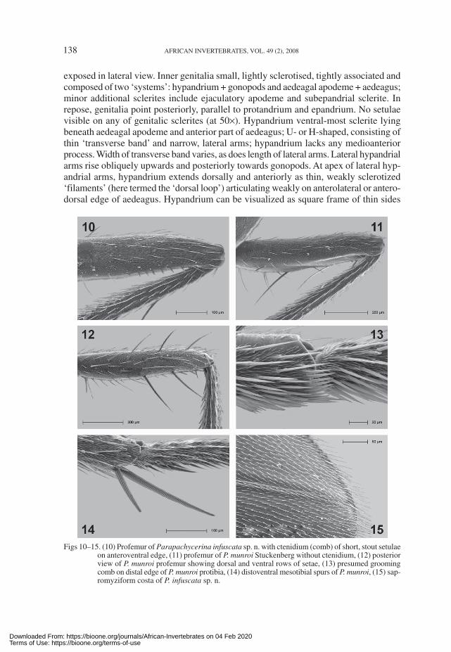
Legs (Figs 10–15): Procoxa has row of 4–6 setae (and a few scattered setulae) onanteroventral margin. Ctenidium on profemur variably present in some species; whenpresent, 4–9 short, stout setulae (rather resembling teeth on a saw). Profemur withposteroventral row of 2 or 3 long setae and posterodorsal row of ca 4 setae. Distoposterioredge of protibia with 2 short spurs, anterior spur ca 2× length of posterior spur. Small(grooming?) comb of ca 10 stout setulae on inner apex of protibia and metatibia (absent onmesotibia). All tibia with dorsal, pre-apical dorsal seta (most robust on mesotibia). Mesocoxahas fan of ca 6 setae (no setulae). Mesofemur with anteromedial row of ca 5 stout, proclinatesetae; mesofemur posteriorly largely bare. Mesotibia with 2 short, divergent, distoventralspurs (posterior spur 0.5× length of anterior spur). Metafemur with anterodorsal, preapical,stout, semi-erect seta. Two flattened spurs on inner face of metatibia, anterior one is longer.
Wing: Hyaline or weakly infuscated (in two species). Costa sapromyziform (stout costalsetulae terminating 3/4 way between R
2+3 and R
4+5). Costal chaetotaxy Type B2 sensu
Hackman & Väisänen (1985: 171). Subcosta and R1(Vein 1) closely associated, but
diverging somewhat when terminating on costal margin. Veins A2 and A
1+CuA
2 parallel,
A2 extending much further towards hind margin of wing (but still falling well short),
A1+CuA
2 shorter than A
2.
Abdomen: Chaetation pattern conservative: row of ca 10–15 flat, black setae on posteriormargins of syntergite 1+2, T3 and T4, on T5 and T6 these posterior rows become erect.T2 with lateral tufts of short setae. Abdomen tapering posteriorly, T6 slightly less thanhalf (transverse) width of syntergite 1+2.
Male terminalia (e.g. Figs 20–22 of type species; Stuckenberg 1971: figs 91, 92;Sasakawa 2003: fig. 2): Protandrium saddle-shaped (i.e. broad dorsally and narrowinglaterally), asetulose and continued ventrally as narrow band (i.e. protandrium is entire).Epandrium displays interspecific variation, but broader laterally than dorsally (cf.protandrium), usually elongated posteriorly as epandrial process (especially in P. bispinaand P. lalitra); sparse cover of scattered vestiture (ca 15 setulae laterally and ca 4strong setae on medio-posterior edge dorsally). Surstylus small, narrow, laterallycompressed, posteriorly hooked lamella or dorsoventrally flattened, blade-like process,fused to inner margin of epandrium; inconspicuous in lateral view, usually resemblingsmall finger projecting posteroventrally. Cerci separate, heavily setulose and partially
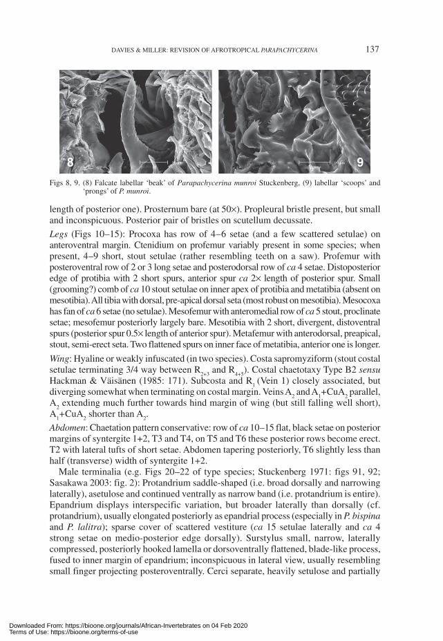
Figs 8, 9. (8) Falcate labellar ‘beak’ of Parapachycerina munroi Stuckenberg, (9) labellar ‘scoops’ and‘prongs’ of P. munroi.
Downloaded From: https://bioone.org/journals/African-Invertebrates on 04 Feb 2020Terms of Use: https://bioone.org/terms-of-use
138 AFRICAN INVERTEBRATES, VOL. 49 (2), 2008
exposed in lateral view. Inner genitalia small, lightly sclerotised, tightly associated andcomposed of two ‘systems’: hypandrium + gonopods and aedeagal apodeme + aedeagus;minor additional sclerites include ejaculatory apodeme and subepandrial sclerite. Inrepose, genitalia point posteriorly, parallel to protandrium and epandrium. No setulaevisible on any of genitalic sclerites (at 50×). Hypandrium ventral-most sclerite lyingbeneath aedeagal apodeme and anterior part of aedeagus; U- or H-shaped, consisting ofthin ‘transverse band’ and narrow, lateral arms; hypandrium lacks any medioanteriorprocess. Width of transverse band varies, as does length of lateral arms. Lateral hypandrialarms rise obliquely upwards and posteriorly towards gonopods. At apex of lateral hyp-andrial arms, hypandrium extends dorsally and anteriorly as thin, weakly sclerotized‘filaments’ (here termed the ‘dorsal loop’) articulating weakly on anterolateral or antero-dorsal edge of aedeagus. Hypandrium can be visualized as square frame of thin sides
Figs 10–15. (10) Profemur of Parapachycerina infuscata sp. n. with ctenidium (comb) of short, stout setulaeon anteroventral edge, (11) profemur of P. munroi Stuckenberg without ctenidium, (12) posteriorview of P. munroi profemur showing dorsal and ventral rows of setae, (13) presumed groomingcomb on distal edge of P. munroi protibia, (14) distoventral mesotibial spurs of P. munroi, (15) sap-romyziform costa of P. infuscata sp. n.
Downloaded From: https://bioone.org/journals/African-Invertebrates on 04 Feb 2020Terms of Use: https://bioone.org/terms-of-use

DAVIES & MILLER: REVISION OF AFROTROPICAL PARAPACHYCERINA 139
that has partially folded back on itself. Articulating with posterior apices of lateralhypandrial arms are sheath-like, ensiculate or gently rounded gonopods. Gonopodsusually closely ensheath apical part of aedeagus. Aedeagus best visualized as tubularstructure (‘aedeagal tube’) comprising two blade-like halves or ‘wings’ joined anteriorlyin narrow ‘dorsal arch’ (dorsal-most part of genitalia, cf. hypandrial transverse band);the two halves taper to pointed apices posteriorly where ensheathed by gonopods.Medially, aedeagus is membranous, but this section may be dissolved by strong KOHtreatment. Anteriorly, two halves of aedeagus may bulge out in anterolateral flanges.Aedeagal apodeme articulates posteriorly with aedeagus, and is fairly narrow, laterallycompressed, Y-shaped, ribbon- or rod-like sclerite, with two short posterior arms. Depen-ding on species, aedeagal apodeme may be longer than aedeagus, approximately equalin length, or slightly shorter. Ejaculatory apodeme is tiny sigmoidal or T-shaped scleritelying dorsally above bifurcation of aedeagal apodeme. Above posterior sections ofaedeagus is subepandrial sclerite, a subrectangular sclerite attached by membranoustissue to posteroventral edge of cercus.
Female terminalia: Unexceptional, short and blunt (not elongated); S9 subrectangularwith straight posterior edge and lightly setulose; subanal and supra-anal plates roughlysemi-circular; cerci dorsoventrally flattened. Inner genitalia not investigated. Stuckenberg(1971: 590) reported three subspherical spermathecae.
Key to Afrotropical Parapachycerina
1 Ctenidium (comb) present on anteroventral edge of profemur; ocellar spot brownor black ................................................................................................................. 2
– Ctenidium (comb) absent on anteroventral edge of profemur; ocellar spot black .... 6
2 With upper parts of occiput black ......................................................................... 3– Without black markings on occiput ...................................................................... 4
3 Dorsal surface of thorax dark chestnut brown to black, mesonotum with silverpruinosity; mesopleuron yellow to orangish brown, no black mark [widespread insub-Saharan Africa] ................................................................... P. infuscata sp. n.
– Dorsal surface of thorax orange with three black stripes, silver pruinosity weaklydeveloped; mesopleuron with large black patch, orange-yellow colouration restrictedto margins of mesopleuron [Comoros] ................................. Undescribed taxon C
4 Part of brown ocellar spot posteriorly with a short medial extension (rod); apex ofpostpedicel darkened, wings anteriorly fumose [Madagascar] ......... P. talea sp. n.
– Brown ocellar spot confined to ocellar triangle, apex of postpedicel not darkened;wings hyaline ........................................................................................................ 5
5 Gonopods long, as long as aedeagus; ventrolateral edge of aedeagus with mucroand small notches [Madagascar] ..................................................... P. lalitra sp. n.
– Gonopods short, only half as long as aedeagus; ventrolateral edge of aedeaguswithout mucro, and largely smooth [Madagascar] ............... Undescribed taxon A
6 Black ocellar spot extends backwards to clearly touch inner margins of postverticalsetal sockets; wing hyaline [southern and western Africa] ............................................................................................................................... P. munroi Stuckenberg
– Black ocellar spot confined to ocellar triangle; wing infuscated anteriorly ......... 7
Downloaded From: https://bioone.org/journals/African-Invertebrates on 04 Feb 2020Terms of Use: https://bioone.org/terms-of-use

140 AFRICAN INVERTEBRATES, VOL. 49 (2), 2008
7 Cross-veins of wing infuscated; ocellar triangle large and ocellar spot deep black[widespread in sub-Saharan Africa] ............................................. P. bispina sp. n.
– Cross-veins of wing not infuscated; ocellar triangle small and ocellar spot darkbrown [Madagascar] ............................................................. Undescribed taxon B
Parapachycerina bispina sp. n.Figs 17, 23
Etymology: From Latin bis (twice) and spina (thorn, spine, spike); refers to the twostout setulae on the apex of the surstylus, which are characteristic of the species.
Diagnosis:
P. bispina is broadly sympatric over much of Africa with P. munroi, and can be separatedfrom that species by: (1) the black ocellar spot not extending back to touch the postverticalsetae; (2) wing with light brown infuscations, shading the outer 3/4 of the costal cell,most of the marginal cell and anterior portion of the submarginal cell, anterior andposterior cross-veins (r–m; m–m) and also M
3+4 (vein 5) (P. bispina is the only Afrotro-
pical species in the genus with marked wings, apart from the Malagasy P. talea); (3) thepaired, black spines on apex of epandrial process; and (4) pale orange-yellow cerci(dark brown in P. munroi). P. bispina is also sympatric in areas with P. infuscata, but thedeep yellow-orange coloration, gently rounded vertex, black ocellar spot and absenceof a ctenidium distinguish P. bispina.
The wing illustration purportedly of P. munroi in Stuckenberg (1971: fig. 42) is infact P. bispina (as it shows infuscation of the cross-veins and anterior portions of thewing). This specimen is noted as a P. munroi paratype in the legend, but no specimenbearing these wing characteristics can be found in the P. munroi paratype series (NMSA),and evidently there was some mix up in the preparation of that figure.
Description:
Colour: Viewed with naked eye and under low magnification, impression is of uniformlyrich yellow-orange fly. Occiput yellow. Ocellar spot deep black. Scape, pedicel andpostpedicel all yellow, slight darkening at base of arista; pedicel may have orange tinge.Maxillary palpus yellow-orange. Thorax yellow-orange with two orange longitudinalstripes (vittae) on scutum (faint in some specimens), stripes are placed slightly mesadof dorsocentral bristles. Scutum sometimes shows darkening on humeral callus andalong border of notopleuron and mesopleuron. Postnotum and postscutellum yellow-orange. Scutellum yellow with broad orange medial stripe. Mesopleuron, pteropleuron,sternopleuron, pleurotergite and meropleuron all yellow-orange. Haltere pale yellow.Legs yellowish. Abdomen yellow-orange to dark brown (generally the former).
Head: Postfrons fairly strongly sloping, wider than long (3:2). Orbital plates weaklydifferentiated from surrounding postfrons. Postfrons ending in narrow lip over proximalportion of scape. Prefrons very weakly tumid. Ocellar triangle large and expanded(relative to other Afrotropical Parapachycerina species), anterior ocellus nearly reachinglevel of posterior fronto-orbital bristle; ocellar spot encloses all three ocelli.Thorax: Five or 6 longitudinal, rather irregular rows of setulae between dorsocentralbristle rows. Posterior pair of acrostichals ca 0.5× length of posteriad dorsocentralbristles. Anterior dorsocentral bristle ca 0.5× length of posterior dorsocentral; middle
Downloaded From: https://bioone.org/journals/African-Invertebrates on 04 Feb 2020Terms of Use: https://bioone.org/terms-of-use
DAVIES & MILLER: REVISION OF AFROTROPICAL PARAPACHYCERINA 141
dorsocentral intermediate in length. Weak, barely visible pruinosity on scutum. Twosternopleural bristles, posterior bristle more robust and longer (area around bristlesgenerally bare of setulae; ventrally usually a patch of ca 20 setulae). Pteropleuron,meropleuron, pleurotergite and prosternum bare of bristles and setulae.Legs: No ctenidium on profemur, in that area ca 8 widely-spaced, weak setulae. Profemurposteriorly with ventral row of ca 3 strong, widely-spaced setae and dorsal row of ca 6equally strong setae. 2 short spurs on posterior, distal edge of protibia, outer spur is ca2× length of inner spur. Procoxa has ventral row of ca 4 or 5 setae and few scatteredsetulae. Mesocoxa has fan of ca 6 setae (no setulae visible). Mesofemur strong, anterome-dial row of 5 robust, proclinate setae, posteriorly largely bare. Metacoxa largely bare,but 2 strong setae on opposite margins of coxa. Metafemur with unexceptional vestitureof ca 6 irregular rows of setulae on anterior face.Wings: Mostly hyaline, but distal 3/4 of costal cell, most of marginal cell and anteriorsection of submarginal cell, anterior and posterior cross-veins (r–m, m–m) and M
3+4(vein 5) light infuscated (in some specimens infuscation very pale and requiring carefuldiscrimination).Male terminalia (Figs 17, 23): Protandrium roughly ring-shaped in transverse view,having small ventral processes, in lateral view fairly narrow dorsally and tapering laterally(slightly less so than in type species, P. munroi). Epandrium moderately broad dorsally,expanding posterolaterally, before tapering to small, rounded epandrial extension, atapex of epandrial extension characteristic stout, paired, black setae, in extracted genitaliainclinate, interdigitate with opposing pair, lower seta of each pair is longer than upperseta (upper seta is ca 0.8× length of lower seta). Surstylus hidden in lateral view, fusedto inner medial margin of epandrium. Surstylus relatively large (in relation to aedeagus),flattened dorsoventrally, blade-like, tapering posteriorly to acute point and with mediola-teral mucro. Surstyli closely associated with aedeagal complex (could on initial examina-tion be considered to be gonopods). Cerci pale yellow. Hypandrium composed of broad‘transverse band’, short, weakly lateroclinate anterior arms and longer, inclinate posteriorarms that curve upward to terminate near base of gonopods and lateral edges of aedeagus(in ventral view, hypandrium resembles a ‘wobbly’ H). Hypandrial ‘transverse band’broader than ‘aedeagal tube’. Aedeagal apodeme approximately same length as aedeagus,bifurcating beneath ‘dorsal arch’ of aedeagus. Aedeagus composed of two halves(‘wings’) that taper posteriorly, halves meet anteriorly in ‘dorsal arch’; posteriorly each‘wing’ is bifurcate, subapically each half (wing) has ventrolateral mucro. Medially,aedeagus is membranous. Gonopods are blade-like, lightly sclerotised, broad basally,emarginate subbasally, widening medially and tapering to acute apex.Measurements: total length – ^(n=7) 3.6 (3.2–4.1), _ (n=2) 3.2 (2.9–3.5); head height– ^(n=7) 0.7 (0.6–0.8), _ (n=2) 0.7; head width – ^(n=5) 1.1, _ (n=1) 1; head length– ^(n=7) 0.5 (0.5–0.7), _ (n=2) 0.6, thorax length – ^(n=7) 1.4 (1.3–1.5), _ (n=2)1.4, wing length – ^(n=5) 3.3 (3.0–3.7), _ (n=2) 3.0; abdomen length – ^(n=7) 1.7(1.3–2.1), _ (n=2) 1.3.Holotype: _ SOUTH AFRICA: Limpopo: Kruger National Park, Pafuri, near Luvuvhu R., 22–23.iv.1981,R.M. Miller & P. Stabbins (NMSA).Other material examined: CAMEROON: 2^Baigom, Bamoun, no date (MNHN); 1_ Lake Barombi (nearKumba), 1939 (MNHN). DEMOCRATIC REPUBLIC OF CONGO: 1^Walikale (39 km S of), 25.xii.1957,E. Ross & R. Leech (CASC). GUINEA: 1_ Mount Nimba, M. Lamotte and R. Roy, vii–xii.1951 (MNHN).
Downloaded From: https://bioone.org/journals/African-Invertebrates on 04 Feb 2020Terms of Use: https://bioone.org/terms-of-use

142 AFRICAN INVERTEBRATES, VOL. 49 (2), 2008
IVORY COAST: 1^Lamto, Bandana, 22.xii.1971, D. Lachaise (MNHN). KENYA: 1_ Kakamega Forest,Isecheno Station, 22.vi.1995, Earthwatch Team (NMSA). NIGERIA: 1_ 1^Bauchi State, nr Tilden Fulani,Kogin Salla, 9.iii.1997, J.C. Deeming (NMWC). SOUTH AFRICA: Limpopo: 1_ 2^same data as holotype(NMSA). Mpumalanga: 2_ 1^Nelspruit (20.5 km S of), Noordkaap R., 1–2.xii.1976, R.M. Miller & P.Stabbins (NMSA). KwaZulu-Natal: 1^Pietermaritzburg, Ukulinga Research Farm, 13.viii.2007, R.M. Miller(NMSA); 1_ 1^same data except 22.viii.2007 (NMSA); 1^Pietermaritzburg, 10.x.1985, R.M. Miller(NMSA); 1^Ndumo Game Reserve, 1–10.xii.1963, B. & P. Stuckenberg (NMSA). TANZANIA: 1_1^Matengo Highlands, WSW of Songea, xii.1935–i.1936, Zerny (NMSA). UGANDA: 1^Budongo Forest,14.xi.1972, H. Gønget (UZMD); 1^Kampala, Tank Hill, 22.xii.1970, H. Gønget (UZMD); 4_ 1^SemlikiForest, 27.viii–3.xi.1952, Fletcher (BMNH); 1^Ankole, Kalinzu Forest, 6–15.ii.1973, H. Gønget (UZMD);1 (sex unknown) Segibwa Falls, 29.iii.1935, E.G. Gibbins (BMNH); 2^Mujenje, viii–ix.1913, K. Kitten-berger (HNHM).
Distribution: Throughout much of sub-Saharan Africa. From KwaZulu-Natal (SouthAfrica) in the south, northwards to Guinea in West Africa and Uganda in East Africa.
Remarks: 1_ 3^Amani, East Usambara Mts, 27.i.1977, H. Enghoff et al. (UZMD) arenotable for their expanded ocellar triangles and may potentially represent a new species.
Figs 16–19. Lateral views of outer _ terminalia: (16) P. munroi Stuckenberg, (17) P. bispina sp. n., (18) P.infuscata sp. n., (19) P. lalitra sp. n. (protandrium of P. lalitra not illustrated). Abbreviations:cer – cercus, epa – epandrium, pro – protandrium, sur – surstylus.
Downloaded From: https://bioone.org/journals/African-Invertebrates on 04 Feb 2020Terms of Use: https://bioone.org/terms-of-use

DAVIES & MILLER: REVISION OF AFROTROPICAL PARAPACHYCERINA 143
Parapachycerina infuscata sp. n.
Figs 10, 15, 18, 24
Etymology: From Latin infuscus (dusky, dark or blackish); refers to the black colorationon head and thorax, immediately setting the species apart from congeners.Diagnosis:Small (ca 2.8–3.5 mm) flies notable for the darkened scapes and pedicels (but somevariation, with certain individuals having yellow scapes and pedicels), black orbitalplates, blackish mesonotum and scutellum, black roughly triangular patches on theocciput, and dark triangular patches on anterolateral corners of postfrons (covered insilver pollinosity; from certain angles giving the impression of reflective ‘head-lights’).Prefrons may also be partially infuscated. This strong melanin deposition is unusual inthe genus, and facilitates identification of the species. Teneral specimens are pale, andmay be confused with P. munroi and P. bispina, which are, however, rich yellow-orangeand lack a profemoral ctenidium.
Description:Colour: Occiput with dark brown or black triangular markings on upper half; lowerhalf yellowish; silvery pruinescence above supracervical setulae. Ocellar spot black.Scape and pedicel may vary from yellowish to dark brown or black. Individuals fromone locality may show complete variation. Postpedicel yellow, base around arista rathertumid and brown. Maxillary palpi yellow to dark brown. Proboscis orange-yellow.Mesonotum and scutellum dark chestnut-brown to blackish, edges yellow to orange,dorsum covered in conspicuous silver pruinescence. Propleuron, mesopleuron, sterno-pleuron, meropleuron and pteropleuron yellow to orangish brown. Postscutellum andpostnotum dark brown. Knob of haltere yellow to orange-brown. Abdomen dark brownor black.Head: Ocellar plates vary from weakly differentiated from remainder of postfrons (i.e.mildly shiny, yellow portions of postfrons) to strongly differentiated, black, shiny longitu-dinal plates (individuals from same population can show this variation). Ocellar trianglesmall and sides approximately equal in length. Vertex/occiput boundary abrupt. Eyesomewhat emarginate ventrally, slightly pointed.Thorax: Two sternopleural bristles. Approximately 6 irregular, transverse rows of setulaebehind suture.Legs: Profemoral ctenidium with 4–8 setulae. Profemur posteriorly with ventral row of2 setae and dorsally with row of 3 setae. Mesofemur with distal medio-anterior row of4 setae.Wings: Hyaline.Male terminalia (Figs 18, 24): Protandrium saddle-shaped, narrowing sharply laterally.Epandrium fairly narrow dorsally, bulging antero- and posterolaterally; posterior epand-rial process short, dorsally hooked. Surstylus thin, laterally compressed lamella withhamate tip, fused to inner edge of epandrium. Cerci dark brown. Hypandrium U-shaped,‘transverse band’ fairly broad (slightly exceeding width of ‘aedeagal tube’), lateralposterior arms very short, ‘dorsal loop’ of hypandrium articulated on anterolateral marginof aedeagus. Gonopods blade-like, tapering unevenly posteriorly to pointed apices,closely ensheathing aedeagus. Aedeagal apodeme short (slightly shorter than aedeagus).
Downloaded From: https://bioone.org/journals/African-Invertebrates on 04 Feb 2020Terms of Use: https://bioone.org/terms-of-use
144 AFRICAN INVERTEBRATES, VOL. 49 (2), 2008
Ejaculatory apodeme lying above aedeagal apodeme, tiny, T-shaped sclerite. Subepand-rial sclerite of irregular outline, lying below cercus.Measurements: total length – ^(n=4) 3.2 (2.8–3.7), _ (n=2) 3; head length – ^(n=4)0.5 (0.5–0.6), _ (n=2) 0.5; head height – ̂ (n=4) 0.6 (0.6–0.7), _ (n=2) 0.6; head width– ^(n=4) 1.0 (0.8–1.1), _ (n=2) 0.9; thorax length – ^(n=4) 1.4 (1.2–1.6), _ (n=2)1.4; abdomen length – ^(n=4) 1.3 (1–1.5), _ (n=2) 1.2; wing length – ^(n=4) 2.8(2.5–3.1), _ (n=2) 2.7.Holotype: _ SOUTH AFRICA: KwaZulu-Natal: Enseleni N.R., near Empangeni, 26.vii.1980, R. M. Miller(NMSA).
Other material examined: DEMOCRATIC REPUBLIC OF CONGO: 1^Virunga National Park, Rwindi,20–24.xi.1934, G.F. de Witte Mission (MRAC); 1^Virunga National Park, Kanyabayongo (Kabasha),7.xii.1934, G. de Witte (MRAC); 1_ Virunga National Park, Kabasha Escarpment, 12.xii.1934, G. de WitteMission (MRAC); 1 (sex unknown) Kivu, Rutshuru, Rutushuru R., 3.vii.1935, de Witte Mission (MRAC);2_ ^Garamba National Park, 13.ii.1952, 8.iv.1952, 31.iii.1952 (MRAC); 3^Garamba National Park, deSaeger Mission, 2.iv.1952 ( )̂, 8.v.1951 (2 )̂ (MRAC); 1_ 1^Garamba, 2.v.1952, 3.i.1952 (MRAC).KENYA: 2_ Matembur, 26–27.v.1980, Malaise trap, B. Lamoral (NMSA). NIGERIA: 1^North West State,Mokwa IAR, viii.1970, P. Ward (BMNH); 1^Zaria, Samaru, 5.ix.1964 (BMNH) and 1 (sex unknown)from same locality, x.1979, J. Deeming (NMWC). SOUTH AFRICA: Mpumalanga: 6^Nelspruit (20 km Sof), Noordkaap R., 1–2.xii.1976, R.M. Miller (NMSA); 2^Ofcolaco, Selati R., 7–8.xii.1976, R.M. Miller(NMSA). KwaZulu-Natal: 3_ 16^Enseleni Nat. Res., 26.vii.1980, R.M. Miller (NMSA); 1_ 8^samelocality, 14.ii.1981, R.M. Miller (NMSA); 2^Dukuduku Forest (between St Lucia and Mtubatuba), 7–8.iv.1960, B. & P. Stuckenberg (NMSA); 1_ 2^Umtentweni R., vii.1951, A.L. Capener (NMSA);1^Krantzkloof Nat. Res., nr Durban, 18.xii.1990, A. Whittington (NMSA); 1^same locality, 24.xi.1999,S. James (NMSA). UGANDA: 1^Mujenje, viii.1913, K. Kittenberger (HNHM). ZIMBABWE: 1^NorthVumba, 27.ii.1965, D. Cookson (NMSA).
Distribution: South Africa (coastal KwaZulu-Natal, escarpment of Mpumalanga), easternZimbabwe (Vumba mountains), Democratic Republic of the Congo, Kenya, Uganda,Nigeria.
Parapachycerina lalitra sp. n.
Figs 3, 19, 25–27
Etymology: From Malagasy lalitra (a fly) (Anonymous 1999).
Diagnosis:
Diminutive (ca 2.5 mm) Malagasy species with yellow-orange head and thorax, anddark brown abdomen. Ocellar triangle small and dark brown, with sides and base oftriangle equal. Postpedicel all yellow with no dark patches or infuscation. Profemoralctenidium with ca 4 setulae. Wings hyaline. P. talea (which can be sympatric, e.g. atAndasibe), is larger (ca 3.7 mm), darker orange in colour, and has fumose wings anddarkened apices to postpedicels. An undescribed Parapachycerina taxon A, representedby a single male from the east coast is extremely similar, and can only be separated bydissection of the male genitalia (see below and Figs 25–28). Another undescribedMalagasy taxon (B) from Amber Mt in the far north of the island, is larger and lacks aprofemoral ctenidium (see below).
Description:
Colour: Head yellow, ocellar spot dark brown and confined to ocellar triangle, scape,pedicel and postpedicel yellow (no darkening, not even at aristal base). Maxillary palpiyellow. Thorax yellow, medial orange-brown stripe on scutum (between dorsocentralbristles) and scutellum, stripe more pronounced on scutellum. Brown wedge on noto-
Downloaded From: https://bioone.org/journals/African-Invertebrates on 04 Feb 2020Terms of Use: https://bioone.org/terms-of-use

DAVIES & MILLER: REVISION OF AFROTROPICAL PARAPACHYCERINA 145
pleuron (may be faint). Haltere pale yellow. Legs all yellow. Abdomen dark brown,first two segments slightly paler. Protandrium, epandrium and genitalia yellow.
Head: Largely as per genus. Orbital plates shining, but not easily differentiated fromsurrounding postfrons at low magnification (6–25×). Ocellar triangle small, all sidesapproximately equal in length, ca 4 ‘fugitive’ setulae between posterior ocelli; vertex/occiput boundary rounded (not abrupt). Prefrons very weakly tumid, slightly translucent.
Thorax: Largely as per genus. Acrostichal bristles very short, only ca 0.2× length ofposterad dorsocentral bristles.
Legs: Profemoral ctenidium with ca 4 setulae. Profemur posteriorly with ventral row ofca 2 setae and dorsal row of ca 4 setae.
Wings: Hyaline.
Male terminalia (Figs 19, 25–27): Protandrium saddle-shaped. Epandrium narrowdorsally and laterally, slight protuberance in posterior lateral margin; epandrium expandsposteriorly in broadly rounded, hirsute epandrial process. Surstylus long thin lamellafused to inner edge of epandrium, dorsally with weakly hamate tip. Cerci dark brown,hirsute. Hypandrium rather complex relative to other species in genus, transverselybroad but longitudinally narrow ‘transverse band’ (broader than ‘aedeagal tube’), fairlyshort lateral posterior arms that rise dorsally at oblique angle then curve back anteriorlyand continue as ‘dorsal loop’ terminating on anterodorsal (not lateral) corner of aedeagus;below ‘transverse band’ is unique (within genus), lightly sclerotised extension or ‘apron’.Aedeagal apodeme stem long (exceeding aedeagus in length), posterior arms very short.Aedeagus composed of two halves (‘wings’) and membranous medial section; aedeagalhalves of unusual shape, externally with noticeable mucro on ventrolateral margin, deepincision subapically and recurved, falcate apices (resembling claws); small spinules(teeth) on posterior subapical margin of aedeagal halves. Gonopods long, unexceptional,broadly tapering to rounded apices; unlike other species in genus, gonopods do not tightlyensheath aedeagus. Subepandrial sclerite rather lunate in shape and lying ventrad of cercus.Ejaculatory apodeme not evident (probably lost during dissection).
Measurements: _ (n=1) ̂ (n = 1): total length – ̂ = 2.5, _ = 2.5; head height – ̂ = 0.7,_ = 0.5; head width – ^= 0.8, _ = 0.8; head length – ^= 0.4, _ = 0.4; thorax length –^= 1.2, _ = 1.1; wing length – ^= 1.4, _ = 1.3; abdomen length – ^= 0.9, _ = 1.1.Holotype: _ MADAGASCAR: Andasibe (= Périnet), xii.1955, B. Stuckenberg (NMSA).
Other material examined: MADAGASCAR: 1^same datum as holotype (NMSA); 2 ^Ranohira, 26.i–4.ii.1958, B. Stuckenberg (NMSA).
Distribution: Restricted to Madagascar. Recorded from eastern escarpment (Andasibe),and western edge of Horombe escarpment in south-central portion of the island(Ranohira).
Parapachycerina munroi Stuckenberg, 1971
Figs 1, 2, 4–9, 11–14, 16, 20–22Parapachycerina munroi Stuckenberg, 1971: 588. Type locality: Nangweshi, Zambesi River, Zambia.
Stuckenberg’s (1971: 588–590, figs 35 (head), 89–92 (male terminalia)) descriptionwas thorough, and does not need to be revisited. However, as mentioned above, thephotograph of the wing purportedly of P. munroi (Stuckenberg 1971: fig. 42) is that of
Downloaded From: https://bioone.org/journals/African-Invertebrates on 04 Feb 2020Terms of Use: https://bioone.org/terms-of-use

146 AFRICAN INVERTEBRATES, VOL. 49 (2), 2008
P. bispina (as it shows infuscation of the anterior portion of the wing and cross-veins).Furthermore, all eight specimens mentioned in Stuckenberg’s description originatedfrom Nangweshi (16°26'S:23°20'E) in western Zambia. This gives an incomplete impres-sion of the distribution of the species. P. munroi is in fact broadly distributed throughoutmuch of southern and western Africa, as the following records show.Material examined: ANGOLA: 1^Villa Luso, Moxico, 25.ix.1949, B. Malkin (CASC); 1_ Dundo, ii.1960, B.Machado (NMSA). DEMOCRATIC REPUBLIC OF CONGO: 1_ Lumbumbashi, Sabenahouse, 1280 m,23.i.1958, E.S. Ross & R.E. Leech (CASC). GAMBIA: 1^at road junction to Situ Sinjang, 1.iii.1977, Cederholmet al. (ZMLU). IVORY COAST: 1^Lamto, 8.v.1971, Q. Flauch (MNHN); 1^Lamto, 3.iv.1971, Q. Flauch(MNHN). NIGERIA: 1 (sex unknown) Ibadan, 16.i.1966, J. Deeming (BMNH); 1, Zungeru, xi.1910, J.W.
Figs 20–24. Inner _ genitalia: (20–22) P. munroi Stuckenberg in dorsal (20), oblique dorsal (21), and ventral(22) views; (23) P. bispina sp. n. and (24) P. infuscata sp. n. in dorsal view.
Downloaded From: https://bioone.org/journals/African-Invertebrates on 04 Feb 2020Terms of Use: https://bioone.org/terms-of-use

DAVIES & MILLER: REVISION OF AFROTROPICAL PARAPACHYCERINA 147
Scott-Macfie (BMNH); 1_ North West State, Mokwa, 14.viii.1970, P. Ward (BMNH); 1_ Kogin Sirikin Aawa,1911, J.W. Scott-Macfie (BMNH); 1^Zaria, Samaru, ix.1979, J.C. Deeming (NMWC). SENEGAL: 1_1^Djibélor (1.5 km NE of), 8.iii.1977, Cederholm et al. (ZMLU). SOUTH AFRICA: Mpumalanga: 18_3^Nelspruit (20 km south of), Noordkaap R., 23.ix.1980 and 18.iv.1981, R.M. Miller (NMSA); 1_ Nelspruit(20.5 km south of), 1–2.xii.1976, R. Miller (NMSA); 4_ 1^Barbeton, Stentor, 22.viii.1924, H.K. Munro(NMSA); 5_ 8^Montrose, 20.ix.1980, R.M. Miller (NMSA). ZIMBABWE: 1_ 4^Umfuli R., Hartley,8.v.1956, C.N. Smithers (NMSA); 1_ half way between Lupane and Hwange, 8.viii.1929, G. van Son (NMSA);1_ Hunyani R., 10.vii.1956, C.N. Smithers (NMSA); 1_ North Vumba, 23.vii.1964, D. Cookson (NMSA).
There appear to be no records from East Africa (i.e. Kenya, Tanzania, Uganda).
Parapachycerina talea sp. n.
Etymology: From Latin talea (small rod), referring to posterior extension of ocellarspot that reaches onto the occiput.
Diagnosis:
Robust Malagasy species only known from female specimens. Ocellar triangle smalland compact. Ocellar spot brown (not black) with narrow extension (‘prong’) posteriad.Postpedicel with darkened apical end. Thick brownish edge to scutum/notopleuron.One sternopleural bristle. Profemoral ctenidium present. Anterior edge of wing fumose(specifically costal cell, marginal cell and anterior part of submarginal cell; no brownish
Figs 25–27. Inner _ genitalia of P. lalitra sp. n.: (25, 26) dorsal and lateral views, (27) aedeagus ‘wing’.
Downloaded From: https://bioone.org/journals/African-Invertebrates on 04 Feb 2020Terms of Use: https://bioone.org/terms-of-use

148 AFRICAN INVERTEBRATES, VOL. 49 (2), 2008
smudges on anterior and posterior cross-veins). Undescribed taxon B closely resemblesP. talea, see below for further details.
Description:
Colour: Mainly yellow-orange. Scape and pedicel yellow. Distal half of postpediceland area at aristal tumidity brown, rest of postpedicel yellow. Ocellar spot brown withcharacteristic brown medial prong extending posteriorly. Maxillary palpi yellow. Thoraxyellow with rather faint orange-brown medial stripe on mesonotum; edges of notopleuroninfuscated orange-brown. Abdomen brownish orange.
Head: Orbital plates weakly differentiated. Basal half of scape recessed under anteriorlip of postfrons. Vertex rounded. Ocellar triangle small, sides slightly longer than base.
Thorax: Chaetation largely as per genus. One sternopleural bristle.
Legs: Ctenidium present on profemur, ca 9 setulae. Profemora posteriorly with ventralrow of ca 2 setae and dorsal row of ca 4 setae. Mesofemur with medioanterior row of 5setae.
Wings: Costal cell, marginal cell and anterior section of submarginal lightly infuscated.
Abdomen: Male terminalia unknown.
Measurements: ^(n=3): total length – 3.8 (3.6–4.1); head length – 0.5; head height –0.8; head width – 1.1; thorax length – 1.8 (1.7–1.9); abdomen length – 1.5 (1.3–1.7);wing length – 3.5 (3.3–3.8).
Figs 28–30. Inner _ genitalia of undescribed taxon A, Ivondro, Madagascar: (28, 29) dorsal and lateralviews, (30) aedeagus ‘wing’. Note that the ventral area of the hypandrial ‘apron’ is not illustrateddue to the extreme transparency of the sclerite following over-treatment in KOH.
Downloaded From: https://bioone.org/journals/African-Invertebrates on 04 Feb 2020Terms of Use: https://bioone.org/terms-of-use

DAVIES & MILLER: REVISION OF AFROTROPICAL PARAPACHYCERINA 149
Holotype: ^MADAGASCAR: Andasibe (= Périnet), 5.xii.1957, F. Keiser (MHNB).
Other material examined: MADAGASCAR: 1^Vohiparara, 13.ix.1958, F. Keiser (MHNB); 1^Ambala-manakana, 18.i.1958, F. Keiser (MHNB).
Distribution: Restricted to Madagascar. Known only from forested areas along easternversant.
Undescribed taxon A
A male from east coast of Madagascar (Ivondro, i.1900, C. Alluaud, MNHN) is extre-mely similar to P. lalitra externally, but differs in genitalic features in having shortergonopods, a weaker hypandrial ‘apron’ and lacking the mucro on the ventrolateral marginof the aedeagus (Figs 28–30). The single specimen available is in rather poor condition,and the genitalia were left too long in KOH during extractive treatment, resulting in thegenitalic sclerites becoming largely translucent and difficult to view. Consequently,this taxon is left undescribed until further material becomes available.
Undescribed taxon B
A single female from the lush forest on Montagne d’Ambre, Antsiranana (Diego-Suarez; 23.xi–4.xii.1958, B. Stuckenberg, NMSA) appears to represent a new species.For want of more material, it is left undescribed. It resembles P. talea in being a robust,deep orange Parapachycerina with very lightly infuscated wings. It differs, however,in lacking the posterior extension from the ocellar spot, and in lacking a profemoralctenidium (a feature which also separates it from the smaller P. lalitra). Other salientfeatures of this specimen are: small ocellar triangle (with sides approximately equal);blackish ocellar spot; postpedicel mostly yellow but darkened apically; sides ofnotopleuron strongly infuscated; dark orange medial stripe on mesonotum; and 2 ster-nopleural bristles. The isolated Amber Mt (a forest-clad late Tertiary volcano) is well-known as a site of local endemism in Madagascar.
Undescribed taxon C
The late Loïc Matile collected a distinctive female specimen from Grande Comore,the largest island in the Comoros archipelago (Grande Comore, La Grille (Guiri), 850–900 m, 15.xi.1973, L. Matile, MNHN). Immediately notable about this specimen arethe large black spot on the mesopleuron, the weakly fumose anterior part of the wings,three broad, black mesoscutal vittae, and the black marks on the lateral sides of theocciput. The profemoral ctenidium is present. The arrangement, size and orientation ofcephalic setation are as per the genus.
This specimen definitely seems to represent a new species, but until further specimensare procured, in particular males, it is left undescribed. The presence of Parapachycerinaon Grande Comore hints at the possibility that the genus may yet be found on theSeychelles. Based on superficial colouration similarity and the presence of a profemoralctenidium, this taxon may be the sister-species of P. infuscata. Interestingly, the threespecies with profemoral ctenidia are the two Malagasy species and P. infuscata of theAfrican mainland. Taxon C is a geographical annectant step joining these ctenidia-bearing Parapachycerina taxa. Whether this is a coincidence, or represents cognatephylogenetic affinity, is still to be determined.
Downloaded From: https://bioone.org/journals/African-Invertebrates on 04 Feb 2020Terms of Use: https://bioone.org/terms-of-use

150 AFRICAN INVERTEBRATES, VOL. 49 (2), 2008
DISCUSSION
Cladogenesis in Parapachycerina
The low number of species in the genus (seven in total, five in the Afrotropics) isimmediately apparent and appears to reflect a limited degree of cladogenesis (‘speci-ation’), but possibly collecting biases or recent extinctions are involved. Although thereremains considerable taxonomic work in the Afrotropical lauxaniid fauna, a total offive Afrotropical species is a circumscribed number when compared to genera such asCestrotus Loew and Homoneura, which have diversified extravagantly in Africa. OtherAfrican lauxaniid genera have a similarly limited degree of ‘speciation’, includingpresumed near-relatives of Parapachycerina such as Neogeomyza. Why Parapachy-cerina should have diversified so weakly is unknown.
Cladogenesis within Parapachycerina has involved rather modest morphologicaland colour pattern changes in: (1) intensity and extent of the dark ocellar spot; (2)shape of ocellar triangle; (3) presence/absence of profemoral ctenidium; (4) wing colour;(5) body size; (6) general coloration; and (7) male terminalia.
Some of the differences enumerated above are possibly the result of sexual selection,e.g. differences in male terminalia and variation in the intensity, size and shape of theocellar spot (a signalling feature?). The inconsistency in the presence of the profemoralctenidium is, at first glance, perplexing. Profemoral ctenidia are found only in the twoMalagasy species (P. lalitra and P. talea) and P. infuscata of Africa, and are absent inP. munroi, P. bispina and P. hirsutiseta (of Asia and northern Australia). The function ofthe ctenidium is unknown, and we are unaware of any serious functional considerationof this morphological feature in the dipterological literature. Profemoral ctenidia arepresent in other acalyptrate fly families such as Campichoetidae (e.g. J.F. McAlpine1962: figs 1, 2), Diastatidae (e.g. Chandler 1987: 4; J.F. McAlpine 1987: fig. 4),Curtonotidae (e.g. J.F. McAlpine 1987: 1008) and Canacidae (e.g. Mathis 1989: figs11, 12), but their function in these acalyptrate flies is also obscure. The ctenidium ismodified in Parapachycerina from a ventral row of ca 10–15 downward-pointing finesetulae. Perhaps it functions to clean the plumose aristae, but there appears to be nounambiguous relationship between the plumosity of the aristae and the presence ofprofemoral ctenidia. Perhaps of significance, the profemora of sepsids bear armature(Meier 1995: 435–436; Eberhard 2001), which are considered to be adaptations forgripping the females’ wings during copulation, but this explanation is unlikely to applyto Parapachycerina as the ctenidium is found in both sexes. Although this paragraphis speculative, we must not lose sight of these functional considerations because featu-res such as the profemoral ctenidium have been given high classificatory and phylo-genetic ‘weight’ by some lauxaniidologists (e.g. Shewell 1987: 953; Yarom 1995).Critical interrogation of the value of presumptive apomorphies is essential to avoidphylogeneticists being misled by uncomplex characters that easily result from conver-gence.
Constructing a phylogeny for Parapachycerina will not be formally attempted here.Reticence is needed because the differences between species are subtle, the charactersthemselves are of low complexity, their distribution amongst the members of the genusis not congruent, and the full diversity of taxa in Asia and Australia is as yet unknown.Evidence from the male terminalia suggests that P. munroi, P. infuscata, P. lalitra and
Downloaded From: https://bioone.org/journals/African-Invertebrates on 04 Feb 2020Terms of Use: https://bioone.org/terms-of-use

DAVIES & MILLER: REVISION OF AFROTROPICAL PARAPACHYCERINA 151
the Oriental P. hirsutiseta (see Sasakawa 2003: fig. 2) form a clade to the exclusion ofP. bispina (P. talea and the Oriental P. cuneifera are ignored here because the maleterminalia are unknown). P. bispina is distinctive by having the two robust setae at theapex of the epandrial process, a unique condition in the genus. P. bispina also has adistinctive surstylus, which is not visible in lateral view. The surstyli in other Para-pachycerina species are narrow, laterally-compressed lamellae with hamate apices,whereas in P. bispina the surstylus is a dorsoventrally flattened blade that lacks thedorsal apical hook. It is unfortunate that the male terminalia of P. talea are unknown,because this species, like P. bispina, has the anterior part of the wing slightly smokyand also resembles P. bispina in general size, shape and colour. In contrast to thepostabdominal evidence, the presence of the profemoral ctenidia would suggest a cladeconsisting of P. lalitra, P. infuscata and P. talea to the exclusion of P. bispina, P. hirsuti-seta, and P. munroi (we are unaware of the condition of the profemora in the OrientalP. cuneifera).
Phylogenetic considerations
Is Parapachycerina a monophylum? Unambiguous apomorphies for the genus areelusive, but the surstylus has an unusual, distinctive shape, arising on the inner marginof the epandrium and resembling a tiny, laterally compressed finger; it is possibly asynapomorphy. For the present, the notion of Parapachycerina as a monophylum appearsa defensible position, given its unique combination of characters.
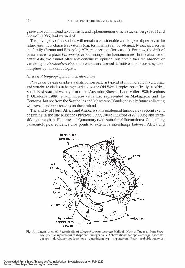
Accepting, pro tempore, Parapachycerina as a monophyletic entity, the next questionis: what is its sister-clade? The senior author was initially strongly impressed by thesimilarity of the genus to the Neotropical Neopachycerina. This South American genuswas described by Malloch (1933: 357, fig. 67c) on the basis of six specimens fromMontevideo, Uruguay (with the single species, N. aristata). Neopachycerina looks alot like Parapachycerina—both are small, orangish lauxaniids with black ocellar spots,plumose aristae, small anterior fronto-orbital bristles, generally hyaline wings andsapromyziform costae. The two genera also key out in the same couplet in Stuckenberg’s(1971) generic key (couplet 32), although Stuckenberg warned that the resemblancewas possibly ‘a case of convergence’.
Differences shown by Neopachycerina include: a more protuberant ocellar hump;dense ocellar setulae (lacking in Parapachycerina); the presence of tiny setulae on theanterolateral portions of the postfrons (absent in Parapachycerina); a conspicuous lunule;a very weak anterior sternopleural bristle; stout, costal setulae terminating well short ofR
4+5 (approximately half way); and densely plumose arista (loosely plumose arista in
Parapachycerina). Compared to Parapachycerina, the male terminalia of Neopachyce-rina (Fig. 31) show noteworthy differences in the shape of the protandria and aedeagalcomplexes. The protandrium of Neopachycerina is bonnet-like, lengthened longitudinallyand shortened laterally in comparison to Parapachycerina species. The protandrium isalso incomplete ventrally in Neopachycerina, in contrast to Parapachycerina. Theepandria are generally similar in both genera (broader laterally than dorsally). What areinterpreted to be the surstyli in Neopachycerina are broadly rounded processes, whichare not articulated or fused to the inner surfaces of the epandria, but meet below thecerci. The aedeagus is a navicular (boat-shaped) sclerite with spinules (teeth) posteriorlyon the inner subapical surfaces. The aedeagal apodeme is very short (<0.5× length of
Downloaded From: https://bioone.org/journals/African-Invertebrates on 04 Feb 2020Terms of Use: https://bioone.org/terms-of-use

152 AFRICAN INVERTEBRATES, VOL. 49 (2), 2008
aedeagus), having an anterior disc and posterior stem (vaguely recalling a ping-pongbat in lateral view), tapering strongly posteriorly to short posterior arms. The hypandrial‘transverse band’ is broad with a small ‘apron’ projecting downwards at an obliqueangle; the lateral hypandrial arms are long, and the ‘dorsal loop’ found in Parapachyce-rina does not occur in Neopachycerina—instead, the lateral arms curve upwards andinwards forming an irregularly shaped convolution bearing a tiny lappet that, ratherunusually for internal genitalic sclerites, has 2 or 3 setulae; the inner edge of this convo-lution then fuses with the base of the gonopods. These differences suggest convergencebetween the two genera. Shewell (1986) had also pointed out convergent similarities inunrelated Old and New World lauxaniid genera.
It was also interesting to note a third row of setulae along the costal margin from betweensubcosta and vein 1 to ca 60 % of the way down the marginal cell in Neopachycerina, acondition not found in Parapachycerina. This extra row of setulae is a strange condition inlauxaniids, judging from Hackman and Väisänen (1985), who found two rows to be uniformthroughout the lauxaniids they examined, but the extra row in Neopachycerina was notedin two co-types examined (part of the type series lodged at the BMNH).
An anonymous referee kindly pointed out to us the genus Tanyura described by Kim(1994: 313–315) from Australia and the Phillipines, which we had overlooked. Wehave not examined Tanyura, but judging from Kim’s description, figures of the terminaliaand comparison with Parapachycerina (Kim 1994: 314–315), this genus emerges as agood candidate for sistership. Kim (1994) gave a list of 11 morphological differencesbetween the two genera. With increased knowledge of Parapachycerina some of thedifferences cited by Kim (1994) are now known to be incorrect; for example, he statesthat Parapachycerina lacks the profemoral ctenidium, whereas it is present in certainAfrotropical species. Differences that continue to hold include: (a) one mesotibial spurin Tanyura (two in Parapachycerina); (b) Tanyura lacking shorter setulae in plumosearista (these shorter macrotrichia present in Parapachycerina); (c) homoneuriform costa(sapromyziform in Parapachycerina); and (d) elongated, ventrally pointing male cerci(rounded, non-elongated male cerci in Parapachycerina). A potential sister-relationshipwith an Oriental and Australasian genus also makes far more sense biogeographicallythan sistership with a South American genus. The senior author must, therefore, concedethat he was mistaken in pursuing Neopachycerina as the potential sister-clade to Parapa-chycerina.
Comprehension of the high-level phylogenetics of the Lauxaniidae is still in a nascentstage, although it has become customary to recognise two subfamilies, the Homoneurinaeand Lauxaniinae (e.g. Shewell 1977, 1987; Miller 1980; Evenhuis & Okadome 1989).Stuckenberg (1971) and Miller (1980) did not include Parapachycerina in the Homoneu-rinae owing to its sapromyziform costa. Shewell (1977) transferred it to the Homoneuri-nae. He did not provide explanation in the Oriental Catalogue for this move, but in aletter to Miller (11 December 1978) explained that ‘the [Parapachycerina] genitaliaseem to me typically oriental homoneurine’ and referred to a manuscript on Nepaleselauxaniids, in which he intended to discuss the matter further. This paper, based on theCoe collection of Nepalese lauxaniids, regrettably was never published (see Shewell’sbibliography in Arnaud 2001).
In an investigation of Homoneurinae monophyly based on parsimony-derived clado-grams, Yarom (1995) also found that Parapachycerina grouped within a monophyletic
Downloaded From: https://bioone.org/journals/African-Invertebrates on 04 Feb 2020Terms of Use: https://bioone.org/terms-of-use

DAVIES & MILLER: REVISION OF AFROTROPICAL PARAPACHYCERINA 153
Homoneurinae, specifically with what he termed the ‘Katalauxania genus-grouplineage’. The latter clade was founded on five synapomorphies. Several of these are,subjectively-speaking, not convincing (e.g. ‘eye not round, posterior margin slanting’,‘face not prominently inflated’) because they are simple characters and appear in otherlauxaniids; furthermore, one of the synapomorphies ‘surstylus freely articulated’ is notcorrect for Parapachycerina. Returning to higher-level divisions, Yarom’s analysisidentified several ‘highly supportive’ synapomorphies for the Homoneurinae: (a)homoneuriform wing; (b) profemoral ctenidium present; and (c) two mesotibial spurs(Yarom 1995: 46–49). These characters had previously been identified as diagnosticfor the homoneurines by Stuckenberg (1971: 516–517) and Shewell (1987: 953).
Parapachycerina does not have a typical homoneuriform wing and the profemoralctenidium is variably present (absent in the type species). The two mesotibial spurs are,however, always present. Although Parapachycerina does not have a typical homo-neurine wing (the original definition by Stuckenberg (1971: 501) is ‘spinules reach orvery nearly reach the apex of R
4+5 where they stop abruptly without marked prior
diminution in size’), the setulae do extend ca 75 % of the way to R4+5
. Other sapro-myziform lauxaniids have the setulae terminating well before that (ca 50 % of the waybetween the two vein apices), as mentioned above with regard to Neopachycerina. It ispossible that the recognition of only two costal setulae categories (sapromyziform andhomoneuriform) is an artificial one, and rather than two distinct states, a messy seriesof intermediates reflects reality better. This is also suggested by Kim’s (1994: 19)comment that ‘in this study numerous species of Trypetisoma with both forms of costahave been identified, and they show different end points of costal spinules between theapices of veins 2 (R
2+3) and 3 (R
4+5)’. Conversely, there are some Homoneura species
where the stout, costal setulae end before R4+5
, notably, H. citreifrons (Malloch, 1920)of the Nearctic (Miller 1977a: 159, fig. 11b) and H. kaszabi Shewell, 1971 and H.amphibola Shatalkin, 1992 of the Palearctic (Shatalkin 2000).
Likewise, the profemoral ctenidium shows variation being absent in homoneurineslike Homoneura tenera (Loew, 1846) (Shatalkin 2000), but present in the sapromyziformLyciella rorida complex in the Palearctic (Shatalkin 2000).
Inconsistency in these ‘strong’ homoneurine synapomorphies reinforces the viewexpressed that lauxaniids display pronounced morphological plasticity (Stuckenberg1971: 500; J.F. McAlpine 1989: 1445). We would also note that these ‘strong’ synapomor-phies comprise morphologically simple characters, respectively: (1) extension by veryshort distances of costal setulae and slight diminution in length of these setulae (thehomoneuriform/sapromyziform dichotomy); (2) strengthening of the row of fine antero-ventral setae of the profemur into shorter, stouter setulae (the profemoral ctenidium);and (3) development of one of the mesotibial setula into a robust but short spur (twomesotibial spurs). Although the genetic and selective processes underlying these changesare unknown, it is a legitimate inference that the changes required are not complex. Asinsightful phylogeneticists have warned (e.g. Bechly 2000: 5–6; Wägele 2004, 2005),erecting phylogenies on the basis of simple characters is a risky enterprise (irrespectiveof whether the cladograms are arrived at by putative ‘objective’, parsimony-aided‘analysis’). As mentioned above, the senior author burnt his fingers badly in contempla-ting Neopachycerina as the sister-clade to Parapachycerina, an example where conver-
Downloaded From: https://bioone.org/journals/African-Invertebrates on 04 Feb 2020Terms of Use: https://bioone.org/terms-of-use
154 AFRICAN INVERTEBRATES, VOL. 49 (2), 2008
gence also can mislead taxonomists, and a phenomenon which Stuckenberg (1971) andShewell (1986) had warned of.
The phylogeny of lauxaniids will remain a considerable challenge to dipterists in thefuture until new character systems (e.g. terminalia) can be adequately assessed acrossthe family (Remm and Elberg’s (1979) pioneering efforts aside). For now, the drift ofconsensus is to place Parapachycerina amongst the homoneurines. In the absence ofbetter data, we cannot offer any conclusive opinion, but note either the absence orvariability in Parapachycerina of the characters deemed definitive homoneurine synapo-morphies by lauxaniidologists.
Historical biogeographical considerations
Parapachycerina displays a distribution pattern typical of innumerable invertebrateand vertebrate clades in being restricted to the Old World tropics, specifically in Africa,South-East Asia and weakly in northern Australia (Shewell 1977; Miller 1980; Evenhuis& Okadome 1989). Parapachycerina is also represented on Madagascar and theComoros, but not from the Seychelles and Mascarene Islands; possibly future collectingwill reveal endemic species on these islands.
The aridity of North Africa and Arabia is (on a geological time-scale) a recent event,beginning in the late Miocene (Pickford 1999, 2000; Pickford et al. 2006) and inten-sifying through the Pliocene and Quaternary (with some brief fluctuations). Compellingpalaeontological evidence also points to extensive interchange between Africa and
Fig. 31. Lateral view of _ terminalia of Neopachycerina aristata Malloch. Note differences from Para-pachycerina in protandrium shape and inner gentialia. Abbreviations: aed apo – aedeagal apodeme;eja apo – ejaculatory apodeme; epa – epandrium; hyp – hypandrium; ? sur – probable surstylus.
Downloaded From: https://bioone.org/journals/African-Invertebrates on 04 Feb 2020Terms of Use: https://bioone.org/terms-of-use

DAVIES & MILLER: REVISION OF AFROTROPICAL PARAPACHYCERINA 155
Eurasian faunae in the Neogene (e.g. Thomas 1985; Pickford 2002). Doubtless proto-Parapachycerina was involved in this mixing of Afrotropical and Eurasian fauna,explaining its presence in much of the paleotropics.
As for Madagascar, geological data show that there has been no terrestrial connectionbetween Africa and Madagascar since the Jurassic (de Wit 2003). The Comoros arevolcanic Cenozoic islands and the presence there of Parapachycerina taxon C, indicatesthat the Comoros may have been used as ‘stepping-stones’ to reach Madagascar.
Acalyptrate flies only began their explosive diversification in the Palaeogene (Hennig1965; Grimaldi & Engel 2005: fig. 12.78), the oldest putative lauxaniids being from theEocene and Oligocene (Evenhuis 1994: 433–434). Given the age of the separation ofMadagascar from Africa, and the constrained dates of acalyptrate diversification, wemust turn to aerial dispersal to explain the occurrence of Parapachycerina on Madagascar.
The circumstantial case for a dispersalist scenario is strong. For example, Chapman etal. (2003: 503, 507) observed that ‘millions of metric tons of insects are aloft in Earth’satmosphere at any given moment’, and that ‘a conservative estimate of the total bioflowover a 1 km stretch of the southern English countryside is an astounding 3 billion insectsper month’. These data underline the potency of aerial dispersal in transporting volantinsects far and wide, as was borne out from the earlier empirical studies of Holzapfel andHarrell (1968) and Wise (1983), among others. Further, in investigations of the bioticrecolonisation of the volcanic island Krakatau (Indonesia), Lauxaniidae were one of the flyfamilies to successfully recolonise the island (Thornton et al. 1990: fig. 13).
In sum, considering these disparate strands of evidence (geological history of Ma-dagascar, potency of aerial dispersal of insects, and rapid colonization of the ‘real-life biogeographical experiment’ of Krakatau by lauxaniids), the most parsimoniousview is that ancestral Parapachycerina reached Madagascar by aerial dispersal. It alsoappears more plausible to posit colonization of Madagascar from Africa, rather thanreverse.
Within Africa itself, Parapachycerina is closely associated with the distribution oftropical and subtropical savannah and moist escarpment grasslands. Current evidenceshows that it is absent from the lowland Guineo-Congo rainforest, although occurringat its edges. The genus is largely tropical and subtropical, its range terminating at about30°S, and at a similar latitude in the north.
Biology of Parapachycerina
Biological information on Parapachycerina is sparse. Most adult specimens have beencaught in grassland and at forest/grassland ecotones by sweeping. Only two specimensexamined were captured in a Malaise trap (both P. infuscata males from Kenya). Theadults are possibly fungivorous, as indicated by the presence of a labellar beak and somepseudotracheal scoops and prongs, which Broadhead (1984, 1989) showed were adaptationsfor feeding on phylloplane fungi. We did not notice any fungal hyphae or spores inproboscides dissected, but our preparations were crude. Consequently, confirmation of afungivorous habit by Parapachycerina calls for further investigation. Perhaps unusually,an adult of P. bispina from Segibwa Falls, Uganda (BMNH) was ‘attracted to human faeces’(label datum). Specimens of P. bispina collected by Deeming in northern Nigeria (TildenFulani) were taken ‘on tomatoes’ at a mixed vegetable farm.
Downloaded From: https://bioone.org/journals/African-Invertebrates on 04 Feb 2020Terms of Use: https://bioone.org/terms-of-use

156 AFRICAN INVERTEBRATES, VOL. 49 (2), 2008
The larvae are unknown, but, it is likely that Parapachycerina larvae feed on deadand decaying grass and other vegetable matter, as is the case with grassland-dwellinglauxaniids in general (Miller 1977b), and phylogenetically-related flies in the Celyphi-dae (Miller 1986) and Natalimyzidae (Miller 1984; Barraclough & D.K. McAlpine2006).
Parapachycerina adults have been collected alongside other small, mostly yellowlauxaniid species of Diplochasma Knab and Chaetolauxania Kertész in South Africangrasslands. Parapachycerina is usually not as commonly encountered as these otheryellowish lauxaniid genera, and the larger, darker grass-inhabiting Calliopum Strand,Lauxania Latrielle and Mycterella Kertész. Being small flies, Parapachycerina are easilyoverlooked by collectors, and possibly are more abundant and widespread than reflectedin the rather small number of specimens in collections (<200).
Most specimens have been collected at low altitudes (<1000 m), but the genus isknown from mid-altitude escarpments in South Africa (e.g. the Mpumalanga Drakens-berg) and mountain ranges in Zimbabwe (e.g. the Vumba Mts) and Tanzania (e.g. theSongea highlands).
Interestingly, several species may be found in sympatry, for example all three Africanspecies (P. bispina, P. infuscata and P. munroi) have been collected in the NoordkaapRiver area (Mpumalanga province) by Miller in lush, damp grassy drainage lines, andthe two Malagasy species (P. lalitra and P. talea) have both been collected at Andasibe(Périnet).
The phenology of the species with sufficient specimens (P. bispina, P. infuscata,P. munroi) shows that there is no seasonality to their flight period, and specimens of allof these species have been collected throughout the year, even at the height of theaustral winter.
ACKNOWLEDGEMENTS
Patsy Birkett (Natal Museum) expertly prepared the illustrations; we greatly appreciateher hard work and attention to detail under tight deadlines. The staff at the Centre forElectron Microscopy (University of KwaZulu-Natal, Pietermaritzburg) are thanked fortheir help with the SEM work. László Papp kindly sent us details of Parapachycerinamaterial held at the HNHM. Constructive criticism on the manuscript was receivedfrom B.R. Stuckenberg, A.I. Shatalkin, L. Papp and an anonymous referee. Lastly, wethank the curators of the institutions from whom lauxaniid material was borrowed, fortheir patience and goodwill.
REFERENCES
ANONYMOUS. 1999. An elementary English–Malagasy Dictionary. Lutheran Press and Antananarivo: AmericanCultural Center.
ARNAUD, P.H. 2001. Guy Eaden Shewell (1913–1996): Diptera bibliography and list of taxonomic namespublished (1938–1996). Myia 6: 187–199.
BARRACLOUGH, D.A. & MCALPINE, D.K. 2006. Natalimyzidae, a new African family of acalyptrate flies(Diptera: Schizophora: Sciomyzoidea). African Invertebrates 47: 117–134.
BECHLY, G. 2000. Mainstream Cladistics versus Hennigian Phylogenetic Systematics. Stuttgarter Beiträgezur Naturkunde, Serie A 316: 1–11.
BROADHEAD, E.C. 1984. Adaptations for fungal grazing in Lauxaniid flies. Journal of Natural History 18:639–649.
––––––1989. The species of Poecilominettia, Homeominettia and Floriminettia (Diptera: Lauxaniidae) inPanama. Bulletin of the British Museum (Natural History), Entomology Ser. 58: 185–226.
Downloaded From: https://bioone.org/journals/African-Invertebrates on 04 Feb 2020Terms of Use: https://bioone.org/terms-of-use

DAVIES & MILLER: REVISION OF AFROTROPICAL PARAPACHYCERINA 157
CHANDLER, P.J. 1987. The families Diastatidae and Camphicoetidae (Diptera, Drosophiloidea) with a revisionof Palaearctic and Nepalese species of Diastata Meigen. Entomologica Scandinavica 18: 1–50.
CHAPMAN, J.W., REYNOLDS, D.R., & SMITH, A.D. 2003. Vertical-looking radar: a new tool for monitoringhigh-altitude insect migration. BioScience 53: 503–511.
DE MEIJERE, J.C.H. 1910. Studien über südostasiatische Dipteren, IV. Die neue Dipteren-Fauna van Krakatau.Tijdschrift voor Entomologie 53: 58–194.
DE WIT, M.J. 2003. Madagascar: heads it’s a continent, tails it’s an island. Annual Review of Earth and Pla-netary Sciences 31: 213–248.
EBERHARD, W.G. 2001. The functional morphology of species-specific clasping structures on the front legsof male Archisepsis and Palaeosepsis (Diptera, Sepsidae). Zoological Journal of the LinnaeanSociety 133: 335–368.
EVENHUIS, N.L. 1994. Catalogue of the fossil flies of the world (Insecta: Diptera). Leiden: BackhuysPublishers.
EVENHUIS, N.L. & OKADOME, T. 1989. Family Lauxaniidae. In: Evenhuis, N.L., ed., Catalog of the Diptera ofthe Australasian and Oriental Regions. Bishop Museum Special Publications 86: 576–589.
GRIMALDI, D. & ENGEL, M.S. 2005. Evolution of the insects. Cambridge: Cambridge University Press.HACKMAN, W. & VÄISÄNEN, R. 1985. The evolution and phylogenetic significance of the costal chaetotaxy in
the Diptera. Annales Zoologicae Fennici 22: 169–203.HENNIG, W. 1965. Die Acalyptratae des baltischen Bernsteins und ihre Bedeutung für die Erforschung der
phylogenetischen Entwicklung dieser Dipteren-Gruppe. Stuttgarter Beiträge zur Naturkunde145: 1–215.
HOLZAPFEL, E.P. & HARRELL, J.C. 1968. Transoceanic dispersal studies of insects. Pacific Insects 10: 115–153.KERTÉSZ, K. 1913. H. Sauter’s Formosa Ausbeute. Lauxaniinae (Dipt.). Annales Historico-naturales Musei
Nationalis Hungarici 11: 88–102.KIM, S.P. 1994. Australian lauxaniid flies: revision of the Australian species of Homoneura van der Wulp,
Trypetisoma Malloch and allied genera (Diptera: Lauxaniidae). Monographs on InvertebrateTaxonomy, Vol. 1. Victoria, Australia: CSIRO.
MALLOCH, J.R. 1933. Diptera of Patagonia and South Chile. Part VI – Fascicle 4, Acalyptrata. London:British Museum (Natural History).
MATHIS, W.N. 1989. A review of the beach flies of the Caribbean and Gulf of Mexico (Diptera: Canacidae).Proceedings of the Biological Society of Washington 102: 590–608.
MCALPINE, D.K. 1973 [1972]. The Australian Platystomatidae (Diptera, Schizophora) with a revision offive genera. The Australian Museum Memoir 15: 1–256.
––––––2007. The surge flies (Diptera: Canacidae: Zaleinae) of Australasia and notes on tethinid-canacidmorphology and relationships. Records of the Australian Museum 59: 27–64.
MCALPINE, D.K. & DE KEYZER, G. 1994. Generic classification of the fern flies (Diptera: Teratomyzidae)with a larval description. Systematic Entomology 19: 305–326.
MCALPINE, J.F. 1962. A revision of the genus Campichoeta Macquart (Diptera: Diastatidae). CanadianEntomologist 94: 1–10.
MCALPINE, J.F. 1981. Morphology and terminology—adults. In: McAlpine, J.F. et al., ed., Manual of NearcticDiptera. Vol. 1. Monograph 27. Ottawa: Research Branch, Agriculture Canada, pp. 9–63.
––––––1987. Chyromyidae. In: McAlpine, J.F. et al., ed., Manual of Nearctic Diptera. Vol. 2. Monograph28. Ottawa: Research Branch, Agriculture Canada, pp. 985–988.
––––––1987. Curtonotidae. In: McAlpine, J.F. et al., ed., Manual of Nearctic Diptera Vol. 2. Monograph28. Ottawa: Research Branch, Agriculture Canada, pp. 1007–1010.
––––––1987. Diastatidae. In: McAlpine, J.F. et al., ed., Manual of Nearctic Diptera Vol. 2. Monograph 28.Ottawa: Research Branch, Agriculture Canada, pp. 1019–1022.
––––––1989. Phylogeny and classification of the Muscomorpha. In: McAlpine, J.F., ed., Manual of NearcticDiptera Vol. 3. Monograph 32. Hull, Quebec: Research Branch, Agriculture Canada, pp. 1397–1518.
MEIER, R. 1995. A comparative SEM study of the eggs of the Sepsidae (Diptera) with a cladistic analysisbased on egg, larval and adult characters. Entomologica Scandinavica 26: 425–438.
MILLER, R.M. 1977a. Taxonomy and biology of the Nearctic species of Homoneura (Diptera: Lauxaniidae). I.Subgenera Mallochomyza and Tarsohomoneura. Iowa State Journal of Research 52: 147–176.
––––––1977b. Ecology of Lauxaniidae (Diptera: Acalyptratae) I. Old and new rearing records with biologicalnotes and discussion. Annals of the Natal Museum 23: 215–238.
––––––1980. Family Lauxaniidae. In: Crosskey, R.W., ed., Catalogue of the Diptera of the AfrotropicalRegion. London: British Museum (Natural History), pp. 603–608.
––––––1984. A new acalyptrate fly from southern Africa, possibly representing a new family. In: XVIIInternational Congress of Entomology, 20–26 August, Hamburg, Federal Republic of Germany,Abstract volume, p. 32.
Downloaded From: https://bioone.org/journals/African-Invertebrates on 04 Feb 2020Terms of Use: https://bioone.org/terms-of-use

158 AFRICAN INVERTEBRATES, VOL. 49 (2), 2008
––––––1986. Preliminary studies on African beetle fly biology and taxonomy. In: Darvas, B. & Papp, L.,eds, First International Congress of Dipterology, 17–24 August, Budapest, Hungary, Abstractvolume, p. 158.
PICKFORD, M. 1999. Aubréville’s hypothesis of a southward shift of Africa’s vegetation belts since the Miocene.In: Maes, F. & Beeckman, H., eds, Wood to Survive, 25, Liber Amicorum Roger Dechamps.Annales Sciences Economiques, Musée Royal de l’Afrique Centrale 25: 195–212.
––––––2000. Crocodiles from the Beglia Formation, Middle/Late Miocene boundary, Tunisia, and theirsignificance for Saharan palaeoclimatology. Annales de Paléontologie 86: 59–67.
––––––2002. Palaeoenvironments and hominoid evolution. Zeitschrift für Morphologie und Anthropologie83: 337–348.
PICKFORD, M., WANAS, H. & SOLIMAN, H. 2006. Indications for a humid climate in the Western Desert ofEgypt 11–10 Myr ago: evidence from Galagidae (Primates, Mammalia). Comptes Rendus Palevol5: 935–943.
REMM, E. & ELBERG, K. 1979. Terminalia of Lauxaniidae (Diptera) found in Estonia, Latvia and Lithuania.In: Elberg, K., ed., Dipteroloogilisi Uurimusi [Diptera Studies]. Tartu: Estonian SSR Academyof Sciences [Eesti NSV Teaduste Akadeemia], pp. 66–177.
SAND-JENSEN, K. 2007. How to write consistently boring scientific literature. Oikos 116: 723–727.SASAKAWA, M. 2003. Oriental Lauxaniidae (Diptera) Part 4. Fauna of the Lauxaniidae of Laos. Scientific
Reports of the Kyoto Prefectural University, Human Environment and Agriculture 55: 57–74.SÉGUY, E. 1938. Diptera 1. Nematocera et Brachycera. Mission Scientifique de l’Omo Vol. 4 Zoologie.
Mémoires du Muséum National d’Histoire Naturelle 8: 319–380.SHATALKIN, A.I. 2000. Keys to the Palaearctic flies of the family Lauxaniidae. Zoological Studies [Zoolo-
gicheskie issledovaniya] 5: 1–102. (in Russian)SHEWELL, G.E. 1977. Family Lauxaniidae. In: Delfinado, M.D. & Hardy, D.E., eds, A Catalogue of the
Diptera of the Oriental Region. Honolulu: University of Hawaii Press, pp. 182–214.––––––1986. Deceptive resemblance between Old and New World genera of Lauxaniidae (Acalyptratae).
In: Darvas, B. & Papp, L., eds, First International Congress of Dipterology, 17–24 August,Budapest, Hungary, Abstract volume, p. 224.
––––––1987. Lauxaniidae. In: McAlpine, J.F. et al., ed., Manual of Nearctic Diptera. Vol. 2. Monograph28. Ottawa: Research Branch, Agriculture Canada, pp. 951–964.
STUCKENBERG, B.R. 1971. A review of the Old World genera of Lauxaniidae (Diptera). Annals of the NatalMuseum 20 (3): 499–610.
––––––1999. Antennal evolution in the Brachycera (Diptera), with a reassessment of terminology relatingto the flagellum. Studia Dipterologica 6: 33–48.
THOMAS, H. 1985. The early and middle Miocene land connection of the Afro-Arabian plate and Asia: amajor event for hominoid dispersal? In: Delson, E., ed., Ancestors: the hard evidence. NewYork: Alan R. Liss, pp. 112–124.
THORNTON, I.W.B., NEW, T.R., ZANN, R.A. & RAWLINSON, P.A. 1990. Colonization of Krakatau Islands byanimals. Philosophical Transactions of the Royal Society, London 328: 131–165.
WÄGELE, J.-W. 2004. Hennig’s Phylogenetic Systematics brought up to date. In: Williams, D.M. & Forey,P.L., eds, Milestones in Systematics. Baton Rouge: CRC Press, pp. 101–125.
––––––2005. Foundations of phylogenetic systematics. Munich: Verlag Dr Friedrich Pfeil.WISE, K. 1983. Trans-oceanic insect dispersal. I. Trapping and collecting on ships in the South Pacific
Ocean, 1974–1979. Records of the Auckland Institute and Museum 20: 223–254.YAROM, I. 1995. Phylogenetic Analysis of Homoneurine flies (Diptera: Lauxaniidae) and revisions of the
genera Dioides, Procestrotus, and Prosopophorella. PhD thesis. Lawrence, USA: University ofKansas.
Downloaded From: https://bioone.org/journals/African-Invertebrates on 04 Feb 2020Terms of Use: https://bioone.org/terms-of-use