extrinsic and intrinsic mechanisms directing epithelial...
TRANSCRIPT

DEVELO
PMENT
367RESEARCH ARTICLE
INTRODUCTIONOne of the main processes leading to the shaping of the three-dimensional body plan of higher organisms is the dynamicrearrangement of cell populations. Whereas in most morphogeneticprocesses cells move individually to new locations over largedistances (i.e. during germ cell migration), in epithelia, duringmorphogenesis, cells reorganize themselves coordinately as a group(i.e. during neural tube folding).
The movement and fusion of epithelial cells during developmentis an essential and general morphogenetic event. Many differentmorphogenetic processes, both in vertebrates and invertebrates, andthe related processes of adult and embryonic vertebrate woundhealing, could be included in this category (reviewed by Martin andWood, 2002). However, although extensive morphologicaldescriptions of these processes represent classical paradigms ofembryology, the genetic basis and the cellular behaviour underlyingthese events remain poorly understood. Thorough studies haveshown that to undertake these processes epithelial cells can employtwo alternative approaches: first, leading edge cells facing a freecellular space become specified and promote the migration ofepithelial sheets acting as the main driving force for migration (inmost cases in the absence of cell proliferation), e.g. Caenorhabditiselegans ventral enclosure (Williams-Masson et al., 1997) andimaginal discs fusion in Drosophila (Pastor-Pareja et al., 2004).Second, overall synchronous action of the whole epithelial cellpopulation can induce global changes in its motility, conformingtissue shape changes, e.g. teleost epiboly (Trinkaus et al., 1992) andextension of the neural plate in Xenopus (Keller et al., 2000).
Two models of unbounded epithelial expansion, in whichepithelial sheets with a free leading edge advance, meet and fuse toequivalent epithelial sheets in order to seal the surface of the animal,have been described in Drosophila. These models are dorsal closureand the fusion of imaginal discs. Dorsal closure is a late embryonic
event that begins with the elongation of the epidermal cells andfinishes when the dorsalmost cells fuse at the midline (reviewed byHarden, 2002). The dimensions of the epidermal layer are notcontrolled by proliferation but are the result of changes in celladhesion and cell shapes. Cells at the leading front that are planarypolarized guide the migration of the epithelium, express specificmarkers and have a very active cytoskeleton. Dorsal closure dependson at least two signalling pathways – the JNK signal transductionpathway and the signalling pathway activated by Dpp (Martin-Blanco et al., 1998; Riesgo-Escovar and Hafen, 1997; Kockel et al.,1997). These two pathways are also involved in the expansion andmerging of excorporated imaginal tissues (imaginal discs) at theexpense of larval cells during metamorphosis (reviewed by Martin-Blanco and Knust, 2001). The imaginal discs evert and later ondifferentiate to give rise to the external structures of the adult. In thisprocess the peripheral cells of the disc expand over the larval cellsand gradually displace them. Finally, the discs recognize and fusewith each other to complete the continuous epithelium that will giverise to the head and the thorax of the adult.
Much less is known about the mechanisms controlling themovements of bounded epithelia without a free edge (e.g. theinvagination of the basal region during sea urchin gastrulation,extension of the neural plate, rearrangement of scale precursorsin the wings of insects, etc.) (see Bard, 1990). A potential modelfor the study of the coordinated expansion of this type of epitheliais the development of the adult abdomen of Drosophila duringmetamorphosis. The adult epidermis is formed by cellsdescending from histoblasts, founder imaginal cells specifiedduring embryonic stages as small incorporated groups organizedin nests. Each adult abdominal segment forms from four pairs ofhistoblast nests: the anterior and posterior dorsal pairs (whichproduce the tergites), the ventral pairs (which produce thesternites and pleurites) and the spiracular pairs (which form thespiracle and the surrounding pleurite tissues). Each anterior dorsaland ventral nest is composed of approximately 16 cells; eachposterior dorsal nest consists of approximately five cells; and eachspiracle nest has approximately three cells (Fig. 1A) (Guerra etal., 1973; Roseland and Schneiderman, 1979; Madhavan andMadhavan, 1980). During larval stages histoblasts are mitoticallyquiescent and arrested in G2 (Garcia-Bellido and Merriam, 1971;Madhavan and Schneiderman, 1977; Roseland and
Extrinsic and intrinsic mechanisms directing epithelial cellsheet replacement during Drosophila metamorphosisNikolay Ninov, Dominic A. Chiarelli* and Enrique Martín-Blanco†
The fusion of epithelial sheets is an essential morphogenetic event. Here, we study the development of the abdomen of Drosophilaas a model of bounded epithelia expansion and uncover a complex multistep process for the generation of the adult epidermisfrom histoblasts, founder cells that replace the larval cells during metamorphosis. We find that histoblasts experience a biphasic cellcycle and emit apical projections that direct their invasive planar intercalation in between larval cells. Coordinately, the larval cellsextrude from the epithelia by apical constriction of an actomyosin ring and as a consequence die by apoptosis and are removed bycirculating haemocytes. We demonstrate that the proliferation of histoblasts and the death of larval cells are triggered by twoindependent extrinsic Ecdysone hormonal pulses. Finally, we show that histoblast spreading and the death of larval cells depend ona mutual exchange of signals and are non-autonomous processes.
KEY WORDS: Drosophila, Metamorphosis, Histoblasts, Morphogenesis, Abdomen, Cell death, Cell replacement
Development 134, 367-379 (2007) doi:10.1242/dev.02728
Instituto de Biología Molecular de Barcelona, Consejo Superior de InvestigacionesCientíficas, Parc Cientific de Barcelona, Josep Samitier 1-5, Barcelona 08028, Spain.
*Present address: Peter MacCallum Cancer Centre, 7 St Andrews pl, EastMelbourne, Victoria, 3002, Australia†Author for correspondence (e-mail: [email protected])
Accepted 2 November 2006

DEVELO
PMENT
368
Schneiderman, 1979). At the onset of metamorphosis histoblastsundertake a rapid proliferation that allows them to expand andfuse, at the expense of the preexisting surrounding polyploidlarval epidermal cells (LECs) that commit programmed cell death(Madhavan and Madhavan, 1980).
Although the general characteristics of the expansion andfusion of histoblast nests are known from studies conducted 25years ago, the molecular, genetic and cellular mechanismsinvolved in the generation of a functional epithelial barrier byhistoblast are unknown. Furthermore, histoblasts represent anexcellent model for in vivo studies at high resolution, as theyconstitute the external sheet of cells in the pupa and can be readilyvisualized across the pupal case. Our cellular characterization ofthe process has given light to numerous unanswered questions.What are the molecular mechanisms triggering abdominalepithelial morphogenesis? How do histoblasts achieve theirmigration within the epithelial layer? And how do LECs becomeremoved in such a way that no gaps are formed in the epitheliumduring their massive cell death? We show that both proliferationof histoblasts and death of larval cells are triggered by extrinsicEcdysone hormonal pulses occurring at different times duringpupariation. Additionally, we have characterized the dynamics ofthe histoblast cell cycle and the cellular mechanisms underlyinghistoblast spreading and LEC death and removal. Finally, we haveuncovered a potential intrinsic strategy for cell replacementdirecting the behaviour of histoblasts and LECs and itscoordination.
MATERIALS AND METHODSFly stocksAll flies were mantained on standard culture media. Fly stocks used were asfollows: Esg-Gal4 (yw; NP5130) (NIG-FLY Stock Center); Esg-Gal4 UAS-GFP (y, w; NP5130, UAS-GFP, UAS-nGFP, UAS-lacZ/CyO) (NIG-FLYStock Center); Hs-FLP; Ay+Gal4 UAS-GFP (hsp70-FLP; Act FRT y+ FRTGal4, UAS-GFP/CyO; MKRS/TM2) (Ito et al., 1997); Esg-Gal4 Ay+Gal4UAS-GFP UAS-FLP (y, w; NP5130, Act FRT y+ FRT Gal4, UAS-GFP/CyO; UAS-FLP/TM6B); Hs-Gal4 (w; Hsp70 Gal4.PB 89-2-1)(Bloomington Stock Center 1799); UAS-FLP (y, w; UAS-FLP.Exel 3)(Bloomington Stock Center 8209); 056-Gal4 – Enhancer trap ubiquitouslyexpressed in the epidermis (y, w; NP0056) (NIG-FLY Stock Center);Ay+Gal4 UAS-GFP (y, w; Act FRT y+ FRT Gal4, UAS-GFP) (BloomingtonStock Center 4411); Srp-Gal4 UAS-GFP [srpHemo-Gal4#16 A3B, UAS-Src-EGFP (II)] (Bruckner et al., 2004); Sqh-GFP (y w sqhAV3 cv; sqh-GFP42) (Royou et al., 2004); UAS-RokCAT (Winter et al., 2001); UAS-P35[w; UAS-p35.H BH1 (II)] (Bloomington Stock Center 5072); UAS-Chickadee (L. Cooley, Yale University Medical School, Department ofGenetics, New Haven, CT, USA); UAS-MbsN300 (Lee and Treisman,2004); UAS-Dap (UAS-Dap II.3) (Lane et al., 1996); UAS-EcR-RNAi(UAS-EcR-AB RNAi) (Schubiger et al., 2005); UAS-EcRDN (w; UAS-EcR.B1-DeltaC655.W650A) (Bloomington Stock Center 6872); UAS-RacV12 (y, w; UAS-Rac1V12) (Bloomington Stock Center 6291); Stb-YFP (w;Act Stb-YFP/TM3) (Strutt et al., 2002); H2-YFP(III) [His2avD–EYFP(II)](Rebollo et al., 2004); Hs-FLP Ay+Gal4 UAS-GFP H2YFP (hsp70-flp; ActFRT y+ FRT Gal4, UAS-GFP/CyO; H2YFP/TM2); Hs-FLP Ay+Gal4 UAS-GFP Stb-YFP (hsp70-flp; Act FRT y+ FRT Gal4, UAS-GFP/CyO; Act Stb-YFP/TM2); UAS-Src-GFP (w; UAS-SrcEGFP M7E) (Bloomington StockCenter 5432); UAS-Actin-GFP (w; UAS-Act5C.GFP 2) (Bloomington StockCenter 7310); DE-Cadherin-GFP [Ubi-DE-Cad-GFP (II)] (Oda andTsukita, 2001); UAS-Tau-GFP (w; UAS-GFP.btau) (Brand, 1995).
Targeted expression in histoblastsThe Gal4/UAS system (Brand and Perrimon, 1993) was used for targetedexpression in the histoblasts. The Esg-Gal4 was used as a histoblast-specificdriver, although expression of this driver can also be detected in salivaryglands, wing discs, eye discs and gut.
Permanent expression of UAS constructs in histoblastsThe Esg-Gal4 driver expression declines during late pupal stages. Toovercome this problem we took advantage of the UAS-FLP/Actin-FRT-y+-FRT-Gal4 system (Struhl and Basler, 1993) by raising flies with the genotypey, w; NP5130, Act FRT y+ FRT Gal4, UAS-GFP/CyO; UAS-FLP/TM6B.
RESEARCH ARTICLE Development 134 (2)
Fig. 1. Abdominal metamorphosis: cell proliferation dynamics ofhistoblasts. (A) During embryonic stages, four nests of abdominalhistoblasts (adult epidermal cells precursors) can be distinguished ineach hemisegment: anterior dorsal (green mask color), posterior dorsal(red), ventral (blue) and spiracular (yellow). During metamorphosis,histoblasts form the different structures that compose the abdominaladult epidermis, tergites (green), intersegmental membranes (red),pleurites and sternites (blue) and spiracle (yellow) (Roseland andSchneiderman, 1979). (B) In vivo time-lapse observation of histoblastproliferation in prepupal stages (1-6 hours APF) shows a synchroniccadence of three cell cycles leading to cell doubling every 2 hours. Thenumber of cells calculated at the shown time points steadily increase(18, 36, 72 and 128). Cell sizes decrease after each mitosis (856, 605,396 and 271 arbitrary 2-dimensional units; see Materials and methods).Histoblasts (anterior dorsal nest) expressed UAS-GFP under the controlof Esg-Gal4 (see Movie 1 in the supplementary material). (C) Earlyhistoblast cell divisions show planar orientation. Histoblasts (anteriordorsal nest) expressed UAS-Tau-GFP under the control of Esg-Gal4. Inthe first cell division, spindles orient predominantly along thedorsoventral axis. (D) Doubling times of histoblasts during pupal stages(from 15 hours APF onwards) increase up to 9 hours. Proliferation isstochastic and coupled to cell growth. Cellular outlines (anterior dorsalnest) were highlighted by ubiquitously expressing a DE-Cadherin-GFPfusion. Mitoses of individual cells were followed by in vivo time-lapse(see Movie 2 in the supplementary material). (E) Schematic of histoblastcell cycle dynamics. During prepupal stages (left), histoblasts do notgrow between cycles (68% cell size decrease in the first three divisions).By contrast, during the pupal stages, histoblasts undergo intermitoticgrowth and their sizes remain constant (right). (F) FACS analysisshowing cell cycle profiles of dissociated histoblasts from prepupal(black) and pupal stages (red). During prepupal stages, histoblasts lackor have a very reduced G1 phase. In pupal stages, the length of G1phase increases by 70%.

DEVELO
PMENT
The larval expression of Esg-Gal4 triggers activation of the UAS-FLPtransgene, which promotes the cis-recombination of FRT sites in the Actin-FRT-y+-FRT-Gal4 cassette generating a FLP-out event in histoblast cells.As a result, the Gal4 gene comes under the control of the Actin promoter,which promotes its permanent expression in histoblasts.
Time- and tissue-specific expression of UAS constructs in LECsTo achieve a tissue- and time-specific expression of UAS constructs in LECswe used a combination of the Gal4/UAS system and the FLP/FRT systemby using the hsp70-FLP; Act FRT y+ FRT Gal4, UAS-GFP/CyO;MRKS/TM2 strain (Ito et al., 1997). This system has been used for thegeneration of positively marked clones in diploid cells where the frequencyof recombination is low (Struhl and Basler, 1993). The larval epidermal cellsare polyploid and contain multiple copies of the genome. As a result therecombination frequency between FRT sites is far greater compared withdiploid cells. A short heat shock treatment of 8-10 minutes leads torecombination, and hence the activation of a UAS-GFP reporter, in 95-100%of the larval epidermal cells 5-7 hours after the heat shock pulse. Heat shockswere performed during late larval stages (wandering larva) when histoblastsare mitotically arrested. In this way, we were able to express distinct UASconstructs in LECs although only small fractions of histoblast were affected(data not shown).
ImmunohistochemistryPrimary antibodies used were: rabbit anti-laminin A antiserum at 1:100dilution (Fessler et al., 1987), mouse anti-Fasciclin III (7G10-HybridomaBank) at 1:1000, mouse anti-GFP at 1:500 (Cell Signalling), rabbit anti-active Caspase 3 at 1:100 (Cell Signalling), mouse anti-EcRB1 at 1:10(AD4.4 Hybridoma Bank) and mouse anti-EcRA at 1:10 (15G1A-Hybridoma Bank). Secondary antibodies were anti-mouse or anti-rabbitFITC, Cy3 or Cy5 conjugated (Molecular Probes) at 1:250 dilutions.Immunohistochemistry was performed using standard procedures. Forpupal staging, white pupae [0 hours after puparium formation (APF)] wereused as reference. After selection, the white puparium prepupa weretransferred to fresh vials and kept at 25°C and standard humidity up todissection. Whole pupae were bisected along the anteroposterior axis insterilized 1� PBS (pH 7.4). The internal organs were cleaned from theepidermis by flushing with 1� PBS using a P10 pipette. The epidermis wasdetached from the pupal case using forceps and transferred to an ependorfftube on ice. Fixation was performed for 10 or 15 minutes in 4%paraformaldehyde. After fixation, the epidermis was rinsed three times in1� PBS and permeabilized in sterilized PBT (0.3% Triton in 1� PBS)(3�15 minutes). After permeabilization, the tissue was blocked for 1 hourusing PBTB [1% bovine serum albumin (BSA) in PBT]. Primary antibodieswere incubated overnight at 4°C with gentle shaking. The epidermis wasrinsed in 1� PBS, and washed 3�15 minutes in PBTB. After 1 hourblocking in PBTB, the secondary antibody was incubated for 3 hours atroom temperature. After rinsing in 1� PBS, the tissue was stained usingDAPI (1 ng/�l) to mark the nuclei and Rhodamine-coupled Phalloidin(Molecular Probes) at a dilution of 1:1000 from a 1 �g/�l stock solutionwas used to visualize polymerized actin. Finally the tissue was washed3�15 minutes in 1� PBS, equilibrated in Vectashield (Vector) and mountedon coverslips. Actin staining using Phalloidin alone was performed as aboveafter 10 minutes fixation and omitting the blocking steps.
In vivo imaging and time-lapse microscopyStaged pupae and prepupae were washed in PBS. Early pupae were directlyimaged through the transparent pupal case. For late pupal imaging (after 12hours APF), a small window was opened into the pupal case on top ofabdominal segments 2 and 3 by careful surgery with a fine needle. At thisstage the pupal case is detached from the epidermis and can be removedwithout disturbing the underlying epidermis. The animals were positionedon a glass bottom microwell dish (MatTek) in a small drop of Voltaleff oil toimprove optics and to avoid desiccation. Images were captured at differenttime intervals using an inverted Leica TCS 4D confocal microscope or aninverted Leica AOBS confocal microscope. Laser intensity was kept at aminimum to avoid photobleaching and to minimize phototoxicity. Eachmovie was repeated at least three times. In most cases, the animal survivedthe dissection and data acquisition and developed to adult stages.
In vivo quantification of cell proliferation rateCell proliferation was recorded using confocal time-lapse imaging in theposterior dorsal histoblast nest from 17 hours APF pupae of the genotypesHs-FLP; Ay+Gal4 UAS-GFP; H2-YFP or Hs-FLP; Ay+Gal4 UAS-GFP/UAS-P35; H2YFP/+, where the expression of the P35 transgene wasactivated by heat shock during late larval stages. To minimize stress duringimage acquisition the pupal case was not removed and imaging wasperformed directly through the puparium at 10 minute intervals. Individualcells were followed from mitosis to mitosis using the ubiquitously expressedHistone2-YFP fusion protein as a marker. The mitosis of mother cells weretaken as time 0 and the consecutive division of one of the two daughter cellsas time 1. Cell doubling time was calculated for a time window of 6 hours(17-23 hours APF). Doubling times for 10 cells in each experiment werecounted.
Flow cytometryTo measure the cell cycle phasing of the abdominal histoblasts, whole pupalcuticles (50 animals for each condition) were dissected, cleaned and thensubjected to an incubation in 9� Tripsin, 1� PBS, with 1 mg/ml Hoechst33342 for 3 hours at room temperature. Histoblasts were positively markedby Esg-Gal4 expression driving UAS-GFP. Prepupae animals were stagedas less than 12 hours APF and greater than 3 hours APF. Early pupae animalswere determined as greater than 12 hours APF and less than 24 hours APF.In order to compare cell cycle profiles, samples of each time point wereprepared and ran simultaneously. We used a MoFlo flow cytometer(DakoCytomation, Fort Collins, Colorado, USA). Excitation was performedwith an argon-ion laser of Coherent Enterprise II and the optical alignmentobtained with fluorescent particles of a diameter of 10 �m (Flowcheck,Coulter Corporation, Miami, Florida, USA). Different populations weredefined combining green (GFP) and blue (Hoechst 33342) emissions andthe refringency parameters FSC and SSC.
Quantification of cell sizeCell size was calculated by dividing the number of cells in a histoblast nestover the total area of the nest. The area of the nest was calculated from z-stack projections covering the whole depth of the nest. Histoblasts werevisualized using Esg-Gal4 driving the expression of a UAS-nuclear-GFP andUAS-cytoplasmic-GFP. Area measurements were carried out using ImageJ(NIH Image).
Image analysisImage analysis was performed with Leica Confocal Software, Imaris 5D(Bitplane) software was used for 3D reconstruction of time-lapse movies,ImageJ (NIH Image) for cell tracking and mounting of time-lapse movies inAVI format, Photoshop 7.0 (Adobe Corporation) for data processing andCorel R.A.V.E. for conversion of movies to QuickTime format.
RESULTSHistoblasts undergo biphasic dynamicallycontrolled cell proliferation duringmetamorphosisThe abdominal histoblasts do not divide during the larval stages andincrease in volume about 60-fold (Madhavan and Madhavan, 1980).Upon pupariation, histoblasts divide rapidly but remain confined totheir original dimensions. A second, slower histoblast proliferationphase begins about 15 hours APF and lasts until about 36 hours APF.During this period, the histoblast nests enlarge, and the histoblastsreplace the adjacent LECs. By time-lapse analysis, we validatedthese distinct phases. We found that during prepupal stages,histoblasts underwent very fast synchronous cell divisions, with acell doubling time of around 2 hours (Fig. 1B; see Movie 1 in thesupplementary material) and preferentially oriented spindles (Tau-GFP labelling, see Fig. 1C). This early stage lasted for three cyclesand ran between 4 and 12 hours APF. A characteristic feature of theprepupal proliferation of histoblasts is the lack of cell growthbetween cycles, resulting in a rapid decrease in cell size (Madhavanand Madhavan, 1980) (Fig. 1E, left panel). We found by FACS
369RESEARCH ARTICLECell replacement in Drosophila metamorphosis

DEVELO
PMENT
370
analysis that the short cell doubling time and lack of growth ofhistoblasts isolated at this stage was associated with a very reducedG1 phase (Fig. 1F).
During pupal stages, histoblasts enter a second phase ofproliferation, in which synchrony gets lost and cell division planesbecome stochastically oriented. Our in vivo observations revealed aprogressive increment of cell doubling times up to 9 hours (Fig. 1D;see Movie 2 in the supplementary material) resulting from alengthening of 70% in the G1 phase (determined by FACS analysis)(Fig. 1F). As a result, histoblast cell size remained constant betweenmitosis (Fig. 1E, right panel). Combining these observations, weconclude that histoblast growth during the long quiescent larvalperiod allows for the rather quick start to their differentiation,leading to a biphasic (quick and then slow) cell cycle profile.
Histoblast nest expansion requires the activeplanar intercalation of histoblasts into the larvalepitheliaDuring the prepupal highly proliferative stage the area occupied byeach histoblast nest did not significantly increase; as a consequenceof this, as revealed by cytoskeleton (Actin) and adherens junction(DE-Cadherin) markers, histoblast nests became organized intopseudostratified monolayers (see Fig. S1A in the supplementarymaterial). Subsequently, from 15 hours APF onwards, in synchronywith histoblast cell cycle slowness, nests initiated expansion andinvaded the territories occupied by polyploid LECs. As an outcome,they rapidly rearranged into unstratified epithelia (see Fig. S1B inthe supplementary material). The anterior and posterior dorsal nestsfused into a single hemitergite nest between 15 and 18 hours APF(see Fig. S1C and Movie 2 in the supplementary material).Hemitergite nests from adjacent segments began to fuse at about 18hours APF. The hemisternite (ventral) histoblast nest and thespiracular anlagen joined up between 18 and 22 hours APF, whilethe hemitergite histoblast nest and the spiracular anlagen joined upbetween 22 and 26 hours APF. The process was completed upon thefusion of left and right nests at the dorsal midline by 36 hours APF(Madhavan and Madhavan, 1980) (and data not shown).
To gain insight into the mechanisms involved in histoblast nestexpansion, we monitored this process in vivo. This analysis revealedthat at its onset, nest spreading proceeded through the intercalationof guiding histoblasts into the surrounding larval epidermal palisade(Fig. 2A; see Movie 3 in the supplementary material). To do so,invading peripheral histoblasts extended dynamic cellularprotrusions in between neighbouring LECs that, by anchoring andshrivelling, promoted traction and the forward movement ofhistoblast cell bodies (Fig. 2B; see Movie 4 in the supplementarymaterial). These structures were actin-rich and developed byeffective actin polymerization at their tips, resembling actin comets(Fig. 2C,D; see Movie 5 in the supplementary material). The initialplanar intercalation was followed by the coordinated expansion ofthe whole nest epithelia. Every cell at the edge of the nests weakly,but reproducibly, downregulated adherens junction markers (see Fig.S1C and Movie 2 in the supplementary material) and emitted bothapical and basal filopodia and lamellipodia, which expanded overthe surface of the LECs (Fig. 2E) and actively advanced over theunderlying extracellular matrix (ECM) in the direction of migration(Fig. 2F). Nest expansion progressed centrifugally for several hoursup to the merge of the adjacent ipsilateral and contralateral nests(data not shown). Finally, the sealing of the epithelia proceeded bythe assembly of an apical purse string that brought together theapices of the leading cells (Fig. 2G; see Movie 6 in thesupplementary material).
Altogether, these results reveal a complex, multistep process thatinvolves the invasion of the larval epithelia by histoblasts (planarintercalation) mediated by active cellular actin-rich protrusions. Totest this model we interfered with actin polymerization in histoblasts.During Drosophila bristle morphogenesis, the activities of theproducts of the cpb (Capping protein beta) and chickadee (Profilin)
RESEARCH ARTICLE Development 134 (2)
Fig. 2. The process of histoblast nest expansion is associatedwith cell shape changes and active cytoskeleton dynamics.(A) Snapshots of the process of histoblast expansion (ubiquitous DE-Cadherin-GFP; see Movie 3 in the supplementary material) show that atearly stages of nest spreading (anterior dorsal nest), leading cellsintercalate within and disjoin LECs. A leading histoblast (red)successively intercalates in between individual LECS (yellow). Overall,leading histoblasts move over a distance of 40 �m in 3 hours.(B) Leading histoblasts (Esg-Gal4/UAS-GFP, posterior dorsal nest) extendinvasive dynamic protrusions (arrowheads), which promote the forwardmovement of histoblast cell bodies (red arrows) (see Movie 4 in thesupplementary material). (C) The long cellular protrusions of histoblastsare enriched with actin at their tips (arrowheads). UAS-Actin-GFP wasexpressed in histoblasts (Esg-Gal4) and visualized with anti-GFPantibodies (green). Cell morphology was revealed by Phalloidin staining.Nuclei are in blue (DAPI). (D) The protruding structures of leadinghistoblasts grow by distal actin filament polymerization (arrowhead).Actin dynamics in vivo were monitored with Actin-GFP (Esg-Gal4) byconfocal time-lapse microscopy (anterior dorsal nest) (see Movie 5 inthe supplementary material). (E) Spreading histoblasts (Esg-Gal4/UAS-GFP, posterior dorsal nest) crawl over the larval epithelia and send outapical cellular projections in the form of lamellipodia and small filopodia(arrowheads). (F) Long filopodia (arrowhead) are observed in thebasolateral membrane of peripheral histoblasts (Esg-Gal4/UAS-GFP,anterior dorsal nest). These structures are enriched in filamentous actinand are highly motile. (G) During the fusion of neighbouring histoblastnests, the apical domains of adjacent histoblasts become organized in apurse string (arrowhead). Histoblasts (Esg-Gal4/UAS-Src-GFP, spiracularand ventral nest) were monitored by time-lapse confocal microscopy(see Movie 6 in the supplementary material).

DEVELO
PMENT
genes hold a tight balance between actin depolymerization andassembly (Hopmann and Miller, 2003). Loss of Cpb or excessiveProfilin activity resulted in actin accumulations and abnormalbristles. In this light, we overexpressed Profilin in histoblasts underthe control of a permanent Esg-Gal4 driver (see Materials andmethods). The overexpression of Profilin disrupted bristlemorphogenesis, as expected, and caused the inefficient expansion ofhistoblast nests, provoking abdominal clefts (Fig. 3A). In thiscondition, actin aggregated within histoblasts (compare Fig. 3B with3C) and, in contrast to wild-type nests, where numerous longprotrusions formed during expansion, Profilin-overexpressinghistoblasts lacked such structures and just exhibited rare shortfilopodia and lamellipodia (Fig. 3D; see Movie 7 in thesupplementary material). Moreover, the transition of histoblast nestsfrom pseudostratified epithelia to an unstratified monolayer wasdelayed (data not shown). Thus, the inability of histoblasts to resortto long cellular protrusions resulted in the delay of their expansionand migration, suggesting that the actin-mediated planarintercalation of histoblasts between LECs is essential for their properexpansion.
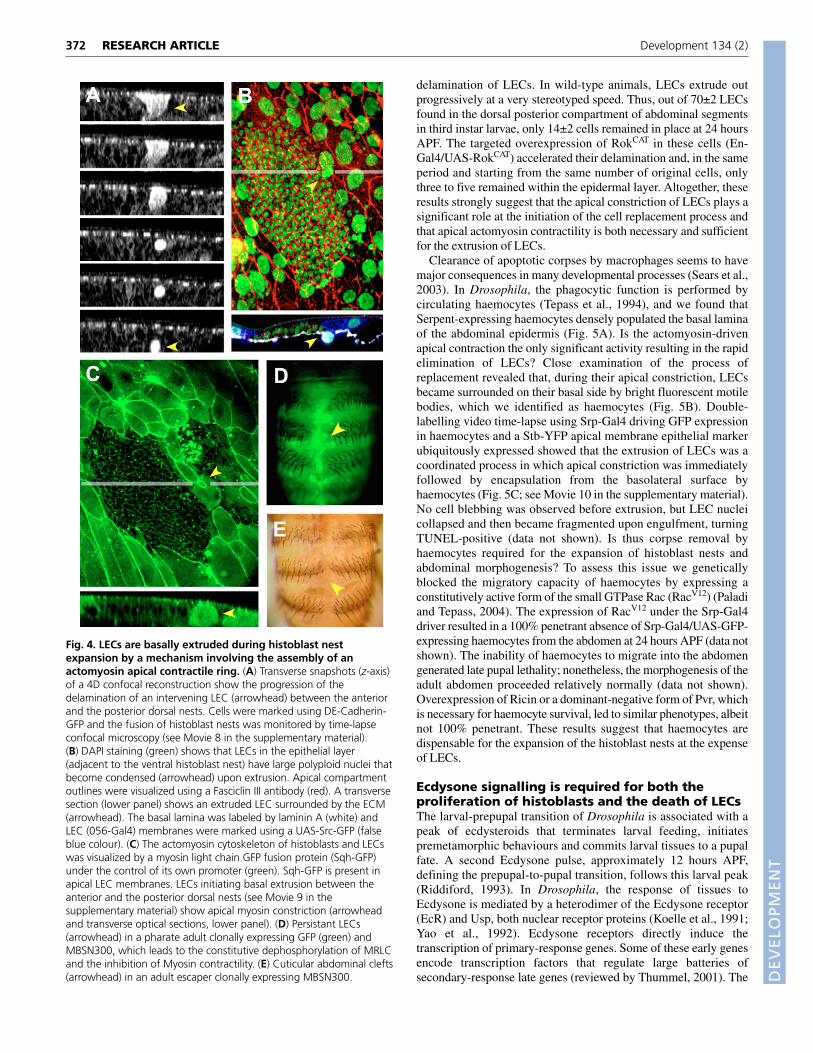
The replacement of LECs relies on their basalextrusion, which is mediated by myosincontractility and is independent of haemocyterecruitmentThe expansion of histoblast nests is directly coupled to thereplacement of the LECs, as no gaps in the epidermis are everdetected histologically. But, how is cell replacement achieved? Bytime-lapse analysis we found that simultaneously with nestexpansion, LECs collapsed, were extruded and died under theepithelial layer (Fig. 4A,B; see Movie 8 in the supplementarymaterial).
What is the mechanism for basal extrusion of LECs? Are thehistoblasts pushing away the polyploid LECs (Fig. 2B)? Or arepulling contractile forces from the LECs driving nest expansion? Ora combination of both? To analyse these issues we evaluatedactomyosin dynamics during the expansion of histoblast nests.Myosin II regulatory light chain (Spaghetti squash) (Royou et al.,
2004) accumulated as perimetral rings in the apical domain of LECs.Assessment of video time-lapse recordings revealed that LECextrusion was initiated by apical constriction, apparently mediatedby actomyosin contraction (Fig. 4C; see Movie 9 in thesupplementary material). The apical constriction of LECs seemed tobe a cell-autonomous event, as it could be observed occasionally inLECs without direct contact with the expanding histoblasts butseveral cell diameters away.
Non-muscle myosin II is a hexamer composed of two of each ofthree subunits: the heavy chain, the regulatory light chain (MRLC)and the essential light chain (Korn and Hammer, 1988). The force-generating activity of actomyosin is mainly controlled through thephosphorylation of the MRLC (Craig et al., 1983). Two kinases,Rho-kinase (Rok; also known as Rock) (Winter et al., 2001) andMyosin light chain kinase (MLCK) (Totsukawa et al., 2000)phosphorylate MRLC in both mammals and Drosophila and activatemyosin contraction. By contrast, dephosphorylation of MRLC bymyosin light chain phosphatase (MLCP) inhibits myosin activity.This serine/threonine protein phosphatase is a heterotrimerconsisting of a catalytic subunit, a 20 kDa protein of unknownfunction, and the myosin binding subunit (MBS) that targets MLCPto MRLC (Fukata et al., 1998). Phosphorylation by Rok of a specificthreonine within a conserved motif in MBS has been shown toinhibit MLCP activity, suggesting that Rok activates MRLC both bydirect phosphorylation and also by inhibition of MBS (Kawano etal., 1999).
To test whether myosin contraction was required for celldelamination, we overexpressed in LECs a truncated constitutivelyactive form of the Drosophila MBS orthologue (MbsN300) thatcannot be phosphorylated by Rok (generalized clonal expression ofUAS-MbsN300; see Materials and methods) and inhibits Myosincontractility (Lee and Treisman, 2004). In this condition, asignificant proportion of LECs did not extrude and remained withinthe abdominal epidermis at postmetamorphosis periods (Fig. 4D).As a consequence, cuticular clefts were observed in the abdomen(Fig. 4E). Further, we found that the overexpression of aconstitutively active form of Rok (RokCAT) (Winter et al., 2001),which upregulates myosin contractility, led to premature
371RESEARCH ARTICLECell replacement in Drosophila metamorphosis
Fig. 3. Actin polymerization directs the expansion of histoblast nests. (A) Bristles with morphogenetic defects (yellow arrowheads) andcuticular abdominal clefts (white arrowheads) in an adult escaper of Permanent-Esg-Gal4/UAS-Profilin genotype. (B) In wild-type histoblasts (22hours APF), most polymerized actin organizes in cortical filaments. Phalloidin-rhodamine staining (red) is shown in the top and bottom panels. UAS-GFP expression (green) under the control of Permanent-Esg-Gal4 is shown in the bottom panel. (C) Actin in histoblasts overexpressing Profilin (22hours APF) hyperpolymerizes and accumulates in intracellular clumps (arrowheads). Top and bottom panels are as in B. (D) Snapshots from Movie 7in the supplementary material. Top panel, wild-type dorsal posterior nest (23 hours APF). Arrowheads point to expanding peripheral protrusions.Bottom panel, dorsal posterior nests from pupae (23 hours APF) overexpressing Profilin (Permanent-Esg-Gal4/UAS-GFP; UAS-Profilin). Note theabsence of long terminal protrusions and the histoblast expansion delay.
DEVELO
PMENT
372
delamination of LECs. In wild-type animals, LECs extrude outprogressively at a very stereotyped speed. Thus, out of 70±2 LECsfound in the dorsal posterior compartment of abdominal segmentsin third instar larvae, only 14±2 cells remained in place at 24 hoursAPF. The targeted overexpression of RokCAT in these cells (En-Gal4/UAS-RokCAT) accelerated their delamination and, in the sameperiod and starting from the same number of original cells, onlythree to five remained within the epidermal layer. Altogether, theseresults strongly suggest that the apical constriction of LECs plays asignificant role at the initiation of the cell replacement process andthat apical actomyosin contractility is both necessary and sufficientfor the extrusion of LECs.
Clearance of apoptotic corpses by macrophages seems to havemajor consequences in many developmental processes (Sears et al.,2003). In Drosophila, the phagocytic function is performed bycirculating haemocytes (Tepass et al., 1994), and we found thatSerpent-expressing haemocytes densely populated the basal laminaof the abdominal epidermis (Fig. 5A). Is the actomyosin-drivenapical contraction the only significant activity resulting in the rapidelimination of LECs? Close examination of the process ofreplacement revealed that, during their apical constriction, LECsbecame surrounded on their basal side by bright fluorescent motilebodies, which we identified as haemocytes (Fig. 5B). Double-labelling video time-lapse using Srp-Gal4 driving GFP expressionin haemocytes and a Stb-YFP apical membrane epithelial markerubiquitously expressed showed that the extrusion of LECs was acoordinated process in which apical constriction was immediatelyfollowed by encapsulation from the basolateral surface byhaemocytes (Fig. 5C; see Movie 10 in the supplementary material).No cell blebbing was observed before extrusion, but LEC nucleicollapsed and then became fragmented upon engulfment, turningTUNEL-positive (data not shown). Is thus corpse removal byhaemocytes required for the expansion of histoblast nests andabdominal morphogenesis? To assess this issue we geneticallyblocked the migratory capacity of haemocytes by expressing aconstitutively active form of the small GTPase Rac (RacV12) (Paladiand Tepass, 2004). The expression of RacV12 under the Srp-Gal4driver resulted in a 100% penetrant absence of Srp-Gal4/UAS-GFP-expressing haemocytes from the abdomen at 24 hours APF (data notshown). The inability of haemocytes to migrate into the abdomengenerated late pupal lethality; nonetheless, the morphogenesis of theadult abdomen proceeded relatively normally (data not shown).Overexpression of Ricin or a dominant-negative form of Pvr, whichis necessary for haemocyte survival, led to similar phenotypes, albeitnot 100% penetrant. These results suggest that haemocytes aredispensable for the expansion of the histoblast nests at the expenseof LECs.
Ecdysone signalling is required for both theproliferation of histoblasts and the death of LECsThe larval-prepupal transition of Drosophila is associated with apeak of ecdysteroids that terminates larval feeding, initiatespremetamorphic behaviours and commits larval tissues to a pupalfate. A second Ecdysone pulse, approximately 12 hours APF,defining the prepupal-to-pupal transition, follows this larval peak(Riddiford, 1993). In Drosophila, the response of tissues toEcdysone is mediated by a heterodimer of the Ecdysone receptor(EcR) and Usp, both nuclear receptor proteins (Koelle et al., 1991;Yao et al., 1992). Ecdysone receptors directly induce thetranscription of primary-response genes. Some of these early genesencode transcription factors that regulate large batteries ofsecondary-response late genes (reviewed by Thummel, 2001). The
RESEARCH ARTICLE Development 134 (2)
Fig. 4. LECs are basally extruded during histoblast nestexpansion by a mechanism involving the assembly of anactomyosin apical contractile ring. (A) Transverse snapshots (z-axis)of a 4D confocal reconstruction show the progression of thedelamination of an intervening LEC (arrowhead) between the anteriorand the posterior dorsal nests. Cells were marked using DE-Cadherin-GFP and the fusion of histoblast nests was monitored by time-lapseconfocal microscopy (see Movie 8 in the supplementary material).(B) DAPI staining (green) shows that LECs in the epithelial layer(adjacent to the ventral histoblast nest) have large polyploid nuclei thatbecome condensed (arrowhead) upon extrusion. Apical compartmentoutlines were visualized using a Fasciclin III antibody (red). A transversesection (lower panel) shows an extruded LEC surrounded by the ECM(arrowhead). The basal lamina was labeled by laminin A (white) andLEC (056-Gal4) membranes were marked using a UAS-Src-GFP (falseblue colour). (C) The actomyosin cytoskeleton of histoblasts and LECswas visualized by a myosin light chain GFP fusion protein (Sqh-GFP)under the control of its own promoter (green). Sqh-GFP is present inapical LEC membranes. LECs initiating basal extrusion between theanterior and the posterior dorsal nests (see Movie 9 in thesupplementary material) show apical myosin constriction (arrowheadand transverse optical sections, lower panel). (D) Persistant LECs(arrowhead) in a pharate adult clonally expressing GFP (green) andMBSN300, which leads to the constitutive dephosphorylation of MRLCand the inhibition of Myosin contractility. (E) Cuticular abdominal clefts(arrowhead) in an adult escaper clonally expressing MBSN300.

DEVELO
PMENT
EcR gene encodes three isoforms: EcR-A, EcR-B1 and EcR-B2,which share common DNA- and ligand-binding domains but differin their N-terminal sequences. During larval stages, imaginal discsexpress high levels of EcR-A, whereas most larval tissues and theimaginal histoblasts predominantly express EcR-B1 (Talbot et al.,1993). This pattern changes upon pupariation, and histoblasts andLECs then express both EcR-A and EcR-B1 isoforms (see Fig. S2in the supplementary material).
Two divergent developmental programmes appear to be activated byEcdysone during metamorphosis: the massive destruction of obsoletelarval tissues (salivary glands, midgut and fat body) and thesimultaneous differentiation of adult tissues (imaginal cells). Toinvestigate whether Ecdysone is responsible for triggering abdominalmorphogenesis, we expressed an RNAi construct (Schubiger et al.,
2005) or a truncated version of its receptor (Cherbas et al., 2003) eitherin histoblasts (Esg-Gal4) or in the LECs at the onset of metamorphosis.Contrary to published reports (Bender et al., 1997), blockingautonomously Ecdysone signalling in histoblasts completely abolishedtheir proliferation up to the time of nest spreading (Fig. 6A), when celldivision resumes. This yielded viable adults with variable abdominaldefects (data not shown). We interpret this variability as being aconsequence of the decay of the Esg-Gal4 driver expression (data notshown), which might fail to sustain EcR inhibition. Conversely, clonalautonomous expression of a truncated (dominant-negative) form ofEcR in LECs completely blocked their ability to apically constrict,extrude and undergo apoptosis. As a result, abdominal morphogenesisdid not succeed, and LECs were still present in the abdomen of pharateadult flies, forming part of the epidermis (Fig. 6B). We conclude thatEcdysone signalling triggers the onset of proliferation in the histoblastsupon pupariation and primes the death of the larval cells in coordinationwith histoblast nest spreading.
Coordination of proliferation and cell deathDuring the early stages of histoblast proliferation, cell division takesplace in the absence of cell growth. By contrast, during late stages,histoblasts couple proliferation with growth. Interestingly, the shiftin the histoblast cell cycle precedes the onset of LEC death,suggesting that histoblast growth might influence non-autonomouslythe triggering of LEC apoptosis.
To explore whether cell growth and cell death during cell sheetreplacement are coordinated and interdependent processes, weperformed two types of analysis. First, we blocked the proliferationof histoblasts and monitored the non-autonomous effects on LECs.Strong inhibition of proliferation by interfering with Ecdysonesignal reception (see above) or by permanent overexpression ofDacapo, an inhibitor of Cyclin E-Cdk2 (Cdc2c – FlyBase)complexes and G1 to S progression (de Nooij et al., 1996), inhistoblasts (Fig. 7A) resulted in a profound delay in LEC death (Fig.7D,E). In none of these cases was the developmental timing ofpupariation and unrelated processes, such as head and imaginal disceversion, affected (data not shown). Pharate adults escapers showedmultiple differentiation and morphogenetic abdominal defects (datanot shown).
In addition, we prevented the death of LECs by interfering in thecaspase cascade. The clonal expression of the apoptosis inhibitorP35 in LECs (see Materials and methods) resulted in LEC survival,impaired extrusion due to the partial inhibition of LEC apicalconstriction (Fig. 7B) and failure in the recruitment of haemocytesand the engulfment process. Those few LECs able to undergoextrusion in the presence of P35 remained viable cells (as judged bytheir nuclear and cellular morphology) under the epithelial layer(Fig. 7C).
In this condition, we found a significant decrease in theprogression of histoblast nest spreading and the average number ofhistoblasts (Fig. 7F). These non-autonomous effects were aconsequence of the delamination and death of numerous histoblasts(Fig. 7G), which does not occur in wild-type animals (data notshown). Histoblast death compensates and surpasses a weak non-autonomous enhancement of their mitotic index (Fig. 7H). Pharateadults could be recovered, but they showed abdominal cuticle cleftswith many LECs still present (data not shown). These resultsstrongly support a mechanism coordinating proliferation andspreading of histoblasts with the programmed death of LECs.Whether this mechanism relies on mutual signalling events,mechanical forces or cell competition for survival factors remains tobe determined.
373RESEARCH ARTICLECell replacement in Drosophila metamorphosis
Fig. 5. Extruded LECs are removed by circulating haemocytes.(A) Macrophage-like haemocytes circulate under the pupal epidermisbasal lamina extending and retracting leading lamellipodia(arrowhead). Haemocytes were visualized in vivo through the pupalcuticle by the expression of membrane-bound Src-GFP (green) usinga haemocyte-specific driver (Srp-Gal4). (B) Travelling cells in thehaemolymph are recruited to the basal surface of LECs. Theactomyosin cytoskeleton of LECs was visualized by the expression ofSqh-GFP using confocal XZT acquisition. The apical constriction ofLECs (small arrowheads) was followed by the attachment of brightfluorescent bodies to their basal side (large arrowhead). (C) Therecruitment of haemocytes to the basolateral surface of LECs occurssequentially to constriction. Simultaneous in vivo visualization ofLECs and histoblasts (ubiquitous expression of Stb-YFP, red) andhaemocytes (Srp-Gal4/UAS-GFP, green) (see Movie 10 in thesupplementary material). Snapshots show LECs undergoing apicalconstriction (arrowhead) and being engulfed from their basal surfaceby haemocytes (asterisk) extending cytoplasmic projections (smallarrowheads). Transverse z-projections show that encapsulationinitiates before LEC delamination is completed and is extremely fast.The whole process (apical constriction, delamination andengulfment) takes place in approximately 45 minutes.

DEVELO
PMENT
374
DISCUSSIONHormonal control of abdominal morphogenesisEcdysone acts as a significant temporal signal in Drosophila,triggering each of the major developmental transitions. Although weknow most of the genetic elements involved in Ecdysone signaltransmission (Thummel, 2002), the difficulty in visualizingmorphogenetic changes in vivo and interfering with signal receptionin individual cells has become a major impediment in ourunderstanding of Ecdysone actions during metamorphosis.
In vitro culture studies have shown that Ecdysone pulses arecrucial for the morphogenesis of adult appendages (Milner, 1977).Other studies have uncovered the ecdysteroid dependence ofmultiple differentiative and maturational responses (Schubiger et al.,1998). Nonetheless, little is known about ecdysteroid control of cellproliferation. A revealing analysis in Manduca showed thatproliferating cells of the optic lobe reversibly arrest in G2 wheneverthe concentration of ecdysteroid drops below a critical threshold(Champlin and Truman, 1998). Furthermore, earlier studies hadshown that the number of histoblasts is reduced in hypomorphmutants for EcR isoforms (Bender et al., 1997). Whether this wasthe result of lack of proliferation or cell death had not been defined.Here, we show in vivo a direct role for Ecdysone in cell proliferation(see Fig. 8). The rapid cell cycles experienced by abdominal
histoblasts at the end of the third larval instar halt if Ecdysone-signalling reception is cell-autonomously compromised (Fig. 6A).Histoblasts remain quiescent in G2 and competent to resumeproliferation in response to late Ecdysone pulses. Our observationssuggest that Ecdysone signalling controls the cell cycle by regulatingthe expression of genes involved in the G2-to-M transition (N.N., M.Grande and E.M.-B., unpublished).
The destruction of larval tissues in Drosophila also results from amajor transcriptional switch triggered by Ecdysone. The anteriorlarval muscles and larval midgut (Lee et al., 2002) and the head andthoracic LECs (N.N. and E.M.-B., unpublished) degenerate duringthe first half of prepupal development (prepupal Ecdysone peak),while the larval salivary glands (Jiang et al., 2000), abdominalmuscles and abdominal LECs (see Fig. 5) histolyze after the secondEcdysone pulse (pupation). Given that the exposure to Ecdysone issystemic, the stage-specific cell death responses of different celltypes to Ecdysone must be differentially regulated.
The death of abdominal LECs shows apoptotic characters andproceeds in two steps: the basal extrusion of cells initiated by thecontraction of an apical actomyosin ring, and their removal byhaemocytes (see Figs 5, 6 and 7). The cell-autonomous inhibition ofEcR activity in LECs led to abortive extrusion and cell survival (Fig.6B). Thus, the death of LECs share with other obsolete larval cellsa common priming hormonal (Ecdysone) input (Fig. 8).
It is still not clear how cell proliferation and cell death aredifferentially controlled by Ecdysone. The trigger of histoblastproliferation seems to be directly dependent on Ecdysone signalling.However, we still do not know how the onset of LEC death is set. Inother words, how do LECs distinguish between the late larval andthe pupal Ecdysone pulses? In a plausible scenario, to avoiddetrimental epithelial gaps at the surface, signalling clues from‘matured’ histoblasts (after their rapid proliferation in response tothe initial prepupal Ecdysone pulse), could assist Ecdysonesignalling to instruct LECs to die. Indeed, LECs do not die inresponse to the pupal Ecdysone pulse if histoblast proliferation (andhence, ‘maturation’) has been experimentally delayed (Fig. 7). Theidentification and characterization of this putative signal awaitsfurther genetic and molecular analysis. Thus, Ecdysone signallingis necessary, but not sufficient, for LEC death.
The control of the cell cycleThe developmental control of cell cycle dynamics and diversityrepresents a key regulatory mechanism that directs cell size, cellnumber and ultimately the organ size of adult individuals. Despitenumerous elegant experiments, the details of how cell division isregulated and coupled to cell growth remain poorly understood.
During abdominal morphogenesis, the trigger of cell proliferationoccurs simultaneously in all histoblast nests within each segment.Cell counting reveals that up to eight cell divisions are required tobuild the complete adult hemitergite (Merriam, 1978). The sameproportions apply to the ventral and spiracular nest. We have foundthat the first three histoblast divisions during pupariation aresynchronous and extremely fast, skip the G1 phase and resemble theearly embryonic blastoderm divisions (Fig. 1). In this early stage,histoblast cleave and progressively reduce their size. We found thatEcdysone signalling is involved in the initiation of the proliferationprogramme (see above). But, how is the histoblast cell cycleregulated to achieve fast proliferation in the absence of cell growth?Does it rely on the storage of preexistent control molecules, as inearly embryos (Knoblich et al., 1994), or is it linked to signalsimpeding their growth? While this issue remains to be unravelled,the extreme growth of histoblasts during previous larval stages
RESEARCH ARTICLE Development 134 (2)
Fig. 6. Histoblast proliferation and LEC death are triggered byEcdysone signaling. (A) Blocking Ecdysone signaling autonomously inhistoblasts abolished their proliferation. The overexpression of anEcdysone receptor EcR-RNAi construct (Esg-Gal4) results in a prolongeddelay in cell division. The left image shows a dorsal anterior nest at theprepupal stage expressing EcR-RNAi. The number of histoblasts in thisnest is similar to equivalent nests in wild-type pupae (see Fig. 1B). Themiddle image shows the same nest 300 minutes later; histoblasts havenot divided. In this period, wild-type animals undergo three rounds ofdivision (right image). (B) Clonal autonomous inhibition of Ecdysonesignalling in LECs (dominant-negative form of the Ecdysone receptorEcR-DN) blocks their death (see Materials and methods). The left imageshows a control animal in which the abdominal epithelial fusionproceeds normally. As a result of the overexpression of EcR-DN (middleimage) the expansion of histoblast nests does not succeed, resulting ina dorsal scar phenotype (arrowheads). Simultaneous expression of aGFP marker reveals that LECs (asterisk) have not been eliminated fromthe abdomen of pharate adult mutant flies (green: right image).

DEVELO
PMENT
makes plausible the accumulation of G1 regulators, which, uponEcdysone signalling, could allow a fast transition through G1 phase.Indeed, we have found that Cyclin E concentration (which regulatesentry into S phase) in histoblasts builds up during the larval period.The observed deceleration of histoblast proliferation could then bethe consequence of the exhaustion of the entire stock of Cyclin E(N.N., M. Grande and E.M.-B., unpublished). Still, the implicationof growth control mechanisms in the regulation of histoblastproliferation cannot be ruled out. Multiple cell types, such as theanimal-cap blastomeres from Xenopus embryos (Wang et al., 2000),change their cell cycles from size-independent to size-dependentafter they become smaller than a critical cell size. Histoblasts mightsense size in an analogous way. Thus, pathways that regulate growth,such as insulin-mediated signalling, Myc and Ras oncoproteins andthe products of the Tuberous sclerosis complex 1 and 2 genes(reviewed by Jorgensen and Tyers, 2004), should be explored toevaluate their potential roles in the coupling mechanism linkinggrowth and cell cycle progression.
The mechanism of histoblast spreadingHistoblast nest spreading initiates with the projection of leadingcomet-like protrusions, followed by apical cytoskeletal activity andactive crawling over the underlying basal membrane, and terminateswith the implementation of an apparent purse string, reminiscent ofthose described during dorsal closure, C. elegans ventral enclosureor wound healing (reviewed by Martin-Blanco and Knust, 2001).
The comet-like protrusions of guiding histoblasts break through theLEC epithelial barrier, leading to planar intercalation of histoblast cellbodies (Fig. 2; see Movie 4 in the supplementary material). Theyaccount for the capacity of histoblasts to achieve migration within thebounded epithelial layer. Indeed, electron micrographs reveal that theadvancing histoblasts form junctions with non-adjacent LECs beforethe adjacent LECs histolyze, thus insuring the continuity of theepidermis (Roseland and Reinhardt, 1982). Time-lapse observations(see Movie 5 in the supplementary material) suggest that theseprotrusions grow by sequential addition of actin molecules at theirforward end (see Bershadsky, 2004). In this sense, they resemble,
375RESEARCH ARTICLECell replacement in Drosophila metamorphosis
Fig. 7. The proliferation of histoblasts and the deathof LECs are coordinated by reciprocal interactions.(A) Histoblast early divisions are autonomously blocked byDacapo. UAS-Dacapo overexpressing clones labelled withGFP (green) were generated using FRT recombination.Dacapo-expressing histoblasts (arrowheads) in the anteriordorsal nest become arrested after two cell cycles andremain enlarged in comparison with wild-type neighbours.Nuclei were labelled by the expression of Histone H2-YFP(red). (B) LECs expressing P35 (induced by FRTrecombination-GFP expressing cells) showed impaired cellextrusion due to partial inhibition of their apicalconstriction. The delamination of LECs expressing P35(right panel) is strongly delayed and LECs persist in theepithelia for at least 3 hours longer than their wild-typecounterparts (arrowheads). Larval cells and histoblasts werelabelled using a DE-Cadherin-GFP fusion (wild type) andStb-YFP, an apical membrane marker. (C) Those few LECsable to undergo extrusion in the presence of P35 remainedas viable cells under the epithelial layer (as judged by theirnuclear and cellular morphology) and were not engulfedby haemocytes. Cell outlines were visualized usingPhalloidin (red) and cell nuclei were labelled with DAPI(blue). Note that the histoblast layer becomes highlypseudo-stratified. (D) The overexpression of Dacapo byheat shock results in the inhibition of cell division in allpupal cells, smaller histoblast nest sizes (DAPI staining) andin the survival of LECs, which remained in the epithelia.Staged heat shocked (right panel) and wild-type (left panel)pupae were dissected at 25 hours APF. Thus, the decreasedproliferation rate of histoblasts correlated with a strongreduction in LEC death rate. (E) To exclude indirect anti-apoptotic effects of Dacapo in LECs (see B), Dacapo wasexclusively and permanently expressed in histoblasts (seeMaterials and methods). In this condition (right panel),histoblast nests (green) are smaller than wild-type ones,with fewer cells (left panel) (25 hours APF). LECs (which donot express Dacapo) are not eliminated from the epithelia (asterisks). Thus, the reduction of LEC death rate caused by inhibition of histoblastproliferation is non-autonomous. (F) The delayed elimination of LECs expressing P35 causes a non-autonomous decrease in the number ofhistoblasts. The average reduction in histoblast numbers (DAPI staining) from ventral nests of animals subjected to FRT recombination (right panel)in comparison with wild-type animals (left panel) at 24 hours APF was approximately 20%. (G) The primary cause of the non-autonomousreduction in histoblast numbers is cell death. P35 expression in LECs (ventral nest, 24 hours APF) results in significant ectopic non-autonomousdelamination and death (arrowheads) of histoblasts. Apoptosis was monitored by activated Caspase-3 antibody staining (red). (H) Inhibition of LECdeath by P35 does not results in a major change in doubling times for histoblast. Quantification of cell division rates in posterior dorsal nests wasperformed by time-lapse analysis (from 17 hours APF) and cell counting (see Materials and methods). Cell doubling times are shown in minutes.

DEVELO
PMENT
376
although being considerably slower, the actin tails employed byListeria to propel through the cytoplasm of infected cells (Skoble etal., 2001), or the actin-rich pseudopodia extended by neutrophils inresponse to chemoattractants (Weiner et al., 1999). Proper actincytoskeleton dynamics appear to be essential to build up theseprotrusions and the full repertoire of activities leading to the expansionof histoblast nests. The equilibrium between actin polymerization anddepolymerization activities should be exquisitely regulated, and theforced polymerization of actin by Profilin overexpression not onlyblocks the cytoskeletal dynamics of single cells, but impedes thespreading of the whole histoblast nest (Fig. 3). Potential roles forfurther actin dynamics regulators, the Arp2-Arp3 (Arp14D-Arp66B– FlyBase) complex, Dynamin (Shibire – FlyBase), membranepolyphosphoinositides, Cdc42, WASp-family proteins and othermolecules (reviewed by Machesky, 1999) in building up theseprojections remain to be explored. Further, although these protrusionsappear to have a mechanical role, they also seem to be involved in therecognition of guidance cues, as they follow stereotyped paths. Indeed,gradients of cell affinity have been described for the patterning of theDrosophila abdomen (Lawrence et al., 1999), and it would be of majorinterest to understand how these cells interpret the larval landscape.
The mechanism of LEC extrusionThe mechanisms involved in the death of LECs have been a matterof debate. While ultrastructural analysis suggests that LECs arephagocytosed (Roseland and Reinhardt, 1982), other studiessuggested that LECs are histolyzed and die by autophagy (Juhaszand Sass, 2005). Our findings are conclusive in this respect. Thedeath of LECs involves a caspase-mediated apoptotic process thatimplicates cytoskeletal remodelling and apical cellular constrictionleading to delamination. The actomyosin mediated contractile forceof dying LECs contributes in bringing together neighbouringhistoblasts. Once the LECs initiate extrusion, they becomeimmediate targets for circulating haemocytes, which extendmembrane projections and engulf them. Finally, LECs are degradedinside haemocytes (Figs 4 and 5).
Apical constriction is a process shared by multiple morphogeneticevents, e.g. Drosophila mesodermal cells accumulate myosin andapically constrict during gastrulation under the control of the smallGTPase Rho (Nikolaidou and Barrett, 2004). Myosin activity is alsosufficient to promote the apical constriction and elimination ofphotoreceptor cells in the Drosophila eye in response to theoverexpression of an activated form of the Rok kinase (Rosenblatt
RESEARCH ARTICLE Development 134 (2)
Fig. 8. Extrinsic and intrinsic signals on the process of generating an epithelial sheet de novo: LEC replacement by histoblasts. Duringlarval stages, histoblasts are arrested in G2 and increase their size. LECs endoreduplicate, become polyploid and secrete the larval cuticle. At theonset of metamorphosis, the histoblasts undergo a series of G1-less synchronous cell divisions and reduce their size. Histoblast nests do not expandand remain confined to their original territories. LECs undergo apolysis, detach from the old larval cuticle and secrete the pupal cuticle. Imagesshow the increment in number and the reduction in size of histoblasts from a ventral nest during pupariation. Histoblasts express GFP under thecontrol of the Esg-Gal4 driver. The cell cytoplasm is marked in red with Propidium Iodide. In pupal stages, histoblasts undertake stochastic celldivisions and nests expand to replace LECs. These extrude from the epithelia, die and are cleared by the action of circulating haemocytes. In theimages, histoblasts and LECs can be distinguished by their size (nuclear DAPI staining). Images show in false colour the spreading of nests in theperiod between 18 and 30 hours APF. Colour coding is as in Fig. 1 (i.e. green, anterior dorsal; red, posterior dorsal; yellow, spiracular; blue, ventralnest). The proliferation and expansion of histoblasts and the death of LECs are very precisely triggered by external (hormonal) inputs. An earlyEcdysone peak of expression activates the synchronous divisions of histoblasts in prepupae. A late peak of Ecdysone correlates with histoblast lossof synchrony and it is essential for the initiation of cell replacement. Nonetheless, intrinsic interactive mechanisms involved in the coordination ofhistoblast proliferation and LEC death are also in place. LECs do not die in the absence of histoblast proliferation; conversely, histoblasts do notexpand when LEC death is blocked. Mutual exchange of distinct signals thus appears to be necessary, beyond hormonal triggering events, toimplement and harmonize the behaviour of histoblasts and LECs during abdominal epithelial morphogenesis

DEVELO
PMENT
et al., 2001). Indeed, we found that the apical contractility of LECsdepends on the level of phosphorylation of the MRLC and could beenhanced or abolished by modulating the counteracting kinase andphosphatase activities of Rok and MLCP (Fig. 4). As a consequence,LEC delamination is either accelerated or delayed. How theseregulatory activities are themselves regulated remains to beestablished. Yet, the LEC extrusion defects observed in weakenedcaspase cascade conditions after P35 overexpression (Fig. 7B,C)strongly suggest that apoptotic signals could be involved in thetrigger of actomyosin contractility in LECs. Apical contractionwould thus be an early event in the LEC apoptotic process. Beingparticularly important to analyse the differences that modulate theactivity of myosin during apical constriction of living cells andduring extrusion of apoptotic cells, the replacement of LECs couldbecome an exceptionally suitable model to unravel how myosinactivity is regulated in apoptotic cells in vivo.
The recruitment of haemocytes to dying LECs during abdominalcell replacement is extremely fast. The apical constriction of LECstakes about 2 hours, but the time that a haemocyte needs to fullyengulf a LEC is less than 10 minutes. This entails a very reliablechemoattracting mechanism. In mammals, caspase 3-dependentlipid attraction signals, released by dying cells, induce the migrationof phagocytes (Lauber et al., 2003). Furthermore, several receptorsare implicated in corpse recognition, including lectins, integrins,tyrosine kinases, the phosphatidylserine receptor (PSR) andscavenger receptors (Krieser and White, 2002). In Drosophila, theelements involved in cell recognition by macrophages are mostlyunknown. Haemocytes express Croquemort, a scavenger receptorhomologue, which is required for the uptake of dead cells (Franc etal., 1999), and Pvr, a homologue of the vertebrate PDGF/VEGFreceptor that seems to affect their motility (Cho et al., 2002). Still,the signals that haemocytes recognize in dying cells and the linksbetween those signals and the apoptotic cascade are essentiallyunknown.
As macrophages are responsible for much of the engulfment ofdead cells in developing animals, an important role for macrophagesin tissue morphogenesis has been suggested (Sears et al., 2003).However, this is not the case during abdominal morphogenesis, asthe inhibition of haemocyte motility, which abrogates the removalof LECs, does not affect their replacement by histoblasts. Our resultsare consistent with studies showing that macrophage removal of celldebris is not required for the regeneration of laser-induced woundsin Drosophila (Stramer et al., 2005).
Cell cooperation or cell competition in epithelialcell replacementHistoblast nest expansion is tightly coordinated with LEC removal.A naive view of the process of LEC extrusion suggests that theirdeath is altruistic – it would promote the expansion of histoblasts.However, several results suggest that LECs do not execute thisprocess autonomously. First, histoblast nests initiate their expansionin the absence of LEC death. Second, histoblast nests, during theirspreading, grow, with no obvious planar orientation, by stochasticcell divisions not restricted to their edges (Fig. 1). Finally, and mostimportantly, the inhibition of histoblast proliferation exerts non-autonomous effects on both extrusion and removal of LECs (Fig. 7).A working model in which histoblast proliferation and LEC deathare synchronized by a spatially and temporally controlled exchangeof signals (secreted ligands or cell-to-cell communication modules)is thus strongly appealing. This potential mechanism forreplacement of LECs by histoblasts somewhat resembles theelimination and death by anoikis of amnioserosa cells upon dorsal
closure completion during Drosophila embryogenesis (Reed et al.,2004). Through this process, physical contacts and intracellularsignalling among epithelial leading cells, the amnioserosa and theyolk sac coordinate the different behaviour of these cell types, whichis essential for the accurate progress of both germ band retractionand dorsal closure. In this scenario, coordinated extrinsic andintrinsic events, hormonal inputs, cell contacts and cell signallingevents will be responsible for the ordered proliferation andexpansion of histoblasts and the extrusion and death of LECs.
An alternative mechanism for the ordered cell substitution takingplace during abdominal morphogenesis involving cell competitioncould also be proposed. Competition can be defined as aninteraction between individuals brought about by a sharedrequirement leading to a reduction in the survivorship, growthand/or reproduction rates. Classical experiments in Drosophilaimaginal discs have shown that cells heterozygous mutant forribosomal protein genes (Minutes) placed beside wild-type cells areoutcompeted and eliminated from the epithelium (Morata andRipoll, 1975). More recent work has shown that imaginal wild-typecells are outcompeted by cells with growth advantageoverexpressing the proto-oncogene Myc (Moreno and Basler,2004). Cell competition does not just apply to the fight for survivalof cells with their ‘fitness’ experimentally altered, but also appliesto the homeostasis of self-renewing cell pools such as lymphocytes(Gett et al., 2003) or stem cells (Oertel et al., 2006). Thesubstitution of LECs by histoblasts closely resembles cellcompetition. Rapidly dividing and expanding histoblasts maybecome competent to displace the surrounding less-metabolically-active LECs. During normal development, having ‘weaker’neighbours, histoblasts do not compete against each other, and cellsfrom Minute clones in the abdomen are not eliminated inheterozygous animals (Morata and Ripoll, 1975). However, whenconfronted with death-resistant LECs (Fig. 7), ‘winner’ histoblastsmay become ‘losers’. Histoblasts in an increasingly crowdedenvironment will compete against each other, and the less fitindividuals (less competent in signalling reception andtransduction, or with slower proliferation rates) would eventuallybecome more sensitive to ‘killing’ signals and would die.
Our findings here demonstrate that the replacement of LECs byhistoblasts, independently of being driven by cooperativemechanisms, cell competition or both, represents an extremelyamenable morphogenetic model for the study of the dynamic controlof the cell cycle and cell death, of the coordination of cytoskeletonactivities and cell adhesion, and for the study of cell invasiveness.
Supplementary materialSupplementary material for this article is available athttp://dev.biologists.org/cgi/content/full/134/2/367/DC1
The authors would like to acknowledge the different groups that haveprovided reagents and fly stocks to perform this work, in particular C.Antoniewski, S. Baumgartner, A. Brand, L. Cooley, S. Hayashi, E. Knust, C.Lehner, E. Marti, E. Rebollo, P. Rorth, D. Strutt and J. Treissman. We also wantto thank C. Thummel, T. Kornberg and D. Shaye and members of thelaboratory for their encouragement and for taking the time to look at andcomment on the manuscript. Special thanks to Antonio LoNigro, who helpedin some critical aspects of this project. NN was supported by a PhDStudentship of FPU programme of the Spanish Ministerio de Educación yCiencia. This work was funded by grants of the DGICYT (Spanish Ministerio deEducación y Ciencia) and a STREP project (WOUND-LSHG-CT-2003-503447) ofthe European Union.
ReferencesBard, J. (1990). Morphogenesis: The Cellular and Molecular Basis of
Developmental Anatomy. Cambridge, New York: Cambridge University Press.Bender, M., Imam, F. B., Talbot, W. S., Ganetzky, B. and Hogness, D. S.
377RESEARCH ARTICLECell replacement in Drosophila metamorphosis

DEVELO
PMENT
378
(1997). Drosophila ecdysone receptor mutations reveal functional differencesamong receptor isoforms. Cell 91, 777-788.
Bershadsky, A. (2004). Magic touch: how does cell-cell adhesion trigger actinassembly? Trends Cell Biol. 14, 589-593.
Brand, A. H. (1995). GFP in Drosophila. Trends Genet. 11, 324-325.Brand, A. H. and Perrimon, N. (1993). Targeted gene expression as a means of
altering cell fates and generating dominant phenotypes. Development 118, 401-415.
Bruckner, K., Kockel, L., Duchek, P., Luque, C. M., Rorth, P. and Perrimon, N.(2004). The PDGF/VEGF receptor controls blood cell survival in Drosophila. Dev.Cell 7, 73-84.
Champlin, D. T. and Truman, J. W. (1998). Ecdysteroid control of cellproliferation during optic lobe neurogenesis in the moth Manduca sexta.Development 125, 269-277.
Cherbas, L., Hu, X., Zhimulev, I., Belyaeva, E. and Cherbas, P. (2003). EcRisoforms in Drosophila: testing tissue-specific requirements by targeted blockadeand rescue. Development 130, 271-284.
Cho, N. K., Keyes, L., Johnson, E., Heller, J., Ryner, L., Karim, F. and Krasnow,M. A. (2002). Developmental control of blood cell migration by the DrosophilaVEGF pathway. Cell 108, 865-876.
Craig, R., Smith, R. and Kendrick-Jones, J. (1983). Light-chain phosphorylationcontrols the conformation of vertebrate non-muscle and smooth muscle myosinmolecules. Nature 302, 436-439.
de Nooij, J. C., Letendre, M. A. and Hariharan, I. K. (1996). A cyclin-dependentkinase inhibitor, Dacapo, is necessary for timely exit from the cell cycle duringDrosophila embryogenesis. Cell 87, 1237-1247.
Fessler, L. I., Campbell, A. G., Duncan, K. G. and Fessler, J. H. (1987).Drosophila laminin: characterization and localization. J. Cell Biol. 105, 2383-2391.
Franc, N. C., Heitzler, P., Ezekowitz, R. A. and White, K. (1999). Requirementfor croquemort in phagocytosis of apoptotic cells in Drosophila. Science 284,1991-1994.
Fukata, Y., Kimura, K., Oshiro, N., Saya, H., Matsuura, Y. and Kaibuchi, K.(1998). Association of the myosin-binding subunit of myosin phosphatase andmoesin: dual regulation of moesin phosphorylation by Rho-associated kinaseand myosin phosphatase. J. Cell Biol. 141, 409-418.
Garcia-Bellido, A. and Merriam, J. R. (1971). Clonal parameters of tergitedevelopment in Drosophila. Dev. Biol. 26, 264-276.
Gett, A. V., Sallusto, F., Lanzavecchia, A. and Geginat, J. (2003). T cell fitnessdetermined by signal strength. Nat. Immunol. 4, 355-360.
Guerra, M., Postlethwait, J. H. and Schneiderman, H. A. (1973). Thedevelopment of the imaginal abdomen of Drosophila melanogaster. Dev. Biol.32, 361-372.
Harden, N. (2002). Signaling pathways directing the movement and fusion ofepithelial sheets: lessons from dorsal closure in Drosophila. Differentiation 70,181-203.
Hopmann, R. and Miller, K. G. (2003). A balance of capping protein and profilinfunctions is required to regulate actin polymerization in Drosophila bristle. Mol.Biol. Cell 14, 118-128.
Ito, K., Awano, W., Suzuki, K., Hiromi, Y. and Yamamoto, D. (1997). TheDrosophila mushroom body is a quadruple structure of clonal units each ofwhich contains a virtually identical set of neurones and glial cells. Development124, 761-771.
Jiang, C., Lamblin, A. F., Steller, H. and Thummel, C. S. (2000). A steroid-triggered transcriptional hierarchy controls salivary gland cell death duringDrosophila metamorphosis. Mol. Cell 5, 445-455.
Jorgensen, P. and Tyers, M. (2004). How cells coordinate growth and division.Curr. Biol. 14, R1014-R1027.
Juhasz, G. and Sass, M. (2005). Hid can induce, but is not required forautophagy in polyploid larval Drosophila tissues. Eur. J. Cell Biol. 84, 491-502.
Kawano, Y., Fukata, Y., Oshiro, N., Amano, M., Nakamura, T., Ito, M.,Matsumura, F., Inagaki, M. and Kaibuchi, K. (1999). Phosphorylation ofmyosin-binding subunit (MBS) of myosin phosphatase by Rho-kinase in vivo. J.Cell Biol. 147, 1023-1038.
Keller, R., Davidson, L., Edlund, A., Elul, T., Ezin, M., Shook, D. andSkoglund, P. (2000). Mechanisms of convergence and extension by cellintercalation. Philos. Trans. R. Soc. Lond. B Biol. Sci. 355, 897-922.
Knoblich, J. A., Sauer, K., Jones, L., Richardson, H., Saint, R. and Lehner, C. F.(1994). Cyclin E controls S phase progression and its down-regulation duringDrosophila embryogenesis is required for the arrest of cell proliferation. Cell 77,107-120.
Kockel, L., Zeitlinger, J., Staszewski, L. M., Mlodzik, M. and Bohmann, D.(1997). Jun in Drosophila development: redundant and nonredundant functionsand regulation by two MAPK signal transduction pathways. Genes Dev. 11,1748-1758.
Koelle, M. R., Talbot, W. S., Segraves, W. A., Bender, M. T., Cherbas, P. andHogness, D. S. (1991). The Drosophila EcR gene encodes an ecdysone receptor,a new member of the steroid receptor superfamily. Cell 67, 59-77.
Korn, E. D. and Hammer, J. A. (1988). Myosins of nonmuscle cells. Annu. Rev.Biophys. Biophys. Chem. 17, 23-45.
Krieser, R. J. and White, K. (2002). Engulfment mechanism of apoptotic cells.Curr. Opin. Cell Biol. 14, 734-738.
Lane, M. E., Sauer, K., Wallace, K., Jan, Y. N., Lehner, C. F. and Vaessin, H.(1996). Dacapo, a cyclin-dependent kinase inhibitor, stops cell proliferationduring Drosophila development. Cell 87, 1225-1235.
Lauber, K., Bohn, E., Krober, S. M., Xiao, Y. J., Blumenthal, S. G., Lindemann,R. K., Marini, P., Wiedig, C., Zobywalski, A., Baksh, S. et al. (2003).Apoptotic cells induce migration of phagocytes via caspase-3-mediated releaseof a lipid attraction signal. Cell 113, 717-730.
Lawrence, P. A., Casal, J. and Struhl, G. (1999). The hedgehog morphogen andgradients of cell affinity in the abdomen of Drosophila. Development 126, 2441-2449.
Lee, A. and Treisman, J. E. (2004). Excessive Myosin activity in mbs mutantscauses photoreceptor movement out of the Drosophila eye disc epithelium. Mol.Biol. Cell 15, 3285-3295.
Lee, C. Y., Cooksey, B. A. and Baehrecke, E. H. (2002). Steroid regulation ofmidgut cell death during Drosophila development. Dev. Biol. 250, 101-111.
Machesky, L. M. (1999). Rocket-based motility: a universal mechanism? Nat. CellBiol. 1, E29-E31.
Madhavan, M. M. and Schneiderman, H. A. (1977). Histological analysis ofthe dynamics of growth of imaginal discs and histoblast nests during thelarval development of Drosophila melanogaster. Dev. Genes Evol. 183, 269-305.
Madhavan, M. M. and Madhavan, K. (1980). Morphogenesis of the epidermisof adult abdomen of Drosophila. J. Embryol. Exp. Morphol. 60, 1-31.
Martin, P. and Wood, W. (2002). Epithelial fusions in the embryo. Curr. Opin. CellBiol. 14, 569-574.
Martin-Blanco, E. and Knust, E. (2001). Epithelial morphogenesis: filopodia atwork. Curr. Biol. 11, R28-R31.
Martin-Blanco, E., Gampel, A., Ring, J., Virdee, K., Kirov, N., Tolkovsky, A. M.and Martinez-Arias, A. (1998). puckered encodes a phosphatase thatmediates a feedback loop regulating JNK activity during dorsal closure inDrosophila. Genes Dev. 12, 557-570.
Merriam, J. R. (1978). Estimating primordial cell numbers in Drosophila imaginaldiscs and histoblasts. Results Probl. Cell Differ. 9, 71-96.
Milner, M. J. (1977). The eversion and differentiation of Drosophila melanogasterleg and wing imaginal discs cultured in vitro with an optimal concentration ofbeta-ecdysone. J. Embryol. Exp. Morphol. 37, 105-117.
Morata, G. and Ripoll, P. (1975). Minutes: mutants of Drosophila autonomouslyaffecting cell division rate. Dev. Biol. 42, 211-221.
Moreno, E. and Basler, K. (2004). dMyc transforms cells into super-competitors.Cell 117, 117-129.
Nikolaidou, K. K. and Barrett, K. (2004). A Rho GTPase signaling pathway isused reiteratively in epithelial folding and potentially selects the outcome of Rhoactivation. Curr. Biol. 14, 1822-1826.
Oda, H. and Tsukita, S. (2001). Real-time imaging of cell-cell adherens junctionsreveals that Drosophila mesoderm invagination begins with two phases of apicalconstriction of cells. J. Cell Sci. 114, 493-501.
Oertel, M., Menthena, A., Dabeva, M. D. and Shafritz, D. A. (2006). Cellcompetition leads to a high level of normal liver reconstitution by transplantedfetal liver stem/progenitor cells. Gastroenterology 130, 507-520.
Paladi, M. and Tepass, U. (2004). Function of Rho GTPases in embryonic bloodcell migration in Drosophila. J. Cell Sci. 117, 6313-6326.
Pastor-Pareja, J. C., Grawe, F., Martin-Blanco, E. and Garcia-Bellido, A.(2004). Invasive cell behavior during Drosophila imaginal disc eversion ismediated by the JNK signaling cascade. Dev. Cell 7, 387-399.
Rebollo, E., Llamazares, S., Reina, J. and Gonzalez, C. (2004). Contribution ofnoncentrosomal microtubules to spindle assembly in Drosophila spermatocytes.PLoS Biol. 2, 54-64.
Reed, B. H., Wilk, R., Schock, F. and Lipshitz, H. D. (2004). Integrin-dependentapposition of Drosophila extraembryonic membranes promotes morphogenesisand prevents anoikis. Curr. Biol. 14, 372-380.
Riddiford, L. M. (1993). Hormone receptors and the regulation of insectmetamorphosis. Receptor 3, 203-209.
Riesgo-Escovar, J. R. and Hafen, E. (1997). Drosophila Jun kinase regulatesexpression of decapentaplegic via the ETS-domain protein Aop and the AP-1transcription factor DJun during dorsal closure. Genes Dev. 11, 1717-1727.
Roseland, C. R. and Reinhardt, C. (1982). Abdominal development. In AHandbook of Drosophila Development (ed. R. Ransom), pp. 215-235.Amsterdam: Elsevier.
Roseland, C. R. and Schneiderman, H. A. (1979). Regulation andmetamorphosis of the abdominal histoblasts of Drosophila melanogaster. Dev.Genes Evol. 186, 235-265.
Rosenblatt, J., Raff, M. C. and Cramer, L. P. (2001). An epithelial cell destinedfor apoptosis signals its neighbors to extrude it by an actin- and myosin-dependent mechanism. Curr. Biol. 11, 1847-1857.
Royou, A., Field, C., Sisson, J. C., Sullivan, W. and Karess, R. (2004).Reassessing the role and dynamics of nonmuscle myosin II during furrowformation in early Drosophila embryos. Mol. Biol. Cell 15, 838-850.
Schubiger, M., Wade, A. A., Carney, G. E., Truman, J. W. and Bender, M.
RESEARCH ARTICLE Development 134 (2)

DEVELO
PMENT
(1998). Drosophila EcR-B ecdysone receptor isoforms are required for larvalmolting and for neuron remodeling during metamorphosis. Development 125,2053-2062.
Schubiger, M., Carre, C., Antoniewski, C. and Truman, J. W. (2005). Ligand-dependent de-repression via EcR/USP acts as a gate to coordinate thedifferentiation of sensory neurons in the Drosophila wing. Development 132,5239-5248.
Sears, H. C., Kennedy, C. J. and Garrity, P. A. (2003). Macrophage-mediatedcorpse engulfment is required for normal Drosophila CNS morphogenesis.Development 130, 3557-3565.
Skoble, J., Auerbuch, V., Goley, E. D., Welch, M. D. and Portnoy, D. A.(2001). Pivotal role of VASP in Arp2/3 complex-mediated actin nucleation, actinbranch-formation, and Listeria monocytogenes motility. J. Cell Biol. 155, 89-100.
Stramer, B., Wood, W., Galko, M. J., Redd, M. J., Jacinto, A., Parkhurst, S. M.and Martin, P. (2005). Live imaging of wound inflammation in Drosophilaembryos reveals key roles for small GTPases during in vivo cell migration. J. CellBiol. 168, 567-573.
Struhl, G. and Basler, K. (1993). Organizing activity of wingless protein inDrosophila. Cell 72, 527-540.
Strutt, D., Johnson, R., Cooper, K. and Bray, S. (2002). Asymmetric localizationof frizzled and the determination of notch-dependent cell fate in the Drosophilaeye. Curr. Biol. 12, 813-824.
Talbot, W. S., Swyryd, E. A. and Hogness, D. S. (1993). Drosophila tissues withdifferent metamorphic responses to ecdysone express different ecdysonereceptor isoforms. Cell 73, 1323-1337.
Tepass, U., Fessler, L. I., Aziz, A. and Hartenstein, V. (1994). Embryonic origin
of hemocytes and their relationship to cell death in Drosophila. Development120, 1829-1837.
Thummel, C. S. (2001). Molecular mechanisms of developmental timing in C.elegans and Drosophila. Dev. Cell 1, 453-465.
Thummel, C. S. (2002). Ecdysone-regulated puff genes 2000. Insect Biochem.Mol. Biol. 32, 113-120.
Totsukawa, G., Yamakita, Y., Yamashiro, S., Hartshorne, D. J., Sasaki, Y. andMatsumura, F. (2000). Distinct roles of ROCK (Rho-kinase) and MLCK in spatialregulation of MLC phosphorylation for assembly of stress fibers and focaladhesions in 3T3 fibroblasts. J. Cell Biol. 150, 797-806.
Trinkaus, J. P., Trinkaus, M. and Fink, R. D. (1992). On the convergent cellmovements of gastrulation in Fundulus. J. Exp. Zool. 261, 40-61.
Wang, P., Hayden, S. and Masui, Y. (2000). Transition of the blastomere cellcycle from cell size-independent to size-dependent control at the midblastulastage in Xenopus laevis. J. Exp. Zool. 287, 128-144.
Weiner, O. D., Servant, G., Welch, M. D., Mitchison, T. J., Sedat, J. W. andBourne, H. R. (1999). Spatial control of actin polymerization during neutrophilchemotaxis. Nat. Cell Biol. 1, 75-81.
Williams-Masson, E. M., Malik, A. N. and Hardin, J. (1997). An actin-mediatedtwo-step mechanism is required for ventral enclosure of the C. eleganshypodermis. Development 124, 2889-2901.
Winter, C. G., Wang, B., Ballew, A., Royou, A., Karess, R., Axelrod, J. D. andLuo, L. (2001). Drosophila Rho-associated kinase (Drok) links Frizzled-mediatedplanar cell polarity signaling to the actin cytoskeleton. Cell 105, 81-91.
Yao, T. P., Segraves, W. A., Oro, A. E., McKeown, M. and Evans, R. M. (1992).Drosophila ultraspiracle modulates ecdysone receptor function via heterodimerformation. Cell 71, 63-72.
379RESEARCH ARTICLECell replacement in Drosophila metamorphosis