extracellular vesicles secreted by hypoxia pre-challenged
TRANSCRIPT
RESEARCH Open Access
Extracellular vesicles secreted by hypoxiapre-challenged mesenchymal stem cellspromote non-small cell lung cancer cellgrowth and mobility as well asmacrophage M2 polarization via miR-21-5pdeliveryWeihua Ren1*, Jianfeng Hou2, Chenguang Yang3, Hanjun Wang4, Shuangting Wu5, Yabin Wu1,Xingpeng Zhao1 and Chao Lu2
Abstract
Objective: To investigate the lung cancer-promoting mechanism of mesenchymal stem cell-secreted extracellularvesicles (MSC-EV).
Methods: EV were isolated from culture media of human bone marrow-derived MSCs that were pre-challengedwith or without hypoxia (referred to as H-EV and N-EV, respectively). After treatment with N-EV or H-EV, A549 andH23 cell proliferation, apoptosis, trans-well invasion and epithelial-to-mesenchymal transition (EMT) were examined.Polarization of human primary monocytes-derived macrophages with or without N-EV or H-EV induction wereanalyzed by flow cytometry and ELISA. PTEN, PDCD4 or RECK gene was overexpressed in A549 cells, while miR-21-5p was knocked down in MSCs, A549 or H23 lung cancer cells or primary monocytes by miR-21-5p inhibitortransfection. Protein level of PTEN, PDCD4, RECK, AKT or STAT3 as well as phosphorylation level of AKT or STAT3protein were assayed by western blot. Tumorigenicity of A549 and H23 cells with or without MSC-EV co-injectionwas assayed on immunocompromised mice. The xenograft tumor were examined for cell proliferation,angiogenesis, apoptosis and intra-tumoral M1/M2 macrophage polarization.
Results: Comparing to N-EV, H-EV treatment significantly increased A549 and H23 cell proliferation, survival,invasiveness and EMT as well as macrophage M2 polarization. MiR-21-5p knocked down significantly abrogated thecancer-promoting and macrophage M2 polarizing effects of H-EV treatment. H-EV treatment downregulated PTEN,PDCD4 and RECK gene expression largely through miR-21-5p. Overexpressing PTEN, PDCD4 and RECK in A549 cellssignificantly reduced the miR-21-5p-mediated anti-apoptotic and pro-metastatic effect of H-EV, whileoverexpressing PTEN in monocytes significantly reduced macrophage M2 polarization after induction with thepresence of H-EV. H-EV co-injection significantly increased tumor growth, cancer cell proliferation, intra-tumoralangiogenesis and M2 polarization of macrophages in vivo partially through miR-21-5p.
(Continued on next page)
* Correspondence: [email protected] Examination Center, Luoyang Central Hospital, ZhengzhouUniversity, No.288, Middle Zhongzhou Rd, Xigong District, Luoyang 471009,ChinaFull list of author information is available at the end of the article
© The Author(s). 2019 Open Access This article is distributed under the terms of the Creative Commons Attribution 4.0International License (http://creativecommons.org/licenses/by/4.0/), which permits unrestricted use, distribution, andreproduction in any medium, provided you give appropriate credit to the original author(s) and the source, provide a link tothe Creative Commons license, and indicate if changes were made. The Creative Commons Public Domain Dedication waiver(http://creativecommons.org/publicdomain/zero/1.0/) applies to the data made available in this article, unless otherwise stated.
Ren et al. Journal of Experimental & Clinical Cancer Research (2019) 38:62 https://doi.org/10.1186/s13046-019-1027-0

(Continued from previous page)
Conclusions: Increased miR-21-5p delivery by MSC-EV after hypoxia pre-challenge can promote lung cancerdevelopment by reducing apoptosis and promoting macrophage M2 polarization.
Keywords: Mesenchymal stem cells, Extracellular vesicles, Hypoxia, miR-21-5p, Lung cancer, Macrophagepolarization
IntroductionMesenchymal stem cells (MSCs, also known as mesenchy-mal stromal cells) are progenitor cells known for theirinflammation resolving, wound healing and homeostasismaintaining properties with high heterogeneity [1]. Thetherapeutic value of MSCs against various inflammatorydiseases and disorders is being tested in clinical trials andpreclinical studies, and multiple researches have demon-strated that these anti-inflammatory and immunoregula-tory effects of MSCs is generally mediated in a non-contactfashion, mostly via extracellular vesicle secretion (EV)[2, 3]. EV is composed of secreted small particles in-cluding exosome and microvesicles with diameter lessthan 1000 nm, mediating intercellular communications bytransferring proteins and nucleotides from donor cells torecipient cells, thus regulating gene expression and intra-cellular signal transduction in the latter [4, 5]. Previousresearches have demonstrated that EV secreted by MSCs(MSC-EV) mediate the anti-inflammatory and cyto-protective effect of MSCs against ischemia-reperfusioninjury or other acute injuries in different tissues byinhibiting apoptosis, immune cell-induced cell deathand pro-inflammatory response via microRNA deliv-ery [6]. A recent research by Luther et al. showedthat MSC-EV protect cardiomyocytes from apoptosis dur-ing myocardial infarction via miR-21-5p transfer, whichinhibits the protein expressions of several pro-apoptoticgenes such as PTEN, PDCD4 and Fas ligand, while Songet al. demonstrated that IL-1β-treated MSCs secreteexosomes with increased miR-146a package, which istransferred into macrophages and promote their M2polarization [7, 8].As a distinct type of inflammatory disease, tumors are
often considered as “wounds that never heal” on the im-munological prospective. In general, MSCs, as they do tothe site of other tissue wound, are found to migrate to thetumor microenvironment, where they transformed intotumor associated MSCs and potently inhibit anti-tumorimmune attack via EV secretion [9, 10]. However, re-searchers might draw different conclusions on the partici-pation of MSCs in oncogenesis and development ofspecific type of cancers. For example, a recent study byPan et al. suggested that treatment of human bone mar-row derived MSCs (hBM-MSC) secretome could inhibitproliferation and metastatic capacity of A549 non-smallcell lung cancer cells, and Kalimuthu et al. reported
that EV secreted by mouse BM-MSC could inhibit lewislung cancer development in vivo, while the lung cancer-promoting role of MSCs or EV secreted by MSCs is alsodemonstrated [11–14]. We believe that these controver-sies might result from the high heterogeneity of MSCs ofdifferent tissue origin, different experimental settingsand the complexity of the tumor microenvironment(TME), where a variety types of non-cancer cells werefound, such as cancer-associated fibroblasts, tumor in-filtrating lymphocytes and, most abundantly, the “M2”like tumor associated macrophages, which is currentlythe best appreciated immunosuppressive and cancerpromoting immune cell in the TME.The present research was inspired by a recent publication
by Lo Sicco et al., who reported that MSC-EV treatmentcould polarize macrophage towards the M2 phenotype, andthis effect could be enhanced when MSCs were challengedwith hypoxic culture condition (1% O2 atmosphere) beforeEV isolation [15]. TME is often composed of hypoxic areadue to the distorted angiogenesis, and hypoxia plays pivotalrole in the establishment of the immunosuppression in theTME [16]. Several other researchers also reported the en-hanced cytoprotective and anti-inflammatory effect of EVsecreted by MSCs after hypoxia pre-challenge in differenttissue damage models [17–19]. Particularly, Cui et al. com-pared the expression level of several miRNA known as hyp-oxia inducible in EV secreted from hypoxia pre-challengedand un-challenged MSCs, showing that miR-21-5p is themost significantly upregulated miRNA in EV after hypoxiachallenge [18]. MiR-21-5p has been demonstrated as apowerful anti-apoptotic and cancer-promoting miRNA,indicated as prognostic marker in lung cancer and othercancer types [20–22]. Canfrán et al. and Xi et al. bothreported that miR-21-5p depletion favors the macrophageM1 polarization, enhancing the macrophage-mediatedpro-inflammatory and tumoricidal response [23, 24].Based on these previous findings we speculated that
MSCs might be reshaped by hypoxia, secreting EV thatcontain increased level of hypoxia inducible miR-21-5p,which can be transferred to recipient cells such as cancercells and macrophages, thus promoting cancer cell sur-vival and immunosuppression in the TME. In the presentresearch we compared the influence of treatment with EVsecreted by MSCs cultured in normal conditions or chal-lenged with hypoxia on A549 and H23 cancer cell growth,metastatic activity as well as macrophage polarization.
Ren et al. Journal of Experimental & Clinical Cancer Research (2019) 38:62 Page 2 of 14

Our data indicated that encountering with hypoxic envir-onment confers the cancer promoting effect on MSC-EV;EV secreted by MSCs after hypoxia pre-challenge couldsignificantly promote cancer cell survival and metastasisin vitro, tumor development in vivo and macrophage M2polarization via miR-21-5p delivery.
Materials and methodsCell culture and preparationA549 and H23 human NSCLC cells as well as humanbone-marrow derived MSCs (hBM-MSCs) were purchasedfrom American type culture collection (Manassas, USA).hBM-MSCs were cultured using a human MSCs prolifera-tion kit (STEMCELL Technologies, Vancouver, Canada)following manufacturer’s instructions. A549 or H23cells were cultured in DMEM with 1000mg/L D-Glucose(STEMCELL Technologies) supplemented with 10% FBS(Thermo Fisher Scientific, Waltham, USA) and 100 U/mlof penicillin/streptomycin (Thermo Fisher Scientific) in ahumidified incubator. MiR-21-5p inhibition in A549, H23,hBM-MSCs or isolated monocytes were performed bystable transfection of miR-21-5p inhibitor (GeneCopoeia,Rockville, USA). A non-targeting miRNA inhibitor control(i-NC) was also transfected as negative control formiR-21-5p inhibition.
Isolation of EV from hBM-MSCs culture mediaBefore EV isolation, hBM-MSCs at about 85% confluentwere thoroughly washed with complete culture mediumand then cultured under 20% O2 (normoxic) OR 1% O2
(hypoxic) conditions for 24 h, followed by EV isolationas described by Lo Sicco et al. [15] with minor modifica-tions. Briefly the cell culture media was centrifuged at300×g for 5 min at room temperature, 2000×g for 30min at 4 °C and 10,000×g for 30 min at 4 °C to removecells and cell debris. The supernatant was centrifuged at100,000×g for 60 min at 4 °C, and the pellet was resus-pended in sterile PBS, followed by another centrifugationat 100,000×g for 60 min at 4 °C to isolate MSC-EV.MSC-EV were re-suspended in sterile PBS before use.MSC-EV aliquots were equilibrated for EV density basedon their total protein concentration measured using aBCA protein assay kit (GeneCopoeia) following manu-facturer’s instructions. EV precipitation from cell culturemedia was confirmed by western blot detecting EVmarker CD9 and CD81 (Additional file 1: Figure S1).
Demonstration of EV uptake by A549 or PBMCsTo demonstrate the uptake of EV by monocytes or A549cells, we incubated the EV secreted by hypoxia-challengedor naïve MSCs with 40 μM of CFSE dye (Thermo FisherScientific) at 30 °C for 1 h, and the EV were pelleted downby centrifugation at 100,000×g for 60min at 4 °C andwashing with PBS. Recipient cells were incubated with
re-suspended EV for 24 h under cell culture condition, andcells with or without EV incubation were subject to flowcytometry detecting cellular fluorescence at 492/517 nm(Additional file 2: Figure S2).
Cell functional assaysCell proliferation assay was performed using a CCK-8cell counting kit (Dojindo, Kumamoto, Japan) followingmanufacturer’s instructions. Briefly, A549 or H23 cellswere seeded on 96 well plate in combination with differ-ent MSC-EV, and viable cells were measured by incuba-tion with CCK-8 solution for 1 h under cell culturecondition followed by measuring the optimal density at450 nm using a microplate reader. Cell apoptosis was ana-lyzed by flow cytometry detecting FITC-Annexin V/PIpositive staining rate on cell surface of A549 or H23 cellsafter indicated treatment. For hypoxia or cisplatin chal-lenge, A549 or H23 cells were pre-treated with differentMSC-EV for 24 h, followed by incubation under 1% O2
(hypoxic) conditions or with 5 μM of cisplatin (TocrisBioscience, Minneapolis, USA) for 24 h, respectively.Cells with Annexin V positive staining were consideredapoptotic cells and those with Annexin V−PI+ being nec-rotic cells. Trans-well invasion assay was performed usingmatrigel invasion chambers (Thermo Fisher Scientific).After treatment with different MSC-EV for 24 h in aserum-free environment, A549 or H23 cells were lifted bytrypsin (Raybiotech, Norcross, USA), and equal amount ofA549 or H23 cells from each treatment group were seededin the invasion chamber, which was inserted in completeculture medium. Cells were incubated for another 12 hbefore A549 or H23 cells migrated to the bottom ofthe chamber being stained with 0.5% methylrosanilinechloride.
Macrophage induction and analysisHuman monocytes were isolated from peripheral blooddonated by healthy individuals. Briefly, peripheral bloodmononuclear cells (PBMC) were first isolated by ficoll 400gradient (Sigma-Aldrich, St. Louis, USA) centrifugation,and monocytes from PBMC were isolated using humanmonocyte enrichment kit (STEMCELL Technologies)following manufacturer’s instructions. Isolated monocyteswere induced with 20 ng/mL of macrophage colonystimulating factor (R&D Systems, Minneapolis, USA)with the presence of different MSC-EV in RPMI-1640medium supplemented with 10% FBS (STEMCELLTechnologies) and 100 U/ml of penicillin/streptomycin(Thermo Fisher Scientific). After 5 days of inductionwith fresh media and induction environment replaced onday 3, cells were subject to flow cytometry detecting theirsurface expression level of CD68 and CD163/CD206 orCD40/CD86 using fluorescent antibody purchased from BDBiosciences (San Jose, USA). After induction, macrophages
Ren et al. Journal of Experimental & Clinical Cancer Research (2019) 38:62 Page 3 of 14
were cultured in fresh medium for 2 days, and protein ex-pression level of IL-10, TGF-β, CCL18 and VEGF-α inmacrophage culture medium were determined by using acustomized ELISA assay kit (MultiSciences, Hangzhou,China) following manufacturer’s instructions.
RT-qPCR and western blotMiR-21-5p expression in cells or isolated EV as well aspre-miR-21 expression in cells were analyzed by RT-qPCRusing U6 spliceosomal RNA as internal reference. Briefly,total RNAs extracted by TRIsol were reversely transcribedinto cDNA using TaqMan Small RNA Assays kits withpre-miR-21-, hsa-miR-21-5p- or RUN6B-specific RTprimers (Invitrogen, Waltham, USA). Primers for pre-miR-21 are as follows: forward, 5’-TACCTCGAGTGTCTGCTTGTTTTGCCT-3′; reverse, 5’-TACGAATTCTGTTTAAATGAGAACATT-3′. Primers for miR-21-5p are asfollows: Forward: 5’-TAGCTTATCAGACTGATGTTGA-3′; reverse: 5’-TGCGTGTCGTGGAGT-3′. Primers forU6 are as follows: forward: 5’-GCTTCGGCAGCACATATACTAAAAT-3′; Reverse: 5’-CGCTTCACGAATTTGCGTGTCAT-3′. Semi-quantification of miR-21-5p or pre-miR-21 expression level was performed using 2-ΔΔCt method.Protein level of N-cadherin, E-cadherin, and Vimentin(NBP1–48309, NBP2–19051 and NBP1–31327, respect-ively, Novus Biologicals), CD9 and CD81 (NBP2–22187and NB100–65805, Novus Biologicals) Arginase-1 and iNOS(P05089 and MAB9502, R&D Systems), PTEN (4C11A11,BioLegend, San Diego, USA), PDCD4 (NBP2–26138, NovusBiologicals, Littleton, USA), RECK (MA5–14781, Invitro-gen), AKT (ab8805, Abcam, Cambridge, USA), STAT3(NBP2–22471, Novus Biologicals), GAPDH (NB300–221,Novus Biologicals) as well as Ser473 phosphorylation of Aktprotein (649,001, BioLegend) and Tyr705 phosphorylationof STAT3 (AF4607, R&D Systems) in cell lysate was ana-lyzed by western blot. Protein expression level of each genewas normalized to that of GAPDH by gray scale analysisusing ImageJ software (Ver. 1.52b).
In vivo assay using xenograft modelAnimal use and experimental methods were approvedby the Ethics review committee of Luoyang CentralHospital Affiliated to Zhengzhou University. Xenograftmodel was established by injecting A549 or H23 cellssubcutaneously on the back of NU/J nude mice (Jacksonlaboratory, Bar Harbor, USA). Briefly, 48 male mice atage of 4–6 weeks with roughly equal body weight wererandomly assigned to 8 groups. MSC-EV from differen-tially treated MSCs were re-suspended in sterile PBS andaliquoted with the same total protein concentration (asdescribed above) and were intravenously injected in eachexperimental mouse every 2 days after A549 or H23 cellinjection, and tumor volume on each animal was moni-tored using a Vernier caliper. Mice were euthanized at
day 30 after A549 or H23 cell transplantation, and tumorswere dissected and weighted. Cell proliferation and intra-tumoral angiogenesis was determined by immunohisto-chemical staining of Ki67 (ab15580, Abcam) and CD31(ab28364, Abcam), respectively, in xenograft tumor derivedtissue section. Cell apoptosis in tumor tissue was detectedusing a terminal deoxynucleotidyl transferase dUTP nickend labeling (TUNEL) Andy Fluor™ 488 apoptosis de-tection kit (Genecopoeia) following manufacturer’s in-structions with minor modifications. Briefly, dissectedtumor tissue buffered in pre-chilled PBS was grindedthrough a cell strainer to make single cell suspension;after centrifugation, cell pellet was resuspended in fixationbuffer, and same number of cells were loaded on 96-wellplate for fluorescent TUNEL staining and assessment witha microplate reader. Murine macrophages within the xeno-graft was isolated from the single cell suspension usinganti-mouse F4/80 microbeads (Miltenyi Biotec, Auburn,USA) following the product manual. Evaluation of M1/M2state of these macrophages was performed by detectingArginase-1 and iNOS protein level by western blot.
Statistical analysisStatistical analysis was performed using GraphPadPrism software (Ver. 7.04). Student’s t-test was used toevaluate the differences between two groups. Dunnett’smultiple comparisons test was used for comparing dif-ferent experimental group to one control group, andTukey’s multiple comparisons test was used for multi-group comparison. P < 0.05 was considered statisticallysignificant. For in vitro experiments, each group of datarepresents 3 independent replicates and were presentedas mean ± standard deviation (SD) unless otherwise in-dicated. For in vivo experiments, each group of datarepresents 6 independent replicates.
ResultsExtracellular vesicles secreted by hypoxia pre-challengedmesenchymal stem cells promoted cell proliferation,survival, mobility and EMT of NSCLC cells as well asmacrophage M2 polarization in vitroInfluences of mesenchymal stem cells on lung cancer de-velopment or the underlying mechanism of action remainsunsettled. In the present research, we first determined theinfluence of treatment with isolated EV secreted by MSCscultured under normoxic condition (N-EV) or challengedby hypoxia for 1 h (H-EV) on cell proliferation, survival,migration and invasion capacity of A549 and H23 NSCLCcells in vitro (Fig. 1). CCK-8 cell counting assay suggestedthat, while treatment with N-EV decreased proliferation ofA549 and H23 cells, treatment with H-EV significantlyincreased A549 and H23 cell proliferation in vitro. Simi-larly, treatment with N-EV showed a trend in increasingapoptosis of A549 and H23 cells, while H-EV treatment
Ren et al. Journal of Experimental & Clinical Cancer Research (2019) 38:62 Page 4 of 14

significantly reduced A549 and H23 cell apoptosis inin vitro culture. We also tested the influence of treat-ment with N-EV or H-EV on apoptosis of A549 andH23 cells cultured with hypoxia or cisplatin challenge.
N-EV treatment showed no significant influence onapoptosis of A549 or H23 cell challenged by hypoxiaor cisplatin, both of which were significantly reducedby H-EV treatment. Trans-well assay results suggested
Fig. 1 Extracellular vesicles (EV) secreted by hypoxia pre-challenged mesenchymal stem cells (MSCs) promote A549 and H23 cell proliferation, survival,mobility and EMT in vitro. Cells were pre-treated with EVs secreted by naïve (N-EV) or hypoxia pre-challenged (H-EV) MSCs. a and b, CCK-8 cellproliferations assay evaluating viable cell count at different timepoints. c–g, A549 and H23 cell apoptosis under normal, hypoxia challenge (1% O2) orcisplatin challenge (5 μM) after N-EV or H-EV treatment. Hypoxia or cisplatin challenge was performed for 24 h before analysis. h–i, trans-well assayevaluating A549 and H23 cells invasion after N-EV or H-EV treatment. A549 or H23 cells treated with PBS vehicle were used as negative control (NC). Barin I indicates 20 μm. j–l, western blot detecting epithelial and mesenchymal marker in A549 and H23 cells after treatment with N-EV or H-EV. Statisticalanalysis results by Student’s t test was marked by # and that by Dunnett’s test were marked by *. * or #, p < 0.05; ** or ##, p< 0.01; ***, p < 0.001
Ren et al. Journal of Experimental & Clinical Cancer Research (2019) 38:62 Page 5 of 14

that treatment with EV secreted by MSCs could signifi-cantly increase migration and invasion capacity of A549and H23 cells in vitro, and these effects were furtherenhanced when MSCs were pre-challenged by hypoxia.Western blot results suggested that treatment with EVsecreted by MSCs that were challenged with hypoxia butnot those from naïve MSCs significantly increasedEMT of A549 and H23 cells, marked by the increase inmesenchymal phenotype marker protein N-cadherin andVimentin. These data suggested that H-EV treatmentcould promote A549 and H23 cell survival in various
conditions in vitro, and cell mobility and EMT-promotingeffect of MSC-EV on A549 and H23 cells can be enhancedby hypoxia pre-challenge on MSCs.The inflammation-resolving and tumor promoting
effect of MSCs has been suggested to be dependent onmodulating macrophage polarization. We therefore in-vestigated the influence of N-EV or H-EV on macrophagepolarization by treating peripheral blood monocytes witheither N-EV or H-EV during induction of their differ-entiation into macrophage using M-CSF (Fig. 2). Flowcytometry results suggested that macrophages induced
Fig. 2 Extracellular vesicles (EV) secreted by normal-cultured (N-EV) or hypoxia pre-challenged mesenchymal stem cells (MSCs) promotemacrophage M2 polarization in vitro. Human monocytes freshly isolated from peripheral blood were induced with 20 ng/ml of M-CSF for 5 dayswith or without the presence of MSC-EV. a–d, flow cytometry determining the percentage of CD163+CD206+ cells or CD40+CD86+ cells amongtotal CD68+ cells after induction. e–h, ELISA detecting protein expression of IL-10, TGF-β, CCL-18 and VEGF-α in macrophage culture mediumafter induction. Macrophages induced without MSC-EV were used as negative control (NC). Statistical analysis results by Student’s t test wasmarked by # and that by Dunnett’s test were marked by *. * or #, p < 0.05; **, p < 0.01; ***, p < 0.001
Ren et al. Journal of Experimental & Clinical Cancer Research (2019) 38:62 Page 6 of 14
with M-CSF in combination with N-EV showed signifi-cant higher expression of M2 macrophage-related cellsurface marker, namely CD163 and CD206, and macro-phages induced with M-CSF in combination with H-EVfurther increased their CD163 and CD206 surface expres-sion, while the M1 macrophage-related marker CD86 andCD40 were decreased by N-EV or H-EV co-induction.Enzyme-linked immuno-sorbent assay further confirmedthe significant upregulation of M2 macrophage relatedsecretory proteins, including IL-10, TGF-β, CCL18 andVEGF-α, in culture medium of macrophages induced byN-EV, which were further increased by H-EV induction.These data clearly suggested that MSC-EV could promotemacrophage M2 polarization, which can be further en-hanced by hypoxia pre-challenge on MSCs.
H-EV treatment promote A549 cell survival and mobilityas well as macrophage M2 polarization in vitro by miR-21-5p deliveryPrecious reports have described the significant upregula-tion of miR-21-5p in EV secreted by MSCs induced byhypoxia challenge. MiR-21-5p is a well-studied “onco-miR” in multiple types of cancers with PTEN, PDCD4and RECK as its best-known target genes. PTEN andPDCD4 have been previously shown to impede cancercell growth and facilitate apoptosis, while RECK couldreduce cancer cell mobility by deactivating matrix metal-loproteinases. To verify whether miR-21-5p was involvedin H-EV promoting cell proliferation, survival and mo-bility of A549 cells, we constructed miR-21-5p knock-down MSCs, A549 and H23 cells by miR-21-5p inhibitortransfection. Hypoxia challenge significantly increasedmiR-21-5p expression level in MSC-EV, which waslargely obliterated by miR-21-5p inhibition in MSCs(Fig. 3a). Treatment with N-EV showed no significant in-fluence on miR-21-5p expression level in A549 or H23cells, but treatment with H-EV significantly increasedmiR-21-5p in these two NSCLC cells, which can be sig-nificantly reduced by miR-21-5p inhibition in eitherMSCs, A549 or H23 cells (Fig. 3b and c). To verify thatthese miR-21-5p increase in A549 and H23 cells wasdue to MSC-EV delivery, we examined pre-miR-21expression level in A549 and H23 cells under varioustreatment in Fig. 3b and c. Treatment with differentMSC-EV showed no significant impact on pre-miR-21expression level in A549 or H23 cells, but miR-21-5pinhibitor transfection significantly reduced pre-miR-21expression in A549 and H23 cells despite H-EV treat-ment (Fig. 3d and e). These data suggested that hypoxiachallenge could significantly increase miR-21-5p expres-sion level in MSC-EV, and treatment with H-EV couldsignificantly increase miR-21-5p in NSCLC cells primar-ily by EV delivery but not by induction of miR-21-5pexpression in the recipient cells.
We next investigated whether miR-21-5p inhibitioncould reduce the effect of H-EV as demonstrated in Fig. 1(Fig. 4). CCK-8 cell counting assay results suggested thatmiR-21-5p inhibition in either MSCs, A549 or H23 cellssignificantly reduced A549 and H23 cell proliferationincreased by H-EV treatment, while reduction of A549and H23 apoptosis under normal, hypoxia or cisplatinchallenging condition by H-EV was largely abrogated bymiR-21-5p inhibition. Trans-well assay results also sug-gested that increase in A549 and H23 cell mobility byH-EV treatment could be inhibited by miR-21-5p inhibi-tor transfection in MSCs, A549 or H23 cells. Western blotresults further demonstrated that inhibition of miR-21-5pin either MSCs or NSCLC cells could significantly abro-gate the EMT-promoting effect of H-EV on A549 or H23cells. These data clearly suggested that H-EV treatmentpromoting cell proliferation, mobility and EMT whileinhibiting apoptosis in NSCLC cells was principally medi-ated by miR-21-5p delivery.Considering that PTEN, PDCD4 and RECK has been
recognized as direct target genes of miR-21-5p, we verifiedwhether inhibition of these genes’ expression was involvedin miR-21-5p-mediated effect of H-EV treatment onNSCLC cells. Western blot results showed that proteinexpression of these three genes were significantly reducedin A549 and H23 cells by H-EV treatment, which couldbe rescued by miR-21-5p inhibition (Fig. 5a-d). Cell func-tional assay further demonstrated that overexpression ofeither PTEN or PDCD4 could dramatically reverse the cellproliferation-promoting and anti-apoptotic effects ofH-EV treatment on A549 and H23 cells (Fig. 5e-i), whileRECK overexpression inhibited the increase in cell mobil-ity of A549 and H23 cells treated by H-EV (Fig. 5j-k).These data further confirmed that the effect of H-EVtreatment on NSCLC cells is based on miR-21-5p tar-geting cancer suppressing genes such as PTEN, PDCD4and RECK.Previous reports also demonstrated that PTEN can
block macrophage M2 polarization by inhibiting Aktand STAT3 activation. We therefor investigated whetherN-EV- and H-EV-induced macrophage M2 polarizationis due to miR-21-5p targeting PTEN (Fig. 6). Western blotresults first showed that PTEN expression in inducedmacrophages was significantly reduced by N-EV treatmentand further reduced by H-EV treatment, which can berescued by miR-21-5p inhibitor transfection in either MSCsor monocytes before induction. MiR-21-5p inhibition orPTEN overexpression in monocytes significantly inhibitedH-EV in promoting macrophage M2 polarization, asrevealed by flow cytometry detecting M2 macrophagesurface marker CD163 and CD206 as well as ELISA evalu-ating M2 macrophage related secretory proteins, includingIL-10, TGF-β and VEGF-α. Western blot results furtherconfirmed that macrophage M2 polarization was associated
Ren et al. Journal of Experimental & Clinical Cancer Research (2019) 38:62 Page 7 of 14

with increased Akt and STAT3 activation, marked bytheir phosphorylation status, and inhibition of miR-21-5p or PTEN overexpression significantly reduced AKTand STAT3 activation induced by H-EV treatment.These data suggested that N-EV or H-EV treatmentinduced macrophage M2 polarization by miR-21-5pdelivery, which targets PTEN and thus liberates AKTand STAT3 activation.
H-EV treatment increased tumor cell growth andintratumoral angiogenesis in vivoTo investigate the influence of H-EV or N-EV treat-ment on tumor growth in vivo, xenograft model wasconstructed by injecting A549 or H23 NSCLC cellssubcutaneously in nude mice. Xenograft model micewere intravenously injected with either N-EV, H-EVor H-EV secreted by miR-21-5p inhibited MSCs every
Fig. 3 MiR-21-5p was delivered into NSCLC cells in vitro by H-EV. a, RT-qPCR detecting miR-21-5p expression level in EV isolated from MSCs aftervarious of treatment. i-miR or i-NC, MSCs were transfected with miR-21-5p inhibitor or microRNA inhibitor negative control before hypoxiachallenge and EV isolation. Vehicle, MSCs were treated with transfection reagent without the vector. MSCs cultured in normoxic condition (Norm.)were used as negative control. b, c, RT-qPCR detecting miR-21-5p expression level in A549 and H23 cells after different treatment. d, e, RT-qPCRdetecting pre-miR-21 expression level in A549 and H23 cells after different treatment. H/i-miR-EV, A549 or H23 cells were treated with EV secretedby miR-21-5p-inhibited, hypoxia pre-challenged MSCs; H-EV +miR, A549 or H23 cells were transfected with miR-21-5p inhibitor before H-EVtreatment. Untreated A549 or H23 cells were used as negative control (NC). Tukey’s test was used for statistical analysis. *, p < 0.05; **, p < 0.01;***, p < 0.001
Ren et al. Journal of Experimental & Clinical Cancer Research (2019) 38:62 Page 8 of 14

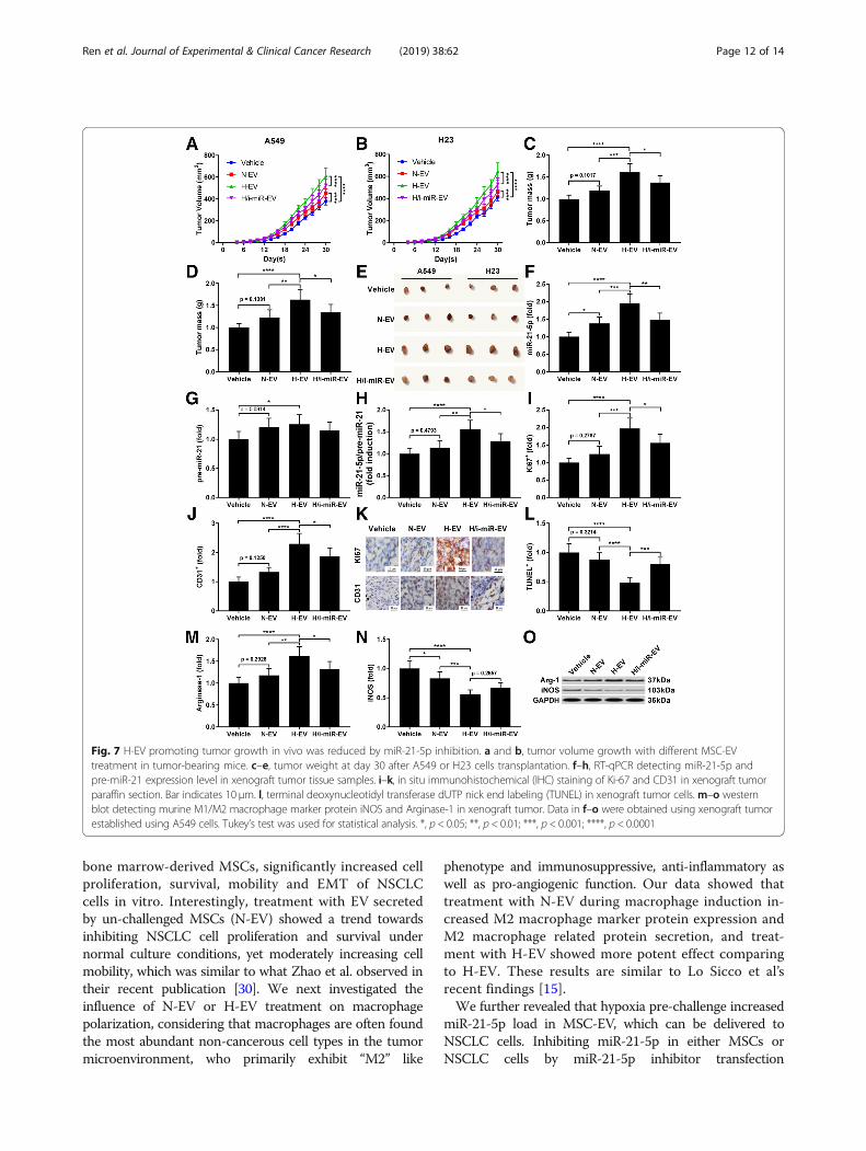
two days, and tumor volume was monitored everytwo days after A549 cell injection using Vernier cali-per. Our data demonstrated that injection with N-EVor H-EV significantly accelerated tumor growth, andH-EV injection showed more potent effect comparingto N-EV; notably, the tumor promoting effect ofH-EV was impaired by miR-21-5p inhibition in MSCs(Fig. 7a and b). Examining the weight of tumorsdissected at day 30 after A549 or H23 cell injectionsuggested that H-EV injection significantly increasedtumor weight, which was impeded by miR-21-5p in-hibition in MSCs, while N-EV injection showed nosignificant effect on tumor weight change (Fig. 7c-e).RT-qPCR detecting miR-21-5p and pre-miR-21 in thexenograft tumors revealed that miR-21-5p in
xenograft tumor cells was significantly increased byN-EV treatment and further increased by H-EV treat-ment, which was largely abrogated by miR-21-5p inhibitionin MSCs, but expression of pre-miR-21 was less signifi-cantly affected, suggesting the delivery of miR-21-5p intothe xenograft tumor cells by MSC-EV (Fig. 7f-h). Immuno-histochemical staining examining the dissected tumorsshowed that H-EV injection significantly increasedA549 or H23 cell proliferation and intra-tumoral angio-genesis in the xenograft model, marked by the signifi-cant increase in Ki67 and CD31 positive staining rate,respectively, with a significant decrease in TUNEL posi-tive staining rate that suggested a markedly reductionof cell apoptosis; these effects of H-EV injection wassignificantly reduced by miR-21-5p inhibition in MSCs,
Fig. 4 H-EV promoted A549 cell proliferation, survival ad mobility in a miR-12-5p-dependent manner. A549 or H23 cells were transfected withmiR-12-5p inhibitor and treated with H-EV. a-k, cell functional assays were performed as described in Fig. 1. Non-treated A549 or H23 cells wereused as negative control (NC). Bar in i indicates 20 μm. Data in h and j were presented as fold change comparing to NC. Tukey's test was usedfor statistical analysis. *, p < 0.05; **, p < 0.01; ***, p < 0.001
Ren et al. Journal of Experimental & Clinical Cancer Research (2019) 38:62 Page 9 of 14

while N-EV injection showed no significant influenceon cell proliferation, apoptosis or intra-tumoral angio-genesis in the xenograft model (Fig. 7i- l). Results ofwestern blot analyzing arginase-1 and iNOS proteinlevel in murine macrophages isolated from the xeno-graft tumors suggested that treatment of MSC-EV sig-nificantly increased M2 polarization while reducing M1polarization of intra-tumoral murine macrophages;H-EV showed more potent effects on screwing macro-phages towards M2 phenotype in vivo, and this effectwas damaged by miR-21-5p inhibition in MSCs (Fig.7m-o).
DiscussionsEV secreted by MSCs has been suggested as possibleanti-cancer drug delivery approach or therapeutic agentby several researches, but different reports on the influ-ence of MSCs or MSC-EV on cancer cell behavior areinconsistent, and the underlying mechanism remains tobe discovered. These controversies might be due to theheterogeneity of MSCs of different tissue origins andcomplexity of the tumor microenvironment. Despite thecomplicated cross-talk among cancer cells, due to thechaotic angiogenesis in tumor mass, hypoxia in insuffi-cient blood supply area can alter the cell function of
Fig. 5 miR-21-5p delivery by H-EV treatment promote NSCLC cell proliferation, survival and mobility by targeting PTEN, PDCD4 and RECK. a–d,western blot detecting PTEN, PDCD4 and RECK protein expression level in A549 or H23 cells after different treatment as described in Fig. 4. e–k,cell functional assay as described in Fig. 1. A549 or H23 cells with or without PTEN, PDCD4 or RECK gene overexpression (O/E) were tested. Wildtype, non-treated A549 or H23 cells were used as negative control (NC). Bar in K indicates 20 μm. Tukey’s test was used for statistical analysis.*, p < 0.05; **, p < 0.01; ***, p < 0.001; ****, p < 0.0001
Ren et al. Journal of Experimental & Clinical Cancer Research (2019) 38:62 Page 10 of 14

cancer cells as well as other cell types residing in thetumor microenvironment. MSCs has been demonstratedto migrate to the tumor microenvironment, where theywere reshaped to tumor associated MSCs and exhibitdistinct immunosuppressive, angiogenic and cancer pro-moting functions, which have been suggested to be largelymediated by EV secretion [10]. Previous researches haveshown that hypoxia pre-challenge on MSCs increasesthe promoting effect of MSC-EV on macrophage M2polarization, and microRNA expression profiles in theMSC-EV can also be significantly altered [15, 18]. Sev-eral previously identified hypoxic-inducible miRNAs werefound upregulated in MSC-EV after hypoxia pre-challenge,including miR-21-5p, miR-210-3p, miR-23a-3p, etc., andthese miRNAs were found to regulate immune cell func-tion and promote cancer development [18]. Caescu et al.found that activation of CSF-1R on murine macrophagefacilitated their M2 polarization by inducing miR-21-5p ex-pression, and Xi et al. further demonstrated that miR-21-5p
inhibition in macrophages favored their M1 polarization,although the detailed molecular mechanism of miR-21-5p’action remains to be clarified [25]. Besides, miR-210-3p andmiR-23a-3p were also found to reduce CD8+ T cell and nat-ural killer cell cytotoxicity towards cancer cells [26–28].MicroRNA delivery via EV from donor cells to regulate
intracellular gene expression and cellular behavior of therecipients has been found under various experimentalconditions [29]. The present research started with ourhypothesis that MSCs pre-challenged by hypoxia mightsecrete cancer promoting and immunosuppressive EV,resulting in accelerated tumor development; consideringthat Cui et al. have demonstrated that miR-21-5p was themiRNA most significantly upregulated in MSC-EV afterhypoxia pre-challenge, we inferred that the potential can-cer promoting and immunosuppressive role of EV secretedby hypoxia pre-challenged MSCs might be at least inpart mediated by miR-21-5p delivery. We first foundthat H-EV, the EV secreted by hypoxia pre-challenged
Fig. 6 N-EV and H-EV treatment promote macrophage M2 polarization by delivering miR-21-5p that targets PTEN. a, western blot analysis ofPTEN protein expression level in induced macrophages. H/i-miR-EV, monocytes were induced with the presence of EV secreted by miR-21-5p-inhibited, hypoxia pre-challenged MSCs; H-EV + i-miR, monocytes were transfected with miR-21-5p inhibitor-expressing vector before inductionwith the presence of H-EV. Macrophages induced without MSC-EV were used as negative control (NC). b, c, flow cytometry determining thepercentage of CD163+CD206+ cells among total CD68+ cells after induction. N-EV + O/E PTEN or H-EV + O/E PTEN, monocytes were transfectedwith PTEN overexpressing vector before N-EV or H-EV treatment, respectively. d–f, western blot detecting Akt and STAT3 protein expression aswell as their activating phosphorylation (p-Ser473 for Akt and p-tyr705 for STAT3) in macrophages after induction. g–i, ELISA evaluating IL-10,TGF-β and VEGF-α in macrophage culture medium after induction. Macrophages induced with the presence of N-EV were used as negativecontrol in b–i. Tukey’s test was used for statistical analysis. *, p < 0.05; **, p < 0.01; ***, p < 0.001; ****, p < 0.0001
Ren et al. Journal of Experimental & Clinical Cancer Research (2019) 38:62 Page 11 of 14
bone marrow-derived MSCs, significantly increased cellproliferation, survival, mobility and EMT of NSCLCcells in vitro. Interestingly, treatment with EV secretedby un-challenged MSCs (N-EV) showed a trend towardsinhibiting NSCLC cell proliferation and survival undernormal culture conditions, yet moderately increasing cellmobility, which was similar to what Zhao et al. observed intheir recent publication [30]. We next investigated theinfluence of N-EV or H-EV treatment on macrophagepolarization, considering that macrophages are often foundthe most abundant non-cancerous cell types in the tumormicroenvironment, who primarily exhibit “M2” like
phenotype and immunosuppressive, anti-inflammatory aswell as pro-angiogenic function. Our data showed thattreatment with N-EV during macrophage induction in-creased M2 macrophage marker protein expression andM2 macrophage related protein secretion, and treat-ment with H-EV showed more potent effect comparingto H-EV. These results are similar to Lo Sicco et al’srecent findings [15].We further revealed that hypoxia pre-challenge increased
miR-21-5p load in MSC-EV, which can be delivered toNSCLC cells. Inhibiting miR-21-5p in either MSCs orNSCLC cells by miR-21-5p inhibitor transfection
Fig. 7 H-EV promoting tumor growth in vivo was reduced by miR-21-5p inhibition. a and b, tumor volume growth with different MSC-EVtreatment in tumor-bearing mice. c–e, tumor weight at day 30 after A549 or H23 cells transplantation. f–h, RT-qPCR detecting miR-21-5p andpre-miR-21 expression level in xenograft tumor tissue samples. i–k, in situ immunohistochemical (IHC) staining of Ki-67 and CD31 in xenograft tumorparaffin section. Bar indicates 10 μm. l, terminal deoxynucleotidyl transferase dUTP nick end labeling (TUNEL) in xenograft tumor cells.m–o westernblot detecting murine M1/M2 macrophage marker protein iNOS and Arginase-1 in xenograft tumor. Data in f–o were obtained using xenograft tumorestablished using A549 cells. Tukey’s test was used for statistical analysis. *, p < 0.05; **, p < 0.01; ***, p < 0.001; ****, p < 0.0001
Ren et al. Journal of Experimental & Clinical Cancer Research (2019) 38:62 Page 12 of 14

significantly decreased the promoting effect of H-EV treat-ment on NSCLC cell proliferation, survival, mobility andEMT, suggesting that these effects of H-EV treatment arelargely mediated by miR-21-5p delivery. We further con-firmed that the cell proliferation and survival-promotingeffect of miR-21-5p on NSCLC cells delivered by H-EV isachieved by PTEN and PDCD4 targeting, and upregulationin NSCLC cell mobility by RECK targeting. Overexpressionof these cancer suppressive genes significantly inhibited theeffect of H-EV on A549 cells. On macrophage differenti-ation, our data suggested that increased miR-21-5p deliveryby MSC-EV after hypoxia pre-challenge can reduce PTENexpression, thus liberating Akt and STAT3 activation andfacilitate macrophage M2 polarization. Using xenograftmodel, we found that intravenous injection of H-EV signifi-cantly increased tumor growth, marked by upregulatedcancer cell proliferation and intra-tumoral angiogenesis aswell as reduced cell apoptosis. Overall, our data suggestedthat encountering with hypoxic environment confers thecancer promoting effect on MSC-EV; EV secreted byMSCs after hypoxia pre-challenge could significantly pro-mote tumor development, possibly by increasing cancercell survival, metastasis and inducing macrophage M2polarization via miR-21-5p delivery.Notably, in most of our experiments, miR-21-5p inhib-
ition not completely but only partially reversed the effectof H-EV treatment, especially in our xenograft tumorformation assay, suggesting that other factors loaded inMSC-EV are involved in the cancer promoting andimmunoregulatory effect of H-EV as our data suggested.In the present research we also compared the effect ofN-EV and H-EV treatment. Besides on macrophagedifferentiation, N-EV generally showed no statisticallysignificant influence on NSCLC cell behavior or xeno-graft tumor growth comparing to vehicle treatment. Inthe present research we found the significant influenceof H-EV treatment on macrophage polarization only inin vitro experiments. We also performed IHC stainingon M2 macrophage marker protein CD163 and CD206in tissue sections derived from xenograft tumor and ad-jacent tissue, but we observed no significant differencein CD163 or CD206 positive staining rate between H-EVtreated groups and non-treated counterparts (data notshown). Further verification on the effect of H-EV inpromoting macrophage M2 polarization is needed underin vivo conditions.
ConclusionCollectively, our data suggested that the cancer promot-ing effect of EV secreted by hypoxia pre-challengedMSCs was mediated at least partially by miR-21-5p de-livery. MiR-21-5p delivered into NSCLC cells by theseEV reduced PTEN, PDCD4 and RECK gene expressionin NSCLC cells, resulting in increased cell proliferation,
survival and mobility. MiR-21-5p delivered into mono-cytes facilitated their M2 polarization during maturation,resulting in the increase in M2 macrophages in thetumor microenvironment.
Additional files
Additional file 1: Figure S1. MSC-EV precipitation from MSCs culturemedia was verified by western blot. Input, raw cell culture media. Output,supernatant after EV precipitation. Pellet, EV that were pelleted down byultra-centrifugation. NC, fresh cell culture media as negative control(processed in parallel). N-EV, EV in naïve MSCs culture media. H-EV, EV inhypoxia pre-challenged MSCs culture media (TIF 2679 kb)
Additional file 2: Figure S2. Uptake of MSC-EV by A549 or humancirculating monocytes as examples of recipients was confirmed by flowcytometry. Cells were treated with N-EV or H-EV that were labeled withCFSE dye before treatment, and cellular fluorescence was detected byflow cytometry. Cells processed in parallel using vehicle (sterile PBS) wasused as negative control. (TIF 4714 kb)
AbbreviationsEV: Extracellular vesicles; hBM-MSCs: Human bone marrow derived MSCs;MSCs: Mesenchymal stem cells; PBMC: Peripheral blood mononuclear cells;TME: Tumor microenvironment
AcknowledgementsNot applicable.
FundingThis work was supported by the Research on Basic and Frontier Technologyin Henan Province (Grant No. 152300410007) and Molecular epidemiologicalstudy of early syphilis (Grant No. 162102310244).
Availability of data and materialsThe datasets used or analyzed during the current study are available fromthe corresponding author on reasonable request.
Author’s contributionsThis work was conceived and designed WR,. The experiments were carriedout by JH, CL, CY, SW and XZ. The manuscript was prepared by WR, JH, YW,CY and HW. The revised manuscript was managed by WR, YW, XPZ. Allauthors read and approved the final manuscript.
Ethics approval and consent to participateOur study was ratified by Ethical and Scientific Committees of LuoyangCentral Hospital Affiliated to Zhengzhou University.
Consent for publicationNot applicable.
Competing interestsThe authors declare that they have no competing interests.
Publisher’s NoteSpringer Nature remains neutral with regard to jurisdictional claims inpublished maps and institutional affiliations.
Author details1Medical Examination Center, Luoyang Central Hospital, ZhengzhouUniversity, No.288, Middle Zhongzhou Rd, Xigong District, Luoyang 471009,China. 2Department of Oncology, Zhengzhou Central Hospital, Zhengzhou,Henan, China. 3Department of Oncology, Kaifeng Central Hospital, Kaifeng475000, China. 4Imaging Department of Luoyang Orthopedic-traumatologicalHospital, Luoyang 471000, China. 5Department of Anesthesiology, LuoyangCentral Hospital, Zhengzhou University, Luoyang 471009, China.
Ren et al. Journal of Experimental & Clinical Cancer Research (2019) 38:62 Page 13 of 14
Received: 23 July 2018 Accepted: 6 January 2019
References1. Bernardo ME, Fibbe WE. Mesenchymal stromal cells: sensors and switchers
of inflammation. Cell Stem Cell. 2013;13:392–402.2. Trounson A, McDonald C. Stem cell therapies in clinical trials: progress and
challenges. Cell Stem Cell. 2015;17:11–22.3. Gyorgy B, Hung ME, Breakefield XO, Leonard JN. Therapeutic applications of
extracellular vesicles: clinical promise and open questions. Annu RevPharmacol Toxicol. 2015;55:439–64.
4. Kourembanas S. Exosomes: vehicles of intercellular signaling, biomarkers,and vectors of cell therapy. Annu Rev Physiol. 2015;77:13–27.
5. Raposo G, Stoorvogel W. Extracellular vesicles: exosomes, microvesicles, andfriends. J Cell Biol. 2013;200:373–83.
6. Ti D, Hao H, Fu X, Han W. Mesenchymal stem cells-derived exosomalmicroRNAs contribute to wound inflammation. Sci China Life Sci. 2016;59:1305–12.
7. Luther KM, Haar L, McGuinness M, Wang Y, Lynch T, Phan A, et al. ExosomalmiR-21a-5p mediates cardioprotection by mesenchymal stem cells. J MolCell Cardiol. 2018.
8. Song Y, Dou H, Li X, Zhao X, Li Y, Liu D, et al. Exosomal mir-146acontributes to the enhanced therapeutic efficacy of interleukin-1beta-primed mesenchymal stem cells against sepsis. Stem Cells. 2017;35:1208–21.
9. Poggi A, Varesano S, Zocchi MR. How to hit mesenchymal stromal cells andmake the tumor microenvironment immunostimulant rather thanimmunosuppressive. Front Immunol. 2018;9:262.
10. Shi Y, Du L, Lin L, Wang Y. Tumour-associated mesenchymal stem/stromalcells: emerging therapeutic targets. Nat Rev Drug Discov. 2017;16:35–52.
11. Pan M, Hou L, Zhang J, Zhao D, Hua J, Wang Z, et al. Inhibitory effect andmolecular mechanism of mesenchymal stem cells on NSCLC cells. Mol CellBiochem. 2018;441:63–76.
12. Kalimuthu S, Gangadaran P, Li XJ, Oh JM, Lee HW, Jeong SY, et al. In vivotherapeutic potential of mesenchymal stem cell-derived extracellularvesicles with optical imaging reporter in tumor mice model. Sci Rep. 2016;6:30418.
13. Dong L, Pu Y, Zhang L, Qi Q, Xu L, Li W, et al. Human umbilical cordmesenchymal stem cell-derived extracellular vesicles promote lungadenocarcinoma growth by transferring miR-410. Cell Death Dis. 2018;9:218.
14. Zhao X, Wu X, Qian M, Song Y, Wu D, Zhang W. Knockdown of TGF-beta1expression in human umbilical cord mesenchymal stem cells reverts theirexosome-mediated EMT promoting effect on lung cancer cells. Cancer Lett.2018;428:34–44.
15. Lo Sicco C, Reverberi D, Balbi C, Ulivi V, Principi E, Pascucci L, et al.Mesenchymal stem cell-derived extracellular vesicles as mediators of anti-inflammatory effects: endorsement of macrophage polarization. Stem CellsTransl Med. 2017;6:1018–28.
16. Terry S, Buart S, Chouaib S. Hypoxic stress-induced tumor and immuneplasticity, suppression, and impact on tumor heterogeneity. Front Immunol.2017;8:1625.
17. Zhu J, Lu K, Zhang N, Zhao Y, Ma Q, Shen J, et al. Myocardial reparativefunctions of exosomes from mesenchymal stem cells are enhanced byhypoxia treatment of the cells via transferring microRNA-210 in annSMase2-dependent way. Artif Cells Nanomed Biotechnol. 2017:1–12.
18. Cui GH, Wu J, Mou FF, Xie WH, Wang FB, Wang QL, et al. Exosomes derivedfrom hypoxia-preconditioned mesenchymal stromal cells amelioratecognitive decline by rescuing synaptic dysfunction and regulatinginflammatory responses in APP/PS1 mice. FASEB J. 2018;32:654–68.
19. Yu J, Liu XL, Cheng QG, Lu SS, Xu XQ, Zu QQ, et al. G-CSF and hypoxicconditioning improve the proliferation, neural differentiation and migrationof canine bone marrow mesenchymal stem cells. Exp Ther Med. 2016;12:1822–8.
20. Yang M, Shen H, Qiu C, Ni Y, Wang L, Dong W, et al. High expression ofmiR-21 and miR-155 predicts recurrence and unfavourable survival in non-small cell lung cancer. Eur J Cancer. 2013;49:604–15.
21. Wu Y, Song Y, Xiong Y, Wang X, Xu K, Han B, et al. Microrna-21 (mir-21)promotes cell growth and invasion by repressing tumor suppressor pten incolorectal cancer. Cell Physiol Biochem. 2017;43:945–58.
22. Li J, Huang H, Sun L, Yang M, Pan C, Chen W, et al. MiR-21 indicates poorprognosis in tongue squamous cell carcinomas as an apoptosis inhibitor.Clin Cancer Res. 2009;15:3998–4008.
23. Xi J, Huang Q, Wang L, Ma X, Deng Q, Kumar M, et al. miR-21 depletion inmacrophages promotes tumoricidal polarization and enhances PD-1immunotherapy. Oncogene. 2018;37:3151–65.
24. Canfran-Duque A, Rotllan N, Zhang X, Fernandez-Fuertes M, Ramirez-Hidalgo C, Araldi E, et al. Macrophage deficiency of miR-21 promotesapoptosis, plaque necrosis, and vascular inflammation during atherogenesis.EMBO Mol Med. 2017;9:1244–62.
25. Caescu CI, Guo X, Tesfa L, Bhagat TD, Verma A, Zheng D, et al. Colonystimulating factor-1 receptor signaling networks inhibit mouse macrophageinflammatory responses by induction of microRNA-21. Blood. 2015;125:e1–13.
26. Noman MZ, Buart S, Romero P, Ketari S, Janji B, Mari B, et al. Hypoxia-inducible miR-210 regulates the susceptibility of tumor cells to lysis bycytotoxic T cells. Cancer Res. 2012;72:4629–41.
27. Noman MZ, Janji B, Berchem G, Chouaib S. miR-210 and hypoxicmicrovesicles: two critical components of hypoxia involved in the regulationof killer cells function. Cancer Lett. 2016;380:257–62.
28. Eichmuller SB, Osen W, Mandelboim O, Seliger B. Immune modulatorymicrornas involved in tumor attack and tumor immune escape. J NatlCancer Inst. 2017;109.
29. Whiteside T. Exosome and mesenchymal stem cell cross-talk in the tumormicroenvironment. Semin Immunol. 2018;35:69–79.
30. Zhao X, Wu X, Qian M, Song Y, Wu D, Zhang W. Knockdown of TGF-β1expression in human umbilical cord mesenchymal stem cells reverts theirexosome-mediated EMT promoting effect on lung cancer cells. Cancer Lett.2018;428:34–44.
Ren et al. Journal of Experimental & Clinical Cancer Research (2019) 38:62 Page 14 of 14